Cofactor Control of a Vital Enzymatic Reaction;the Effect of Factor Va on Thrombin Formation During Blood Coagulation
|
|
|
- Asher Hampton
- 5 years ago
- Views:
Transcription


1 Cleveland State University ETD Archive 2009 Cofactor Control of a Vital Enzymatic Reaction;the Effect of Factor Va on Thrombin Formation During Blood Coagulation Jamila Hirbawi Cleveland State University Follow this and additional works at: Part of the Chemistry Commons How does access to this work benefit you? Let us know! Recommended Citation Hirbawi, Jamila, "Cofactor Control of a Vital Enzymatic Reaction;the Effect of Factor Va on Thrombin Formation During Blood Coagulation" (2009). ETD Archive This Dissertation is brought to you for free and open access by EngagedScholarship@CSU. It has been accepted for inclusion in ETD Archive by an authorized administrator of EngagedScholarship@CSU. For more information, please contact library.es@csuohio.edu.
2 COFACTOR CONTROL OF A VITAL ENZYMATIC REACTION: THE EFFECT OF FACTOR VA ON THROMBIN FORMATION DURING BLOOD COAGULATION JAMILA HIRBAWI Bachelor of Science in Chemistry Cleveland State University May, 2003 Submitted in partial fulfillment of the requirements For the degree DOCTOR OF PHILOSOPHY IN CLINICAL/BIOANLYTICAL CHEMISTRY At the CLEVELAND STATE UNIVERSITY December, 2009
3 This dissertation has been approved for the Department of Chemistry And the College of Graduate Studies by: Dr. Michael Kalafatis Dissertation Committee Chairperson Department & Date Dr. Edward Plow Dissertation Committee Member Department & Date Dr. Alvin Schmaier Dissertation Committee Member Department & Date Dr. David Anderson Dissertation Committee Member Department & Date Dr. Anton Komar Dissertation Committee Member Department & Date Dr. Stan Duraj Dissertation Committee Member Department & Date
4 ACKNOWLEDGEMENT I would like to dedicate this dissertation to my brother, Kamal Sulaiman ( ), who stood by me through all these years and supported me along the way. God bless your soul and rest in peace. Secondly, this dissertation is dedicated to my loving husband, Ziyad Hirbawi, and my children, Abdallah, Yasmine, and Sabrin. For all the love and support you provided. It was a long road for all of us and I thank you all. In addition, I want to thank my parents, Abed and Concepcion Sulaiman, for all the help throughout the years. This would not be possible if it were not for them. A special thanks also goes out to my brother, Jamal Sulaiman, and my sister, Yaminah Sulaiman. You guys were always there when I needed you. There were many people that helped and guided me throughout the course of my studies; First, I would like to thank my research advisor, Dr. Michael Kalafatis, for all the time and insight he put into my research project. His knowledge and expertise in coagulation was the backbone to this research. It is an honor and a privilege to graduate from his research group. I wish him all the best in the future to come. I would like to thank the members of my dissertation committee for their helpful advice during my research project: Drs. Edward Plow, Alvin Schmaier, David Anderson, Anton Komar, and Stan Duraj. I would also like to thank Richelle Emery and Michelle Jones in the Chemistry office for all their administrative support over the years. I would especially like thank the members of my research group: Dr. Melissa Barhoover, Dr. Evrim Erdogan, Dr. Michael Bukys, Dr. Tivadar Orban, Dr. Alieta
5 Ciocea, and Kerri Smith for all the emotional and technical support. I would also like to thank all the fellow graduate students and staff at Cleveland State University.
6 COFACTOR CONTROL OF A VITAL ENZYMATIC REACTION: THE EFFECT OF FACTOR VA ON THROMBIN FORMATION DURING BLOOD COAGULATION JAMILA HIRBAWI ABSTRACT The LONG-TERM goal of our research is to study and analyze the structure and function of the factor V molecule in order to understand its regulatory effects on the natural process of hemostasis and its role in the life-threatening development of deep venous thrombosis. The SHORT-TERM goal of our research is to identify the amino acid residue(s) of factor V that interact with prothrombin during the assembly and function of the prothrombinase complex in order to fully understand its particular role in maintaining the integrity of the blood coagulation cascade. The final goal of the coagulation cascade is the formation of a fibrin clot that is catalyzed by the serine protease, thrombin. The proteolytic conversion of prothrombin to thrombin is catalyzed by the prothrombinase complex composed of the enzyme, factor Xa, its cofactor, factor Va, assembled on a membrane surface in the presence of divalent metal ions. Although factor Xa alone can activate prothrombin, it is at a rate that is not compatible for survival. Incorporation of factor Va into the prothrombinase complex results in a 300,000-fold increase in the catalytic efficiency of factor Xa for thrombin generation and plays an important role in v
7 regulating prothrombin activation. Thus, it is crucial to identify which specific amino acid residue(s) are responsible for the interaction of factor Va with prothombin and factor Xa in the formation of thrombin, which is absolutely vital for maintaining normal hemostasis in a healthy individual. The specific aims of the present study are; To identify the specific amino acid region of the factor Va heavy chain that promotes optimal cofactor function during prothrombin activation; To identify key acidic amino acid sequences in the factor Va molecule that control the rate of prothrombin cleavage; To identify the specific amino acids of the factor V/Va heavy chain that regulate enzyme-substrate interaction during prothrombin activation. vi
8 TABLE OF CONTENTS ABSTRACT LIST OF TABLES LIST OF FIGURES v x xi CHAPTER I INTRODUCTION 1.1 Hemostasis The Coagulation Cascade Blood Coagulation Factor V Factor Va inactivation Factor Va Interaction with Factor Xa FVa Interaction with Prothrombin/Thrombin Prothrombin Activation Factor V Leiden Thrombin Inhibition Reference List 17 CHAPTER II ROLE OF THE ACIDIC HIRUDIN-LIKE COOH- TERMINAL AMINO ACID REGION OF FACTOR Va HEAVY CHAIN IN THE ENHANCED FUNCTION OF PROTHROMBINASE 2.1 Abstract Introduction Experimental Procedures Results 41 vii
9 2.5 Discussion Reference List 68 CHAPER III AMINO ACID REGION OF FACTOR Va HEAVY CHAIN IS INDISPENSABLE FOR OPTIMAL ACTIVITY OF FACTOR XA WITHIN PROTHROMBINASE 3.1 Abstract Introduction Experimental Procedures Results Discussion Reference List 124 CHAPTER IV THE COFACTOR EFFECT OF AMINO ACIDS695 AND 696 OF FACTOR Va HEAVY CHAIN ON THE ENZYMATIC ACTIVITY OF THE PROTHROMBINASE COMPLEX 4.1 Abstract Introduction Experimental Procedures Results Discussion Reference List 156 CHAPTER V OVERALL CONCLUSION 5.1 Conclusion 159 viii
10 5.2 Reference List 163 ix
11 LIST OF TABLES CHAPTER II. Table I. Characteristics of various factor Va molecules when assembled into prothrombinase. 47 Table II. Summary of results obtained with factor Va molecules truncated at the COOHterminus of the heavy chain in a clotting assay and in a prothrombinase assay. 64 CHAPTER III. Table I. Functional properties of various factor Va molecules. 98 Table II. Rate of activation of native plasma-derived prothrombin and recombinant mutant rmz-ii in the presence of prothrombinase assembled with various recombinant factor Va species. 104 Table III. Rates of activation of recombinant prothrombin rp2-ii and plasma-derived FPR-meizothrombin in the presence of various recombinant factor Va species. 116 CHAPTER IV. Table I. Clotting assay with factor Va WT and recombinant mutant molecules. 145 x
12 LIST OF FIGURES Figure 1.1. The blood coagulation pathway. 5 Figure 1.2. Human factor V. 8 Figure 1.3. Prothrombin activation pathway. 13 Figure 2.1. Mutants of human factor V. 34 Figure 2.2. Electrophoretic analysis of wild-type factor V and recombinant factor V molecules. 42 Figure 2.3A. Raw data used for the determination of the parameters of prothrombinase complex assembly and function. 45 Figure 2.3B. Raw data used for the determination of the parameters of prothrombinase complex assembly and function. 46 Figure 2.4. Analysis of the activation of plasma-derived prothrombin by prothrombinase. 50 Figure 2.5. Reaction profiles for the activation of prothrombin by prothrombinase. 51 xi
13 Figure 2.6A. Analysis of the activation of rmz-ii. 55 Figure 2.6B. Kinetic analysis of the activation of rmz-ii. 56 Figure 2.7. Gel electrophoresis analysis for cleavage of FPR-meizothrombin. 57 Figure 3.1. Factor V structure and mutant molecules 81 Figure 3.2. Comparison of the acidic COOH-terminal amino acid sequences and from factor Va heavy chain among species (numbering from the human molecule, top sequence). 82 Figure 3.3. Electrophoretic analyses of wild-type factor V and recombinant factor V molecules. 93 Figure 3.4A. Factor Va titrations to determine the affinity of the recombinant factor Va molecules for factor Xa. 94 Figure 3.4B. Prothrombin titration to determine the kinetic parameters of prothrombinase assembled with various recombinant factor Va species. 95 Figure 3.4C. Determination of the kinetic parameters of prothrombinase assembled with the various recombinant factor Va species. 97 xii
14 Figure 3.5. Analysis of the activation of plasma-derived prothrombin by prothrombinase. 100 Figure 3.6. Analysis of prothrombin consumption by prothrombinase assembled with recombinant factor Va molecules. 103 Figure 3.7 Electrophoretic analyses of the activation of rmz-ii by prothrombinase assembled with mutant factor Va molecules. 109 Figure 3.8. Electrophoretic analyses of the activation of rp2-ii by prothrombinase assembled with mutant factor Va molecules. 110 Figure 3.9. Analysis of recombinant prothrombin consumption by prothrombinase assembled with recombinant factor Va molecules. 111 Figure Gel electrophoresis analyses for cleavage of FPR-meizothrombin. 112 Figure Analysis of FPR-meizothrombin consumption by prothrombinase assembled with recombinant factor Va molecules. 115 Figure 4.1. Factor V structure and mutant molecules. 143 xiii
15 Figure 4.2. Electrophoretic analyses of wild-type factor V and recombinant factor V molecules. 144 Figure 4.3. Factor Va titrations to determine the affinity of the recombinant factor Va molecules for factor Xa. 149 Figure 4.4. Prothrombin titration to determine the kinetic parameters of prothrombinase assembled with various recombinant factor Va species. 150 Figure 4.5. Analysis of the activation of plasma-derived prothrombin by prothrombinase. 151 Figure 4.6. Gel electrophoresis analyses for cleavage of FPR-meizothrombin. 152 xiv
16 CHAPTER I INTRODUCTION An estimated eighty million Americans have one or more forms of cardiovascular disease. Among these numbers are individuals who suffer from high blood pressure, coronary heart disease, and ischemic stroke. An important factor involved in the evolution of these disorders, is the process of hemostasis. Hemostasis is the body s natural mechanism for the control of excessive bleeding after injury, as well as prevention of a hypercoagulative state during the coagulation response. This procedure involves a series of enzymatic reactions that are all tightly regulated in order for proper clot formation to occur at the sight of injury [1, 2]. Without proper control, a range of thrombotic and hemorrhagic difficulties can arise. These tendencies can be attributed to genetic disorders or can be acquired through various outside factors. Thrombosis is the formation of a blood clot within the vasculature such as an artery or a vein that causes the blockage of blood flow through the circulatory system. There are two forms of veins present in an individual s legs. Superficial veins are located just beneath the surface of the skin, while deep veins are found deep within the muscles of the leg. 1
17 Deep venous thrombosis (DVT) is a condition where an unwanted blood clot forms in the thigh or lower leg [3]. This pathological state can progress to a more fatal stage once a small portion of the clot (embolus) is dislodged, circulates in the system through the heart and remains lodged in the lung. A pulmonary embolism will cause blood supply to the lung to be blocked therefore, depleting it of oxygen and carbon dioxide exchange and ultimately causing necrosis of the lung tissue followed by death. In contrast, hemophilia is an inherited disorder that involves prolonged bleeding after injury [4]. There are two major forms of the disease caused by an x-linked recessive trait. Hemophilia A is caused by a deficiency in the clotting factor, factor VIII and hemophilia B is due to a deficiency in factor IX [5]. Individuals affected with these deficiencies usually suffer bleeding episodes that range from mild to severe. In addition, acquired hemophilia is an autoimmune disorder, in which a person with normal haemostatic activity spontaneously develops autoantibodies against the blood clotting factors, more frequently factor VIII. This condition generally occurs as a result of various medical issues such as pregnancy, autoimmune disorders, diabetes, respiratory disease, and a variety of malignancies. Another frequently seen clotting disorder involves the inability of the coagulation response to halt in a timely manner. APC (activated protein C) is the enzyme responsible for the inactivation of the clotting factors, FVa and FVIIIa, when bound to its cofactor, protein S [6]. Factors Va and VIIIa are important facilitators in the formation of a blood clot. APC resistance occurs in about 25-40% of patients with a family history of thrombotic conditions and in about 3-5% of the normal population [7]. Deficiency in 2
18 either protein C or protein S will cause the continued accumulation of thrombus formation due to inadequate quantities present to inactivate FVa and FVIIIa. 1.1 Hemostasis Primary hemostasis is the body s initial response to injury to the endothelial surface of a blood vessel. This initial phase is characterized by constriction of the blood vessel, blood platelet adhesion to the surface, and the formation of a soft plug [8]. Once damage occurs, vasoconstriction limits the flow of blood to the damaged area. Platelets then become activated by thrombin and soon aggregate at the site of injury. Cell adhesion occurs when glycoproteins present on the platelet surface bind to von Willebrand (vwd) factor that attaches to the endothelim. Platelets then clump by binding to collagen that is exposed during initial rupture of the blood vessel lining. Individuals, who have decreased amounts of circulating vwd factor, suffer from bleeding of the skin and mucous membranes due to interruption of primary hemostais. In addition, vwd factor also binds and stabilizes circulating factor VIII, which is involved in the intrinsic pathway of secondary hemostasis. There are three types of von Willebrand disease, with type III being the most severe and rare form. Although the initial plug arrests bleeding, it must be stabilized through secondary hemostasis. This process involves a series of reactions induced by coagulation factors that serve as enzymes and coenzymes, the presence of calcium, and a phospholipid surface that is usually presented by the activated platelets [9]. Defects in this system may lead to serious bleeding disorders including internal bleeding of the cavities and subcutaneous 3
19 hematomas. In addition, deficiencies in circulating blood factors and/or genetic mutations can incur risk of thrombotic episodes. 1.2 The Coagulation Cascade The coagulation response is of utmost importance in the maintenance of normal hemostasis in healthy individuals. Any slight disruption of this mechanism can lead to abnormalities that eventually cause heart disease, stroke, or blood pressure variations. The penultimate goal of the coagulation cascade is the generation of thrombin, which in turn converts fibrinogen to fibrin to generate a fibrin plug. Thrombin generation is divided into three separate segments: the initiation phase where low levels of α-thrombin are produced; the propagation phase when a considerable amount of thrombin is created and the fibrin plug is formed; and the termination phase where the inhibition of thrombin and many of the serine proteases involved in the coagulation cascade takes place due to the inhibitors TFPI (tissue factor pathway inhibitor) and AT-III (antithrombin III); and inactivation of the cofactors (fva and fviiia) by activated Protein C (APC)/Protein S [10]. This response is initiated by cellular and vascular injury and can be initially divided into two distinct pathways, followed by the common pathway [11] (Fig.1.1). The extrinsic pathway (tissue factor pathway) is initiated with the exposure of tissue factor (TF) after vascular damage. Tissue factor is a cell surface glycoprotein that serves as a cofactor for factor VIIa [12, 13]. TF will bind to factor VIIa to form the extrinsic tenase complex that will activate factor X [14]. In addition, factor VII can also be activated by factor Xa and thrombin. Activated factor X (fxa) will then go on to the common pathway where the prothrombinase complex is formed with its cofactor, factor 4
20 Fig. 1.1: The Blood Coagulation Pathway. The blood coagulation cascade includes the extrinsic and extrinsic pathways, which combine at the common pathway where the prothrombinase complex forms to continue prothrombin activation. This process will eventually lead to the formation of the fibrin plug after vascular injury. (from 5
21 Va, on a membrane surface in the presence of Ca 2+ to activate the substrate, prothrombin. Consequently, the TF-FVII complex will also activate factor IX that will play a role in the intrinsic pathway [15]. The extrinsic pathway is rapidly inhibited by tissue factor pathway inhibitor (TFPI), a serine protease inhibitor that is associated to lipoprotein molecules. Although this pathway is inhibited quickly, sufficient quantities of thrombin are formed to activate factor XI [12]. Factor XIa will participate in the intrinsic pathway, inducing the amplification of the coagulation response. The intrinsic pathway, involves the conversion of factor XI to XIa by factor XIIa. Activated factor XII is a key component in the generation of bradykinin and implementation of fibrinolysis and is the initial activator of the intrinsic pathway. Factor XIa will convert factor IX to factor IXa. This enzyme will associate on a membrane surface with its cofactor, factor VIIIa in the presence of calcium to form the intrinsic tenase complex [16]. Intrinsic tenase will activate factor X that will be incorporated into prothrombinase for thrombin generation. The intrinsic pathway is not important for initiation of the coagulation cascade. However, it is responsible for amplification of the process due to production of large amounts of factor Xa and thrombin. Once enough factor Xa is generated through the extrinsic and intrinsic pathways, the common pathway takes part in the generation of human alpha thrombin through conversion of prothrombin by the prothrombinase complex. This complex is composed of the enzyme, factor Xa, with its non-enzymatic cofactor, factor Va, on a phospholipid surface, (provided by the activated platelets), in the presence of Calcium ions [17]. Although factor Xa can activate prothrombin with an initial cleavage at Arg 271 followed 6
22 by cleavage at Arg 320 to yield the intermediates Fragment 1.2 and Prethrombin 2, incorporation of factor Va into the prothrombinase complex results in a reversal of cleavages and a 300,000-fold increase in the catalytic efficiency of factor Xa for thrombin generation [18]. Thrombin will proceed to convert fibrinogen to fibrin to produce a stable clot. 1.3 Blood Coagulation Factor V Coagulation factor V circulates in plasma at a concentration of 20 nm, as a single chain protein of M r 330,000 consisting of three protein domains (A, B, & C) that are arranged in the order of A1-A2-B-A3-C1-C2 [1] (Fig. 1.2). In addition, factor V can be found in a partially active state in the α-granules of platelets. It is cleaved by α-thrombin (fiia) at Arg 709, Arg 1018, and Arg 1545 to release the B domain (containing a large number of asparagines-linked oligosaccharides) and generate an active cofactor (fva Iia ) composed of a light chain of M r 74 kda (A3-C1-C2 domains) and a heavy chain of M r 105 kda (A1-A2 domains) that is involved in prothrombinase complex formation and function [19, 20]. The amino-terminal heavy chain (amino acids 1-709) is associated via Ca 2+ ions to the carboxy-terminal light chain (amino acids ). The B domain has been shown to have sequences recognized by α-thrombin for activation of the cofactor [21]. While thrombin is a major activator of the human factor V molecule, it also has been implicated in the inactivation of the cofactor. In previous studies, it was determined that cleavage at Arg 643 of the COOH-terminal region of the heavy chain resulted in a partially inactive molecule [22]. In addition to thrombin, various proteases can cleave factor V to 7


23 Fig. 1.2: Human Factor V. Schematic of the factor V molecule with various modifications and activation/deactivation sites. 8
24 induce activation. The procofactor can be activated by Russell s viper venom (RVV-V) through cleavage at Arg 1018 and Arg 1045, resulting in a cofactor with approximately the same activity as factor Va IIa. Proteolytic cleavage of the procofactor can also be induced by the serine proteases found in polymorphonuclear leukocytes (PMN), Cathepsin G and Human Neutrophil Elastase (HNE). These enzymes give result to a cofactor with a truncated heavy chain missing vital amino acid residues necessary for interaction with prothrombin during thrombin formation. Cathepsin G will cleave factor V at Phe 1031, Leu 1447, Tyr 1518, and Tyr 696 to yield a 103 kda heavy chain and 80 kda light chain. Elastase will activate factor V through cleavage at Ile 708, Ile 819, Ile 1484, and Thr 678 to produce a 102 kda heavy chain and 90 kda light chain. Other proteases involved in factor V cleavage include the protease from the venom of the snake naja naja oxiana, factor Xa, and the prothrombin activation intermediate, meizothrombin [18]. 1.4 Factor Va Inactivation Factor Va can be inactivated by the anticoagulant, activated protein C (APC). Protein C is a 62,000 M r protein that circulates in plasma at a concentration of 60 nm. It consists of a heavy chain and light chain, which are held together by a single disulfide bridge[23]. When cleaved at Arg 169 of the heavy chain by the thrombin-thrombomodulin complex, protein C is converted to the active enzyme, APC. APC cleaves factor Va sequentially at Arg 506, Arg 306, and Arg 679 in the presence of its cofactor protein S to yield an inactive factor Va molecule that can no longer bind to factor Xa or prothrombin [24]. Cleavages at Arg 506 and Arg 306 occur with the involvement of a phospholipid surface with the latter being strictly dependent on the presence of a membrane surface [25]. 9
25 Initial cleavage occurs at Arg 506 to yield a factor Va molecule that is partially active and has a lower affinity for the enzyme, factor Xa. Once cleavage at Arg 306 takes place, total deactivation of the cofactor is obtained [26]. In addition to APC inactivation, factor Va can also be cleaved at Arg 643 by human alpha thrombin in the presence of endothelial cells to yield an inactive product [22]. Factor Va will also be inactivated by plasmin which will cleave at Lys 309, Lys 310, Arg 313, and Arg 348 in the presence of a membrane surface and cause the A2 domain to dissociate from the rest of the molecule [27]. 1.5 Factor Va interaction with factor Xa. Appropriate binding of factor Va to factor Xa during prothrombinase function is crucial to the proper activation of the substrate, prothrombin. Factor Va increases the catalytic efficiency of factor Xa for prothrombin activation by a great magnitude. It has been shown that factor Va increases the k cat and decreases the K m of the reaction along with inducing sequential cleavages at Arg 320 and Arg 271 to induce the meizothrombin pathway in the presence of phospholipids [28]. The increase in K cat value is attributed to cofactor interaction with the enzyme, while the decrease in K m is due to the γ- carboxyglutamic acid-dependent (Gla domain) interaction of factor Xa and prothrombin with the phospholipid suface. Factor Va binds to both the light and heavy chains of factor Xa including amino acids and of the catalytic domain of factor Xa [29, 30]. Cofactor binding to factor Xa has also been found to expose cryptic exosites on fxa that are recognition sites for its substrate, prothrombin [31]. Previous data has indicated that amino acids located on various portions of the heavy chain have had enormous effects on prothrombinase formation and function. A nonapeptide (AP4 ) with 10
26 amino acid sequence Glu 323 -Val 331 of the factor Va A2 domain of the heavy chain was found to have inhibitory effects on prothrombinase assembly and function by interfering in factor Xa-Va interaction [32]. It has also been found that the carboxy-terminus of the factor Va heavy chain has a dramatic effect on its binding to factor Xa and prothrombin. A factor Va molecule lacking the 27 amino acids at the carboxyl end of the heavy chain produced by cleavage at Asp 683 by the protease purified from the venom of the snake Naja naja oxiana experienced a reduction in clotting activity. Additionally, this factor Va variant (factor Va NO ) was found to have a lower affinity for factor Xa within the prothrombinase complex than wild-type factor Va with K D values of 4 nm and 0.5 nm, respectively [33]. A pentapeptide (DYDYQ) consisting of the region Asp 695 -Gln 699 was also found to inhibit prothrombinase function by competitively inhibiting the prothrombinase complex during thrombin formation with a K i of 850 nm [34, 35]. 1.6 FVa interaction with prothrombin/thrombin There has been substantiated evidence that there are certain acidic amino acid residues in the carboxyl-terminal end of the heavy chain of factor Va that are extremely important for cofactor activity. The region between amino acids has been the basis for investigation into the functional role of factor Va in prothrombinase complex formation and function [34, 36]. These acidic residues may be directly involved in the interaction of the cofactor with factor Xa or Thrombin through positively charged amino acids. It was recently demonstrated that there is a thrombin-binding site in the last 13 amino acids of the heavy chain. Thrombin contains two electro-positively charged binding regions (anion binding exosite I (ABE-I) and anion binding exosite II (ABE II)), 11
27 which are crucial for protein function [37, 38]. ABE-I is responsible for binding many of the proteins involved in the coagulation cascade including factors V, Va, fibrinogen, PAR-I (the platelet thrombin receptor), thrombomodulin, and heparin cofactor II [39]. ABE-II, located just above the active site of the molecule, serves as a heparin-binding site [40]. Factor Va will also bind to the second kringle domain of prothrombin to enhance prothrombinase function [41]. Additional studies involving factor Va interactions with prothrombin (fii) have implied that Cys 539 of the heavy chain interacts with residues of the serine protease domain of factor II [42]. A peptide (N42R) consisting of amino acid region of the heavy chain of factor Va was found to be a good inhibitor of prothrombinase with an IC 50 value of 1.3 um, leading to believe that this may be a factor Xa and/or prothrombin binding site on factor Va [27]. Previous studies have also demonstrated that a factor Va molecule cleaved initially with either cathepsin G (fva CG ) or human neurtrophil elastase (fva HNE ) followed by cleavage with thrombin giving rise to a factor Va molecule with a truncated heavy chain (fv CG/IIa or fva HNE/IIa ), resulted in K d values of.2nm, similar to fva IIa and k cat values that were higher than the wild-type factor Va [43]. Surprisingly, clotting assays performed with these species in the presence of factor V deficient plasma resulted in a loss of clotting activity of about 60%. 1.7 Prothrombin Activation The activation of prothrombin to α-thrombin by factor Xa alone is not considered a very efficient reaction. Factor Xa will convert prothrombin at a rate of 5 orders of 12


28 Fig.1.3: Prothrombin Activation Pathway. Conversion of prothrombin to thrombin occurs by two distinct pathways. Prothrombin can be activated by fxa alone, as shown in pathway I or it can be converted to thrombin by the prothrombinase complex. Pathway I will initially induce cleavage at Arg 271 followed by cleavage at Arg 320, to produce the intermediates fragment 1.2 and prethrombin 2. Pathway II, which is essential for normal clotting in healthy individuals, catalyzes an initial cleavage at Arg 320, followed by cleavage at Arg 271 that produces the intermediate, meizothrombin. 13
29 magnitude lower than when factor Va is incorporated into the prothrombinase complex [44]. Factor Va promotes an increase in catalytic efficiency of prothrombinase along with inducing sequential cleavage of Arg 320 and Arg 271 (Fig. 1.3). Both cleavages are phospholipid-dependent, but only cleavage of Arg 320 is dependent on factor Va. [45] [46]. The catalytic efficiency is highly improved as a result of decreasing K m by 100-fold and increasing K cat values by 3,000-fold, corresponding to substrate concentration and enzyme efficiency, respectfully [47]. Kinetic analyses has revealed the Ca 2+ dependent interaction of factor Va with factor Xa, in solution, has a dissociation constant value (K d ) of.8 μm, while the K d in the presence of a phospholipid surface is decreased to1 nm, showing a decrease of 1000-fold [15, 18]. The K d value of the calcium-independent factor Va-prothrombin interaction is 1 μm. Factor Va provides binding sites for proexosite 1 and the Gla domain of prothrombin, explaining one of the possible mechanisms by which the cofactor functions to increase enzyme efficiency [48]. It has been demonstrated that a factor V molecule activated with the purified protease from the venom of the snake Naja nigricollis nigricollis (NN) has reduced clotting activity and an increased K d value of 4 nm when compared to factor V activated with thrombin (fva IIa ) (K d 0.5nM) [27]. When factor V IIa is additionally cleaved by NN, cofactor activity is reduced by 60-80%. This reduction in activity has been hypothesized to be a direct result from the loss of crucial amino acids of the heavy chain that are released when cleaved by NN. 14
30 1.8 Factor V Leiden Disruptions in the coagulation cascade can lead to life-threatening circumstances, such as bleeding and clotting disorders. Factor V Leiden is an autosomal dominant mutation in the factor V gene that inhibits degradation of the molecule by APC [25]. This occurs through a substitution of Arg 506 by a glutamine that prevents proteolytic cleavage of the cofactor by the APC/Protein S complex. Factor Va is inactivated by APC through three sequential cleavages at Arg 506, Arg 306, and Arg 679. In factor Va Leiden, cleavage of Arg 506 does not occur, but cleavage at Arg 306 and Arg 679 cause inactivation of the cofactor at a slower rate [49]. Studies have shown that cleavage at Arg 506 promotes cleavage at the other two sites for proper inactivation of the cofactor in order to prevent thrombosis [26]. Excessive venous clotting occurs from this disorder and eventually leads to a condition known as deep venous thrombosis (DVT). Individuals with this syndrome are at risk of pulmonary embolism that occurs when a venous blot clot dislodges and travels to the lungs. Patients who are homozygous for the disease are more likely to suffer from these episodes than heterozygous ones who only have one defective gene. 1.9 Thrombin Inhibition Human alpha-thrombin is the main weapon in the battle to halt excessive bleeding, after injury. In order for this process to be carried out effectively, all aspects of the coagulation response must be tightly regulated. In cases where individuals suffer from thrombotic disorders, the inhibition of thrombin is executed. Thrombin can be inhibited directly through its catalytic active site and/or through its exosites [50]. One of the main thrombin inhibitors used in medicine is heparin [51]. It indirectly inhibits 15
31 thrombin by forming the heparin-thrombin-antithrombin complex, which involves binding of heparin to exosite II of thrombin [52]. Direct inhibition of thrombin occurs when an inhibitor binds to the active site of the enzyme or includes binding of one of the exosites. Hirudin, a leech derived inhibitor, was one of the first discovered inhibitors of exosite I. It leads to the impairment of the enzyme to recognize some of its macromolecular substrates by binding to both the active site and exosite I. Bothrojaracin, isolated from a Brazilian snake vanom, is another thrombin inhibitor that will inhibit both, exosites I and II [53]. In addition to outside inhibitors, thrombin can be inhibited physiologically by antithrombin, once it forms a complex with a heparin sulfate [54]. Although there are a variety of antithrombotic drugs available, many of them lead to undesirable side-effects and levels must constantly be monitored during therapy. It would be of great advantage to develop alternative methods to aid individuals with thrombotic tendencies. 16
32 1.10 REFERENCE LIST 1. Kalafatis, M., et al., The Regulation of Clotting Factors. Critical Reviews in Eukaryotic Gene Expression, (3): p Rand, M.D., et al., Blood Clotting in Minimally Altered Whole Blood. Blood, (9): p Castoldi, E., et al., Combinations of 4 mutations (FVR506Q, FV H1299R, FV Y1702C, PT 20210G/A) affecting the prothrombinase complex in a thrombophilic family. Haemostasis, Thrombosis, and Vascular Biology, (4): p Mannucci, P.M., Hemophilia and Related Bleeding Disorders: A Story of Dismay and Success. Hematology, 2002: p Awidi, A., et al., Study of mutations in Jordanian patients with haemophilia A: identification of five novel mutations. Haemophilia, Kalafatis, M., et al., Isolation and Characterization of an antifactor V antibody causing activated protein C resistance from a patient with severe thrombotic manifestations. Haemostasis, Thrombosis, and Vascular Biology, (11). 17
33 7. Dahlback, B., Resistance to activated protein C caused by the R506Q mutation in the gene for factor V is a common risk factor for venous thrombosis. Journal of Internal Medicine Supplement, : p Tripodi, a., m. Primignani, and P.M. Mannucci, Abnormalities of hemostasis and bleeding in chronic liver disease: the paradigm is challenged. Internal Emergency Medicine, lawson, j.h., et al., A model for the tissue factor pathway to thrombin. The Journal of Biological Chemistry, (37): p Butenas, S., C.v.t. Veer, and K. Mann, Evaluation of the Initiation phase of blood coagulation using ultrasensitive assays for serine proteases. The Journal of Biological Chemistry, (34): p Davie, E.W., K. Fujikawa, and W. Kisiel, The Coagulation Cascade: Initiation, Maintenance, and Regulation. Biochemistry, (43): p Orfeo, T., et al., The Tissue Factor Requirement in Blood Coagulation. The Journal of Biological Chemistry,
34 13. Brummel, K.E., et al., Thrombin Functions during Tissue Factor-induced Blood Coagulation. Hemostasis, Thrombosis, and Vascular Biology, (1): p Stone, M.D., et al., Large Enhancement of Functional Activity of Active Site- Inhibited Factor VIIa Due to Protein Dimerization: Insights into Mechanism of Assembly/Disassembly from Tissue Factor. Biochemistry, Hathcock, J.J., et al., Phospholipid Regulates the Activation of Factor X by Tissue Factor/Factor VIIa (TF-VIIa) via Substrate and Product Interactions. Biochemistry, Yuan, Q.-P., E.N. Walke, and J.P. Sheehan, The Factor IXa Heparin-Binding Exosite is a Cofactor Interactive Site: Mechanism for Antithrombin-Independent Inhibition of Intrinsic Tenase Heparin. Biochemistry, Bukys, M.A., et al., Incorportation of Factor Va into Prothrombinase is Required for Coordinated Cleavage of Prothrombin by Factor Xa. The Journal of Biological Chemistry, (29): p Kalafatis, M., D.O. Beck, and K.G. Mann, Structural Requirements for Expression of Factor Va Activity. The Journal of Biological Chemistry,
35 19. Kalafatis, M., Coagulation Factor V: a Plethora of Anticoagulant Molecules. Current Opinion in Hematology, : p Keller, F.G., et al., Thrombin-Catalyzed Activation of Recombinant Human Factor V. Biochemistry, : p Thorelli, E., R.J. Kaufman, and B. Dahlback, The C-terminal Region of the Factor V B-domain is Crucial for the Anticoagulant Activity of Factor V. The Journal of Biological Chemistry, (26): p Erdogan, E., et al., Identification of an inactivating cleavage site for alphathrombin on the heavy chain of factor Va. thrombosis haemostasis, (5): p Kalafatis, M., et al., Regulation and Regulatory Role of χ-carboxyglutamic Acid Containing Clottin Factors. Critical Reviews in Eukaryotic Gene Expression, (1): p Yegneswaran, S., et al., Prothrombin Amino Terminal Region Helps Protect Coagulation Factor Va From Proteolytic Inactivation by Activated Protein C. thrombosis haemostasis, : p
36 25. Veer, C.v.t., et al., An In Vitro Analysis of the Combination of Hemophilia A and Factor V Leiden. Blood, (8): p Bajzar, L., et al., An Antifibrinolytic Mechanism Describing the Prothrombotic Effect Associated with Factor V Leiden. The Journal of Biological Chemistry, (38): p Kalafatis, M. and K.G. Mann, The Role of the Membrane in the Inactivation of Factor Va by Plasmin. The Journal of Biological Chemistry, (21): p Kalafatis, M. and K. Mann, Factor V: a combination of Dr. Jekyll and Mr Hyde. Blood, (1). 29. Yegneswaran, S., R.M. Mesters, and J.H. Griffin, Identification of Distinct Sequences in Human Blood Coagulation Factor Xa and Prothrombin Essential for Substrate and Cofactor Recognition in the Prothrombinase Comples. The Journal of Biological Chemistry, (35): p Rezaie, A.R. and F.S. Kittur, The Critical Role of the Loop in the Factor Xa Interaction with Na + and Factor Va in the Prothrombinase Complex. The Journal of Biological Chemistry, (46): p
37 31. Manithody, C. and A.R. Rezaie, Functional Mapping of Charged Residues of the Sequence in Factor Xa. Evidence that Lysine 96 is a Factor Va Independent Recognition Site for Prothrombin in the Prothrombinase Complex. Biochemistry, Singh, L.S., et al., Amino Acids Glu 323, Tyr 324, Glu 330, and Val 331 if Factor Va Heavy Chain are Essential for Expression of Cofactor Activity. The Journal of Biological Chemistry, (30): p Mann, K.G. and M. Kalafatis, Factor V: A Combination of Dr. Jekyll and Mr. Hyde. Blood, (1): p Beck, D.O., et al., The Contribution of Amino Acid Region Asp 695 -Tyr 698 of Factor V to Procofactor Activation and Factor Va Function. The Journal of Biological Chemistry, (4): p Bukys, M.A., et al., A control Switch for Prothrombinase. The Journal of Biological Chemistry, (51): p Suzuki, K., B. Dahlback, and J. Stenflo, Thrombin-catalyzed Activatin of Human Coagulation Factor V. The Journal of Biological Chemistry, (11): p
38 37. Anderson, P.J., et al., Role of proexosite I in factor Va-dependent substrate interactions of prothrombin activation. The Journal of Biological Chemistry, (22). 38. Bukys, M.A., et al., The Structural Integrity of Anion Binding Exosite I of Thrombin is Required and Sufficient for Timely Cleavage and Activation of Factor V and Factor VIII. The Journal of Biological Chemistry, (27): p Liu, L.-W., et al., Proteolytic Formation of Either of the Prothrombin Activation Intermediates Results in Formation of a Hirugen-binding Site. The Journal of Biological Chemistry, (35): p Rezaie, A., Identification of Basic Residues in the Heparin-binding Exosite of Factor Xa Critical for Heparin and Factor Va Binding. The Journal of Biological Chemistry, (5): p Blostein, M.D., et al., The Gla Domain of Human Prothrombin has a Binding Site for Factor Va. The Journal of Biological Chemistry, (48): p Yegneswaran, S., et al., Prothrombin residues contribute to factor Va 23
39 binding in the prothrombinase complex. The Journal of Biological Chemistry, (47). 43. Camire, R.M., M. Kalafatis, and P.B. Tracy, Proteolysis of factor V by cathepsin G and elastase indicates that cleavage at Arg1545 optimizes cofactor function by facilitating factor Xa binding. Biochemistry, (34). 44. Brufatto, N. and M.E. Nesheim, Analysis of the Kinetics of Prothrombin Activation and Evidence that Two Equilibrating Forms of Prothrombinase are Involved in the Process. The Journal of Biological Chemistry, (9): p Stone, M.D. and G.L. Nelsestuen, Efficacy of Soluble Phospholipids in the Prothrombinase Reaction. Biochemistry, Rosing, J., et al., The Role of Phospholipids and Factor Va in the Prothrombinase Comples. The Journal of Biological Chemistry, (1): p Majumder, R., M.A. Quinn-Allen, and W.H. Kane, The Phosphotidylserine Binding Site of the Factor Va C2 Domain Accounts for Membrane Binding but does not Contribute to the Assembly or Activity of Human Factor Xa-Factor Va Complex. Biochemistry, Barry R. Lentz. 44: p
40 48. Myles, T., et al., An Extensive Interaction Interface between Thrombin and Factor V is Required for Factor V Activation. The Journal of Biological Chemistry, (27): p Veer, C.v.t., et al., Increased Tissue Factor-initiated Prothrombin Activation as a Result of the Arg 506 to Glu Mutation in Factor V Leiden. The Journal of Biological Chemistry, (33): p Mann, K., S. Butenas, and K.E. Brummel, The dynamics of thrombin formation. Arteriosclerosis Thrombosis Vascular Biology, (17). 51. Yang, L., C. Manithody, and A. Rezaie, Localization of the Heparin Binding Exosite of Factor IXa. The Journal of Biological Chemistry, (52): p Neuenschwander, P.F., Exosite Occupation by Heparin Enhances the Reactivity of Blood Coagulation Factor IXa. Biochemistry, : p Arocas, V., et al., Inhibition of Thrombin-catalyzed Factor V Activation by Bothrojaracin. thrombosis haemostasis, Izaguirre, G., et al., Localization of an Antithrombin Exosite that Promotes Rapid 25
41 Inhibition of Factors Xa and IXa Dependent on Heparin Activation of the Serpin. The Journal of Biological Chemistry, (51): p
42 CHAPTER II ROLE OF THE ACIDIC HIRUDIN-LIKE COOH-TERMINAL AMINO ACID REGION OF FACTOR VA HEAVY CHAIN IN THE ENHANCED FUNCTION OF PROTHROMBINASE 2.1 Abstract Prothrombinase activates prothrombin through initial cleavage at Arg 320 followed by cleavage at Arg 271. This pathway is characterized by the generation of an enzymatically active transient intermediate, meizothrombin that has increased chromogenic substrate activity but poor coagulant activity. The heavy chain of factor Va contains an acidic region at the COOH-terminus (residues ). We have shown that a pentapeptide from this region (DYDYQ) inhibits prothrombin activation by prothrombinase by inhibiting meizothrombin generation. To ascertain the function of these regions we have created a mutant recombinant factor V molecule that is missing the last 30 amino acids from the heavy chain (factor V Δ ) and a mutant molecule with the substitution 695 DYDY 698 AAAA (factor V 4A ). The clotting activities of both recombinant mutant 27
43 factor Va molecules were impaired compared to the clotting activity of wild type factor Va (factor Va Wt ). Using an assay employing purified reagents, we found that prothrombinase assembled with factor Va Δ had a ~39% increase in the k cat, while prothrombinase assembled with factor Va 4A showed a ~20% increase in k cat for the activation of prothrombin as compared to prothrombinase assembled with factor Va Wt. Gel electrophoresis analyzing prothrombin activation by prothrombinase assembled with the mutant molecules revealed a delay in prothrombin activation with persistence of meizothrombin. Our data do demonstrate that the COOH-terminal region of factor Va heavy chain is indeed crucial for coordinated prothrombin activation by prothrombinase because it regulates meizothrombin cleavage at Arg 271 and suggest that this portion of the molecule is partially responsible for the enhanced coagulant function of prothrombinase. 28
44 2.2 Introduction Blood coagulation is initiated at the site of vascular injury and results in the activation of prothrombin to thrombin by the prothrombinase complex. Prothrombinase is composed of the enzyme factor Xa bound to its cofactor, factor Va, on a phospholipid surface in the presence of Ca 2+ (1,2). Prothrombin and α-thrombin have two distinct exosites (anion binding exosite I, ABE-I, and anion binding exosite II, ABE-II) that are responsible for the functions of the molecules. The role of (pro)exosite I of thrombin within prothrombinase is dependent on the incorporation of factor Va into the complex as independently suggested by several laboratories (3-7). Two activation pathways for prothrombin activation are possible: membrane-bound factor Xa alone activates prothrombin following initial cleavage at Arg 271 followed by cleavage at Arg 320, while the fully assembled prothrombinase complex activates prothrombin following the opposite pathway, initial cleavage at Arg 320 followed by cleavage at Arg 271 (8-15). Activation of prothrombin via this latter pathway is characterized by the generation of an intermediate, meizothrombin, that has proteolytic activity and results in a significant increase in the catalytic efficiency of factor Xa with respect to thrombin formation (16). Initial cleavage of prothrombin at Arg 320 that is absolutely factor Va-dependent and results in meizothrombin generation is required and sufficient for the formation of the active site of α-thrombin and complete exposure of ABE-I of the molecule (17-19). However, meizothrombin does not have a fully exposed ABE-II which is required for proper fibrinogen binding and timely fibrin formation. Exposure of this exosite, which is partially covered by fragment 2 of prothrombin, requires cleavage at Arg 271 (5,20,21). 29
45 As a consequence, most functions associated with ABE-II of thrombin are impaired in meizothrombin. For this reason, meizothrombin has reduced fibrinogen clotting activity (22,23). Human factor V circulates in plasma as a 330,000 single-chain protein that consists of three domains in the order A1-A2-B-A3-C1-C2. Proteolytic cleavage of the cofactor by α-thrombin occurs sequentially at Arg 709, Arg 1018, and Arg 1045 to produce a heterodimer consisting of a heavy chain (M r ~105,000) and a light chain (M r ~74,000) associated through divalent metal ions (24-28) (Fig.2.1). The heavy chain of the cofactor contains an acidic amino acid region that has been shown to be important for cofactor function (amino acid ) (29). Early data have suggested that this region is implicated in the productive interaction of factor Va with prothrombin (30-32). Various proteases can cleave this acidic region to produce a cofactor with a truncated heavy chain (33-36). Gerads et al first showed that it is possible to selectively eliminate the acidic region from factor Va heavy chain by an enzyme purified from the venom of the snake Naja Naja Oxiana (33). Using proteins of bovine origin, the authors showed that while elimination of the COOH-terminal region of factor Va results in a molecule with severely impaired clotting activity, incorporation of the truncated cofactor molecule into prothrombinase resulted in an increased k cat for the activation of prothrombin (33). The same authors also showed that cleavage of the human factor Va heavy chain by the same enzyme results in a cofactor with impaired clotting activity (33). Subsequently, Bakker et al using the same purified enzyme identified the cleavage site of the enzyme at His 682 in the human factor Va heavy chain (this amino acid is conserved in the bovine cofactor) and demonstrated that prothrombinase assembled with 30
46 a human factor Va molecule missing the Asp 683 -Arg 709 portion has increased k cat for the activation of prothrombin (34). Afterward, Camire et al using cathepsin G (CG) and human neutrophil elastase demonstrated that prothrombinase assembled with factor Va molecules missing the COOH-terminal domain of the heavy chain result in enzymes that consistently express higher k cat values suggesting that these molecules are more active cofactors than purified plasma factor Va activated with thrombin in an assay using purified reagents (35). Surprisingly, and in line with the initial findings of Gerads et al (33) the same factor Va molecules showed a significant decrease in clotting activity (35). More recently, using a purified enzyme from the snake venom of Naja Naja Nigricollis, we have also shown that a factor Va molecule missing a portion of the COOH-terminus of the heavy chain has decreased clotting activity (36). Altogether these studies reveal that removal of the acidic COOH-terminal portion of factor Va heavy chain results in a cofactor molecule that is deficient in its clotting activity. However, prothrombinase assembled with cofactor molecules missing the acidic COOH-terminus produces significant higher k cat for prothrombin activation when assessed in assays using purified reagents and a chromogenic substrate specific for thrombin. A molecular explanation for these paradoxical observations has not yet been provided. We have recently used overlapping peptides from the region of the factor Va molecule to show that a pentapeptide with the sequence DYDYQ inhibits prothrombin activation by prothrombinase in a competitive manner with respect to substrate (36,37). We have further demonstrated that DYDYQ inhibits prothrombinase activity by inhibiting meizothrombin generation (38). Using data obtained with 31
47 recombinant proteins, Toso and Camire have recently suggested that the COOHterminal region of the factor Va heavy chain has no detectable effect on prothrombinase function (39). This conclusion was surprising since their data showed that: 1) prothrombinase assembled with recombinant factor Va molecules missing a portion or the entire hirudin-like carboxyl- terminal end of the heavy chain have increased k cat for the activation of prothrombin (from 129%-150%) compared to prothrombinase assembled with the wild type molecule in an assay using purified reagents and a chromogenic substrate specific for thrombin; 2) initial velocity measurements using the same assay demonstrated a 20-25% increase in the rate of thrombin formation by prothrombinase assembled with the same recombinant mutant cofactor molecules that were truncated at their carboxyl-terminal end; and 3) a recombinant factor Va molecule that is missing 17 amino acids from the carboxy-terminal portion of the heavy chain had decreased clotting activity (39). All these data are in complete accord with all earlier findings using plasma-derived factor Va molecules truncated at their heavy chain and demonstrate a crucial but yet undetermined contribution of the acidic COOH-terminal region of the heavy chain of the cofactor to prothrombinase activity during prothrombin activation. It has been well established that while meizothrombin has poor clotting activity, its amidolytic activity is increased compared to thrombin towards small fluorescent and chromogenic substrates specifically used to assess thrombin activity (23,40). A logical hypothesis to reconcile all the findings described above is that the acidic COOH-terminus of factor Va heavy chain, and more precisely the sequence DYDYQ, regulates meizothrombin concentration during the factor Xa catalyzed prothrombin activation by 32
48 prothrombinase. Thus, activation of prothrombin by prothrombinase assembled with a cofactor that is missing the acidic region will result in increased and stable meizothrombin production. This result will be translated by a factor Va molecule that is deficient in its clotting activity but produces an increase in k cat when introduced into prothrombinase. In contrast, in the presence of an excess of the acidic region (represented by DYDYQ, (36-38)) no meizothrombin is produced by prothrombinase resulting in the generation of thrombin through the alternative pathway characterized by initial cleavage of prothrombin at Arg 271. The present work was undertaken to test these hypotheses and to elucidate the role of the acidic COOH-terminal portion of factor Va heavy chain during activation of prothrombin by prothrombinase. 33
49 FV Δ( ) 680 KMHDRLEPEDEESDADYDYQNRLAAALGIR 709 NH 2 FV 4A B Domain A1 A2 71, ,000 A3 C1 C2 COOH Arg 709 Arg 1018 Arg 1545 (α-iia) (α-iia) (α-iia) (RVV-V) (RVV-V) Figure 2.1. Mutants of human factor V. Factor V is activated following three sequential cleavages by thrombin at Arg 709, Arg 1018, and Arg These cleavages release the active cofactor composed of heavy and light chains associated in the presence of divalent metal ions, and two activation fragments. The COOH-terminus of the heavy chain contains an acidic hirudin-like amino acid region that is important for its cofactor functions. The mutations (deletions and point mutations) within the heavy chain are indicated together with the designation for the recombinant mutant factor V molecules created and used throughout the manuscript. 34
50 2.3 Experimental Procedures Materials reagents, and proteins. Diisopropyl-fluorophosphate (DFP), O- phenylenediamine dihydrochloride (OPD), N-[2-Hydroxyethyl]piperazine-N'-2- ethanesulfonic acid (Hepes), Trizma (Tris base), and Coomassie Blue R-250 were purchased from Sigma (St. Louis, Mo). Factor V-deficient plasma was from Research Proteins Inc (Essex Junction VT). L-α-phosphatidylserine (PS) and L-aphosphatidylcholine (PC) were from Avanti Polar Lipids (Alabaster, AL). Normal reference plasma and the chromogenic substrate H-D-Hexahydrotyrosol-alanyl-arginyl-pnitroanilide diacetate (Spectrozyme-TH) were purchased from American Diagnostica Inc. (Greenwich, CT). H-D-Phenylalanyl-L-pipecolyl-L-arginyl-p-nitroaniline dihydrochloride (Chromogenix, S-2238) was purchased from Diapharma Group, Inc. (West Chester, OH) and its concentration in solution (water) was verified as described (41). RecombiPlasTin for the clotting assays was purchased from Instrumentation Laboratory Company (Lexington, MA). The reversible fluorescent α-thrombin inhibitor dansylarginine N,N-(3-ethyl-1,5-pentanediyl)amide (DAPA), human prothrombin, RVV- V activator, and human α-thrombin, were from Haematologic Technologies Inc. (Essex Junction, VT). Active-site blocked human meizothrombin (obtained following digestion of prothrombin with the purified component from the venom of the snake Echis Carinatus as described (42,43), FPR-meizothrombin) was provided by Dr. Rick Jenny (Haematologic Technologies Inc, Essex Junction VT). Human factor Xa was from Enzyme Research Laboratories (South Bend, IN). Human cathepsin G was from Calbiochem (EMD Chemicals, Inc. San Diego, CA). All molecular biology and tissue 35
51 culture reagents and media were from Gibco, Invitrogen Corporation (Grand Island, NY). Human plasma factor V was purified and concentrated using methodologies previously described employing the monoclonal antibody αhfv#1 coupled to Sepharose (44). Digestion of factor Va by a-thrombin and cathepsin G (to obtain factor Va II/CG ) and/or the purified enzyme from Naja Naja Nigricollis (factor Va NN ) were performed as described (35,36). The clotting activities of all factor Va preparations was measured by a clotting assay using factor V deficient plasma and standardized to the percentage of control as described (44) using an automated coagulation analyzer (START-4, Diagnostica Stago, Parsippany, NJ). Recombinant wild type prothrombin and prothrombin rmz-ii that has only one cleavage site for factor Xa (i.e. Arg 320 ) were prepared and purified as previously described (17,22,45,46). Phospholipid vesicles composed of 75% PC and 25% PS (referred to as PCPS vesicles throughout the manuscript) were prepared as previously described (47). Construction of Recombinant FV Molecules. Mutant factor V with the substitutions 695 DYDY 698 AAAA (factor V 4A ) was constructed using the QuickChange II XL site-directed mutagenesis kit (Stratagene, La Jolla, CA, USA) according to the manufacturer s instructions with the following primers (underlined nucleotides represent the mismatch): 5 -GATGAAGAGAGTGATGCTGCCGCTGCTGCCCAGAACAGA- 3 (sense) and 5 -TCTGTTCTGGGCAGCAGCGGCAGCATCACTCTCTTCATC-3 (anti-sense). The deletion mutant (factor V D ) ) was constructed with the same kit. Primers for factor V D were 5 - CCTCCAGAATCTACAGTCATGGCTACACGGTCATTCCGAAACTCATCATTGAA TCAGG-3 (sense) and 5-36
52 CCTGATTCAATGATGAGTTTCGGAACGACCGTGTAGCCATGACTGTAGATTCT GGAGG-3 (anti-sense). PCR products were transformed into competent E. Coli cells and positive ampicillin-resistant clones were selected. Before transfection, all mutant constructs were verified following sequencing in the Cleveland State University DNA Analysis Facility using a Beckman Coulter CEQ 8000 Genetic Analysis System (Beckman, Fullerton CA) with factor-v sequence-specific primers. The wild type pmt2- FV and mutant pmt2-fv plasmids were isolated from the bacterial culture by the QIAfilter High Speed plasmid Midi Kit (Qiagen Inc., Valencia, CA). Expression of Recombinant Wild Type and Mutant Factor V in Mammalian Cells. COS-7L and COS-7 cells (Invitrogen) were maintained in Dulbecco s modified Eagle s medium supplemented with 10% fetal bovine serum, 2mM L-glutamine, and antibiotics (100 μg/ml streptomycin and 100 IU/ml penicillin) in a humidified atmosphere of 5% CO 2 and 95% air at 37 C. Purified factor V Wt, factor V D , and factor V 4A plasmids were transfected into the cells as described (48). Purification of all recombinant factor V molecules was performed as described (49). The concentration of all molecules was assessed by ELISA as detailed (48). The activity and integrity of the recombinant molecules was verified before and after activation with RVV-V activator or thrombin by clotting assays using factor V-deficient plasma and and in several experiments by sodium dodecyl sulfate-polyacrylamide gel electrophoresis (SDS-PAGE) followed by Western blotting using monoclonal and polyclonal antibodies. Analysis of Prothrombin Activation and FPR-Meizothrombin Cleavage at Arg 271 by Gel Electrophoresis. Prothrombin (1.4μM) was incubated with PCPS vesicles (20μM), DAPA (50μM), and factor Va (10-30nM) in a buffer composed of 5 mm Ca 2+ in 37
53 20 mm Tris, 0.15 M NaCl, ph 7.4. The reaction was initiated with the addition of factor Xa (0.5-1nM) at room temperature over a 1 h time course. Aliquots (50 μl ) from the reaction were removed at selected time intervals (as indicated in the legend to the figures) treated as described (38) and analyzed using 9.5% SDS-PAGE. Prothrombin and prothrombin-derived fragments were visualized by Coomassie Blue staining. Scanning densitometry and calculation of the rates of prothrombin consumption were performed as described (38,50,51). FPR-meizothrombin cleavage at Arg 271 was assessed in a similar manner. Gel Electrophoresis and Western Blotting. SDS-PAGE analyses of recombinant proteins following activation were performed using 5-15% gradient gels according to the method of Laemmli (52). Proteins were transferred to polyvinylidene difluoride (PVDF) membranes according to the method described by Towbin et al. (53). After transfer to nitrocellulose, factor Va heavy and light chain(s) were detected using the appropriate monoclonal and polyclonal antibodies (54-57). Immunoreactive fragments were visualized with chemiluminescence. Measurement of Rates of Thrombin Formation in a Prothrombinase Assay. rmz-ii and recombinant prothrombin were activated by prothrombinase prior to the experiment using conditions previously described (58). Subsequent gel electrophoresis analyses under reducing conditions were performed to verify that both rmz-ii and the recombinant prothrombin preparations were activated to the same extent. The enzymes (rmz-iia and recombinant a-thrombin) used for the titration of Spetrozyme-TH and S were assayed at a constant concentration (4.3 nm), as previously described using 38
54 serial dilutions of chromogenic substrate (35,50,59,60). The absorbance was monitored with a Thermomax microplate reader (Molecular Devices, Sunnyvale, CA). Functionally defined apparent dissociation constants (K Dapp ) for factor Va binding to factor Xa-PCPS were obtained from plots measuring the rate of thrombin generation as a function of factor Va concentration in the presence of a limiting, (constant) concentration of factor Xa. Throughout all experiments the assumption was n=moles of factor Xa bound/mole of factor Va at saturation; throughout this study 1=1; the stoichiometry of the factor Va-factor Xa interaction was fixed at 1. The initial rate of the formation of thrombin (initial velocity in nm IIa min -1 ) was calculated, and the data were analyzed and plotted using the software Prizm (Graphpad Software Inc, San Diego CA) according to the one binding site model. Dissociation constants were extracted directly from the graphs. The assay using purified reagents and verifying the activity of the recombinant factor V molecules was conducted under conditions where all factor Xa was saturated with factor Va, as described by measuring α-thrombin formation by the change in the absorbance of a chromogenic substrate at 405 nm (Spectrozyme-TH, 0.4 mm) (48). All factor V molecules were activated with RVV-V or thrombin as described (36,37). Knowing the (K Dapp ) of each factor Va species for factor Xa, the amount necessary to saturate factor Xa was calculated using the quadratic equation described in the literature (61,62) before each experiment. The total concentration of ligand (Va T ) used was modified as appropriate to obtain between 95-98% saturation of the factor Xa molecule. The absorbance was monitored with a Thermomax microplate reader and compared to an α-thrombin standard prepared daily using purified plasma-derived α-thrombin (35,37,61-39
55 63). The data were analyzed and plotted using the software Prizm according to the Michaelis-Menten equation. Kinetic constants provided throughout the manuscript were extracted directly from the graphs. 40
56 2.4 Results Activation of Recombinant Human Factor V Molecules. To ascertain the function of the acidic region from the COOH-terminus of factor Va heavy chain for cofactor activity, we constructed a molecule that is missing the entire amino acid region (factor V D , Fig. 2.1). We have also constructed a factor V molecule with the mutation 695 DYDY 698 AAAA (factor V 4A ). The recombinant molecules were expressed in mammalian cells and purified to homogeneity as described (49). Because factor V D ( ) is missing Arg 709 that is the activating cleavage site for thrombin (Fig.2.1), in the functional assays, the recombinant mutant molecule was activated with RVV-V activator. In contrast, factor V 4A was activated with either RVV-V activator or α- thrombin. SDS-PAGE analyses followed by immunoblotting with specific monoclonal antibodies to the heavy and light chain of the cofactor demonstrate that the mutant recombinant proteins were intact and homogeneous and migrated according to their expected molecular weights (Fig.2.2). Cofactor Function of Recombinant Human Factor Va Molecules. Recent work using recombinant factor Va molecules demonstrated that all cofactors lacking portions or the entire acidic COOH-terminus of the heavy chain encompassing amino acid region , had K Dapp values for membrane-bound factor Xa that are similar to the values for the bimolecular interaction found with wild type or the intact plasma cofactor (39). These values are similar to the K Dapp obtained with truncated plasma-derived factor Va molecules for factor Xa (35,36). Our studies with factor Va D( ) also demonstrate that the mutant cofactor has similar affinity for plasma-derived factor Xa as the wild type molecule (Fig.2.3A). Similar results were found for factor Va 4A (not shown). Thus, 41
57 elimination of the acidic COOH-terminal region of factor Va heavy chain has no consequence on the affinity of the cofactor for its interaction with factor Xa. We next evaluated the ability of all recombinant factor Va molecules to function as a cofactor for prothrombinase. A clotting assay using factor V-deficient plasma and a α- thrombin generation assay using spectrozyme-th and purified reagents were employed. We have also followed the activation of prothrombin by gel electrophoresis. Figure 2.3B show the fitted hyperbolic plots used to extract the kinetic constants obtained following plasma-derived prothrombin activation by prothrombinase assembled with all control or modified cofactor molecules, and summarized in Table 2.I. Because we wanted to prevent the possibility that differences between prothrombinase assembled with factor Va Wt and prothrombinase assembled with factor Va D ) or factor Va 4A may be attributed to subtle differences in the K Dapp of factor Va for factor Xa, which would result in a lesser amount of prothrombinase formed, all experiments described below were conducted under condition where more than 95% of factor Xa was saturated with factor Va. The amount of factor Xa saturation by each cofactor was calculated using the quadratic equation provided in the literature (61,62). The percent saturation of factor Xa by each cofactor species as well as the correlation coefficient (R 2 ) of the hyperbolas used to extract the values of V max and K m, are provided in the legend to figure 2.3B together with the actual reagent concentrations used in each experiment. Factor Va RVV D ) had a 39.2% higher k cat (figure 2.3B, open circles) and 73% lower clotting activity than factor Va RVV Wt (figure 2.3B, open inverse triangles), while factor Va IIa/CG had a 23.7% higher k cat and 62% lower clotting activity than factor Va IIa PLASMA. In separate experiments, we have found that under similar experimental 42
58 conditions, factor Va IIa/NN that has 60% lower clotting activity than the plasma cofactor (36) had a 20.5% increased k cat than factor Va PLASMA IIa (Table 2.I). All these data are in complete agreement with earlier and recent findings (33-36,39) and demonstrate that elimination of the COOH-terminal region of factor Va heavy chain produces a molecule with poor clotting activity yet, when the truncated cofactor is incorporated into prothrombinase, it produces an enzyme with increased catalytic efficiency for prothrombin activation as assessed in an assay using purified components and under conditions in which the majority of membrane-bound cofactor is bound to factor Xa. Overall the data shown in Table 2.I also demonstrate that the plasma and wild type molecule behave similarly following activation by either RVV-V activator or a-thrombin with respect to prothrombin activation. Thus, the two molecules are interchangeable and throughout the manuscript control experiment were conducted with only one of the two molecules as indicated. We next assessed the capability of prothrombinase assembled with the truncated molecules to activate prothrombin by gel electrophoresis and the data are presented in figure 2.4. Under the conditions employed, prothrombinase assembled with factor Va Wt RVV activates prothrombin quickly, with meizothrombin as a short-lived intermediate as demonstrated by the brief half-life of fragment 1 2-A (Fig. 2.4A, panel A). Scanning densitometry demonstrated a peak of meizothrombin early in the reaction at 80 sec and 43
 were activated with RVV-V activator as described (36); panel B, factor V Wt and factor V 4A were activated with thrombin as described in the")

59 A B FVa Wt FVa Δ( ) FVa Wt FVa 4A HC RVV-V HC IIa LC RVV-V LC IIa Figure 2.2. Electrophoretic analyses of wild type factor V and recombinant factor V molecules. Panel A, factor V Wt and factor V ) were activated with RVV-V activator as described (36); panel B, factor V Wt and factor V 4A were activated with thrombin as described in the Experimental Procedures section and analyzed by SDS- PAGE. Following transfer to a PVDF membrane, immunoreactive fragments were detected with monoclonal antibodies αhfva HC 17 (recognizing an epitope on the heavy chain of the cofactor between amino acid residues ) and αhfva HC 9 (recognizing the light chain). At the right the positions of the heavy/light chains of factor Va are shown. 44
60 30 25 nm IIa/min FVa Wt (K DappXa = 0.53 ± 0.18 nm) FVa Δ( ) (K DappXa = 0.45 ± 0.22 nm) Factor Va (nm) Figure 2.3A. Raw data used for the determination of the parameters of prothrombinase complex assembly and function. Panel A. Determination of the affinity of recombinant factor Va molecules for plasma-derived factor Xa. Initial rates of thrombin generation were determined as described under Experimental Procedures. Prothrombinase assembled with factor Va Wt is shown by filled squares (R 2 = 0.979) while prothrombinase assembled with FVa D( ) is depicted by filled triangles (R 2 = 0.978) Titrations were carried out to 20 nm factor Va; however, for graphical purposes the data show the titration for up to 10 nm cofactor. The solid lines represent a nonlinear regression fit of the data as detailed under Experimental Procedures using the software Prizm and the model for one binding site. The apparent dissociation constant (K Dapp ) for each species was derived from each titration performed at least in triplicate with at least two different preparations of recombinant proteins and is listed in the inset. 45
61 12 10 nm IIa/min Prothrombin (μm) Figure 2.3B. Panel B. Determination of kinetic parameters of prothrombinase assembled with various factor Va species. Initial rates of thrombin generation were determined using the K D for factor Xa found in panel A as described under Experimental Procedures in the presence of 20 um PCPS vesicles. The reaction was initiated by the addition of factor Xa (5 pm). Prothrombinase assembled with two different concentrations of factor Va Wt RVV is shown by open triangles (10 nm, 95% factor Xa saturation, R 2 = 0.97) and open inverse triangles (20 nm, 97% factor Xa saturation, R 2 = 97). Prothrombinase assembled with two different concentrations of factor Va D( ) is depicted by filled diamonds (10 nm, 96% factor Xa saturation, R 2 = 0.98) and open circles (20 nm, 98% factor Xa saturation, R 2 = 0.99) while prothrombinase assembled with factor Va IIa PLASMA is depicted by filled squares (10 nm, 98% factor Xa saturation, R 2 = 0.97) and factor Va RVV PLASMA is shown by open diamonds (10 nm, 98% factor Xa saturation, R 2 = 0.97). Prothrombinase assembled with factor Va RVV 4A is depicted by filled inversed triangles (10 nm, 96.5% factor Xa saturation, R 2 = 0.98). The values of the K m and V max /E T (=k cat ) extracted directly from these graphs are listed in Table
62 FVa IIa PLASMA FVa RVV PLASMA FVa IIa Wt FVa RVV Wt FVa RVV D(680- FVa IIa/CG d FVa IIa/NN FVa IIa 4A FVa RVV 4A 709) II Consum b (moles sec -1 mole fxa - 1 ) 16.2 ± 1.8 ND 17.3 ± ± ± ± 0.6 ND 4.4 ± ± 0.4 K m (mm) 0.1 ± ± ± ± 0.24 ± ± 0.15 ± 0.31 ± ± k cat (min - 1 ) 1715 ± ± ± ± ± ± ± ± ± 75 Specific Activity 3337 ± ± ± ± 869 ± ± ND e 663 ± ± 120 (Units/mg) c Table 2.I Characteristics of various factor Va molecules when assembled into prothrombinase a a The rate of thrombin formation following activation of prothrombin by prothrombinase assembled with the various factor Va species was calculated as described in the Experimental Procedures section by knowing the dissociation constant of each factor Va species for factor Xa. In each case more than 95% of factor Xa was saturated with factor Va. Some of the data in the table were extracted directly from the graphs shown in figure 2.3B. b The rate of prothrombin consumption was determined following quantitative scanning densitometry of several gels stained with Coomassie Blue as described in the Experimental Procedures section. Some of the gels used are shown in figure 2.4. c All clotting activities were determined in a two-stage clotting assay following activation of factor V species by RVV-V activator or α-thrombin as described (58,59). d Plasma factor V was activated with α-thrombin and treated with cathepsin G (CG) as described (49). e The clotting activity of the truncated cofactor is ~50% that of plasma-derived factor Va (50). ND, not determined in the present study. 47
63 no meizothrombin was detected following 180 sec (Fig.2.5). In contrast, in the presence of prothrombinase assembled with factor Va D( ), prothrombin is activated with a rate that is approximately 3-fold slower than the rate of activation by prothrombinase assembled with the wild type molecule (Table 2.I), with persistence of meizothrombin as indicated by the lingering of fragment 1 2-A even at the late time-points of the reaction (Fig.2.4, panel B). The lower extent of prothrombin consumption most likely reflects the result of product inhibition by the accumulating meizothrombin. Comparison of the data shown in figures 2.4, panels A and B demonstrates that meizothrombin was produced with similar initial rates but was more persistent. Scanning densitometry demonstrated a peak of meizothrombin late in the reaction at 250 sec. Meizothrombin persisted for up to 6 minutes in the time course (Fig.2.5). In addition, appearance of the B chain is also delayed when prothrombin is activated by prothrombinase assembled with factor Va D( ), compared with the appearance of the B chain obtained following incubation of prothrombin with factor Va Wt RVV. Similar results were obtained with factor Va IIa/CG (Table 2.I). In contrast, in the presence of DYDYQ, no meizothrombin is observed following prothrombin activation by prothrombinase assembled with factor Va D( ) and α-thrombin is formed through the alternate pathway characterized by initial cleavage at Arg 271 and formation of prethrombin 2 as intermediate (Fig.2.5, panel C). Overall, the data demonstrate that elimination of amino acid region from factor Va results in a cofactor molecule that when incorporated into prothrombinase produces an enzyme responsible for persistence of meizothrombin during activation of prothrombin. In preliminary experiments using several preparations of recombinant proteins we observed that while the truncated factor Va molecules are impaired in their clotting 48
64 activity, factor Va 4A is also deficient in its clotting activity. To understand the properties of this molecule and the effect of the mutations on cofactor activity, we used the same preparation of recombinant protein to perform three different experiments. Factor V 4A was first activated with α-thrombin and the solution was split into three separate samples. One sample was used for assessment of clotting activity, one sample was used to measure the kinetic parameters of prothrombinase assembled with saturating concentration of factor Va 4A IIa, and the third sample was used for analysis of prothrombin activation by gel electrophoresis. The results reveal that while factor Va 4A IIa is severely impaired in its clotting activity (~22% that of factor Va Wt IIa ), prothrombinase assembled with the mutant molecule shows a 19.7% increased k cat (Table 2.I). Gel electrophoresis followed by scanning densitometry analysis demonstrated that prothrombinase assembled with factor Va 4A IIa activates prothrombin with a rate that is approximately 3.8-fold slower that the rate of activation of prothrombin assembled with factor Va Wt IIa (Fig.2.4 panels A ande, Table 2.1). Scanning densitometry of several gels studying prothrombin activation by prothrombinase assembled with factor Va IIa 4A revealed a peak of meizothrombin at approximately 240 sec with significant amounts of meizothrombin remaining for up to 10 minutes into the time course (not shown). These findings provide strong evidence in favor of our previous conclusion that amino acid sequence , regulates meizothrombin formation by prothrombinase (37,38). These data also demonstrate that, in the presence of prothrombinase assembled with factor Va 4A the excess meizothrombin formed as assessed functionally by clotting assays can compensate for the absence of α- thrombin in the assay using purified reagents because the increased amidolytic activity of meizothrombin towards the chromogenic substrate is read as α-thrombin activity. 49
65 FVaRVV Wt C A M M FVaRVV Δ DYDYQ II FVaRVVΔ F 1.2A F 1.2 B M M FVaIIaPLASMA II P1 F 1.2 P2 B P2 B D II F 1.2A F 1.2 B II F 1.2A F 1.2 B E FVaIIa4A M II F 1.2A F 1.2 B Figure 2.4. Analysis of the activation of plasma-derived prothrombin by prothrombinase. Plasma-derived prothrombin (1.4 um) was incubated in different mixtures with PCPS vesicles (20 nm), and prothrombinase assembled with either wild type factor Va (panel A, 10 nm) or factor Va D( ) (panel B, 10 nm) as described in the Experimental Procedures section. Panel C, prothrombinase assembled with factor Va ) in the presence of 20 um DYDYQ (same conditions as in panel B); panel D, prothrombinase assembled with plasma-derived factor Va (10 nm); panel E, prothrombinase assembled with factor Va 4A (10 nm). At selected time intervals aliquots of the reactions were withdrawn and treated as described in the Experimental Procedures section. M represents the lane with the molecular weight markers (from top to bottom): M r 98,000, M r 64,000, M r 50,000, M r 36,000, M r 22,000. Lanes 1-19 represent samples from the reaction mixture before (0 min) the addition of factor Xa and 20 sec, 40 sec, 60 sec, 80 sec, 100 sec, 120 sec, 140 sec, 160 sec, 180 sec, 200 sec, 220 sec, 240 sec, 5 min, 6 min, 10 min, 20 min, 30 min, and 60 min respectively following the addition of factor Xa. The prothrombin derived fragments are shown as follows: II, prothrombin (amino acid residues 1-579); prethrombin-1 (amino acid residues ); F1 2-A, fragment 1 2-A chain (amino acid residues 1-320); F1 2, fragment 1 2 (amino acid residues 1-271); P2, prethrombin-2 (amino acid residues ); P2, prethrombin-2 cleaved at Arg 284 ; B, B chain of α-thrombin (amino acid residues ). 50
66 1.5 A 1.5 B Concentration (μm) Time (sec) Time (sec) Figure 2.5. Reaction profiles for the activation of prothrombin by prothrombinase. Progress curves for products and reactants for the activation of prothrombin by prothrombinase assembled with factor Va Wt (panel A) or factor Va D( ) (panel B) were obtained by quantitative densitometry of gels shown in Fig.2.4A and 2.4B, as described in the Experimental Procedures section. The graphs illustrate the disappearance of prothrombin (filled squares), the transient formation of meizothrombin (open circles), and the accumulation of the B chain of α-thrombin (filled circles). There is a three-fold difference in the x-axis between the two panels, because prothrombinase assembled with factor Va D( ) consumes prothrombin with a ~3-fold slower rate than prothrombinase assembled with factor Va Wt. The lines for the disappearance of prothrombin were drawn according to the equation of a one phase exponential decay (factor Va Wt, R 2 = 0.985, and factor Va D( ), R 2 = 0.968). The lines depicting the formation of meizothrombin and the accumulation of the B chain of α-thrombin were arbitrarily drawn. Additional data points extending to 1h of incubation (shown in figure 4) have been omitted for clarity. 51
67 Role of the Acidic COOH-terminal Portion of Factor Va Heavy Chain: a Potential Mechanism. The data obtained thus far with the mutant cofactor molecules indicate that meizothrombin is generated with similar rates, however the intermediate accumulates to more persistent levels and lingers throughout the time course. Appearance of the B-chain of α-thrombin also appears to be delayed when prothrombin is activated by prothrombinase assembled with the mutant cofactor (Fig.2.5). Consequently, analysis of the results suggests that cleavage of meizothrombin at Arg 271 by prothrombinase assembled with factor Va D ( ) RVV maybe be slower than cleavage Wt of meizothrombin at the same site by prothrombinase assembled with factor Va RVV resulting in less conversion of meizothrombin to α-thrombin. Since during the course of the assay using plasma-derived prothrombin we are measuring the sum of meizothrombin and α-thrombin, a more rigorous test to verify which cleavage in prothrombin is affected by the deletion at the COOH-terminus of factor Va heavy chain, involves the study of the activation of a prothrombin molecule that cannot be cleaved at Arg 271 such as rmz-ii (22). In addition, while cleavage of rmz-ii can be followed by gel electrophoresis, generation of rmz-iia can be also assessed with the chromogenic substrate used to assess α-thrombin formation. We thus followed activation of rmz-ii by prothrombinase assembled with either factor Va D( ) RVV or factor Va Wt RVV by gel electrophoresis (Fig. 2.6A) and activity assays using spetrozyme-th (Fig.2.6B). Both sets of data demonstrate that there are no significant differences between the rates of activation of rmz-ii by either enzyme. The rates of rmz-ii consumption were similar as assessed by scanning densitometry of the gels depicted in figure 2.6A (not shown), and the profiles of both titrations are coincident (similar V ma x and similar K m values of 91±9 mm and 121±18 52
68 mm, Fig.2.6B). Comparable results were found in two independent measurements using two different preparations of recombinant factor Va molecules and two separate preparations of rmz-ii. The data demonstrate that cleavage at Arg 320 in prothrombin is not affected by the COOH-terminal portion of factor Va heavy chain. Earlier data has demonstrated that incorporation of factor Va in prothrombinase has a differential influence on the rates of cleavage of the two bonds responsible for the activation of prothrombin. In particular, the binding of factor Va to factor Xa within prothrombinase results in an 3-5-fold increase in the rate of cleavage of meizothrombin at Arg 271 as compared to cleavage at the same site by factor Xa alone (13,14). We next compared the rate of cleavage of FPR-meizothrombin at Arg 271 by prothrombinase assembled with various factor Va molecules (Fig.2.7). The data demonstrate a delay for cleavage of FPR-meizothrombin at Arg 271 by prothrombinase assembled with factor Va D( ) RVV (panel C) or factor Va 4A RVV (panel D) as compared to the same reaction catalyzed by prothrombinase assembled with factor Va PLASMA RVV (panel B). A direct comparison between the rates of cleavage of FPR-meizothrombin by membrane-bound factor Xa alone (panel A) and cleavage of the substrate by prothrombinase assembled with factor Va RVV D ) (panel C) or factor Va 4A do not show any significant differences. These data imply that following incorporation of factor Va in prothrombinase it is the COOH-terminal acidic hirudin-like portion of factor Va heavy chain and in particular amino acid sequence 695 DYDY 698 that is responsible for the increase in the rate of cleavage of meizothrombin at Arg 271. Quantitative scanning densitometry of fragment 1 2-A present on the gels shown in figure 2.7, demonstrated a ~3-4-fold delay in cleavage of FPR-meizothrombin at Arg 271 by either membrane-bound 53
69 factor Xa alone or prothrombinase assembled with the recombinant mutant factor Va molecules, compared to cleavage at Arg 271 by prothrombinase assembled with the plasma-derived cofactor (Fig.2.7, panel E). These data are in complete agreement with previous findings (13,14) and demonstrate that the lingering of meizothrombin during plasma-derived prothrombin activation by prothrombinase assembled with factor Va D ) RVV or factor Va 4A is the result of impaired cleavage at Arg 271. Overall, the data suggest that amino acid sequence 695 DYDY 698, regulates meizothrombin concentration during activation of prothrombin by prothrombinase. 54
 Figure 2.6A. Analysis of the activation of rmz-ii. Panel A.")
 was incubated in different mixtures with PCPS vesicles (20 µm), DAPA (3")
.")

, or following 0.5 min, 1 min, 2.")

70 M M II F1 2 B Factor Va RVV Wt Factor Va RVV Δ( ) Figure 2.6A. Analysis of the activation of rmz-ii. Panel A. Gel electrophoresis analyses. rmz-ii (1.4 µm) was incubated in different mixtures with PCPS vesicles (20 µm), DAPA (3 µm), and factor Va Wt (left panel, 20nM) or factor Va D( ) (right panel, 20nM). The reaction was started by the addition of factor Xa and the samples were treated as detailed in the Experimental Procedures section. Lanes 1-9, represent samples of the reaction mixture following incubation of prothrombinase with rmz-ii, before (lane 1), or following 0.5 min, 1 min, 2.5 min, 4min, 6min, 10min, 20min, and 30min incubation with factor Xa respectively. Positions of prothrombin-derived fragments are indicated at right as detailed in the legend to figure2.4. For the easy reading of the manuscript, the factor Va species used for the reconstitution of prothrombinase are shown under each panel. 55
71 nm IIa/min rmz (μm) Figure 2.6B. Kinetic analyses of the activation of rmz-ii. Initial rates of α-thrombin generation were determined as described under Experimental Procedures and the data were plotted according to the Michaelis-Menten equation. Activation of rmz-ii by prothrombinase assembled with factor Va Wt is shown by filled squares (R 2 = 0.98) while activation of rmz-ii by prothrombinase assembled with factor Va D( ) is depicted by filled triangles (R 2 = 0.96). Kinetic constants reported in the text were extracted directly from the graphs. 56
72 A M B M F1 2-A F1 2 B Control, no FVa FVa RVV PLASMA C M D M F1 2-A F1 2 B FVa RVV Δ( ) FVa RVV 4A Figure 2.7. Gel electrophoresis analyses for cleavage of FPR-meizothrombin. FPRmeizothrombin (1.4 µm) was incubated in different mixtures with PCPS vesicles (20 µm) and factor Va as described in the legend to figure 2.4. The reaction and the samples were further treated as detailed in the Experimental Procedures section. The gels were scanned and quantification of fragment 1 2-A was performed as described (65, 66). Panel A, control, no factor Va (13.4 moles FPR-meizo consumed s -1 mole factor Xa -1 ); panel B, factor Va RVV PLASMA (42.6 moles FPR-meizo consumed s -1 mole factor Xa -1 ); panel C, factor Va RVV D( ) (8.4 moles FPR-meizo consumed s -1 mole factor Xa -1 ); panel D, factor Va RVV 4A (14.6 moles FPR-meizo consumed s -1 mole factor Xa -1 ). M represents the lane with the molecular weight markers (from top to bottom): M r 50,000, M r 36,000, M r 22,000. Lanes 1-19 represent samples from the reaction mixture before (0 min) the addition of factor Xa and 20 sec, 40 sec, 60 sec, 80 sec, 100 sec, 120 sec, 140 sec, 160 sec, 180 sec, 200 sec, 220 sec, 240 sec, 5 min, 6 min, 10 min, 20 min, 30 min, and 60 min respectively following the addition of factor Xa. The prothrombin derived fragments are shown as detailed in the legend to figure 2.4. For the easy reading of the manuscript, the factor Va species used for the reconstitution of prothrombinase are also shown under each panel. 57
73 2.5 Discussion Our data demonstrate that the acidic COOH-terminus of factor Va heavy chain is responsible for coordinate activation of prothrombin resulting in timely a-thrombin formation at the place of vascular injury. Furthermore, our findings appear to resolve many apparently conflicting results and explain the observation that was initially reported 15 years ago (33) and verified several times with either truncated plasma-derived factor Va (34-36) or more recently with recombinant factor Va molecules (39). Namely, incorporation of cofactor molecules with a truncated heavy chain into prothrombinase results in enhanced stability of meizothrombin during prothrombin activation. Our findings assign an important physiological role to the COOH-terminus of factor Va heavy chain for efficient prothrombin activation and demonstrate that the sequence 695 DYDY 698, regulates meizothrombin formation by factor Xa within prothrombinase. In addition, we have recently demonstrated that meizothrombin formation by prothrombinase assembled with the plasma-derived cofactor is inhibited in the presence of an excess of an acidic region from the carboxyl-terminal portion of factor Va (represented by mm concentrations of DYDYQ). Since meizothrombin formation by prothrombinase assembled with factor Va D ( ) is also completely inhibited by an excess of DYDYQ, the combined findings demonstrate that this amino acid sequence is solely responsible for the regulation of meizothrombin processing, and provide a logical explanation for the anticoagulant effect of the DYDYQ peptide, (explicitly, the acidic pentapeptide controls meizothrombin concentration during prothrombin activation). Keeping in mind the data shown in figure 2.7, we can thus speculate that DYDYQ inhibits meizothrombin generation by accelerating the cleavage rate at Arg 271. These findings are in complete 58
74 accord with our recent data demonstrating that DYDYQ increases the rate of cleavage of prothrombin by factor Xa alone with accumulation of prethrombin 2 (38), and warrant the need for further investigation of the mode of action and site of interaction with prothrombin of the pentapeptide as well as of the molecular mechanism of inhibition of prothrombinase by DYDYQ. Approximately 35 years ago Heldebrant and Mann first demonstrated that incubation of fragment 2 with α-thrombin results in a significant increase in the esterolytic activity of the enzyme towards Tos-L-Arg-OMe (TAME) as compared to the activity of α- thrombin alone towards the same substrate (66). This increase in the esterase activity of a-thrombin was concomitant with a decrease in its clotting activity (66). Subsequently, Franza et al (65), Morita et al (67) and Kornalik and Blombäck (68) reported that meizothrombin (and meizothrombin desfragment 1) generated by a component purified from the venom of Echis Carinatus has poor clotting activity and very high hydrolytic activity towards TAME. Franza et al specifically demonstrated that the meizothrombin species generated by Echis Carinatus has much higher hydrolytic activity towards TAME than α-thrombin (between 30-50% higher activity) (65). Concomitantly, Myrmel et al provided complete evidence demonstrating that addition of fragment 2 to α-thrombin increases significantly the catalytic efficiency of the enzyme towards TAME, compared to the efficiency of α-thrombin alone against the same substrate (69). All these findings were later corroborated by two independent studies using either TAME or S-2238 that reported that meizothrombin has higher catalytic efficiency than α-thrombin towards small peptidyl substrates (43,70), and it was hypothesized that there may be subtle but significant differences between the active sites of α-thrombin and meizothrombin (70). 59
75 This conclusion was strengthened by earlier findings demonstrating that while DAPA interacts with prethrombin 2 with an affinity that is ~30-times lower than that for α- thrombin, the inhibitor doesn t bind prothrombin and prethrombin 1 (64), suggesting an important effect of cleavage at Arg 271 on the progressive formation of the active-site of α- thrombin. More recently, using electron spin resonance to probe the active sites of meizothrombin and α-thrombin, significant conformational differences between the apolar binding region of the active-site of the two enzymes have been reported (71). Abundant structural data obtained in recent years from the crystal structure of several prothrombin derivatives, has established that meizothrombin is impaired in its clotting activity because it has not yet exposed ABE-II (which is covered by fragment 2). Exposure of this site that is required for optimum fibrinogen binding and processing, necessitates cleavage of meizothrombin at Arg 271 resulting in the release of fragment 2 (5,20,21,72). Several studies have shown independently that plasma-derived or recombinant meizothrombin has 25-60% higher catalytic efficiency towards S-2238 (15,23,40,73,74) or TAME (43), and 25-50% higher fluorescence intensity with DAPA (14,64). The data shown in Fig.2.6B confirm all previous findings obtained using recombinant or plasma-derived meizothrombin, and demonstrate that rmz-iia has higher catalytic efficiency than α-thrombin towards the peptidyl substrates generally used to assess for α-thrombin activity (75-77). At this point, it is important to underline the fact that while a sulfated peptide representing the last 12 amino acids of hirudin (Hir (SO 3 )) is a very potent inhibitor of α-thrombin s function in vivo and in vitro because it interacts with ABE-I of the enzyme with high affinity (78), the dodecapeptide increases considerably the activity of α-thrombin towards peptidyl chromogenic substrates 60
76 (between 30%-50%, (4,79)). The correlation of this observation with the increased activity of meizothrombin towards small peptidyl chromogenic and fluorescent substrates as compared to the activity of α-thrombin alone against the same synthetic substrates remains to be established. Nevertheless, the bulk of published data strongly supports the concept that any interaction of ABE-I and/or ABE-II of α-thrombin with their respective ligands will result in allosteric modulations of the active site of the enzyme (80,81). It is thus expected that meizothrombin as well as meizothrombin desfragment 1 (ABE-II occupied by fragment 2) may have a different configuration of the active-site than the active-site of α-thrombin. Blood clotting enzymes, with the exception of factor IX and prothrombin, are activated following single proteolytic cleavage (1). It is thus assumed that results obtained following the activation of a zymogen either by gel electrophoresis or by assays that measure the generation of enzymatic activity with chromogenic substrates should be concordant. However, prothrombin activation is different from other zymogens activation because: 1) prothrombin can be activated through two different pathways that have different requirements and significantly different rates; and 2) activation of prothrombin through the meizothrombin pathway generates an intermediate that has higher amidolytic activity than α-thrombin towards chromogenic substrates that are used to assess for a-thrombin generation. Thus, while following prothrombin activation by both gel electrophoresis and activity assays simultaneously may appear to be a duplication of the same result, drawing conclusions from activity assays alone without analyzing the pathway to prothrombin activation and the intermediates formed, is an oversimplification that can lead to erroneous conclusions. 61
77 Two assays are used worldwide for the determination of factor Va cofactor activity: a clotting assay using factor V-deficient plasma and an assay using purified reagents. The former assay is performed at low (limiting) concentrations of factor Va, while the latter assay is conducted with high (saturating) concentrations of factor Va with respect to factor Xa. Both assays indirectly report on factor Va cofactor activity through the activation of prothrombin to α-thrombin assuming that α-thrombin is the final and main product of the reaction. The clotting time measures fibrin formation in factor V-deficient plasma, while the prothrombinase assay measures α-thrombin s amidolytic activity as it is generated towards a chromogenic substrate. Initial cleavage of prothrombin at Arg 320 by prothrombinase, which is absolutely factor Va-dependent, results in rapid meizothrombin generation. Depending on the ratio of meizothrombin to α-thrombin formed at the moment the measurement is taken, the results reflect the clotting or amidolytic activity of either α-thrombin, or meizothrombin, or the sum of both. Thus, if generation of α-thrombin is slower than formation of meizothrombin, the measurements will mostly reflect the properties of meizothrombin (i.e. impaired clotting and increased chromogenic activity). Work performed independently in several laboratories worldwide has demonstrated that while elimination of the COOH-terminal region of factor Va heavy chain results in a cofactor molecule with poor clotting activity (retaining 20-40% clotting activity of normal factor Va), the same molecule as part of prothrombinase produces a substantial increase in the k cat of the enzyme in an assay using a chromogenic substrate to assess α- thrombin generation (33-36) (Table 2.II). Similar results were recently obtained with recombinant factor Va molecules missing portions of the COOH-terminus of factor Va 62
78 heavy chain (39). All these results could be partially explained if the truncated cofactor molecules had diminished binding capabilities for membrane-bound factor Xa. However, earlier data has demonstrated that all truncated factor Va molecules have similar K D for factor Xa as the plasma-derived cofactor (35,36), and recent experiments with recombinant factor Va molecules missing the entire hirudin-like COOH-terminal portion of factor Va heavy chain (amino acids , rfva 678 ) confirm our findings shown in Fig.2.3A with factor Va D ( ), and demonstrate that the recombinant mutant cofactor binds factor Xa with similar affinity as the wild type molecule (39). Thus, while every study performed (worldwide) was remarkably consistent in showing poor clotting and increased k cat for the same truncated factor Va molecules (up to ~50% higher k cat, Table 2.II), no satisfactory explanation has been yet provided to explain this seeming paradox at best (33-35), or at worst the observations were simply discarded as relatively modest changes (39). Our data, put in the context of the literature, provide a logical explanation for these observations and clearly demonstrate that the acidic COOH-terminal portion of factor Va heavy chain controls the rate of α-thrombin generation by factor Xa within prothrombinase. Crystallographic evidence has determined that cleavage at Arg 320 in prothrombin is required to induce the conformational transitions for the conversion of the zymogen to proteinase (82,83). In meizothrombin, the two factor Xa cleavage sites are separated by 36 Å (21). Before cleavage at Arg 320, the two cleavage sites in prethrombin 1 are separated by 42 Å (79). These combined findings alone verify the change in 63
79 Table 2.II Summary of results obtained with factor Va molecules truncated at the COOHterminus of the heavy chain in a clotting assay and in a prothrombinase assay Impaired Clotting a Increased k cat b Gerads et al (ref.18) + + (17%) c Bakker et al (ref.19) + d + (12%) e Camire et al (ref.20) + + (18-23%) Kalafatis et al (ref.21) + + (19%) f Toso and Camire (ref.37) + + (29-50%) g a The plus sign indicates that the molecules missing part or the entire COOH-terminal portion of the heavy chain are impaired in clotting activity. b The plus sign indicates that the factor Va molecules missing part or the entire COOHterminal portion of the heavy chain have increase catalytic efficiency when assembled in prothrombinase. In parenthesis is the % increase in k cat compared to prothrombinase assembled with plasma-derived factor Va or to wild type recombinant factor Va reported in each study. c All results reported were obtained with proteins of bovine origin. d From reference 18; the authors also report results obtained with human factor Va and clotting assays with factor V-deficient plasma e All work was performed with proteins of human origin and are complementary to the work reported in reference 18. f The k cat of factor Va IIa/NN in the assay for using purified reagents and a chromogenic substrate to assess for α-thrombin formation is reported in the present manuscript (Table 2.I) g All results were obtained with recombinant proteins. The factor V construct used expression was missing a major portion of the B domain of the molecule. 64
80 conformation associated with cleavage of prothrombin at Arg 320 since we can expect that the two scissile bonds are separated by ~42 Å in intact prothrombin. We show that while rmz-ii is cleaved with equivalent rates by either, prothrombinase assembled with factor Va Wt or prothrombinase made with factor Va D ( ), active-site blocked meizothrombin was processed with a slower rate by prothrombinase assembled with factor Va D ( ) or factor Va 4A, compared to prothrombinase assembled with factor Va PLASMA. Altogether, these findings demonstrate that the rate of the first cleavage in prothrombin (i.e. Arg 320 ) does not appear to be significantly affected by the deletion of the COOH-terminal portion of factor Va heavy chain. The data rather suggest that in the presence of prothrombinase assembled with factor Va D( ) (or factor Va 4A ), meizothrombin has a different conformation than meizothrombin formed by prothrombinase assembled with factor Va Wt since the molecule is not cleaved with equivalent rates by the two enzymes. These data confirm earlier findings showing a 4-5-fold increase in the rate of cleavage of meizothrombin by prothrombinase at Arg 271 as compared to cleavage of the same substrate by factor Xa alone (13,14), and clearly demonstrate that the COOH-terminal acid region from factor Va heavy chain alone is responsible for the acceleration of the rate of cleavage of prothrombin at Arg 271 by prothrombinase. Recently, it has been shown that following cleavage at Arg 320, meizothrombin undergoes ratcheting prior to cleavage at Arg 271, whereby, meizothrombin changes conformation prior to subsequent processing to product by prothrombinase (84). A prothrombin molecule with the mutations Ile 321 -Val 322 -Glu 323 Thr-Ala-Thr (prothrombin TAT ) was impaired in the ratcheting process and meizothrombin TAT persisted throughout the prothrombinase-mediated activation process. Remarkably, these latter 65
81 data resemble our data shown in figure 2.4 Panels B and E obtained with factor Va D ( ) RVV and factor Va 4A IIa. However, ratcheting and efficient cleavage at Arg 271 in meizothrombin TAT could be restored in the presence of an excess of DAPA (64,84), implying that the active site of meizothrombin is involved in the conformational change of the molecule required for efficient cleavage at Arg 271. Furthermore, an incomplete conversion of conformationally activated prothrombin bound to FPRck, from zymogen to proteinase, was observed in the presence of an excess of DAPA, most likely because DAPA interaction with the active-site involves both the site occupied by FPR-ck and the site that is available on prethrombin 2 (64) in order for the complete transition (ratcheting) to occur. Since it is not clear yet whether it is the ratcheting of meizothrombin from the zymogen to proteinase form, or cleavage at Arg 271 per se, or both, that induce a change in the active site of α-thrombin resulting in an enzyme with diminished k cat for peptidyl chromogenic and fluorescent substrates as compared to meizothrombin, our data put in the context of the literature suggest that slow ratcheting and/or impaired cleavage at Arg 271 are the cause of meizothrombin lingering during activation of prothrombin by prothrombinase assembled with factor Va D ) RVV. Our data provide strong evidence pointing to the fact that the COOH-terminal acidic region of factor Va heavy chain is crucial for efficient α-thrombin generation at the place of vascular injury. Bearing in mind the poor fibrinogen clotting properties of meizothrombin, we can hypothesize that following injury individuals with a factor Va molecule lacking the COOH-terminal region of the heavy chain will produce more meizothrombin, rather than α-thrombin at the place of vascular injury resulting in bleeding tendencies. This hypothetical situation is verified by clinical data obtained from 66
82 patients that are homozygous for an amino acid substitution in the prothrombin gene at Arg 271 (85-89). All these patients that have a prothrombin molecule unable to be cleaved at Arg 271 circulating in their plasma, were identified because of their hemorrhagic syndrome clinically resembling mild hemophilia. Thus, while meizothrombin can competently substitute for most of α-thrombin s procoagulant/anticoagulant functions locally at the place of vascular injury in the presence of an adequate membrane surface (22,40,58,90-93), the molecule still lacks the ultimate procoagulant function (i.e. efficient fibrin formation). All these observations together with the fact that prothrombin can be activated through two distinct pathways that differ in terms of rate constants and intermediates, may allow for the design of novel anticoagulant molecules with the potential to act as a control switch and able to modulate specific events during α- thrombin (and meizothrombin) formation according to a precise need. DYDYQ may be the prototype for such a class of anticoagulants and we have recently reported that DYDYQ inhibits prothrombin activation on the surface of endothelial cells (79) attesting of its physiological potency. Thus, if the requirement for anticoagulant therapy is to slow down generation of α-thrombin activity without eliminating its production, a low concentration of DYDYQ will be enough to alleviate α-thrombin formation through the meizothrombin pathway (38). In contrast, if complete arrest of α-thrombin formation is required, higher concentrations of DYDYQ will completely eliminate α-thrombin generation through the prethrombin 2 pathway as well. In conclusion, pentapeptide DYDYQ provides an ideal backbone for exosite directed anticoagulant molecules that could attenuate or fully suppress α-thrombin formation in individuals with thrombotic tendencies. 67
83 2.6 Reference List 1. Kalafatis, M., Egan, J. O., van't Veer, C., Cawthern, K. M., and Mann, K. G. (1997) Crit. Rev. Eukar. Gene. 7, Mann, K. G., and Kalafatis, M. (2003) Blood 101(1), Anderson, P. J., and Bock, P. E. (2003) J. Biol. Chem. 278, Anderson, P. J., Nesset, A., Dharmawardana, K. R., and Bock, P. E. (2000) J. Biol. Chem. 275, Anderson, P. J., Nesset, A., and Bock, P. E. (2003) J. Biol. Chem. 278, Chen, L., and Rezaie, A. R. (2004) J. Biol. Chem. 279, Chen, L., Yang, L., and Rezaie, A. R. (2003) J. Biol. Chem. 278, Mann, K. G., Bajaj, S. P., Heldebrant, C. M., Butkowski, R. J., and Fass, D. N. (1973) Sem Haematol. 6, Heldebrant, C. M., Butkowski, R. J., Bajaj, S. P., and Mann, K. G. (1973) J. Biol. Chem. 248, Esmon, C. T., Owen, W. G., and Jackson, C. M. (1974) J. Biol. Chem. 249, Esmon, C. T., and Jackson, C. M. (1974) J. Biol. Chem. 249,
84 12. Rosing, J., Tans, G., Govers-Riemslag, J. W., Zwaal, R. F., and Hemker, H. C. (1980) J. Biol. Chem. 255, Nesheim, M. E., and Mann, K. G. (1983) J. Biol. Chem. 258, Krishnaswamy, S., Church, W. R., Nesheim, M. E., and Mann, K. G. (1987) J. Biol. Chem. 262, Brufatto, N., and Nesheim, M. E. (2003) J. Biol. Chem. 278, Nesheim, M. E., Taswell, J. B., and Mann, K. G. (1979) J. Biol. Chem. 254, Côté, H. C. F., Bajzar, L., Stevens, W. K., Samis, J. A., Morser, J., MacGillivray, R. T. A., and Nesheim, M. E. (1997) J. Biol. Chem. 272, Rosing, J., Zwaal, R. F. A., and Tans, G. (1986) J. Biol. Chem. 261, Van de Locht, A., Stubbs, M., Bauer, M., and Bode, W. (1996) J. Biol. Chem. 271, Arni, R. K., Padmanabhan, K., Padmanabhan, K. P., Wu, T. P., and Tulinsky, A. (1993) Biochemistry 32, Martin, P. D., Malkowski, M. G., Box, J., Esmon, C. T., and Edwards, B. F. (1997) Structure 5, Côté, H. C. F., Stevens, W. K., Bajzar, L., Banfield, D. K., Nesheim, M. E., and MacGillivray, T. A. (1994) J. Biol. Chem. 269,
85 23. Shim, K., Zhu, H., Westfield, L. A., and Sadler, J. E. (2004) Blood 104, Esmon, C. T. (1979) 254(3), Nesheim, M. E., Foster, W. B., Hewick, R., and Mann, K. G. (1984) J. Biol. Chem. 259, Suzuki, K., Dahlbäck, B., and Stenflo, J. (1982) J. Biol. Chem. 257, Adams, T. E., Hockin, M. F., Mann, K. G., and Everse, S. J. (2004) Proc. Natl. Acad. Sci. U.S.A. 101, Orban, T., Kalafatis, M., and Gogonea, V. (2005) Biochemistry 44, Hortin, G. L. (1990) Blood 76, Esmon, C. T., Owen, W. G., Duiguid, D. L., and Jackson, C. M. (1973) Biochim Biophys Acta 310(1), Guinto, E. R., and Esmon, C. T. (1984) J. Biol. Chem. 259, Luckow, E. A., Lyons, D. A., Ridgeway, T. M., Esmon, C. T., and Laue, T. M. (1989) Biochemistry 28, Gerads, I., Tans, G., Yukelson, L. Y., Zwaal, R. F., and Rosing, J. (1992) Toxicon 30(9), Bakker, H. M., Guido, T., Thomassen, M. C. L. G. D., Yukelson, L. Y., Ebberink, R., Hemker, H. C., and Rosing, J. (1994) J. Biol. Chem. 269(12),
86 35. Camire, R. M., Kalafatis, M., and Tracy, P. B. (1998) Biochemistry 37, Kalafatis, M., Beck, D. O., and Mann, K. G. (2003) J. Biol. Chem. 278(35), Beck, D. O., Bukys, M. A., Singh, L. S., Szabo, K. A., and Kalafatis, M. (2004) J. Biol. Chem. 279, Bukys, M. A., Kim, P. Y., Nesheim, M. E., and Kalafatis, M. (2006) J. Biol. Chem. 281, Toso, R., and Camire, R. M. (2006) J. Biol. Chem. 281, Doyle, M. F., and Mann, K. G. (1990) J. Biol. Chem. 265(18), Lottenberg, R., and Jackson, C. M. (1983) Biochim Biophys Acta 742(3), Morita, T., Iwanaga, S., and Suzuki, T. (1976) J. Biochem. (Tokyo) 79, Morita, T., and Iwanaga, S. (1981) Methods in Enzymology 90, Nesheim, M. E., Katzmann, J. A., Tracy, P. B., and Mann, K. G. (1980) Methods Enzymol. 80, Stevens, W. K., Côté, H. C. F., MacGillivray, R. T. A., and Nesheim, M. E. (1996) J. Biol. Chem. 271, Yan, S. C., Razzano, P., Chao, Y. B., Walls, J. D., Berg, D. T., McClure, D. B., and Grinnell, B. W. (1990) Biotechnology (NY) 8(7),
87 47. Barenholz, Y., Gibbs, D., Litmann, B. J., Goll, J., Thompson, T., and Carlson, D. (1977) Biochemistry 16, Singh, L. S., Bukys, M. A., Beck, D. O., and Kalafatis, M. (2003) J. Biol. Chem. 278(30), Erdogan, E., Bukys, M. A., Orfeo, T., Mann, K. G., and Kalafatis, M. (2007) Thromb Haemost 98, Bukys, M. A., Blum, M. A., Kim, P. Y., Brufatto, N., Nesheim, M. E., and Kalafatis, M. (2005) J. Biol. Chem. 280, Kim, P. Y., and Nesheim, M. E. (2007) J. Biol. Chem. 282(45), Laemmli, U. K. (1970) Nature 227, Towbin, H., Staehlin, T., and Gordon, J. (1979) Proc. Natl. Acad. Sci. 76, Kalafatis, M., Bertina, R. M., Rand, M. D., and Mann, K. G. (1995) J. Biol. Chem. 270, Kalafatis, M., Haley, P. E., Lu, D., Bertina, R. M., Long, G. L., and Mann, K. G. (1996) Blood 87, Van"t Veer, C., Kalafatis, M., Bertina, R. M., Simioni, P., and Mann, K. G. (1997) J. Biol. Chem. 272, Van"t Veer, C., Golden, N. J., Kalafatis, M., and Mann, K. G. (1997) J. Biol. Chem. 272,
88 58. Bukys, M. A., Orban, T., Kim, P. Y., Beck, D. O., Nesheim, M. E., and Kalafatis, M. (2006) J. Biol. Chem. 280, Krishnaswamy, S., and Walker, R. K. (1997) Biochemistry 36, Camire, R. M. (2002) J. Biol. Chem. 277(40), Krishnaswamy, S., Williams, E. B., and Mann, K. G. (1986) J. Biol. Chem. 261, Krishnaswamy, S. (1992) J. Biol. Chem. 267, Singh, L. S., Bukys, M. A., Beck, D. O., and Kalafatis, M. (2003) J. Biol. Chem. 278, Hibbard, L. S., Nesheim, M. E., and Mann, K. G. (1982) Biochemistry 1982(21), Franza, B. R. J., Aronson, D. L., and Finlayson, J. S. (1975) J. Biol. Chem Heldebrant, C. M., and Mann, K. G. (1973) J. Biol. Chem. 248(10), Morita, T., Iwanaga, S., and Suzuki, T. (1976) Thromb Res 8(2 suppl), Kornalik, F., and Blombäck, B. (1975) Thromb Res 6, Myrmel, K. H., Lundblad, R. L., and Mann, K. G. (1976) Biochemistry 15, Doyle, M. F., and Haley, P. E. (1993) Methods in Enzymology 222, Boxrud, P. D., and Berliner, L. J. (1996) J. Prot. Chem. 15(2),
89 72. Bode, W., Mayr, I., Baumann, U., Huber, R., Stone, S. R., and Hofsteenge, J. (1989) EMBO J. 8, Orcutt, S. J., and Krishnaswamy, S. (2004) J. Biol. Chem. 279, Tans, G., Janssen-Claessen, T., Hemker, H. C., Zwaal, R. F., and Rosing, J. (1991) J. Biol. Chem. 266(32), Lottenberg, R., Christensen, U., Jackson, C. M., and Coleman, P. L. (1981) Methods in Enzymology 80, Coleman, P. L., Perry, J. F., and Wehrly, J. A. (1983) Clin. Chem. 29, Sonder, S. A., and Fenton, J. W. n. (1986) Clin. Chem. 32, Anderson, P. J., Nesset, A., Dharmawardana, K. R., and Bock, P. E. (2000) J. Biol. Chem. 275, Bukys, M. A., Orban, T., Kim, P. Y., Nesheim, M. E., and Kalafatis, M. (2008) Thromb Haemost 80. Fredenburgh, J. C., Stafford, A. R., and Weitz, J. I. (1997) J. Biol. Chem. 272, Verhamme, I. M., Olson, S. T., Tollefsen, D. M., and Bock, P. E. (2002) J. Biol. Chem. 277, Vijayalakshmi, J., Padmanabhan, K. P., Mann, K. G., and Tulinsky, A. (1994) Protein Sci. 3,
90 83. Khan, A. R., and James, M. N. G. (1998) Protein Sci. 7, Bianchini, E. P., Orcutt, S. J., Panizzi, P., Bock, P. E., and Krishnaswamy, S. (2005) Proc. Natl. Acad. Sci. U S A. 102(29), Rabiet, M. J., Benarou, R., Labie, D., and Josso, F. (1978) FEBS Lett. 87(1), Diuguid, D. L., Rabiet, M. J., Furie, B. C., and Furie, B. (1989) Blood 74, Rabiet, M. J., Furie, B. C., and Furie, B. (1986) J. Biol. Chem. 261, Miyata, T., Zheng, Y. Z., Kato, A., and Kato, H. (1995) Brit J Haemat 90, Guillin, M. C., and Bezeaud, A. (1981) Annals NY Acad Sci 370, Orfeo, T., Brufatto, N., Nesheim, M. E., Xu, H., Butenas, S., and Mann, K. G. (2004) J. Biol. Chem. 279, Tans, G., Nicolaes, G. A. F., Thomassen, M. C. L. G. D., Hemker, H. C., van Zonneveld, A.-J., Pannekoek, H., and Rosing, J. (1994) J. Biol. Chem. 269, Kroh, H. K., Tans, G., Nicolaes, G. A., Rosing, J., and Bock, P. E. (2007) J. Biol. Chem. 82, Hackeng, T. M., Tans, G., Koppelman, S. J., de Groot, P. G., Rosing, J., and Bouma, B. N. (1996 Oct 15;319 ( Pt 2): ) Biochem. J. 319 ( Pt 2),
91 76
92 Chapter III AMINO ACID REGION OF FACTOR VA HEAVY CHAIN IS INDISPENSABLE FOR OPTIMAL ACTIVITY OF FACTOR XA WITHIN PROTHROMBINASE 3.1 Abstract Factor Va, the cofactor of prothrombinase, is composed of heavy and light chains associated non-covalently in the presence of divalent metal ions. The COOH-terminal region of the heavy chain contains acidic amino acid clusters that are important for cofactor activity. We have investigated the role of amino acid region that contains five consecutive acidic amino acid residues. To ascertain the function of this region site-directed mutagenesis was performed to generate factor V molecules in which all residues were mutated to either lysine (factor V 5K ) or alanine (factor V 5A ). We have also constructed a mutant molecule with this region deleted (factor V Δ ). The recombinant molecules along with wild type factor V (factor V WT ) were transiently 76
93 expressed in COS7L cells, purified, and assessed for cofactor activity. Two-stage clotting assays revealed that the mutant molecules had reduced clotting activities compared to factor Va WT. Kinetic analyses studying prothrombinase assembled with the mutant molecules demonstrated diminished k cat values, while the affinity of all mutant molecules for factor plasma-derived Xa was similar to factor Va WT. Gel electrophoresis analyzing plasma-derived and recombinant mutant prothrombin activation demonstrated delayed cleavage of prothrombin at both Arg 320 and Arg 271 by prothrombinase assembled with the mutant molecules. However, cleavage at Arg 271 by prothrombinase assembled with factor Va Δ , factor Va 5K and factor V 5A was slower compared to cleavage at Arg 320 by prothrombinase assembled with the same recombinant cofactor molecules, resulting in lingering of meizothrombin throughout the activation process. These results were confirmed following analysis of the cleavage of FPR-meizothrombin by prothrombinase assembled with the mutant cofactor molecules. Our data suggest that amino acid region from factor Va heavy chain plays an important role during prothrombin activation by prothrombinase. 77
94 3.2 Introduction The proteolytic conversion of prothrombin to thrombin is catalyzed by the prothrombinase complex composed of the enzyme, factor Xa, and the cofactor, factor Va, assembled on a membrane surface in the presence of Ca 2+ (1, 2). Factor Xa alone can activate prothrombin following sequential cleavages at Arg 271 and Arg 320 yielding the transient inactive intermediate prethrombin 2. However, the interaction of factor Va with factor Xa on a membrane/cell surface in the presence of divalent metal ions and formation of the prothrombinase complex results in the reversal of the order of cleavages and a 300,000-fold increase in the catalytic efficiency of factor Xa for thrombin generation. A first cleavage of prothrombin by prothrombinase at Arg 320 produces the active intermediate meizothrombin, while the second cleavage at Arg 271 produces thrombin (3-6). Thrombin and prothrombin contain two positively charged binding regions (anion binding exosite I, ABE-I and anion binding exosite II, ABE II), that are crucial for protein function. Initial cleavage of prothrombin at Arg 320 by prothrombinase which is absolutely factor Va dependent, entirely exposes (pro)exosite I (7). Factor Va is required for the specific recognition of prothrombinase by (pro)exosite I of prothrombin (8, 9). Proteolytic elimination of fragment 1 of prothrombin eliminates the accelerating effect of the membrane surface for initial cleavage at Arg 320 by prothrombinase (10, 11). Coagulation factor V circulates in plasma at a concentration of 20 nm, as a single chain inactive precursor of M r 330,000 consisting of three subdomains in the order A1- A2-B-A3-C1-C2) (Fig.3.1). The molecule is activated by thrombin following sequential cleavages at Arg 709, Arg 1018, and Arg 1545 to generate the active cofactor (factor Va) 78
95 composed of a heavy chain of M r 105,000 (A1-A2 domains) and a light chain of M r 74,000 (A3-C1-C2 domains). The two chains are associated via non-covalent bonds in the presence of divalent metal ions (12). The light chain contains the domains that interact with the cell membrane surface at the place of vascular injury while the heavy chain possesses specific amino acid motifs that are involved in prothrombinase complex formation and function. We have demonstrated that a binding site for factor Xa is contained within the heavy chain of the cofactor (13, 14) and we have recently shown that two residues from the central portion of the heavy chain of factor Va (amino acid residues ) are crucial for cofactor function (15). The COOH-terminal region of factor Va heavy chain is involved in the interaction with prothrombin (16, 17). We have recently demonstrated that a factor Va molecule lacking the last 30 amino acids from the carboxyl-terminal end of the heavy chain (amino acid residues ) displayed a reduction in clotting activity and a delay in prothrombin consumption, leading to the accumulation of the intermediate meizothrombin during prothrombin activation. Prothrombinase assembled with the same mutant also demonstrated an increased k cat for prothrombin activation when compared to prothrombinase assembled with wild-type factor Va (18). We have established that this acidic region is essential for optimal expression of cofactor activity because it promotes a productive interaction with prothrombin regulating the rate of cleavage at Arg 271 by prothrombinase (18-21). This region of the cofactor is highly acidic in nature and contains several tyrosine residues that have been shown to be involved in factor V activation by α-thrombin and proper cofactor function (22). This part of the molecule and, more precisely, the motif DYDYQ (amino acid residues ) is highly 79
96 conserved in 20 different mammal species (Fig.3.2). The heavy chain of the cofactor contains another cluster of acidic amino acids (DDDED, amino acid region ) that may also be involved in the regulation of prothrombinase activity. This region is also conserved among species a likely indicator of its physiological significance (Fig.3.2). The present study was undertaken to evaluate the importance of amino acid region of factor Va heavy chain for prothrombinase complex assembly and function. Our results show that this region exerts a profound and unexpected effect on prothrombin activation by prothrombinase. 80
, a connecting B")


97 Figure 3.1. Factor V structure and mutant Molecules. The procofactor, factor V, is composed of three A domains (red), a connecting B region, and two C domains (blue). Factor Va is generated following three sequential cleavages of factor V by α-thrombin at Arg 709, Arg 1018, and Arg The mutations within the acidic, hirudin-like COOHterminus region of the heavy chain (amino acid residues ), are indicated together with the designation of the recombinant mutant factor V molecules created and used throughout the manuscript. The acidic region , recently shown to be involved in factor Va cofactor function is also illustrated (18). 81
98 Humf5.Pep CIP--DDDEDSYEI--FEPPESTVMATRKMHDRLEPEDEESDADYDYQNRLAAALGIRS chimpf5_pep_ CIP--DDDEDSYEI--FEPPESTVMATRKMHDRLEPEDEESDADYDYQNRLAAALGIRS orangf5tot_pep_ CIP--DDDEDSYEI--FEPPESTVMATRKMHDRLEPEDEESDADYDYQNRLAAALGIRS nomaleucf5_pep_ CIP--DDDEDSYEI--FEPPESTVMATRKMHDRLEPEDEESDADYDYQSRLAAALGIRS macacaf5.pep_ CIT--DDDEDSYEI--FEPPESTVIATRKMHDPLETEDEESDTDYDYQSRLAAALGIRS papiohamaf5_pep_ CIT--DDDEDSYEI--FEPPESTVIATRKMHDRLETEDEEGDTDYDYQSRLAAALGIRS calljaccf5.pep_ CIR--DDYEDSYEI--YEPLESSVTATRKMHTPSENEDEESDADYDYQTRLASALGIRS otogarf5.pep_ CIL--DDGDNSYEI--YEPPSFTPMETRKMHDFPDYEDEETKIEDYYQYMLASEFGIRS Bovf5.Pep CIR--NDDDDSYEI-IYEPSGSTAMTTKKIHDSSE-IEDENDADSDYQDELALILGLRS pigf5.pep_ CIR--DDDEDSYEI-IYEPSSSTTLTTRKMHDSSENKEEENDDEYDYQDLLASVLGIRS dogf5.pep_ CIR--DDYEDSYEI-IYESLAPTVMTTRKMRDSPEDNGDENDADYDYQNNLASWLGIRS myotlucif5_pep_ CNR--NDENEEYEFEIYKPPPSTPMTTRKVHEFPENQGEIDETEDDYDSHLASLYGIRS horsef5_pep_ CIL--DDGEGSYEF-IFQPPESTAITTRKMHDSSENSNEEFDADYDYQNRLASLFGIRS dasnovf5_pep_ CIR-DYDDEDSYQI-IYEHKTSSTMDTRKMHDSSEDKSEMDDTDSDYQDTLASLLGIRS elephf5.pep_ CIR--DDYEGSYEI-MYGPPTSIPMDTRKMHDSLENKSGEDATEYDYQDSLASSLGIRS dipordif5_pep_ CNRNDDDNDDSY-I--YKPVESTVMETRKMRYSAENEQEEDDNESDYQDELATSLGIRS rabbitf5_pep_ CNR--DDDDDSYEI--YQPPTSSPIDTRKMRDSSENRDEEYDAEYDYQNSLASSLGIRS caviaf5_pep_ CIG--DDDEDSYKI-YAPPEVSTPMDVRKIKFPSENEHEEINPDDDYQDDLASALGLRS Musf5.Pep CNR-DYDNEDSYEI-YEPP-APTSMTTRRIHDSLENEFGIDNEDDDYQYLLASSLGIRS ratf5_pep_ CNRNDDDDEDSYEI-YQPL-EPTSMTTRKIHDSVENDFGIENEDDDYQYELASTLGIRS Figure 3.2. Comparison of the acidic COOH-terminal amino acid sequences and from factor Va heavy chain among species (numbering from the human molecule, top sequence). Sequences were derived from various database sources, such as GenBank and the NCBI Trace Archive. The species shown are as follows (from top to bottom): Homo sapiens, human; Pan troglodytes, chimpanzee; Pongo pygmaeus, orangutan; Nomascus leucogenys, white-cheeked gibbon; Macaca mulatta, rhesus monkey; Papio hamadryas, hamadryas baboon; Callithrix jacchus, whitetufted-ear marmoset; Otolemur garnettii, small-eared galago; Bos taurus, cattle; Sus scrofa, pig; Canis lupus familiaris, dog; Myotis lucifugus, little brown bat; Equus caballus horse; Dasypus novemcinctus, nine-banded armadillo; Loxodonta africana, African elephant; Dipodomys ordii, Ord s kangaroo rat; Oryctolagus cuniculus, rabbit; Cavia porcellus, domestic guinea pig; Mus musculus, western European house mouse; Rattus norvegicus, Norway rat. 82
99 3.3 Experimental Procedures Materials and Reagents. Diisopropyl-fluorophosphate (DFP), O- phenylenediamine (OPD)-dihydrochloride, and Coomassie Blue R-250, were purchased from Sigma (St. Louis, MO). Factor V-deficient plasma was from Research Protein Inc. (Essex Junction, VT). Secondary anti-mouse and anti-sheep IgG coupled to peroxidase was purchased from Southern Biotechnology Associates Inc. (Birmingham, AL). L-αphosphatidylserine (PS) and L-α-phosphatidylcholine (PC) were obtained from Avanti Polar Lipids (Alabaster, AL). Chemiluminescent reagent ECL + and Heparin Sepharose were obtained from AmershamPharmacia Biotech Inc. (Piscataway, NJ). Normal reference plasma and the chromogenic substrate H-D-Hexahydrotyrosol-alanyl-arginyl-pnitroanilide diacetate (Spectrozyme-TH) were purchased from American Diagnostica Inc. (Greenwich, CT). RecombiPlasTin used in the clotting assays was purchased from Instrumentation Laboratory Company (Lexington, MA). The reversible fluorescent α- thrombin inhibitor dansylarginine-n- (3-ethyl-1,5-pentanediyl) amide (DAPA), human α- thrombin, human prothrombin and active-site blocked human meizothrombin (obtained following digestion of prothrombin with the purified component from the venom of the snake Echis Carinatus as described (23), FPR-meizothrombin) were purchased from Haematologic Technologies Inc. (Essex Junction, VT). Human factor Xa was from Enzyme Research Laboratories (South Bend, IN). Human factor V cdna was obtained from American Type Tissue Collection (ATCC# pmt2-v, Manassas, VA). All restriction enzymes were obtained from New England Biolabs (Beverly, MA). All molecular biology and tissue culture reagents, specific primers, and medium were purchased from Gibco, Invitrogen Corp. (Grand Island, NY) or as indicated. 83
100 Recombinant wild type prothrombin, prothrombin rmz-ii that has only one cleavage site for factor Xa (i.e. Arg 320 ) and prothrombin rp2-ii that has only one cleavage site for factor Xa (i.e. Arg 271 ) were prepared and purified as previously described (24) and provided by Dr. Michael Nesheim (Queen s University, Kingston, Ontario Canada). Human factor V monoclonal antibodies (αhfv HC 17 and αhfv LC 9) used for immunoblotting experiments and monoclonal antibody αhfv1 coupled to Sepharose used to purify plasma and recombinant factor V molecules were provided by Dr. Kenneth G. Mann (Department of Biochemistry, University of Vermont, Burlington, VT). Mutagenesis and Transient Expression of Recombinant Factor V Molecules. The factor V cdna consists of a 6,909 bp long fragment inserted into the pmt2 mammalian expression vector at the SalI site. Mutant factor V molecules consisting of point mutations and various deletions to the COOH-terminus of the heavy chain were synthesized using the QuickChange Site-Directed Mutagenesis Kit (Stratagene, La Jolla, CA, USA) according to the manufacturer s instructions. The mutagenic primers used for the deletions were 58-base pair primers on the sense and antisense strands of the recombinant factor V molecule. The primers for factor V 5K were 5 - TGAGGCTGAAATTCAGGGATGTTAAATGTATCCCAAAGAAGAAGAAAAAGTC ATATGAGATTTTTGAACCTCCAGAATC-3 (sense) and 5 - GATTCTGGAGGTTCAAAAATCTCATATGACTTTTTCTTCTTCTTTGGGATACAT TTAACATCCCTGAATTTCAGCCTCA-3 (antisense). Primers for factor V were, 5 - AGGCTGAAATTCAGGGATGTTAAATGTATCCCATCATATGAGATTTTTGAACC TC-3 (sense), and 5-84
101 GAGGTTCAAAAATCTCATATGATGGGATACATTTAACATCCCTGAATTTCAGC CT-3 (antisense). Primers for factor V 5A were 5 - GCTGAAATTCAGGGATATTAAATGTATCCCAGCGGCGGCGGCGGCGTCATAT GAGATTTTTGAACCTCCAGA-3 (sense), 5 - TCTGGAGGTTCAAAAATCTCATATGACGCCGCCGCCGCCGCTGGGATACATTT AACATCCCTGAATTTCAGC-3 (antisense), respectively. The mutagenized primers were transformed into competent E.coli cells and positive ampicillin-resistant clones were selected to screen for mutants. Wild-type factor V and mutant factor V clones were cultured and isolated using the PureLink Quick Plasmid miniprep Kit (Invitrogen, Carlsbad, CA). The incorporation of the mutations in the cdna was verified by DNA sequence analysis, using factor V-specific primers. Transfection and harvesting of the media was performed as described in detail by our laboratory (14, 25). All media containing the recombinant factor V molecules were concentrated using the Vivaflow 50 Complete System (Vivascience AG, Hannover, Germany) according to manufacturer s instructions. All recombinant factor V molecules were purified according to the detailed protocol previously described by our laboratory (25). The concentration of the recombinant proteins was determined by ELISA as previously described (14). The activity and integrity of the recombinant molecules was verified before and after activation with thrombin by clotting assays using factor V-deficient plasma and by sodium dodecyl sulfate-polyacrylamide gel electrophoresis (SDS-PAGE) followed by Western Blotting using both monoclonal and polyclonal antibodies. In some instances factor Va fragments were also visualized following staining with silver. 85
102 Gel Electrophoresis and Western Blotting. SDS-PAGE analyses of recombinant factor V molecules were performed using 4-12% gradient gels according to the method of Laemmli (26). Proteins were transferred to polyvinylidene difluoride (PVDF) membranes according to the method described by Towbin et al.(27). After transfer to PVDF, factor V heavy and light chain(s) were detected using the appropriate monoclonal and polyclonal antibodies (28, 29). Immunoreactive fragments were visualized with chemiluminescence. In several instances recombinant factor V and factor Va fragments obtained following activation of the procofactor with thrombin were visualized following staining of the gels with silver as described (30). Analysis of Prothrombin or Recombinant Mutant Prothrombin Activation and FPR-Meizothrombin Cleavage at Arg 271 by Gel Electrophoresis. Prothrombin or recombinant mutant prothrombin molecules (1.4μM) were incubated with PCPS vesicles (20μM), DAPA (50μM), and factor Va (10-20nM) in the presence of 5 mm Ca 2+ in 20 mm Tris, 0.15 M NaCl, ph 7.4. The reaction was initiated upon addition of factor Xa (0.5-1 nm) at room temperature over the time course indicated in the legend to the figures. Aliquots (50 μl ) from the reaction were removed at selected time intervals (as indicated in the legend to the figures), diluted into 2 volumes of 0.2 M glacial acetic acid and concentrated using a Centrivap concentrator attached to a Centrivap cold trap (Labconco, Kansas City, MO). The dried samples were dissolved in 0.1 M Tris-base, ph 6.8, 1% SDS, 1% β-mercaptoethanol, heated for exactly 75s at 90 C, mixed, and subjected to SDS-PAGE using 9.5% gels prepared according to the method of Laemmli (26); 6μg of protein per lane was applied. FPR-meizothrombin cleavage at Arg 271 was assessed in a similar manner using 12% SDS-PAGE. Protein bands were visualized 86
103 following staining with Coomassie Brilliant Blue R and destained by diffusion in a methanol/acetic acid/water solution. Scanning densitometry of SDS-PAGE and calculation of the rate of prothrombin consumption. Scanning densitometry of the gels was performed as described (31). Briefly, the stained gels were scanned and imported into the software UN-SCAN-IT gel (Silk Scientific, Orem UT). The numbers were normalized to the initial concentration of prothrombin (1.4 μm) and adjusted for the capability of each fragment to be stained by Coomassie as described (32). Following scanning densitometry, the data representing prothrombin consumption as a function of time (sec) were subsequently plotted using nonlinear regression analysis and the equation representing a first-order exponential decay using the software Prizm (GraphPad, San Diego, CA). The apparent first-order rate constant, k (s -1 ), obtained directly from the fitted data was divided by the molar concentration of factor Xa used in each experiment. The number obtained was subsequently multiplied by the starting concentration of prothrombin. The final numbers reported throughout the manuscript characterizing the effect of the mutations on plasmaderived prothrombin consumption by prothrombinase assembled with various recombinant factor Va molecules, represent moles of prothrombin consumed per mole of factor Xa per second for a given experiment. The consumption rates reported in the manuscript are representative of experiments performed at least in triplicate using 2-4 different preparations of recombinant proteins. All constants reported in Tables 3.II and 3.III were extracted directly from the fitted data. The tables also report the standard deviation for each measurement. In the legend to the figures we are also providing the goodness of the fit to the equation representing a first-order exponential decay (R 2 ). 87
104 Measurement of Rates of Thrombin Formation in a Prothrombinase Assay. All factor V molecules, both recombinant and plasma, were activated with human α- thrombin as described (33). The assay verifying the activity of the recombinant molecules was conducted as described by measuring thrombin formation by the change in the absorbance of a chromogenic substrate at 405 nm (Spectrozyme-TH) monitored with a Thermomax microplate reader (Molecular Devices, Sunnyvale, CA) (14, 20). Absorbance at 405 nm was compared with a standard curve prepared daily using purified thrombin (0-50 nm). For the functional calculation of the apparent dissociation constants (K Dapp ) between the recombinant factor Va molecules and factor Xa, experiments were performed in the presence of a limited concentration of factor Xa (15 pm) and varying concentrations of factor Va (between 30 pm and 5 nm). Throughout all experiments the assumption was n =moles of factor Xa bound/mole of factor Va at saturation; throughout this study n = 1; the stoichiometry of the factor Va-factor Xa interaction was fixed at 1. The initial rate of thrombin formation was calculated, and the data were analyzed and plotted using nonlinear regression analysis and the software Prizm (Graphpad, GraphPad, San Diego, CA) according to the one binding site model. Dissociation constants reported in the manuscript were extracted directly from the fitted data. In the legend to the figures we are also providing the goodness of the fit to the one binding site model (R 2 ). The assay using purified reagents and verifying the cofactor activity of the recombinant factor V molecules for prothrombin activation was conducted under conditions where all factor Xa was saturated with factor Va, as described by measuring α-thrombin formation by the change in the absorbance of a chromogenic substrate at
105 nm (Spectrozyme-TH, 0.4 mm) (14). All factor V molecules were activated with thrombin as described (19, 20). Knowing the K Dapp of each factor Va species for factor Xa, the amount necessary to saturate factor Xa was calculated using the quadratic equation described in the literature (34) before each experiment. The absorbance was monitored with a Thermomax microplate reader and compared to a α-thrombin standard prepared daily using purified plasma-derived α-thrombin. The data were analyzed and plotted using nonlinear regression analysis and the software Prizm according to the Henri Michaelis-Menten equation. Kinetic constants provided throughout the manuscript were extracted directly from the fitted data. In addition, in the legend to the figures we are reporting the goodness of the fit to the Henri Michaelis-Menten equation (R 2 ). 89
106 3.4 Results Transient Expression and Activation of Recombinant Factor V Molecules. In order to assess the importance of amino acid region from the heavy chain of factor Va, we constructed three mutant molecules. We prepared recombinant factor V Δ , factor V 5A, and factor V 5K (Fig.3.1). Recombinant factor V WT and the mutant molecules were expressed in mammalian cells and purified by immunoaffinity chromatography as previously described in detail by our laboratory (25). All recombinant mutant molecules were activated with thrombin. Figure 3.3 illustrates a typical quality control procedure performed prior to all experiments. Panels A-C show the subunit composition the recombinant molecules prior and after activation by thrombin. SDS-PAGE analyses followed by immunoblotting with specific monoclonal antibodies to the heavy and light chain of the cofactor demonstrate that the mutant recombinant proteins are intact and composed of heavy and light chains that migrated in accord with their expected molecular weights (panel D). In addition, experiments using a polyclonal antibody to factor V demonstrate that all factor V molecules were fully activated prior to use (not shown). The recombinant molecules were assessed for their clotting activity in a two-stage clotting assay. Thrombin activation of factor V WT resulted in a cofactor with similar clotting activity as the plasma-derived molecule (Table 3.I). Under similar experimental conditions factor Va Δ , factor Va 5K and factor V 5A displayed 3.5-, 10-, and 24-fold less clotting activities respectively compared to the clotting activity of factor Va WT (Table 3.I). These data demonstrate that amino acid region is important for expression of optimal factor Va clotting activity. 90
107 Kinetic Analyses of Recombinant Factor Va Molecules. We next examined the capability of the recombinant factor Va molecules to bind factor Xa and to assemble in prothrombinase using an assay employing purified reagents and a chromogenic substrate to probe for thrombin generation. The assay was performed under conditions of limiting factor Xa concentrations while varying the concentration of recombinant factor Va molecules. Figure 3.4A and Table 3.I show the results of the kinetic studies. The data demonstrate that under the experimental conditions used, factor Va WT has similar affinity for the enzyme, factor Xa, as its plasma counterpart. Likewise, the three recombinant mutant factor Va molecules have similar affinities for plasma-derived factor Xa which are indistinguishable from the affinity of factor Va WT for factor Xa. These results are in complete accord with our recent findings (18) and demonstrate that the acidic hirudin-like amino acid regions from the COOH-terminal of the heavy chain of factor Va do not participate in the interaction of the cofactor with plasma-derived factor Xa (Table 3.I). We subsequently evaluated the effect of the mutations on the K m and k cat of prothrombinase. The raw data are displayed in Figure 3.4B, the values of the k cat and K m for each set of titration are reported in Table 3.I, and a comparison of the second order rate constants is displayed in figure 3.4C. Since we wanted to prevent the possibility that differences between prothrombinase assembled with factor Va WT and prothrombinase assembled with the two recombinant mutant factor Va molecules may be attributed to subtle differences in the K Dapp of factor Va for factor Xa, which would result in a lesser amount of prothrombinase formed, all experiments shown in figure 3.4B were conducted under conditions where factor Xa was saturated with factor Va. Calculations were performed using the dissociation constants of all recombinant molecules for factor Xa 91
108 shown in Table 3.I as extensively described (34). Under the experimental conditions employed the mutations had no significant effect on the K m of the reaction, while the catalytic efficiencies of prothrombinase assembled with the three mutant recombinant cofactor molecules were decreased compared to the catalytic efficiency of prothrombinase assembled with factor Va WT (Fig. 3.4B, Table 3.I). The data show that prothrombinase assembled with factor Va 5K or factor Va 5A have approximately 50% reduced catalytic efficiency when compared to the catalytic efficiency of factor Va WT, while factor Va Δ has a 40% reduced catalytic efficiency, compared to the value obtained for prothrombinase assembled with the wild type cofactor. Comparison of the second-order rate constant between prothrombinase assembled with the recombinant mutant molecules (k cat /Km) with the second-order rate constant obtained with prothrombinase assembled with the wild type or the plasma-derived cofactor demonstrates that the turnover number for prothrombinase assembled with the mutant molecules is 40-50% reduced (Fig.3.4C). The inability of prothrombinase assembled with the mutant cofactor molecules to function optimally coupled to the lack of effect of the mutations on the dissociation constant of the recombinant mutant cofactor molecules for plasma factor Xa, can be explained by the inability of factor Xa to efficiently convert prothrombin to thrombin because of diminished productive collisions between the enzyme and prothrombin. Overall, the data demonstrate that amino acid region of factor Va has an important regulatory role during prothrombin activation by factor Xa within prothrombinase. 92
109 Figure 3.3. Electrophoretic analyses of wild type factor V and recombinant factor V molecules. Factor V WT, factor V Δ , factor V 5K, and factor V 5A were activated with thrombin as described in the Experimental Procedures section and analyzed by SDS- PAGE followed by staining with silver (panels A-C). Lane 1, factor V prior to activation by thrombin; lane 2, recombinant molecules following incubation with thrombin. Panel D, following SDS-PAGE and transfer to a PVDF membrane, immunoreactive fragments were detected with monoclonal antibodies αhfva HC 17 (recognizing an epitope on the heavy chain of the cofactor between amino acid residues ) and αhfva HC 9 (recognizing the light chain). At the right of all panels the positions of factor V as well as of the heavy/light chains of factor Va are shown. 93

 model.")
110 Figure 3.4A. Factor Va titrations to determine the affinity of the recombinant factor Va molecules for factor Xa. Thrombin generation experiments were carried out as described under Experimental Procedures. Factor V WT, factor V Δ , factor V 5K, and factor V 5A were activated by thrombin and assayed for factor Xa binding as described in the Experimental Procedures. The solid lines represent a nonlinear regression fit of the data using Prizm GraphPad software according to the equation of a one binding site (hyperbola) model. Prothrombinase complex was assembled with varying concentrations of recombinant factor Va WT (filled squares, R 2 =0.98), factor Va Δ (filled circles, R 2 =0.96), factor Va 5A (filled diamonds, R 2 =0.99) and factor Va 5K (filled inverse triangles, R 2 =0.97). The dissociation constants reported in Table 3.1 were extracted directly from the fitted data. 94
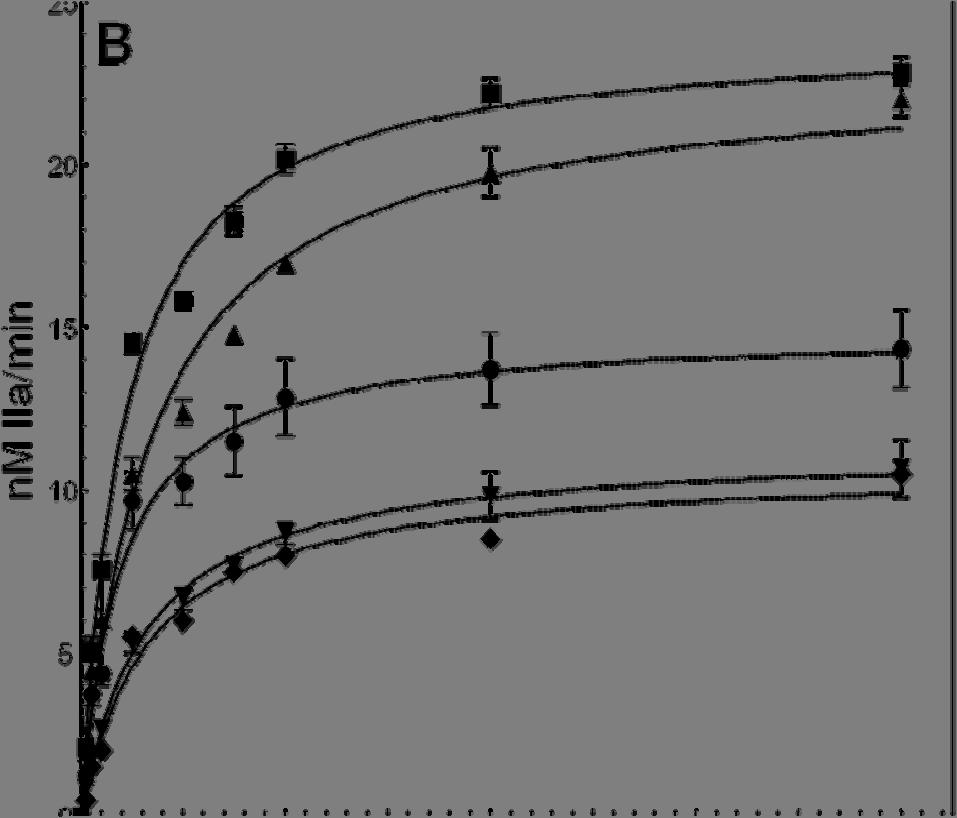
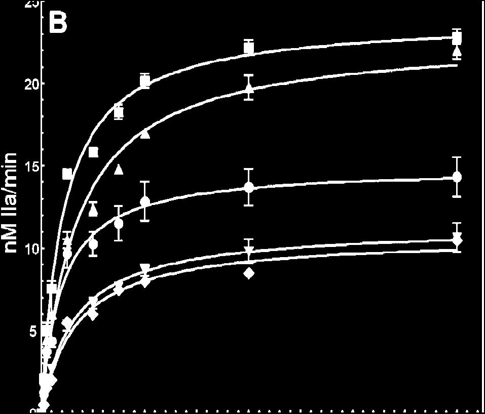
111 95
112 Figure 3.4B. Prothrombin titration to determine the kinetic parameters of prothrombinase assembled with various recombinant factor Va species. Thrombin generation experiments were carried out as described under Experimental Procedures by varying the substrate concentration (25 nm to 4 μm) with factor Xa saturated with each of the recombinant factor Va species. Factor V WT, factor V PLASMA, factor V Δ , factor V 5K, and factor V 5A were activated with thrombin as described in the Experimental Procedures section. The solid lines represent the nonlinear regression fit of the data using Prizm GraphPad software according to the Henri Michaelis-Menten equation. Prothrombinase was assembled with recombinant factor Va WT (filled squares, R 2 =0.99), factor Va PLASMA (filled triangles, R 2 =0.97), factor Va Δ (filled circles, R 2 =0.94), factor Va 5K (filled inverse triangles, R 2 =0.97), and factor Va 5A (filled diamonds, R 2 =0.98). The data with factor Va WT, factor Va PLASMA, factor Va Δ , and factor V 5K show the average results obtained from at least three different titrations with three different preparations of purified proteins. The data with factor Va 5A are representative of two titrations obtained in duplicate using two different preparations of recombinant protein. Kinetic constants reported in the text and in Table 3.I were extracted directly from fitted data. 96


113 Figure 3.4C. Determination of the kinetic parameters of prothrombinase assembled with the various recombinant factor Va species. Second-order rate constants for prothrombinase assembled with all cofactor molecules (M -1 s -1 ). 97
114 Factor Va Species Table 3.I Functional properties of various factor Va molecules Clotting Activity (U/mg) a K Dapp (nm) b k cat (min -1 ) c K m (μm) c Factor Va PLASMA 3124 ± ± ± ± 0.04 Factor Va WTa 2926 ± ± ± ± 0.01 Factor Va Δ ± ± ± ± 0.03 Factor Va 5K 275 ± ± ± ± 0.03 Factor Va 5A 120 ± ± ± ± 0.05 Table 3.I a Two-stage clotting assays of recombinant factor V molecules was performed as described in the Experimental Procedures section. b Apparent dissociation constants of recombinant factor Va for plasma-derived factor Xa (K Dapp ) were determined as described in the Experimental Procedures section at limiting factor Xa concentrations (15 pm) according to the binding model assuming one binding site using the software Prizm. Dissociation constants were derived directly from the fitted data. c The Km and kcat of prothrombinase assembled with saturating concentrations of recombinant factor Va molecules were determined as described in the Experimental Procedures section according to the Michaelis-Menten equation using the software Prizm. Kinetic constants were derived directly from the fitted data. 98
115 The findings obtained thus far suggest that prothrombin cleavage and activation by prothrombinase assembled with the recombinant mutant molecules is deficient. These data indicate that the rate of one or both of the two prothrombin activation cleavages are impaired when prothrombinase is assembled with a cofactor molecule mutated at the amino acid region of the heavy chain. To better understand the deficiency in prothrombin cleavage and activation by prothrombinase assembled with the recombinant mutant cofactor molecules, we have studied prothrombin activation by gel electrophoresis. The results demonstrate a delay in prothrombin activation by prothrombinase assembled with either Va Δ , factor Va 5K, or factor Va 5A as compared to the activation of prothrombin by prothrombinase assembled with either factor Va PLASMA or factor Va WT (Fig. 3.5, panels A-E). Scanning densitometry of the gels shown in figure 3.5 demonstrated a 3- and 7-fold delay in prothrombin consumption by prothrombinase assembled with factor Va Δ and factor Va 5K respectively, as compared to the consumption of prothrombin assembled with factor Va WT (Fig.3.6 and Table 3.II). Interestingly, prothrombin consumption by prothrombinase assembled with factor Va 5A was most severely impaired and no meizothrombin was visible during the activation process (Figs 3.5E). In addition, when prothrombin is activated by prothrombinase assembled with either factor Va Δ or factor Va 5K, there is persistence (lingering) of meizothrombin throughout the activation process (Figs 3.5C and 3.5D). Scanning densitometry demonstrated that a peak for meizothrombin is observed at 120 sec when prothrombin is activated by prothrombinase assembled with factor Va WT, while a peak for meizothrombin is detected at 200 sec and 360 sec when prothrombin is activated by prothrombinase assembled with either factor Va Δ or 99

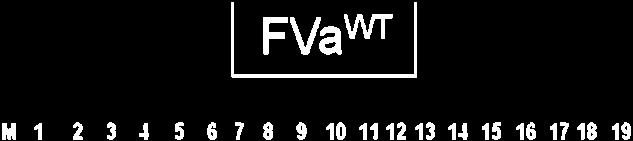
116 100
117 101
118 Figure 3.5. Analysis of the activation of plasma-derived prothrombin by prothrombinase assembled with various mutant factor Va molecules. Plasma-derived prothrombin (1.4 μm) was incubated in different mixtures with PCPS vesicles (20 μm), and prothrombinase assembled with either factor Va WT (panel A) or factor Va PLASMA (panel B) as described in the Experimental Procedures section. Panel C, prothrombinase assembled with factor Va Δ ; panel D, prothrombinase assembled with factor Va 5K ; panel E, prothrombinase assembled with factor Va 5A. The reactions were started by the addition of factor Xa. At selected time intervals aliquots of the reaction mixtures were withdrawn and treated as described in the Experimental Procedures section. M represents the lane with the molecular weight markers (from top to bottom): M r 98,000, M r 64,000, M r 50,000, M r 36,000. Lanes 1-19 represent samples from the reaction mixture before (0 min) the addition of factor Xa and 20 sec, 40 sec, 60 sec, 80 sec, 100 sec, 120 sec, 140 sec, 160 sec, 180 sec, 200 sec, 220 sec, 240 sec, 5 min, 6 min, 10 min, 20 min, 30 min, and 60 min respectively following the addition of factor Xa. 102


.")


, factor Va 5K (open squares, R 2 =0.")
119 Figure 3.6. Analysis of prothrombin consumption by prothrombinase assembled with recombinant factor Va molecules. The gels shown in figure 3.5 were scanned and prothrombin consumption was recorded as described in the Experimental Procedures section. Following scanning densitometry, the numbers were normalized to the initial concentration of prothrombin (1.4 μm). The data representing prothrombin consumption (μm) as a function of time (sec) were subsequently plotted using non-linear regression analysis according to the equation representing a first-order exponential decay using the software Prizm (GraphPad, San Diego, CA). Prothrombinase was assembled with recombinant factor Va WT (filled triangles, R 2 =0.98), factor Va PLASMA (filled circles, R 2 =0.98), factor Va Δ (filled squares, R 2 =0.99), factor Va 5K (open squares, R 2 =0.93), and factor Va 5A (open inverse triangles, the data were not a good fit to a first order exponential decay model, see Table 3.2). The apparent first-order rate constant, k (s -1 ), obtained directly from the fitted data was divided by the molar concentration of factor Xa used in each experiment. The number obtained was subsequently multiplied by the starting concentration of prothrombin. The resulting numbers representing prothrombin consumption are reported in Table 3.II. 103
120 Table 3.II Rate of activation of native plasma-derived prothrombin and recombinant mutant rmz-ii in the presence of prothrombinase assembled with various recombinant factor Va species Enzyme Membrane-bound factor Xa alone Plasma-derived Prothrombin * (Initial cleavage at Arg 320 ) (moles consumed sec -1 mole factor Xa -1 ) rmz-ii * (Cleavage at Arg 320 ) (moles consumed sec -1 mole factor Xa -1 ) ± Prothrombinase assembled with factor Va PLASMA 19 ± ± 0.8 Prothrombinase with 21.8 ± ± 1.1 factor Va WT Prothrombinase with 7.7 ± ± 0.6 factor Va Δ Prothrombinase with 3.3 ± ± 0.8 factor Va 5K Prothrombinase with factor Va 5A NF a NSC b Table 3.II. The rates of plasma-derived prothrombin and recombinant mutant prothrombin rmz-ii consumption were obtained following scanning densitometry of gels shown in Figs.3.5 and 3.7 respectively. The final rate of prothrombin consumption in the presence of prothrombinase assembled with various factor Va species, was extracted following plotting of the data according to the equation representing a first-order exponential decay as described in the Experimental Procedures section (Figs.3.6 and 3.9). The apparent first-order rate constants were obtained directly from the fitted data. a NF, no fit; the data could not be fitted to a first-order exponential decay (R 2 = ). The concentration of plasma-derived prothrombin varied from 1400 nm at time 0 to 200 nm following 1h incubation with prothrombinase assembled with factor Va 5A. b NSC, not significant consumption. The concentration of rmz-ii varied from 1400 nm at time 0 to 1260 nm following 2h incubation with prothrombinase assembled with factor Va 5A 104
121 factor Va 5K respectively. It is important to note, that in the latter case the detrimental effect of the mutations is more pronounced on prothrombin activation than when amino acid sequence is deleted. However, mutating all amino acids from this region to alanine appears to abolish the capability of factor Xa within prothrombinase for cleavage of prothrombin at Arg 320. These data are in complete accord with the data obtained in the clotting assays and the combined data suggest that both prothrombin activation cleavages (Arg 320 and Arg 271 ) appear to be affected to various degrees when prothrombinase is assembled with factor Va Δ , factor Va 5K, or factor Va 5A However, because meizothrombin lingers throughout the activation process, it appears that the rate of cleavage of prothrombin at Arg 271 is considerably more affected by a deletion of the amino acid region or a D K substitution, than the rate of cleavage of prothrombin at Arg 320. In contrast, the D A substitution in the hirudin-like acidic region appears to be equally detrimental to prothrombinase for cleavage of prothrombin at either Arg 320 or Arg 271. Overall, the data demonstrate that 1) amino acid region is important for optimum activity of prothrombinase and 2) the nature of the mutations in this region has an impact on the pathway and rate of prothrombin activation by prothrombinase. Activation of Recombinant Mutant Prothrombin by Prothrombinase Assembled with Mutant Factor Va molecules. The data obtained thus far studying plasma-derived prothrombin activation by prothrombinase assembled with the mutant cofactor molecules, demonstrate that 1) meizothrombin lingers throughout the time course, 2) appearance of the B-chain of thrombin is also delayed when prothrombin is activated by prothrombinase assembled with two of the three mutant cofactor molecules; and 3) initial cleavage of 105
122 prothrombin by prothrombinase assembled with factor Va 5A is severely impaired. In order to verify which cleavage in prothrombin is specifically affected by the modifications in the amino acid region of factor Va heavy chain, we first used prothrombin molecules that cannot be cleaved at either Arg 271 such as rmz-ii (Fig.3.7) or at Arg 320 (rp-ii, Fig.3.8) (24). The data demonstrate that there is a significant difference between the rates of activation of rmz-ii by prothrombinase assembled with factor Va Δ , factor Va 5K, or factor Va 5A as compared to rmz-ii activated by prothrombinase made with factor Va WT or factor Va PLASMA (Fig.3.7). Scanning densitometry of the gels shown in figure 3.7 demonstrate that cleavage at Arg 320 of rmz-ii is delayed by ~2-fold when prothrombinase is assembled with factor Va Δ and 4-fold when prothrombinase is assembled with factor Va 5K compared to cleavage of the recombinant mutant prothrombin molecule by prothrombinase assembled with factor Va WT (Table 3.II, and Fig.3.9). In contrast, no significant prothrombin consumption was observed when rmz-ii was incubated with prothrombinase assembled with factor Va 5A (Fig.3.7) The data confirm our overall findings with plasma-derived prothrombin and demonstrate that prothrombinase-mediated cleavage at Arg 320 in prothrombin is affected by the modifications/deletion of amino acids from the COOH-terminal portion of factor Va heavy chain. The data shown in figures 3.8 and 3.9, and in Table 3.III demonstrate that the rate of cleavage of rp2-ii at Arg 271 by prothrombinase assembled with factor Va Δ , factor Va 5K, or factor Va 5A is undetectable compared to the rate of cleavage of rp2-ii at Arg 271 by prothrombinase assembled with either factor Va WT or factor Va PLASMA. In fact, these 106
123 mutations in factor Va heavy chain practically abolish the capability of prothrombinase assembled with any of the mutant cofactor molecules to cleave rp2-ii at Arg 271. Overall, the data shown in figures demonstrate that mutations in the acidic amino acid segment differentially affect the rate of the two activating cleavage sites of prothrombin by prothrombinase. These data demonstrate that this portion of factor Va heavy chain controls the catalytic efficiency of factor Xa within the enzymatic complex. Activation of FPR-Meizothrombin by Prothrombinase Assembled with Mutant Factor Va molecules. Analysis of the results obtained thus far with plasma-derived and recombinant prothrombin suggests that activation of prothrombin by prothrombinase assembled with factor Va Δ , factor Va 5K, and factor Va 5A is severely impaired compared to cleavage of the recombinant molecule by prothrombinase assembled with factor Va WT because of delayed cleavage at both Arg 320 and Arg 271 resulting in both slow prothrombin consumption and less conversion of meizothrombin to thrombin. It has been shown that a change in conformation of meizothrombin is associated with cleavage of prothrombin at Arg 320 (35). To ascertain the effect of the mutations of the factor Va heavy chain on the cleavage of prothrombin at Arg 271 following the transition that occurs after cleavage at Arg 320, we compared the rate of cleavage of FPR-meizothrombin by prothrombinase assembled with either factor Va WT or the recombinant mutant cofactor molecules (Fig.3.10). The data demonstrate a delay for cleavage of FPR-meizothrombin at Arg 271 by prothrombinase assembled with factor Va Δ (panel B), factor Va 5K (panel C), or factor Va 5A (panel D) as compared to the same reaction catalyzed by prothrombinase assembled with factor Va WT (panel A). Quantitative scanning densitometry of fragment 1 2-A present on the gels shown in figure 3.10, demonstrated a 107
124 ~2-fold delay in cleavage of FPR-meizothrombin at Arg 271 by prothrombinase assembled with all mutant cofactor molecules, compared to cleavage at Arg 271 by prothrombinase assembled with factor Va WT (Fig.3.11, Table 3.III). However, the rates for cleavage of FPR-meizothrombin at Arg 271 are 2-fold faster than the rate of cleavage of FPRmeizothrombin by factor Xa alone. Thus, deletion or substitutions of amino acid residues within region has a similar effect on prothrombinase, namely, significantly impedes acceleration of the rate of cleavage at Arg 271 of meizothrombin attributed to the interaction of factor Va with factor Xa (3, 36). These data are in complete agreement with all the findings presented herein and overall our data demonstrate that amino acid sequence 659 DDDED 663 from the factor Va heavy chain is indispensable for optimal rates of thrombin formation during activation of prothrombin by prothrombinase. 108


125 Figure 3.7. Electrophoretic analyses of the activation of rmz-ii by prothrombinase assembled with mutant factor Va molecules. rmz-ii was incubated in different mixtures with PCPS vesicles, DAPA, and various factor Va molecules. The reaction was started by the addition of factor Xa and the samples were treated as detailed in the Experimental Procedures section. The gels were scanned and quantified as detailed under Experimental Procedures. Lanes 1-9, represent samples of the reaction mixture following incubation of prothrombinase with rmz-ii, before (lane 1), or following 1min, 3min, 5min, 10min, 20min, 45min, 60min, and 120min incubation with factor Xa respectively; Panel A, prothrombinase assembled with factor Va WT ; panel B prothrombinase assembled with plasma-derived factor Va; panel C, prothrombinase assembled with factor Va Δ ; panel D, prothrombinase assembled with factor Va 5K ; panel E, prothrombinase assembled with factor Va 5A ; panel F, is a schematic of the recombinant prothrombin molecule used (rmz-ii). This molecule is a triple mutant with only one cleavage site for prothrombinase remaining at Arg 320. Positions of prothrombinderived fragments are indicated at right as detailed in the legend to figure 3.5. The factor Va species used for the reconstitution of prothrombinase are shown under each panel. 109


126 Figure 3.8. Electrophoretic analyses of the activation of rp2-ii by prothrombinase assembled with mutant factor Va molecules. rp2-ii was incubated in different mixtures with PCPS vesicles, DAPA, and various factor Va molecules. The reaction was started by the addition of factor Xa and the samples were treated as detailed in the Experimental Procedures section. The gels were scanned and quantified as detailed under Experimental Procedures. Lanes 1-9, represent samples of the reaction mixture following incubation of prothrombinase with rp2-ii, before (lane 1), or following 1min, 3min, 5min, 10min, 20min, 45min, 60min, and 120min incubation with factor Xa respectively; Panel A, prothrombinase assembled with factor Va WT ; panel B prothrombinase assembled with plasma-derived factor Va; panel C, prothrombinase assembled with factor Va Δ ; panel D, prothrombinase assembled with factor Va 5K ; panel E, prothrombinase assembled with factor Va 5A ; panel F, is a schematic of the recombinant prothrombin molecule used (rp2-ii). This molecule is a triple mutant with only one cleavage site for prothrombinase remaining at Arg 271. Positions of prothrombinderived fragments are indicated at right as detailed in the legend to figure 3.5. The factor Va species used for the reconstitution of prothrombinase are shown under each panel. 110

 as described in the legend to figure 6.")
127 Figure 3.9. Analysis of recombinant prothrombin consumption by prothrombinase assembled with recombinant factor Va molecules. The gels shown in figures 3.7 and 3.8 were scanned and prothrombin consumption was recorded as described in the Experimental Procedures section. Following scanning densitometry, the data representing recombinant mutant prothrombin consumption as a function of time (sec) were plotted using non-linear regression analysis according to the equation representing a first-order exponential decay using the software Prizm (GraphPad, San Diego, CA) as described in the legend to figure 6. Prothrombinase was assembled with recombinant factor Va WT (filled triangles), factor Va PLASMA (filled circles), factor Va Δ (filled squares), factor Va 5K (open squares), and factor Va 5A (open inverse triangles). The apparent first-order rate constant, k (s -1 ) was obtained directly from the fitted data. The resulting numbers representing recombinant mutant prothrombin consumption are reported in Table 3.2 and table 3.3. Panel A; shows the data obtained following cleavage of rmz-ii: factor Va WT, R 2 =0.99, factor Va PLASMA, R 2 =0.99, factor Va Δ , R 2 =0.99, and factor Va 5K, R 2 =0.94. Panel B; shows the data obtained following cleavage of rp2-ii: factor Va WT, R 2 =0.99, factor Va PLASMA, R 2 =
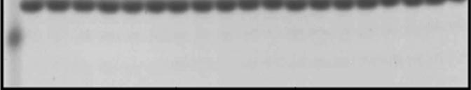
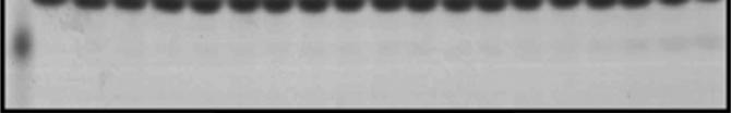
128 112
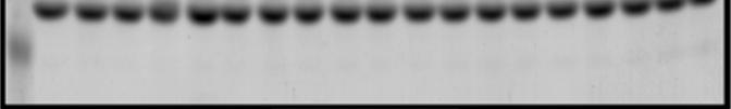
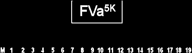
129 113
130 Figure Gel electrophoresis analyses for cleavage of FPR-meizothrombin. FPRmeizothrombin (1.4 μm) was incubated in different mixtures with PCPS vesicles (20 μm) and factor Va as described in the legend to figure 3.5. The reactions were started by the addition of factor Xa and the samples were further treated, scanned and quantified as detailed in the Experimental Procedures section. Panel A, factor Va WT ; panel B, factor Va Δ ; panel C, factor Va 5K, panel D, factor Va 5A, M represents the lane with the molecular weight markers (from top to bottom): M r 50,000, M r 36,000, M r 22,000. Lanes 1-19 represent samples from the reaction mixture before (0 min) the addition of factor Xa and 20 sec, 40 sec, 60 sec, 80 sec, 100 sec, 120 sec, 140 sec, 160 sec, 180 sec, 200 sec, 220 sec, 240 sec, 5 min, 6 min, 10 min, 20 min, 30 min, and 60 min respectively following the addition of factor Xa. The prothrombin derived fragments are shown as detailed in the legend to figure 3.5. The recombinant factor Va species used for the reconstitution of prothrombinase are shown under each panel. 114

 as described in the legend to figure 3.6.")
131 Figure Analysis of FPR-meizothrombin consumption by prothrombinase assembled with recombinant factor Va molecules. The gels shown in figure 3.10 were scanned and FPR-meizothrombin consumption was recorded as described in the Experimental Procedures section. Following scanning densitometry, the data representing prothrombin consumption as a function of time (sec) were plotted using nonlinear regression analysis according to the equation representing a first-order exponential decay using the software Prizm (GraphPad, San Diego, CA) as described in the legend to figure 3.6. The apparent first-order rate constant, k (s -1 ) was obtained directly from the fitted data. Prothrombinase was assembled with recombinant factor Va WT (filled triangles), factor Va Δ (filled squares), factor Va 5K (open squares), and factor Va 5A (open inverse triangles). The resulting numbers representing FPR-meizothrombin consumption are reported in Table 3.III. 115
132 Table 3.III Rates of activation of recombinant prothrombin rp2-ii and plasma-derived FPR- Meizothrombin in the presence of various recombinant factor Va species Enzyme rp2-ii * (Cleavage at Arg 271 ) (moles of rp2-ii consumed sec -1 mole factor Xa -1 ) FPR-Meizothrombin * (Cleavage at Arg 271 ) (moles of fragment 1 2-A consumed sec -1 mole factor Xa - Factor Xa alone ± a Prothrombinase with factor Va PLASMA Prothrombinase with factor Va WT Prothrombinase with factor Va Δ Prothrombinase with factor Va 5K Prothrombinase with factor Va 5A 3.3 ± a 3.4 ± ± 14 NSC b 23.5 ± 2.1 c NSC d NSC 1 ) 20.1 ± ± 2.2 Table 3.3. * Rates of rp2-ii consumption in the presence of wild-type or plasma factor Va and recombinant mutant factor Va Δ , factor Va 5K and factor Va 5A were measured following scanning densitometry of gels shown in Fig.3.8. Calculation of the apparent first-order rate constant was achieved as detailed in Experimental Procedures. The rate of fragment 1 2-A consumption in the presence of prothrombinase assembled with factor Va WT, factor Va Δ , factor Va 5K and factor Va 5A was measured following scanning densitometry of the gels shown in Fig.3.10 and calculation of the apparent first-order rate constant as detailed in Experimental Procedures. The rates of rp2-ii and fragment
133 A consumption were extracted from the fitted data (shown in Figs. 3.9 and 3.11 respectively). a From reference (18). b NSC, not significant consumption. The concentration of rp2-ii varied from 1400 nm at time 0 to 1250 nm following 2h min incubation with prothrombinase assembled with factor Va Δ c NSC, not significant consumption. The concentration of rp2-ii varied from 1400 nm at time 0 to 1220 nm following 2h min incubation with prothrombinase assembled with factor Va 5K. d NSC, not significant consumption. The concentration of rp2-ii varied from 1400 nm at time 0 to 1100 nm following 2h min incubation with prothrombinase assembled with factor Va 5A. 117
134 3.5 Discussion Our data demonstrate that the acidic region located at the COOHterminus of factor Va heavy chain is responsible for coordinated activation of prothrombin by prothrombinase resulting in timely thrombin formation at the place of vascular injury. To our knowledge, this is the first time that the role of this specific amino acid region of the cofactor has ever been investigated. The data presented here, assign an important physiological role to the acidic region from the COOHterminus of factor Va heavy chain for efficient prothrombin activation. Namely, amino acid sequence 659 DDDED 663 regulates the rate of cleavage of prothrombin by prothrombinase at Arg 320 and Arg 271. This acidic amino acid sequence together with sequence 695 DYDYQ 699 (18, 20, 21) are both located on the surface of the factor Va molecule (37), and are conserved among species (Fig.3.2) attesting to their physiological significance. Since both sequences have no effect on the direct interaction between factor Va and factor Xa, our findings are consistent with the interpretation that these acidic amino acid sequences contribute significantly to the macromolecular substrate recognition of prothrombin by prothrombinase and demonstrate that factor Va indeed regulates the activity of factor Xa within the enzymatic complex. Proteolytic inactivation of factor Va by activated protein C (APC) following cleavages at Arg 506, Arg 306, and Arg 679 (38, 39) results in the arrest of the contribution of the cofactor to the catalytic efficiency of factor Xa. Earlier data have demonstrated that factor Xa and prothrombin protect the cofactor from APC inactivation (40-42). Functional data obtained with plasma or natural and/or recombinant mutant factor Va molecules have established that cleavages at Arg 306 and Arg 506 result in the dissociation of the A2 domain of the cofactor from the rest of the molecule (37, 43-46). The A2 118
135 domain of factor Va contains the major factor Xa binding domains (13, 14, 47, 48). Thus, dissociation of the A2 domain from the rest of the molecule results in impaired factor Xa-factor Va interaction. However, a specific role for cleavage at Arg 679 has not been yet determined and has been mystifying scientists since the initial identification of the specific inactivating cleavage sites of the human cofactor approximately 15 years ago (39). Our data demonstrate that the acidic hirudin-like sequences from the COOHterminal portion of the heavy chain of factor Va direct the rate of prothrombin activation by factor Xa because they are part of a regulatory group of amino acids from the cofactor controlling the rate of the two prothrombin activation cleavage sites. Our data put in the context of the literature suggest that cleavage of the cofactor by APC at Arg 679 destabilizes the structure of the COOH-terminus of the heavy chain of factor Va and eliminates its regulatory role on the two prothrombin activation cleavages, resulting in uncontrolled meizothrombin generation by the prothrombinase complex. This conclusion is also strengthened by the fact that the acidic regions from the COOH-terminal region of factor Va heavy chain together with the Arg 679 APC-cleavage site are conserved among species (Fig.3.2). Overall, the data strongly suggest that proteolytic inactivation of factor Va by APC results in the inability of the cofactor to bind factor Xa and to provide a productive regulatory interaction with prothrombin. Thus, our findings also offer for the first time a logical explanation for the role of cleavage of factor Va by APC at Arg 679. Data obtained with plasma-derived prothrombin show delay in prothrombin consumption when prothrombinase was assembled with factor Va Δ , factor Va 5K, or factor Va 5A, compared to plasma-derived prothrombin activation by prothrombinase assembled with factor Va WT. Clotting assays using factor Va Δ , factor Va 5K, and 119
136 factor Va 5A showed a comparable decrease in clotting activity compared to the activity of factor Va WT. Similarly, experiments using rmz-ii demonstrate that cleavage at Arg 320 and rmz-ii consumption is delayed significantly when prothrombinase is assembled with factor Va Δ , factor Va 5K, and factor Va 5A, as compared to cleavage of rmz-ii by prothrombinase assembled with factor Va WT. These data together with the data shown in figures 3.8 and 3.10 demonstrating that elimination of the amino acid region in factor Va heavy chain or substitution of all amino acids from this region by either lysine or alanine, result in the elimination of the acceleration of the rate of cleavage of rp2-ii and FPR-meizothrombin at Arg 271 due to the interaction of factor Va with factor Xa. These data suggest that lingering of meizothrombin during prothrombin activation by prothrombinase assembled with the mutant cofactor molecules is the consequence of delayed cleavage at and Arg 271. However, we must note a differential effect of the mutations on the two prothrombin activation cleavage sites. Thus, while prothrombinase assembled with factor Va 5A is severely impaired for initial cleavage at Arg 320 of prothrombin or rmz-ii as compared to cleavage of the two molecules by prothrombinase assembled with either factor Va Δ or factor Va 5K, prothrombinase assembled with factor Va 5A has similar activity for cleavage of either rp2-ii or FPR-meizothrombin as prothrombinase assembled with factor Va Δ or factor Va 5K. Overall, our findings convincingly demonstrate that prothrombinase assembled with factor Va Δ is a better enzyme than prothrombinase assembled with factor Va 5K, which in turn is a better enzyme than prothrombinase assembled with factor Va 5A for cleavage at Arg 320. These data strongly suggest a differential effect of the mutations within amino acid region of factor Va heavy chain on the rate of the two prothrombin activating cleavage sites. 120
137 It is well established that cleavage of prothrombin at Arg 271 has an important effect on the progressive formation of the active site of thrombin (49, 50), and that meizothrombin has higher amidolytic activity than thrombin towards several chromogenic substrates usually employed to assess thrombin esterase activity (23, 51-53). In addition, it has been demonstrated following analysis of the crystal structure of meizothrombin, that ABE-II of meizothrombin has not yet been exposed, because it is covered by fragment 2 (54). This fact alone explains the poor clotting activity of meizothrombin since ABE-II is part of the binding site of thrombin for fibrinogen and is required for optimal rate of fibrin formation during blood clotting. ABE-II is exposed following cleavage of meizothrombin at Arg 271 and release of fragment 2. However, the catalytic activity of prothrombinase when assembled with the mutant cofactor molecules is only moderately affected by the mutations, when the activity of factor Va is measured in an assay using a chromogenic substrate to assess for thrombin activity (half to 1/3 of the values found with prothrombinase assembled with the wild type cofactor). Indeed, the k cat and second order rate constants of prothrombinase assembled with the mutant factor Va molecules are approximately 50% of that of prothrombinase assembled with factor Va PLASMA or factor Va WT. In contrast, the clotting activity of the mutant cofactor molecules is severely impaired and gel electrophoresis experiments combined with densitometric analyses reveal a significant delay in prothrombin activation with meizothrombin being more stable throughout the activation process when prothrombinase is assembled with the mutant molecules as compared to prothrombin activation by prothrombinase assembled with either factor Va WT or factor Va PLASMA. The data demonstrate a delay in prothrombin activation and thrombin generation because of impaired rates of cleavage at both Arg
138 and Arg 271 with the rate of cleavage at Arg 271 being affected the most, and with the 659 DDDED 663 AAAAA substitution being the most detrimental to factor Va cofactor activity. The findings presented herein are entirely consistent with earlier findings obtained with plasma-derived proteins (55-57), recent findings obtained with recombinant proteins (58) and our data (18, 19). Explicitly, when prothrombinase is assembled with a factor Va molecule possessing a heavy chain which is truncated at the acidic hirudin-like COOH-terminal region, a discrepancy is observed between the activity of factor Va measured by clotting assay as compared to the activity of the cofactor measured in an assay using a chromogenic substrate to assess for thrombin activity. The data provided herein and in our recent manuscript detailing the properties of factor V Δ (18), explain these paradoxical findings. Because we have studied prothrombin, rmz- II, rp2-ii, and FPR-meizothrombin activation by gel electrophoresis, we can conclude that meizothrombin lingers throughout the activation of prothrombin by prothrombinase assembled with the recombinant mutant molecules. The excess meizothrombin present in the assays while having poor clotting activity can compensate for the lack of thrombin activity because of its increased amidolytic activity towards chromogenic substrates (51, 52) that are usually employed to assess thrombin activity, thus creating the false impression that the mutations have a minimal effect on prothrombinase activity. As a consequence, and because factor Va is a cofactor molecule devoid of enzymatic activity, when studying recombinant mutant factor Va molecules, drawing conclusions from activity assays only, without visualizing the pathway to thrombin generation, would lead to incorrect interpretations of the prothrombin intermediates formed and the rate of the specific prothrombin activating cleavages. We therefore conclude that following the 122
139 activity of various recombinant mutant factor Va molecules by clotting assays, gel electrophoresis analyses, and assays using chromogenic substrates is by no means redundant and prevents oversimplification that in turn might lead to flawed conclusions. 123
140 3.6 Reference List 1. Kalafatis, M., Egan, J. O., van't Veer, C., Cawthern, K. M., and Mann, K. G. (1997) The regulation of clotting factors, Crit. Rev. Eukar. Gene. 7, Mann, K. G., and Kalafatis, M. (2003) Factor V: a combination of Dr Jekyll and Mr Hyde, Blood 101, Nesheim, M. E., and Mann, K. G. (1983) The kinetics and cofactor dependence of the two cleavages involved in prothrombin activation, J. Biol. Chem. 258, Nesheim, M. E., Taswell, J. B., and Mann, K. G. (1979) The contribution of bovine factor V and factor Va to the activity of prothrombinase, J. Biol. Chem. 254, Heldebrant, C. M., Butkowski, R. J., Bajaj, S. P., and Mann, K. G. (1973) The activation of prothrombin. II. Partial reactions, physical and chemical characterization of the intermediates of activation, J. Biol. Chem. 248, Esmon, C. T., Owen, W. G., and Jackson, C. M. (1974) A plausible mechanism for prothrombin activation by factor Xa, factor Va, phospholipid, and calcium ions, J. Biol. Chem. 249, Anderson, P. J., and Bock, P. E. (2003) Role of prothrombin fragment 1 in the pathway of regulatory exosite I formation during conversion of human prothrombin to thrombin, J. Biol. Chem. 278, Anderson, P. J., Nesset, A., Dharmawardana, K. R., and Bock, P. E. (2000) Role of proexosite I in factor Va-dependent substrate interactions of prothrombin activation, J. Biol. Chem. 275,
141 9. Chen, L., Yang, L., and Rezaie, A. R. (2003) Proexosite-1 on prothrombin is a factor Va-dependent recognition site for the prothrombinase complex, J. Biol. Chem. 278, Walker, R. K., and Krishnaswamy, S. (1994) The activation of prothrombin by the prothrombinase complex. The contribution of the substrate-membrane interaction to catalysis, J. Biol. Chem. 269, Bukys, M. A., Orban, T., Kim, P. Y., Nesheim, M. E., and Kalafatis, M. (2008) The interaction of fragment 1 of prothrombin with the membrane surface is a prerequisite for optimum expression of factor Va cofactor activity within prothrombinase, Thromb Haemost 99, Adams, T. E., Hockin, M. F., Mann, K. G., and Everse, S. J. (2004) The crystal structure of Activated Protein C-inactivated bovine factor Va: Implications for cofactor function, Proc. Natl. Acad. Sci. U.S.A. 101, Kalafatis, M., and Beck, D. O. (2002) Identification of a binding site for blood coagulation factor Xa on the heavy chain of factor Va. Amino acid residues of factor V represent an interactive site for activated factor X, Biochemistry 41, Singh, L. S., Bukys, M. A., Beck, D. O., and Kalafatis, M. (2003) Amino acids Glu323, Tyr324, Glu330, and Val331 of factor Va heavy chain are essential for expression of cofactor activity, J. Biol. Chem. 278, Barhoover, M. A., Orban, T., Beck, D. O., Bukys, M., and Kalafatis, M. (2008) Contribution of Amino Acid Region from Factor Va Heavy Chain to the Catalytic Efficiency of Prothrombinase, Biochemistry 47,
142 16. Esmon, C. T., Owen, W. G., Duiguid, D. L., and Jackson, C. M. (1973) The action of thrombin on blood clotting factor V: conversion of factor V to a prothrombin-binding protein, Biochim Biophys Acta 310, Luckow, E. A., Lyons, D. A., Ridgeway, T. M., Esmon, C. T., and Laue, T. M. (1989) Interaction of clotting factor V heavy chain with prothrombin and prethrombin 1 and role of activated protein C in regulating this interaction: analysis by analytical ultracentrifugation, Biochemistry 28, Hirbawi, J., Bukys, M., Barhoover, M. A., Erdogan, E., and Kalafatis, M. (2008) Role of the acidic hirudin-like COOH-terminal amino acid region of factor Va heavy chain in the enhanced function of prothrombinase, Biochemistry 47, Kalafatis, M., Beck, D. O., and Mann, K. G. (2003) Structural requirements for expression of factor Va activity, J. Biol. Chem. 278, Beck, D. O., Bukys, M. A., Singh, L. S., Szabo, K. A., and Kalafatis, M. (2004) The contribution of amino acid region ASP695-TYR698 of factor V to procofactor activation and factor Va function, J. Biol. Chem. 279, Bukys, M. A., Kim, P. Y., Nesheim, M. E., and Kalafatis, M. (2006) A control switch for prothrombinase: characterization of a hirudin-like pentapeptide from the COOH terminus of factor Va heavy chain that regulates the rate and pathway for prothrombin activation, J. Biol. Chem. 281, Pittman, D. D., Tomkinson, K. N., Michnick, D., Selighsohn, U., and Kaufman, R. J. (1994) Posttranslational sulfation of factor V is required for efficient 126
143 thrombin cleavage and activation and for full procoagulant activity, Biochemistry 33, Morita, T., and Iwanaga, S. (1981) Prothrombin activator from Echis carinatus venom, Methods in Enzymology 90, Côté, H. C. F., Stevens, W. K., Bajzar, L., Banfield, D. K., Nesheim, M. E., and MacGillivray, T. A. (1994) Characterization of a stable form of human meizothrombin derived from recombinant prothrombin (R155A, R271A, and R284A), J. Biol. Chem. 269, Erdogan, E., Bukys, M. A., Orfeo, T., Mann, K. G., and Kalafatis, M. (2007) Identification of an inactivating cleavage site for α-thrombin on the heavy chain of factor Va, Thromb Haemost 98, Laemmli, U. K. (1970) Cleavage of structural proteins during the assembly of the head of bacteriophage T4, Nature 227, Towbin, H., Staehlin, T., and Gordon, J. (1979) Electrophoretic transfer of proteins from polyacrylamide gels to nitrocellulose sheets: procedure and some applications, Proc. Natl. Acad. Sci. U S A. 76, van 't Veer, C., Golden, N. J., Kalafatis, M., and Mann, K. G. (1997) Inhibitory mechanism of the protein C pathway on tissue factor-induced thrombin generation. Synergistic effect in combination with tissue factor pathway inhibitor, J. Biol. Chem. 272, Kalafatis, M., Haley, P. E., Lu, D., Bertina, R. M., Long, G. L., and Mann, K. G. (1996) Proteolytic events that regulate factor V activity in whole plasma from 127
144 normal and activated protein C (APC)-resistant individuals during clotting: an insight into the APC-resistance assay, Blood 87, Merril, C. R., Dunau, M. L., and Goldman, D. (1981) A rapid sensitive silver stain for polypeptide in polyacrylamide gels, Analytical Biochemistry 110, Bukys, M. A., Orban, T., Kim, P. Y., Beck, D. O., Nesheim, M. E., and Kalafatis, M. (2006) The structural integrity of anion binding exosite-i of thrombin is required and sufficient for timely cleavage and activation of factor V and factor VIII, J. Biol. Chem. 281, Kim, P. Y., and Nesheim, M. E. (2007) Further evidence for two functional forms of prothrombinase each specific for either of the two prothrombin activation cleavages, J. Biol. Chem. 282, Erdogan, E., Bukys, M. A., and Kalafatis, M. (2008) The contribution of amino acid residues of factor V to light chain generation, J Thromb Haemost. 6, Krishnaswamy, S., Williams, E. B., and Mann, K. G. (1986) The binding of activated protein C to factors V and Va, J. Biol. Chem. 261, Bianchini, E. P., Orcutt, S. J., Panizzi, P., Bock, P. E., and Krishnaswamy, S. (2005) Racheting of the substrate from the zymogen to proteinase conformations directs the sequential cleavage of prothrombin by prothrombinase, Proc. Natl. Acad. Sci. U S A. 102, Krishnaswamy, S., Church, W. R., Nesheim, M. E., and Mann, K. G. (1987) Activation of human prothrombin by human prothrombinase. Influence of factor Va on the reaction mechanism, J. Biol. Chem. 262,
145 37. Orban, T., Kalafatis, M., and Gogonea, V. (2005) Completed three-dimensional model of human coagulation factor Va. Molecular dynamics simulations and structural analyses, Biochemistry 44, Kalafatis, M., and Mann, K. G. (1993) Role of the membrane in the inactivation of factor Va by activated protein C, J. Biol. Chem. 268, Kalafatis, M., Rand, M. D., and Mann, K. G. (1994) The mechanism of inactivation of human factor V and human factor Va by Activated Protein C, J. Biol. Chem. 269, Nesheim, M. E., Canfield, W. M., Kisiel, W., and Mann, K. G. (1982) Studies of the capacity of factor Xa to protect factor Va from inactivation by activated protein C, J. Biol. Chem. 257, Solymoss, S., Tucker, M. M., and Tracy, P. B. (1988) Kinetics of inactivation of membrane-bound factor Va by activated protein C. Protein S modulates factor Xa protection, J. Biol. Chem. 263, Smirnov, M. D., Safa, O., Esmon, N. L., and Esmon, C. T. (1999) Inhibition of activated protein C anticoagulant activity by prothrombin, Blood 94, Rosing, J., Hoekema, L., Nicolaes, G. A., Thomassen, M. L. G. D., Hemker, H. C., Varadi, K., Schwartz, H. P., and Tans, G. (1995) Effects of Protein S and factor Xa on peptide bond cleavages during inactivation of factor Va and factor VaR506Q by Activated Protein C, J. Biol. Chem. 270, Tran, S., Norstrøm, E., and Dahlbäck, B. (2008) Effects of prothrombin on the individual activated protein C-mediated cleavages of coagulation factor Va, J. Biol. Chem. 283,
146 45. Yegneswaran, S., Nguyen, P. M., Gale, A. J., and Griffin, J. H. (2009) Prothrombin amino terminal region helps protect coagulation factor Va from proteolytic inactivation by activated protein C, Thromb Haemost 101, Mann, K. G., Hockin, M. F., Begin, K. J., and Kalafatis, M. (1997) Activated Protein C cleavage of factor Va leads to dissociation of the A2 domain, J. Biol. Chem. 272, Bukys, M. A., Blum, M. A., Kim, P. Y., Brufatto, N., Nesheim, M. E., and Kalafatis, M. (2005) Incorporation of factor Va into prothrombinase is required for coordinated cleavage of prothrombin by factor Xa, J. Biol. Chem. 280, Heeb, M. J., Kojima, Y., Hackeng, T. M., and H., G. J. (1996) Binding sites for blood coagulation factor Xa and protein S involving residues in factor Va, Protein Sci. 9, Hibbard, L. S., Nesheim, M. E., and Mann, K. G. (1982) Progressive development of a thrombin inhibitor binding site, Biochemistry 1982, Boxrud, P. D., and Berliner, L. J. (1996) Comparison of the active-site conformations of bovine a-thrombin and meizothrombin(desf1) by electron spin resonance, J. Prot. Chem. 15, Doyle, M. F., and Mann, K. G. (1990) Multiple active forms of thrombin. Relative activities of meizothrombins, J. Biol. Chem. 265, Doyle, M. F., and Haley, P. E. (1993) Meizothrombin: active intemediate formed during prothrombin-catalyzed activation of prothrombin, Methods in Enzymology 222,
147 53. Shim, K., Zhu, H., Westfield, L. A., and Sadler, J. E. (2004) A recombinant murine meizothrombin precursor, prtothrombin R157A/R268A, inhibits thrombosis in a model of acute carotide artery injury, Blood 104, Martin, P. D., Malkowski, M. G., Box, J., Esmon, C. T., and Edwards, B. F. (1997) New insights into the regulation of the blood clotting cascade derived from the X-ray crystal structure of bovine meizothrombin des F1 in complex with PPACK, Structure 5, Gerads, I., Tans, G., Yukelson, L. Y., Zwaal, R. F., and Rosing, J. (1992) Activation of bovine factor V by an activator purified from the venom of Naja naja oxiana, Toxicon 30, Bakker, H. M., Tans, G., Thomassen, M. C. L. G. D., Yukelson, L. Y., Ebberink, R., Hemker, H. C., and Rosing, J. (1994) Functional properties of human factor Va lacking Asp683-Arg709 domain of the heavy chain, J. Biol. Chem. 269, Camire, R. M., Kalafatis, M., and Tracy, P. B. (1998) Proteolysis of factor V by cathepsin G and elastase indicates that cleavage at Arg1545 optimizes cofactor function by facilitating factor Xa binding, Biochemistry 37, Toso, R., and Camire, R. M. (2006) Role of Hirudin-like factor Va heavy chain sequences in prothrombinase function, J. Biol. Chem. 281,
148 CHAPTER IV THE COFACTOR EFFECT OF AMINO ACIDS 695 AND 696 OF FACTOR VA HEAVY CHAIN ON THE ENZYMATIC ACTIVITY OF THE PROTHROMBINASE COMPLEX 4.1 Abstract Blood coagulation is initiated after vascular injury, promoting formation of the fibrin clot. Without the proper regulation of this process, serious life threatening conditions, such as DVT (deep vein thrombosis), can occur. The proteolytic conversion of prothrombin to thrombin is catalyzed by the prothrombinase complex composed of the enzyme, factor Xa (fxa), the cofactor, factor Va (fva), assembled on a membrane surface in the presence of Ca 2+ [1]. The incorporation of fva into the prothrombinase complex results in a 300,000-fold increase in the catalytic efficiency of fxa for thrombin generation. Prothrombinase activates prothrombin through initial cleavage at Arg 320 followed by cleavage at Arg 271 to yield human alpha-thrombin [2]. This pathway is 132
149 responsible for the generation of a transient intermediate, meizothrombin, which is enzymatically active with increased chromogenic substrate activity, but yields poor clotting activity [3]. Factor Va is composed of heavy and light chains that play a crucial role during thrombin formation. Portions of the fva heavy chain have been found to act as fxa binding sites and to interact with proexosite 1 (pro1) of prothrombin to increase prothrombinase activity [4, 5]. It has been recently demonstrated that deletion of the COOH-terminal region of the factor Va heavy chain causes accumulation of meizothrombin due to delayed cleavage of prothrombin at Arg 271 [3]. Site-directed mutagenesis was performed to generate recombinant mutant molecules to identify the specific amino acids of this terminal region that regulate cleavage. Mutants with the 695 DYDY 698 DFDY(fVa DFDY ), KFDY(fVa KFDY ), DEDE(fVa DEDE ), DFDF(fVa DFDF ) substitutions were constructed. SDS-PAGE analyses of prothrombin activation time courses revealed that the overall cleavage of prothrombin by prothrombinase assembled with fva KFDY and fva DFDY was delayed, while fva DEDE, and fva DFDF had no significant effects when compared to fva WT. Two- stage clotting assays (PT times) revealed that prothrombinase assembled with fva KFDY had reduced clotting activity when compared to fva WT, while fva DEDE, fva DFDY, and fva DFDF gave similar clotting results as fva WT. Determination of k cat values for prothrombinase assembled with the various recombinant molecules revealed that fva KFDY and fva DFDY lead to an increase in the catalytic efficiency of the enzyme, while fva DEDE, and fva DFDF gave values that were normal. The data presented suggests that the 695 DY 696 portion of the acidic cluster found in the COOH-terminus of the fva heavy chain plays a significant role in enzyme-substrate interaction during thrombus formation. 133
150 4.2 Introduction The penultimate goal of the coagulation response is the generation of thrombin, which in turn converts fibrinogen to fibrin to produce a fibrin plug. This cascade of enzymatic reactions begins immediately after vascular injury and is a tightly regulated process involving a multitude of proteins and enzyme complexes. The proteolytic conversion of prothrombin to thrombin is catalyzed by the prothrombinase complex composed of the enzyme, factor Xa, its cofactor, factor Va, assembled on a membrane surface in the presence of divalent metal ions [6]. Although factor Xa can activate prothrombin with an initial cleavage at Arg 271 followed by cleavage at Arg 320 to yield the intermediates Fragment 1.2 and Prethrombin 2, incorporation of factor Va into the prothrombinase complex results in a reversal of cleavages and a 300,000-fold increase in the catalytic efficiency of factor Xa for thrombin generation [7]. A first cleavage of prothrombin by prothrombinase at Arg 320 produces the active intermediate meizothrombin (Fragment 1.2A), while the second cleavage at Arg 271 produces thrombin. The incorporation of factor Va into the prothrombinase complex is of absolute importance to the production of thrombin during the coagulation response. It exists in the human body at a physiological concentration of 20nM and presents itself as a single chain protein with a M r of 330,000 [8]. The cofactor is composed of three protein domains (A, B, & C) that are arranged in the order of A1-A2-B-A3-C1-C2. It is cleaved by α-thrombin (fiia) at Arg 709, Arg 1018, and Arg 1545 to release the B domain (containing a large number of asparagine-linked oligosaccharides) and generate an active cofactor (fva Iia ) composed of a light chain of M r 74 kda (A3-C1-C2 domains) and a heavy chain of M r 105 kda (A1-A2 domains) [9, 10]. It has been frequently demonstrated that factor 134
151 Va will increase the efficiency of factor Xa by decreasing the K m by 100-fold and increasing K cat values by 3,000-fold [11]. This decrease in K m is due to tighter binding of factor Xa to the membrane surface, allowing higher concentrations of the enzyme to localize while the K cat value is improved due to factor Va interaction with factor Xa [12]. Although the factor Va heavy chain interacts with factor Xa and anion exosite I of prothrombin during its conversion to thrombin, the exact mechanism by which this occurs has not been clarified [13]. Anion exosite I is known to be rich in basic amino acid residues that may interact with the factor Va molecule [14]. The carboxy-terminal region of the factor Va heavy chain contains small clusters of acidic amino acids, which are believed to play a role in the cofactor effect of the molecule on the prothrombinase complex [15]. We recently showed that amino acid region of the heavy chain regulates proteolytic cleavage of the substrate, prothrombin, at Arg 271 by decreasing the rate of cleavage [3, 16]. In addition, a pentapeptide (DYDYQ) consisting of the region Asp 695 -Gln 699 was also found to inhibit prothrombinase function as well as inhibiting activation of factor V by human alpha thrombin. The following studies were undertaken to further understand the importance of the COOH-terminal region of the factor Va heavy chain during proteolytic cleavage of prothrombin by prothrombinase. 135
152 4.3 Experimental Procedures Materials reagents, and proteins. Diisopropyl-fluorophosphate (DFP), O- phenylenediamine dihydrochloride (OPD), N-[2-Hydroxyethyl]piperazine-N'-2- ethanesulfonic acid (Hepes), Trizma (Tris base), and Coomassie Blue R-250 were purchased from Sigma (St. Louis, Mo). Factor V-deficient plasma was from Research Proteins Inc (Essex Junction VT). L-α-phosphatidylserine (PS) and L-aphosphatidylcholine (PC) were from Avanti Polar Lipids (Alabaster, AL). Normal reference plasma and the chromogenic substrate H-D-Hexahydrotyrosol-alanyl-arginyl-pnitroanilide diacetate (Spectrozyme-TH) were purchased from American Diagnostica Inc. (Greenwich, CT). H-D-Phenylalanyl-L-pipecolyl-L-arginyl-p-nitroaniline dihydrochloride (Chromogenix, S-2238) was purchased from Diapharma Group, Inc. (West Chester, OH) and its concentration in solution (water) was verified as described (41). RecombiPlasTin for the clotting assays was purchased from Instrumentation Laboratory Company (Lexington, MA). The reversible fluorescent α-thrombin inhibitor dansylarginine N,N-(3-ethyl-1,5-pentanediyl)amide (DAPA), human prothrombin, RVV- V activator, and human α-thrombin, were from Haematologic Technologies Inc. (Essex Junction, VT). Active-site blocked human meizothrombin (obtained following digestion of prothrombin with the purified component from the venom of the snake Echis Carinatus as described). FPR-meizothrombin was provided by Dr. Rick Jenny (Haematologic Technologies Inc, Essex Junction VT). Human factor Xa was from Enzyme Research Laboratories (South Bend, IN). Human cathepsin G was from Calbiochem (EMD Chemicals, Inc. San Diego, CA). All molecular biology and tissue culture reagents and media were from Gibco, Invitrogen Corporation (Grand Island, NY). 136
153 Human plasma factor V was purified and concentrated using methodologies previously described employing the monoclonal antibody αhfv#1 coupled to Sepharose. Digestion of factor Va by a-thrombin and cathepsin G (to obtain factor Va II/CG ) and/or the purified enzyme from Naja Naja Nigricollis (factor Va NN ) were performed as described. The clotting activities of all factor Va preparations was measured by a clotting assay using factor V deficient plasma and standardized to the percentage of control as described using an automated coagulation analyzer (START-4, Diagnostica Stago, Parsippany, NJ). Recombinant wild type prothrombin and prothrombin rmz-ii that has only one cleavage site for factor Xa (i.e. Arg 320 ) were prepared and purified as previously described Phospholipid vesicles composed of 75% PC and 25% PS (referred to as PCPS vesicles throughout the manuscript) were prepared as previously described. Construction of Recombinant FV Molecules. Mutant factor V with the substitutions 695 DYDY 698 DEDE (factor V DEDE ) was constructed using the QuickChange II XL site-directed mutagenesis kit (Stratagene, La Jolla, CA, USA) according to the manufacturer s instructions with the following primers (underlined nucleotides represent the mismatch): 5 -GAGAGTGATGCTGACGAAGATGAACAG 3 (sense) and 5 -CTGTTCATCTTCGTCAGCATCACTCTC-3 (anti-sense). The deletion mutant (factor V DFDF) ) was constructed with the same kit. Primers for factor V DFDF were 5 -GAGAGTGATGCTGACTTTGATTTCCAG-3 (sense) and 5 - CTGGAAATCAAAGTCAGCATCACTCTC-3 (anti-sense). In addition, the mutant factor V KFDY with the following primers was constructed: 5 - GAGAGTGATGCTAAGTTTGATTACCAG-3 (sense) and 5 - CTGGTAATCAAACTTAGCATCACTCTC -3 (antisense). One more mutant was also 137
154 made, factor V DFDY with primers: 5 -GAGAGTGATGCTGACTTTGATTACCAG-3 (sense) and 5 -CTGGTAATCAAAGTCAGCATCACTCTC-3 (antisense). PCR products were transformed into competent E. Coli cells and positive ampicillin-resistant clones were selected. Before transfection, all mutant constructs were verified following sequencing in the Cleveland State University DNA Analysis Facility using a Beckman Coulter CEQ 8000 Genetic Analysis System (Beckman, Fullerton CA) with factor-v sequence-specific primers. The wild type pmt2-fv and mutant pmt2-fv plasmids were isolated from the bacterial culture by the QIAfilter High Speed plasmid Midi Kit (Qiagen Inc., Valencia, CA). Expression of Recombinant Wild Type and Mutant Factor V in Mammalian Cells. COS-7L and COS-7 cells (Invitrogen) were maintained in Dulbecco s modified Eagle s medium supplemented with 10% fetal bovine serum, 2mM L-glutamine, and antibiotics (100 μg/ml streptomycin and 100 IU/ml penicillin) in a humidified atmosphere of 5% CO 2 and 95% air at 37 C. Purified factor V Wt, factor V D , and factor V 4A plasmids were transfected into the cells as described. Purification of all recombinant factor V molecules was performed as described. The concentration of all molecules was assessed by ELISA as detailed. The activity and integrity of the recombinant molecules was verified before and after activation with thrombin by clotting assays using factor V-deficient plasma and and in several experiments by sodium dodecyl sulfate-polyacrylamide gel electrophoresis (SDS-PAGE) followed by Western blotting using monoclonal and polyclonal antibodies. Analysis of Prothrombin Activation and FPR-Meizothrombin Cleavage at Arg 271 by Gel Electrophoresis. Prothrombin (1.4μM) was incubated with PCPS vesicles 138
155 (20μM), DAPA (50μM), and factor Va (10-30nM) in a buffer composed of 5 mm Ca 2+ in 20 mm Tris, 0.15 M NaCl, ph 7.4. The reaction was initiated with the addition of factor Xa (0.5-1nM) at room temperature over a 1 h time course. Aliquots (50 μl) from the reaction were removed at selected time intervals (as indicated in the legend to the figures) treated as described and analyzed using 9.5% SDS-PAGE. Prothrombin and prothrombin-derived fragments were visualized by Coomassie Blue staining. Scanning densitometry and calculation of the rates of prothrombin consumption were performed as described. FPR-meizothrombin cleavage at Arg 271 was assessed in a similar manner. Gel Electrophoresis and Western Blotting. SDS-PAGE analyses of recombinant proteins following activation were performed using 5-15% gradient gels according to the method of Laemmli. Proteins were transferred to polyvinylidene difluoride (PVDF) membranes according to the method described by Towbin et al. After transfer to nitrocellulose, factor Va heavy and light chain(s) were detected using the appropriate monoclonal and polyclonal antibodies. Immunoreactive fragments were visualized with chemiluminescence. Measurement of Rates of Thrombin Formation in a Prothrombinase Assay. rmz-ii and recombinant prothrombin were activated by prothrombinase prior to the experiment using conditions previously described. Subsequent gel electrophoresis analyses under reducing conditions were performed to verify that both rmz-ii and the recombinant prothrombin preparations were activated to the same extent. The enzymes (rmz-iia and recombinant a-thrombin) used for the titration of Spetrozyme-TH and S were assayed at a constant concentration (4.3 nm), as previously described using 139
156 serial dilutions of chromogenic substrate. The absorbance was monitored with a Thermomax microplate reader (Molecular Devices, Sunnyvale, CA). Functionally defined apparent dissociation constants (K Dapp ) for factor Va binding to factor Xa-PCPS were obtained from plots measuring the rate of thrombin generation as a function of factor Va concentration in the presence of a limiting, (constant) concentration of factor Xa. Throughout all experiments the assumption was n=moles of factor Xa bound/mole of factor Va at saturation; throughout this study n=1; the stoichiometry of the factor Va-factor Xa interaction was fixed at 1. The initial rate of the formation of thrombin (initial velocity in nm IIa min -1 ) was calculated, and the data were analyzed and plotted using the software Prizm (Graphpad Software Inc, San Diego CA) according to the one binding site model. Dissociation constants were extracted directly from the graphs. The assay using purified reagents and verifying the activity of the recombinant factor V molecules was conducted under conditions were all factor Xa was saturated with factor Va, as described by measuring a-thrombin formation by the change in the absorbance of a chromogenic substrate at 405 nm (Spectrozyme-TH, 0.4 mm). All factor V molecules were activated with thrombin as describe. Knowing the (K Dapp ) of each factor Va species for factor Xa, the amount necessary to saturate factor Xa was calculated using the quadratic equation described in the literature before each experiment. The total concentration of ligand (Va T ) used was modified as appropriate to obtain between 95-98% saturation of the factor Xa molecule. The absorbance was monitored with a Thermomax microplate reader and compared to an a-thrombin standard prepared daily using purified plasma-derived a-thrombin. The data were analyzed and plotted using the 140
157 software Prizm according to the Michaelis-Menten equation. Kinetic constants provided throughout the manuscript were extracted directly from the graphs. 141
158 4.4 Results Expression of Recombinant Factor V Molecules and Activation. To analyze the importance of amino acids 695 and 696 of the heavy chain of factor V, four recombinant mutant molecules were synthesized using site-directed mutagenesis. Factor V DEDE and factor V DFDF (DY 696 DY 698 DEDE, DFDF) were constructed along with factor V KFDY and factor V DFDY (D 695 FDY KFDY, DFDY) (Fig. 4.1). These recombinant factor V molecules along with wild-type factor V were transiently expressed in COS 7L cells and the recombinant proteins were harvested and purified. The concentration of the various proteins was determined by ELISA [11]. Western blot analysis was first done to determine the integrity of the recombinant proteins using anti-factor V antibodies that recognize an epitope on the light (α-fv #9) and heavy (α-fv #17) chains of the factor V molecule (Fig.4.2). All molecules were activated with human α-thrombin and clotting activity was assessed in a two-stage clotting assay (Table 4.1). Factor V wild-type (fv WT ) showed clotting activity that was normal along with factor V DFDF (fv DFDF ), factor V DFDY, and factor V DEDE (fv DEDE ). In contrast, fv KFDY had clotting activity that was decreased and resulted in a prolonged clotting time (30.3s)(Table 4.I). The results obtained indicate that the mutations at amino acid residues D 695 Y 696 KF have a profound effect on the cofactor. 142
159 FV DEDE,FV DFDF,FV DFDY,FV KFDY 680 KMHDRLEPEDEESDADYDYQNRLAAALGIR 709 NH 2 B Domain A1 A2 71, ,000 A3 C1 C2 COOH Arg 709 Arg 1018 Arg 1545 (α IIa) (α IIa) (α IIa) (RVV V) (RVV V) Figure 4.1. Factor V structure and mutant Molecules. The procofactor, factor V, is composed of three A domains, a connecting B region, and two C domains. Factor Va is generated following three sequential cleavages of factor V by α-thrombin at Arg 709, Arg 1018, and Arg The mutations within the acidic hirudin-like COOH-terminus region of the heavy chain (amino acid residues ) are indicated together with the designation of the recombinant mutant factor V molecules created and used throughout the manuscript. 143


160 Figure 4.2. Electrophoretic analyses of wild type factor V and recombinant factor V molecules. Factor V Wt, factor V KFDY, factor V DFDY, factor V DFDF, and factor V DEDE were activated with thrombin as described in the Experimental Procedures section and analyzed by SDS-PAGE. Following transfer to a PVDF membrane, immunoreactive fragments were detected with monoclonal antibodies αhfva HC 17 (recognizing an epitope on the heavy chain of the cofactor between amino acid residues ) and αhfva HC 9 (recognizing the light chain). At the right the positions of the heavy/light chains of factor Va are shown. 144
161 Factor Va Clotting Time(s) FVa WT 14.0±2.02 FVa DEDE 19.8±0.39 FVa DFDF 14.4±0.43 FVa DFDY 13.6±1.11 FVa KFDY 30.3±4.60 Table 4.I. Clotting assay with factor Va WT and recombinant mutant molecules. Two-stage clotting assays of recombinant factor V molecules was performed as described in the Experimental Procedures section. As seen above, FVa DFDF, and FVa DFDY, and FVa DEDE all had clotting times that were in the normal range as compared to FVa WT, while FVa KFDY revealed a prolonged clotting time. 145
162 Kinetic Analysis of Prothrombinase Assembled with Recombinant Factor Va molecules. The cofactor effect on prothrombin activation when assembled in the prothrombinase complex has been a topic of heated discussion for the past decade. It has been repeatedly shown by our lab that the factor Va carboxy-terminal region of the heavy chain contains acidic amino acid residues that are important for interaction of prothrombinase with its substrate, prothrombin [16, 3]. The exact mechanism of how this takes place is still not known. To establish what specific effects the residues at 695 and 696 of the heavy chain have on prothrombinase activity, the following kinetic assays were performed. Factor Va titrations were performed using a limiting amount of factor Xa (15 pm) to assess the ability of the factor Va mutants to bind factor Xa (fxa) during prothrombinase complex formation. Varying concentrations of fva were titrated in a mixture consisting of all components of the prothrombinase complex and the substate, prothrombin. Thrombin generation was measured by cleavage of a chromogenic substrate. The data obtained demonstrates that fva WT has an affinity for fxa that is the same as plasma-derived fva. In analyzing results for fva DEDE, fva DFDF, fva DFDY, and fva KFDY, it was discovered that all of these mutant fva molecules also have the same affinity for fxa as wild-type with K d values that were less than 1.0 nm (Fig. 4.3). These results indicate that mutation of these critical amino acids does not have any effect on fva-fxa binding. To further evaluate the effect of the mutations on the heavy chain of the fva molecules, prothrombin titrations were performed to determine K cat and K m values. A limiting concentration of fxa (10 pm) was combined with 20 nm fva in the presence of 146
163 varying amounts of prothrombin (25nM-4uM). Experiments done with fva WT showed that the catalytic efficiency of prothrombinase during proteolytic cleavage of prothrombin was normal when compared to values obtained with plasma fva. Titrations done with prothrombinase assembled with fva DEDE, and fva DFDF resulted in catalytic efficiencies that were normal when compared to wild-type values. In contrast, studies performed with fva KFDY and fva DFDY revealed a catalytic efficiency that was increased by 15% and 10%, respectively, when compared to fva WT (Fig. 4.4). Meizothrombin, one of the intermediates during prothrombin activation, has been found to cleave the substrate, Spectrozyme-TH, at a higher rate than thrombin. Accumulation of this intermediate caused by fva KFDY, leads to the increase in K cat (2355 min -1 ) value that was observed. Analysis of Prothrombin Activation by Prothrombinase Assembled with FVa WT, FVa DEDE, FVa DFDF, FVa DFDY, and FVa KFDY. To visualize the activation of prothrombin by prothrombinase, assembled with the various recombinant factor V molecules, analysis by SDS-PAGE was done. Each cofactor molecule was included in a mixture consisting of 1.4 um prothrombin, a membrane surface (20 um PC/PS), and calcium. Factor Xa was added to start the reaction and 50 ul samples were pulled at time points over a 1 hour time course. Results show that prothrombin activation occurred through the meizothrombin pathway for all of the recombinant fva molecules (Fig. 4.5). This pathway is characterized by initial cleavage of prothrombin at Arg 320 to reveal fragment 1.2A, also known as meizothrombin. Gel analysis showed that proteolytic cleavage of prothrombin by prothrombinase assembled with fva WT, fva DEDE, and fva DFDF occured in a timely fashion and was normal. On the other hand, prothrombinase consisting of fva KFDY and fva DFDY resulted in accumulation of meizothrombin, 147
164 represented by fragment 1.2A, with fva KFDY showing a more prominent effect and fva DFDY showing slight accumulation. These results lead to the conclusion that fva KFDY leads to impairment of proteolytic processing of the prothrombin molecule. Analysis of Prothrombin Cleavage Rates by the Prothrombinase Assembled with Recombinant Mutant FVa Molecules. To understand the effect of the mutations at amino acids of the factor Va heavy chain on cleavage at Arg 271 of prothrombin, FPR-meizothrombin activation was done by prothrombinase assembled with fva WT and the recombinant mutant fva molecules. Experimental analysis of FPRmeizothrombin cleaved with prothrombinase assembled with fva WT, fva DEDE, and fva DFDF,all revealed no effect on cleavage at Arg 271, while the rate of cleavage by prothrombinase assembled with fva KFDY and fva DFDY was delayed as displayed in figure 4.6. The prolonged presence of fragment 1.2A during the reaction with fva KFDY points to the fact that 695 DY 696 of the factor Va heavy chain has a regulating effect on proteolytic processing of the prothrombin intermediate at the Arg 271 cleavage site, leading to a delay in thrombin formation. From the data obtained through this assay, we can therefore conclude that mutation of amino acid residues 695 and 696 of the COOH-terminal region of the factor Va heavy chain has a negative effect on the proteolytic conversion of meizothrombin to human α-thrombin. In addition, it can also be assumed that the sequence 695 DY 696 plays an important role in factor Va regulation of prothrombinase function towards the activation of prothrombin. 148
165 Figure 4.3. Factor Va titrations to determine the affinity of the recombinant factor Va molecules for factor Xa. Thrombin generation experiments were carried out as described under Experimental Procedures. Prothrombinase complex was assembled with varying concentrations of recombinant factor Va WT (filled squares), factor Va DFDY (filled triangles), factor Va KFDY (filled inverse triangles), factor Va DEDE (filled diamonds), and fva DFDF (filled circles). The dissociation constants (K d ) were obtained using the GraphPad Prism software. 149
166 Figure 4.4. Prothrombin titration to determine the kinetic parameters of prothrombinase assembled with various recombinant factor Va species. Thrombin generation experiments were carried out as described under Experimental Procedures by varying the substrate concentration (25 nm to 4 µm) with 10 pm of factor Xa saturated with the factor Va species. Prothrombinase complex was assembled with recombinant factor Va WT (filled squares), factor Va KFDY (filled triangles), factor Va DFDY (filled inverse triangles), factor Va DEDE (filled diamonds), and fva DFDF (filled circles). The solid lines represent a nonlinear regression fit of the data using Prizm GraphPad software for a one binding site model. 150
167 Figure 4.5. Analysis of the activation of plasma-derived prothrombin by prothrombinase. Plasma-derived prothrombin (1.4 μm) was incubated in different mixtures with PCPS vesicles (20 μm), and prothrombinase assembled with either factor Va WT, factor Va KFDY, factor Va DFDY, factor Va DEDE, and factor Va DFDF as described in the Experimental Procedures section. 151


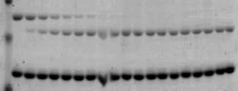
168 Factor Va WT II F1.2A B Factor Va KFDY II F1.2A B Factor Va DFDY II F1.2A B Factor Va DFDF Factor Va DEDE II F1.2A B II F1.2A B 152
Blood coagulation and fibrinolysis. Blood clotting (HAP unit 5 th )
") Blood coagulation and fibrinolysis Blood clotting (HAP unit 5 th ) Vessel injury Antithrombogenic (Favors fluid blood) Thrombogenic (Favors clotting) 3 Major systems involved Vessel wall Endothelium ECM
Blood coagulation and fibrinolysis Blood clotting (HAP unit 5 th ) Vessel injury Antithrombogenic (Favors fluid blood) Thrombogenic (Favors clotting) 3 Major systems involved Vessel wall Endothelium ECM
Ch. 45 Blood Plasma proteins, Coagulation and Fibrinolysis Student Learning Outcomes: Describe basic components of plasma
 Chapt. 45 Ch. 45 Blood Plasma proteins, Coagulation and Fibrinolysis Student Learning Outcomes: Describe basic components of plasma Inheritance of X-linked gene for Factor VIII hemophilia A Explain the
Blood clotting. Subsequent covalent cross-linking of fibrin by a transglutaminase (factor XIII) further stabilizes the thrombus.
 further stabilizes the thrombus.") Blood clotting It is the conversion, catalyzed by thrombin, of the soluble plasma protein fibrinogen (factor I) into polymeric fibrin, which is deposited as a fibrous network in the primary thrombus. Thrombin
Blood clotting It is the conversion, catalyzed by thrombin, of the soluble plasma protein fibrinogen (factor I) into polymeric fibrin, which is deposited as a fibrous network in the primary thrombus. Thrombin
Part IV Antithrombotics, Anticoagulants and Fibrinolytics
 Part IV Antithrombotics, Anticoagulants and Fibrinolytics "The meaning of good and bad, of better and worse, is simply helping or hurting" Emerson Chapter 16: Blood Coagulation and Fibrinolytic System
Chapter 1. General introduction
 Chapter 1 General introduction 8 Haemostasis All organs and tissues of higher organisms are provided with nutrients and oxygen through the bloodstream. The bloodstream is an extensive vascular system that
Primary Exam Physiology lecture 5. Haemostasis
 Primary Exam Physiology lecture 5 Haemostasis Haemostasis Body s response for the prevention and cessation of bleeding. Broadly consists of: Primary Haemostasis - vascular spasm and platlet plug formation
Chapter 1 Introduction
 Chapter 1 Introduction There are several disorders which carry an increased risk of thrombosis, clots that interfere with normal circulation, including: venous thromboembolism (VTE), comprising both deep
Diagnosis of hypercoagulability is by. Molecular markers
 Agenda limitations of clinical laboratories to evaluate hypercoagulability and the underlying cause for thrombosis what is the INR the lupus anticoagulant and the antiphospholipid antibody syndrome hassouna
Introduction to coagulation and laboratory tests
 Introduction to coagulation and laboratory tests Marc Jacquemin Special Haemostasis Laboratory Center for Molecular and Vascular Biology University of Leuven Coagulation in a blood vessel: fibrin stabilises
Molecular Mechanism of Incorporation of Factor Va into Prothrombinase
 Cleveland State University EngagedScholarship@CSU ETD Archive 2007 Molecular Mechanism of Incorporation of Factor Va into Prothrombinase Melissa Barhoover Cleveland State University Follow this and additional
Factor Va Directs Catalysis by Factor Xa During Prothrombin Activation
 Cleveland State University EngagedScholarship@CSU ETD Archive 2008 Factor Va Directs Catalysis by Factor Xa During Prothrombin Activation Michael Anthony Bukys Cleveland State University Follow this and
Studies of Coagulation and Fibrinolysis
 Studies of Coagulation and Fibrinolysis by Paul Y. Kim A thesis submitted to the Department of Biochemistry in conformity with the requirements for the degree of Doctor of Philosophy Queen s University
Hemostasis. Learning objectives Dr. Mária Dux. Components: blood vessel wall thrombocytes (platelets) plasma proteins
 plasma proteins") Hemostasis Learning objectives 14-16 Dr. Mária Dux Components: blood vessel wall thrombocytes (platelets) plasma proteins Hemostatic balance! procoagulating activity anticoagulating activity 1 Thrombocytes
Hemostasis Learning objectives 14-16 Dr. Mária Dux Components: blood vessel wall thrombocytes (platelets) plasma proteins Hemostatic balance! procoagulating activity anticoagulating activity 1 Thrombocytes
UNIT VI. Chapter 37: Platelets Hemostasis and Blood Coagulation Presented by Dr. Diksha Yadav. Copyright 2011 by Saunders, an imprint of Elsevier Inc.
 UNIT VI Chapter 37: Platelets Hemostasis and Blood Coagulation Presented by Dr. Diksha Yadav Hemostasis: Prevention of Blood Loss Vascular constriction Formation of a platelet plug Formation of a blood
This slide belongs to iron lecture and it is to clarify the iron cycle in the body and the effect of hypoxia on erythropoitein secretion
 This slide belongs to iron lecture and it is to clarify the iron cycle in the body and the effect of hypoxia on erythropoitein secretion Topics of today lectures: Hemostasis Meaning of hemostasis Mechanisms
Modeling of human factor Va inactivation by activated protein C
 Bravo et al. BMC Systems Biology 2012, 6:45 RESEARCH ARTICLE Modeling of human factor Va inactivation by activated protein C Maria Cristina Bravo 1, Thomas Orfeo 2, Kenneth G Mann 2 and Stephen J Everse
On a Minimal Mathematical Model for Regulation of Factor Va by Activated Protein C
 UNIVERSITY OF VERMONT DEPARTMENT OF MATHEMATICS AND STATISTICS On a Minimal Mathematical Model for Regulation of Factor Va by Activated Protein C Laura B. Balzer Advisor: Daniel Bentil, Ph.D. Please Note:
Chapter 19. Hemostasis
 Chapter 19 Hemostasis Hemostasis Hemostasis is the cessation of bleeding stopping potentially fatal leaks important in small blood vessels not effective in hemorrhage excessive bleeding from large blood
Biochemical Reaction Networks and Blood Clotting
 Biochemical Reaction Networks and Blood Clotting Yasmeen Hussain, Aaron Fogelson Abstract Following an injury to the walls of a blood vessel, platelet aggregation and blood coagulation are induced. Through
Thursday, February 26, :00 am. Regulation of Coagulation/Disseminated Intravascular Coagulation HEMOSTASIS/THROMBOSIS III
 REGULATION OF COAGULATION Introduction HEMOSTASIS/THROMBOSIS III Regulation of Coagulation/Disseminated Coagulation necessary for maintenance of vascular integrity Enough fibrinogen to clot all vessels
Topics of today lectures: Hemostasis
 Topics of today lectures: Hemostasis Meaning of hemostasis Mechanisms of hemostasis - Vascular contraction - Platelets plug - Blood coagulation (clotting) - Structure and functions of platelets - Blood
Thrombosis. By Dr. Sara Mohamed Abuelgasim
 Thrombosis By Dr. Sara Mohamed Abuelgasim 1 Thrombosis Unchecked, blood coagulation would lead to dangerous occlusion of blood vessels if the protective mechanisms of coagulation factor inhibitors, blood
Cover Page. The handle holds various files of this Leiden University dissertation.
 Cover Page The handle http://hdl.handle.net/1887/28736 holds various files of this Leiden University dissertation. Author: Debeij, Jan Title: The effect of thyroid hormone on haemostasis and thrombosis
Bleeding and Haemostasis. Saman W.Boskani HDD, FIBMS Maxillofacial Surgeon
 Bleeding and Haemostasis Saman W.Boskani HDD, FIBMS Maxillofacial Surgeon 1 Beeding Its escaping or extravasation of blood contents from blood vessels Types: - Arterial - Venous - Capillary Differences
Outline Anti-coagulant and anti-thrombotic drugs Haemostasis and Thrombosis Year 3 Dentistry
 Outline Anti-coagulant and anti-thrombotic drugs Year 3 Dentistry Professor Yotis Senis Cellular Haemostasis y.senis@bham.ac.uk I. Haemostasis and II. Coagulation and anti-coagulants III. Platelets and
Elucidating the interaction between the Fragment 2 domain of Prothrombin and Factor Va
 Elucidating the interaction between the Fragment 2 domain of Prothrombin and Factor Va by Joanne Elizza Berridge A thesis submitted to the Graduate Program in Biochemistry in the Department of Biomedical
THROMBOTIC DISORDERS: The Final Frontier
 THROMBOTIC DISORDERS: The Final Frontier Jeffrey I. Weitz, MD, FRCP(C), FACP Professor of Medicine and Biochemistry McMaster University Canada Research Chair in Thrombosis Heart & Stroke Foundation/ J.F.
WHITE PAPERS PRESENTATION VIDEO DOCUMENTATION EXPERIMENT WO NDCLOT. The WoundClot Principals for Effective Bleeding Control PRESENTATION
 WHITE PAPERS PRESENTATION VIDEO DOCUMENTATION EXPERIMENT ARTICLES OUR STUDY BLEEDING CONTROL 5 POINT MODEL WO NDCLOT The WoundClot Principals for Effective Bleeding Control PRESENTATION Harnessing SCIENCE
Thrombosis. Jeffrey Jhang, M.D.
 Thrombosis Jeffrey Jhang, M.D. Introduction The human hemostatic system has evolved to maintain blood flow under normal physiologic conditions while remaining primed to rapidly respond to vascular injury
Chapter 3. Haemostatic abnormalities in patients with liver disease
 Chapter 3 Haemostatic abnormalities in patients with liver disease Ton Lisman, Frank W.G. Leebeek 1, and Philip G. de Groot Thrombosis and Haemostasis Laboratory, Department of Haematology, University
Hemostasis. Clo)ng factors and Coagula4on NORMAL COAGULATION. Overview of blood coagula4on. The Cascade Theory 5/1/12. Clot
ng factors and Coagula4on NORMAL COAGULATION. Overview of blood coagula4on. The Cascade Theory 5/1/12. Clot") Hemostasis Clo)ng factors and Coagula4on Dr Badri Paudel www.badripaudel.com Hemostasis is defined as a property of circula4on whereby blood is maintained within a vessel and the ability of the system
Hemostasis Clo)ng factors and Coagula4on Dr Badri Paudel www.badripaudel.com Hemostasis is defined as a property of circula4on whereby blood is maintained within a vessel and the ability of the system
Approach to bleeding disorders &treatment. by RAJESH.N General medicine post graduate
 Approach to bleeding disorders &treatment by RAJESH.N General medicine post graduate 2 Approach to a patient of bleeding diathesis 1. Clinical evaluation: History, Clinical features 2. Laboratory approach:
Thrombophilia. Diagnosis and Management. Kevin P. Hubbard, DO, FACOI
 Thrombophilia Diagnosis and Management Kevin P. Hubbard, DO, FACOI Clinical Professor of Medicine Kansas City University of Medicine and Biosciences-College of Osteopathic Medicine Kansas City, Missouri
COPYRIGHTED MATERIAL. Overview of h emostasis. Kathleen Brummel Ziedins and Kenneth G. Mann University of Vermont, College of Medicine, VT, USA
 1 Overview of h emostasis Kathleen Brummel Ziedins and Kenneth G. Mann University of Vermont, College of Medicine, VT, USA Introduction The maintenance of blood fluidity and protection from blood leakage
-Hashim ahmed is the one who wrote this sheet. I just edited it according to our record.
 * Subjects of this lecture : - Hemostasis - Platelets, general information, their ultrastructure and role in hemostasis. - Definitions: Thrombus, Embolus, Arteriosclerosis and Atherosclerosis. *NOTE: Prof
Hemostasis and. Blood Coagulation
 Hemostasis and Blood Coagulation Events in Hemostasis The term hemostasis means prevention of blood loss. Whenever a vessel is severed or ruptured, hemostasis is achieved by several mechanisms: (1) vascular
What are blood clots?
 What are blood clots? Dr Matthew Fay GP Principal The Willows Medical Practice- Queensbury GPwSI and Co-Founder Westcliffe Cardiology Service GP Partner Westcliffe Medical Group Created 5/31/18 Dr. Matthew
Hemostasis and Thrombosis
 Hemostasis Hemostasis and Thrombosis Normal hemostasis is a consequence of tightly regulated processes that maintain blood in a fluid state in normal vessels, yet also permit the rapid formation of a hemostatic
Anatomy and Physiology
 Anatomy and Physiology For The First Class 2 nd Semester Thrombocytes = Platelets Thrombocytes = Platelets Blood platelets are non-nucleated disc like cell fragments 2-4 µm in diameter. Platelets are not
Physiology of. The Blood hemostasis. By prof. Israa f. jaafar
 Physiology of The Blood hemostasis By prof. Israa f. jaafar Learning objectives Understand the Platelet structure and function Explane the Platelet production Understand the phases of hemostasis: vascular
PHASES OF HAEMOSTASIS
 HAEMOSTASIS Maintains the integrity of a closed, highpressure circulatory system after vascular damage Vessel Wall Injury events in the vessel wall and in the blood which seal breach Delicate balance exists
Bleeding Disorders. Dr. Mazen Fawzi Done by Saja M. Al-Neaumy Noor A Mohammad Noor A Joseph Joseph
 Bleeding Disorders Dr. Mazen Fawzi Done by Saja M. Al-Neaumy Noor A Mohammad Noor A Joseph Joseph Normal hemostasis The normal hemostatic response involves interactions among: The blood vessel wall (endothelium)
Hemodynamic Disorders, Thrombosis, and Shock. Richard A. McPherson, M.D.
 Hemodynamic Disorders, Thrombosis, and Shock Richard A. McPherson, M.D. Edema The accumulation of abnormal amounts of fluid in intercellular spaces of body cavities. Inflammation and release of mediators
The anticoagulant function of coagulation factor V
 Review Article Schattauer 2012 15 The anticoagulant function of coagulation factor V Thomas J. Cramer; Andrew J. Gale Department of Molecular and Experimental Medicine, The Scripps Research Institute,
Proexosite-1 on Prothrombin is a Factor Va-dependent Recognition Site for the. Prothrombinase Complex
 JBC Papers in Press. Published on May 14, 2003 as Manuscript M302707200 Proexosite-1 on Prothrombin is a Factor Va-dependent Recognition Site for the Prothrombinase Complex Lin Chen, Likui Yang and Alireza
L iter diagnostico di laboratorio nelle coagulopatie congenite emorragiche
 L iter diagnostico di laboratorio nelle coagulopatie congenite emorragiche Armando Tripodi Angelo Bianchi Bonomi Hemophilia and Thrombosis Center Dept. of Clinical Sciences and Community Health University
Chapter 19 Cardiovascular System Blood: Functions. Plasma
 Chapter 19 Cardiovascular System Blood: Functions 19-1 Plasma Liquid part of blood. Colloid: liquid containing suspended substances that don t settle out of solution 91% water. Remainder proteins, ions,
Disseminated Intravascular Coagulation. M.Bahmanpour MD Assistant professor IUMS
 به نام خدا Disseminated Intravascular Coagulation M.Bahmanpour MD Assistant professor IUMS Algorithm for Diagnosis of DIC DIC Score factor score Presence of known underlying disorder No= 0 yes=2 Coagolation
The R-subunit would not the able to release the catalytic subunit, so this mutant of protein kinase A would be incapable of being activated.
 1. Explain how one molecule of cyclic AMP can result in activation of thousands of molecules of glycogen phosphorylase. Technically it takes four molecules of cyclic AMP to fully activate one molecule
HEME 10 Bleeding Disorders
 HEME 10 Bleeding Disorders When injury occurs, three mechanisms occur Blood vessels Primary hemostasis Secondary hemostasis Diseases of the blood vessels Platelet disorders Thrombocytopenia Functional
Inherited Thrombophilia Testing. George Rodgers, MD, PhD Kristi Smock MD
 Inherited Thrombophilia Testing George Rodgers, MD, PhD Kristi Smock MD Prevalence and risk associated with inherited thrombotic disorders Inherited Risk Factor % General Population % Patients w/ Thrombosis
4/23/2009. September 15, 2008
 The Current Treatment of Deep Venous Thrombosis: Are We Doing Enough? George H. Meier, MD Professor and Chief Division of Vascular Surgery University of Cincinnati College of Medicine Cincinnati, Ohio
Branch of medicine that deals with blood, its formation and disorders is called. Three main functions of cardiovascular system are,, and.
 Chapter 19 The Blood Human body must maintain a balance called. Body fluid inside the cells is called fluid; that outside is called or fluid. Two major fluid networks that help in connecting cells are
Hemodynamics. Objectives. Filtration across the endothelium 1/17/2017. K[(P cap - P int )-σ(π cap π int )]
![Hemodynamics. Objectives. Filtration across the endothelium 1/17/2017. K[(P cap - P int )-σ(π cap π int )]](/thumbs/90/103064695.jpg "Hemodynamics. Objectives. Filtration across the endothelium 1/17/2017. K[(P cap - P int )-σ(π cap π int )]") Hemodynamics Amir Kol DVM, Ph.D, Dip ACVP (Clinical Pathology) UC Davis, School of Veterinary Medicine akol@ucdavis.edu Objectives Understand the mechanisms that promote extravascular fluid accumulation
Hemodynamics Amir Kol DVM, Ph.D, Dip ACVP (Clinical Pathology) UC Davis, School of Veterinary Medicine akol@ucdavis.edu Objectives Understand the mechanisms that promote extravascular fluid accumulation
Approach to Thrombosis
 Approach to Thrombosis Theera Ruchutrakool, M.D. Division of Hematology Department of Medicine Siriraj Hospital Faculty of Medicine Mahidol University Approach to Thrombosis Thrombosis: thrombus formation
Congenital bleeding disorders
 Congenital bleeding disorders Overview Factor VIII von Willebrand Factor Complex factor VIII von Willebrand factor (vwf) complex circulate as a complex + factor IX intrinsic pathway Platelets bind via
Hemostasis. PHYSIOLOGICAL BLOOD CLOTTING IN RESPONSE TO INJURY OR LEAK no disclosures
 Hemostasis PHYSIOLOGICAL BLOOD CLOTTING IN RESPONSE TO INJURY OR LEAK no disclosures Disorders of Hemostasis - Hemophilia - von Willebrand Disease HEMOPHILIA A defect in the thrombin propagation phase
PAPER No. : 16, Bioorganic and biophysical chemistry MODULE No. : 22, Mechanism of enzyme catalyst reaction (I) Chymotrypsin
 Chymotrypsin") Subject Paper No and Title 16 Bio-organic and Biophysical Module No and Title 22 Mechanism of Enzyme Catalyzed reactions I Module Tag CHE_P16_M22 Chymotrypsin TABLE OF CONTENTS 1. Learning outcomes 2.
Subject Paper No and Title 16 Bio-organic and Biophysical Module No and Title 22 Mechanism of Enzyme Catalyzed reactions I Module Tag CHE_P16_M22 Chymotrypsin TABLE OF CONTENTS 1. Learning outcomes 2.
Coagulation Disorders. Dr. Muhammad Shamim Assistant Professor, BMU
 Coagulation Disorders Dr. Muhammad Shamim Assistant Professor, BMU 1 Introduction Local Vs. General Hematoma & Joint bleed Coagulation Skin/Mucosal Petechiae & Purpura PLT wound / surgical bleeding Immediate
True/False: Idarucizumab can be utilized for the management of bleeding associated with dabigatran.
 Discuss the role of idarucizumab for the management of bleeding associated with dabigatran Understand dosing, preparation and administration of idarucizumab I have no financial interest/arrangement or
Continuing Medical Education
 Continuing Medical Education WHY DO HEMOPHILIACS BLEED? B.R. Agarwal Z.E. Currimbhoy Deficiency of coagulation factor VIII (antihemophilic factor) and coagulation factor IX (Christmas factor) result in
Anticoagulants. Pathological formation of a haemostatic plug Arterial associated with atherosclerosis Venous blood stasis e.g. DVT
 Haemostasis Thrombosis Phases Endogenous anticoagulants Stopping blood loss Pathological formation of a haemostatic plug Arterial associated with atherosclerosis Venous blood stasis e.g. DVT Vascular Platelet
Student number. University of Guelph Department of Chemistry and Biochemistry Structure and Function In Biochemistry
 University of Guelph Department of Chemistry and Biochemistry 19356 Structure and Function In Biochemistry Midterm Test, March 3, 1998. Time allowed, 90 min. Answer questions 120 on the computer scoring
Coagulation Pathway and Physiology
 Chapter 1 Coagulation Pathway and Physiology Russell A. Higgins, M Introduction Our understanding of blood clotting is intimately tied to the history of civilization. With the advent of writing 5000 years
Chapter 19: Cardiovascular System: Blood
 Chapter 19: Cardiovascular System: Blood I. Functions of Blood A. List and describe the seven major homeostatic functions of blood: 1. 2. 3. 4. 5. 6. 7. II. Plasma A. Composition 1. It is a fluid consisting
Hemodynamic Disorders, Thromboembolic Disease, and Shock
 Hemodynamic Disorders, Thromboembolic Disease, and Shock Kumar et al: Robbins & Cotran Pathologic Basis of Disease 7E Figure 4-1 Factors affecting fluid balance across capillary walls. Capillary hydrostatic
Blood Lecture Test Questions Set 2 Summer 2012
 Blood Lecture Test Questions Set 2 Summer 2012 1. Leukocytes are attracted to a site of injury or disease by: a. diapedesis b. chemotaxis c. leukocytosis d. heparin e. leukomotosis 2. Leukocytes leave
COAGULATION INHIBITORS LABORATORY DIAGNOSIS OF PROTHROMBOTIC STATES REGULATION OF. ANTICOAGULANT PROTEIN DEFICIENCY Disease entities COAGULATION
 LABORATORY DIAGNOSIS OF PROTHROMBOTIC COAGULATION INHIBITORS Tissue Factor Pathway Inhibitor (TFPI) Lipoprotein Associated Coagulation Inhibitor (LACI) Extrinsic Pathway Inhibitor (EPI) Complexes with
Cooperative Regulation of the Activity of Factor Xa within Prothrombinase by Discrete Amino Acid Regions from Factor Va Heavy Chain
 Biochemistry 2008, 47, 12835 12843 12835 Cooperative Regulation of the Activity of Factor Xa within Prothrombinase by Discrete Amino Acid Regions from Factor Va Heavy Chain Melissa A. Barhoover,, Tivadar
Elsamani Ismail Abdelfadiel. All Rights Reserved
 Elsamani Ismail Abdelfadiel All Rights Reserved i INVESTIGATION OF ANTICOAGULATION PROPERTIES OF SULFATED GLYCOSAMINOGLYCAN MIMETICS A dissertation submitted in partial fulfillment of the requirements
Discuss the role of idarucizumab for the management of bleeding associated with dabigatran
 Discuss the role of idarucizumab for the management of bleeding associated with dabigatran Understand dosing, preparation and administration of idarucizumab I have no financial interest/arrangement or
PICT ANTICOAGULANT MONITORING 1. PEFAKIT PICT. Clotting Assay. PEFAKIT PiCT. PEFAKIT PICT Calibrators UFH. PEFAKIT PICT Controls UFH
 1 2 3 4 PEFAKIT PiCT PEFAKIT PICT Calibrators UFH PEFAKIT PICT Controls UFH PEFAKIT PICT Calibrators LMWH Clotting Assay PICT 1. PEFAKIT PICT Package size 8-505-01 kit 80 3 vials of PiCT Activator (2mL)
Bleeding and Thrombotic Disorders. Kristine Krafts, M.D.
 Bleeding and Thrombotic Disorders Kristine Krafts, M.D. Bleeding and Thrombotic Disorders Bleeding disorders von Willebrand disease Hemophilia A and B DIC TTP/HUS ITP Thrombotic disorders Factor V Leiden
Do some of physiotherapy and rehabilitation programs improve the health state of patients suffering from cerebral clot?
 International Journal of Science Culture and Sport (IntJSCS) September 2014: 2(3) ISSN : 2148-1148 Doi : 10.14486/IJSCS63 Do some of physiotherapy and rehabilitation programs improve the health state of
Laboratory Evaluation of Venous Thrombosis Risk
 Laboratory Evaluation of Venous Thrombosis Risk Dorothy M. Adcock, MD Volume 17, Number 12 December 2003 Objective: The reader will be able to discuss the concepts of risk factor, risk potential and thrombotic
Rethinking events in the haemostatic process: role of factor V and TFPI
 Haemophilia (2016), 22 (Suppl. 5), 3 8 DOI: 10.1111/hae.13004 REVIEW ARTICLE Rethinking events in the haemostatic process: role of factor V and TFPI R. M. CAMIRE Division of Hematology, Department of Pediatrics,
Chapter 19 Blood Lecture Outline
 Chapter 19 Blood Lecture Outline Cardiovascular system Circulatory system Blood 1. distribution 2. regulation 3. protection Characteristics: ph 7.4 38 C 4-6 L Composition: Plasma Formed elements Erythrocytes
Student number. University of Guelph Department of Chemistry and Biochemistry Structure and Function In Biochemistry
 University of Guelph Department of Chemistry and Biochemistry 19356 Structure and Function In Biochemistry Midterm Test, March 3, 1998. Time allowed, 90 min. Answer questions 120 on the computer scoring
ACQUIRED COAGULATION ABNORMALITIES
 ACQUIRED COAGULATION ABNORMALITIES ACQUIRED COAGULATION ABNORMALITIES - causes 1. Liver disease 2. Vitamin K deficiency 3. Increased consumption of the clotting factors (disseminated intravascular coagulation
Haemostasis & Coagulation disorders Objectives:
 Haematology Lec. 1 د.ميسم مؤيد علوش Haemostasis & Coagulation disorders Objectives: - Define haemostasis and what are the major components involved in haemostasis? - How to assess the coagulation status?
Hemostasis and fibrinolysis the hemostatic balance
 Hemostasis and fibrinolysis the hemostatic balance Bleeding Clotting The hemostatic balance Reference: Boron & Boulpaep Medical Physiology, 3 rd Ed, Chapter 18 Dr. Ana-Maria Zagrean Carol Davila University
PHRM 836 September 1, 2015
 PRM 836 September 1, 2015 Protein structure- function relationship: Catalysis example of serine proteases Devlin, section 9.3 Physiological processes requiring serine proteases Control of enzymatic activity
REVIEW. Blood Coagulation. S. Butenas and K. G. Mann*
 Biochemistry (Moscow), Vol. 67, No.,, pp. -. Translated from Biokhimiya, Vol. 67, No.,, pp. 5-5. Original Russian Text Copyright by Butenas, Mann. REVIEW Blood Coagulation S. Butenas and K. G. Mann* University
Recent studies have found that elevated levels of coagulation
 Elevated Hemostatic Factor Levels as Potential Risk Factors for Thrombosis Wayne L. Chandler, MD; George M. Rodgers, MD, PhD; Jason T. Sprouse, MD; Arthur R. Thompson, MD, PhD Recent studies have found
Blood Thinner Agent. Done by: Meznah Al-mutairi Pharm.D Candidate PNU Collage of Pharmacy
 Blood Thinner Agent Done by: Meznah Al-mutairi Pharm.D Candidate PNU Collage of Pharmacy Outline: Blood thinner agent definition. anticoagulants drugs. Thrombolytics. Blood thinner agent Therapeutic interference
The role of Ca² + ions in the complex assembling of protein Z and Z-dependent protease inhibitor: A structure and dynamics investigation
 www.bioinformation.net Hypothesis Volume 8(9) The role of Ca² + ions in the complex assembling of protein Z and Z-dependent protease inhibitor: A structure and dynamics investigation Zahra Karimi 1 *,
Mousa Suboh. Osama Alzoubi (OZ) Malik Zuhlof. 1 P a g e
 Malik Zuhlof. 1 P a g e") 3 Mousa Suboh Osama Alzoubi (OZ) Malik Zuhlof 1 P a g e In the last lecture we talked about the anticoagulants, we talked about heparin and we said that it has 2 types: one that has long stretch of polysaccharides
3 Mousa Suboh Osama Alzoubi (OZ) Malik Zuhlof 1 P a g e In the last lecture we talked about the anticoagulants, we talked about heparin and we said that it has 2 types: one that has long stretch of polysaccharides
BLEEDING DISORDERS Simple complement:
 BLEEDING DISORDERS Simple complement: 1. Select the statement that describe the thrombocytopenia definition: A. Marked decrease of the Von Willebrandt factor B. Absence of antihemophilic factor A C. Disorder
Prothrombin Complex Concentrate- Octaplex. Octaplex
 Prothrombin Complex Concentrate- Concentrated Factors Prothrombin Complex Concentrate (PCC) 3- factor (factor II, IX, X) 4-factor (factors II, VII, IX, X) Activated 4-factor (factors II, VIIa, IX, X) Coagulation
Chapter 11. Lecture and Animation Outline
 Chapter 11 Lecture and Animation Outline To run the animations you must be in Slideshow View. Use the buttons on the animation to play, pause, and turn audio/text on or off. Please Note: Once you have
Procoagulant Adaptation of a Blood Coagulation Prothrombinase-like Enzyme Complex in Australian Elapid Venom
 Toxins 2010, 2, 1554-1567; doi:10.3390/toxins2061554 Review OPEN ACCESS toxins ISSN 2072-6651 www.mdpi.com/journal/toxins Procoagulant Adaptation of a Blood Coagulation Prothrombinase-like Enzyme Complex
PART I. Introduction COPYRIGHTED MATERIAL
 PART I Introduction COPYRIGHTED MERIAL CHAPTER 1 Overview of h emostasis Kathleen Brummel Ziedins and Kenneth G. Mann University of Vermont, College of Medicine, VT, USA Introduction The maintenance of
Amino acids. Side chain. -Carbon atom. Carboxyl group. Amino group
 PROTEINS Amino acids Side chain -Carbon atom Amino group Carboxyl group Amino acids Primary structure Amino acid monomers Peptide bond Peptide bond Amino group Carboxyl group Peptide bond N-terminal (
Blood. Plasma. The liquid part of blood is called plasma. 1. Pale yellow fluid; forms more than half the blood volume.
 11 Blood FOCUS: Blood consists of plasma and formed elements. The plasma is 91% water with dissolved or suspended molecules, including albumin, globulins, and fibrinogen. The formed elements include erythrocytes,
Chapter 10. Regulatory Strategy
 Chapter 10 Regulatory Strategy Regulation of enzymatic activity: 1. Allosteric Control. Allosteric proteins have a regulatory site(s) and multiple functional sites Activity of proteins is regulated by
HEART HEALTH WEEK 2 SUPPLEMENT. A Beginner s Guide to Cardiovascular Disease ATHEROSCLEROSIS. Fatty deposits can narrow and harden the artery
 WEEK 2 SUPPLEMENT HEART HEALTH A Beginner s Guide to Cardiovascular Disease ATHEROSCLEROSIS FIGURE 1 Atherosclerosis is an inflammatory process where cholesterol is deposited in the wall of arteries and
Hemostasis Haemostasis means prevention of blood loss from blood vessels.
 ١ Hemostasis Haemostasis means prevention of blood loss from blood vessels. Bleeding is stopped by several mechanisms, which are: 1. Local vasoconstriction 2. Formation of platelet plug 3. Blood coagulation
Human coagulation factor Xa, FXa ELISA Kit
 Human coagulation factor Xa, FXa ELISA Kit Catalog No: E1613h 96 Tests Operating instruction www.eiaab.com FOR RESEARCH USE ONLY; NOT FOR THERAPEUTIC OR DIAGNOSTIC APPLICATIONS! PLEASE READ THROUGH ENTIRE
THROMBOPHILIA SCREENING
 THROMBOPHILIA SCREENING Introduction The regulation of haemostasis Normally, when a clot occurs, it exactly occurs where it has to be and does not grow more than necessary due to the action of the haemostasis
Page 1 of 6 THROMBOCYTES
 Page 1 of 6 THROMBOCYTES Platelets are not cells in the strict sense. About one-fourth the diameter of a lymphocyte, they are cytoplasmic fragments of extraordinarily large cells (up to 60 µm in diameter)