HSV-1 REMODELS PI3-KINASE/AKT SIGNALING
|
|
|
- Evangeline Hodge
- 6 years ago
- Views:
Transcription
1 UNIVERSITY OF ALBERTA HSV-1 REMODELS PI3-KINASE/AKT SIGNALING by KEVIN QUACH A thesis submitted to the Faculty of Graduate Studies and Research in partial fulfillment of the requirements for the degree of MASTER OF SCIENCE in VIROLOGY Department of Medical Microbiology and Immunology Kevin Quach Spring 2013 Edmonton, Alberta Permission is hereby granted to the University of Alberta Libraries to reproduce single copies of this thesis and to lend or sell such copies for private, scholarly or scientific research purposes only. Where the thesis is converted to, or otherwise made available in digital form, the University of Alberta will advise potential users of the thesis of these terms. The author reserves all other publication and other rights in association with the copyright in the thesis and, except as herein before provided, neither the thesis nor any substantial portion thereof may be printed or otherwise reproduced in any material form whatsoever without the author's prior written permission.
2 ABSTRACT AKT inhibits apoptosis and stimulates cap-dependent translation by phosphorylating key downstream cellular proteins. Many viruses therefore activate the PI3-kinase-AKT signaling pathway to promote cell survival and viral protein synthesis. HSV-1 activates AKT during lytic infection and the abundant tegument protein VP11/12 is required for this effect. Although VP11/12 is essential for AKT activation, deleting VP11/12 has no detectable effect on HSV-1 induced phosphorylation of the examined downstream target proteins. Rather, the protein kinase U S 3 is required for the phosphorylation of AKT targets and thus serves as an AKT mimic. Our data indicate that VP11/12 and U S 3 do not collaborate to provide redundant coverage of key AKT targets during infection. Interestingly, HSV-1 attenuates signals from activated AKT induced by external growth factors. Remarkably, neither VP11/12 nor U S 3 is required for this effect.
3 ACKNOWLEDGEMENTS I would like to thank my supervisor, Dr. James R. Smiley for providing an exceptional learning environment for the development of scientific writing and critical thinking skills. Thank you to past and current lab members for sharing your scientific ideas with me and for making graduate school an exceptionally enjoyable time. I would like to specifically thank Holly Saffran for making me feel welcome and providing support in the lab and personal life. Thank you to my committee members Dr. David Evans, Dr. Rob Ingham, and Dr. Richard Lamb for your assistance with science and academics. Additionally, many thanks to Anne Giles, Debbie Doudiet, Tabitha Vasquez, and the rest of the office staff for their support. Thank you to all my friends in the Department of Medical Microbiology and Immunology for your support and friendly conversations. I cannot begin to thank enough those I ve developed close relationships with: Nancy Hu, Andre Deslauriers, Kinola Williams and Bettina Bareiss. Thank you to Nancy Hu with whom I have shared some of my best memories and who has made the last few years special. Furthermore, your scientific contribution to this thesis was invaluable. Thank you to Kinola Williams for providing the support that I needed in multiple situations. Thanks to all who have helped make Edmonton my home. In closing, I would like to dedicate this thesis to my sister Tara; you have taught me everything I know and have never left my side.
4 TABLE OF CONTENTS
5 CHAPTER 1: INTRODUCTION Overview of Herpes Simplex Virus-1 (HSV-1) A Brief Introduction Taxonomy The HSV Virion HSV-1 Lytic Cycle HSV-1 Latency Cellular initiation of PI3K-AKT signaling A Brief Introduction on PI3-Kinase Activation of PI3-Kinase Propagating PI3-Kinase Signals AKT AKT Signaling Viruses modulate PI3K-AKT signaling HSV-1 U L UL46 activates SFKs and PI3-Kinase to induce AKT activation HSV-1 U S US3 promotes cell survival by circumventing cell signaling pathway U S 3 modulates AKT signaling Thesis rationale...26 CHAPTER 2: MATERIALS AND METHODS Mammalian cell cultures Virus strains and growth conditions Preparation of viral stocks Virus titrations Infecting cells studies of HSV-induced host signaling modifications Addition of drugs and external growth ligands Preparation of cell extracts SDS-polyacrylamide gel 32
6 2.9 Western blot Antibodies.. 33 CHAPTER 3: RESULTS HSV-1 Infection Causes Maximal AKT Activation at late times during infection HSV-1 Requires mtorc2 for AKT phosphorylation U S 3 Mediates the Phosphorylation of AKT Targets HSV-1 also activates AKT in Human Foreskin Fibroblasts U S 3 Mediates the phosphorylation of AKT targets despite activation of AKT by U L Serum induces AKT activation, but there is no significant activation signal transduced to AKT targets at late times of infection PDGF BB induces AKT activation, but the signal is not transduced to AKT targets Titration of PDGF AA, AB and BB PDGF ligands do not perform equally at activating AKT during infection.85 CHAPTER 4: DISCUSSION Thesis summary The HSV-1 kinase U S 3 masquerades as an AKT mimic to regulate cellular processes HSV-1 promotes cap-dependent translation A potential role for U L 46 in promoting cell survivalals HSV-1 encodes proteins other than U L 46 and U S 3 to modulate AKT signaling HSV-1 permits signaling to AKT from specific receptors Other aspects of AKT activation that are modulated by HSV A mechanism for the phosphorylation of AKT by HSV U S 3 negatively regulates the phosphorylation of the T308 residue of AKT.102
7 4.8 Future directions BIBLIOGRAPHY APPENDICES 128
8 LIST OF FIGURES
9 Figure 1.1: Structure of PI3K. 10 Figure 1.2: PDGF-receptor activates PI3K 12 Figure 1.3: Transduction of an activated PI3K signal 14 Figure 1.4: Activation of AKT and propagation of an activated AKT signal 17 Figure 1.5: AKT acts on multiple proteins 20 Figure 1.6: HSV-1 U L 46 binds SFK and PI3K to activate AKT...24 Figure 3.1: HSV-1 activates AKT in a U L 46-dependent manner..37 Figure 3.2: mtorc2 is required for AKT activation during HSV-1 infection.41 Figure 3.3: DMSO does not dampen AKT activation during HSV-1 infection...43 Figure 3.4: The AKT targets, GSK3β and FoxO1, are phosphorylated in a U L 46- independent fashion, but require U S 3 46 Figure 3.5: HSV-1 activates AKT in a U L 46-dependent manner at early and late times post infection 49 Figure 3.6: Despite U L 46-activated AKT, GSK3β is mainly phosphorylated in a U S 3-dependent fashion at early times post infection.55 Figure 3.7: Multiple AKT targets are phosphorylated in a U S 3-dependent fashion at late times post infection Figure 3.8: 10% serum induces maximal AKT activation after 20 minutes..65 Figure 3.9: Serum induces AKT activation and downstream signaling in infected cells at early infection 68 Figure 3.10: Serum induces AKT activation and, but not downstream signaling in infected cells at late times post infection...70 Figure 3.11: PDGF BB activates AKT 75 Figure 3.12: PDGF BB induces AKT activation, but the signal is not propagated to AKT targets during late times post infection. 77 Figure 3.13: Differing concentrations of PDGF AA, AB and BB induce comparable levels of AKT activation 81 Figure 3.14: PDGF ligands do not perform equally at phosphorylating S473 AKT during infection 88
10 Figure 3.15: PDGF ligands do not perform equally at phosphorylating T308 AKT during infection.. 91 Figure 4.1: U L 46 promotes AKT activation, while U S 3 negatively regulates AKT signaling.96 Figure 4.2: HSV-1 blocks AKT activation downstream of specific receptors..97
11 ABBREVIATIONS
12 4EBP1 Eukaryotic translation initiation factor 4E-binding protein 1 α Immediate-early β Early γ Late AGC Protein kinase A, protein kinase G, protein kinase C AKT/PKB Protein kinase B BAC Bacterial artificial chromosome BCR B cell receptor BH3 Bcl-2 homology domain 3 CTL Cytotoxic lymphocytes DAG Diacylglycerol DMEM Dulbecco s modified eagle medium EGFR Epidermal growth factor receptor g- Glycoprotein FBS Fetal bovine serum FoxO1 Forkhead box O1 GSK3β Glycogen synthase kinase 3 β HCF Host cell factor HCMV/HHV-5 Human cytomegalovirus/human herpes virus-5 HEL Human embryonic lung fibroblast HFF Human foreskin fibroblasts HSV-1 Herpes simplex virus-1 HSV-2 Herpes simplex virus-2 IGFR Insulin growth factor receptor INPP5B Type II phosphatidylinositol 5 phosphate MHC Major histocompatibility complex mtorc1, mtorc2 Mammalian target of rapamycin complex 1, 2 NGFR Nerve growth factor receptor NK Natural killer OBB Odyssey blocking buffer
13 Oct-1 Octamer transcription factor-1 ORF Open reading frame P Penicillin PDGFR Platelet-derived growth factor receptor PDK1 Phosphoinositide-dependent protein kinase-1 PH Pleckstrin homology PI3K Phosphoinositide-3-Kinase PIP2 Phosphatydylinosital-4,5-diphosphate PIP3 Phosphatydylinosital-3,4,5-triphosphate PKA Protein kinase A PLC Phospholipase C PP2A Protein phosphatase 2A PTEN Phosphatase and tensin homolog on chromosome TEN RTK Receptor tyrosine kinase S Streptomycin S- Serine S6K S6 kinase SFK Src family kinase SH- Src homology- T308 Threonine 308 TAP Transporter associates with antigen presentation TBST TBS-Tween TCR T cell receptor TSC2 Tuber sclerosis complex 2 U S 3PK U S 3 protein kinase Vero American monkey kidney cells VHS Virion host shut-off VP11/12 Viral protein 11 and viral protein 12 VZV Varicella zoster virus
14 CHAPTER 1: INTRODUCTION 1
15 This thesis focuses on the role of the Herpes Simplex Virus (HSV)-1 proteins VP11/12 and U S 3 in modulating the host cell PI3K-AKT signaling pathway. Below, I have provided the context for this research by outlining the HSV life cycle, reviewing the PI3K-AKT pathway, and summarizing previous knowledge about how HSV and other viruses manipulate this system. 1.1 Overview of Herpes Simplex Virus A Brief Introduction HSV-1 is a prevalent human pathogen found in 70-95% of the population depending on geographic location, and this percentage increases with age (255). Herpes simplex viruses are neurotropic, and infection causes the appearance of fluid-filled blisters or sores on the skin around the oral cavity (cold sores) or genitals that are caused by either HSV-1 or 2. Although clinical symptoms are mild in the majority of the population, HSV-1 can cause a variety of diseases such as infection of the mouth and gums (gingivostomatitis) and pharyngitis (80, 154). People with eczema have an increased risk of herpes infection outbreaks around eczema-prone areas of the body (eczema herpeticum) (213). Herpes simplex viruses are also known to cause other disorders such as encephalitis, herpetic whitlow, herpes gladiatorum, and herpes keratitis (198). HSV is transmitted through direct skin contact or through the exchange of bodily fluids from an infected individual. HSV must be actively replicating to be transmitted. Active replication is phenotypically characterized by the appearance of blisters that contain infectious virus particles. However, herpes can be asymptomatic while viral shedding occurs (198). Most people who harbor latent HSV shed virus asymptomatically. Viral shedding occurs in 2-9% of adults and 5-8% of children at any given time (255). Following initial infection and lytic replication at the epithelia, HSV virions migrate to innervating sensory nerve endings and travel retrograde into nerve cell bodies (52, 197). It is here that the virus enters latency and can remain dormant throughout life. Alternatively, the virus can cycle between periods of lytic and latent replication. Recurrent active lytic infections have an unknown 2
16 etiology; however, immune system repression may be an initiating event. Repression of the immune system may occur during cancer, stress, infection, and certain drug therapies. During this time, the virus will infect epithelial cells linked to the site of latency by innervating neurons (reviewed in (197, 198)). Tingling, itching and pain at the site of cold sores are signs of an oncoming viral reactivation (71, 210, 211). Unfortunately, herpes is a life-long infection and no cure has been developed. Effective antiviral treatments such as acyclovir can reduce viral shedding and alleviate the severity and duration of symptoms Taxonomy Within the Herpesvirales order is a family of large, double-stranded, linear DNA viruses that are known to infect mammals, fish, birds, or molluscs (reviewed in (198)). Based on their sequence homology and the species they infect, the order can be divided into families, including Alloherpesviridae, Herpesviridae, and Malacoherpesviridae. The Herpesviridae family can be divided into three subfamilies: Alphaherpesvirinae, Betaherpesvirinae, and Gammaherpesvirinae (198). Alphaherpesvirinae have a broad host range, short replication cycle, and can establish latency in sensory neurons. Betaherpesvirinae generally have a more restricted host range, long reproductive cycle, grow slowly in culture and cause infected cells to enlarge. They often establish latency in tissues such as secretory glands and hematopoietic progenitor cells (198). Gammaherpesvirinae have a narrow host range, replicate in epithelial cells, and establish latency in T or B lymphocytes, which can lead to cellular immortalization. There are more than 200 different Herpesviruses, but only eight distinct types are known to cause disease in humans (253). The Alphaherpesvirinae subfamily includes Herpes Simplex Virus types 1 and 2 (HSV-1 and HSV-2), which are also called Human Herpes Viruses 1 and 2 (HHV-1 and HHV-2); and varicella zoster virus (VZV/HHV-3). HSV-1 and HSV-2 cause oral and genital herpes respectively, and severe ocular damage leading to blindness (Herpes keratitis) (15), while VZV is the causative agent of chicken pox and shingles. The Betaherpesvirinae subfamily includes 3
17 human cytomegalovirus (HCMV/HHV-5), herpes lymphotropic virus (HHV-6), and roseolovirus (HHV-7). HCMV can be associated with birth defects (223). and fatal diseases in immune-compromised individuals, including organ transplant recipients and individuals with AIDS (101). Lastly, the Gammaherpesvirinae subfamily is comprised of Epstein-Barr virus (EBV/HHV-4), which causes infectious mononucleosis, and Kaposi s sarcoma-associated herpesvirus (KSHV/HHV-8), which is more often associated with Castleman s disease (199) The HSV Virion The herpes virion consists of four essential parts: the genome, capsid, tegument and envelope. The size of herpes virions ranges from 120 to 300 nm depending on the variability in the thickness of the tegument layer (198). Herpesvirus virions have their DNA enclosed within an icosahedral capsid, which is wrapped by a proteinaceous layer called the tegument, and contained within a host-cell-derived lipid envelope (197, 256). HSV-1 is a large DNA virus that contains approximately 90 open reading frames (ORFs). The genome is divided into two covalently linked regions known as the unique long (U L ) and unique short (U S ) regions, which are both flanked by inverted repeats.(150, 151) Of the 90 proteins, there are a reported 30 known and 10 suspected structural proteins distributed throughout the capsid, tegument and envelope (97, 198, 254). The icosahedral capsid packages the viral DNA and is composed of 162 capsomers that can be broken down into seven proteins. There are only six genes to encode all seven proteins: U L 18 (VP23), U L 19 (VP5), U L 26 (VP21 and VP24), U L 26.5 (VP22a), U L 35 (VP26), and U L 38 (VP19C) (231). VP5, VP19C, VP23, and VP26 are structural components of the outer capsid while VP21, VP22a, and VP24 form the inner scaffold. A protein encoded by U L 26 has proteolytic activity to auto-cleave creating the byproducts VP21 and VP24. The U L 26 proteolytic enzyme also cleaves pre-vp22a into VP22a (97, 168). Additionally, there are four more gene products that form a minor component of the HSV capsid (U L 6, U L 15, U L 25) (25, 175, 233, 266). 4
18 Between the capsid and envelope lies an amorphous proteinaceous layer called the tegument (133). The tegument contains 14 identified proteins that aid the initial stages of infection, but the majority of tegument genes are not transcribed until the late stages (272). Some tegument proteins are VP16, virion host shut-off (VHS) protein, VP22, VP13/14, VP11/12, and VP1/2 (198). VP16 is an essential trans-activating factor of viral immediate-early promoters. VHS is important to viral replication since it has a role in shutting off host protein synthesis, degrading host mrna, aiding viral replication and regulating viral gene expression (146). VP22 is used as a vehicle for intracellular trafficking of proteins, and reorganizes microtubules (59, 60). VP13/14 remains largely unexplored, but may influence modulation of the activity of VP16 (271). VP11/12 may have a role in regulation host cell signaling and T cell activation (247, 248, 269). VP1/2 is thought to influence release of HSV DNA into the nucleus (111). The envelope is composed of a lipid bilayer that is acquired during egress from the cell membranes and is dotted with glycoprotein spikes. Glycoproteins are required for host cell recognition and binding, resulting in viral uptake into cells. There are 10 characterized glycoproteins found on the surface of the virion: gb, gc, gd, ge, gg, gh, gi, gk, gl, and gm (198). Additionally, the virion contains non-glycosylated membrane proteins that are encoded by U L 20, U S 9, U L 24, U L 43, and U L 34 (198). Several of these glycoproteins have essential roles in viral uptake by cells and the initial stages of infection HSV-1 Lytic Cycle Lytic infection begins with viral attachment and entry into a cell that is mediated by host surface ligands and their cognate viral receptors. Once the virus enters the cell, gene transcription occurs in a sequential order. Immediate early (α) genes are the first to be transcribed and their products are required for the activation of other viral gene promoters. Expression of IE genes is necessary for the expression of early (β) genes and the onset of viral DNA replication. This is followed by the expression of leaky late (γ 1 ) and true late (γ 2 ) genes, and virion assembly and egress. Each step will be described in more detail below. 5
19 Preceding viral gene expression and viral DNA replication, HSV must first recognize and bind to cell surface receptors, and penetrate the cell membrane. Viral glycoproteins are required for recognition of cell receptors and entry of HSV into the cell (49, 205, 245). There are a total of five viral glycoproteins that are involved in viral entry: gb, gc, gd, gh, and gl (93, 219). Glycoproteins -B and -C mediate an initial weak binding interaction to cell surface herparan sulfate chains on cell surface proteoglycans like glycosaminoglycan (93, 209, 228, 261). Even though the initial binding is a weak interaction, virions lacking both gb and gc are severely impaired and unable to bind to cells (92). The second step of viral attachment involves an association of viral gd to a plethora of entry receptors. On the cell surface, there are three main receptors to which HSV can bind: herpes virus entry mediator (HVEM) (159), nectin-1 and nectin-2 (74, 250), and specific sites in herparan sulfate. gd has the potential to bind to any one of the three cell surface molecules and mediate viral entry. However, the binding preference depends on the cell type and serotype of HSV. The last step in viral entry is fusion of the viral envelope and host cell membrane. Glycoproteins -B, -D, -H, and -L act in concert to induce membrane fusion between the viral envelope and cell membrane (67, 136, 205, 212). Following fusion, the nucleocapsid and proteinaceous tegument are released into the cytosol. The tegument contains proteins that act immediately to aid viral gene expression and evade the immune response. The nucleocapsid interacts with dynein microtubular networks and the cofactor dynactin (56) for transportation to the nucleus (126, 215). The nucleocapsid interacts with importin-β and the nuclear pore complex (9, 215), triggering the release of viral DNA into the nucleus (107). Upon lytic infection, α genes are transcribed by host RNA polymerase II at the periphery of nuclear domain (ND) 10 structures (37, 149), α gene expression does not require prior viral protein synthesis since proteins found in the tegument mediate the transcription of α genes (140), For example, a viral transcription activator, VP16, forms a complex with the host cell factor (HCF) (116, 130) and Octamer transcription factor 1 (Oct-1) (127, 128, 171, 182). This 6
20 complex binds upstream regulatory elements common to α genes, such as TAATGArATT sequences (139, 140, 237). The VP16/HCF/Oct-1 aids in the transcriptional activation of five α gene products: infected cell protein (ICP) 0, ICP4, ICP22, ICP27, and ICP47 (98, 252, 257). Most α genes are required for subsequent gene expression (100). For example, ICP0 is an E3 ubiquitin ligase that induces proteasome-dependent degradation of components found in ND10 structures (PML, ATRx, SP100, and Daxx) that are thought to repress viral gene expression (63). ICP4 is a significant regulatory protein of HSV-1 transcription that mediates the transcriptional activation of β and γ genes (251). ICP27 is required for many aspects of viral translation and repressing host protein synthesis (88, 194, 216, 222, 238). The majority of genes expressed early in an infection are required for viral DNA replication and nucleotide metabolism. The viral genome contains three origins of replication. Replication can begin at any one of the locations (232). Attached to the viral genome are several early proteins that promote DNA replication (129). U L 9 is a viral origin-binding protein that binds the replication origin to bend the DNA and cause a single-stranded (ss) stem loop structure (58). ICP8 binds the ss-dna stem loop (36), stimulating the helicase activity of U L 9 (19). U L 5, U L 8, and U L 52 interact to make a helicase-primase complex that has 5-3 activity (40). DNA polymerase is composed of U L 30/U L 42 and possesses 3 to 5 exonuclease activity for proofreading. U L 42 activity provides increased processivity (183). There are additional cellular enzymes such as DNA ligases and topoisomerases that aid in viral DNA replication (232). Late genes are expressed shortly after viral DNA synthesis (99), so it makes sense their products are structural proteins, and are involved in the assembly of virions, or modification of cellular membranes. Late gene transcription occurs in replication compartments (178) and is promoted by the immediate early gene products ICP0 (30), ICP4, and ICP27 (201) as well as the early gene product, ICP8 (72). Late gene products can be divided into two classes (leaky-late and true-late), based on the kinetics of their expression. Leaky-late 7
21 gene expression occurs before DNA replication. Therefore, DNA replication is not required, but does enhance leaky-late gene expression (198). Capsid assembly and DNA packaging are required to create infectious progeny. DNA packaging and capsid formation occurs in viral replication compartments found in the nucleus (48, 75). Cleavage and DNA packaging are tightly linked. These processes require six viral proteins: U L 6, U L 15, U L 25, U L 28, U L 32, and U L 33 (97). Viral DNA replication forms head-to-tail concatemeric complexes composed of tandem repeats of the viral genome. Concatermers are cleaved into unit-monomers and packaged into viral nucleocapsids (97). Viral nucleocapsids bud through the nuclear membrane to become virions (162). The accepted model of virion assembly involves a multi-stage process of envelopment, de-envelopment and re-envelopment. It is thought that capsids with viral genomes egress through the nucleoplasm, and acquire a primary envelope at the inner nuclear membrane. The primary envelope fuses with the outer leaflet of the nuclear membrane or endoplasmic reticulum, forcing the loss of the primary envelope (reviewed in (156)). Non-enveloped nucleocapsids travel to Trans Golgi membranes, where they acquire mature glycoproteins and tegument (110). After maturation, virus particles bud into cytoplasmic vesicles derived from the trans- Golgi network or endosomes, and egress from the cell by exocytosis (156) HSV-1 Latency HSV-1 is a lifelong neurotropic virus of the sensory neural ganglia. Once the virus has replicated in epithelial cells, a progeny virus migrates and infects sensory neurons. In the sensory neural ganglia, the virus often enters a latent state (224). Latency occurs when the episomal viral genome is maintained in a circular conformation and non-replicating state in the neuronal nuclei (51, 155, 195, 196). The number of viral genomes found in a latent neuron can range from one to thousands (206). During latency, the expression of genes is highly restricted, with the exemption of latency-associated RNA transcripts (LATs) that accumulate to high levels in neuron nuclei (55). LATs are thought to influence chromatin 8
22 modulation of the viral episome, inhibit apoptosis and produce micro-rnas that suppress lytic gene expression (18). 1.2 Cellular initiation of PI3K-AKT signaling Viruses manipulate numerous host signaling pathways in order to enhance virus replication, suppress cellular death pathways and modulate the innate and adaptive immune responses. One cellular signaling pathway that is almost invariably targeted is the phosphoinositide 3-kinase (PI3K)-Protein Kinase B (AKT) axis, which as described below promotes cell survival and cap-dependent mrna translation. Previous work has demonstrated that HSV-1 modulates this pathway, and identified two viral tegument proteins that contribute to this regulation. Melany Wagner in our lab showed that the U L 46 gene product, VP11/12, is essential for AKT activation during infection (247), while Benetti and Roizman provided evidence that the serine-threonine protein kinase encoded by gene U S 3 dampens AKT activation (13). Below I review the PI3K-AKT pathway, and describe the properties of HSV U S 3 and VP11/ A Brief Introduction to PI3-Kinase PI3Ks are a family of intracellular enzymes involved in critical signaling pathways. The PI3K family is divided into three classes (I, II, and III) (262) based on primary structure, regulation and in vitro lipid substrate specificity (132). Class I PI3Ks are subgrouped into class IA and class IB (260), but for the purpose of this thesis, PI3K class IA (henceforth referred to as PI3K) is thought to be the only relevant PI3K molecule for AKT signaling. PI3Ks are heterodimers that consist of a regulatory subunit, p85, and a catalytic subunit, p110 (32, 113). The p85 subunit consists of several domains, including two proline-rich fragments, a Src Homology (SH) 3 domain, and two SH2 domains (Fig. 1.1A). The SH3 and SH2 domains serve as docking sites for proteins that promote PI3K activation (114). The two SH2 domains are separated by the inter-sh2 sequence, which connects the p85 and p110 subunits (Fig. 1.1B) (113). 9
 and the inter-sh2 sequence mediates an interaction between the p85 and p110 subunits. 10")
23 Figure 1.1: Structure of PI3K. A) The p85 subunit contains several domains, including proline rich domains, an SH3 domain and two SH2 binding domains. The SH2 domains are separated by the inter-sh2 sequence, B) and the inter-sh2 sequence mediates an interaction between the p85 and p110 subunits. 10
24 1.2.2 Activation of PI3-Kinase PI3K activation is initiated downstream of several receptor tyrosine kinase (RTK) and cytoplasmic tyrosine kinase (non-rtk) signaling pathways. One example is signaling through platelet-derived growth factor receptor (PDGFR) (10, 163), a RTK that dimerizes and activates when a PDGF dimer-ligand is bound to the receptor (Fig. 1.2). This leads to auto-trans-phosphorylation on tyrosine residues in the cytoplasmic domain, including YXXM motifs, which serve as a binding site for the N-terminal SH2 domain of p85 (243). When the RTK is not activated, the p85 regulatory subunit renders the catalytic subunit inactive (268). Early studies demonstrated that the amino (N)-terminal SH2 domain is required to inhibit p110 activity (267). Binding of the SH2 domain of p85 induces a conformational change that leads to the activation of the p110 catalytic subunit. Src Family Kinases (SFKs) have also been implicated in the activation of PI3K. The SFK Lck phosphorylates Y688 on the C-terminal SH2 domain, which enhances p110 catalytic activity. Furthermore, Lck is a non-rtk that has been shown to phosphorylate YXXM motifs on CD28, resulting in recruitment and activation of PI3K downstream of T-cell receptor signaling. Binding of p85 to phosphorylated tyrosine motifs not only causes a substantial increase in phosphotransferase activity (ability to transfer phosphates) (270), but also increases the affinity of PI3K for phospholipids (28). 11
25 Figure 1.2: PDGF-receptor activates PI3K. PDGF receptors have two types of subunits, α and β, which both contain tyrosine kinase activity required for signal transduction (259, 265). When the growth factor binds to PDGFα and β receptors, it induces receptor homo- or heterodimerization and subsequent autophosphorylation of tyrosine residues on the receptor (120, 265). Translocating PI3K to these activated growth factor receptors stimulates p110 catalytic activity via binding of phospho-tyrosines to the SH2 domains of the PI3K p85 subunit (38, 96, 115, 118, 125). 12
26 1.2.3 Propagating PI3-Kinase Signals In the active conformation, the p110 subunit of PI3K preferentially phosphorylates phosphatidylinositol-4,5-biphosphate (PIP2) on the 3 -OH of its inositol ring, producing phosphatydylinositol-3,4,5-triphosphate (PIP3) at the plasma membrane (95, 239, 240). This allows some proteins containing pleckstrin homology (PH) domains, including AKT and phosphoinositide-dependent kinase 1 (PDK1), to be recruited to the plasma membrane (Fig. 1.3) (239). The PH domains of AKT and PDK1 preferentially bind to PIP3 compared to other phosphoinositols (20, 106). Phosphorylation of PIP2 by PI3K is counteracted by the phosphatase PTEN, which catalyzes the dephosphorylation of PIP3 to PIP2 (Fig 1.3) (141). PTEN deficiency results in constitutive PI3K/AKT signaling, highlighting the critical role of PTEN in regulating the AKT pathway (112, 141). In addition to PI3K, PIP2 is a substrate for phospholipase C (PLC) enzymes that hydrolyze inositol phospholipids into membrane-bound diacylglycerol and soluble inositol phosphates (IP3) (50, 202) (reviewed in(124)). The production of IP3 is often associated with subsequent cytosolic calcium release, and DAG remains bound to the membrane to trigger PKC activation (14, 54, 95, 192). 13
 producing phosphatydylinositol- 3,4,5-triphosphate (PIP3) on the")
27 Figure 1.3: Transduction of an activated PI3K signal. Following activation, PI3K preferentially phosphorylates the 3`-OH inositol ring of phosphatydylinositol-4,5-diphosphate (PIP2) producing phosphatydylinositol- 3,4,5-triphosphate (PIP3) on the cytoplasmic face of the plasma membrane. The generation of phosphorylated phosphoinositides by PI3K serves as docking sites for a plethora of effector molecules, including AKT and PDK1. 14
28 1.2.4 AKT The AGC kinases are a family of protein kinases that regulate important cellular processes such as survival, translation, and cell-cycle progression. Members of the AGC kinase family include AKT, PDK1, S6 kinase (S6K), protein kinase C (PKC), and serum- and glucocorticoid-induced protein kinase (SGK) to name a few (6, 22, 131, 169). AGC kinases share sequence similarity in their catalytic domains (161) and are activated by phosphorylation in the activation loop (177). As downstream effectors of PI3K, AKT and PDK1 are activated by a plethora of ligands including growth factors, hormones, and immune signals (81). AKT is a serine/threonine kinase that has been implicated in promoting cell survival, inhibiting apoptosis, regulating cellular proliferation and metabolism, and translation initiation (1, 44, 121, 217). The crucial role of AKT in regulating these important cellular processes is reflected in its phylogenetic conservation within invertebrate and vertebrate species (26, 241, 242). Dysregulation of AKT signaling has been linked to aberrant cell growth (208), diabetes, cardiovascular disease and neurological disorders (94). AKT activation is characterized by phosphorylation on two residues: Serine 473 (S473) of the hydrophobic motif and Threonine 308 (T308) of the activation loop. The kinase that phosphorylates S473 is often referred to as PDK2. Several PDK2 candidates have been proposed including PDK1 (8), integrin-linked kinase (176), AKT itself (236), and DNA-PK (65). Mammalian target of rapamycin complex 2 (mtorc2) is the most widely accepted candidate responsible for the phosphorylation of S473, which can be blocked by RNA interference of mtorc2 (203, 204). The phosphorylation of S473 is thought to precede and be required for the phosphorylation of T308 (2, 204, 207, 264). Phosphorylated S473 has been proposed to act as a docking site for the phosphate-binding pocket of PDK1, which helps to bring PDK1 in close proximity to the activation loop of AKT (264). More importantly, binding of the PH domain of PDK1 to PIP3 brings 15
29 PDK1 in close proximity to AKT (3, 16). The PH domain of AKT is thought to inhibit T308 phosphorylation by masking this residue, and an interaction between the PH domain and PIP3 releases this inhibition (17, 225), allowing PDK1 to phosphorylate T308 (3, 31, 157, 225) (Fig. 1.4). 16
30 Figure 1.4: Activation and propagation of an activated AKT signal. AKT is activated by phosphorylation once bound to the plasma membrane. Phosphorylating AKT at its activating residues requires an interaction between PIP 3 and the PH domain on AKT. AKT activation is characterized by phosphorylation of two residues, S473 of the hydrophobic motif by mtorc2 and T308 of the activation loop by PDK1. Following activation, AKT phosphorylates the following substrate to inhibit their activity: GSK3β, BAD, FoxO1, and TSC2. If not inhibited, TSC2 forms a heterodimeric complex with TSC1 and prevents activation of mtorc1. By inhibiting the TSC2/TSC1 complex, mtorc1 is able to phosphorylate downstream targets 4EBP1 and S6K. 17
31 1.2.5 AKT signaling Following AKT activation, AKT acts on a multitude of effector molecules at the plasma membrane, cytosol, and nucleus (reviewed in (23)), until AKT is inactivated through dephosphorylation by protein phosphatase 2A (66). AKT is a major signaling arm of PI3K that acts on numerous proteins (Fig. 1.5, Cell Signaling), but only a small subset of these proteins will be discussed (Fig. 1.4). AKT targets are involved in important cellular processes such as cell survival, growth and translation (reviewed in (142)) (Fig. 1.4). For example, the AKT target glycogen synthase kinase 3β (GSK3β) is a serine/threonine kinase that regulates metabolism, cell survival, growth, differentiation, and migration (103, 104, 158, 214). GSK3β, a negatively regulator of β-catenin, is phosphorylated by AKT on serine 9 (S9) (39). Phosphorylated GSK3β inhibits the kinase, allowing β-catenin to drive gene transcription (50). AKT also promotes cell survival by inhibiting the pro-apoptotic protein Bcl-2-associated death (BAD) (45) and the transcription factor forkhead box O1 (FoxO1). Through physical interaction, BAD inhibits pro-survival Bcl-2 proteins at the mitochondria to promote the release of cytochrome c and apoptosis (reviewed in (82)). AKT phosphorylates BAD on serine 136 (S136) (45), enabling proteins to sequester BAD in the cytoplasm, inhibiting its activity (45, 46). BAD can also be inactivated by phosphorylation on Serine 112 and 155 by kinases other than AKT (244, 246). FoxO1 is a member of a family of transcription factors that induces genes involved in promoting apoptosis, cell cycle arrest, and metabolic processes (27). Activated AKT translocates to the nucleus and phosphorylates FoxO1 on serine 256 (S256), which promotes translocation of FoxO1 out of the nucleus (27). Aside from cell survival, AKT is known for its function in promoting cell growth and translation. This can occur through AKT-mediated phosphorylation and inhibition of the tuberous sclerosis complex 2/1 (TSC2/TSC1) complex at serine 939 of TSC2 (105, 181). When TSC2 is inhibited, Ras-related small G protein, Rheb-GTP, acts as an activator for mtorc1 (73, 143, 230). 18
32 mtorc1 and mtorc2 are protein complexes that contain the kinase mtor. mtorc2 contains the subunits mtor, mlst8/gβl, Sin1 and rictor (70, 83, 84, 138, 203), while mtorc1 contains mtor, mlst8/gβl, PRAS40, deptor and raptor (87, 122, 123, 138). Both mtorc1 and mtorc2 are inhibited by torin1, while selective inhibition of mtorc1 is achieved with rapamycin. mtorc1 inhibition is reflected in reduced phosphorylation of eukaryotic translation initiation factor (eif) 4E binding protein 1 (4EBP1) and the hydrophobic motif of S6K (29) (77, 234) (Fig. 1.4). The phosphorylation of S6K by mtorc1 results in its activation, which allows S6K to phosphorylate the 40S ribosomal protein S6, leading to enhanced translation (108). 4EBP1 inhibits the incorporation of the cap-binding protein eif4e (79) into the translation initiation complex eif4f, which is also composed of the scaffolding protein (eif4g) and an RNA helicase (eif4a) (78). The eif4f complex binds and promotes the translation of methylated-5 -capped mrna, including most HSV-1 and cellular mrnas. mtorc1 phosphorylation of 4EBP1 on multiple sites dissociates 4EBP1 from eif4e, permitting the formation of the translation initiation complex (77). 19

33 Figure 1.5: AKT acts on numerous proteins. AKT is a major signaling arm of PI3K. AKT acts on multiple proteins at the plasma membrane, and in the cytosol and nucleus (Cell Signaling). 20
34 1.3 Viruses modulate PI3K-AKT signaling Given the importance of PI3K-AKT signaling in regulating cellular processes, it is of no surprise that viruses manipulate this signaling pathway. For example, the EBV immediate-early protein BRLF1 and latent membrane protein LMP1 activate PI3K, which promotes viral gene transcription and survival of infected cells (43, 47). Similarly, the polyoma virus protein middle T antigen (mtag) also activates PI3K. mtag is a membrane-bound protein that becomes tyrosine phosphorylated and recruits c-src (190). This interaction leads to a conformational change in the mtag-c-src complex that creates a binding site for PI3K (229), leading to activation of PI3K-AKT signaling and cellular transformation (102). Similar to mtag, our lab has shown the HSV-1 protein VP11/12 recruits SFK and PI3K, leading to activation of AKT (247) HSV-1 U L 46 VP11/12 was originally thought to be two proteins, VP11 and VP12 (220). However, in-depth analysis suggested that VP11/12 is one protein encoded by the ORF UL46 (133, 167, 271). VP11/12 is one of the most abundant structural proteins found in the virion tegument (258). As a structural protein of the virion, VP11/12 is produced in the late stages of infection and its synthesis is highly dependent on viral DNA replication (117). During infection, it is located throughout the cytoplasm, along membranous structures (167), and the periphery of the nucleus (117, 258). VP11/12 was originally thought to enhance α-gene transcription in collaboration with VP16, while suppressing α-gene transcription in the absence of VP16 (117, 152). However, this was brought into question when no detectable effect on immediate early gene expression was seen in a U L 46- virus infection (269) U L 46 activates SFKs and PI3-Kinase to induce AKT activation The first evidence that VP11/12 might influence cell signaling pathways emerged from studies designed to examine HSV-inactivated lymphocytes (NK, T and B cells), which were found to contain a 90 kda tyrosine-phosphorylated protein identified as VP11/12. Tyrosine phosphorylation of VP11/12 is cell-type 21
35 specific, with weaker phosphorylation observed in fibroblasts and epithelial cells compared to lymphocytes (269). Accordingly, the lymphocyte-specific SFK Lck was found to be required for tyrosine phosphorylation of VP11/12 in Jurkat cells (269). In reciprocation, VP11/12 was required for tyrosine phosphorylation of Lck on the activation residue (Y394) (248). Subsequent work by Dr. Wagner suggested another purpose, other than T- cell signaling, for VP11/12-Lck interactions. First, HSV mutants lacking VP11/12 retained the ability to block TCR signaling and inhibit cytolytic activity of an NK cell line. This indicated that VP11/12 is not essential for immune cell inactivation (269). Second, Lck expression is largely restricted to T and NK cells (145), yet a Western blot using anti-sfk phosphotyrosine antibody detected signals in B cells and weaker signals in epithelial cells and fibroblasts during HSV-1 infection (269). B cells express Lck-related SFKs, including Lyn, which is analogous to Lck in signaling from the BCR (reviewed in (263)). However, HSV does not appear to alter signaling from the BCR (86). Furthermore, epithelial cells and fibroblasts do not express Lck. These data suggest that VP11/12 serves as a substrate for other cellular tyrosine kinases (269). Because SFKs have been reported to activate PI3K, Dr. Wagner examined the role of VP11/12 in regulating PI3K-AKT signaling during HSV-1 infection (247). Using a selective inhibitor of SFKs, PP2, Dr. Wagner discovered that SFKinduced tyrosine phosphorylation of VP11/12 is essential for an interaction between VP11/12 and the p85 regulatory subunit of PI3K (247). Using a scansite algorithm, Dr. Wagner s model predicts that VP11/12 is tyrosine phosphorylated on a predicted SH2 binding motif (YEEI) at position Y624, allowing for interaction with SFKs (248). Subsequent activation of SFK induces tyrosine phosphorylation of VP11/12 on predicted p85 SH2 (Y519, YTHM) binding motifs. Phosphorylation of the predicted p85 binding motifs on VP11/12 is thought to allow for binding of p85 and activation of PI3K (172, 247). By recruiting SFKs and PI3K to the plasma membrane, VP11/12 is hypothesized to 22
36 mimic an activated receptor, allowing HSV-1 to induce PI3K-AKT signaling (Fig 1.5) independently of activation of cellular receptors. Indeed, AKT was found to be activated at late times post-infection in a VP11/12 and SFK-dependent manner. Furthermore, the phosphorylation of AKT is reduced in the presence of PI3K inhibitors, Ly and Wortmannin, providing evidence for PI3K-dependent AKT activation (247). As expected, downstream AKT targets, S6K, 4EBP1 and GSK3β, were also phosphorylated. Unexpectedly, although AKT was activated in a VP11/12-dependent fashion, AKT targets remained phosphorylated during infection in the absence of VP11/12. This suggests that another HSV-1 protein is involved in modulation of the AKT pathway. 23
, serving as a docking site for Lck.")
, which is a putative p85 SH2 binding motif.")
37 Figure 1.6: HSV-1 U L 46 binds SFK and PI3K to activate AKT. VP11/12 is thought to form a complex with Lck (or other SFKs) to initiate PI3K signaling. VP11/12 is tyrosine phosphorylated on a putative SH2-binding motif at residue Y624 (YEEI), serving as a docking site for Lck. Lck is tyrosine phosphorylated on an activating residue. Active Lck promotes tyrosine phosphorylation of VP11/12 on residue Y519 (YTHM), which is a putative p85 SH2 binding motif. Tyrosine phosphorylation of VP11/12 at residue YTHM serves as a docking site for the p85 subunit of PI3K, activating the lipid kinase. Activated PI3K signals are then propagated to phosphorylate AKT. 24
38 1.3.3 HSV-1 U S 3 U S 3 is a 68 kda tegument protein encoded by the ORF U S 3 and expressed during early infection (68, 197). U S 3 is a protein kinase that phosphorylates several viral gene products including U S 9, U L 12, U L 31, U L 34, and U L 46 (41, 42, 147, 185, 187, 200). Although the effect of phosphorylation on most of these proteins has yet to be determined, the phosphorylation of U L 31 and U L 34 is thought to affect primary envelopment of the virus and egress of nucleocapsids (164, 191, 200). In addition to viral targets, U S 3 also phosphorylates the cellular proteins histone-deacetylating enzymes 1 and 2, resulting in enhanced gene expression (179, 180) U S 3 promotes cell survival through PKA-signaling While U S 3 is not required for viral spread in cell culture (184), U S 3- deficient viral mutants are severely restricted for growth in mice (4). The impairment in growth was attributed to increased apoptosis of infected cells (4), strongly suggesting that U S 3 functions to promote survival of host cells. Furthermore, U S 3 was shown to block apoptosis in cultured cells (11, 89, 173), and that its kinase activity is required for this function (166, 173, 200). One mechanism used by U S 3 to prevent apoptosis is the promotion of cyclic AMPdependent protein kinase (PKA) signaling (12, 161), which is a pathway that regulates metabolism, survival, and proliferation of eukaryotic cells (174). U S 3 promotes PKA signaling by phosphorylating the regulatory subunit of PKA as well as PKA targets (12). Consequently, apoptosis is blocked by the concerted action of both activated PKA and U S 3. One protein inhibited by both PKA and U S- 3 is BAD (12, 166). U S 3 inhibits apoptosis by promoting BAD phosphorylation at S112 and S136, which leads to sequestration of BAD by proteins (166). Interestingly, phosphorylation of BAD at these two sites is not essential for the anti-apoptotic activity of U S 3 (11, 173), which indicates that U S 3 can inhibit apoptosis through PKA-independent mechanisms as well U S 3 modulates AKT signaling 25
39 While U S 3 promotes PKA signaling, it negatively regulates AKT activity (13). This was demonstrated by enhanced and prolonged AKT phosphorylation in cells infected with U S 3-virus (13). It is peculiar that U S 3 negatively regulates AKT activation, given that both AKT and PKA have pro-survival roles. Although U S 3 negatively regulates AKT activation, it was found to act as an AKT mimic by phosphorylating AKT targets, during the course of this thesis (reviewed in (33)). This information, taken together with that of section 1.3.2, provides evidence that HSV-1 encodes both positive and negative regulators of AKT signaling. 1.4 Thesis Rationale Regulation of the PI3K-AKT pathway by HSV-1 has only begun to be unraveled. Recent work by Dr. Wagner has raised two key questions. First, it is unclear whether VP11/12-PI3K interactions alone are sufficient to induce AKT activation. Activation of PI3K by VP11/12 is thought promote the recruitment of PDK1 to the plasma membrane, which subsequently phosphorylates the activation loop of AKT. Activation of AKT also involves phosphorylation of its hydrophobic motif by mtorc2. Phosphorylation of the hydrophobic motif of AKT has also been shown to be dependent on VP11/12 (247). However, it is currently unknown whether the VP11/12-dependent phosphorylation of the hydrophobic motif is mediated by mtorc2. Second, it is unclear whether VP11/12-induced activation of AKT results in modification of downstream AKT targets in a VP11/12-dependent manner. Early evidence from Dr. Wagner suggests that HSV-1 infection induces S6K and GSK3β phosphorylation on the same sites as AKT, but surprisingly, in a VP11/12-independent manner (247). Therefore, AKT targets may be phosphorylated by an AKT-independent mechanism during HSV-1 infection. We considered the idea that AKT targets are regulated by a viral protein kinase. HSV- 1 encodes two protein kinases, U S 3 and U L 13. Of these, U S 3 is known to promote the phosphorylation of PKA targets such as BAD. Thus, I chose to examine whether U S 3 is involved in the phosphorylation of AKT targets. 26
40 MATERIALS AND METHODS 27
41 2.1 Mammalian cell cultures Human Embryonic Lung (HEL) fibroblast, African monkey kidney epithelial (Vero) cells, and HeLa cervical carcinoma cells were obtained from the American Type Culture Collection. Telomerase immortalized human foreskin fibroblasts (HFF-Tel12, referred to as HFF below) were a gift from W. Bresnahan (24). Cells were cultured and passaged in 150 cm 2 tissue culture flasks (Corning) with 20 ml of Dulbecco s Modified Eagle Medium (DMEM) (Gibco) supplemented with 10% fetal bovine serum (FBS), 50 units/ml penicillin (P), 5 µg/ml streptomycin (S), 2mM L-glutamine and 1mM Sodium Pyruvate. Cultures were incubated at 37 C supplemented with 5% CO 2 in a humidified atmosphere. Cells were passaged when confluent (usually every 2-3 days dependent on the cell type) by trypsinizing (Trypsin, Gibco) until cells were no longer attached to the flask, re-suspending in DMEM/FBS and reseeding to approximately 25% confluence. HEL cells are not immortal and were therefore passed up to a maximum of 15 times, while the immortal Vero, HeLa, and HFFs were passed indefinitely. Cells were trypsinized and their concentration was determined using a hemacytometer. Monolayers of confluent cells for infection were prepared by seeding 5x10 5 cells (6-well plate) in tissue culture plates (Corning) that have a surface area of 9.5 cm 2 per well. The plates were incubated at 37 C supplemented with 5% CO 2 in a humidified atmosphere until confluence was observed under a light microscope, which was usually one day after seeding plates. 2.2 Virus strains and growth conditions The following virus strains were used during the course of this investigation; all were grown and titered on Vero cells: KOS37: is derived from the KOS37 BAC, an infectious bacterial artificial chromosome (BAC) that contains the entire HSV-1 strain KOS genome (76). KOS37ΔU L 46-GalK(269): is a derivative of KOS37 in which the U L 46 open reading frame (ORF) has been precisely deleted and replaced with an Escherichia coli galk selective cassette. 28
42 KOS37ΔU L 46(269): KOS37ΔU L 46 is a derivative of KOS37ΔU L 46-GalK in which the galk cassette has been precisely removed. KOS37-RUL46(269): is an U L 46 mutant virus where the U L 46 ORF mutation present in KOS37ΔU L 46-GalK has been restored to wild type. The mutant virus is a derivative of KOS37ΔU L 46-GalK in which the galk cassette has been precisely removed and replaced with the KOS37 U L 46 ORF. KOS37ΔU S 3-GalK and KOS37ΔU S 3 (Wu, F. unpublished data): KOS37ΔU S 3-GalK is a derivative of KOS37 in which the U S 3 ORF has been precisely removed and replaced by an E. coli galk selective cassette. KOS37ΔU S 3-GalK under went further selection to remove galk and generate KOS37ΔU S 3 constructs bearing a complete U S 3 deletion. KOS37ΔU S 3ΔU L 46-GalKand KOS37ΔU S 3ΔU L 46 (Wu, F. unpublished data): KOS37ΔU S 3ΔU L 46-GalK is a derivative of KOS37ΔU L 46 in which the U S 3 ORF has precisely removed and replaced by an E. coli galk selective cassette. KOS37ΔU L 46-ΔU S 3-GalK underwent further selection to remove galk and generate KOS37ΔU L 46ΔU S 3 constructs bearing a complete U L 46 and U S 3 deletion. F(57): is a wild type HSV-1 virus isolated from individuals with skin lesions and characterized based on its adhesion and fusion properties to Hep-2 cells. R7041 (FΔU S 3)(185): is an F strain mutant in which the U S 3 ORF that has been removed. R7306 (F-RU S 3)(187): is a derivative of R7041 in which the U S 3 ORF has been restored by co-transfection with a plasmid containing the entire U S 3 gene with flanking sequences. R7356 (FΔU L 13)(185, 186): is an F strain mutant in which the U L 13 ORF that has been removed. 2.3 Preparation of viral stocks Confluent monolayers of Vero cells grown in 150 cm 2 flasks were infected with virus at an MOI of 0.05 PFU/cell in 5 ml serum-free DMEM for 1 hour at 37 C and rocked every minutes. After an hour of incubation, medium was aspirated and replaced with 25 ml of medium P/S (Gibco) supplemented 29
43 with 10% FBS at 37 C in a humidified atmosphere supplemented with 5% CO 2. When the majority of cells exhibited total cytopathic effect (3-5 days post infection), they were harvested and pelleted by centrifugation (Beckman G5-R swinging bucket centrifuge) at 3000 revolutions per minute (rpm) for 10 minutes at 4 C. The medium was aspirated and samples were re-suspended in 1 ml serum free DMEM/flask, then subjected to a triplicate process of freezing at -80 C and thawing in a 37 C water bath in conjunction with vortexing in between freezethaws cycles. Cells were disrupted by three sonication cycles of 20 seconds (550 Sonic Dismembrator, Fisher Scientific) in conjunction with vortexing between cycles. Cell debris was separated by centrifugation at 3000 rpm for 10 minutes at 4 C. The supernatant was re-suspended in 100 µl DMEM/flask and aliquots of 100 µl were stored in cryovials (Nalgene) at -80 C. 2.4 Virus titrations One day prior to infection, Vero cells were seeded in 6-well plates (Corning) to create a confluent monolayer of permissive cells. Virus stocks were titrated by plating ten-fold dilutions (ranging from 10-2 to 10-8 ) in serum-free DMEM. Cells were infected with 0.5 ml of each dilution for 1 hour at 37 C and rocked every 15 minutes. The inoculum was replaced with 2 ml DMEM containing 10% human serum. Infections were incubated at 37 C in a humidified atmosphere supplemented with 5% CO 2 until plaques were visible (~2-5 days) by light microscopy. Medium was aspirated and cells were fixed with methanol for 10 minutes at room temperature. Samples were stained with 10% Geimsa stain (Sigma) for at least 1 hour. Geimsa stain was removed and plaques were counted for dilutions that produced plaques. 2.5 Infecting Cells for studies of HSV-induced host signaling modifications Once cells formed a confluent monolayer in a 6-well plate (Corning), they were serum-starved by incubation in 2 ml serum-free DMEM for 24 hours prior to infection. Immediately prior to infection, one well was sacrificed to determine the cell number. For the cell count, medium of the well was aspirated and the well was washed with 1 ml of 1xPBS. Cells were trypsinized and their concentration was determined using a hemacytometer. Virus was appropriately diluted in serum- 30
44 free DMEM and 0.5 ml was used to infect cells at an MOI of 10. The infection proceeded for 1 hour at 37 C with rocking every 15 minutes. The medium was aspirated and replaced with 2 ml serum-free DMEM. The plates were incubated for varying times at 37 C in a humidified atmosphere and supplemented with 5% CO Addition of drugs and growth factors Inhibitory drugs were used to assess the requirement of specific kinases on AKT activation. Torin1 (NS Grey, Dana-Farber Cancer Institute) and rapamycin (Sigma-Aldrich) were stored in DMSO at -20 C at 250 nm and 1mM respectively. Rapamycin was used at a working concentration of 100nM in 1 ml serum-free DMEM for either the entire course of the infection or for the last 3 hours of post infection. Torin1 was used at a working concentration of 50nM in 1 ml serum-free DMEM for either the entire course of the infection or for the last 3 hours of post infection. External growth factor ligands were used to assess the transduction of activated AKT signals. PDGF A-chain A-chain (AA), A-chain B-chain (AB) and B-chain B-chain (BB) (PDGF PeproTech) were reconstituted in water. Confluent monolayers of cells were serum-starved for 24 hours and treated with an increasing amount of serum (FBS), PDGF, or recombinant human PDGF AA, AB or BB that was diluted in DMEM. As described in the results, cells were treated with an amount of external ligand that induces detectable AKT phosphorylation at residues S473 and T Preparation of cell extracts Medium was removed into a conical centrifuge tube and the monolayers were washed with 0.5 ml 1xPBS, treated with 0.5 ml trypsin at 37 C and cells scraped off and the flask was washed with PBS once more. All of the washes were combined with the cells and original medium, and the cells were then pelleted at 1500 rpm for 5 minutes at 4 C. The supernatant was aspirated and the pellet resuspended in 0.5 ml 1xPBS. The resuspended pellet was placed under centrifugative force at 1681 rpm (Eppendorf Centrifuge 5417C) for 5 minutes. The supernatant was aspirated and the pellet was incubated in 200 ul lysis buffer 31
45 (1%Nonidet, 0.25% sodium deoxycholate, 150 mm NaCl, 1 mm EGTA, 1 mm sodium fluoride (NaF), 1 mm sodium vanadate (Na 3 VO 4 ), 1mM β- glycerophosphate, 50 mm Tris-HCl ph 7.4) supplemented Complete protease inhibitor cocktail (Roche). After addition of 50 ul 6x SDS-Page lysis buffer (1M Tris-HCl ph 6.8, 30% glycerol, 10% SDS, 0.012% bromophenol blue, 6% 2- mercaptoethanol) for each sample, the samples were incubated at 4 C for minutes and stored at -80 C. 2.8 SDS-polyacrylamide gel A SDS-polyacrylamide gel consisted of a separating and stacking gel portion made from 5 and 1 ml solutions respectively. The separating gel portion was either made 8 or 15% from a 40% polyacrylamide (Fisher Bioreagents) mix. An 8% acrylamide-sds gel consists of 2.65 ml dh 2 O, 1 ml 40% polyacrylamide mix, 1.3 ml 1.5 M Tris ph 8.8, 0.05 ml 10% SDS, 75 µl 10% ammonium persulfate (Bio-Rad), and 4.5 µl TEMED (EMD). A 15% acrylamide-sds gel consists of 1.77 ml, 1.88 ml 40% polyacrylamide mix, 1.3 ml 1.5 M Tris ph 8.8, 0.05 ml 10% SDS, 75 µl 10% ammonium persulfate, and 4.5 µl TEMED. The separating gels were loaded into cleaned 1 mm glass gel plates (Bio-Rad) and an unmeasured small amount of water was pipetted on top to smoothly level the separating gel. The gels were left for minutes to polymerize and the water was carefully removed. The stacking gel portion consists of 0.70 ml H 2 O, 0.13 ml 40% polyacrylamide mix, 0.13 ml 1.0 M Tris ph 6.8, 0.01 ml 10% SDS, 30 µl 10% ammonium persulfate, and 3.0 µl TEMED. Immediately after loading the stacking solution onto the separating gel, a 10 or 15-well comb (Bio-Rad) was inserted and the gel was left to polymerize for 30 minutes. Upon polymerization, gels were loaded into a western blot apparatus (Bio-Rad) and the apparatus was filled with 1xSDS page buffer. 1xSDS page buffer is composed of g Tris (Invitrogen), 57.6 g Glycine (Fisher Scientific), 4 g SDS (Bio-Rad), 1 L ddh 2 O. The combs were carefully removed and a syringe (BD) filled with 1xSDS page buffer was used to remove any particulates left in the wells. Samples were boiled at 95 C for 5 minutes and 15 µl was loaded into an 8% or 15% acrylamide-sds gel along with 3 ul pre-stained SDS-Page standards, 32
46 Low Range (Fermentas). The gel was electrophoresed (Thermo) for 1-2 hours at 100 V in 1xSDS PAGE buffer. 2.9 Wet transfer Nitrocellulose membranes (Hybond ECL, Ambersham Pharmacia) were incubated in western transfer buffer (800 ml methanol (Fisher Scientific), 2.8 L ddh 2 O, 400 ml 10x transfer buffer [121.2 g Tris, 575 g Glycine, 4 L ddh 2 O]) for 10 minutes. Protein was transferred onto a nitrocellulose membrane using a wet protein transfer apparatus (Bio-Rad Trans-blot cell) at 75 V for 1.5 hours. After transfer, the membrane was incubated in Odyssey Blocking Buffer (OBB) (Aerius) and TBS-Tween (TBST) (25 mm Tris, ph 8, 150 mm NaCl, 0.1% Tween-20) in a 1:1 ratio at room temperature for 1 hour Antibodies The following dilutions of primary antibody were used for western blotting in this investigation: monoclonal anti-vp16 (LP1) (153), a kind gift of A. Minson, 1:15000; monoclonal anti-icp27 1:7000 (Cat.# P1113, Virusys); polyclonal anti-actin 1:5000 (Cat.# A2066, Sigma-Aldrich); monoclonal GSK3β 27C10 1:1000 (Cat.# 9315, Cell Signaling); polyclonal phospho-gsk3β S9 1:1000 (Cat.# 9336, Cell Signaling); polyclonal AKT 1:1000 (Cat.# 9272, Cell Signaling); polyclonal phospho-akt Thr308 1:1000 (Cat.# 9275, Cell Signaling); monoclonal phospho-akt Ser473 1:1000 (Cat. #9271, Cell Signaling); monoclonal phospho-akt Ser473 (D9E) 1:1000 (Cat. #4060, Cell Signaling); polyclonal BAD 1:1000 (Cat.# 5286, Cell Signaling); monoclonal phospho-bad Ser136 1:1000 (Cat.# 9292, Cell Signaling); monoclonal FoxO1 C29H4 1:1000 (Cat.# 2880, Cell Signaling); polyclonal phospho-foxo1 Ser256 1:1000 (Cat.# 9461, Cell Signaling); and polyclonal 4EBP1 1:1000 (Cat.# 9452, Cell Signaling). The membrane was incubated with primary antibody diluted in OBB:TBST in a 1:1 ratio overnight at 4 C. Membranes were washed in TBST 4-5 times for 10 minutes each at room temperature. For infrared imaging, the membrane was incubated in secondary antibody in OBB:TBST in a 1:1 ratio for 1 hour at room temperature. The following dilutions of secondary antibody were used in this investigation: polyclonal donkey 33
47 anti-mouse IgG IRDye 800 diluted 1:10000 (Rockland); Alexa Fluor 680 goat anti-rabbit IgG (H+L) diluted 1:15000 (Invitrogen). Following, washes in TBST 4-5 times for 10 minutes at room temperature, membranes were visualized using an infrared imager (Aerius). FoxO1 and BAD antibody staining were visualized by enhanced chemiluminescence followed by detecting using X-ray film. Membranes that were incubated with primary FoxO1 or BAD antibody staining were washed in TBST 3-5 times for 10 minutes each. The membranes were incubated with the secondary antibody in TSBT/5% skim milk for 1 hour at room temperature: rabbit antimouse IgG-horseradish peroxidase (HRP) (Sigma) diluted 1:3000 and goat antirabbit IgG-HRP diluted 1: After washing five times as before, the membrane was developed using ECL-plus system (Ambersham Biosciences) according to the manufacturer s instructions. The signal was detected via exposure to Fuji X-Ray Medical Film (Fuji) on a M135A X-OMAT processor (Kodak). 34
48 CHAPTER 3: RESULTS 35
49 3.1 HSV-1 infection causes maximal AKT activation at late times during infection As summarized in the rationale of Section 2.2, I sought to address two key questions raised by Dr. Wagner s previous research on HSV-1 induced phosphorylation of AKT. First, there was strong evidence for a role of PI3K (and by implication PDK1) in the VP11/12-dependent phosphorylation of AKT during HSV-infection, but her work did not address whether mtorc2 is also involved. Second, although HSV-1 strongly promotes phosphorylation of several AKT targets, such phosphorylation was independent of VP11/12. Therefore, one or more additional viral proteins influences phosphorylation of at least some AKT targets during infection. I examined if there are other viral proteins that regulate AKT signaling. I first confirmed Dr. Wagner s findings that VP11/12 is required for HSV- 1 induced AKT phosphorylation at late times post-infection in non-transformed human embryonic lung (HEL) fibroblasts (Fig. 3.1). To this end, confluent monolayers of cells were serum-starved 24 hours prior to infection and during the course of infection. They were mock-infected or infected with wild type HSV-1 KOS37, the VP11/12-null mutantδu L 46, or the VP11/12-repaired virus RU L 46 at a MOI of 10 PFU. Cell lysates were collected at 18 hpi and analyzed for the activating phosphorylation of AKT at residues S473 (anti-s473) and T308 (anti- T308), and total AKT by Western blotting. Actin served as a loading control and HSV-1 VP16 was used as a control for virus infection. As expected, wild type virus enhanced phosphorylation on both AKT residues relative to the levels observed in mock-infected cells and this effect was abrogated in cells infected with the VP11/12-null mutant ΔU L 46, and restored repairing the VP11/12 gene in RU L 46 (Fig. 3.1). 36

50 Figure 3.1: HSV-1 activates AKT in a U L 46-dependent manner. Confluent monolayers of HEL cells were serum-starved and mock-infected or infected with wild type HSV-1 KOS37, the VP11/12-null mutant ΔUL46, or the VP11/12- repaired virus RUL46. Cell lysates were collected at 18 hpi and analyzed for the activating phosphorylation of AKT at residues S473 (anti-s473) and T308 (anti- T308), total AKT, VP16, and β-actin by Western blotting. 37
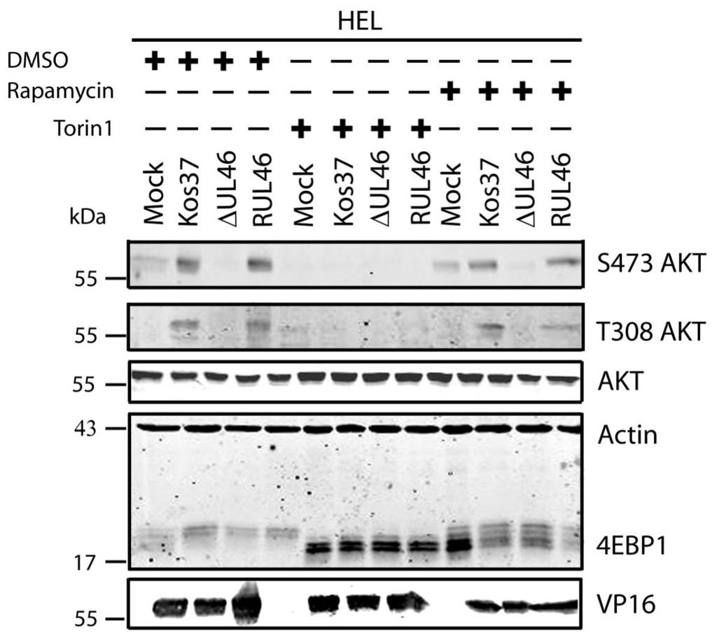
51 3.2 HSV-1 requires mtorc2 for AKT phosphorylation In uninfected cells, growth factor receptor signaling activates PI3K (sometimes with the assistance of SFKs), leading to activation of PDK1, which subsequently phosphorylates the activation loop of AKT. Activation of AKT also involves phosphorylation of its hydrophobic motif by PDK2, recently identified as mtorc2. The mechanisms that lead to the activation of mtorc2 remain largely unknown. HSV-induced activation of AKT involves VP11/12- and SFKdependent activation of PI3K, strongly suggesting that in HSV-infected cells, PDK1 mediates phosphorylation of AKT as in uninfected cells. Phosphorylation of the hydrophobic motif of AKT has also been shown to be dependent on VP11/12. However, it is currently unknown whether the VP11/12-dependent phosphorylation of the hydrophobic motif is mediated by mtorc2. To address this question I examined the effects of inhibiting mtorc2 activity. As described in the introduction, the mtor kinase exists in two complexes, mtorc1 and mtorc2, that differ in their subunit composition and sensitivity to rapamycin. Both kinases contain the mtor subunit that requires phosphatidic acid (PA) for mtor activity. PA and rapamycin compete to interact with the FKBP12-rapamycin binding domain (FRB) of mtor (64). Low concentrations of rapamycin are able to outcompete PA interactions with mtorc1. In contrast to mtorc1, mtorc2 is a more stable structure, so higher concentrations of rapamycin are required to dissociate the mtorc2 complex. Thus, rapamycin is an effective inhibitor of mtorc1. Another inhibitor of the mtor complexes is torin1, which inhibits the mtor catalytic subunit found in both mtorc1 and mtorc2 (235). Therefore, if mtorc2 is required for HSVinduced AKT activation, then one would predict that torin1 will block AKT activation, while rapamycin will have no effect. To test this prediction, serum-starved confluent monolayers of HEL cells were mock-infected or infected with wild type HSV-1 KOS37, the VP11/12-null mutantsδu L 46 and ΔU L 46-GalK, or the VP11/12-repaired virus RU L 46. Samples were treated with either 250 nm of torin1 or 100 nm of rapamycin for the entire course of infection (Fig. 3.2A) or the last three hours only (Fig. 3.2B). Cell lysates 38
52 were collected at 18 hpi and analyzed for the activating phosphorylation of AKT at residues S473 and T308, as well as for total AKT, 4EBP1, VP16, and β-actin by Western blotting. An equivalent concentration of the solvent DMSO (1µL/mL) was also used to discount any non-specific effects. DMSO had no detectable effect (either positive or negative) on AKT signaling or downstream phosphorylation of 4EBP1 in either mock-infected or infected cells (Fig. 3.3). 4EBP1 is a direct target of mtorc1 that can exist in several states of phosphorylation, resulting in the appearance of multiple bands on a Western blot. Both rapamycin and torin1 inhibited the phosphorylation of 4EBP1 in mockinfected cells. Reduction in the slowly migrating hyperphosphorylated forms of 4EBP1 is indicative of the effectiveness of these drugs. AKT phosphorylation remained undetectable in mock-infected cells treated with either drug (Fig. 3.2). As expected, U L 46 was required for HSVinduced AKT phosphorylation, which was restored in RU L 46-infected cells. In the presence of rapamycin, AKT was strongly phosphorylated at residues S473 and T308 during wild type and RU L 46 infection, but not during infection with ΔU L 46 virus. This was expected given that mtorc1 is positioned downstream of AKT and does not positively regulate AKT phosphorylation (79). Regardless of the length of drug treatment, torin1 prevented phosphorylation at both activating residues of AKT during infection with wild type HSV-1 and RU L 46. These data suggest that AKT phosphorylation during HSV-1 infection requires mtorc2. Taken in combination with the results of Wagner and Smiley (247), these results are consistent with the hypothesis that HSV employs VP11/12 to trigger AKT phosphorylation via mechanisms similar to those used by activated growth factor receptors in uninfected cells. As previously reported (247), in the absence of inhibitors, HSV-1 infection resulted in 4EBP1 hyperphosphorylation. Treatment of infected HEL cells with torin1 or rapamycin caused 4EBP1 to become hypophosphorylated, though this effect was more pronounced in torin1-treated cells. Also as previously reported, 4EBP1 hyperphosphorylation was VP11/12-independent (247). The simplest interpretation of this result is that HSV-1 encodes one or more proteins 39
53 other than VP11/12 that promote 4EBP1 hyperphosphorylation in an AKTindependent manner (Fig. 3.2) (247). I next focused my efforts on identifying the additional viral protein(s) involved. 40
54 Figure 3.2: mtorc2 is required for AKT activation during HSV-1 infection. Serum-starved HEL cells were either mock-infected or infected with HSV-1 KOS37, the VP11/12-null mutant ΔUL46, and the VP11/12-repaired virus RUL46 for 18 hours. A) During infection, cells were either left in serum-free media, treated with 250 nm torin1 or 100 nm of rapamycin. B) Alternatively, for the last three hours of infection, samples were either left in serum-free media, treated with 250 nm Torin1 or 100 nm of rapamycin. Cell lysates were collected at 18 hpi and analyzed for the activating phosphorylation of AKT at residues S473 (anti-s473) and T308 (anti-t308), total AKT, 4EBP1, VP16, and β-actin by Western blotting. 41
55 A. B. 42

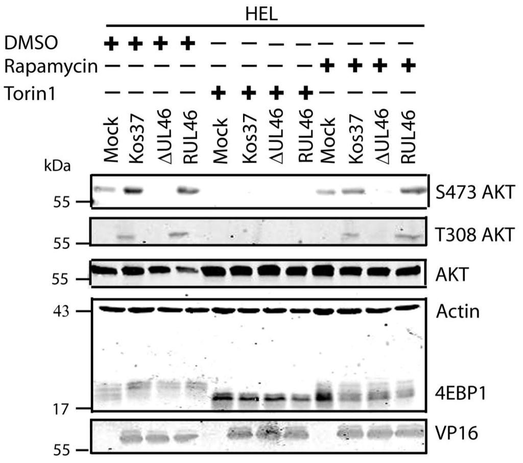
56 Figure 3.3: DMSO does not dampen AKT activation during HSV-1 infection. Serum-starved HEL cells were mock-infected or infected with wild type HSV-1 KOS37, the VP11/12-null mutants ΔUL46GalK and ΔUL46, or the VP11/12-repaired virus RUL46. During the course of the infection, cells were either left in serum-free media or treated with 1µL of DMSO in 1 ml of serum. Cell lysates were collected at 18 hpi and analyzed for the activating phosphorylation of AKT at residues S473 (anti-s473) and T308 (anti-t308), total AKT, 4EBP1, VP16, and β-actin by Western blotting. 43
57 3.3 US3 mediates the phosphorylation of AKT targets Since U L 46 is required for AKT phosphorylation during HSV-1 infection, the phosphorylation of AKT targets was expected to be U L 46-dependent (247). However, although the AKT targets GSK3β and 4EBP1 were phosphorylated in HSV-1 infected cells, their phosphorylation did not require U L 46 (247). Therefore, HSV-1 likely encodes one or more proteins other than VP11/12 that promote phosphorylation of AKT targets independently of AKT activation. We considered the possibility that an HSV-1 protein kinase directly phosphorylates AKT targets. HSV-1 encodes two protein kinases, U S 3PK and U L 13PK. Of these, U S 3 has previously been shown to regulate the phosphorylation of BAD on residue S136, the same site that AKT phosphorylates (165). Therefore, I asked whether U S 3 is also required for the phosphorylation of other AKT targets. Serum-starved HEL cells were mock infected or infected with wild type HSV-1 KOS37 and F strains, the KOS37 strain VP11/12-null mutants ΔU L 46 and ΔU L 46-GalK, the VP11/12-repaired virus RU L 46, the F strain U S 3PK-null mutant R7041, the U S 3PK-repaired virus R7306 or the F strain U L 13-null mutant R7356 virus. Cell lysates were collected at 18 hpi and analyzed for the phosphorylation of GSK3β and FoxO1 at residues S9 and S256 respectively, as well as for total GSK3β and FoxO1, VP16, and β-actin by Western blotting (Fig. 3.4). Compared to mock-infected cells, infection with wild type (KOS37 and F), VP11/12-null mutants (ΔU L 46 and ΔU L 46-GalK) or VP11/12 repaired virus strongly increased GSK3β and FoxO1 phosphorylation. Phosphorylation of AKT targets during infection was not due to increased protein expression since total GSK3β was unchanged and total FoxO1 decreased. Phosphorylated FoxO1 was not dramatically increased in ΔU L 46-GalK infected cells, which may be a consequence of gel-loading errors or poor film exposure. In contrast to wild type HSV-1 infection, deletion of U L 13 did not reduce the phosphorylation of AKT targets, whereas deletion of U S 3 abrogated the phosphorylation of FoxO1 and GSK3β. This result was not due to differences in protein loading as assessed by β- actin levels or a change in the progression of the virus s lytic life cycle assessed by VP16 levels. Phosphorylated GSK3β and FoxO1 was restored in cells infected 44
58 with repaired-u S 3 virus. These data suggest that U S 3 is required for phosphorylation of AKT targets (Fig. 3.4). During the course of these experiments, the Mohr group published similar results on the phosphorylation of AKT targets by U S 3 (34). These authors showed that U S 3 promoted phosphorylation of AKT targets FoxO1 and GSK3B. Furthermore, like AKT, U S 3 directly phosphorylates TSC2, which led to constitutive activation of mtorc1 and subsequent phosphorylation of S6K and 4EBP1. Phosphorylation of S6K and 4EBP1 resulted in enhanced translation and viral replication. Interestingly, S6K and 4EBP1 phosphorylation was only partially attenuated in ΔU S 3-infected cells, and was further reduced in the presence of an AKT drug inhibitor. These data suggest that HSV-1 activates mtorc1 through both AKT-dependent and AKT-independent mechanisms (34). From Dr. Wagner s findings, we know that AKT is activated by U L 46 (247). Thus, I examined whether U L 46-activated AKT and U S 3 signal in parallel to phosphorylate AKT targets. HEL cells are a difficult cell line to culture because they had a finite lifespan. Therefore, I developed a new cell system before studying the effects of HSV on AKT signaling. 45

59 Figure 3.4: The AKT targets, GSK3β and FoxO1, are phosphorylated in a U L 46-independent fashion, but require U S 3. Serum-starved HELs were mock infected or infected with wild type HSV-1 KOS37 and F, the VP11/12-null mutants ΔUL46GalK and ΔUL46, the VP11/12-repaired virus RUL46, the US3- null mutant R7041ΔUS3, the US3-repaired virus R7306 RUS3, and the UL13-null mutant R7356ΔUL13. Cell lysates were collected at 18 hpi and analyzed for the inhibiting phosphorylation of GSK3β and FoxO1 at residues S9 (anti- GSK3β) and S256 (anti-foxo1) respectively, total GSK3β and FoxO1, VP16, and β-actin by Western blotting. 46
60 3.4 HSV-1 also activates AKT in Human Foreskin Fibroblasts in a U L 46- dependent fashion HEL cells have a finite lifespan and therefore cannot be passaged indefinitely in cell culture. In order to provide a more convenient model for AKT activation in non-transformed human fibroblasts, I asked if HSV is also able to promote AKT phosphorylation in a VP11/12-dependent fashion during infection of telomerase-immortalized human foreskin fibroblasts (HFF-tel12) (24), kindly provided Wade Bresnahan). HFF cells were used by the Mohr group to examine the phosphorylation of AKT targets by U S 3 (34). Like HEL cells, HFF-tel12 cells (hereafter referred to as HFF cells) are non-transformed fibroblasts that lack constitutively active AKT. However, unlike HEL cells, they are immortal and therefore able to proliferate indefinitely. To examine AKT phosphorylation during infection, serum-starved HFFs were mock-infected or infected with wild type HSV-1 KOS37 or the VP11/12- null mutantδu L 46. Cell lysates were collected at regular intervals of 3 hours until 15 hpi and analyzed for the activating phosphorylation of AKT at residue S473 (anti-s473), total AKT, VP16, and β-actin by Western blotting. As previously observed for HEL cells, wild type HSV-1 KOS37 enhanced phosphorylation on residue S473 of AKT relative to the levels observed in mock-infected cells and this effect was abrogated by the VP11/12-null mutation. Interestingly, there was a noticeable induction of AKT phosphorylation detected as early as 3 hpi that diminished by 6 hpi and reappeared by 15 hpi (Fig. 3.5A). I next examined the kinetics of AKT activation at early times postinfection in greater detail by harvesting cells every hour during the first 5 hpi (Fig. 3.5B). A sample was also harvested at 15 hpi. Samples were analyzed by western blot as before. Actin served as a loading control, and HSV-1 ICP27 was used as a control for early virus infection. In infected HFFs, VP11/12-dependent AKT phosphorylation was not sustained during infection but rather occurred as a biphasic induction. AKT activation was seen at 3 and 4 hpi, diminished at 5 hpi and reappeared at 15 hpi. In this experiment, AKT activation was strongest at late 47
61 times post-infection. This was likely due to an accumulation of de novo VP11/12 during late infection. Despite the presence of VP11/12 in the virion tegument, there was no increase in AKT phosphorylation at 1 hpi, suggesting that the phosphorylation of AKT depends on newly synthesized VP11/12 (Fig. 3.5B). However, the experiments performed were not designed to examine whether VP11/12 molecules from incoming virions are sufficient to induce AKT phosphorylation. Overall these data indicate that HSV-1 triggers bi-phasic AKT phosphorylation in a VP11/12-dependent fashion during infection of HFF cells. HFF cells are a suitable cell model to examine AKT signaling during HSV-1 infection. Thus, I was able to examine if U L 46-activated AKT and U S 3 signal in parallel to phosphorylate AKT targets. AKT activation by HSV-1 was a common phenomenon seen in multiple cell types, including HeLa and Vero cells (Appendix A). 48
62 Figure 3.5: HSV-1 activates AKT in a U L 46-dependent manner at early and late times post infection. Confluent monolayers of HFF cells were serum-starved and mock-infected or infected with wild type HSV-1 KOS37 or the VP11/12-null mutants ΔUL46. Cell lysates were collected at various times and analyzed for the activating phosphorylation of AKT at residues S473 (anti-s473) and T308 (anti- T308), total AKT, VP16 or ICP27, and β-actin by Western blotting. 49
63 A. B. 50
64 3.5 U S 3 mediates the phosphorylation of AKT targets despite activation of AKT by U L 46 In HSV-1 infected HEL cells, AKT is phosphorylated at late times in a U L 46-dependent manner (247). In addition to HEL cells, HFFs can be used to study U L 46-dependent AKT activation. Infected HFFs have a biphasic induction (4 and 15 hpi) of AKT phosphorylation that is dependent upon U L 46. Evidence obtained using a combination of HSV-1 strains F and KOS and their mutants, suggested that the phosphorylation of AKT targets requires U S 3 (Fig. 3.4). However, it is unclear if both U L 46-activated AKT and U S 3 signal in parallel to promote the phosphorylation of AKT targets. To compare the roles of U S 3 and U L 46, ΔU S 3- and ΔU S 3ΔU L 46-mutant viruses were constructed in the same KOS37 background as the U L 46 mutants (as described in the Methods. Wu, Fred, unpublished data). If U L 46-activated AKT signals are transduced to downstream targets, then the phosphorylation of AKT targets should be reduced in cells infected with a ΔU S 3ΔU L 46-virus compared to cells infected with a ΔU S 3-virus. I examined the relative contributions of U S 3 and U L 46 over the course of infection. Due to the biphasic induction of AKT phosphorylation, I looked at AKT signaling at 4 and 15 hpi. Serum-starved HFFs were mock-infected or infected with wild type HSV- 1 KOS37, the KOS37 strains VP11/12-null mutant ΔU L 46, the U S 3PK-null mutant U S 3, or the VP11/12 and U S 3PK-double null mutant ΔU L 46 U S 3 virus. Cell lysates were collected at 4 hpi and 15 hpi, and analyzed for the phosphorylation of AKT at residues S473 and T308, the phosphorylation of GSK3β and BAD at residues S9 and S136 respectively, as well as for total AKT, GSK3β and BAD, and 4EBP1 (Fig. 3.6, 3.7). Lysates harvested at the late times were also analyzed for the phosphorylation of FoxO1 at residue S256 and total FoxO1 (Fig. 3.7). Actin was used as a loading control and HSV-1 ICP27 or VP16 was used as a control for virus infection (Fig. 3.6, 3.7). For each virus treatment, the level of phosphorylated GSK3β and 4EBP1 were measured in order to determine whether U L 46-activated AKT signals could reach AKT targets. Phosphorylated GSK3β levels were then divided by the values 51
65 obtained by anti-β-actin to give normalized values; this accommodated for any changes in cellular protein levels. Phosphorylation of 4EBP1 was determined by the ratio of hyperphosphorylated 4EBP1 to hypophosphorylated forms. Experiments pertaining to the first phase of AKT activation were replicated three times, while the experiments pertaining to the second phase of AKT activation were replicated seven times. The quantified values from the infrared signals were statistically analyzed by a two tailed un-paired t-test. At early (Fig. 3.6A) and late (Fig. 3.7A) times of infection, AKT was strongly phosphorylated in an U L 46-dependent manner; thus, AKT phosphorylation was attenuated in ΔU L 46- and ΔU L 46ΔU S 3-infections. AKT was phosphorylated in ΔU S 3-infected cells compared to wild type-infected cells (Fig. 3.6A, 3.7A). In fact, a previous finding states that U S 3 negatively regulates AKT phosphorylation, and the absence of U S 3 enhances and prolongs AKT activation during infection (13). Surprisingly, the phosphorylation of some AKT targets was reduced in ΔU S 3-infected cells even though AKT was phosphorylated. At early times of infection, AKT targets (GSK3β, BAD and 4EBP1) were not overtly phosphorylated in mock-infected cells (Fig. 3.6A). Similar to HSV-1 infection in HEL cells, HFFs infected with wild type-virus exhibited a significant increase in phosphorylated GSK3β (p<0.001), as well as, an increase in phosphorylated BAD and 4EBP1 and total BAD (Fig 3.6A, B). Although ΔU L 46- infected cells did not have activated AKT, the AKT targets 4EBP1, BAD and GSK3β (p<0.001) were strongly phosphorylated. The absence of U S 3 resulted in a reduction in total protein levels of BAD and both phosphorylated BAD and GSK3β (p<0.05), despite the fact AKT was phosphorylated. These data imply that activated AKT was not required for the phosphorylation of AKT targets. Cells infected with a ΔU L 46ΔU S 3-virus displayed a weak induction of phosphorylated GSK3β and total BAD that was comparable to levels seen in mock-infected cells. The amount of phosphorylated GSK3β was statistically similar between ΔU S 3- and ΔU L 46ΔU S 3-infections (p>0.05) (Fig. 3.6A, B). This indicates that the phosphorylation of GSK3β was primarily dependent on U S 3, with no significant contribution from U L 46-activated AKT. 4EBP1 was also hyperphosphorylated 52
66 during wild-type virus infection, but it was difficult to discern whether 4EBP1 was phosphorylated in the absence of U L 46 and U S 3. If 4EBP1 was hyperphosphorylated in ΔU L 46ΔU S 3-infected cells, then the data suggests HSV-1 encodes one or more proteins (other than U L 46 and U S 3) that promote 4EBP1 phosphorylation at early times of infection. During late infection with wild-type virus, the phosphorylation of AKT targets was enhanced compared to mock-infected cells. The AKT targets GSK3β (p<0.005) and 4EBP1 (p<0.001) were significantly more phosphorylated in wildtype and ΔU L 46-virus infection (Fig. 3.7A, B, C). In ΔU S 3- or ΔU L 46ΔU S 3- infection, there was an attenuation of both total BAD and phosphorylation of AKT targets, even though AKT was phosphorylated (Fig. 3.7A). ΔU S 3- and ΔU L 46ΔU S 3- infected cells had significantly less phosphorylated 4EBP1 and GSK3β (p<0.05) compared to wild-type infection. Less phosphorylated 4EBP1 could be a result of poor recognition of hyperphosphorylated 4EBP1 species. Cells infected with ΔU S 3-virus showed reduced phosphorylation of GSK3β and 4EBP1, and these levels were further diminished during ΔU L 46ΔU S 3-infection. However, this reduction did not correspond to a significant difference between ΔU S 3- and ΔU L 46ΔU S 3-virus infections (p>0.05, Fig. 3.7B, C). Two conclusions were drawn from these data. First, the phosphorylation of 4EBP1 and GSK3β are primarily U S 3-dependent with no significant contribution from U L 46-activated AKT. Second, at early times of infection, U S 3 selectively promotes the phosphorylation of some AKT targets and at late times of infection this selection more clearly includes 4EBP1 (Fig. 3.7). Mohr and colleagues reported that 4EBP1 was degraded during infection in a U S 3 dependent manner (34). My data confirm this finding showing that total 4EBP1 proteins levels were reduced in wild-type and ΔU L 46- infection (p<0.05) and U S 3 was required for this reduction (p>0.05). Cells infected with a ΔU- L46ΔU S 3- or ΔU S 3-virus had comparable 4EBP1 protein levels (p>0.05) indicating U L 46 does not affect 4EBP1 protein levels (Fig. 3.7D). In summary, these data indicate that U L 46-activated AKT signals do not contribute to the phosphorylation of AKT targets. Furthermore, the 53
67 phosphorylation of AKT targets relies upon U S 3, regardless of whether AKT is phosphorylated. Thus, phosphorylated AKT signals are either blocked or redirected during HSV-1 infection. 54
68 Figure 3.6: Despite U L 46-activated AKT, GSK3β is mainly phosphorylated in a U S 3-dependent fashion at early times post infection. Serum-starved HFF cells were mock infected or infected with wild type HSV-1 KOS37, the VP11/12-null mutant ΔUL46, the US3-null mutant ΔUS3, and the VP11/12-US3-null mutant ΔUL46 ΔUS3. A) Cell lysates were collected at 15 hpi and analyzed for the activating phosphorylation of AKT at residues S473 (anti-s473) and T308 (anti- T308), the inhibiting phosphorylation of GSK3β, FoxO1 and BAD at residues S9 (anti- GSK3β), S256 (anti-foxo1), and S136 (anti-bad) respectively, 4EBP1, total proteins AKT, GSK3β, FoxO1 and BAD, VP16, and β-actin by Western blotting. B) For each sample, the infrared signal obtained with the anti-gsk3β phospho-s9 antibody for panel A was divided by the signal obtained with the antiβ-actin antibody, and the ratios were divided to yield a proportional value. Compared to mock infected cells, KOS37- and ΔUL46-infected cells had significantly (p<0.001) more detectable phosphorylated GSK3β on residue S9. Additionally, ΔUS3-infection had significantly (p<0.05) more phosphorylated GSK3β on residue S9. Relative to ΔUS3-infected cells, ΔUL46ΔUS3 infection did not have significantly less phosphorylated GSK3β on residue S9 (p>0.05). 55
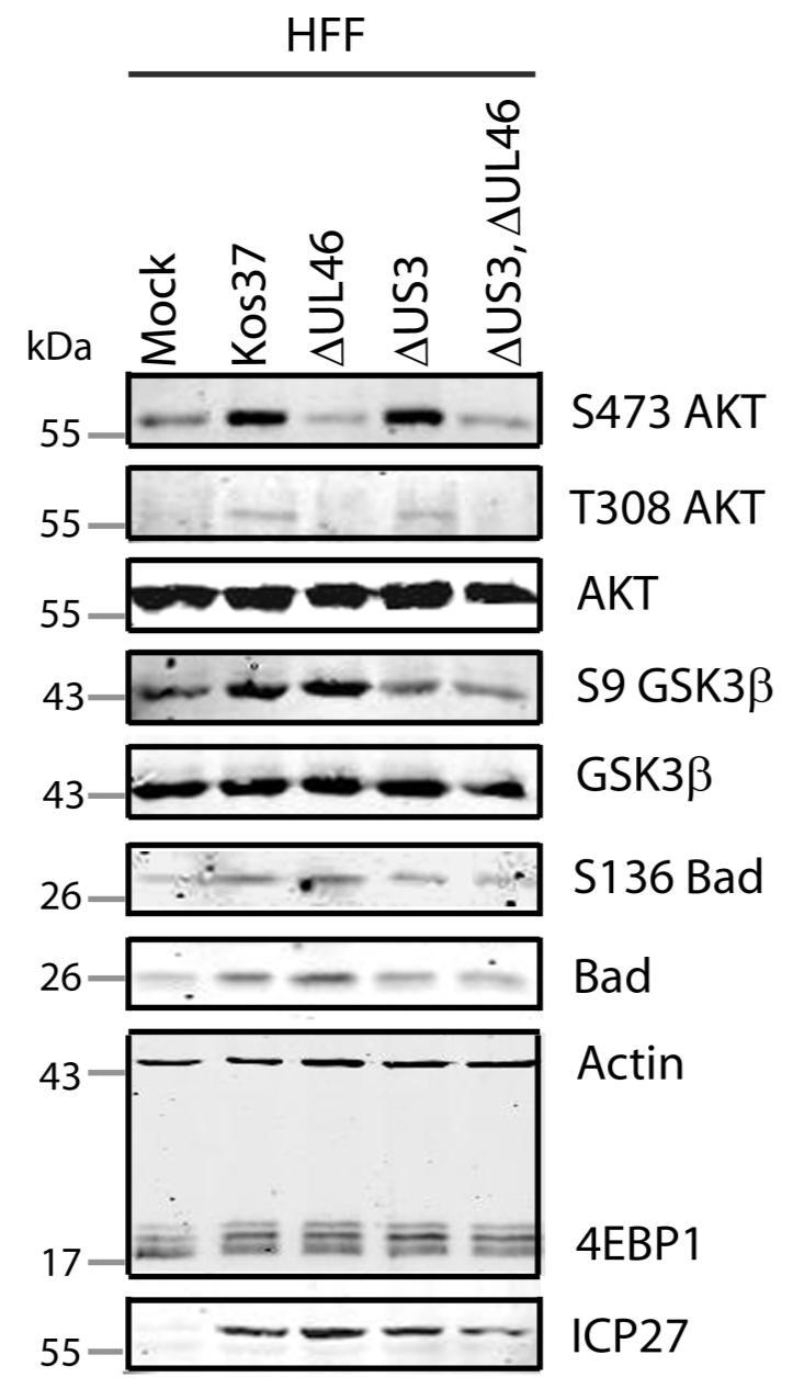
69 A. 56
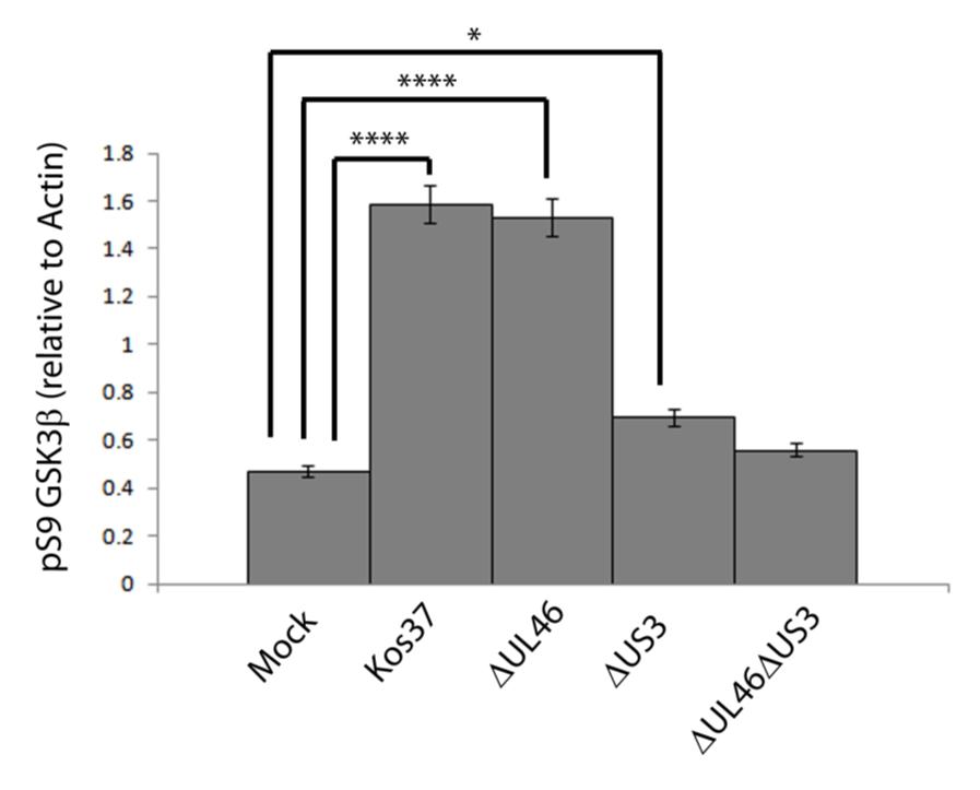
70 B. 57
71 Figure 3.7: Multiple AKT targets are phosphorylated in a U S 3-dependent fashion at late times post infection. Serum-starved HFF cells were mock infected or infected with wild type HSV-1 KOS37, the VP11/12-null mutant ΔUL46, the US3-null mutant ΔUS3, and the VP11/12-US3-null mutant ΔUL46ΔUS3. Cell lysates were collected at 15 hpi and A) analyzed for the activating phosphorylation of AKT at residues S473 (anti-s473) and T308 (anti- T308), the inhibiting phosphorylation of GSK3β, FoxO1 and BAD at residues S9 (anti- GSK3β), S256 (anti-foxo1), and S136 (anti-bad) respectively, 4EBP1, total proteins AKT, GSK3β, FoxO1 and BAD, VP16, and β-actin by Western blotting. B) For each sample, the infrared signal obtained with the anti-gsk3β phospho-s9 for panel A was divided by the signal obtained with the anti-β-actin antibody, and the ratios were normalized to yield a proportional value. Compared to mock infected cells, Kos37- and ΔUL46-infected cells had significantly (p<0.005) more detectable phosphorylated GSK3β at residue S9. Additionally, ΔUS3-infection had significantly (p<0.05) more phosphorylated GSK3β at residue S9. Relative to ΔUS3-infected cells, ΔUL46ΔUS3 infection did not have significantly less phosphorylated GSK3β at residue S9 (p>0.05). C) The infrared signal for the two upper and two lower bands of 4EBP1 were divided to yield a proportional value of phosphorylated 4EBP1. Compared to mock infected cells, KOS37- and ΔUL46-infected cells had significantly (p<0.001) more phosphorylated 4EBP1. Relative to ΔUS3-infected cells, ΔUL46ΔUS3 infection had similar levels of phosphorylated 4EBP1 (p>0.05). D) The infrared signals obtained with anti-4ebp1 from panel A was divided by the signal obtained with anti-β-actin, and the ratios were normalized to yield a proportional value. Compared to mock infected cells, KOS37- and ΔUL46-infected cells had significantly (p<0.05) more detectable 4EBP1. Compared to wild type infeted cells, both ΔUS3- and ΔUL46ΔUS3- infection had significantly less 4EBP1 (p>0.05). 58
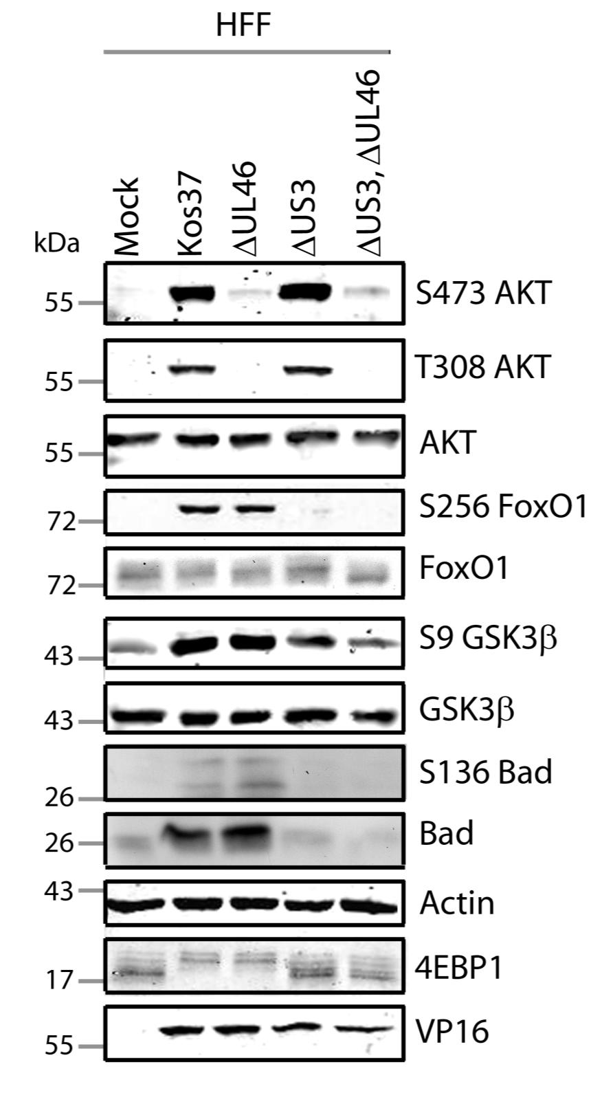
72 A. 59
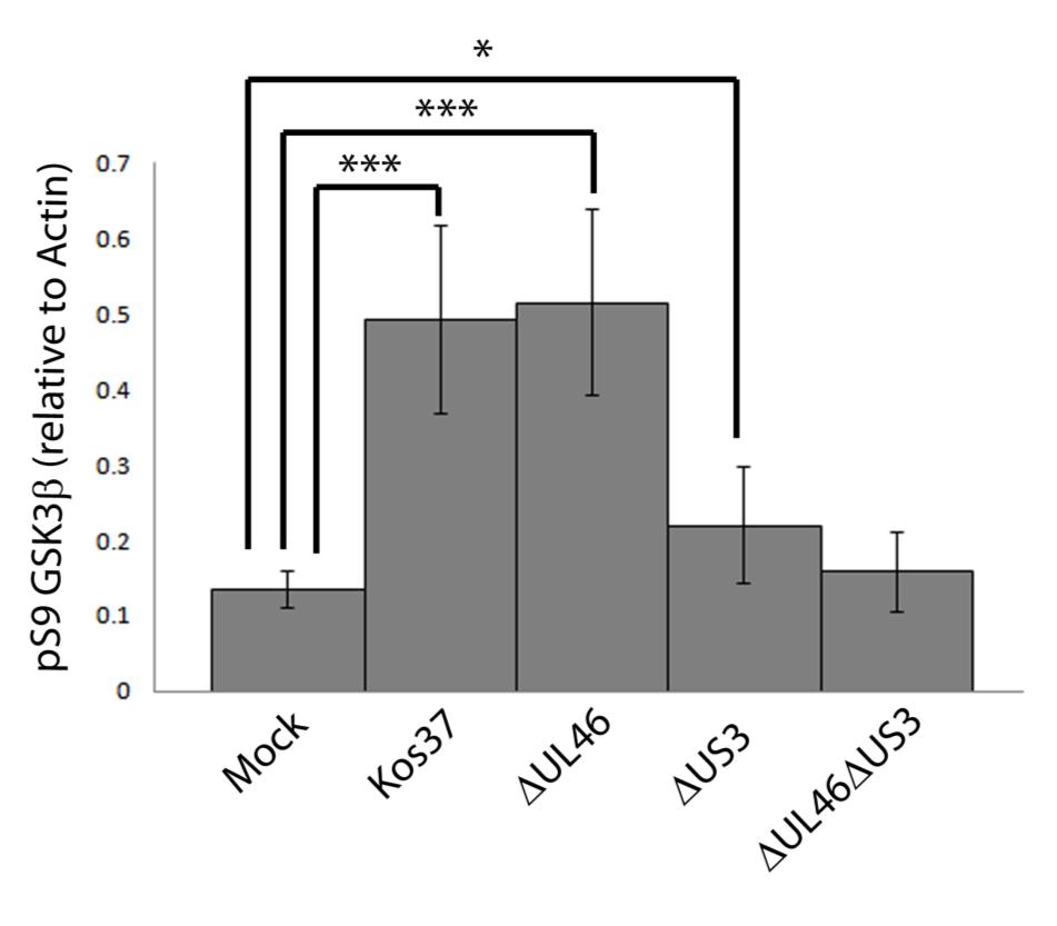
73 B. 60
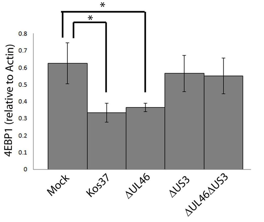
74 C. D. 61
75 3.6 Serum induces AKT activation, but there is no significant activation signal transduced to AKT targets at late times of infection HSV-1 infection requires U L 46 for the phosphorylation of AKT, while U- S3 is required for the phosphorylation of AKT targets. Deletion of U L 46, and hence activated AKT, does not affect the phosphorylation of AKT targets. These data indicate that AKT is unable to phosphorylate some of its targets during HSV- 1 infection, implying that its activity is either blocked or redirected. If there is a signaling block between AKT and its targets, then stimulating the pathway with external growth factors should not enhance the phosphorylation of AKT targets. The AKT pathway can be stimulated by serum, which contains numerous growth factors that bind and activate cell receptors. In uninfected cells, serum activates some receptors that recruit PI3K to the plasma membrane, which is followed by the phosphorylation of AKT and AKT targets. Since there are two phases of AKT phosphorylation (4 and 15 hpi), I asked whether the signaling dynamics of the AKT pathway changed over the course of infection. Prior to examining activated AKT signaling during infection, serum was titrated to determine a concentration that would yield maximal AKT phosphorylation. Serum-starved HFFs were exposed to an increasing amount of serum from 0% to 15%. Cell lysates were collected after 20 minutes and analyzed for AKT phosphorylation at residue S473, phosphorylated GSK3β at residue S9, as well as, total proteins AKT and GSK3β. β-actin served as a loading control. Serum-starved cells lacked phosphorylated AKT and GSK3β. As serum concentrations increased, so did the amount of phosphorylated AKT, however, serum concentrations greater than 10% did not induce more phosphorylated AKT (Fig. 3.8B). Phosphorylated GSK3β was also enhanced with greater serum concentrations (Fig. 3.9A). Additional control experiments were done to determine the optimal duration of serum exposure that would maximize AKT phosphorylation. Serumstarved HFFs were exposed to 10% serum for increasing lengths of time between 5 minutes and 2 hours. Cell lysates were analyzed for the same proteins noted in Fig AKT was most strongly phosphorylated between 15 and 30 minutes of 62
76 serum treatment, and AKT phosphorylation decreased after 30 minutes of exposure (Fig. 3.9). After 5 minutes of serum treatment, GSK3β was phosphorylated and its phosphorylation was not enhanced with longer serum exposure. Therefore, AKT is phosphorylated most strongly with 10% serum between 15 and 30 minutes. As mentioned previously, I examined if a signaling block existed between AKT and its targets during infection. If there is a block in signaling between AKT and its targets, then stimulating cells with serum should not result in the phosphorylation of AKT targets. To test this, serum-starved HFFs were infected with the viruses shown in Fig and Samples were incubated for either 4 or 15 hpi (Fig. 3.10, 3.11). Prior to harvesting lysates, cells were treated with 10% serum for 20 minutes. The lysates were collected after 20 minutes of serum exposure, and analyzed for the phosphorylation of AKT, GSK3β and 4EBP1 by western blotting as before (Fig. 3.10, 3.11). β-actin served as a loading control and HSV-1 VP16 or ICP27 were used as controls for virus infection. To discern whether activated growth factor signals could be propagated to AKT targets, the level of phosphorylated GSK3β was measured. Phospho-GSK3β levels were then divided by the values obtained by anti-β-actin to give normalized values. The quantified values were statistically analyzed using a two-tailed t-test from three replicates. During early infection, HSV-1 induced AKT phosphorylation was U L 46- dependent. U S 3 was required for the phosphorylation of AKT targets, excluding 4EBP1, which remained in a hyperphosphorylated form in the absence of U S 3 and U L 46. Therefore, in ΔU L 46ΔU S 3-infected cells, neither AKT nor the majority of its targets were unphosphorylated in the absence of serum. In the presence of serum, AKT and its targets were phosphorylated in mock, wild-type, ΔU L 46, ΔU S 3 and ΔU L 46ΔU S 3-infections. Since the addition of serum caused AKT and its targets to become phosphorylated in ΔU L 46ΔU S 3-infected cells, the data indicates serum-activated AKT signals were propagated to AKT targets. This suggests a signaling block between AKT and its targets is non-existent at early times of infection (Fig. 3.10). 63
77 During late infection, AKT was strongly phosphorylated in a U L 46- dependent manner. In the presence of serum, AKT was strongly phosphorylated at the hydrophobic motif in all samples. Contrary to the hydrophobic motif, ΔU L 46- infected cells had less AKT phosphorylation at residue T308 compared to mockinfection. There were similar levels of phosphorylated T308 of AKT between mock- and ΔU L 46ΔU S 3-infected cells. Thus, only in the absence of U S 3 does serum stimulation result in the phosphorylation of AKT at residue T308. These data suggest that U S 3 is negatively regulating AKT phosphorylation, specifically at the T308 residue, and this effect only exists during late infection (Fig. 3.11). Since serum was able to induce activated AKT in ΔU L 46ΔU S 3-infected cells, I was able to test whether activated AKT signals could be transduced to downstream targets. At late times of infection, phosphorylated 4EBP1 and GSK3β were significantly enhanced in wild-type and ΔU L 46-infected cells (p<0.001), but not in ΔU S 3- or ΔU L 46ΔU S 3- infections. This supports the theory that U S 3 is required for the phosphorylation of AKT targets during infection. Serum stimulation resulted in comparable levels of phosphorylated 4EBP1 and GSK3β between mock, wild-type and ΔU L 46-infected cells. Although serum-stimulation enhanced the phosphorylation of 4EBP1 and GSK3β in ΔU S 3- and ΔU L 46ΔU S 3-infected cells, these virus infections had significantly less phosphorylated GSK3β than mock and wild-type infections (p>0.05). Because AKT is phosphorylated in ΔU L 46ΔU S 3-infected cells, but the signal is not significantly transduced to AKT targets, activated AKT signals are attenuated at late times during infection. Furthermore, this signaling block is independent of U S 3 and U L 46 (Fig. 3.11A, B). Serum contains numerous growth factors that bind a variety of cell receptors and activate cell signaling pathways. These cell receptors can potentially cross-talk with the AKT pathway. Hence, it is unclear which specific growth factor is contributing to AKT signaling. Thus, an alternative method of testing a signaling block during infection is by using defined growth factors of the PI3K- AKT pathway. 64
78 Figure 3.8: 10% serum induces maximal AKT activation after 20 minutes. Confluent monolayers HFF cells were serum-starved for 24 hours and A) titrated with an increasing concentration of serum in 1 ml DMEM. Cell lysates were collected after 20 minutes of treatment and analyzed for the activating phosphorylation of AKT at residues S473 (anti-s473), the inhibiting phosphorylation of GSK3β at residue S9 (anti- GSK3β), total proteins AKT and GSK3β, and β-actin by Western blotting. B) The infrared signal obtained with the anti-s473 AKT for panel A was divided by the signal obtained with the anti-akt antibody, and the ratios were normalized to yield a proportional value. 65
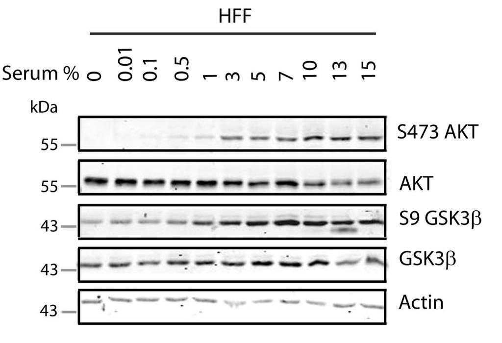
79 A. B. 66
80 C. 67
81 Figure 3.9: Serum induces AKT activation and downstream signaling in infected cells at early infection. Serum-starved HFF cells were mock infected or infected with wild type HSV-1 KOS37, the VP11/12-null mutant ΔUL46, the US3-null mutant ΔUS3, and the VP11/12-US3-null mutant ΔUL46 ΔUS3. Samples were either left in serum-free media or treated with 10% serum-dmem for 20 minutes prior to harvesting cells. Cell lysates were collected at 15 hpi and analyzed for the activating phosphorylation of AKT at residues S473 (anti-s473) and T308 (anti-t308), the inhibiting phosphorylation of GSK3β at residue S9 (anti- GSK3β), 4EBP1, total proteins AKT and GSK3β, ICP27, and β-actin by Western blotting. 68
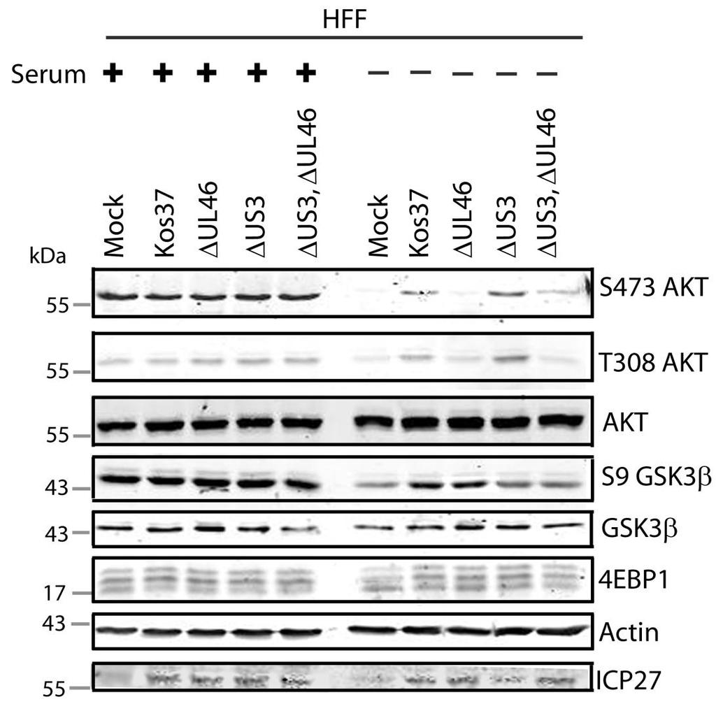
82 69
83 Figure 3.10: Serum induces AKT activation and, but not downstream signaling in infected cells at late times post infection. Serum-starved HFF cells were mock infected or infected with wild type HSV-1 KOS37, the VP11/12-null mutant ΔUL46, the US3-null mutant ΔUS3, and the VP11/12-US3-null mutant ΔUL46 ΔUS3. Samples were either left in serum-free media or treated with 10% serum-dmem for 20 minutes prior to harvesting cells. A) Cell lysates were collected at 15 hpi and analyzed for the activating phosphorylation of AKT at residues S473 (anti-s473) and T308 (anti-t308), the inhibiting phosphorylation of GSK3β at residue S9 (anti- GSK3β), 4EBP1, total proteins AKT and GSK3β, ICP27, and β-actin by Western blotting. B) The infrared signal obtained with the anti-gsk3β phospho-s9 antibody for panel A was divided by the signal obtained with the anti-β-actin antibody, and the ratios were normalized to yield a proportional value. Compared to mock infected cells, KOS37- and ΔUL46- infected cells had significantly (p<0.001) more detectable phosphorylated GSK3β at residue S9. Under serum-stimulation mock infected cells were significantly (p<0.05) more phosphorylated than stimulated ΔUS3- and ΔUL46 ΔUS3 infected cells. 70
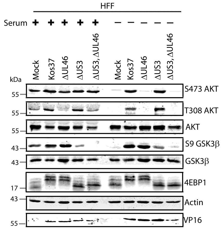
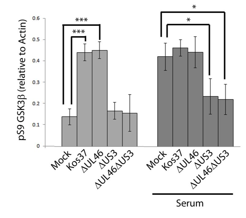
84 A. B. 71
85 3.7 PDGF BB induces AKT activation, but the signal is not transduced to AKT targets Activated PI3K leads to the phosphorylation of AKT and AKT targets. U L 46 is required for AKT activation, but U L 46-activated AKT signals are not transduced to AKT targets during infection; this indicates that AKT signals are either blocked or redirected. As shown in section 3.6, activated AKT signals are not propagated during late times of infection even in the presence of serum, providing evidence for a signaling block. Serum contains a plethora of growth factors and several of these factors could contribute to AKT signaling. Thus, I asked if HSV infection inhibits signaling downstream of AKT when AKT is activated by a defined growth factor, in the same way that HSV blocks signaling downstream of AKT when AKT is activated by serum. PDGF BB liagnds are one of many growth factors that activate the PI3K-AKT pathway. When PDGF BB binds to its cognate receptor, it induces dimerization, which is followed by the recruitment and activation of PI3K (5, 119, 259). I examined AKT signaling during late times of infection when cells are stimulated with PDGF BB. PDGF BB was titrated to give a maximal amount of AKT activation, which happened to be equal to the amount of phosphorylated AKT induced by 10% serum (Fig. 3.8). Serum-starved HFFs were exposed to an increasing concentration of PDGF BB from 1.0 pg/µl to 4.0 pg/µl, or 10% serum (Fig. 3.12A). Since 20 minutes of serum treatment was sufficient to induce maximal AKT phosphorylation (Fig. 3.8), cell lysates were collected after 20 minutes of PDGF BB treatment. Cell lysates were analyzed for the phosphorylation of AKT and GSK3β (Fig. 3.12A). In the absence of PDGF BB, AKT and GSK3β were not phosphorylated. Stimulating cells with 2.5 pg/µl of PDGF BB was sufficient to induce phosphorylation of AKT that was equivalent to the levels given by 10% serum (Fig. 3.12B). Phosphorylated GSK3β was seen in cells treated with a minimum of 1.0 pg/µl PDGF BB (Fig. 3.12A). AKT is phosphorylated most strongly with a minimum of 2.5 pg/µl PDGF BB for 20 minutes. As previously stated, if PDGF BB signals are not propagated to AKT targets, then there is evidence for a signaling block. To test this, serum-starved 72
86 HFFs were infected with the viruses shown in Fig Cells were incubated for 15 hours and treated with 20.0 pg/µl of PDGF BB for 20 minutes; this concentration was more than sufficient to induce phosphorylation of AKT and AKT targets. Cell lysates were collected after exposure to PDGF BB for 20 minutes and analyzed for the phosphorylation of AKT and GSK3β, and total proteins AKT, GSK3β and 4EBP1 (Fig. 3.13). β-actin was used as a loading control, and HSV-1 VP16 served as a control for virus infection. For each virus treatment, the level of phosphorylated GSK3β was measured in order to determine whether PDGF BB activation signals could reach AKT targets. Phospho-GSK3β levels were then divided by the values obtained by anti-β-actin to give normalized values; this accommodated for any changes in cellular protein levels. The quantified values were statistically analyzed using a two-tailed t-test from three replicates. As expected, in the absence of PDGF BB, AKT was phosphorylated in a U L 46-dependent fashion. In all samples, PDGF BB treatment greatly enhanced AKT phosphorylation at both activating residues. Treatment with PDGF BB resulted in more phosphorylated AKT in wild-type infected cells compared to unstimulated HSV-1 infected cells. An excessive amount of PDGF BB was added to cells, which could have caused the enhancement of AKT phosphorylation. Interestingly, under the influence of PDGF BB, ΔU L 46-infected cells had equivalent levels of phosphorylated AKT at residue T308 when compared to mock-infected cells (Fig. 3.13A). This result was different from ΔU L 46-infected cells that were treated with serum, which showed less T308 phosphorylation compared to mock-infection (Fig. 3.11A). ΔU L 46-virus reduces T308 phosphorylation in the presence of serum, while PDGF BB-treated cells did not display this inhibition. An excessive amount PDGF BB was added to infected cells, which may compensate for any negative effect on AKT signaling. Since PDGF BB was able to stimulate AKT phosphorylation in ΔU S 3- and ΔU L 46ΔU S 3- infected cells, I asked if these activation signals could be propagated downstream. As expected, U S 3 was required for the phosphorylation of AKT targets during HSV-1 infection. AKT targets were phosphorylated in mock, wild-type 73
87 and ΔU L 46-infected cells that were treated with PDGF BB. In the presence of PDGF BB, ΔU S 3- and ΔU L 46ΔU S 3-infected cells displayed an increase in phosphorylated GSK3β compared to their unstimulated counterparts (Fig. 3.13A). However, this increased GSK3β phosphorylation was significantly less than levels seen in mock-infected cells treated with PDGF BB (p<0.005) (Fig. 3.13B). In ΔU S 3- and ΔU L 46ΔU S 3-infected cells, 4EBP1 remained in a hypophosphorylated form in the presence of PDGF BB (Fig. 3.13A). Because PDGF BB-activated AKT signals were not significantly propagated downstream during infection, there is a block in signaling between AKT and some of its targets. Furthermore, PDGF signals were attenuated in virus infections lacking U S 3 and U L 46, which infers that these two viral proteins are not responsible for the signaling block. It would be interesting to examine whether or not HSV-1 blocks activation signals from other defined growth factors. PDGF BB is one member from a family of PDGF ligands that are specific for activating the PI3K-AKT pathway. Thus, I examined AKT signaling in the presence of other PDGF ligands during HSV-1 infection. 74
88 Figure 3.11: PDGF BB activates AKT. Confluent monolayers of HFF cells were serum-starved for 24 hours and titrated with an increasing concentration of PDGF BB in 1 ml DMEM. A) Cell lysates were collected after 20 minutes of treatment and analyzed for the activating phosphorylation of AKT at residue S473 (anti- S473), the inhibiting phosphorylation of GSK3β at residue S9 (anti- GSK3β), total protein AKT by Western blotting. B) The infrared signal obtained with the anti-s473 AKT for panel A was divided by the signal obtained with the anti- β- actin antibody, and the ratios were normalized to yield a proportional value. 75
89 A. B. 76
90 Figure 3.12: PDGF BB induces AKT activation, but the signal is not propagated to AKT targets during late times post infection. Serum-starved HFF cells were mock infected or infected with wild type HSV-1 KOS37, the VP11/12-null mutant ΔUL46, the US3-null mutant ΔUS3, and the VP11/12-US3- null mutant ΔUL46 ΔUS3. Samples were either left in serum-free media or treated with 20.0 pg/µl PDGF BB for 20 minutes prior to harvesting cells. A) Cell lysates were collected at 15 hpi and analyzed for the activating phosphorylation of AKT at residues S473 (anti-s473) and T308 (anti-t308), the inhibiting phosphorylation of GSK3β at residue S9 (anti- GSK3β), 4EBP1, total proteins AKT and GSK3β, VP16, and β-actin by Western blotting. B) The infrared signal obtained with the anti-s9 GSK3β for panel A was divided by the signal obtained with the anti- β-actin antibody, and the ratios were normalized to yield a proportional value. Kos37- and ΔUL46-infected cells had significantly (p<0.005) more detectable phosphorylated GSK3β at residue S9 then mock-infection. Upon PDGF BB stimulation mock- infected cells were significantly more phosphorylated than stimulated ΔUS3- (p<0.005) and ΔUL46 ΔUS3 (p<0.05) infected cells. 77
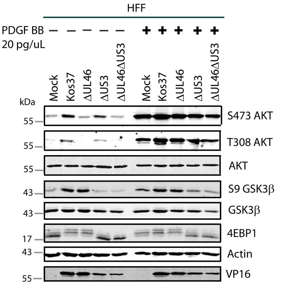
91 A. \ 78

92 B. 79
93 3.8 Titration of PDGF AA, AB and BB PDGF ligands exist in three isoforms, (AA, AB and BB) and each of them differs in their receptor-binding specificity (85, 226). AA has the ability to bind the αα- receptor; AB the αβ- and αα- receptor; BB the ββ-, αβ-, and αα-receptor (62, 69, 90, 91, 218). Once the dimer-ligands are bound to their receptors, PDGFreceptors undergo homo- or heterodimerization and auto-tyrosine phosphorylation, which triggers the recruitment and activation of PI3K (5, 119, 259). The PDGF ligands were titrated for an equal level of AKT phosphorylation at both activating residues. To accomplish this, serum-starved HFFs were treated with increasing amounts of PDGFs between 0.5 pg/µl and 50.0 pg/µl for 20 minutes (Fig. 3.14A, B, C). Cell lysates were collected after 20 minutes of PDGF treatment and analyzed for the phosphorylation of AKT and GSK3β (Fig. 3.14). β-actin was used as a loading control. These experiments were replicated three times for both phosphorylated residues of AKT, S473 and T308. When cells were treated with equal concentrations of PDGFs, PDGF AA was unable to induce S473- and T308- phosphorylation as strongly as, PDGF AB and BB (Fig. 3.14D, E). Stimulating cells with PDGF AB produced the most phosphorylated AKT compared to PDGF AA and BB (Fig A, B, C). It is interesting that PDGF AB triggers stronger AKT phosphorylation than PDGF BB at the same concentration, even though PDGF BB binds all three PDGF receptors and PDGF AB only binds two (62, 69, 90, 91, 218). Equivalent levels of phosphorylated AKT at residue S473 were achieved at 30.0 pg/µl PDGF AA, 10.0 pg/µl PDGF AB, and 20.0 pg/µl PDGF BB (Fig. 3.14D). Equivalent levels of phosphorylated AKT at residue T308 were achieved at 20.0 pg/µl PDGF AA, 5.0 pg/µl PDGF AB, and 10.0 pg/µl PDGF BB (Fig. 3.14E). Regardless of the type of PDGF, the phosphorylation of GSK3β was enhanced with greater PDGF concentrations (Fig A, B, C). Using PDGF concentrations that gave equal levels of phosphorylated AKT, I asked whether PDGF AA, AB and BB signals could be propagated to AKT and AKT targets during infection. 80
94 Figure 3.13: Differing concentrations of PDGF AA, AB and BB induce comparable levels of AKT activation. Confluent monolayers of HFF cells were serum-starved for 24 hours and titrated with an increasing concentration of PDGF in 1 ml DMEM. A), B), C) Cell lysates were collected after 20 minutes of treatment and analyzed for the activating phosphorylation of AKT at residues S473 (anti-s473) and T308 (anti-t308), the inhibiting phosphorylation of GSK3β at residue S9 (anti- GSK3β), total proteins AKT and GSK3β, and β-actin by Western blotting. The infrared signal obtained with the D) anti-s473 and E) anti- T308 AKT for panel D was divided by the signal obtained with the anti- β-actin antibody, and the ratios were normalized to yield a proportional value. 81
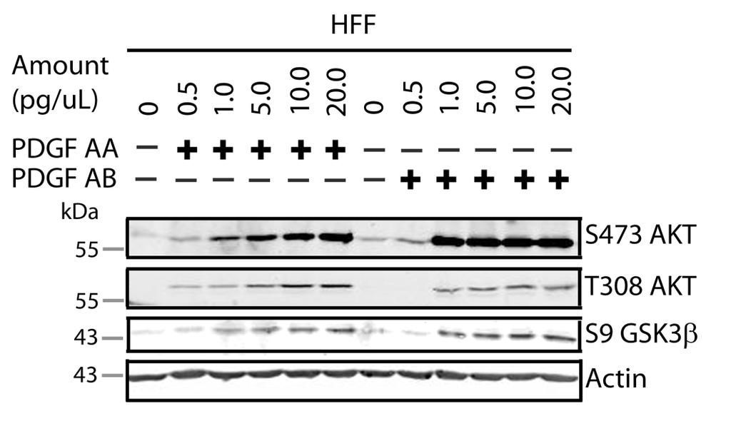
95 A. B. 82
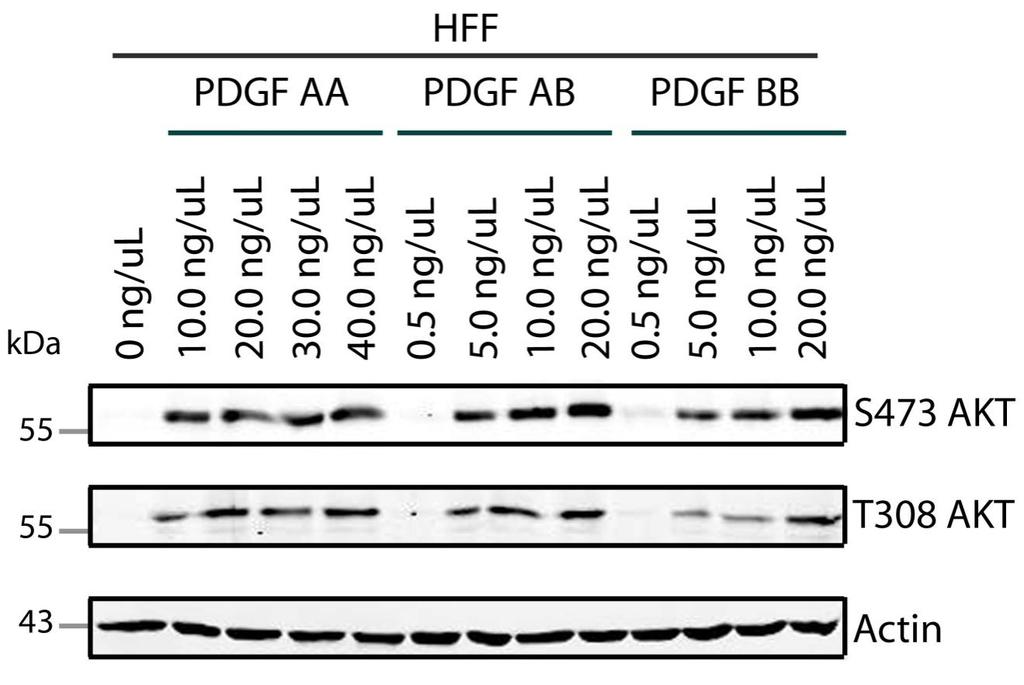
96 C. 83
97 E. F. 84
98 3.9 PDGF ligands do not perform equally at activating AKT during infection In uninfected cells, the presence of PDGF BB results in AKT activation and the successive phosphorylation of AKT targets. In HSV-1 infected cells, PDGF BB signals enhanced AKT phosphorylation, but those phosphorylated signals were not propagated downstream (Fig. 3.13). Thus, there is a block in signaling between AKT and its targets, which is observed when infected cells are stimulated with PDGF BB. I examined whether HSV-1 blocks signaling between AKT and AKT targets when cells are stimulated with other types of PDGF ligands (AA, AB and BB). Serum-starved cells were infected with the viruses indicated in Fig and Cells were incubated for 15 hours and then treated with PDGF AA, AB or BB. Because the PDGF ligands do not perform equally at stimulating AKT phosphorylation on either residue, cells were treated with PDGFs that gave comparable levels of AKT phosphorylation either at S473 or T308 (as noted in Section 3.9, Fig. 3.14). Cell lysates were collected after 20 minutes of PDGF treatment and analyzed for the phosphorylation of AKT and AKT targets (Fig. 3.15, 3.16). HSV-1 VP16 was used as a control for viral infection, and β-actin was used as a loading control. These experiments were replicated three times using PDGF concentrations that gave equal levels of AKT phosphorylation either at residue S473 or T308. In mock-infected cells, all three PDGFs gave equal levels of phosphorylated AKT at residues S473 and T308. Since the PDGFs gave similar levels of phosphorylated AKT, any change in AKT signaling would likely be a result of virus infection. As expected, phosphorylated AKT was increased during HSV-1 infection in a U L 46-dependent fashion. In wild-type and ΔU S 3-infected cells, all three PDGF ligands were able to further enhance the phosphorylation of AKT in contrast to their unstimulated counterparts. Interestingly, during HSV-1 infection, PDGF AA produced less phosphorylated AKT compared to PDGF AB or BB stimulation, while PDGF BB gave the greatest amount of phosphorylated AKT. This observation was most apparent in cells infected with a virus lacking U L 46. Cells infected with ΔU L 46- or ΔU L 46ΔU S 3-virus displayed a lack of AKT 85
99 phosphorylation, and stimulation with PDGF AA or AB had either little (Fig. 3.15) or no effect (Fig. 3.16) at inducing AKT phosphorylation. In the event that AKT phosphorylation was enhanced by PDGFs during infection, PDGF AB had a stronger effect at inducing AKT phosphorylation compared to PDGF AA (Fig. 3.15). On the other hand, phosphorylated AKT was strongest in cells stimulated with PDGF BB (Fig. 3.15, 3.16). Therefore, these data suggest that HSV-1 infection dampens signaling from certain PDGF receptors to AKT. It is unclear whether the block is at the receptor, or between the receptor and AKT. Interestingly, treatment with PDGF AB induced less AKT phosphorylation in ΔU L 46-infected cells compared to ΔU L 46ΔU S 3-infected cells. Thus, U S 3 may have a marginal role in modulating growth factor signaling at the receptor. However, U L 46 and U S 3 were obviously not the main factors for this signaling block, which indicates other viral protein(s) are involved. As a result of this receptor-block, PDGF AA and AB were not suitable candidates to assess signaling between AKT and AKT targets. In the presence of each PDGF, AKT targets were phosphorylated in mockinfected cells. In wild type and ΔU L 46-infected cells, AKT targets were noticeably phosphorylated and this phosphorylation was not enhanced in the presence of an external ligand. As expected, the phosphorylation of AKT targets were dependent on U S 3, thus phosphorylation was attenuated in the ΔU S 3- and ΔU L 46ΔU S 3-infected cells. In ΔU S 3- and ΔU L 46ΔU S 3-infected cells, PDGF AA and PDGF AB had no apparent effect on the phosphorylated levels of 4EBP1 and GSK3β. Stimulating cells with PDGF BB increased phosphorylated GSK3β in ΔU S 3- and ΔU L 46ΔU S 3-infected cells, but as witnessed in Section 3.8, this phosphorylation was less than those levels seen in mock-infected cells that were stimulated with PDGF BB. As stated previously in Section 3.8, activated AKT signals are attenuated during HSV-1 infection and this is independent of U L 46 and U S 3 (Fig. 3.15, 3.16). The initial aim of this experiment was to examine if PDGF AA, AB and BB signals could be propagated to AKT targets during infection. Rather, I witnessed that HSV-1 inhibits signaling through some receptors for unknown 86
100 reasons (Fig. 3.15, 3.16). Regardless of this inhibition, HSV-1 encodes U L 46 to activated AKT, yet these activated signals are not propagated to the examined downstream targets. Rather, HSV-1 encodes U S 3 to cherry-pick and phosphorylate AKT targets. These data indicate that activated AKT signals are either attenuated or redirected during infection. 87
101 Figure 3.14: PDGF ligands do not perform equally at phosphorylating S473 AKT during infection. Serum-starved HFF cells were mock infected or infected with wild type HSV-1 KOS37, the VP11/12-null mutant ΔUL46, the US3-null mutant ΔUS3, and the VP11/12-US3-null mutant ΔUL46 ΔUS3. Samples were either left in serum-free media or treated with 30.0 pg/µl PDGF AA, 1.0 pg/µl PDGF AB, and 20.0 pg/µl PDGF BB for 20 minutes prior to harvesting. Cell lysates were collected at 15 hpi and analyzed for the activating phosphorylation of AKT at residues S473 (anti-s473) and T308 (anti-t308), the inhibiting phosphorylation of GSK3β at residue S9 (anti- GSK3β), 4EBP1, total proteins AKT and GSK3β, VP16, and β-actin by Western blotting. 88
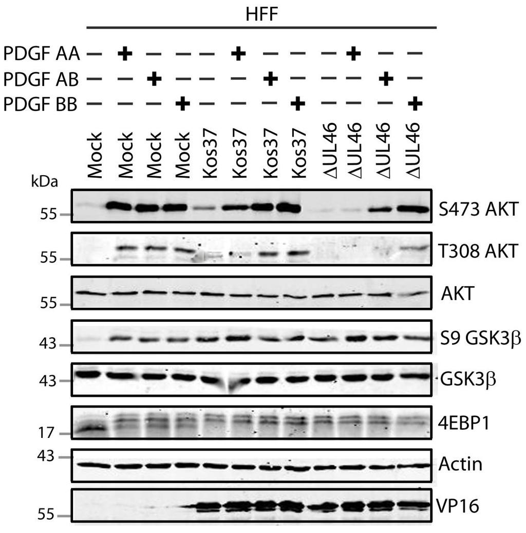
102 89
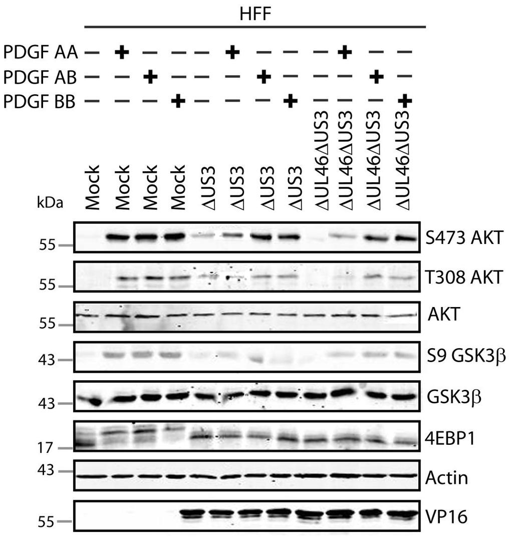
103 90
104 Figure 3.15: PDGF ligands do not perform equally at phosphorylating T308 AKT during infection. Serum-starved HFF cells were mock infected or infected with wild type HSV-1 KOS37, the VP11/12-null mutant ΔUL46, the US3-null mutant ΔUS3, and the VP11/12-US3-null mutant ΔUL46 ΔUS3. Samples were either left in serum-free media or treated with 20.0 pg/µl PDGF AA, 5.0 pg/µl PDGF AB, and 10.0 pg/µl PDGF BB for 20 minutes prior to harvesting. Cell lysates were collected at 15 hpi and analyzed for the activating phosphorylation of AKT at residues S473 (anti-s473) and T308 (anti-t308), the inhibiting phosphorylation of GSK3β at residue S9 (anti- GSK3β), 4EBP1, total proteins AKT and GSK3β, VP16, and β-actin by Western blotting. 91
105 92
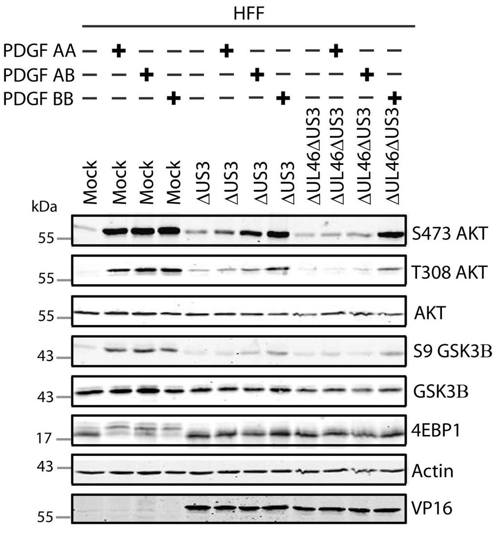
106 93
107 CHAPTER 4: DISCUSSION 94
108 4.1 Thesis Summary The results in this communication have highlighted the complexity in AKT signaling during HSV-1 infection. The data support a model where the HSV-1 tegument protein VP11/12 is required for the phosphorylation of AKT (247). Although a viral protein is devoted to activating AKT, signaling from activated AKT is severely attenuated. Rather, the phosphorylation of AKT targets in HSV-infected cells depends predominantly on the viral AKT mimic, U S 3 (Fig. 4.1). In addition to attenuating activated-akt signals, HSV-1 prevents signaling from select PDGF receptors that normally initiate AKT signaling (Fig. 4.2). Although HSV-1 infection induces two blocks in AKT signaling, AKT is still activated for unknown reasons. Interestingly, neither U S 3 nor U L 46 are required for either of these signaling blocks indicating that other viral proteins may be involved in AKT signaling. My work in this thesis has shed light on many aspects of AKT regulation. 95
109 Figure 4.1: U L 46 promotes AKT activation, while U S 3 negatively regulates AKT signaling. AKT is activated in a U L 46-dependent manner during HSV-1 infection, while the phosphorylation of AKT targets is primarily U S 3-dependent. During infection, the phosphorylation of AKT targets does not require U L 46, but rather depends on U S 3. Since AKT is activated, but the signal is not propagated, HSV-1 appears to attenuate signaling between AKT and its downstream targets. Interestingly, neither U L 46 nor U S 3 are required for attenuation of an activated AKT signal. Although Us3 does not block the signaling between AKT and its targets, U S 3 appears to inhibit the phosphorylation of residue T308 on AKT. 96
110 Figure 4.2: HSV-1 blocks AKT activation downstream of specific receptors. AKT is phosphorylated during HSV-1 infection. During infection, the phosphorylation of AKT can be further enhanced when cells are stimulated with PDGF ββ, and to a lesser extent PDGF αβ. However, stimulating infected cells with PDGF αα does not enhance the phosphorylation of AKT. Thus, HSV-1 selectively blocks signaling to AKT from select receptors. 97
111 4.2 The HSV-1 kinase U S 3 masquerades as an AKT mimic to regulate cellular processes While U S 3 negatively regulates AKT activation, I, as well as the Mohr group (34), found that U S 3 is required for the phosphorylation of several AKT targets (Fig. 3.6, 3.7). Though U S 3 has no sequence homology to AKT, Mohr and colleagues (34) found that it behaves analogously to AKT by directly phosphorylating the targets TSC2, FoxO1, and GSK3β on the same residues as AKT. A similar phenomenon is found in another Herpesvirus. The VZV U S 3 homolog ORF 66 phosphorylates the same residues and targets as PKA (61). These data suggest that it is advantageous for a virus to bypass cellular signaling to directly regulate selective target proteins. For example, the activation of AKT normally requires external growth factors, while U S 3 is able to phosphorylate AKT targets in serum-starved conditions (Fig. 3.6, 3.7). Furthermore, the activation of AKT is biphasic and transient during infection (Fig. 3.5), whereas U S 3 promotes the phosphorylation of AKT targets as early as 3 hours after infection and continues until as late as 21 hpi (Appendix C). Furthermore, it is possible that U S 3 does not undergo the same negative regulation as AKT. Firstly, inhibition from PLC and the phosphatase PTEN must be overcome in order for AKT to be phosphorylated (124, 141). Secondly, activated AKT is dephosphorylated, and thus inactivated, by active PP2A (13). Thirdly, AKT exists in a negative feedback loop with downstream proteins. For example, it was suggested that U S 3 is unaffected by the AKT-S6K-IRS1 negative feedback loop (34, 188). Mohr suggested (34) that since U S 3 is unrelated to AKT at the sequence level, it is unresponsive to cellular pathways that limit AKT activity. Another possible advantage of encoding a viral AKT mimic is that U S 3 can selectively phosphorylate specific AKT targets (34). By modulating AKT targets, U S 3 hijacks cellular regulation of processes such as apoptosis and protein translation. 4.3 HSV-1 promotes cap-dependent translation 98
112 HSV-1 protein synthesis relies on host translational machinery. We and the Mohr group (34) have independently found that U S 3 promotes the phosphorylation and degradation of the translational repressor 4EBP1 (Fig. 3.7) by phosphorylating an upstream regulator, TSC2 (34). Phosphorylated TSC2 leads to the activation of mtorc1, which is required for the hyperphosphorylation of 4EBP1 (29). Mohr and colleagues (249) previously found a complete reduction in the hyperphosphorylation of 4EBP1 when HSV-1-infected cells were treated with rapamycin, which is consistent with viral manipulation of TSC2 upstream of mtorc1. On the other hand, HCMV continually induces hyperphosphorylation of 4EBP1 without being impaired by rapamycin, indicating that other Herpesviruses can affect translation initiation downstream of mtorc1 (160). We found that treating cells with rapamycin partially reduced 4EBP1 phosphorylation (Appendix B), suggesting that HSV-1 manipulates both upstream regulators of 4EBP1 and 4EBP1 directly, reconciling these reports. Interestingly, although 4EBP1 was hyperphosphorylated at both early (Fig. 3.6) and late (Fig. 3.7) time points during wildtype infection, U S 3 was required for 4EBP1 hyperphosphorylation only at late times. It was unclear whether U S 3 was required for 4EBP1 hyperphosphorylation at early times. Thus, it is possible that another viral protein maintains 4EBP1 hyperphosphorylation at early times of infection. ICP0 is a likely candidate that has been shown to enhance the assembly of translational machinery (249). ICP0 not only stimulates 4EBP1 phosphorylation, but also targets 4EBP1 for proteasomal degradation (21, 249). Although the phosphorylation of 4EBP1 is ICP0-dependent at 8 hpi (249), it would be interesting to determine if ICP0 is required for the phosphorylation of 4EBP1 that we observed at 4 hpi. 4.4 A potential role for U L 46 in promoting cell survival In addition to regulating protein translation, it has long been known that U S 3 promotes cell survival in response to variety of apoptotic stimuli, including the stress of virus infection (4, 134). Accordingly, we found that infection with 99
113 U S 3-virus resulted in cells bearing an apoptotic morphology (Appendix D, Kevin Quach and Heather Eaton, unpublished data). Though U L 46 is required for AKT activation, U L 46-activated AKT signals are not transduced to GSK3β, FoxO1, and 4EBP1 during HSV-1 infection (Fig. 3.11). This is a stark contrast to other Herpesviruses. For instance, VZV encodes ORF 12 to activate AKT, resulting in phosphorylation of FoxO1 (XueQiao, et al., unpublished data). Given this information, the purpose of U L 46-activated AKT is a conundrum that has yet to be solved. Given that U S 3 and U L 46 both appear to promote AKT signaling, we examined whether they collaborate to suppress apoptosis. Using light-microscope analysis to examine cell morphology, we found that, in contrast to U S 3-infection, U L 46-infected cells were not phenotypically apoptotic. However, deleting both ΔU L 46 and ΔU S 3 increased the frequency of apoptotic cells compared to ΔU S 3- infection alone. These data indicate that U L 46 may also contribute to cell survival. If U L 46 promotes cell survival, then cells infected with U L 46-virus should be more sensitive to pro-apoptotic stimuli (such as UV exposure or treatment with drugs that induce apoptosis) compared to wild type-infected cells. For example, we would expect to find increased cleavage of the cellular proteins PARP and caspase-3 by Western blot analysis, DNA fragmentation by TUNEL staining, and the appearance of apoptotic cell bodies using immunofluorescence imaging. In order to address whether U L 46 inhibits apoptosis through an AKTdependent mechanism, we could test whether the presence of an AKT inhibitor, AKT VIII, impairs cell survival in a U L 46-competent virus infection. If the putative pro-survival function of U L 46 requires AKT, then this may be directed to selective AKT targets such as p53 and MDM2 (reviewed in (144, 221). Alternatively, if inhibiting AKT does not impair cell survival, then U L 46 may promote cell survival through another signaling pathway. For example, the VZV U L 46 homolog ORF12 activates ERK signaling to prevent apoptosis (137). 4.5 HSV-1 encodes proteins other than U L 46 and U S 3 to modulate AKT signaling External growth stimuli resulted in activation of AKT, but these signals were not transduced to the examined AKT targets in ΔU S 3- or ΔU L 46ΔU S 3-100
114 infected cells (Fig. 3.11, 3.13). Therefore, both U S 3 and U L 46 are not required for HSV-1 to attenuate AKT signaling, suggesting that another viral protein(s) prevents activated AKT signals from being transduced downstream. One potential candidate is the HSV-1 protein kinase U L 13, as the VZV orthologs of U L 13 and U S 3, ORF 47 and ORF 66 respectively, have been shown to collaborate to modulate AKT signaling (189). Interestingly, the U L 13 gene product is conserved between HSV-1 and VZV with a 79% sequence similarity. In contrast to VZV, we found that U L 13 deficiency did not affect the phosphorylation of AKT targets (Fig. 3.4). However, the results do not exclude the possibility that U L 13 is involved in AKT signaling. It would be interesting to examine if serum-induced activated-akt signals result in the phosphorylation of AKT targets during infection with viral mutants lacking U L 13. These viral mutants must also be deficient in U S 3 in order to observe the downstream effects of activated AKT. 4.6 HSV-1 permits signaling to AKT from specific receptors HSV-1 has previously been shown to downregulate expression of EGFR (135). In addition to EGFR, our studies show that HSV-1 regulates signaling from other growth factor receptors. Specifically, we found that HSV-1 infection selectively impairs signaling from PDGF receptors that include the α-subunit (Fig. 3.15, 3.16, 4.2). Neither U L 46 nor U S 3 are the main contributors to this signaling impairment through the α-chain, suggesting that another viral protein is involved. ICP0 is a potential candidate given the precedence for ICP0-mediated inhibition of EGFR signaling. In addition to transcriptional regulation of the EGFR gene, ICP0 was found to form a complex with the cellular proteins Cin85 and Cbl to mediate a reduction in the levels of cell surface and total EGFR through internalization and degradation (135). Similar to EGFR, PDGFRs are receptor tyrosine kinases that can form a complex with Cin85 and Cbl (227). Consequently, PDGFRs undergo negative regulation by endocytosis and degradation or recycling (53). Thus, it would be interesting to determine whether ICP0 mediates downregulation of the α-pdgfr chain during HSV-1 infection. Though it remains unknown what the biological outcomes of EGFR or PDGFR downregulation are during HSV-1 infection, it is interesting to speculate that 101
115 excessive signaling or selective signaling pathways are not beneficial for viral infection. Our findings that HSV-1 utilizes U S 3 to bypass AKT signaling and directly regulate AKT targets lend further support to this idea. 4.7 Other aspects of AKT activation that are modulated by HSV-1 My work has also shed light on the molecular mechanisms by which U L 46 and U S 3 modulate the PI3K-AKT pathway A mechanism for the phosphorylation of AKT by HSV-1 We have previously shown that the phosphorylation of AKT is U L 46- dependent. However, the role of mtorc2 in phosphorylating the hydrophobic motif of AKT during HSV-1 infection was unclear. We found that HSV-1 induced AKT phosphorylation was prevented in the presence of torin1. Since use of drugs could have non-specific effects on cell signaling, RNA silencing of mtorc2 subunits can be used to verify that mtorc2 is required for AKT phosphorylation during infection. Knockdown of the mtorc2 subunits mtor and rictor were previously used by Sabatini and colleagues to prevent AKT phosphorylation in uninfected cells (204). Though mtorc2 is required for the phosphorylation of AKT, these results do not tell us whether U L 46 activates AKT by promoting mtorc2 activity. To get an indication of this, we can examine whether infection with U L 46-virus reduces rictor expression levels, impairs the association between rictor and mtor, or impairs the phosphorylation of rictor U S 3 negatively regulates the phosphorylation of the T308 residue of AKT Though we and others have shown that U S 3 antagonizes AKT phosphorylation at both the S473 and T308 residues (13), the mechanism by which this occurs was unclear. By using serum to stimulate AKT phosphorylation, we have shown that U S 3 specifically antagonizes phosphorylation of T308. This implies that U S 3 interferes with a step upstream of PDK1 phosphorylation of AKT (Fig. 3.11, 4.2). For instance, U S 3 may be interfering with the generation of PIP3 species by activating PLC, inhibiting PI3K, or activating phosphatases such as PTEN, and inositol polyphosphate 5 -phosphatases (INPP5) (including SHIP-2 and type II inositol polyphosphate 5-phosphatase) (7, 35, 193). Potential activation of INPP5 species by U S 3 would increase cleavage of PIP3 (109, 148, 102
116 170), preventing binding of AKT and PDK1 at the plasma membrane, and consequently phosphorylation of T308 on AKT. Alternatively, U S 3 may disrupt interactions of the PH domain of PDK1 and AKT with PIP3, or impair PDK1 activity. 4.8 Future Directions Previous work done by Dr. Melany Wagner examined the interactions between U L 46 and SFK, and their effect on AKT signaling (247). Her data has clearly shown that U L 46-dependent activated AKT signals are not required for the phosphorylation of downstream targets. In fact, we found that U L 46-activated AKT signals are attenuated during infection. Future work should aim to identify cellular or viral factors that prevent activated AKT signals from being transduced. Although U L 46 is required for AKT activation, we have yet to determine the biological function(s) of U L 46 during infection. Although we suspect that U L 46 promotes cell survival, future studies should verify this possibility and determine whether U L 46 promotes cell survival through an AKT-dependent mechanism. HSV-1 not only attenuates signaling between AKT and its targets, but also inhibits signaling from select growth factor receptors. The viral proteins involved in attenuating PDGFRα-receptor signaling have not been explored. Future experiments should examine gene and cell surface expression of PDGFRs during infection. The investigations from this thesis add to our knowledge of how HSV infection induces changes to cell signaling. Work in our lab presently examines other signaling pathways targeted by U L 46, the anti-apoptotic roles played by U L 46 and U S 3, the molecular mechanisms governing U L 46-mediated activation of SFKs, and inhibition of AKT phosphorylation by U S 3. Although these future directions should add to our understanding of HSV-modulated AKT signaling, many questions remained unanswered, such as reasons for encoding an AKT mimic, resolving how AKT signals are attenuated, the purpose of attenuating AKT signaling, and other viral proteins involved in AKT signaling. 103
117 BIBLIOGRAPHY 104
118 1. Ahmed, N. N., H. L. Grimes, A. Bellacosa, T. O. Chan, and P. N. Tsichlis Transduction of interleukin-2 antiapoptotic and proliferative signals via Akt protein kinase. Proceedings of the National Academy of Sciences of the United States of America 94: Alessi, D. R., F. B. Caudwell, M. Andjelkovic, B. A. Hemmings, and P. Cohen Molecular basis for the substrate specificity of protein kinase B; comparison with MAPKAP kinase-1 and p70 S6 kinase. FEBS Lett 399: Alessi, D. R., M. Deak, A. Casamayor, F. B. Caudwell, N. Morrice, D. G. Norman, P. Gaffney, C. B. Reese, C. N. MacDougall, D. Harbison, A. Ashworth, and M. Bownes Phosphoinositide-dependent protein kinase- 1 (PDK1): structural and functional homology with the Drosophila DSTPK61 kinase. Current biology : CB 7: Asano, S., T. Honda, F. Goshima, D. Watanabe, Y. Miyake, Y. Sugiura, and Y. Nishiyama US3 protein kinase of herpes simplex virus type 2 plays a role in protecting corneal epithelial cells from apoptosis in infected mice. The Journal of general virology 80 ( Pt 1): Auger, K. R., L. A. Serunian, S. P. Soltoff, P. Libby, and L. C. Cantley PDGF-dependent tyrosine phosphorylation stimulates production of novel polyphosphoinositides in intact cells. Cell 57: Avruch, J., C. Belham, Q. Weng, K. Hara, and K. Yonezawa The p70 S6 kinase integrates nutrient and growth signals to control translational capacity. Prog Mol Subcell Biol 26: Backers, K., D. Blero, N. Paternotte, J. Zhang, and C. Erneux The termination of PI3K signalling by SHIP1 and SHIP2 inositol 5-phosphatases. Advances in enzyme regulation 43: Balendran, N., R. L. Clough, J. R. Arguello, R. Barber, C. Veal, A. B. Jones, J. L. Rosbotham, A. M. Little, A. Madrigal, J. N. Barker, S. H. Powis, and R. C. Trembath Characterization of the major susceptibility region for psoriasis at chromosome 6p21.3. J Invest Dermatol 113: Batterson, W., and B. Roizman Characterization of the herpes simplex virion-associated factor responsible for the induction of alpha genes. Journal of virology 46: Baxter, R. M., J. P. Secrist, R. R. Vaillancourt, and A. Kazlauskas Full activation of the platelet-derived growth factor beta-receptor kinase involves multiple events. The Journal of biological chemistry 273: Benetti, L., J. Munger, and B. Roizman The herpes simplex virus 1 US3 protein kinase blocks caspase-dependent double cleavage and activation of the proapoptotic protein BAD. Journal of virology 77:
119 12. Benetti, L., and B. Roizman Herpes simplex virus protein kinase US3 activates and functionally overlaps protein kinase A to block apoptosis. Proceedings of the National Academy of Sciences of the United States of America 101: Benetti, L., and B. Roizman Protein kinase B/Akt is present in activated form throughout the entire replicative cycle of deltau(s)3 mutant virus but only at early times after infection with wild-type herpes simplex virus 1. Journal of virology 80: Berridge, M. J., and R. F. Irvine Inositol phosphates and cell signalling. Nature 341: Binder, P. S Herpes simplex keratitis. Surv Ophthalmol 21: Biondi, R. M Phosphoinositide-dependent protein kinase 1, a sensor of protein conformation. Trends in biochemical sciences 29: Biondi, R. M., P. C. Cheung, A. Casamayor, M. Deak, R. A. Currie, and D. R. Alessi Identification of a pocket in the PDK1 kinase domain that interacts with PIF and the C-terminal residues of PKA. The EMBO journal 19: Bloom, D. C., N. V. Giordani, and D. L. Kwiatkowski. Epigenetic regulation of latent HSV-1 gene expression. Biochim Biophys Acta 1799: Boehmer, P. E., and I. R. Lehman Physical interaction between the herpes simplex virus 1 origin-binding protein and single-stranded DNA-binding protein ICP8. Proceedings of the National Academy of Sciences of the United States of America 90: Bottomley, M. J., K. Salim, and G. Panayotou Phospholipid-binding protein domains. Biochim Biophys Acta 1436: Boutell, C., S. Sadis, and R. D. Everett Herpes simplex virus type 1 immediate-early protein ICP0 and is isolated RING finger domain act as ubiquitin E3 ligases in vitro. Journal of virology 76: Brazil, D. P., and B. A. Hemmings Ten years of protein kinase B signalling: a hard Akt to follow. Trends in biochemical sciences 26: Brazil, D. P., Z. Z. Yang, and B. A. Hemmings Advances in protein kinase B signalling: AKTion on multiple fronts. Trends in biochemical sciences 29: Bresnahan, W. A., G. E. Hultman, and T. Shenk Replication of wildtype and mutant human cytomegalovirus in life-extended human diploid fibroblasts. J Virol 74:
120 25. Bronstein, J. C., S. K. Weller, and P. C. Weber The product of the UL12.5 gene of herpes simplex virus type 1 is a capsid-associated nuclease. Journal of virology 71: Burgering, B. M., and P. J. Coffer Protein kinase B (c-akt) in phosphatidylinositol-3-oh kinase signal transduction. Nature 376: Burgering, B. M., and R. H. Medema Decisions on life and death: FOXO Forkhead transcription factors are in command when PKB/Akt is off duty. J Leukoc Biol 73: Burke, J. E., O. Vadas, A. Berndt, T. Finegan, O. Perisic, and R. L. Williams. Dynamics of the phosphoinositide 3-kinase p110delta interaction with p85alpha and membranes reveals aspects of regulation distinct from p110alpha. Structure 19: Burnett, P. E., R. K. Barrow, N. A. Cohen, S. H. Snyder, and D. M. Sabatini RAFT1 phosphorylation of the translational regulators p70 S6 kinase and 4E-BP1. Proceedings of the National Academy of Sciences of the United States of America 95: Cai, W., and P. A. Schaffer Herpes simplex virus type 1 ICP0 regulates expression of immediate-early, early, and late genes in productively infected cells. Journal of virology 66: Calleja, V., D. Alcor, M. Laguerre, J. Park, B. Vojnovic, B. A. Hemmings, J. Downward, P. J. Parker, and B. Larijani Intramolecular and intermolecular interactions of protein kinase B define its activation in vivo. PLoS Biol 5:e Carpenter, C. L., B. C. Duckworth, K. R. Auger, B. Cohen, B. S. Schaffhausen, and L. C. Cantley Purification and characterization of phosphoinositide 3-kinase from rat liver. The Journal of biological chemistry 265: Chuluunbaatar, U., and I. Mohr A herpesvirus kinase that masquerades as Akt: you don't have to look like Akt, to act like it. Cell Cycle 10: Chuluunbaatar, U., R. Roller, M. E. Feldman, S. Brown, K. M. Shokat, and I. Mohr. Constitutive mtorc1 activation by a herpesvirus Akt surrogate stimulates mrna translation and viral replication. Genes & development 24: Cockcroft, S., and G. M. Thomas Inositol-lipid-specific phospholipase C isoenzymes and their differential regulation by receptors. The Biochemical journal 288 ( Pt 1): Conley, A. J., D. M. Knipe, P. C. Jones, and B. Roizman Molecular genetics of herpes simplex virus. VII. Characterization of a temperature-sensitive 107
121 mutant produced by in vitro mutagenesis and defective in DNA synthesis and accumulation of gamma polypeptides. Journal of virology 37: Costanzo, F., G. Campadelli-Fiume, L. Foa-Tomasi, and E. Cassai Evidence that herpes simplex virus DNA is transcribed by cellular RNA polymerase B. Journal of virology 21: Coughlin, S. R., J. A. Escobedo, and L. T. Williams Role of phosphatidylinositol kinase in PDGF receptor signal transduction. Science 243: Cross, D. A., D. R. Alessi, P. Cohen, M. Andjelkovich, and B. A. Hemmings Inhibition of glycogen synthase kinase-3 by insulin mediated by protein kinase B. Nature 378: Crute, J. J., E. S. Mocarski, and I. R. Lehman A DNA helicase induced by herpes simplex virus type 1. Nucleic acids research 16: Daikoku, T., R. Kurachi, T. Tsurumi, and Y. Nishiyama Identification of a target protein of US3 protein kinase of herpes simplex virus type 2. The Journal of general virology 75 ( Pt 8): Daikoku, T., Y. Yamashita, T. Tsurumi, and Y. Nishiyama The US3 protein kinase of herpes simplex virus type 2 is associated with phosphorylation of the UL12 alkaline nuclease in vitro. Arch Virol 140: Darr, C. D., A. Mauser, and S. Kenney Epstein-Barr virus immediateearly protein BRLF1 induces the lytic form of viral replication through a mechanism involving phosphatidylinositol-3 kinase activation. Journal of virology 75: Datta, S. R., A. Brunet, and M. E. Greenberg Cellular survival: a play in three Akts. Genes & development 13: Datta, S. R., H. Dudek, X. Tao, S. Masters, H. A. Fu, Y. Gotoh, and M. E. Greenberg Akt phosphorylation of BAD couples survival signals to the cell-intrinsic death machinery. Cell 91: Datta, S. R., A. Katsov, L. Hu, A. Petros, S. W. Fesik, M. B. Yaffe, and M. E. Greenberg proteins and survival kinases cooperate to inactivate BAD by BH3 domain phosphorylation. Mol Cell 6: Dawson, C. W., G. Tramountanis, A. G. Eliopoulos, and L. S. Young Epstein-Barr virus latent membrane protein 1 (LMP1) activates the phosphatidylinositol 3-kinase/Akt pathway to promote cell survival and induce actin filament remodeling. The Journal of biological chemistry 278: de Bruyn Kops, A., S. L. Uprichard, M. Chen, and D. M. Knipe Comparison of the intranuclear distributions of herpes simplex virus proteins involved in various viral functions. Virology 252:
122 49. DeLuca, N., D. J. Bzik, V. C. Bond, S. Person, and W. Snipes Nucleotide sequences of herpes simplex virus type 1 (HSV-1) affecting virus entry, cell fusion, and production of glycoprotein gb (VP7). Virology 122: Desbois-Mouthon, C., A. Cadoret, M. J. Blivet-Van Eggelpoel, F. Bertrand, G. Cherqui, C. Perret, and J. Capeau Insulin and IGF-1 stimulate the beta-catenin pathway through two signalling cascades involving GSK-3beta inhibition and Ras activation. Oncogene 20: Deshmane, S. L., and N. W. Fraser During latency, herpes simplex virus type 1 DNA is associated with nucleosomes in a chromatin structure. Journal of virology 63: Diefenbach, R. J., M. Miranda-Saksena, M. W. Douglas, and A. L. Cunningham Transport and egress of herpes simplex virus in neurons. Rev Med Virol 18: Dikic, I., and S. Giordano Negative receptor signalling. Curr Opin Cell Biol 15: Divecha, N., and R. F. Irvine Phospholipid signaling. Cell 80: Dobson, A. T., F. Sederati, G. Devi-Rao, W. M. Flanagan, M. J. Farrell, J. G. Stevens, E. K. Wagner, and L. T. Feldman Identification of the latencyassociated transcript promoter by expression of rabbit beta-globin mrna in mouse sensory nerve ganglia latently infected with a recombinant herpes simplex virus. Journal of virology 63: Dohner, K., A. Wolfstein, U. Prank, C. Echeverri, D. Dujardin, R. Vallee, and B. Sodeik Function of dynein and dynactin in herpes simplex virus capsid transport. Mol Biol Cell 13: Ejercito, P. M., E. D. Kieff, and B. Roizman Characterization of herpes simplex virus strains differing in their effects on social behaviour of infected cells. The Journal of general virology 2: Elias, P., M. E. O'Donnell, E. S. Mocarski, and I. R. Lehman A DNA binding protein specific for an origin of replication of herpes simplex virus type 1. Proceedings of the National Academy of Sciences of the United States of America 83: Elliott, G., and P. O'Hare Herpes simplex virus type 1 tegument protein VP22 induces the stabilization and hyperacetylation of microtubules. Journal of virology 72: Elliott, G., and P. O'Hare Intercellular trafficking and protein delivery by a herpesvirus structural protein. Cell 88:
123 61. Erazo, A., and P. R. Kinchington. Varicella-zoster virus open reading frame 66 protein kinase and its relationship to alphaherpesvirus US3 kinases. Current topics in microbiology and immunology 342: Eriksson, A., A. Siegbahn, B. Westermark, C. H. Heldin, and L. Claesson- Welsh PDGF alpha- and beta-receptors activate unique and common signal transduction pathways. The EMBO journal 11: Everett, R. D., P. Freemont, H. Saitoh, M. Dasso, A. Orr, M. Kathoria, and J. Parkinson The disruption of ND10 during herpes simplex virus infection correlates with the Vmw110- and proteasome-dependent loss of several PML isoforms. Journal of virology 72: Fang, Y., M. Vilella-Bach, R. Bachmann, A. Flanigan, and J. Chen Phosphatidic acid-mediated mitogenic activation of mtor signaling. Science 294: Feng, J., J. Park, P. Cron, D. Hess, and B. A. Hemmings Identification of a PKB/Akt hydrophobic motif Ser-473 kinase as DNA-dependent protein kinase. The Journal of biological chemistry 279: Ferguson, K. M., M. A. Lemmon, P. B. Sigler, and J. Schlessinger Scratching the surface with the PH domain. Nat Struct Biol 2: Forrester, A., H. Farrell, G. Wilkinson, J. Kaye, N. Davis-Poynter, and T. Minson Construction and properties of a mutant of herpes simplex virus type 1 with glycoprotein H coding sequences deleted. Journal of virology 66: Frame, M. C., F. C. Purves, D. J. McGeoch, H. S. Marsden, and D. P. Leader Identification of the herpes simplex virus protein kinase as the product of viral gene US3. The Journal of general virology 68 ( Pt 10): Fretto, L. J., A. J. Snape, J. E. Tomlinson, J. J. Seroogy, D. L. Wolf, W. J. LaRochelle, and N. A. Giese Mechanism of platelet-derived growth factor (PDGF) AA, AB, and BB binding to alpha and beta PDGF receptor. The Journal of biological chemistry 268: Frias, M. A., C. C. Thoreen, J. D. Jaffe, W. Schroder, T. Sculley, S. A. Carr, and D. M. Sabatini msin1 is necessary for Akt/PKB phosphorylation, and its isoforms define three distinct mtorc2s. Current biology : CB 16: Friedman, E., A. H. Katcher, and V. J. Brightman Incidence of recurrent herpes labialis and upper respiratory infection: a prospective study of the influence of biologic, social and psychologic predictors. Oral Surg Oral Med Oral Pathol 43:
124 72. Gao, M., and D. M. Knipe Potential role for herpes simplex virus ICP8 DNA replication protein in stimulation of late gene expression. Journal of virology 65: Gao, X., and D. Pan TSC1 and TSC2 tumor suppressors antagonize insulin signaling in cell growth. Genes & development 15: Geraghty, R. J., C. Krummenacher, G. H. Cohen, R. J. Eisenberg, and P. G. Spear Entry of alphaherpesviruses mediated by poliovirus receptor-related protein 1 and poliovirus receptor. Science 280: Gibson, W Structure and assembly of the virion. Intervirology 39: Gierasch, W. W., D. L. Zimmerman, S. L. Ward, T. K. Vanheyningen, J. D. Romine, and D. A. Leib Construction and characterization of bacterial artificial chromosomes containing HSV-1 strains 17 and KOS. J Virol Methods 135: Gingras, A. C., S. P. Gygi, B. Raught, R. D. Polakiewicz, R. T. Abraham, M. F. Hoekstra, R. Aebersold, and N. Sonenberg Regulation of 4E-BP1 phosphorylation: a novel two-step mechanism. Genes & development 13: Gingras, A. C., B. Raught, and N. Sonenberg eif4 initiation factors: effectors of mrna recruitment to ribosomes and regulators of translation. Annu Rev Biochem 68: Gingras, A. C., B. Raught, and N. Sonenberg Regulation of translation initiation by FRAP/mTOR. Genes & development 15: Glezen, W. P., G. W. Fernald, and J. A. Lohr Acute respiratory disease of university students with special reference to the etiologic role of Herpesvirus hominis. Am J Epidemiol 101: Greenberg, M. E., S. R. Datta, and A. Brunet Cellular survival: a play in three Akts. Genes & development 13: Gross, A., J. M. McDonnell, and S. J. Korsmeyer BCL-2 family members and the mitochondria in apoptosis. Genes Dev 13: Guertin, D. A., and D. M. Sabatini Defining the role of mtor in cancer. Cancer Cell 12: Guertin, D. A., and D. M. Sabatini An expanding role for mtor in cancer. Trends Mol Med 11: Hammacher, A., U. Hellman, A. Johnsson, A. Ostman, K. Gunnarsson, B. Westermark, A. Wasteson, and C. H. Heldin A major part of platelet- 111
125 derived growth factor purified from human platelets is a heterodimer of one A and one B chain. The Journal of biological chemistry 263: Han, J. Y., D. D. Sloan, M. Aubert, S. A. Miller, C. H. Dang, and K. R. Jerome Apoptosis and antigen receptor function in T and B cells following exposure to herpes simplex virus. Virology 359: Hara, K., Y. Maruki, X. Long, K. Yoshino, N. Oshiro, S. Hidayat, C. Tokunaga, J. Avruch, and K. Yonezawa Raptor, a binding partner of target of rapamycin (TOR), mediates TOR action. Cell 110: Hardwicke, M. A., and R. M. Sandri-Goldin The herpes simplex virus regulatory protein ICP27 contributes to the decrease in cellular mrna levels during infection. Journal of virology 68: Hata, S., A. H. Koyama, H. Shiota, A. Adachi, F. Goshima, and Y. Nishiyama Antiapoptotic activity of herpes simplex virus type 2: the role of US3 protein kinase gene. Microbes Infect 1: Heidaran, M. A., J. H. Pierce, R. A. Jensen, T. Matsui, and S. A. Aaronson Chimeric alpha- and beta-platelet-derived growth factor (PDGF) receptors define three immunoglobulin-like domains of the alpha-pdgf receptor that determine PDGF-AA binding specificity. The Journal of biological chemistry 265: Heidaran, M. A., J. C. Yu, R. A. Jensen, J. H. Pierce, and S. A. Aaronson A deletion in the extracellular domain of the alpha platelet-derived growth factor (PDGF) receptor differentially impairs PDGF-AA and PDGF-BB binding affinities. The Journal of biological chemistry 267: Herold, B. C., R. J. Visalli, N. Susmarski, C. R. Brandt, and P. G. Spear Glycoprotein C-independent binding of herpes simplex virus to cells requires cell surface heparan sulphate and glycoprotein B. The Journal of general virology 75 ( Pt 6): Herold, B. C., D. WuDunn, N. Soltys, and P. G. Spear Glycoprotein C of herpes simplex virus type 1 plays a principal role in the adsorption of virus to cells and in infectivity. Journal of virology 65: Hers, I., E. E. Vincent, and J. M. Tavare. Akt signalling in health and disease. Cell Signal 23: Hinchliffe, K., and R. Irvine Inositol lipid pathways turn turtle. Nature 390: Holt, K. H., L. Olson, W. S. Moye-Rowley, and J. E. Pessin Phosphatidylinositol 3-kinase activation is mediated by high-affinity interactions between distinct domains within the p110 and p85 subunits. Molecular and cellular biology 14:
126 97. Homa, F. L., and J. C. Brown Capsid assembly and DNA packaging in herpes simplex virus. Rev Med Virol 7: Honess, R. W., and B. Roizman Proteins specified by herpes simplex virus. XIII. Glycosylation of viral polypeptides. Journal of virology 16: Honess, R. W., and B. Roizman Regulation of herpesvirus macromolecular synthesis. I. Cascade regulation of the synthesis of three groups of viral proteins. Journal of virology 14: Honess, R. W., and B. Roizman Regulation of herpesvirus macromolecular synthesis: sequential transition of polypeptide synthesis requires functional viral polypeptides. Proc Natl Acad Sci U S A 72: Huang, E. a. K., T. (ed.) Molecular Aspects of Human Cytomegalovirus Diseases. Springer, Berlin Ichaso, N., and S. M. Dilworth Cell transformation by the middle T- antigen of polyoma virus. Oncogene 20: Ingham, P. W Hedgehog signaling: a tale of two lipids. Science 294: Ingham, P. W., and A. P. McMahon Hedgehog signaling in animal development: paradigms and principles. Genes & development 15: Inoki, K., Y. Li, T. Zhu, J. Wu, and K. L. Guan TSC2 is phosphorylated and inhibited by Akt and suppresses mtor signalling. Nature cell biology 4: Isakoff, S. J., T. Cardozo, J. Andreev, Z. Li, K. M. Ferguson, R. Abagyan, M. A. Lemmon, A. Aronheim, and E. Y. Skolnik Identification and analysis of PH domain-containing targets of phosphatidylinositol 3-kinase using a novel in vivo assay in yeast. The EMBO journal 17: Jackson, S. A., and N. A. DeLuca Relationship of herpes simplex virus genome configuration to productive and persistent infections. Proceedings of the National Academy of Sciences of the United States of America 100: Jefferies, H. B., S. Fumagalli, P. B. Dennis, C. Reinhard, R. B. Pearson, and G. Thomas Rapamycin suppresses 5'TOP mrna translation through inhibition of p70s6k. The EMBO journal 16: Jefferson, A. B., and P. W. Majerus Properties of type II inositol polyphosphate 5-phosphatase. The Journal of biological chemistry 270: Johnson, D. C., and M. T. Huber Directed egress of animal viruses promotes cell-to-cell spread. Journal of virology 76:
127 111. Jovasevic, V., L. Liang, and B. Roizman Proteolytic cleavage of VP1-2 is required for release of herpes simplex virus 1 DNA into the nucleus. Journal of virology 82: Kandel, E. S., and N. Hay The regulation and activities of the multifunctional serine/threonine kinase Akt/PKB. Experimental cell research 253: Kapeller, R., and L. C. Cantley Phosphatidylinositol 3-kinase. BioEssays : news and reviews in molecular, cellular and developmental biology 16: Kapeller, R., K. V. Prasad, O. Janssen, W. Hou, B. S. Schaffhausen, C. E. Rudd, and L. C. Cantley Identification of two SH3-binding motifs in the regulatory subunit of phosphatidylinositol 3-kinase. The Journal of biological chemistry 269: Kaplan, D. R., M. Whitman, B. Schaffhausen, D. C. Pallas, M. White, L. Cantley, and T. M. Roberts Common elements in growth factor stimulation and oncogenic transformation: 85 kd phosphoprotein and phosphatidylinositol kinase activity. Cell 50: Katan, M., A. Haigh, C. P. Verrijzer, P. C. van der Vliet, and P. O'Hare Characterization of a cellular factor which interacts functionally with Oct-1 in the assembly of a multicomponent transcription complex. Nucleic acids research 18: Kato, K., T. Daikoku, F. Goshima, H. Kume, K. Yamaki, and Y. Nishiyama Synthesis, subcellular localization and VP16 interaction of the herpes simplex virus type 2 UL46 gene product. Arch Virol 145: Kazlauskas, A., and J. A. Cooper Autophosphorylation of the PDGF receptor in the kinase insert region regulates interactions with cell proteins. Cell 58: Kazlauskas, A., and J. A. Cooper Phosphorylation of the PDGF receptor beta subunit creates a tight binding site for phosphatidylinositol 3 kinase. The EMBO journal 9: Kelly, J. D., B. A. Haldeman, F. J. Grant, M. J. Murray, R. A. Seifert, D. F. Bowen-Pope, J. A. Cooper, and A. Kazlauskas Platelet-derived growth factor (PDGF) stimulates PDGF receptor subunit dimerization and intersubunit trans-phosphorylation. The Journal of biological chemistry 266: Kennedy, S. G., A. J. Wagner, S. D. Conzen, J. Jordan, A. Bellacosa, P. N. Tsichlis, and N. Hay The PI 3-kinase/Akt signaling pathway delivers an anti-apoptotic signal. Genes & development 11:
128 122. Kim, D. H., D. D. Sarbassov, S. M. Ali, J. E. King, R. R. Latek, H. Erdjument-Bromage, P. Tempst, and D. M. Sabatini mtor interacts with raptor to form a nutrient-sensitive complex that signals to the cell growth machinery. Cell 110: Kim, D. H., D. D. Sarbassov, S. M. Ali, R. R. Latek, K. V. Guntur, H. Erdjument-Bromage, P. Tempst, and D. M. Sabatini GbetaL, a positive regulator of the rapamycin-sensitive pathway required for the nutrient-sensitive interaction between raptor and mtor. Mol Cell 11: Kim, M. J., E. Kim, S. H. Ryu, and P. G. Suh The mechanism of phospholipase C-gamma1 regulation. Exp Mol Med 32: Klippel, A., J. A. Escobedo, W. J. Fantl, and L. T. Williams The C- terminal SH2 domain of p85 accounts for the high affinity and specificity of the binding of phosphatidylinositol 3-kinase to phosphorylated platelet-derived growth factor beta receptor. Molecular and cellular biology 12: Kristensson, K., E. Lycke, M. Roytta, B. Svennerholm, and A. Vahlne Neuritic transport of herpes simplex virus in rat sensory neurons in vitro. Effects of substances interacting with microtubular function and axonal flow [nocodazole, taxol and erythro-9-3-(2-hydroxynonyl)adenine]. The Journal of general virology 67 ( Pt 9): Kristie, T. M., J. H. LeBowitz, and P. A. Sharp The octamer-binding proteins form multi-protein--dna complexes with the HSV alpha TIF regulatory protein. The EMBO journal 8: Kristie, T. M., and B. Roizman Host cell proteins bind to the cis-acting site required for virion-mediated induction of herpes simplex virus 1 alpha genes. Proceedings of the National Academy of Sciences of the United States of America 84: Kristie, T. M., and B. Roizman Separation of sequences defining basal expression from those conferring alpha gene recognition within the regulatory domains of herpes simplex virus 1 alpha genes. Proceedings of the National Academy of Sciences of the United States of America 81: Kristie, T. M., and P. A. Sharp Interactions of the Oct-1 POU subdomains with specific DNA sequences and with the HSV alpha-transactivator protein. Genes & development 4: Lang, F., and P. Cohen Regulation and physiological roles of serum- and glucocorticoid-induced protein kinase isoforms. Sci STKE 2001:re Leevers, S. J., B. Vanhaesebroeck, and M. D. Waterfield Signalling through phosphoinositide 3-kinases: the lipids take centre stage. Current Opinion in Cell Biology 11:
129 133. Lemaster, S., and B. Roizman Herpes simplex virus phosphoproteins. II. Characterization of the virion protein kinase and of the polypeptides phosphorylated in the virion. Journal of virology 35: Leopardi, R., C. Van Sant, and B. Roizman The herpes simplex virus 1 protein kinase US3 is required for protection from apoptosis induced by the virus. Proceedings of the National Academy of Sciences of the United States of America 94: Liang, Y., A. Kurakin, and B. Roizman Herpes simplex virus 1 infected cell protein 0 forms a complex with CIN85 and Cbl and mediates the degradation of EGF receptor from cell surfaces. Proceedings of the National Academy of Sciences of the United States of America 102: Ligas, M. W., and D. C. Johnson A herpes simplex virus mutant in which glycoprotein D sequences are replaced by beta-galactosidase sequences binds to but is unable to penetrate into cells. Journal of virology 62: Liu, X., Q. Li, K. Dowdell, E. R. Fischer, and J. I. Cohen Varicella- Zoster virus ORF12 protein triggers phosphorylation of ERK1/2 and inhibits apoptosis. Journal of virology 86: Loewith, R., E. Jacinto, S. Wullschleger, A. Lorberg, J. L. Crespo, D. Bonenfant, W. Oppliger, P. Jenoe, and M. N. Hall Two TOR complexes, only one of which is rapamycin sensitive, have distinct roles in cell growth control. Mol Cell 10: Mackem, S., and B. Roizman Differentiation between alpha promoter and regulator regions of herpes simplex virus 1: the functional domains and sequence of a movable alpha regulator. Proceedings of the National Academy of Sciences of the United States of America 79: Mackem, S., and B. Roizman Regulation of herpesvirus macromolecular synthesis: transcription-initiation sites and domains of alpha genes. Proceedings of the National Academy of Sciences of the United States of America 77: Maehama, T., and J. E. Dixon PTEN: a tumour suppressor that functions as a phospholipid phosphatase. Trends Cell Biol 9: Manning, B. D., and L. C. Cantley AKT/PKB signaling: navigating downstream. Cell 129: Manning, B. D., and L. C. Cantley Rheb fills a GAP between TSC and TOR. Trends in biochemical sciences 28: Marine, J. C., S. Francoz, M. Maetens, G. Wahl, F. Toledo, and G. Lozano Keeping p53 in check: essential and synergistic functions of Mdm2 and Mdm4. Cell death and differentiation 13:
130 145. Marth, J. D., R. Peet, E. G. Krebs, and R. M. Perlmutter A lymphocyte-specific protein-tyrosine kinase gene is rearranged and overexpressed in the murine T cell lymphoma LSTRA. Cell 43: Matis, J., and M. Kudelova Early shutoff of host protein synthesis in cells infected with herpes simplex viruses. Acta Virol 45: Matsuzaki, A., Y. Yamauchi, A. Kato, F. Goshima, Y. Kawaguchi, T. Yoshikawa, and Y. Nishiyama US3 protein kinase of herpes simplex virus type 2 is required for the stability of the UL46-encoded tegument protein and its association with virus particles. The Journal of general virology 86: Matzaris, M., S. P. Jackson, K. M. Laxminarayan, C. J. Speed, and C. A. Mitchell Identification and characterization of the phosphatidylinositol-(4, 5)-bisphosphate 5-phosphatase in human platelets. The Journal of biological chemistry 269: Maul, G. G Nuclear domain 10, the site of DNA virus transcription and replication. BioEssays : news and reviews in molecular, cellular and developmental biology 20: McGeoch, D. J., M. A. Dalrymple, A. J. Davison, A. Dolan, M. C. Frame, D. McNab, L. J. Perry, J. E. Scott, and P. Taylor The complete DNA sequence of the long unique region in the genome of herpes simplex virus type 1. The Journal of general virology 69 ( Pt 7): McGeoch, D. J., A. Dolan, S. Donald, and F. J. Rixon Sequence determination and genetic content of the short unique region in the genome of herpes simplex virus type 1. Journal of molecular biology 181: McKnight, J. L., P. E. Pellett, F. J. Jenkins, and B. Roizman Characterization and nucleotide sequence of two herpes simplex virus 1 genes whose products modulate alpha-trans-inducing factor-dependent activation of alpha genes. Journal of virology 61: McLean, C., A. Buckmaster, D. Hancock, A. Buchan, A. Fuller, and A. Minson Monoclonal antibodies to three non-glycosylated antigens of herpes simplex virus type 2. The Journal of general virology 63: McMillan, J. A., L. B. Weiner, A. M. Higgins, and V. J. Lamparella Pharyngitis associated with herpes simplex virus in college students. Pediatr Infect Dis J 12: Mellerick, D. M., and N. W. Fraser Physical state of the latent herpes simplex virus genome in a mouse model system: evidence suggesting an episomal state. Virology 158: Mettenleiter, T. C Herpesvirus assembly and egress. Journal of virology 76:
131 157. Milburn, C. C., M. Deak, S. M. Kelly, N. C. Price, D. R. Alessi, and D. M. Van Aalten Binding of phosphatidylinositol 3,4,5-trisphosphate to the pleckstrin homology domain of protein kinase B induces a conformational change. The Biochemical journal 375: Miller, J. R The Wnts. Genome Biol 3:REVIEWS Montgomery, R. I., M. S. Warner, B. J. Lum, and P. G. Spear Herpes simplex virus-1 entry into cells mediated by a novel member of the TNF/NGF receptor family. Cell 87: Moorman, N. J., and T. Shenk. Rapamycin-resistant mtorc1 kinase activity is required for herpesvirus replication. Journal of virology 84: Mora, A., D. Komander, D. M. van Aalten, and D. R. Alessi PDK1, the master regulator of AGC kinase signal transduction. Semin Cell Dev Biol 15: Morgan, C., H. M. Rose, M. Holden, and E. P. Jones Electron microscopic observations on the development of herpes simplex virus. J Exp Med 110: Mori, S., L. Ronnstrand, K. Yokote, A. Engstrom, S. A. Courtneidge, L. Claesson-Welsh, and C. H. Heldin Identification of two juxtamembrane autophosphorylation sites in the PDGF beta-receptor; involvement in the interaction with Src family tyrosine kinases. The EMBO journal 12: Mou, F., E. Wills, and J. D. Baines Phosphorylation of the U(L)31 protein of herpes simplex virus 1 by the U(S)3-encoded kinase regulates localization of the nuclear envelopment complex and egress of nucleocapsids. Journal of virology 83: Munger, J., A. V. Chee, and B. Roizman The U(S)3 protein kinase blocks apoptosis induced by the d120 mutant of herpes simplex virus 1 at a premitochondrial stage. Journal of virology 75: Munger, J., and B. Roizman The US3 protein kinase of herpes simplex virus 1 mediates the posttranslational modification of BAD and prevents BADinduced programmed cell death in the absence of other viral proteins. Proceedings of the National Academy of Sciences of the United States of America 98: Murphy, M. A., M. A. Bucks, K. J. O'Regan, and R. J. Courtney The HSV-1 tegument protein pul46 associates with cellular membranes and viral capsids. Virology 376: Newcomb, W. W., B. L. Trus, F. P. Booy, A. C. Steven, J. S. Wall, and J. C. Brown Structure of the herpes simplex virus capsid. Molecular 118
132 composition of the pentons and the triplexes. Journal of molecular biology 232: Newton, A. C Regulation of the ABC kinases by phosphorylation: protein kinase C as a paradigm. The Biochemical journal 370: Nishizuka, Y Protein kinase C and lipid signaling for sustained cellular responses. FASEB J 9: O'Hare, P., C. R. Goding, and A. Haigh Direct combinatorial interaction between a herpes simplex virus regulatory protein and a cellular octamer-binding factor mediates specific induction of virus immediate-early gene expression. The EMBO journal 7: Obenauer, J. C., L. C. Cantley, and M. B. Yaffe Scansite 2.0: Proteome-wide prediction of cell signaling interactions using short sequence motifs. Nucleic acids research 31: Ogg, P. D., P. J. McDonell, B. J. Ryckman, C. M. Knudson, and R. J. Roller The HSV-1 Us3 protein kinase is sufficient to block apoptosis induced by overexpression of a variety of Bcl-2 family members. Virology 319: Ojeda, F., H. Folch, M. I. Guarda, B. Jastorff, and H. A. Diehl Induction of apoptosis in thymocytes: new evidence for an interaction of PKC and PKA pathways. Biol Chem Hoppe Seyler 376: Patel, A. H., F. J. Rixon, C. Cunningham, and A. J. Davison Isolation and characterization of herpes simplex virus type 1 mutants defective in the UL6 gene. Virology 217: Persad, S., S. Attwell, V. Gray, N. Mawji, J. T. Deng, D. Leung, J. Yan, J. Sanghera, M. P. Walsh, and S. Dedhar Regulation of protein kinase B/Akt-serine 473 phosphorylation by integrin-linked kinase: critical roles for kinase activity and amino acids arginine 211 and serine 343. The Journal of biological chemistry 276: Peterson, R. T., and S. L. Schreiber Kinase phosphorylation: Keeping it all in the family. Current biology : CB 9:R Phelan, A., J. Dunlop, A. H. Patel, N. D. Stow, and J. B. Clements Nuclear sites of herpes simplex virus type 1 DNA replication and transcription colocalize at early times postinfection and are largely distinct from RNA processing factors. Journal of virology 71: Poon, A. P., H. Gu, and B. Roizman ICP0 and the US3 protein kinase of herpes simplex virus 1 independently block histone deacetylation to enable gene expression. Proceedings of the National Academy of Sciences of the United States of America 103:
133 180. Poon, A. P., Y. Liang, and B. Roizman Herpes simplex virus 1 gene expression is accelerated by inhibitors of histone deacetylases in rabbit skin cells infected with a mutant carrying a cdna copy of the infected-cell protein no. 0. Journal of virology 77: Potter, C. J., L. G. Pedraza, and T. Xu Akt regulates growth by directly phosphorylating Tsc2. Nature cell biology 4: Preston, C. M., M. C. Frame, and M. E. Campbell A complex formed between cell components and an HSV structural polypeptide binds to a viral immediate early gene regulatory DNA sequence. Cell 52: Purifoy, D. J., R. B. Lewis, and K. L. Powell Identification of the herpes simplex virus DNA polymerase gene. Nature 269: Purves, F. C., A. D. Deana, F. Marchiori, D. P. Leader, and L. A. Pinna The substrate specificity of the protein kinase induced in cells infected with herpesviruses: studies with synthetic substrates [corrected] indicate structural requirements distinct from other protein kinases. Biochim Biophys Acta 889: Purves, F. C., R. M. Longnecker, D. P. Leader, and B. Roizman Herpes simplex virus 1 protein kinase is encoded by open reading frame US3 which is not essential for virus growth in cell culture. Journal of virology 61: Purves, F. C., and B. Roizman The UL13 gene of herpes simplex virus 1 encodes the functions for posttranslational processing associated with phosphorylation of the regulatory protein alpha 22. Proceedings of the National Academy of Sciences of the United States of America 89: Purves, F. C., D. Spector, and B. Roizman The herpes simplex virus 1 protein kinase encoded by the US3 gene mediates posttranslational modification of the phosphoprotein encoded by the UL34 gene. Journal of virology 65: Radimerski, T., J. Montagne, M. Hemmings-Mieszczak, and G. Thomas Lethality of Drosophila lacking TSC tumor suppressor function rescued by reducing ds6k signaling. Genes Dev 16: Rahaus, M., N. Desloges, and M. H. Wolff Varicella-zoster virus requires a functional PI3K/Akt/GSK-3alpha/beta signaling cascade for efficient replication. Cell Signal 19: Raptis, L., A. L. Boynton, and J. F. Whitfield Protein kinase C promotes the phosphorylation of immunoprecipitated middle T antigen from polyoma-transformed cells. Biochemical and biophysical research communications 136:
134 191. Reynolds, A. E., E. G. Wills, R. J. Roller, B. J. Ryckman, and J. D. Baines Ultrastructural localization of the herpes simplex virus type 1 UL31, UL34, and US3 proteins suggests specific roles in primary envelopment and egress of nucleocapsids. Journal of virology 76: Rhee, S. G., and Y. S. Bae Regulation of phosphoinositide-specific phospholipase C isozymes. The Journal of biological chemistry 272: Rhee, S. G., and K. D. Choi Regulation of inositol phospholipid-specific phospholipase C isozymes. The Journal of biological chemistry 267: Rice, S. A., and D. M. Knipe Genetic evidence for two distinct transactivation functions of the herpes simplex virus alpha protein ICP27. Journal of virology 64: Rock, D. L., and N. W. Fraser Detection of HSV-1 genome in central nervous system of latently infected mice. Nature 302: Rock, D. L., and N. W. Fraser Latent herpes simplex virus type 1 DNA contains two copies of the virion DNA joint region. Journal of virology 55: Roizman, B. (ed.) In Fields Virology, 3 ed. Lippincott-Raven Publishers, Philidelphia Roizman, B., and D. Knipe (ed.) In Field s virology (eds. D.M. Knipe and P.M. Howley), 4th eds. ed, vol. 2. Lippincott, Williams and Wilkins, Philadelphia Ryan K.J., R. C. G. (ed.) Sherris medical microbiology an introduction to infectious diseases, 4th e.d. ed. McGraw Hill, New York Ryckman, B. J., and R. J. Roller Herpes simplex virus type 1 primary envelopment: UL34 protein modification and the US3-UL34 catalytic relationship. Journal of virology 78: Samaniego, L. A., A. L. Webb, and N. A. DeLuca Functional interactions between herpes simplex virus immediate-early proteins during infection: gene expression as a consequence of ICP27 and different domains of ICP4. Journal of virology 69: Sano, K., Y. Takai, J. Yamanishi, and Y. Nishizuka A role of calciumactivated phospholipid-dependent protein kinase in human platelet activation. Comparison of thrombin and collagen actions. The Journal of biological chemistry 258: Sarbassov, D. D., S. M. Ali, D. H. Kim, D. A. Guertin, R. R. Latek, H. Erdjument-Bromage, P. Tempst, and D. M. Sabatini Rictor, a novel 121
135 binding partner of mtor, defines a rapamycin-insensitive and raptorindependent pathway that regulates the cytoskeleton. Current biology : CB 14: Sarbassov, D. D., D. A. Guertin, S. M. Ali, and D. M. Sabatini Phosphorylation and regulation of Akt/PKB by the rictor-mtor complex. Science 307: Sarmiento, M., M. Haffey, and P. G. Spear Membrane proteins specified by herpes simplex viruses. III. Role of glycoprotein VP7(B2) in virion infectivity. Journal of virology 29: Sawtell, N. M Comprehensive quantification of herpes simplex virus latency at the single-cell level. Journal of virology 71: Scheid, M. P., P. A. Marignani, and J. R. Woodgett Multiple phosphoinositide 3-kinase-dependent steps in activation of protein kinase B. Molecular and cellular biology 22: Sheppard, K., K. M. Kinross, B. Solomon, R. B. Pearson, and W. A. Phillips. Targeting PI3 kinase/akt/mtor signaling in cancer. Crit Rev Oncog 17: Shieh, M. T., D. WuDunn, R. I. Montgomery, J. D. Esko, and P. G. Spear Cell surface receptors for herpes simplex virus are heparan sulfate proteoglycans. The Journal of cell biology 116: Ship, II, V. J. Brightman, and L. L. Laster The patient with recurrent aphthous ulcers and the patient with recurrent herpes labialis: a study of two population samples. J Am Dent Assoc 75: Ship, II, M. F. Miller, and C. Ram A retrospective study of recurrent herpes labialis (RHL) in a professional population, Oral Surg Oral Med Oral Pathol 44: Shukla, D., and P. G. Spear Herpesviruses and heparan sulfate: an intimate relationship in aid of viral entry. J Clin Invest 108: Simmons, A Clinical manifestations and treatment considerations of herpes simplex virus infection. J Infect Dis 186 Suppl 1:S Smalley, M. J., and T. C. Dale Wnt signalling in mammalian development and cancer. Cancer Metastasis Rev 18: Sodeik, B., M. W. Ebersold, and A. Helenius Microtubule-mediated transport of incoming herpes simplex virus 1 capsids to the nucleus. The Journal of cell biology 136: Soliman, T. M., R. M. Sandri-Goldin, and S. J. Silverstein Shuttling of the herpes simplex virus type 1 regulatory protein ICP27 between the nucleus and 122
136 cytoplasm mediates the expression of late proteins. Journal of virology 71: Song, G., G. Ouyang, and S. Bao The activation of Akt/PKB signaling pathway and cell survival. J Cell Mol Med 9: Soriano, P The PDGF alpha receptor is required for neural crest cell development and for normal patterning of the somites. Development 124: Spear, P. G., and R. Longnecker Herpesvirus entry: an update. Journal of virology 77: Spear, P. G., and B. Roizman Proteins specified by herpes simplex virus. V. Purification and structural proteins of the herpesvirion. Journal of virology 9: Speidel, D. Transcription-independent p53 apoptosis: an alternative route to death. Trends Cell Biol 20: Spencer, C. A., M. E. Dahmus, and S. A. Rice Repression of host RNA polymerase II transcription by herpes simplex virus type 1. Journal of virology 71: Stagno, S., R. F. Pass, G. Cloud, W. J. Britt, R. E. Henderson, P. D. Walton, D. A. Veren, F. Page, and C. A. Alford Primary cytomegalovirus infection in pregnancy. Incidence, transmission to fetus, and clinical outcome. JAMA 256: Stevens, J. G., and M. L. Cook Latent herpes simplex virus in spinal ganglia of mice. Science 173: Stokoe, D., L. R. Stephens, T. Copeland, P. R. Gaffney, C. B. Reese, G. F. Painter, A. B. Holmes, F. McCormick, and P. T. Hawkins Dual role of phosphatidylinositol-3,4,5-trisphosphate in the activation of protein kinase B. Science 277: Stroobant, P., and M. D. Waterfield Purification and properties of porcine platelet-derived growth factor. The EMBO journal 3: Szymkiewicz, I., K. Kowanetz, P. Soubeyran, A. Dinarina, S. Lipkowitz, and I. Dikic CIN85 participates in Cbl-b-mediated down-regulation of receptor tyrosine kinases. The Journal of biological chemistry 277: Tal-Singer, R., C. Peng, M. Ponce De Leon, W. R. Abrams, B. W. Banfield, F. Tufaro, G. H. Cohen, and R. J. Eisenberg Interaction of herpes simplex virus glycoprotein gc with mammalian cell surface molecules. Journal of virology 69:
137 229. Talmage, D. A., R. Freund, A. T. Young, J. Dahl, C. J. Dawe, and T. L. Benjamin Phosphorylation of middle T by pp60c-src: a switch for binding of phosphatidylinositol 3-kinase and optimal tumorigenesis. Cell 59: Tapon, N., N. Ito, B. J. Dickson, J. E. Treisman, and I. K. Hariharan The Drosophila tuberous sclerosis complex gene homologs restrict cell growth and cell proliferation. Cell 105: Tatman, J. D., V. G. Preston, P. Nicholson, R. M. Elliott, and F. J. Rixon Assembly of herpes simplex virus type 1 capsids using a panel of recombinant baculoviruses. The Journal of general virology 75 ( Pt 5): Taylor, T. J., M. A. Brockman, E. E. McNamee, and D. M. Knipe Herpes simplex virus. Front Biosci 7:d Tengelsen, L. A., N. E. Pederson, P. R. Shaver, M. W. Wathen, and F. L. Homa Herpes simplex virus type 1 DNA cleavage and encapsidation require the product of the UL28 gene: isolation and characterization of two UL28 deletion mutants. Journal of virology 67: Thomas, G., and M. N. Hall TOR signalling and control of cell growth. Curr Opin Cell Biol 9: Thoreen, C. C., S. A. Kang, J. W. Chang, Q. Liu, J. Zhang, Y. Gao, L. J. Reichling, T. Sim, D. M. Sabatini, and N. S. Gray An ATP-competitive mammalian target of rapamycin inhibitor reveals rapamycin-resistant functions of mtorc1. The Journal of biological chemistry 284: Toker, A., and A. C. Newton Akt/protein kinase B is regulated by autophosphorylation at the hypothetical PDK-2 site. The Journal of biological chemistry 275: Triezenberg, S. J., K. L. LaMarco, and S. L. McKnight Evidence of DNA: protein interactions that mediate HSV-1 immediate early gene activation by VP16. Genes & development 2: Uprichard, S. L., and D. M. Knipe Herpes simplex ICP27 mutant viruses exhibit reduced expression of specific DNA replication genes. Journal of virology 70: Vanhaesebroeck, B., and D. R. Alessi The PI3K-PDK1 connection: more than just a road to PKB. Biochemical Journal 346: Vanhaesebroeck, B., S. J. Leevers, K. Ahmadi, J. Timms, R. Katso, P. C. Driscoll, R. Woscholski, P. J. Parker, and M. D. Waterfield Synthesis and function of 3-phosphorylated inositol lipids. Annu Rev Biochem 70:
138 241. Vanhaesebroeck, B., S. J. Leevers, G. Panayotou, and M. D. Waterfield Phosphoinositide 3-kinases: A conserved family of signal transducers. Trends in biochemical sciences 22: Vanhaesebroeck, B., S. J. Leevers, G. Panayotou, and M. D. Waterfield Phosphoinositide 3-kinases: a conserved family of signal transducers. Trends in biochemical sciences 22: Vanhaesebroeck, B., and M. D. Waterfield Signaling by distinct classes of phosphoinositide 3-kinases. Experimental cell research 253: Virdee, K., P. A. Parone, and A. M. Tolkovsky Phosphorylation of the pro-apoptotic protein BAD on serine 155, a novel site, contributes to cell survival. Curr Biol 10: Volsky, D. J., and A. Loyter Role of Ca++ in virus-induced membrane fusion. Ca++ accumulation and ultrastructural changes induced by Sendai virus in chicken erythrocytes. The Journal of cell biology 78: von Gise, A., P. Lorenz, C. Wellbrock, B. Hemmings, F. Berberich-Siebelt, U. R. Rapp, and J. Troppmair Apoptosis suppression by Raf-1 and MEK1 requires MEK- and phosphatidylinositol 3-kinase-dependent signals. Mol Cell Biol 21: Wagner, M. J., and J. R. Smiley. Herpes simplex virus requires VP11/12 to activate Src family kinase-phosphoinositide 3-kinase-Akt signaling. Journal of virology 85: Wagner, M. J., and J. R. Smiley Herpes simplex virus requires VP11/12 to induce phosphorylation of the activation loop tyrosine (Y394) of the Src family kinase Lck in T lymphocytes. Journal of virology 83: Walsh, D., and I. Mohr Phosphorylation of eif4e by Mnk-1 enhances HSV-1 translation and replication in quiescent cells. Genes & development 18: Warner, M. S., R. J. Geraghty, W. M. Martinez, R. I. Montgomery, J. C. Whitbeck, R. Xu, R. J. Eisenberg, G. H. Cohen, and P. G. Spear A cell surface protein with herpesvirus entry activity (HveB) confers susceptibility to infection by mutants of herpes simplex virus type 1, herpes simplex virus type 2, and pseudorabies virus. Virology 246: Watson, R. J., and J. B. Clements A herpes simplex virus type 1 function continuously required for early and late virus RNA synthesis. Nature 285: Watson, R. J., C. M. Preston, and J. B. Clements Separation and characterization of herpes simplex virus type 1 immediate-early mrna's. Journal of virology 31:
139 253. Whitley, R. J. (ed.) Herpesviruses. in: Baron's Medical Microbiology, 4th ed. ed. Univ of Texas Medical Branch, Galveston, Texas Whitley, R. J., D. W. Kimberlin, and B. Roizman Herpes simplex viruses. Clin Infect Dis 26: ; quiz Whitley, R. J. a. G., J.W. (ed.) The epidemiology and clinical manifestations of herpes simplex virus infections. Raven Press, New York Wildy, P., W. C. Russell, and R. W. Horne The morphology of herpes virus. Virology 12: Wilkie, N. M., R. Cortini, and J. B. Clements Structural studies and physical maps for the herpes simplex virus genome. The Journal of antimicrobial chemotherapy 3 Suppl A: Willard, M Rapid directional translocations in virus replication. Journal of virology 76: Williams, L. T Signal transduction by the platelet-derived growth factor receptor. Science 243: Woscholski, R., and P. J. Parker Inositol lipid 5-phosphatases--traffic signals and signal traffic. Trends in biochemical sciences 22: WuDunn, D., and P. G. Spear Initial interaction of herpes simplex virus with cells is binding to heparan sulfate. Journal of virology 63: Wymann, M. P., and L. Pirola Structure and function of phosphoinositide 3-kinases. Biochim Biophys Acta 1436: Xu, Y., K. W. Harder, N. D. Huntington, M. L. Hibbs, and D. M. Tarlinton Lyn tyrosine kinase: accentuating the positive and the negative. Immunity 22: Yang, J., P. Cron, V. Thompson, V. M. Good, D. Hess, B. A. Hemmings, and D. Barford Molecular mechanism for the regulation of protein kinase B/Akt by hydrophobic motif phosphorylation. Mol Cell 9: Yarden, Y., and A. Ullrich Growth factor receptor tyrosine kinases. Annu Rev Biochem 57: Yu, D., A. K. Sheaffer, D. J. Tenney, and S. K. Weller Characterization of ICP6::lacZ insertion mutants of the UL15 gene of herpes simplex virus type 1 reveals the translation of two proteins. Journal of virology 71: Yu, J., C. Wjasow, and J. M. Backer Regulation of the p85/p110alpha phosphatidylinositol 3'-kinase. Distinct roles for the n-terminal and c-terminal SH2 domains. The Journal of biological chemistry 273:
140 268. Yu, J., Y. Zhang, J. McIlroy, T. Rordorf-Nikolic, G. A. Orr, and J. M. Backer Regulation of the p85/p110 phosphatidylinositol 3'-kinase: stabilization and inhibition of the p110alpha catalytic subunit by the p85 regulatory subunit. Molecular and cellular biology 18: Zahariadis, G., M. J. Wagner, R. C. Doepker, J. M. Maciejko, C. M. Crider, K. R. Jerome, and J. R. Smiley Cell-type-specific tyrosine phosphorylation of the herpes simplex virus tegument protein VP11/12 encoded by gene UL46. Journal of virology 82: Zhang, X., O. Vadas, O. Perisic, K. E. Anderson, J. Clark, P. T. Hawkins, L. R. Stephens, and R. L. Williams. Structure of lipid kinase p110beta/p85beta elucidates an unusual SH2-domain-mediated inhibitory mechanism. Mol Cell 41: Zhang, Y., and J. L. McKnight Herpes simplex virus type 1 UL46 and UL47 deletion mutants lack VP11 and VP12 or VP13 and VP14, respectively, and exhibit altered viral thymidine kinase expression. Journal of virology 67: Zhou, Z. H., D. H. Chen, J. Jakana, F. J. Rixon, and W. Chiu Visualization of tegument-capsid interactions and DNA in intact herpes simplex virus type 1 virions. Journal of virology 73:
141 APPENDICES 128
142 Appendix A: AKT is activated in HSV-1 infected HeLa and Vero cells. Confluent monolayers of a) HeLa and b) Vero cells were serum-starved and mock-infected or infected with wild type HSV-1 KOS37. Cell lysates were collected at various times and analyzed for the activating phosphorylation of AKT at residues S473 (anti-s473) and T308 (anti-t308), total AKT, VP16, and β-actin by Western blotting. 129
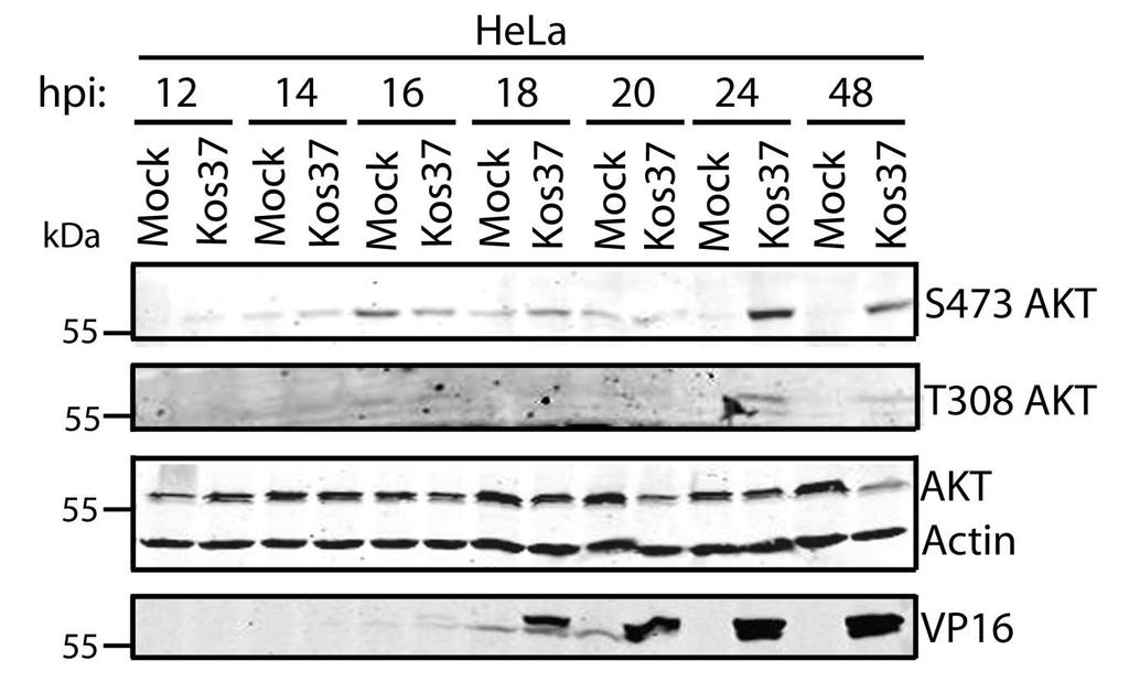
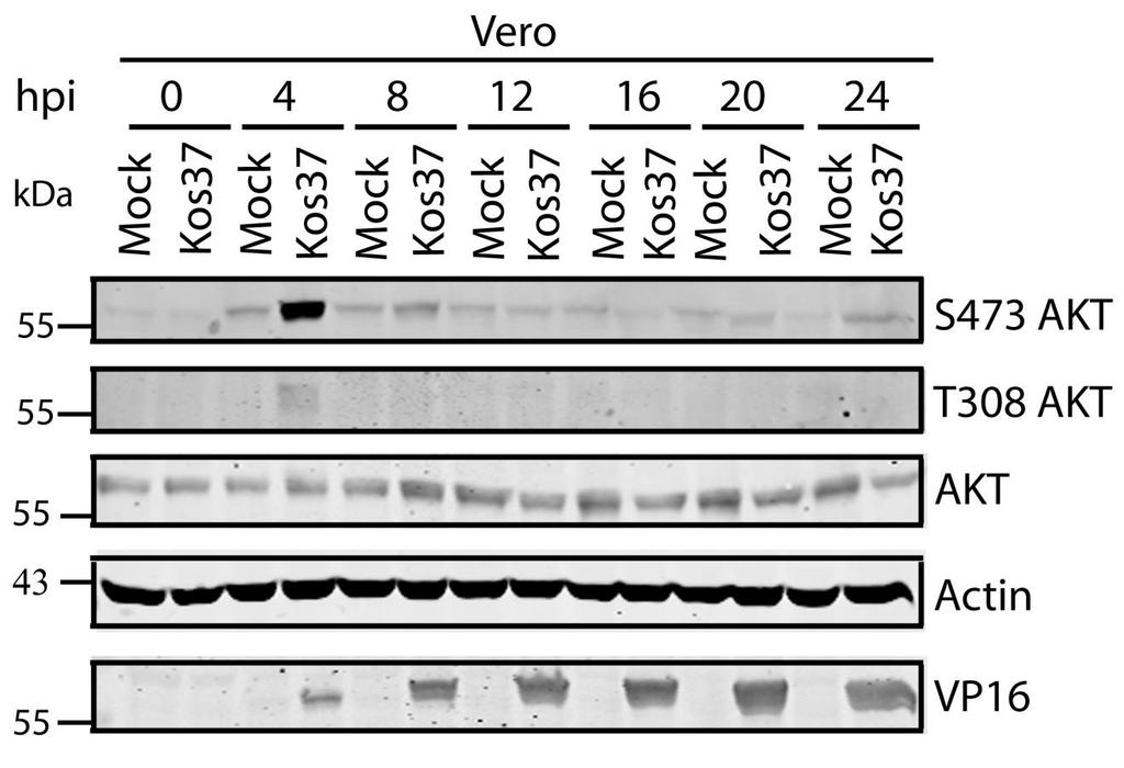
143 A. B. 130

144 Appendix B: Torin1 and rapamycin reduce 4EBP1 hyperphosphorylated patterns regardless of infection. Confluent monolayers of HEL cells were serum-starved and infected with mock, wild type HSV-1 KOS37, or the VP11/12- null mutant ΔUL46. For the last three hours of infection, cells were either left in serum-free media or treated with 250nM of torin1 or 100nM of rapamycin diluted in 1 ml of serum free DMEM. Cell lysates were collected at 18 hpi and analyzed for 4EBP1 and β-actin by Western blotting. 131
145 Appendix C: U S 3 promotes the phosphorylation of AKT targets as early as 6 hpi. Confluent monolayers of HEL cells were serum-starved and mock-infected or infected with wild type HSV-1 F or the U S 3-null mutant. Cell lysates were collected at various times and analyzed for the activating phosphorylation of AKT at residue S473 (anti-s473), phosphorylated FoxO1 and GSK3β at serine 256 and serine 9 respectively, total AKT, FoxO1 and GSK3β, VP16, and β-actin by Western blotting. 132
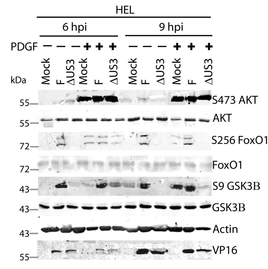
146 A. B. 133
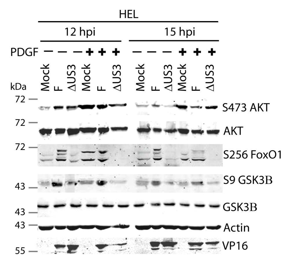
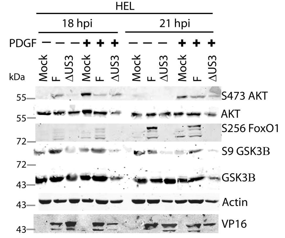
147 C. D. 134
148 Appendix D: Cells infected with ΔU S 3- and ΔU L 46ΔU S 3- virus are more apoptotic than wild type infection. Using light-microscope analysis to examine cell morphology, cells infected with ΔU S 3-virus were more apoptotic compared to wild type-infected cells. Cells that were infected with ΔU L 46ΔU S 3-virus appeared even more apoptotic than ΔU S 3-infected cells. These data indicated that U L 46 and U S 3 are potentially involved in preventing apoptosis 135
149 136
Herpesviruses. Virion. Genome. Genes and proteins. Viruses and hosts. Diseases. Distinctive characteristics
 Herpesviruses Virion Genome Genes and proteins Viruses and hosts Diseases Distinctive characteristics Virion Enveloped icosahedral capsid (T=16), diameter 125 nm Diameter of enveloped virion 200 nm Capsid
Enzyme-coupled Receptors. Cell-surface receptors 1. Ion-channel-coupled receptors 2. G-protein-coupled receptors 3. Enzyme-coupled receptors
 Enzyme-coupled Receptors Cell-surface receptors 1. Ion-channel-coupled receptors 2. G-protein-coupled receptors 3. Enzyme-coupled receptors Cell-surface receptors allow a flow of ions across the plasma
Receptor mediated Signal Transduction
 Receptor mediated Signal Transduction G-protein-linked receptors adenylyl cyclase camp PKA Organization of receptor protein-tyrosine kinases From G.M. Cooper, The Cell. A molecular approach, 2004, third
Lipids and Membranes
 Lipids and Membranes Presented by Dr. Mohammad Saadeh The requirements for the Pharmaceutical Biochemistry I Philadelphia University Faculty of pharmacy Membrane transport D. Endocytosis and Exocytosis
Cell Signaling part 2
 15 Cell Signaling part 2 Functions of Cell Surface Receptors Other cell surface receptors are directly linked to intracellular enzymes. The largest family of these is the receptor protein tyrosine kinases,
G-Protein Signaling. Introduction to intracellular signaling. Dr. SARRAY Sameh, Ph.D
 G-Protein Signaling Introduction to intracellular signaling Dr. SARRAY Sameh, Ph.D Cell signaling Cells communicate via extracellular signaling molecules (Hormones, growth factors and neurotransmitters
Chapter 15: Signal transduction
 Chapter 15: Signal transduction Know the terminology: Enzyme-linked receptor, G-protein linked receptor, nuclear hormone receptor, G-protein, adaptor protein, scaffolding protein, SH2 domain, MAPK, Ras,
Propagation of the Signal
 OpenStax-CNX module: m44452 1 Propagation of the Signal OpenStax College This work is produced by OpenStax-CNX and licensed under the Creative Commons Attribution License 3.0 By the end of this section,
7.012 Problem Set 6 Solutions
 Name Section 7.012 Problem Set 6 Solutions Question 1 The viral family Orthomyxoviridae contains the influenza A, B and C viruses. These viruses have a (-)ss RNA genome surrounded by a capsid composed
Large DNA viruses: Herpesviruses, Poxviruses, Baculoviruses and Giant viruses
 Large DNA viruses: Herpesviruses, Poxviruses, Baculoviruses and Giant viruses Viruses are the only obstacles to the domination of the Earth by mankind. -Joshua Lederberg Recommended reading: Field s Virology
KEY CONCEPT QUESTIONS IN SIGNAL TRANSDUCTION
 Signal Transduction - Part 2 Key Concepts - Receptor tyrosine kinases control cell metabolism and proliferation Growth factor signaling through Ras Mutated cell signaling genes in cancer cells are called
Signaling. Dr. Sujata Persad Katz Group Centre for Pharmacy & Health research
 Signaling Dr. Sujata Persad 3-020 Katz Group Centre for Pharmacy & Health research E-mail:sujata.persad@ualberta.ca 1 Growth Factor Receptors and Other Signaling Pathways What we will cover today: How
Polyomaviridae. Spring
 Polyomaviridae Spring 2002 331 Antibody Prevalence for BK & JC Viruses Spring 2002 332 Polyoma Viruses General characteristics Papovaviridae: PA - papilloma; PO - polyoma; VA - vacuolating agent a. 45nm
Signal Transduction Cascades
 Signal Transduction Cascades Contents of this page: Kinases & phosphatases Protein Kinase A (camp-dependent protein kinase) G-protein signal cascade Structure of G-proteins Small GTP-binding proteins,
MedChem401 Herpesviridae. Herpesviridae
 MedChem401 Herpesviridae Members of the herpesvirus family have been identified in more than 80 different animal species Eight have been identified as human pathogens Herpes viruses are a leading cause
Chapter 25. 바이러스 (The Viruses)
") Chapter 25 바이러스 (The Viruses) Generalized Structure of Viruses 2 2 Virus Classification Classification based on numerous characteristics Nucleic acid type Presence or absence of envelope Capsid symmetry
Chapter 25 바이러스 (The Viruses) Generalized Structure of Viruses 2 2 Virus Classification Classification based on numerous characteristics Nucleic acid type Presence or absence of envelope Capsid symmetry
Signal Transduction: G-Protein Coupled Receptors
 Signal Transduction: G-Protein Coupled Receptors Federle, M. (2017). Lectures 4-5: Signal Transduction parts 1&2: nuclear receptors and GPCRs. Lecture presented at PHAR 423 Lecture in UIC College of Pharmacy,
VZV, EBV, and HHV-6-8
 VZV, EBV, and HHV-6-8 Anne Gershon Common Features of Herpesviruses Morphology Basic mode of replication Primary infection followed by latency Ubiquitous Ability to cause recurrent infections (reactivation
Signaling Through Immune System Receptors (Ch. 7)
") Signaling Through Immune System Receptors (Ch. 7) 1. General principles of signal transduction and propagation. 2. Antigen receptor signaling and lymphocyte activation. 3. Other receptors and signaling
Signaling Through Immune System Receptors (Ch. 7) 1. General principles of signal transduction and propagation. 2. Antigen receptor signaling and lymphocyte activation. 3. Other receptors and signaling
Medical Virology. Herpesviruses, Orthomyxoviruses, and Retro virus. - Herpesviruses Structure & Composition: Herpesviruses
 Medical Virology Lecture 2 Asst. Prof. Dr. Dalya Basil Herpesviruses, Orthomyxoviruses, and Retro virus - Herpesviruses Structure & Composition: Herpesviruses Enveloped DNA viruses. All herpesviruses have
Signal Transduction Pathways. Part 2
 Signal Transduction Pathways Part 2 GPCRs G-protein coupled receptors > 700 GPCRs in humans Mediate responses to senses taste, smell, sight ~ 1000 GPCRs mediate sense of smell in mouse Half of all known
Introduction to Viruses That Infect Humans: The DNA Viruses
 Chapter 24 Introduction to Viruses That Infect Humans: The DNA Viruses Copyright The McGraw-Hill Companies, Inc. Permission required for reproduction or display. 24.1 Viruses in Human Infections and Diseases
Human Herpesviruses. VZV, EBV, and HHV-6-8. The rash of VZV is vesicular. MID 34
 VZV, EBV, and HHV-6-8 Anne Gershon Human Herpesviruses Replication (lytic infection) occurs in a cascade Latency occurs when the cascade is interrupted Transcription of viral genome and protein synthesis
Introduction to viruses. BIO 370 Ramos
 Introduction to viruses BIO 370 Ramos 1 2 General Structure of Viruses Size range most
Chapter13 Characterizing and Classifying Viruses, Viroids, and Prions
 Chapter13 Characterizing and Classifying Viruses, Viroids, and Prions 11/20/2017 MDufilho 1 Characteristics of Viruses Viruses Minuscule, acellular, infectious agent having either DNA or RNA Cause infections
Human Herpes Viruses (HHV) Mazin Barry, MD, FRCPC, FACP, DTM&H Assistant Professor and Consultant Infectious Diseases KSU
 Mazin Barry, MD, FRCPC, FACP, DTM&H Assistant Professor and Consultant Infectious Diseases KSU") Human Herpes Viruses (HHV) Mazin Barry, MD, FRCPC, FACP, DTM&H Assistant Professor and Consultant Infectious Diseases KSU HERPES VIRUS INFECTIONS objectives: ØTo know the clinically important HHVs. ØTo
Human Herpes Viruses (HHV) Mazin Barry, MD, FRCPC, FACP, DTM&H Assistant Professor and Consultant Infectious Diseases KSU HERPES VIRUS INFECTIONS objectives: ØTo know the clinically important HHVs. ØTo
Chapter 11. Cell Communication
 Chapter 11 Cell Communication Overview: The Cellular Internet Cell-to-cell communication Is absolutely essential for multicellular organisms Concept 11.1: External signals are converted into responses
A particular set of insults induces apoptosis (part 1), which, if inhibited, can switch to autophagy. At least in some cellular settings, autophagy se
, which, if inhibited, can switch to autophagy. At least in some cellular settings, autophagy se") A particular set of insults induces apoptosis (part 1), which, if inhibited, can switch to autophagy. At least in some cellular settings, autophagy serves as a defence mechanism that prevents or retards
A particular set of insults induces apoptosis (part 1), which, if inhibited, can switch to autophagy. At least in some cellular settings, autophagy serves as a defence mechanism that prevents or retards
Dr. Ahmed K. Ali Attachment and entry of viruses into cells
 Lec. 6 Dr. Ahmed K. Ali Attachment and entry of viruses into cells The aim of a virus is to replicate itself, and in order to achieve this aim it needs to enter a host cell, make copies of itself and
Lab 3: Pathogenesis of Virus Infections & Pattern 450 MIC PRACTICAL PART SECTION (30397) MIC AMAL ALGHAMDI 1
 MIC AMAL ALGHAMDI 1") Lab 3: Pathogenesis of Virus Infections & Pattern 450 MIC PRACTICAL PART SECTION (30397) 2018 450 MIC AMAL ALGHAMDI 1 Learning Outcomes The pathogenesis of viral infection The viral disease pattern Specific
Lab 3: Pathogenesis of Virus Infections & Pattern 450 MIC PRACTICAL PART SECTION (30397) 2018 450 MIC AMAL ALGHAMDI 1 Learning Outcomes The pathogenesis of viral infection The viral disease pattern Specific
Effects of Second Messengers
 Effects of Second Messengers Inositol trisphosphate Diacylglycerol Opens Calcium Channels Binding to IP 3 -gated Channel Cooperative binding Activates Protein Kinase C is required Phosphorylation of many
Lecture 2: Virology. I. Background
 Lecture 2: Virology I. Background A. Properties 1. Simple biological systems a. Aggregates of nucleic acids and protein 2. Non-living a. Cannot reproduce or carry out metabolic activities outside of a
Viruses. Poxviridae. DNA viruses: 6 families. Herpesviridae Adenoviridae. Hepadnaviridae Papovaviridae Parvoviridae
 Viruses DNA viruses: 6 families Poxviridae Herpesviridae Adenoviridae Hepadnaviridae Papovaviridae Parvoviridae Human herpesviruses Three subfamilies (genome structure, tissue tropism, cytopathologic effect,
Name Section Problem Set 6
 Name Section 7.012 Problem Set 6 Question 1 The viral family Orthomyxoviridae contains the influenza A, B and C viruses. These viruses have a (-)ss RNA genome surrounded by a capsid composed of lipids
MCB*4010 Midterm Exam / Winter 2008
 MCB*4010 Midterm Exam / Winter 2008 Name: ID: Instructions: Answer all 4 questions. The number of marks for each question indicates how many points you need to provide. Write your answers in point form,
Cell Communication. Chapter 11. PowerPoint Lectures for Biology, Seventh Edition. Lectures by Chris Romero. Neil Campbell and Jane Reece
 Chapter 11 Cell Communication PowerPoint Lectures for Biology, Seventh Edition Neil Campbell and Jane Reece Lectures by Chris Romero Overview: The Cellular Internet Cell-to-cell communication Is absolutely
History electron microscopes
 Viruses History Through the 1800s, many scientists discovered that something smaller than bacteria could cause disease and they called it virion (Latin word- poison) In the 1930s, after the invention of
1. Activated receptor tyrosine kinases (RTKs) phosphorylates themselves
 phosphorylates themselves") Enzyme-coupled receptors Transmembrane proteins Ligand-binding domain on the outer surface Cytoplasmic domain acts as an enzyme itself or forms a complex with enzyme 1. Activated receptor tyrosine kinases
Enzyme-coupled receptors Transmembrane proteins Ligand-binding domain on the outer surface Cytoplasmic domain acts as an enzyme itself or forms a complex with enzyme 1. Activated receptor tyrosine kinases
Herpesviruses. -Recurrence: clinically obvious disease due to reactivation. **Reactivation and recurrence are used interchangeably.
 *Herpesviruses: A large group of viruses (100 strains), but we are concerned with only 8 strains as they are the only ones to infect human beings *herpesviruses groups: HSV-1 HSV-2 VZV CMV EBV HHV-6 HHV-7
Signal Transduction I
 Signal Transduction I Prof. Tianhua Zhou Department of Cell Biology Zhejiang University School of Medicine Office hours by appointment tzhou@zju.edu.cn Signal transduction: Key contents for signal transduction:
RAS Genes. The ras superfamily of genes encodes small GTP binding proteins that are responsible for the regulation of many cellular processes.
 ۱ RAS Genes The ras superfamily of genes encodes small GTP binding proteins that are responsible for the regulation of many cellular processes. Oncogenic ras genes in human cells include H ras, N ras,
Properties of Herpesviruses
 Herpesviruses Properties of Herpesviruses Structure and Composition Spherical icosahedron, 150-200 nm Double-stranded DNA, linear More than 35 proteins Enveloped Replication from nucleus (budding) Features
Regulation of cell function by intracellular signaling
 Regulation of cell function by intracellular signaling Objectives: Regulation principle Allosteric and covalent mechanisms, Popular second messengers, Protein kinases, Kinase cascade and interaction. regulation
CYTOKINE RECEPTORS AND SIGNAL TRANSDUCTION
 CYTOKINE RECEPTORS AND SIGNAL TRANSDUCTION What is Cytokine? Secreted popypeptide (protein) involved in cell-to-cell signaling. Acts in paracrine or autocrine fashion through specific cellular receptors.
Lecture: CHAPTER 13 Signal Transduction Pathways
 Lecture: 10 17 2016 CHAPTER 13 Signal Transduction Pathways Chapter 13 Outline Signal transduction cascades have many components in common: 1. Release of a primary message as a response to a physiological
Fayth K. Yoshimura, Ph.D. September 7, of 7 HIV - BASIC PROPERTIES
 1 of 7 I. Viral Origin. A. Retrovirus - animal lentiviruses. HIV - BASIC PROPERTIES 1. HIV is a member of the Retrovirus family and more specifically it is a member of the Lentivirus genus of this family.
Page 32 AP Biology: 2013 Exam Review CONCEPT 6 REGULATION
 Page 32 AP Biology: 2013 Exam Review CONCEPT 6 REGULATION 1. Feedback a. Negative feedback mechanisms maintain dynamic homeostasis for a particular condition (variable) by regulating physiological processes,
The Tissue Engineer s Toolkit
 The Tissue Engineer s Toolkit Stimuli Detection and Response Ken Webb, Ph. D. Assistant Professor Dept. of Bioengineering Clemson University Environmental Stimulus-Cellular Response Environmental Stimuli
General information. Cell mediated immunity. 455 LSA, Tuesday 11 to noon. Anytime after class.
 General information Cell mediated immunity 455 LSA, Tuesday 11 to noon Anytime after class T-cell precursors Thymus Naive T-cells (CD8 or CD4) email: lcoscoy@berkeley.edu edu Use MCB150 as subject line
Part I. Content: History of Viruses. General properties of viruses. Viral structure. Viral classifications. Virus-like agents.
 Viruses Part I Content: History of Viruses. General properties of viruses. Viral structure. Viral classifications. Virus-like agents. History Through the 1800s, many scientists discovered that something
Viruses. CLS 212: Medical Microbiology Miss Zeina Alkudmani
 Viruses CLS 212: Medical Microbiology Miss Zeina Alkudmani History Through the 1800s, many scientists discovered that something smaller than bacteria could cause disease and they called it virion (Latin
Virology Introduction. Definitions. Introduction. Structure of virus. Virus transmission. Classification of virus. DNA Virus. RNA Virus. Treatment.
 DEVH Virology Introduction Definitions. Introduction. Structure of virus. Virus transmission. Classification of virus. DNA Virus. RNA Virus. Treatment. Definitions Virology: The science which study the
Cellular Signaling Pathways. Signaling Overview
 Cellular Signaling Pathways Signaling Overview Signaling steps Synthesis and release of signaling molecules (ligands) by the signaling cell. Transport of the signal to the target cell Detection of the
What would you observe if you fused a G1 cell with a S cell? A. Mitotic and pulverized chromosomes. B. Mitotic and compact G1 chromosomes.
 What would you observe if you fused a G1 cell with a S cell? A. Mitotic and pulverized chromosomes. B. Mitotic and compact G1 chromosomes. C. Mostly non-compact G1 chromosomes. D. Compact G1 and G2 chromosomes.
Common Characteristics and Distinct Features of Human Pathogenic Herpesviruses
 Common Characteristics and Distinct Features of Human Pathogenic Herpesviruses Hartmut Hengel Chapter 1 1.1 Hallmarks of Herpesvirus Infections The members of the family of the herpesviridae are phylogenetically
T cell maturation. T-cell Maturation. What allows T cell maturation?
 T-cell Maturation What allows T cell maturation? Direct contact with thymic epithelial cells Influence of thymic hormones Growth factors (cytokines, CSF) T cell maturation T cell progenitor DN DP SP 2ry
October 26, Lecture Readings. Vesicular Trafficking, Secretory Pathway, HIV Assembly and Exit from Cell
 October 26, 2006 Vesicular Trafficking, Secretory Pathway, HIV Assembly and Exit from Cell 1. Secretory pathway a. Formation of coated vesicles b. SNAREs and vesicle targeting 2. Membrane fusion a. SNAREs
Chapter 20. Cell - Cell Signaling: Hormones and Receptors. Three general types of extracellular signaling. endocrine signaling. paracrine signaling
 Chapter 20 Cell - Cell Signaling: Hormones and Receptors Three general types of extracellular signaling endocrine signaling paracrine signaling autocrine signaling Endocrine Signaling - signaling molecules
AP Biology. Viral diseases Polio. Chapter 18. Smallpox. Influenza: 1918 epidemic. Emerging viruses. A sense of size
 Hepatitis Viral diseases Polio Chapter 18. Measles Viral Genetics Influenza: 1918 epidemic 30-40 million deaths world-wide Chicken pox Smallpox Eradicated in 1976 vaccinations ceased in 1980 at risk population?
Cell Biology Lecture 9 Notes Basic Principles of cell signaling and GPCR system
 Cell Biology Lecture 9 Notes Basic Principles of cell signaling and GPCR system Basic Elements of cell signaling: Signal or signaling molecule (ligand, first messenger) o Small molecules (epinephrine,
BIO 5099: Molecular Biology for Computer Scientists (et al)
") BIO 5099: Molecular Biology for Computer Scientists (et al) Lecture 15: Being a Eukaryote: From DNA to Protein, A Tour of the Eukaryotic Cell. Christiaan van Woudenberg Being A Eukaryote Basic eukaryotes
BIO 5099: Molecular Biology for Computer Scientists (et al) Lecture 15: Being a Eukaryote: From DNA to Protein, A Tour of the Eukaryotic Cell. Christiaan van Woudenberg Being A Eukaryote Basic eukaryotes
number Done by Corrected by Doctor Ashraf
 number 4 Done by Nedaa Bani Ata Corrected by Rama Nada Doctor Ashraf Genome replication and gene expression Remember the steps of viral replication from the last lecture: Attachment, Adsorption, Penetration,
Chapter 19: Viruses. 1. Viral Structure & Reproduction. 2. Bacteriophages. 3. Animal Viruses. 4. Viroids & Prions
 Chapter 19: Viruses 1. Viral Structure & Reproduction 2. Bacteriophages 3. Animal Viruses 4. Viroids & Prions 1. Viral Structure & Reproduction Chapter Reading pp. 393-396 What exactly is a Virus? Viruses
Intrinsic cellular defenses against virus infection
 Intrinsic cellular defenses against virus infection Detection of virus infection Host cell response to virus infection Interferons: structure and synthesis Induction of antiviral activity Viral defenses
General Principles of Endocrine Physiology
 General Principles of Endocrine Physiology By Dr. Isabel S.S. Hwang Department of Physiology Faculty of Medicine University of Hong Kong The major human endocrine glands Endocrine glands and hormones
Chapter 19: Viruses. 1. Viral Structure & Reproduction. What exactly is a Virus? 11/7/ Viral Structure & Reproduction. 2.
 Chapter 19: Viruses 1. Viral Structure & Reproduction 2. Bacteriophages 3. Animal Viruses 4. Viroids & Prions 1. Viral Structure & Reproduction Chapter Reading pp. 393-396 What exactly is a Virus? Viruses
Introduction. Biochemistry: It is the chemistry of living things (matters).
.") Introduction Biochemistry: It is the chemistry of living things (matters). Biochemistry provides fundamental understanding of the molecular basis for the function and malfunction of living things. Biochemistry
Introduction Biochemistry: It is the chemistry of living things (matters). Biochemistry provides fundamental understanding of the molecular basis for the function and malfunction of living things. Biochemistry
STRUCTURE, GENERAL CHARACTERISTICS AND REPRODUCTION OF VIRUSES
 STRUCTURE, GENERAL CHARACTERISTICS AND REPRODUCTION OF VIRUSES Introduction Viruses are noncellular genetic elements that use a living cell for their replication and have an extracellular state. Viruses
Viruses defined acellular organisms genomes nucleic acid replicate inside host cells host metabolic machinery ribosomes
 The Viruses Viruses Viruses may be defined as acellular organisms whose genomes consist of nucleic acid, obligately replicate inside host cells using host metabolic machinery and ribosomes to form a pool
CELLS. Cells. Basic unit of life (except virus)
") Basic unit of life (except virus) CELLS Prokaryotic, w/o nucleus, bacteria Eukaryotic, w/ nucleus Various cell types specialized for particular function. Differentiation. Over 200 human cell types 56%
Basic unit of life (except virus) CELLS Prokaryotic, w/o nucleus, bacteria Eukaryotic, w/ nucleus Various cell types specialized for particular function. Differentiation. Over 200 human cell types 56%
Protein Trafficking in the Secretory and Endocytic Pathways
 Protein Trafficking in the Secretory and Endocytic Pathways The compartmentalization of eukaryotic cells has considerable functional advantages for the cell, but requires elaborate mechanisms to ensure
Cell cycle, signaling to cell cycle, and molecular basis of oncogenesis
 Cell cycle, signaling to cell cycle, and molecular basis of oncogenesis MUDr. Jiří Vachtenheim, CSc. CELL CYCLE - SUMMARY Basic terminology: Cyclins conserved proteins with homologous regions; their cellular
Human Herpesviruses. Medical Virology, 27 Nov 2015.
 Human Herpesviruses Assoc.Prof. Murat Sayan Kocaeli Üniversitesi, Rutin PCR Lab. Sorumlu Öğt.Üyesi Yakın Doğu Üniversitesi, DESAM Kurucu Öğrt. Üyesi sayanmurat@hotmail.com 0533 6479020 Medical Virology,
Under the Radar Screen: How Bugs Trick Our Immune Defenses
 Under the Radar Screen: How Bugs Trick Our Immune Defenses Session 7: Cytokines Marie-Eve Paquet and Gijsbert Grotenbreg Whitehead Institute for Biomedical Research HHV-8 Discovered in the 1980 s at the
Phospho-AKT Sampler Kit
 Phospho-AKT Sampler Kit E 0 5 1 0 0 3 Kits Includes Cat. Quantity Application Reactivity Source Akt (Ab-473) Antibody E021054-1 50μg/50μl IHC, WB Human, Mouse, Rat Rabbit Akt (Phospho-Ser473) Antibody
Growth and Differentiation Phosphorylation Sampler Kit
 Growth and Differentiation Phosphorylation Sampler Kit E 0 5 1 0 1 4 Kits Includes Cat. Quantity Application Reactivity Source Akt (Phospho-Ser473) E011054-1 50μg/50μl IHC, WB Human, Mouse, Rat Rabbit
Cell Communication. Chapter 11. Biology Eighth Edition Neil Campbell and Jane Reece. PowerPoint Lecture Presentations for
 Chapter 11 Cell Communication PowerPoint Lecture Presentations for Biology Eighth Edition Neil Campbell and Jane Reece Lectures by Chris Romero, updated by Erin Barley with contributions from Joan Sharp
Lecture 7: Signaling Through Lymphocyte Receptors
 Lecture 7: Signaling Through Lymphocyte Receptors Questions to Consider After recognition of its cognate MHC:peptide, how does the T cell receptor activate immune response genes? What are the structural
Sarah Jaar Marah Al-Darawsheh
 22 Sarah Jaar Marah Al-Darawsheh Faisal Mohammad Receptors can be membrane proteins (for water-soluble hormones/ligands) or intracellular (found in the cytosol or nucleus and bind to DNA, for lipid-soluble
Revision. camp pathway
 االله الرحمن الرحيم بسم Revision camp pathway camp pathway Revision camp pathway Adenylate cyclase Adenylate Cyclase enzyme Adenylate cyclase catalyses the formation of camp from ATP. Stimulation or inhibition
Viral structure م.م رنا مشعل
 Viral structure م.م رنا مشعل Viruses must reproduce (replicate) within cells, because they cannot generate energy or synthesize proteins. Because they can reproduce only within cells, viruses are obligate
Chapter 13B: Animal Viruses
 Chapter 13B: Animal Viruses 1. Overview of Animal Viruses 2. DNA Viruses 3. RNA Viruses 4. Prions 1. Overview of Animal Viruses Life Cycle of Animal Viruses The basic life cycle stages of animal viruses
Persistent Infections
 Persistent Infections Lecture 17 Biology 3310/4310 Virology Spring 2017 Paralyze resistance with persistence WOODY HAYES Acute vs persistent infections Acute infection - rapid and self-limiting Persistent
Chapter 6- An Introduction to Viruses*
 Chapter 6- An Introduction to Viruses* *Lecture notes are to be used as a study guide only and do not represent the comprehensive information you will need to know for the exams. 6.1 Overview of Viruses
Lecture 15. Signal Transduction Pathways - Introduction
 Lecture 15 Signal Transduction Pathways - Introduction So far.. Regulation of mrna synthesis Regulation of rrna synthesis Regulation of trna & 5S rrna synthesis Regulation of gene expression by signals
Chapter 9. Cellular Signaling
 Chapter 9 Cellular Signaling Cellular Messaging Page 215 Cells can signal to each other and interpret the signals they receive from other cells and the environment Signals are most often chemicals The
Principles of Genetics and Molecular Biology
 Cell signaling Dr. Diala Abu-Hassan, DDS, PhD School of Medicine Dr.abuhassand@gmail.com Principles of Genetics and Molecular Biology www.cs.montana.edu Modes of cell signaling Direct interaction of a
Chapter 11: Enzyme Catalysis
 Chapter 11: Enzyme Catalysis Matching A) high B) deprotonated C) protonated D) least resistance E) motion F) rate-determining G) leaving group H) short peptides I) amino acid J) low K) coenzymes L) concerted
Unit 13.2: Viruses. Vocabulary capsid latency vaccine virion
 Unit 13.2: Viruses Lesson Objectives Describe the structure of viruses. Outline the discovery and origins of viruses. Explain how viruses replicate. Explain how viruses cause human disease. Describe how
Nucleic acid: singled stranded, double stranded, RNA, or DNA, linear or circular. Capsid: protein coat that is most of the mass of the virus.
 Viruses General Characteristics of Viruses 1. Depending on view may be regarded as exceptionally complex aggregates of nonliving chemicals or as exceptionally simple living microbes. 2. Contain a single
MOLECULAR CELL BIOLOGY
 1 Lodish Berk Kaiser Krieger scott Bretscher Ploegh Matsudaira MOLECULAR CELL BIOLOGY SEVENTH EDITION CHAPTER 13 Moving Proteins into Membranes and Organelles Copyright 2013 by W. H. Freeman and Company
EBV infection B cells and lymphomagenesis. Sridhar Chaganti
 EBV infection B cells and lymphomagenesis Sridhar Chaganti How EBV infects B-cells How viral genes influence the infected B cell Differences and similarities between in vitro and in vivo infection How
HSV DNA replication. Herpesvirus Latency. Latency and Chemotherapy. Human Herpesviruses - subtypes. Acyclovir (acycloguanosine) {Zovirax}
 {Zovirax}") Human Herpesviruses - subtypes Herpes Simplex I (HSVI) - herpes labialis (cold sores) herpes keratitis (eye infections) HSVII - herpes genitalis (genital herpes) Varicella Zoster virus (VZV) Chicken pox
Human Herpesviruses - subtypes Herpes Simplex I (HSVI) - herpes labialis (cold sores) herpes keratitis (eye infections) HSVII - herpes genitalis (genital herpes) Varicella Zoster virus (VZV) Chicken pox
Viruses. Properties. Some viruses contain other ingredients (e.g., lipids, carbohydrates), but these are derived from their host cells.
, but these are derived from their host cells.") Viruses Properties They are obligate intracellular parasites. Probably there are no cells in nature that escape infection by one or more kinds of viruses. (Viruses that infect bacteria are called bacteriophages.)
Viruses Properties They are obligate intracellular parasites. Probably there are no cells in nature that escape infection by one or more kinds of viruses. (Viruses that infect bacteria are called bacteriophages.)
Reoviruses. Virion. Genome. Genes and proteins. Viruses and hosts. Diseases. Distinctive characteristics
 Reoviruses Virion Genome Genes and proteins Viruses and hosts Diseases Distinctive characteristics Virion Naked icosahedral capsid (T=13), diameter 60-85 nm Capsid consists of two or three concentric protein
Translation. Host Cell Shutoff 1) Initiation of eukaryotic translation involves many initiation factors
 Initiation of eukaryotic translation involves many initiation factors") Translation Questions? 1) How does poliovirus shutoff eukaryotic translation? 2) If eukaryotic messages are not translated how can poliovirus get its message translated? Host Cell Shutoff 1) Initiation
Translation Questions? 1) How does poliovirus shutoff eukaryotic translation? 2) If eukaryotic messages are not translated how can poliovirus get its message translated? Host Cell Shutoff 1) Initiation
Human Immunodeficiency Virus
 Human Immunodeficiency Virus Virion Genome Genes and proteins Viruses and hosts Diseases Distinctive characteristics Viruses and hosts Lentivirus from Latin lentis (slow), for slow progression of disease
Bacteriophage Reproduction
 Bacteriophage Reproduction Lytic and Lysogenic Cycles The following information is taken from: http://student.ccbcmd.edu/courses/bio141/lecguide/unit3/index.html#charvir Bacteriophage Structure More complex
Cell Communication. Local and Long Distance Signaling
 Cell Communication Cell to cell communication is essential for multicellular organisms Some universal mechanisms of cellular regulation providing more evidence for the evolutionary relatedness of all life
Size nm m m
 1 Viral size and organization Size 20-250nm 0.000000002m-0.000000025m Virion structure Capsid Core Acellular obligate intracellular parasites Lack organelles, metabolic activities, and reproduction Replicated