Computational Molecular Modeling Studies of the Interactions of Estrogens with Their Receptors and Intracellular Binding Protein PDIp
|
|
|
- August Cook
- 6 years ago
- Views:
Transcription
1 Computational Molecular Modeling Studies of the Interactions of Estrogens with Their Receptors and Intracellular Binding Protein PDIp BY Pan Wang B.S. Tsinghua University, Beijing, China 2005 Submitted to the graduate degree program in Pharmacology and the Graduate Faculty of the University of Kansas in partial fulfillment of the requirements for the degree of Doctor of Philosophy Dissertation Committee members: Bao Ting Zhu, Ph.D. (Chair) Bruno Hagenbuch, Ph.D. Curtis D. Klaassen, Ph.D. Brian K. Petroff, Ph.D. Dissertation Defended 09/27/2010 Scott J. Weir, Pharm. D., Ph.D.
2 The Dissertation Committee for Pan Wang certifies that this is the approved version of the following dissertation: Computational Molecular Modeling Studies of the Interactions of Estrogens with Their Receptors and Intracellular Binding Protein PDIp Pan Wang B.S. Tsinghua University, Beijing, China 2005 Dissertation Committee: Bao Ting Zhu, Ph.D. (Chair) Bruno Hagenbuch, Ph.D. Curtis D. Klaassen, Ph.D. Brian K. Petroff, Ph.D. Scott J. Weir, Pharm. D., Ph.D. Dissertation Defended 09/27/2010 ii
3 DEDICATION I would like to dedicate the work herein to my beloved parents Guanglian Wu and Xingwu Wang iii
4 ACKNOWLEDGEMENTS I am heartily thankful to the many people who helped me complete this dissertation. First and foremost, I owe my deepest gratitude to my mentor, Dr. Bao Ting Zhu, for all of his guidance, support, encouragement, and advice throughout my graduate study. This dissertation would not have been possible without the great amount of effort and time he has put in to guide me through the last five years. It has been a great honor for me to work with Dr. Zhu to learn not only how to be a better researcher but also how to be a better person. I am also grateful to the rest of my dissertation committee members, Dr. Bruno Hagenbuch, Dr. Curtis D. Klaassen, Dr. Brian Petroff, and Dr. Scott Weir, for all of their scientific advice, as well as for being so generous with their time in reviewing my dissertation proposal and draft. It would not have been possible for me to complete this project without the help of my committee. I would also like to thank other faculty members in the department, Dr Grace Guo, Dr Xiao-bo Zhong, Dr Xiaochao Ma, Dr. Thomas L. Pazdernik, and a visiting professor Dr. Lirong Zhang, who provided me lots of support and encouragement along the way. Thanks also go to the previous and current members of Dr. Zhu s laboratory, especially Dr. Masayuki Fukui, Dr. Hye-Joung Choi, Stephanie Bishop, Angelia Choi, iv
5 Jihoon Song, Dr. Hyoung-Woo Bai, Jina Yu, Dr. Jian Ding, Dr. Xiangchen Dai, Dr. Jun Liu, Elisabeth Yan Zhang, Aaron Yun Chen, and Dr. Xiang-Rong Jiang who have provided me with great guidance and help. They are all kind, generous, and wonderful people to work with. I would like to acknowledge the suggestions given by Dr. Campbell McInnes from University of South Carolina on my computational modeling study. Dr. Gerry Lushington from University of Kansas, Lawrence helped with the installation of SYBYL software used for the molecular modeling study. Dr. Carolyn L. Smith from Baylor College of Medicine kindly helped us with the estrogen receptor reporter assay. Dr Benyi Li from Department of Urology generously provided us the androgen receptor reporter plasmid. Thanks also go to the people in the flow cytometry facility, electron microscope facility and laboratory animal resources for their technical help with some of my other projects. I would also like to thank my colleagues in the Department of Pharmacology, Toxicology and Therapeutics for their friendship and academic support. Special thanks go to Connie Kai Wu, Youcai Zhang, Dr. Yi Miao Weaver, Dr. Pengli Bu, Dr. Dan Li, Dr. Chieko Saito, Huimin Yan, Dr. Min Yang, Dr. Bo Kong, Dr. Chunshan Gui, Dr. Jie Lu, Xiaohong Lei, Dr. Shary Shelton, Dr. Shuang Mei, and Ruipeng Wang who generously provided lots of mental and technical help whenever I needed them. v
6 I also appreciate the help from the Pharmacology department administration staffs, particularly Ms. Rosa Meagher, Ms. Dorothy McGregor and Mr. Cody Tully. They not only keep the department running efficiently, but also have the best interest of the students in mind. I am also grateful to the people from the International program for their help, especially Ms. Julia Shaw. I would like to express my deepest appreciation to my family who have always trusted and believed in me. I want to thank my mother Guanglian Wu, my father Xingwu Wang, and my husband Dr. Xinmiao Fu, who have always emotionally supported me with their everlasting love, which is what keeps me moving on through difficult times. For this reason, I share this degree with them. Thanks also go to the National Institutes of Health for the funding support that my mentor Dr. Zhu has received in the past few years which were used to support my graduate study and research, as well as the Department of Defense Breast Cancer Research Program for awarding me the Predoctoral Fellowship. vi
7 ABSTRACT The endogenous estrogens are vitally important female sex hormones with diverse biological functions. Disruptions of their actions contribute to the pathogenesis of a number of disease states in humans, such as endocrine malfunction, infertility, and development of cancers. Therefore, it is important to be able to predict whether a given chemical may have the potential to alter estrogen s actions through binding to the estrogen receptors (ERs) or other estrogen-binding proteins in the body. As opposed to some of the widely-used methods such as biochemical assays and crystallography, computational molecular modeling methods have the potential advantages of low cost, high speed, and high throughput. My dissertation research sought to explore the potential usefulness of computational molecular modeling tools in studying the interactions of various estrogen derivatives (e.g., endogenous estrogen metabolites, non-aromatic steroids, and synthetic antiestrogens) with human ERα and ERβ as well as with a recently-identified intracellular estrogen-binding protein. As described in SECTION I of this dissertation, I estabolished and validated a molecular docking approach for studying the binding interactions of estrogens with human ERα and ERβ, and then I used this method to determine the binding characteristics of 27 structurally-similar estrogen derivatives with the ligand binding domains (LBDs) of human ERs. I found that while the binding modes of these estrogen derivatives are very similar to that of 17β-estradiol (E 2 ), there are distinct subtle vii
8 differences. In the case of A-ring E 2 derivatives, for instance, the small differences in the length of the hydrogen bonds formed between the phenolic 3-hydroxyl group of the estrogens and the ERs were found to be a major determinant of their overall binding affinity. This study reveals some of the structural features of the binding interactions of steroidal ligands with human ERs. All previously characterized endogenous estrogens are steroids with their A-ring being an aromatic ring. Based on the computational analysis of the binding characteristics of various aromatic estrogens for human ERs as described above, I tested an intriguing hypothesis that some of the non-aromatic androgen metabolites or precursors may also be able to bind to human ERs. This work is described in SECTION II of my dissertation. With the aid of the computational molecular modeling tools together with a number of in vitro ER binding assays, I identified, for the first time, several non-aromatic metabolites or synthetic precursors of endogenous androgens that can bind to human ERα and ERβ with physiologically-relevant binding affinity, and these non-aromatic steroids can also activate the ERs to elicit estrogenic responses in human cancer cell lines. These results lead to the suggestion that some of the endogenously-formed androgen precursors or metabolites may serve as male-specific ER modulators. It is known that compounds such as ICI-172,780, which possesses a long linear side chain attached to the C-7α position of E 2, could serve as effective ER pure antagonists. The studies described in SECTION III of my dissertation sought to test the viii
9 hypothesis that estrogen analogs with a shorter but bulky side chain may also be able to function as effective ER antagonists, and theoretically, these compounds may be structurally more stable. Our laboratory designed and synthesized nine of these novel estrogen analogs. Four of them were identified as having a strong antiestrogenic activity in ER-positive human breast cancer cells. Computational molecular modeling studies found that these compounds could tightly bind the ERα LBD similar to ICI-182,780, which helps to explain the mechanism of their antiestrogenic actions. Recently, our laboratory identified, for the first time, the pancreas-specific protein disulfide isomerase (PDIp) as a novel intracellular E 2 -binding protein with the binding site located in its b-b domain. As described in SECTION IV of my dissertation, I used computational molecular modeling methods to determine the detailed E 2 -binding site structures of the human PDIp protein. Molecular docking analysis predicted the binding site in the hydrophobic pocket between the b and b domains. The hydrogen bond formed between the 3-hydroxyl group of E 2 and PDIp-His278 is indispensable for the binding interaction. Selective point mutations of relevant amino acid residues and selective modifications of the ligand structures both confirmed this predicted binding mode. Altogether, these results precisely define, for the time, the E 2 -binding site structure of human PDIp. As a whole, the results of my dissertation projects offer important insights into the three-dimensional structural characteristics of the binding interactions of various estrogen ix
10 analogs with the human ERα, ERβ, and PDIp. These studies provide a platform for the future development of an automated docking-based computational approach that can screen numerous environmental compounds for their potential ability to bind human ERs as well as other estrogen binding proteins in the body. x
11 TABLE OF CONTENTS Acceptance Page... ii Dedication... iii Acknowledgements... iv Abstract... vii Table of Contents... xi List of Figures... xiii List of Tables... xvi List of Abbreviations... xvii Chapter 1: General Introduction....1 Chapter 2: Statement of Purpose Chapter 3: Materials and Methods Chapter 4: Structural Characterization of the Binding Interactions of Various Estrogen Metabolites with Human Estrogen Receptor α and β Subtypes Chapter 5: Characterization of the Estrogenic Activity of Non-aromatic Steroids: Implications for the Presence of Estrogen Receptor Modulators in Men Chapter 6: Characterization of the Interactions of Newly-synthesized E 2 -based C-7α Derivatives with Human Estrogen Receptors Chapter 7: Structural Characterization of the Estradiol-Binding Site of Human PDIp: Indispensable Role of the Hydrogen Bond between PDIp-His278 and the 3-Hydroxyl Group of Estradiol Chapter 8: General Summary and Conclusions xi
12 Literature Cited xii
13 LIST OF FIGURES Figure 1 Comparison of positioning of Helix12 in ERα structures bound with an agonist and an antagonist Figure 2 Scheme of the steps for developing QSAR models Figure 3 The chemical structures of E 2 and the 26 E 2 derivatives Figure 4 Overlay of docked structure and the crystal structure of ERα LBD in complex with DES or E Figure 5 Interactions of A-ring derivatives with ERα LBD determined by the molecular docking method Figure 6 Correlation of hydrogen bond length and logrba of A-ring and B/C ring derivatives with ERs LBDs Figure 7 Interactions of B/C-ring derivatives and D-ring derivatives with ERα LBD determined by the molecular docking method Figure 8 Correlation between logrba and binding energy calculated according to equation (3) and data in Table Figure 9 Relationship between the correlation coefficient r value and x/y value for estrogen derivatives listed in Table Figure 10 Potential precursors and metabolic intermediates produced during the biosynthesis and metabolism of endogenous androgens and chemical structures of E 2, E 1, and compounds Figure 11 Competition of [ 3 H]E 2 binding to human ERα and ERβ by various non- xiii
14 aromatic steroids (compounds 1 6) and also by aromatic steroids E 2 and E Figure 12 ERα and AR transcriptional activity of various non-aromatic steroids Figure 13 Comparison of the estrogenic activity of compounds 1 6 with E Figure 14 3-D QSAR/CoMFA analysis of 58 non-aromatic steroids for their binding with ERα and ERβ Figure 15 Comparison of ERα and ERβ binding sites in complex with E 2 or compounds 1 6 in the docking models developed in this study Figure 16 Structures of newly-synthesized E 2 -based C-7α derivatives J1-J Figure 17 Comparison of the relative ER binding affinity of J1-J9 with that of E Figure 18 ERα reporter assay with J1-J9 in HeLa cells Figure 19 Effect of J1-J9 on the proliferation of breast cancer cell lines Figure 20 Molecular docking of the ER antagonists with human ERα LBD Figure 21 Human PDIp b-b fragment contains the E 2 -binding site Figure 22 Determination of the dissociation constant (K d ) of the human b-b fragment and the full-length human PDIp for E Figure 23 Isolation of PDIp from cos-7 cell lysates by SEC Figure 24 Determination of the E 2 -binding sites in PDIp using computational modeling and docking analyses Figure 25 Docking analysis of the binding mode of E 2 inside human PDIp b-b fragment Figure 26 The H278L mutant protein lacks E 2 -binding activity Figure 27 PDIP Q265L mutant protein retains similar E 2 -binding activity xiv
15 Figure 28 Relative binding activity of PDIp for several E 2 derivatives Figure 29 Docking analysis of the interaction of PDIp with E 2 analog C Figure 30 Docking analysis of the interaction of PDIp with ICI-172, xv
16 LIST OF TABLES Table 1 Summary of current structures of ER with various ligands listed in chronological order of publication Table 2 Hydrogen bond lengths (Å) and calculated van der Waals interaction energy (ΔE VDW, kcal/mol) and Coulomb interaction energy (ΔE Coulomb, kcal/mol) between the 27 estrogen derivatives and ERα and ERβ...53 Table 3 Inhibition of [ 3 H]E 2 binding to human ERα and ERβ in vitro by a total of sixty non-aromatic steroids from several steroid classes Table 4 The IC 50 and RBA values of J1-J9 for ERα and ERβ xvi
17 LIST OF ABBREVIATIONS AR ARE CoMFA DES ER ERE ERT E 1 E 2 E 3 GPR Helix 12 LBD PC PDI PDIp QSAR RAL RMS RBA SEC SERM SHBG androgen receptor androgen response element comparative molecular field analysis diethylstilbestrol estrogen receptor estrogen response element estrogen replacement therapy estrone 17β-estradiol estriol G protein-coupled receptor H12 ligand binding domain principle component protein disulfide isomerase pancreas-specific PDI homolog quantitative structure-activity relationship raloxifene root mean square relative binding affinity size exclusion chromatography selective estrogen receptor modulator sex hormone binding globulin xvii
18 T 3 TAM VDW 3,3,5-triiodo-L-thyronine tamoxifen van der Waals xviii
19 CHAPTER ONE GENERAL INTRODUCTION 1
20 ESTROGENS AND ANTIESTROGENS Biosynthesis and metabolism of estrogens. In women, estrogens are synthesized from cholesterol mainly in the theca and granulosa cells of the ovary. Theca cells secrete androgens that diffuse into the granulosa cells where the androgens are aromatized by aromatase to estrogens (Hillier et al., 1994). Some estrogens are also produced in smaller amounts by other tissues such as the liver, adrenal glands, and the breasts. Estrogens are metabolized mainly in liver by oxidation (largely hydroxylations by cytochrome P450 enzymes) (Martucci and Fishman, 1993), glucuronidation (Lucier and McDaniel, 1977), sulfation (Brooks and Horn, 1971), and/or O-methylation (Ball and Knuppen, 1980). The hydrophilic metabolites are excreted into the bile or urine. Hydroxylation of estrogens yields 2-hydroxyestrogens, 4-hydroxyestrogens, and 16αhydroxyestrogens. The unique functions of these estrogen metabolites have been reviewed (Zhu and Conney, 1998). Notably, 4-hydroxyestradiol and 16αhydroxyestradiol are considered to be carcinogenic. 4-Hydroxyestradiol is capable of undergoing metabolic redox cycling, a process that yields semiquinone/quinone intermediates, formation of free radicals, and covalent binding to DNA (Liehr et al., 1986). In addition, catechol estrogens have relatively weaker estrogenic activity compared to E 2 and they may inhibit catechol-o-methyltransferase. Physiological functions of estrogens. Estrogens have a wide array of 2
21 physiological functions. They promote the development of female secondary sexual characteristics, such as breasts. In young women, the lobular units of their breast terminal ducts are highly responsive to estrogens. Estrogens can stimulate the growth and differentiation of ductal epithelium, induce mitotic activity of ductal cylindric cells, and stimulate the growth of connective tissue (Porter, 1974). Estrogens are also involved in the thickening of the endometrium and other aspects of regulating the menstrual cycle. Estrogens can serve as feedback control to inhibit production of GnRH by hypothalamus. Besides these reproductive functions, estrogens have beneficial effects on the bone, cardiovascular system, and central nervous system. Estrogen deficiency accelerates bone loss and increases risk of fractures. Estrogen therapy diminishes bone loss (Christiansen et al., 1981) and reduces the risk of fracture in women with osteoporosis and in those without this condition for the duration of therapy. Both osteoclasts and osteoblasts express ERs (Eriksen et al., 1988) and are the targets of estrogen actions. Estrogens directly inhibit the function of osteoclasts. In oophorectomized mice, estrogen deficiency increases the production of interleukin-6, interleukin-1, and tumor necrosis factor in osteoblasts and other bone-derived stromal cells (Manolagas et al., 1993). Estrogens are natural vasoprotective agents. ERs have been detected in smooth muscle cells of coronary arteries and vascular endothelial cells at various locations (Karas et al., 1994). Estrogens cause short-term vasodilation by increasing the formation and 3
22 release of nitric oxide and prostacyclin in endothelial cells (Kim et al., 1999). They also reduce vascular smooth-muscle tone by opening specific calcium channels mediated by second messenger cgmp (White et al., 1995). On the cellular level, estrogens inhibit apoptosis of endothelial cells (Spyridopoulos et al., 1997) and promote their angiogenic activity in vitro (Morales et al., 1995). Recently, estrogens were found to have neuroprotective effects. In brain tissue from adult rats, estrogens have been reported to induce synaptic and dendritic remodeling (Naftolin et al., 1990) and cause glial activation (Garcia-Segura et al., 1999). In neurons of the hippocampus, an area involved in memory, estrogens increase the density of N- methyl-d-aspartate receptors and increase neuronal sensitivity to input mediated by these receptors (Woolley et al., 1997). In cultured human neuroblastoma cells, estrogens have neuroprotective effects (Green et al., 1997) and reduce the generation of beta-amyloid peptides (Xu et al., 1998). Some epidemiologic data suggest that in postmenopausal women, estrogen deficiency is associated with a decline in cognitive function and an increased risk of Alzheimer s disease (Paganini-Hill and Henderson, 1996). Carcinogenic effects of estrogens. Several of the most common cancers in western societies occur in hormonally responsive tissues, including breast, endometrium, and ovary in women, and prostate in men. These cancers have been causally linked to exposure to synthetic or endogenous steroidal hormones or their metabolites (Henderson et al., 1991). Breast cancer has also been linked to exposures to various environmental 4
23 xenobiotics or their metabolites with estrogenic or androgenic activities (Falck et al., 1992; Lewis, 1994). Exposure in utero to diethylstilbestrol (DES) resulted in an increased incidence of cervicovaginal clear-cell carcinomas in young women and of testicular cancer in men (Bern et al., 1992). The great interest in treating and preventing these cancers has generated research on the mechanisms involved and spawned development of the field of hormonal carcinogenesis. The most widely appreciated and investigated effect of estrogens is increased cell proliferation (Nandi et al., 1995). Moreover, it has become clear that several hydroxylated estrogen metabolites can, directly or indirectly through redox cycling processes that generate reactive radical species, cause oxidative DNA damage (Li et al., 1994). Thus, the mechanism of estrogen carcinogenesis involves both nongenotoxic, i.e. cell proliferative, as well as direct and indirect genotoxic effects, with the importance of either or both being a function of the tissue and cell type (Yager and Liehr, 1996). Environmental estrogens. There is mounting evidence that increased exposure to certain hormone-mimicking environmental chemicals increases the risk of breast cancer (Brody et al., 2007; Rudel et al., 2007). They can also disrupt reproductive system functions (Abaci et al., 2009; Ma, 2009; Rogan and Ragan, 2003) and development (Colborn et al., 1993). It is estimated tens of thousands of chemicals existing in our environment have varying degrees of estrogen-like activity. Polychlorinated biphenyls, dioxins, pesticides, polycyclic aromatic hydrocarbons from vehicle exhaust and air 5
24 pollution, and disinfection products from chlorinated drinking water are among the wellknown examples (Bernstein, 2002). Selective estrogen receptor modulators (SERMs) and antiestrogens. Estrogen replacement therapy (ERT) is used to treat the syndromes associated with menopause. For example, estrogens can inhibit bone resorption and be used for the treatment and prevention of osteoporosis (Turner et al., 1994). In addition, many studies have indicated that estrogens have beneficial activities in the cardiovascular system and decrease the incidence of coronary heart disease by as much as 50% (Zumoff, 1993). ERT may also have beneficial activities in the central nervous system, as several studies have shown improvements in cognitive function and delaying the onset of Alzheimer s disease by ERT (Grady et al., 1992). However, several large studies have shown that the incidence of breast cancer is increased in patients taking ERT (Dupont and Page, 1991). Most studies put the relative risk somewhere between 1.1 and 1.3. Thus pharmaceutical companies have developed Selective ER Modulators (SERMs) that retain the beneficial effect of estrogens but have no stimulatory effect in breast. One example of an approved SERM is Raloxifene for the prevention of osteoporosis (Delmas et al., 1997). Raloxifene is an ER agonist in the bone and ER antagonist in the breast. Another example of SERMs is Tamoxifen, which is used as an adjuvant hormonal therapy for ER-positive human breast cancer (Jordan, 2003). In addition, it is also effective in preventing estrogen-inducible breast cancer in high risk populations (Fisher et al., 2005). Tamoxifen has predominant antiestrogenic activity (i.e., ER antagonist 6
25 activity) in the breast; it also has a weak estrogenic activity (i.e., ER agonist activity) in bone and cardiovascular systems. The pure ER antagonists, such as ICI-182,780 (fulvestrant) and ICI-164,384, which are devoid of any ER agonistic activity, have been developed as effective alternatives to tamoxifen (Howell et al., 2000). Studies have shown that human breast cancer cells that became resistant to tamoxifen were still sensitive to the anticancer effect of fulvestrant. 7
26 ESTROGEN RECEPTORS AND ESTROGEN BINDING PROTEINS Estrogen receptors (ERs). Many of the hormonal actions of estrogens are mediated by specific estrogen receptors (ERs), which are members of the nuclear receptor superfamily. ERs have several functional domains. The DNA-binding domain contains two zinc fingers that are involved in DNA binding and dimerization. The ligandbinding domain (LBD) contains different sets of amino acids that bind different ligands and this domain also interacts with coregulatory proteins. The N-terminal domain has a high degree of variability and normally contains a transactivation domain that can interact directly with factors of the transcriptional machinery. The C-terminal domain contributes to the transactivation capacity of the receptor (Rollerova and Urbancikova, 2000). ERs can form dimers upon binding with estrogens and then migrate into the nucleus, bind to estrogen response elements, recruit coactivators, and induce downstream gene expression. There are two subtypes of ERs, and for each ER subtype, there are several known isoforms and splice variants. The first subtype, the classic ERα, was first cloned in 1986 (Green et al., 1986). The second subtype, ERβ, was discovered more recently (Kuiper and Gustafsson, 1997). The DNA-binding domains of ERα and ERβ are very similar, but the ligand-binding domains of these two ERs exhibit only 55 percent amino acid sequence homology (Witkowska et al., 1997). As a result, most ligands can bind to these two ERs with different binding affinities (Zhu et al., 2006). The SERM raloxifene binds with higher affinity for ERα, whereas several environmental pollutants, such as the 8
27 alkylphenols, have higher affinity for ERβ. The activated ERα and ERβ (i.e. receptor bound with an agonist such as E 2 ) can form homodimers or heterodimers (ERα + ERβ), and these dimerized ERs can bind to various estrogen response elements in highly similar fashions (Katzenellenbogen et al., 1993). The tissue distributions of ERα and ERβ are different. Endometrium, breastcancer cells, and ovarian stroma contain mostly ERα. In contrast, ERβ is present in several nonclassic estrogen target tissues, including the kidney, intestinal mucosa, lung parenchyma, bone marrow, bone, brain, endothelial cells, and prostate gland (Enmark et al., 1997). In addition to their different tissue distributions, ERα and ERβ also have very different biological functions. A number of studies have shown that ERβ often counteracts the actions of ERα in many systems. For instance, while ERα mediates the proliferative effect of estrogens in breast cancer cells, ERβ appears to be antiproliferative and negatively regulates the transactivation of ERα in human breast cancer cells (Strom et al., 2004). In agreement with this suggestion, it was shown that induced expression of ERβ in T47D human breast cancer cells reduced E 2 -stimulated proliferation of these cells when the levels of ERβ mrna equaled the levels of ERα. Induction of ERβ suppressed the growth of rapidly-proliferating cells with a concomitant decrease in cell cycleregulatory proteins, namely, cyclin E, Cdc25A, p45, and an increase in the Cdk inhibitor p27. Similarly, in the prostate, whereas ERα predominatly mediated the proliferative 9
28 effect of the estrogens, ERβ mediated the antiproliferative effect of the estrogens (Ellem and Risbridger, 2007). Mechanistically, studies in human breast cancer cells have led to the suggestion that ERβ may negatively regulate the transactivation of ERα (Zhao et al., 2007). Crystal structures of ERs ligand binding domain in complex with several agonist or antagonist revealed the structural mechanism of antagonism (Brzozowski et al., 1997; Tanenbaum et al., 1998). As shown in Figure 1, in the E 2 -liganded complex, Helix12 (H12) sits snugly over the ligand binding cavity, forms the lid of the binding cavity, and projects its inner hydrophobic surface towards the bound hormone. This precise positioning of H12 is a prerequisite for transcriptional activation by generating a competent transactivation domain that is capable of interacting with coactivators. Upon binding with antagonist, the ligand binding domain undergoes drastic conformational change. The alignment of H12 over the cavity is prevented by the antagonist and H12 undergoes rotation of 130 degrees combined with a 10-Å rigid-body shift towards the amino terminus of the LBD compared with the agonist-induced conformation, which renders the transactivation domain incompetent. 10
 or green (RAL complex). The remainder of the ER LBD is shown in red. Dotted lines indicate unmodelled regions of the structures.")
29 A B Figure 1 Comparison of positioning of Helix12 (H12) in ERα structures bound with an agonist and an antagonist. H12 position is shown in A, the ER LBD E 2 complex; and B, the ERLBD RAL complex. H12 is drawn as a cylinder and colored blue (E 2 complex) or green (RAL complex). The remainder of the ER LBD is shown in red. Dotted lines indicate unmodelled regions of the structures. Hydrophobic residues located in the groove between H3 and H5 (yellow) and Lys362 (K362, pink) are depicted in space-filling form. The locations of Asp538, Glu542, and Asp545 are highlighted (brown spheres) along with the helices that interact with H12 in the two complexes. 11
30 Estrogen binding proteins. Estrogens in humans are synthesized mainly in the ovary and transported through blood circulation to various target organs for hormonal actions and also to liver for metabolic disposition. During this coordinated biological process, many factors affect the half-life and concentration of estrogens in the blood and particularly in target tissues/cells, and also affect the levels of ERα and ERβ and their ratios (ERα/ERβ). Although most of the hormonal functions of estrogens are mediated by their intracellular ERs (ERα and ERβ), here I will discuss a few well-known examples of estrogen-binding proteins with a focus on their unique biological functions in modulating and/or diversifying endogenous estrogens actions in the body. (1) Sex hormone-binding globulin (SHBG). It has been known for decades that SHBG is a plasma glycoprotein that can tightly bind steroids, in particular estrogens. It is perhaps the most powerful regulator of the concentrations of free (unbound) estrogens and androgens in the blood as well as their availability to target tissues or cells (Zhang et al., 2005). In addition to this important function as a circulating estrogen-binding protein, studies in recent years have surprisingly revealed that SHBG may have its specific membrane-bound receptor (SHBG-R). This SHBG-R was found to be expressed in MCF- 10A cells (non-neoplastic human mammary cells), MCF-7 cells (ER-positive human breast cancer cells), and breast tissue samples from breast cancer patients, but it was not present in estrogen-insensitive MDA-MB-231 cells (Fortunati et al., 1998). Although the precise mechanism of its action is still unclear, studies of cultured MCF-7 cells revealed that the estrogen-bound SHBG interacted with a specific binding site on the cell 12
31 membrane, which resulted in activation of protein kinase A and subsequent synthesis of camp (Fortunati et al., 1999; Fortunati et al., 1996). The changes in intracellular camp lead to inhibition of ERα-mediated activation of ERK (Fortunati and Catalano, 2006). Similarly, it was reported that estrogens and 5α-androstan-3α,17β-diol stimulated the production of intracellular camp by binding to SHBG which then formed complex with its membrane receptor (SHBG-R) present on prostate cells (Fortunati et al., 1999). (2) G protein-coupled receptor 30 (GPR30). GPR30 is a seven-transmembrane intracellular receptor, and it was reported that GPR30 has the binding and signaling characteristics of a membrane ER. The membrane proteins prepared from SKBR3 breast cancer cells that expressed GPR30 but lacked nuclear ERs were found to have a binding site with high affinity (K d = 2.7 nm), limited binding capacity, displaceable, and single binding site kinetics for estrogens (Thomas et al., 2005). The activation of GPR30 bby estrogen resulted in intracellular calcium mobilization and synthesis of phosphatidylinositol 3,4,5-trisphosphate in the nucleus. Based on this observation, it is likely that GPR30 represents an important intracellular transmembrane ER that may contribute to the diversification of normal estrogen physiology as well as pathophysiology in various target tissues (Filardo et al., 2007). (3) Protein disulfide isomerases. Protein disulfide isomerase (PDI) is a member of the thioredoxin superfamily (Freedman et al., 2002). During the maturation of extracellular proteins, disulfide bonds that chemically cross-link specific cysteines are 13
32 often added to stabilize a protein or to join it covalently to other proteins (Ellgaard and Ruddock, 2005). PDI interacts with unfolded/misfolded protein substrates and facilitates the arrangement of disulfide bonds by either introducing disulfide bonds into proteins (oxidase activity) or by re-arranging incorrect disulfides (isomerase activity). In addition, PDI also acts as a chaperone by binding to certain cellular proteins to prevent them from aggregation and/or to slow down their degradation by the ubiquitin proteasome pathway (Grune et al., 2002). PDI is constitutively expressed in most tissues and organs and is retained at a concentration approaching the mm range in the endoplasmic reticulum by its retention sequence signal, KDEL (Gruber et al., 2006). At present, more than a dozen homologs of the human PDI have been characterized and the best studied examples include PDI, PDIp (pancreas-specific PDI), ERp57, ERp72, ERp28, PDIR, and P5 (Ellgaard and Ruddock, 2005; Gruber et al., 2006; Maattanen et al., 2006; Wilkinson and Gilbert, 2004). Among this family of proteins, PDI is perhaps the most abundant member. Structurally, PDI and many of its homologs all comprise two thioredoxin-like catalytic domains, a and a', which are separated by two non-catalytic domains, b and b'. The catalytic domains contain a characteristic CXXC active-site motif, and the two amino acids that lie between the cysteine residues have a major role in determining the redox potential of the enzyme, and hence it functions as a disulfide reductase, oxidase, or isomerase. The b' domain provides the principal peptide-binding site of protein disulfide 14
33 isomerase but all domains contribute to binding of misfolded proteins (Klappa et al., 1998). In addition to the well-known functions of PDI and its homologs described above, certain members of this protein family have also been reported to have other unique biological functions. Studies using mammalian cell models have shown that PDI is a protein that is strongly up-regulated in response to hypoxia/brain ischemia for the protection against apoptotic neuronal cell death (Tanaka et al., 2000; Uehara et al., 2006). In the context of estrogen, recent studies by us (Fu et al., 2008; Fu and Zhu, 2009) and also by others (Primm and Gilbert, 2001) have shown that PDI has estrogen-binding activity. Our recent data showed that (i) PDI and PDIp each can bind estrogens in vitro; (ii) We further demonstrated that PDI can help concentrate estrogens inside the mammalian cells and augment ERα-mediated transcriptional activity and growth stimulation. (iii) It was reported earlier that PDI is associated with ERα (Landel et al., 1997; Schultz-Norton et al., 2006), and this observation was confirmed in our recent study. (iv) We found that PDI knockdown in MCF-7 cells resulted in a strong downregulation of ERα protein but a marked up-regulation of ERβ protein, which resulted in a drastic change in the ratio of ERα to ERβ. This is the first observation that an intracellular protein can alter the levels of human ERα and ERβ in a bi-directional manner. As briefly discussed above, it has been recognized in recent years that the ERα and ERβ ratio is a crucial determinant for the estrogen-responsiveness of breast and prostate cancer cells (Hall and McDonnell, 1999). 15
34 COMPUTATIONAL MOLECULAR MODELING Interest in computer aided methods for studying biological problems has increased significantly in recent years due to the development of computational tools as their cost droped and the speed of calculation increased. There are mainly two categories of computational methods to study receptor-ligand interactions. One is the more traditional Quantitative Structure Activity Relationship (QSAR) approach, and the other one is the molecular docking approach. The QSAR approach relies solely on the structures of ligands to predict the three-dimensional properties of the receptor binding site as well as the binding affinity of a test ligand. Molecular docking analysis is based on the three-dimensional structures of both the receptor binding pocket and the ligand to predict the preferred binding conformations of the ligand in the receptor s binding pocket. The binding affinity of a ligand can then be predicted by computing the binding energy values. These two approaches have their own advantages and disadvantages. The QSAR model usually can achieve a good prediction when the unknown test compounds are structurally similar to the training set of compounds used. In comparison, the molecular docking analysis theoretically can predict the binding conformations of structurally diverse compounds. I chose to focus primarily on the molecular docking analysis with QSAR models as a complementary approach. 16
35 QSAR. The general workflow of the QSAR process is schematically illustrated in Figure 2. The assumption behind a QSAR model is that there is a quantitative relationship between the molecular features of a chemical and its biological activity. There are mainly three steps to develop a QSAR model: (1) Calculation of structural descriptors of small molecules. For 3 dimensional (3D) QSAR, Molecular Interaction Field is used to calculate the structural descriptors. By placing the molecule within a lattice divided into small grids, we can calculate the interaction energy (steric and electrostatic energies) of that molecule with a probe such as a sp3 carbon atom at each grid point. Because Molecular Interaction Field descriptors are directionally dependent, a critical step in 3D-QSAR analysis is the alignment of all the molecular 3D optimized structures in the dataset by superposition. For each chemical in the dataset, each descriptor column contains the energy value of the interaction field at a certain point of the grid. (2) Input target properties to be studied as Y-block variable. In principle these can be any physicochemical or biochemical property as well as more biological activities such as binding affinity to estrogen receptors. (3) Statistical analysis. This is the central step of the modeling task. It includes variable selection of relevant descriptors and the application of specific algorithms to find the relationship between variables and the target property. Principal Component Analysis 17
36 using the Partial Least Square method is most widely used. A regression map can be created by mapping the regression coefficient of the model back onto the grid created around the molecules. Therefore, the map identifies the most important regions around the molecule responsible for modulating the target properties. And these regions can also reflect the property of the receptor binding site. The most popular method in 3D-QSAR studies is Comparative Molecular Field Analysis (CoMFA) (Cramer et al., 1989) but other methods have been developed based on different force fields adopted to calculate the energy. (4) Validation of the model. This includes the assessment of goodness-of-fit and robustness. Goodness-of-fit describes the model s prediction ability in the training set whereas robustness provides an indication of the model s stability in terms of its sensitivity to perturbation in the training set. The main statistical parameter for assessing the goodness-of-fit is the coefficient of determination R 2, reported in equation (1): (1) where,, and are, respectively, the observed, calculated, and mean values of the Y dependent variable. The closer R 2 is to 1 the better the model fits. Robustness is usually assessed by the cross-validation procedure: the training set is iteratively perturbed by excluding one compound and the other compounds are used to generate a model predicting the excluded chemicals with this sub-model. This procedure is repeated for all compounds. Statistical parameters similar to accuracy or r 2 (usually called q 2 ) are then 18
37 calculated based on the predicted values and should maintain a considerable value, usually larger than 0.5. Figure 2 Scheme of the steps for developing QSAR models. 19
38 Molecular docking. Molecular docking computationally predicts the binding between a protein and a small molecule (ligand). For this kind of study, the 3D structure of the protein and the binding site must be known. Usually, the best source is the structure provided by X-ray crystallography. If this is not available, a structure determined with NMR spectroscopy or by homology modeling may be used. Homology modeling involves reconstructing the 3D structure of the protein of interest based on other proteins whose structure is known. There must be amino acid sequence similarities between the protein of interest and template proteins. The majority of current docking programs take into account ligand flexibility in contrast with rigid docking (where both the ligand and the protein are considered as rigid), but protein flexibility has not yet been fully integrated into docking protocols. The Affinity module of InsightII I used in this study was among the first to integrate protein flexibility in the docking protocols. Among the searching strategies for the optimal binding of the ligand to the binding site, there are molecular dynamic simulations, Monte Carlo methods, genetic algorithms, and fragment-based methods (Taylor et al., 2002). Once a pool of ligand protein complexes has been generated, scoring functions are used by docking programs to indicate the likelihood that the conformation offers a favorable binding interaction. The scoring functions may rely on force fields to calculate energies or on knowledge-based or empirical functions (including QSAR relationships). Although the molecular docking method is a powerful tool in identifying the 20
39 favorable binding conformations, its drawback lies on the accuracy of energy calculation. Although fairly accurate ways exist for estimating these binding energies based on free energy perturbation or thermodynamic integration methods, the accuracy levels are usually not high enough for general purpose use, but may be useful in certain special cases. The most common application of the molecular docking approach is in virtual high throughput screening, where large virtual libraries of compounds are screened to find a subset of molecules with high binding affinities to a target receptor. This approach is often used in the drug discovery process to identify new lead compounds. The typical level of accuracy reached by docking programs in virtual high throughput screening usually does not allow direct correlation of their binding energies with the binding affinity. 21
40 CHAPTER TWO STATEMENT OF PURPOSE 22
41 Estrogens have diverse physiological and pathophysiological functions in different organ systems, including reproductive organs, cardiovascular system, central nervous system, bone, and others. Disruption of their actions contributes to the pathogenesis of a number of disease states in humans, such as endocrine malfunctions, infertility, and cancers. There is evidence showing that increased exposure to certain hormone-mimicking environmental chemicals that can activate the human estrogen receptor α (ERα) also increases the risk of breast cancer and disrupt normal reproductive functions. In addition, some of the synthetic estrogen receptor antagonists (e.g., tamoxifen, reloxifene, and ICI-182,780) are found to be highly effective in the treatment and prevention of human breast cancer by antagonizing the actions of endogenous estrogens. Whereas most of the existing studies on the actions of estrogens focus on the interactions of estrogens with the known ERα and ERβ, studies in recent years have also shown that their interactions with other estrogen-binding proteins such as the sex hormone-binding globulin (SHBG) may also play important roles not only in modulating the bioavailability of the free estrogen concentrations in blood and target tissues, but also in diversifying the biological actions of estrogens. At present, there are very few studies on other cellular estrogen-binding proteins besides ERα and ERβ. Needless to say, it is important to have the ability to predict the estrogenic potency and efficacy of any given chemical and also to predict whether other cellular 23
42 proteins can bind estrogens. Compared to the widely-used methods such as the receptor binding assay, receptor activity assay and crystallography, computational molecular modeling methods have the potential advantages of low cost, high speed, and high throughput. Progress in this area of research will not only help develop the ability to predict and/or identify new environmental estrogens so that the general public can be informed and avoid excessive exposure to these chemicals, but it will also help upgrade our ability to rationally design more effective antiestrogens for treatment and prevention of human breast cancer as well as other estrogen-sensitive cancers. The identification of proteins with estrogen-interacting ability will facilitate the discovery of novel estrogenbinding proteins that may modulate cellular estrogen concentrations as well as biological activities. Two main types of molecular computational modeling approaches are employed in my dissertation research: One is the more traditional Quantitative Structure Activity Relationship (QSAR) approach, and the other one is the molecular docking approach. The basic assumption behind the QSAR modeling approach is that there is a quantitative relationship between the structural features of the ligands and their biological activities. QSAR models rely solely on the structures of ligands to predict the three-dimensional properties of the receptor binding site as well as the potential binding affinity of a given test ligand. This method was the predominantly-used modeling method when the crystallographic structures of most receptors were not available. On the contrary, molecular docking analysis is based on the three-dimensional structures of both the 24
43 receptor binding pocket and the ligand to predict the preferred binding conformations of the ligand inside the receptor s ligand binding pocket. The binding affinity of a ligand theoretically can then be predicted by computing the binding energy values. These two approaches have their own advantages and disadvantages. The QSAR model usually can achieve a very good prediction when the unknown test compounds are structurally similar to the training set of compounds used. However, this model will not reliably predict the binding interactions of a compound with completely different structural features. In comparison, the molecular docking analysis theoretically can predict the binding mode of structurally-diverse compounds, but usually this computational method is less accurate in predicting the binding affinity of a compound compared to the QSAR method. In my studies, I chose to focus more on the molecular docking analysis whereas the QSAR approach was basically used occasionally as a complementary approach for comparison. Therefore, the overall objective of my dissertation research work was to apply computational molecular modeling tools to study the interactions of a number of representative estrogen analogs with human ERα and ERβ as well as with a recentlyidentified intracellular estrogen-binding protein, the human PDIp (pancreas-specific protein disulfide isomerase). The chief significance of these studies is that they will provide insights into the 25
44 three-dimensional structural characteristics during the binding interactions of estrogen analogs with these estrogen-binding proteins. These studies also enhance our ability to develop computational tools for the prediction of structurally-diverse compounds for their binding affinities with these endogenous receptors and estrogen-interacting cellular proteins. Specifically, my dissertation research project has two specific aims. The studies described under SPECIFIC AIM 1 focused on determining the structural characteristics of the binding interactions of human ERα and ERβ with three representative classes of estrogen analogs (i.e., the endogenous metabolites of E 2, non-aromatic steroids, and synthetic E 2 -based C-7α antagonists). The studies described under SPECIFIC AIM 2 mainly sought to determine the E 2 -binding pocket structure in a recently-identified estrogen-binding protein PDIp. SPECIFIC AIM 1: To study the structural characteristics of the interactions of representative estrogen analogs with the human ERα and ERβ. This aim has three closely-related sub-aims. AIM 1A: To determine the three dimensional structural characteristics of the binding interactions of endogenous estrogen metabolites with human ERα and ERβ. We first used the well-characterized estrogen metabolites as model compounds to 26
45 study their interactions with human ERα and ERβ. The reason we chose estrogen metabolites is that they are structurally very similar to E 2 with only one more functional group added. However, their binding affinities for ERs vary drastically. By studying the interaction of these compounds with ERs, we can understand the detailed structural features of a ligand that are required in order to bind ERs. In this aim, I used molecular docking analysis to study the binding interactions of the E 2 derivatives with ERα and ERβ, and also the relationship between the calculated binding energy values and the experimentally-determined binding affinities. AIM 1B: To determine the three dimensional structural characteristics of the binding interactions of non-aromatic steroids with human ERα and ERβ. All currently-known endogenously-formed estrogens are steroids with their A-ring being aromatic. Based on the results from AIM 1A, i.e. the computational analysis of the binding characteristics of aromatic estrogens for human ERα and ERβ, we hypothesized that some of the non-aromatic androgen metabolites or precursors with hydroxyl groups at the C-3 and/or C-17 positions may also be able to bind ERα and ERβ with relatively high binding affinities, and thereby may serve as non-aromatic endogenous ER ligands. Based on this hypothesis, I employed computational molecular modeling tools coupled with in vitro ER-binding assays, ER-driven reporter assays, and cell proliferation assays, and characterized the interactions of several non-aromatic endogenous steroids with the human ERα and ERβ. The results of this part of the study led to the discovery that some 27
46 of the non-aromatic androgen derivatives that are preferentially formed in men can serve as male-specific estrogens. AIM 1C: To determine the three dimensional structural characteristics of the interactions of E 2 -based C-7α derivatives with human ERα and ERβ. ER pure antagonists are useful in the treatment of ER-positive human breast cancer refractory to tamoxifen. ER pure antagonists, such as ICI-172,780 that have a long linear side chain attached to the C-7α position of their E 2 core, can displace helix 12 of the ER LBD and prevent dimerization and activation of the receptor. So we hypothesized that estrogen analogs with a shorter but bulky side chain attached to the C-7α position of E 2 may be ER antagonists with more stable steric structures. We designed and synthesized nine of them and studied their interactions with human ERα by using molecular docking analysis coupled with in vitro ER binding assays, ER-driven reporter assays, and cell proliferation assays. SPECIFIC AIM 2: To study the binding interactions of E 2 with a novel estrogen binding protein, PDIp. Recently, our laboratory identified an intracellular protein called pancreasspecific protein disulfide isomerase (PDIp) as a novel intracellular estrogen binding protein. The structure of its estrogen binding site is not known at present. Based on what 28
47 we learned from SPECIFIC AIM 1 on the three-dimensional interactions of estrogens with ERs, I then used computational molecular modeling methods to predict the binding site structure of PDIp for the endogenous estrogen, E 2. Its predicted binding site structure was then confirmed using a series of biochemical experiments (performed by another researcher in Dr. Zhu s lab). 29
48 CHAPTER THREE MATERIALS AND METHODS 30
49 CHEMICALS AND REAGENTS Estrone (E 1 ), 17β-estradiol (E 2 ), and all non-aromatic steroids were obtained from Steraloids (Newport, RI). The non-aromatic steroids selected for testing in the present study all contained hydroxyl groups at their C-3 and/or C-17 position, which are critical for them to potentially form C-3 and C-17 hydrogen bonds with ERs. [ 3 H]E 2 (specific activity of 115 Ci/mmol), [ 3 H]methyltrienolone (specific activity of 85 Ci/mmol), and non-radiolabeled methyltrienolone were obtained from PerkinElmer (Waltham, MA). The recombinant human ERα, ERβ, and AR proteins were obtained from Invitrogen (Carlsbad, CA). According to the supplier, the purities of the human ERα, ERβ, and AR proteins were higher than 75%. Hydroxylapatite was obtained from Calbiochem (through EMD Biosciences, Inc. San Diego, CA). Eagle s modified minimum essential medium (EMEM, phenol red-free) and RPMI-1640 medium were obtained from Sigma Chemical Co. (St. Louis, MO). Fetal bovine serum (FBS) used in cell culture was purchased from Hyclone Corporation (Logan, UT). The charcoal-stripping method was employed to remove the endogenous estrogens present in the serum by using dextran-coated charcoal (Sigma Chemical Co., St. Louis, MO) as described earlier (Liu and Zhu, 2004). The plasmid containing pgl-basic + ERE (estrogen response element) + E1b + luciferase was a gift from Dr. Carolyn L. Smith at Baylor College of Medicine (Houston, TX). The plasmid containing ARE (androgen response element) + luciferase was kindly provided by Dr. Benyi Li at the 31
50 University of Kansas Medical Center (Kansas City, KS). The renilla plasmid used in the ARE-based reporter assay was purchased from Promega (Madison, WI). Lipofectamine 2000 was purchased from Invitrogen (Carlsbad, CA) and the luciferase assay system was obtained from Promega (Madison WI). Specific rabbit antibodies against a number of proteins were used in this study, and they were obtained from the following sources: PDI was from Sigma-Aldrich (catalog No. P7372, dilution of 1:2000 for western blotting), ERα from Santa Cruz (Santa Cruz, CA, sc-543, 1:200), and ERβ from Invitrogen (Carlsbad, CA, , 1:100). The mouse anti-pdip antiserum (dilution of 1:2500 for Western blotting) was raised in our laboratory. The human pancreatic tissue specimen was obtained from the National Disease Research Interchange (catalog No ). 32
51 ERα AND ERβ BINDING ASSAYS ERα and ERβ competitive binding assays were performed according to the method described in our recent study (Zhu et al., 2006). The ER binding buffer used for dilution of the receptor preparations consisted of 10% glycerol, 2 mm dithiothreitol, 1 mg/ml Bovine Serum Albumin, and 10 mm Tris HCl (ph 7.5). The ERα washing buffer contained 40 mm Tris-HCl and 100 mm KCl (ph 7.4), but the ERβ washing buffer contained only 40 mm Tris-HCl (ph 7.5). The 50% (v/v) hydroxylapatite (HAP) slurry was prepared by using the 50 mm Tris-HCl solution (ph 7.4). The reaction mixture contained 50 µl of varying concentrations of the test compound in the ER binding buffer, and 45 µl of [ 3 H]E 2 solution (at nm) with a final [ 3 H]E 2 concentration of 10 nm. Then 5 µl of ERα or ERβ protein (at final concentration of 2.5 nm) was added and mixed gently. Nonspecific binding by [ 3 H]E 2 was determined by addition of a 400-fold excess non-radioactive E 2 (at 8 μm with final concentration 4 μm). The binding mixture was incubated at room temperature for 2 h. At the end of the incubation, 100 µl of the HAP slurry was added and the tubes were incubated on ice for 15 min with three times of brief vortexing. An aliquot (1 ml) of the washing buffer was added. The tubes were vortexed, centrifuged at 10,000 g for 1 min at 4 o C and the supernatants were discarded. This wash step was repeated twice. The HAP pellets were then resuspended in 200 µl ethanol and transferred to scintillation vials for measurement of the 3 H-radioactivity in a liquid scintillation counter (Packard Tri-CARB 2900 TR, Downers Grove, IL). The IC 50 value for each competing ligand was calculated according to the 33
52 sigmoidal inhibition curve, and the relative binding affinity (RBA) was calculated against E 1 or E 2 using the equation (2): IC 50 for E 1 (or E 2 ) RBA = (2) IC 50 for the test compound ANDROGEN RECEPTOR BINDING ASSAY The same procedures as described above for the ER binding assay were used for assaying the relative AR binding affinity of various non-aromatic steroids. The nonspecific binding by [ 3 H]methyltrienolone was determined by addition of a 400-fold concentration (8 μm) of the non-radioactive methyltrienolone. 34
53 CULTURE OF HUMAN CANCER CELL LINES The ER-positive human breast cancer cell line MCF-7, the ER-negative human breast cancer cell line MDA-MB-231 and the ER-positive human prostate cancer cell line LNCaP were obtained from ATCC (Manassas, VA) and cultured according to the instructions of the suppliers under 37 o C and 5% CO 2. For cell proliferation assay with non-aromatic steroids, cells were cultured with medium containing 10% dextran-coated charcoal (DCC)-stripped FBS for 3 days and then were seeded in 96-well plates with 10 4 cells/well. Cells were treated on the second and the fifth days. On the eighth day after seeding the cells, the cell density was determined by using the crystal violet staining method (Liu and Zhu, 2004). The absorbance values of each well were measured at 560 and 405 nm with a UVmax microplate reader (Molecular Device, Palo Alto, CA) and the difference in the absorbance values at these two wavelengths were used to represent the cell density. For cell proliferation assay with newly-synthesized E 2 -based C-7α derivatives, MCF-7 and MDA-MB-231 were cultured and treated in medium containing normal FBS. 35
54 REPORTER ASSAYS ERE-based reporter assay. MCF-7 cells were cultured in medium containing dextran-coated charcoal-stripped FBS for 3 days. Then the cells were seeded in 24-well plates with cells/well. After 24 h, the cells were transfected with a plasmid containing pgl-basic + ERE + E1b + luciferase by using lipofectamine After another 24 h, various concentrations of compounds were added to each well. Cells were harvested 20 h later and used for luciferase assay by using the luciferase assay system. Protein concentration of each well was quantified with the protein assay reagent (Bio-Rad, Hercules, CA). The lucifierase activity in each well was normalized by the protein concentration. Notably, in this ERE-based luciferase reporter assay, little or no baseline luciferase expression level was detected when human ERα was not expressed in ER negative cells regardless of whether E 2 was present or not (Peterson et al., 2007). ARE-based reporter assay. LNCaP cells were seeded in 24-well plates at a density of cells/well. After 24 h, the cell culture medium was replaced with a medium containing 10% FBS that was pre-treated with dextran-coated charcoal to remove endogenous hormones. After an additional 24 h, the ARE-Luciferase and renilla plasmids were transfected into LNCaP cells with lipofectamine 2000 according to manufacturer s instructions. Twenty-four hours later, cells were treated with each of the testing compounds. The firefly luciferase and renilla luciferase activities were determined 24 h later using a dual-luciferase system obtained from Promega (Madison, WI). 36
55 CONSTRUCTION OF PLASMIDS Human PDIp coding sequence was cloned from cdna ordered (ATCC no ) into pet-19b vector at the sites of 5'-NdeI/XhoI-3' without the signal peptide and also cloned into the pcdna3.1 vector with the signal peptide at the sites of 5'-HindIII/XhoI-3'. The plasmids for the expression of histidine-tagged truncated PDIp proteins (a-b, b-b', b'- x-a'-c fragments) were constructed by cloning the corresponding cdna sequences of the fragments into a modified pet-19b vector (as described earlier (Fu and Zhu, 2009)). Purification of the recombinant histidine-tagged proteins from E. coli was performed as described earlier (Fu and Zhu, 2009; Fu and Zhu, 2010). Fragment b, b', x-a'-c were cloned into a modified pgex-4t-1 vector containing an introduced NdeI site at 5'- NdeI/SalI-3' and expressed as GST-tagged fusion proteins, which were purified by using Glutathione Sepharose 4 Fast Flow (GE Healthcare, Waukesha, WI). Site-directed mutagenesis was performed as described earlier (Fu and Zhu, 2010). 37
56 OVEREXPRESSION OF PDIP IN MAMMALIAN CELLS Cos-7 cells were obtained from ATCC (Manassas, VA) and cultured according to the instructions of the suppliers. The plasmids pcdna3.1 and pcdna3.1-pdip were used for overexpression of PDIp in cos-7 cells. Lipofectamine 2000 and Opti-MEM I reduced serum medium (both obtained from Invitrogen) were used for transfection according to manufacturer s instructions. Cells were harvested 36 h after transfection and used for [ 3 H]E 2 -binding activity measurements, protein concentration determination, western blotting, or protein purification. PROTEIN PURIFICATION OF PDIP FROM COS-7 CELLS PDIp recombinant proteins over-expressed in cos-7 cells were partially purified using the following steps: Cos-7 cell pellets from 6-well plates overexpressing PDIp were re-suspended in 10 mm sodium phosphate buffer (ph 7.4) plus protease inhibitors. After sonication for 5 min on ice and centrifugation (12,000g for 16 min at 4 C), the supernatants were then loaded onto the Superdex /300 GL column (GE Healthcare, Waukesha, WI) for size exclusion chromatography (SEC), which was performed on an ÄKTA FPLC system and eluted with 10 mm sodium phosphate buffer (ph 7.4), at a flow rate of 0.6 ml/min at room temperature. The fractions were collected at 1 ml/tube in the ice bath and subjected to protein concentration assay, [ 3 H]E 2 -binding assay and native gel electrophoresis followed by western blotting analysis. 38
57 [ 3 H]E 2 -BINDING ASSAY FOR PURIFIED PDIP PROTEIN We explored the desalting procedure to separate free [ 3 H]E 2 and protein-bound [ 3 H]E 2 as described earlier (Fu et al., 2008) with modifications. Each purified protein, or each fraction from sex exclusion chromatography (SEC) and ion exchange chromatography (IEC), was incubated with 4.5 nm [ 3 H]E 2 (final concentration) in 10 mm sodium phosphate buffer (ph 7.4) at 4 C overnight. The sample (100 μl) was desalted using a PD minitrap G-25 column (obtained from GE healthcare) that was preequilibrated with 10 mm sodium phosphate buffer (ph 7.4, 0.15 M NaCl). Eluted fractions collected from 0.5 to 1.1 ml were collected and mixed with 5-volumes of scintillation cocktail (Fisher Scientific, Pittsburgh, PA) for radioactivity measurement on a liquid scintillation counter (Perkin Elmer, Waltham, MA). For saturation experiments, increasing concentrations of [ 3 H]E 2 were present during the incubation, and the same assay method as described above was used. The nonspecific binding was determined in the presence of 10 μm excess cold E 2. 39
58 QUANTITATIVE STRUCTURE-ACTIVITY RELATIONSHIP (QSAR) STUDY All calculations described in the present study were carried out using the SYBYL molecular modeling program (V7.1, Tripos Inc., St. Louis, MO) installed on a Dell Precision 690 workstation running Red Hat Enterprise Linux WS4.0 (Red Hat Inc. Raleigh, NC) operating system. Molecular models and structural alignment. A total of 58 non-aromatic endogenous steroids were used in this study. Note that 5α-androstan-3β-ol-16,17-dione- 16-oxime was excluded for alignment because of its unique side chain, whereas 4- androsten-3β,17β-diol and 4,9(11)-androstadien-17β-ol-3-one were considered to be outliner compounds for ERα and ERβ, respectively. All molecules were constructed using the building tools of the SYBYL7.1 molecular modeling software. The geometry of each molecule was optimized using the standard Tripos force field with the conjugategradient minimization to an energy change convergence criterion of kcal/mol. All atomic partial charges were computed using the Gasteiger-Marsili method. All molecules were aligned by using 5-androsten-3β,17β-diol (compound 1) as template with the rigidbody least-squares fitting method. After alignment, the molecules were placed in a 3-D cubic lattice with 2 Å spacing. Any calculated steric and electrostatic energies that were >30 kcal/mol were truncated to this value. 3-D QSAR/CoMFA analysis. For 3-D QSAR/CoMFA analysis, the method of 40
59 partial least squares regression was used to analyze the compounds by correlating the percent inhibition of [ 3 H]E 2 binding for each compound and the CoMFA fields. 4- Androsten-3β,17β-diol and 4,9(11)-androstadien-17β-ol-3-one were the outline compounds for ERα and ERβ, respectively, and were excluded in the analysis. The optimum number of principal components (PCs) was determined by the leave-one-out cross-validation procedure. In this method, each compound was systematically excluded once from the data set, after which its activity was predicted by a model derived from the remaining compounds. Then the q 2 value (i.e., the cross-validated r 2 value) was calculated based on these predictions. This q 2 value measures a model s predictive ability. By setting the number of PCs to this optimum number, the final partial least squares (PLS) analysis was carried out without cross-validation with correlation coefficient r 2. This r 2 tells the model s goodness of fit to the activity data of the compounds. A model for which r 2 > 0.9 and q 2 > 0.4 is normally considered to be predictive. 41
60 MOLECULAR DOCKING AND SIMULATION STUDY Selection of the human ERα and ERβ 3-D structures as templates. The use of molecular docking approach to study the binding interaction of a ligand with its receptor protein is usually based on the known 3-D structure of the receptor protein. As listed in Table 1, at present, about a dozen crystal structures of the ligand binding domains (LBDs) of the human ERα and ERβ are available in the PDB database (discussed by (Pike et al., 2000). While some of the receptor proteins are in complex with agonists (such as E 2 or diethylstilbestrol), others are in complex with antagonists (such as raloxifene and tamoxifen). We compared the ERα structures in complex with three ER agonists, namely, E 2 (PDB codes 1ERE and 1A52), DES (PDB code 3ERD), and genistein (GEN) (PDB code 1X7R) by superimposing these structures on each other. The Root Mean Square (RMS), a parameter that is commonly used to reflect the average distance between the same atoms in different structures, was found to be only approximately 0.5 Å for these ERα structures when they were complexed with different agonists. This indicates that the receptor structures resolved by different research groups have very similar overall structures. 42
61 Table 1 Summary of current structures of ER with various ligands listed in chronological order of publication. ER isoform Ligands PDB code ERα-LBD E 2 1ERE (Brzozowski et al. 1997) ERα-LBD Raloxifene (RAL) 1ERR (Brzozowski et al. 1997) ERα-LBD E 2 1A52 (Tanenbaum et al. 1998) References Comments First steroid receptor structure with agonist bound First ER structure with SERM bound, a model for receptor antagonism Similar structure as 1ERE ERα-LBD DES/peptide 3ERD (Shiau et al. 1998) First ER structure with agonist and coactivator peptide bound ERα-LBD 4-hydroxyl tamoxifene (OHT) 3ERT (Shiau et al. 1998) Ligand induces a similar orientation in H12 as 1ERR ERβ-LBD RAL 1QKN (Pike et al. 1999) First ERβ structure with antagonist bound ERβ-LBD ERB041 1X7B (Manas et al. 2004) ERβ structure with agonist bound ERα-LBD Genistein (GEN) 1X7R (Manas et al. 2004) ERα structure with agonist bound ERβ-LBD GEN 1X7J (Manas et al. 2004) ERβ structure with partial agonist bound. GEN has 40 fold higher binding affinity to ERβ than ERα 43
62 Energy minimization and molecular docking were performed with InsightII modeling program (Version 2005, Accelrys Inc. San Diego, CA) installed on a Dell Precision 690 workstation running Red Hat Enterprise Linux WS4.0 (Red Hat Inc. Raleigh, NC) operating system. Consistent valence force field was used for energy minimization. ERα The structures of six non-aromatic male-specific steroids were built with the Builder module in InsightII and minimized with the Discover module. The ligands were superimposed onto E 2 which was taken from the the x-ray crystal structure of ERα in association with E 2 (PDB code: 1ERE) (Brzozowski et al., 1997). The water molecule in the binding pocket forming a hydrogen bond with the 3-OH group of E 2 was also included. All other water molecules were deleted from the crystal structure. Energy minimization was carried out with the the Polak and Ribiere conjuate gradients method in the Discover module in InsightII until the final convergence criterion reached the kcal/molå. The heavy atoms of the receptor were fixed during the minimization. Because the crystal structure of human ERα in complex with a full antagonist is not available, the crystallographic structure of ERα in complex with RAL (PDB code: 1ERR) (Brzozowski et al., 1997), which is a partial agonist/antagonist, was thus used as a template for the docking study of ICI-182,780 and the newly synthesized ER antagonists. The structures of ICI-182,780 and ER antagonists were first built using the Builder module and then minimized with the Discover module of the InsightII. In brief, the 44
63 flexible docking procedures were carried out using the Simulated Annealing Docking method in Affinity module of InsightII. The binding pocket was defined to include all residues within the 7-Å reach of the original ligand RAL. A combination of Monte Carlo and simulated annealing methods was used to explore all possible conformations of the ligands. One hundred conformations were obtained and the one with the lowest potential energy was chosen for further minimization using the Standard Dynamics Cascade protocol in the Discovery Studio. The backbone of the protein and its key residues in the binding pocket (namely, E353, R394 and H524) were fixed during the docking procedures. ERβ The x-ray crystal structure of ERβ in complex with ERB-041 (PDB code: 1X7B) (Manas et al., 2004) was used as template for docking its binding interaction with E 2. The flexible docking procedures were carried out using the Simulated Annealing Docking method in the Affinity module of InsightII. The binding pocket was defined to include all residues within the 7-Å reach of the binding ligand ERB-041. The water molecule in the binding pocket was also included, which forms a hydrogen bond with one of the hydroxyl groups of ERB-041. All other water molecules were deleted from the crystal structure. A combination of Monte Carlo and simulated annealing methods was used to explore all the possible binding conformations of E 2. One hundred conformations were obtained and the one with the lowest potential energy was chosen for further minimization by Discover. The other ligands were superimposed onto E 2 in the minimized structure and energy minimization was carried out with the Polak and Ribiere 45
64 conjugate gradients method in the Discover module of InsightII until the final convergence criterion reached the kcal/molå. The heavy atoms of the receptor were fixed during the minimization. Calculation of binding energy value. With the InsightII program, ΔE binding was calculated by using the Evaluate_Intermolecular_Energy function of the Docking module. The E value includes two components, namely, the van der Waals (VDW) interaction energy (ΔE VDW ) and the Coulomb interaction energy (i.e., electrostatic interaction energy ΔE Coulomb ). Hydrogen bond energy was included as part of the Coulomb interaction energy. Equation (3) is commonly used to calculate the total interaction energy ΔE binding : ΔE binding = ΔE VDW + ΔE Coulomb (3) Theoretically, ΔE binding can be used to reflect the experimentally-determined logrba, as expressed in equation (4) below: ΔE binding = RT ln (K d ) RT ln (IC 50 ) = a logrba (4) Here, a higher value of E would correlate with a lower binding affinity. A higher degree of correlation between ΔE binding and logrba would mean that the computational docking model has a higher degree of accuracy to predict a compound s relative binding affinity for the ERs. 46
65 HOMOLOGY MODELING OF HUMAN PDIP B-B FRAGMENT Since the x-ray structures of human PDIp b-b' fragment are not available at present, the PDI b-b' domain structure (PDB code: 2k18) (Denisov et al., 2009) was used as a template to build the structure of human PDIp b-b' fragment. Protein sequence alignment showed that the sequence identity between the PDIp b-b' fragment and PDI b- b' fragment is approximately 40%. The Homology Modeling module of Insight II was used to generate the 3-D structure model for PDIp b-b' domain. The side chain was manually optimized to minimize the intramolecular bump and then the protein structure was further optimized by the Discover module of Insight II. BINDING SITE DETERMINATION AND MOLECULAR DOCKING FOR HUMAN PDIP B-B' FRAGMENT The binding sites on the PDIp b-b' domain model for E 2 were determined by using the Active-Site-Search function in the Binding-Site module of Insight II. The site-opensize parameter was set at 5 Å and the site-cut-off-size parameter was set at 150 Å 3. The binding pocket included amino acid residues within 5 Å around the candidate binding site. The Simulated Annealing docking method in the Affinity module was used to dock E 2 into the candidate binding pockets. One hundred docking modes were calculated and the ones with the lowest binding energy were selected for further minimization. 47
66 CHAPTER FOUR STRUCTURAL CHARACTERIZATION OF THE BINDING INTERACTIONS OF VARIOUS ESTROGEN METABOLITES WITH HUMAN ESTROGEN RECEPTOR ALPHA AND ΒETA SUBTYPES 48
67 INTRODUCTION The endogenous estrogens are a group of vitally important female sex hormones with diverse biological functions. Disruption of their actions contributes to the pathogenesis of a number of disease states in humans, including the malfunctions of reproductive functions (Abaci et al., 2009; Ma, 2009; Rogan and Ragan, 2003), development of breast cancer (Brody et al., 2007; Falck et al., 1992; Henderson et al., 1991; Lewis, 1994; Rudel et al., 2007), and many other conditions (Colborn et al., 1993; Diamanti-Kandarakis et al., 2009). Undoubtedly, it is of considerable scientific interest to develop the ability to predict, beforehand, the estrogenic potency and efficacy of a chemical. Compared to the widely-used methods such as receptor binding assay, receptor activity assay, and crystallography, computational molecular modeling methods have the potential advantages of low cost and high speed. Progress in this area of research will help develop the ability to predict and/or identify new environmental estrogens so that the general public can be informed and avoid potential dangerous exposure to these chemicals. In an earlier study (Zhu et al., 2006), we compared the relative binding affinity (RBA) of some 50 estrogen derivatives (mostly the endogenous metabolites of 17βestradiol, E 2 ) for the human estrogen receptor α (ERα) and ERβ. These estrogen derivatives, while sharing the same core structure as E 2 with only one or two small functional group(s) added (their structures are shown in Figure 3), have vastly different RBAs for human ERα and ERβ, ranging from having a similar binding affinity as E 2 to 49
68 having little or no binding affinity at all (Table 2). In the present study, these wellcharacterized E 2 derivatives were used as model compounds to study their binding interactions with human ERα and ERβ using the molecular docking approach. There are two reasons why we chose these structurally-similar estrogen derivatives as model compounds. First, although the crystallographic structure of ERα in complex with E 2 is known, the interactions of the receptor with various E 2 metabolites are actually not known. Studying the interactions of these compounds with human ERα and ERβ may shed light on how a small modification in the E 2 structure can drastically disrupt its binding interactions with these two ERs. These results, in turn, may help us to better understand the detailed structural characteristics of the binding interaction of an estrogen with various amino acid residues in the LBDs of ERs. Second, we have successfully used the QSAR approach in an earlier study (Zhu et al., 2006) to predict the binding affinities of these estrogen derivatives for human ERs. However, as mentioned above, the drawback of the QSAR models is their lack of ability to predict the binding affinity of structurally-diverse chemicals. Therefore, in the present study, we sought to use the molecular docking approach to determine whether it could predict the binding interactions with the human ERα and ERβ as well as the relative binding affinities of these estrogen derivatives. Success of this approach may provide a useful platform for the future development of an automated docking-based computational approach that can screen many environmental compounds for their potential human ER-binding affinities. 50
69 The experimental strategies used in this study were as follows: We first evaluated whether the molecular docking method could be used to correctly predict the binding interaction of a ligand with the ligand binding domain (LBD) of the human ERα. Next, using the validated molecular docking method, we determined the binding interactions of a total of 27 representative E 2 metabolites/derivatives (structures shown in Figure 3) with the LBDs of human ERα and ERβ. Lastly, we explored ways to achieve a better prediction of the binding affinity of estrogen derivatives to ERs by modifying the calculation method for the binding energy ( E) values. 51
70 Figure 3 The chemical structures of E 2 and the 26 E 2 derivatives. The number of each carbon is labeled next to the atom in the E 2 structure. The names of the E 2 derivatives are shown in the upper left corner of each frame. 52
71 Table 2 Hydrogen bond lengths (Å) and calculated van der Waals interaction energy (ΔEVDW, kcal/mol) and Coulomb interaction energy (ΔECoulomb, kcal/mol) between estrogen derivatives and ERα or ERβ. Hydrogen bond lengths were quantified by measuring distances between the hydrogen atoms of the 3-hydroxyl group of estrogen derivatives and the Oε of ERα-E353 or ERβ-E305, between oxygen atoms of the 3- hydroxyl group of estrogen derivatives and the Hη of ERα-R394 or ERβ-R346 and between the hydrogen atoms of 17-hydroxyl group of estrogen derivatives and Nδ of ERα-H524 or ERβ-H475. For the D-ring derivatives, two hydrogen bond lengths were listed. The first is formed by hydrogen atoms of 17-hydroxyl groups and the second is formed by hydrogen atoms of 16-hydroxyl groups. Relative binding affinity (RBA) was also listed for comparison. Estrogen derivatives names ERα ERβ RBA E353 R394 H524 ΔEVDW ΔECoulomb RBA E305 R346 H475 ΔEVDW ΔECoulomb E OH-E Br-E MeO-E A-ring 1-Methyl-E OH-E Methyl-E Br-E MeO-E α-OH-E β-OH-E Keto-E B/C-ring 6-Dehydro-E D-ring 7-Dehydro-E (11)-Dehydro-E α-OH-E β-OH-E Keto-E2 (E1) α-OH-E2 (E3) / / β-OH-E / / Keto-E α-OH-E α-OH-E α-OH-E Keto-E α-OH-E2-17α / / β-OH-E2-17α / /
72 RESULTS AND DISCUSSION Validation of the molecular docking method The 3-D structure of the ligand binding domain (LBD) of human ERα in complex with diethylstilboestrol (DES), a non-steroidal estrogen, has been previously determined (PDB code: 3ERD). To determine whether the computational docking approach can produce the same binding mode for DES as observed in the earlier crystallographic study, we used another crystal structure of the human ERα LBD in complex with E 2 (PDB code: 1ERE) as a template and then docked DES into this ERα structure. The docking model of ERα-DES complex was then compared with the known crystal structure of the ERα-DES complex (PDB code: 3ERD) by superimposing these two structures. As shown in Figure 4A, the docked structure of ERα-DES complex has nearly the same overall structure as the known 3-D structure of this complex. The overall binding conformation of the DES ligand matched exactly the same as the known crystal structure. Also, both structures showed that the two aromatic rings of DES have the same steric orientation, forming four hydrogen bonds with the amino acid residues E353, R394 and H524 in the binding pocket. These three amino acids have less than 0.4Å RMS between the docked structure and the crystal structure. In addition, several other important amino acid residues in the binding pocket, namely, A350, L346, M343, L525, I424, L402, L391, F404 and L346 also form the same hydrophobic interactions with DES, 54
73 and their steric configurations (positions and orientations) are exactly the same in both modeled and crystal structures. Similarly, by using the same method, we also took the crystal structure of ERα LBD in complex with DES (PDB code: 3ERD) as a template and then docked E 2 into its LBD. The docking results were compared with the known crystal structure of the ERα-E 2 complex (PDB code: 1ERE). As shown in Figure 4B, the docking method produced a nearly identical structure of the ERα-E 2 complex compared to the known 3-D structure of this complex. Three identical hydrogen bonds were identified in both docked and known crystal structures, which were formed between the two hydroxyl groups of E 2 and the amino acid residues E353, R394, and H524 in the binding pocket. Also, all hydrophobic amino acids in the binding pocket had nearly the same steric orientation and positioning in these two structures. Altogether, these two examples showed that the docking method that we used predicted the binding mode of a ligand in the LBDs of human ERs with a high degree of reliability and accuracy. 55


74 56
75 Figure 4 Overlay of docked structure and the crystal structure of ERα LBD in complex with DES or E 2. A. Superimposed structures of docking result and the crystal structure of ERα LBD in complex with DES. The known crystal structure of ERα LBD in complex with DES (PDB code: 3ERD) was colored in yellow with DES colored in green. The docked DES was colored in orange and ERα LBD was colored in magenta. B. Superimposed structures of docking result and the crystal structure of ERα LBD in complex with E 2. The known crystal structure of ERα LBD in complex with E 2 (PDB code: 3ERE) was colored in yellow with E 2 colored in green. The docked E 2 was colored in orange and ERα LBD was colored in magenta. The green dashes indicated the hydrogen bonds formed. All structures were rendered in ball and stick format. The atoms involved with hydrogen bond formation were colored according to the atom type, i.e. white for hydrogen, red for oxygen, and blue for nitrogen. Hydrogen atoms in other amino acids were not shown. 57
76 Prediction of the binding mode of E 2 derivatives in the LBDs of human ERα and ERβ By using the same docking approach as described above, we docked a total of 27 E 2 derivatives (including E 2 itself) into the LBDs of human ERα and ERβ. These 27 chemicals were selected from a pool of some 50 estrogen derivatives tested in our earlier study (Zhu et al., 2006). They were divided into three groups according to their structural relationship with E 2 (see Figure 3): A-ring derivatives (9 compounds including E 2 itself), B/C-ring derivatives (8 compounds), and D-ring derivatives (10 compounds). These 27 chemicals all share the same core structure as E 2 with only one or two small functional group(s) added to the steroid core. The purpose of the docking study was to determine how these subtle modifications in the ligand structures can drastically alter the binding conformation and binding energy values (which reflect the binding affinity) of these ligands. While the crystal structure of ERα LBD in complex with E 2 is available (PDB code: 1ERE), the crystal structure of ERβ LBD in complex with E 2 has not been resolved. Therefore, we chose to use the known crystal structure of the human ERβ LBD in complex with a synthetic ERβ agonist ERB041 (PDB code: 1X7B) as a template to dock E 2 into its binding pocket. We found that E 2 shares a very similar binding mode when it is inside the LBDs of human ERα and ERβ. Hydrogen bonds are formed between the 3- and 17-hydroxyl groups of E 2 with amino acid residues E353, R394, and H524 in ERα and 58
77 with E305, R346 and H475 in ERβ (data not shown). A-ring derivatives. The docking results of various A-ring derivatives of E 2 with ERα LBD are shown in Figure 5. Overall, the binding modes of these A-ring derivatives were very similar to that of E 2. Two hydrogen bonds were formed between their 3- hydroxyl groups and the amino acid residues E353 and R394 in ERα or E305 and R346 in ERβ. A third hydrogen bond was formed between the 17-hydroxyl group of E 2 and ERα-H524 or ERβ-H475. All other hydrophobic amino acids in the binding pocket had nearly the same configuration and thus were not shown in Figure 5. 59

78 60
79 Figure 5 Interactions of A-ring derivatives with ERα LBD determined by the molecular docking method. The green dashes indicate the hydrogen bonds formed. All the structures were rendered in ball and stick format. The amino acids were colored according to the atom type, i.e. green for carbon, red for oxygen, blue for nitrogen and white for hydrogen. Among the amino acids in the binding site, only E353, R394 and H524 were shown in this figure. E 2 was colored in red; 1-methyl-E 2 and 4-methyl-E 2 were colored in magenta; 2-OH-E 2 and 4-OH-E 2 were colored in green; 2-Br-E 2 and 4- Br-E 2 were colored in blue; 2-MeO-E 2 and 4-MeO-E 2 were colored in yellow. A. Overlay of all the A-ring derivatives. B. Overlay of E 2, 1-methyl-E 2, 2-MeO-E 2, 2-OH-E 2 and 2- Br-E 2. C. Overlay of E 2, 4-methyl-E 2, 4-MeO-E 2, 4-OH-E 2 and 4-Br-E 2. 61
80 For most A-ring derivatives, there are only subtle differences between their binding modes with that of E 2. The small differences as observed in the docking models are believed to contribute importantly to their differential binding affinity for the ERs. One of the most notable differences in the binding of these A-ring estrogen derivatives is that the hydrogen bonds formed by their 3-hydroxyl group are of different lengths. It is hypothesized that the difference in the hydrogen bond distance, which would affect the hydrogen bond strength, is an important determinant of the binding affinity of a given ligand. To test this hypothesis, I computed for comparison the distances between the hydrogen atoms of the 3-hydroxyl group of various estrogen derivatives and the O ε of ERα-E353 or ERβ-E305, the distances between the oxygen atoms of the 3-hydroxyl group of the estrogen derivatives and the H η of ERα-R394 or ERβ-R346, and also the distances between the hydrogen atoms of the 17-hydroxyl group of estrogen derivatives and N δ of ERα-H524 or ERβ-H475. The values are summarized in Table 2. To study the role of hydrogen bonds in determining the binding affinity of estrogen derivatives, we tried to correlate the hydrogen bond length with the experimentally logrba values. Because the hydrogen bond interaction is generally thought to be an electrostatic interaction by nature and electrostatic potential is in an inverse first order relationship with the distance between the two atoms, I correlated logrba values with hydrogen bond length with the inverse first order curve regression. For the A-ring derivatives, two hydrogen bonds are formed between the 3- hydroxyl groups of these estrogen derivatives and the amino acid residues E353 and 62
81 R394 in ERα or E305 and R346 in ERβ. As shown in Figure 6, for ERα, the inverse correlation for the distance of the hydrogen bond with R394 is stronger than that for E353, suggesting that the hydrogen bond between the 3-hydroxyl group of E 2 and R394 is more important for the binding interaction than the bond with E353. For ERβ, the correlation for the length of the hydrogen bond formed with E305 is stronger than that for R346, again suggesting that these two hydrogen bonds contribute differentially to the binding affinity. The substantial correlation between the experimentally-determined logrba values and the hydrogen bond lengths indicates the vital importance of the hydrogen bonds with the 3-hydroxyl group of E 2 in determining the overall binding affinity of a ligand. This correlation also suggests that computing the lengths of the hydrogen bonds formed between the 3-hydroxyl group of a steroidal estrogen and ERs can be used as an important predictor of the binding affinity of a given A-ring derivative of E 2. The hydrogen bonds formed between the 17-hydroxyl groups of A-ring derivatives of E 2 with H524 in ERα or H475 in ERβ were of similar lengths and less important to determine their binding affinities. Earlier studies suggested that 4-OH-E 2 had a very low dissociation constant for the ERα compared to E 2, which means that 4-OH-E 2 could bind very tightly in the ERα LBD. Our computational docking model showed that 4-OH-E 2 can form a perfect hydrogen bond between its 3-hydroxyl group and E353 and R394 in the same way as seen with E 2. In addition, 4-OH-E 2 can also form an additional hydrogen bond between its 4-hydroxyl group and ERα-L387. In the case of ERβ, a similar additional hydroxyl 63
82 group is also formed with L339. This additional hydrogen bond is believed to account for its tight binding interactions with the ERs. In this context, it is also of note that our docking results show that 2-OH-E 2 can also form an additional hydrogen bond between its 2-hydroxyl group and ERα-E353 or ERβ-E305. Interestingly, despite the presence of additional hydrogen bond, 2-OH-E 2 actually has a much lower binding affinity for both ERα and ERβ compared to E 2. Our docking model of 2-OH-E 2 provides unique insights. Presence of an additional 2-hydroxyl group causes a slight interference with the hydrogen bond formation between its 3-hydroxyl group and ERα-E353 or ERβ-E305, which may reduce its binding affinity for the ERs. Among the A-ring derivatives tested, 2-MeO-E 2 and 4-MeO-E 2 caused the most drastic shift of the ligand position relative to E 2 due to their bulkier methoxyl substituents, which interfered with the formation of hydrogen bonds between their 3-hydroxyl groups and ERα-E353 & R394 or ERβ-E305 & R346, ultimately resulting in low binding affinity. 64
83 ER A-ring E ER A-ring E r = r = R R346 A-ring 2.1 A-ring r = r = logrba E E305 1 B/C-ring 1 B/C-ring R R346 1 B/C-ring 1 B/C-ring Hydrogen Bond Length (A)
84 Figure 6 Correlation of hydrogen bond length and logrba of A-ring and B/C ring derivatives with ERs LBDs. The hydrogen bond length data are shown in Table 2. The amino acids shown in the upper right corner of each figure indicated that the hydrogen bonds were formed between 3-hydroxyl groups of the A-ring derivatives and this specific amino acid in the binding pocket. The curve regression was performed according to the Inverse First Order equation y = y 0 + a/x. 66
85 B/C-ring and D-ring derivatives. Docking results of B/C-ring and D-ring derivatives with ERα LBD are shown in Figure 7. Similar to A-ring derivatives, we can see that their binding modes are very close to those of E 2. Two hydrogen bonds are formed between their 3-hydroxyl groups with amino acid residues E353 and R394 in ERα or E305 and R346 in ERβ. No hydrogen bond was formed with either 6- or 11- hydroxyl groups of the B/C-ring derivatives. For B/C-ring derivatives, the correlation coefficient r values between their hydrogen bond lengths and logrba values are 0.1, 0.2, and 0.4 for the hydrogen bonds with E353, R394, and H524 of ERα, respectively. For ERβ, the r values are 0.3, 0.1, and 0.4 for the hydrogen bonds with E305, R346, and H475, respectively. C-6 and C-11 are surrounded by hydrophobic amino acid residues, and the addition of a hydroxyl group reduces the binding affinity because of the repulsive force at play. In such cases, the hydrogen bond formation with the 3- and 17- hydroxyl groups may not change much. This is probably the main reason why the correlation of their binding affinities to logrba is poor. For some D-ring derivatives with a hydroxyl group at the C-16 position, one more hydrogen bond can be formed with H524, which may contribute to their relatively higher binding affinities for the ERs as compared to other estrogen metabolites. The correlation coefficient r 2 values for the D-ring derivatives are 0.13, 0.06, and 0.04 for the hydrogen bonds with E353, R394, and H524 of ERα, respectively. For ERβ, the r 2 values are 67
86 0.00,(which indicated that no correlation was observed between the hydrogen bond lengths and logrbas), 0.00, and 0.08 for the hydrogen bonds with E305, R346, and H475, respectively. For the D-ring derivatives, which form two hydrogen bonds with histidine, only the shorter hydrogen bond was used for the correlation. Similarly to the B/C ring derivatives, the poor correlation for the D-ring derivatives suggested that the repulsive force around D-ring area may be more important than hydrogen bond formation in determining the binding affinities of the ligands. 68

87 69
88 Figure 7 Interactions of B/C-ring derivatives (A) and D-ring derivatives (B) with ERα LBD determined by the molecular docking method. The green dashes indicate the hydrogen bonds formed. All the structures were rendered in ball and stick format. The amino acids were colored according to the atom type, i.e. green for carbon, red for oxygen, blue for nitrogen, and white for hydrogen. Among the amino acids in the binding site, only E353, R394, and H524 are shown in this figure. E 2 was colored in white. The ligands are shown in the following colors: in panel A, 6α-OH-E 2 (yellow), 6β-OH-E 2 (orange), 6-keto-E 2 (pink), 6-dehydro-E 2 (red), 7-dehydro-E 2 (magenta), 9(11)-dehydro- E 2 (light blue), 11α-OH-E 2 (purple), and 11β-OH-E 2 (green); in panel B, E 1 (magenta), estriol (16α-OH-E 2 ) (yellow), 16β-OH-E 2 (orange), 16-keto-E 2 (pink), 17α-OH-E 2 (red), 15α-OH-E 3 (dark blue), 16α-OH-E 1 (light blue), 16-keto-E 1 (purple), 16α-OH-E 2-17α (brown), and 16β-OH-E 2-17α (grey). 70
89 Prediction of binding affinities of estrogen derivatives with ERα and ERβ LBDs based on binding energy calculation According to the docking models developed in this study for each of the 27 E 2 derivatives, I computed their binding energy ( E) values in complex with ERα or ERβ (listed in Table 2). The correlation between the computed ΔE binding values and the experimentally-determined logrba values for various E 2 derivatives is shown in Figure 8. For both ERα and ERβ, the correlation is highest for A-ring derivatives, with r values of 0.87 and 0.78, respectively. In contrast, there was no meaningful correlation observed between the computed values and experimental values for the B/C-ring derivatives or D- ring derivatives. The poor correlation between the calculated binding energy values and the experimentally-determined RBAs for B/C-ring derivatives or D-ring derivatives may be, in part, due to the inherent problems associated with many of the currently-used molecular docking methods (Roncaglioni and Benfenati, 2008), which is the relative inaccuracy of the molecular mechanics approach in computing the energy levels. Here, we sought to modify the binding energy calculation by varying the weight of VDW interaction energy and Coulomb interaction energy through simply converting the commonly-used equation (3) to the following equation (5): ΔE binding = x ΔE VDW + y ΔE Coulomb (5) 71
90 where x and y are assigned parameters to adjust the weights of ΔE VDW and ΔE Coulomb in the total binding energy. Based on the above equation, by altering the x/y value, we can calculate the corresponding r value for the relationship between the computed ΔE binding and experimentally-determined logrba. The relationship between x/y value and r value for the A-ring, B/C-ring, and D-ring derivatives is shown in Figure 9 with x/y value shown in log scale. The optimal r value was achieved with different x/y values for A-ring and B/C ring derivatives with ERα or ERβ. For A-ring derivatives, in the case of ERα, when x/y is 2, the optimal r value is 0.9. For ERβ, the curve is basically flat, which means that VDW and Coulomb interaction energy are equally important in determining the RBA of a ligand. For B/C-ring derivatives with ERβ, the optimal r is 0.8 when x is 0, which indicates that Coulomb energy is more important to determine RBA. Interestingly, when x/y is 1, the correlation is the worst. However, for B/C- and D-ring derivatives, the prediction is still very low for both ERs. Thus the method used to calculate binding energy for B/C ring and D-ring derivatives merits further investigation. 72
91 Computed Binding Energy E binding Value (kcal/mol) ER r = r = 0.32 r = 0.07 A-ring B/C-ring D-ring r = 0.78 ER r = 0.02 r = 0.46 A-ring B/C-ring D-ring log RBA Figure 8 Correlation between logrba and binding energy calculated according to equation (3) and data in Table 2. The correlation coefficient r value is shown inside the figure. 73
92 1.0 ER 1.0 ER Coefficiency (r) between the computed binding energy E binding value and log RBA A-ring B/C-ring D-ring A-ring B/C-ring D-ring x/y ratio 74
93 Figure 9 Relationship between the correlation coefficient r value and x/y value for the estrogen derivatives listed in Table 2. For each x/y value, the total binding energy was calculated according to equation (5) and the correlation coefficient r value was calculated by correlating the total binding energy for each chemical with its logrba. The x/y value was shown in log scale. 75
94 CONCLUSIONS This study sought to use the molecular docking method to characterize the interactions of E 2 derivatives with human ERα and ERβ. First, we tested the suitability of the molecular docking method for predicting the correct binding mode of a ligand in the binding pockets of the human ERα and ERβ. Using DES and E 2 as examples, we demonstrated that the docked structures are almost exactly the same as the known crystal structures of ERα LBD in complex with these two ligands. Using the same docking approach, we also docked 27 structurally-similar E 2 derivatives into the LBDs of human ERα and ERβ. Whereas their binding modes are very similar to that of E 2, there are notable subtle differences. These small differences contribute importantly to their different ER binding affinity. In the case of the A-ring estrogen derivatives analyzed, there is a strong inverse relationship between the length of the hydrogen bonds formed with ERs and the binding affinity. In the study, we optimized our approach s ability to predict the relative binding affinity of a given A-ring derivative of E 2 through re-adjusting the relative contribution of the van der Waals (VDW) interaction energy and Coulomb interaction energy in computing the overall binding energy ( E) value. 76
95 CHAPTER FIVE CHARACTERIZATION OF THE ESTROGENIC ACTIVITY OF NON- AROMATIC STEROIDS: IMPLICATIONS FOR THE PRESENCE OF ESTROGEN RECEPTOR MODULATORS IN MEN 77
96 INTRODUCTION The endogenous estrogens in women are biosynthesized from androgens through enzymatic aromatization of their A-rings, which occurs mostly in ovaries and, to a smaller extent, also in extragonadal tissues (Simpson and Davis, 2001). In comparison, aromatization of androgens to estrogens is quantitatively less important in men, and the blood levels of aromatic estrogens in men are usually very low (de Ronde et al., 2003). Nevertheless, studies in animals and humans have shown that the ERα and ERβ are present in many tissues or cell types in males, such as prostate, bladder, certain regions of the brain, lymphatic tissues, and heart (Adams et al., 2002; Prins et al., 2001). In addition, ERs present in various male tissues (e.g., pituitary, breast, and prostate) are functionally active and can be readily activated by administration of an estrogen to elicit strong biological effects. Among all classes of endogenous steroids, only estrogens (such as E 1 and E 2 ) contain an aromatic A-ring. For decades, it was generally thought that the aromatic A-ring in a steroid is a critical structural feature for its specific ER-binding activity. However, based on the results from AIM 1A, i.e. the computational analysis of the binding characteristics of aromatic estrogens for human ERα and ERβ, we hypothesized that some of the non-aromatic androgen metabolites or precursors with hydroxyl groups at the C-3 and/or C-17 positions may also be able to bind ERα and ERβ with relatively high binding affinities and thereby may serve as non-aromatic endogenous ER ligands. To confirm this hypothesis, we set out to test a total of sixty non-aromatic steroids 78
97 for their ability to bind and activate the human ERα and ERβ in several in vitro bioassays. These non-aromatic steroids are selected from the steroid families of androstens, androstans, androstadiens, estrens, and estrans (some of the representative structures of non-aromatic steroids are listed in Figure 10), and they have a hydroxyl group at their C- 3 and/or C-17 position, which are known to be important in forming hydrogen bonds with the ERs. Notably, all these non-aromatic steroids are potential precursors, intermediates, or metabolites that may be produced during the process of steroid biosynthesis and metabolism. A number of enzymes are known to catalyze the formation of some of these steroids (see Figure 10). For instance, the 3- and 17β-hydroxylsteroid dehydrogenases (HSD) can catalyze the interconversion of the ketone and hydroxyl groups at the C-3 and C-17 positions, respectively (Peltoketo et al., 1999; Simard et al., 2005). Whereas it is well-documented that an androsten can be readily converted to its 5α-androstan product in some of the male target tissues by 5α-reductase (Makridakis et al., 2000), testosterone (an androsten) and epiandrosterone (an androstan) can also be converted to 19- norandrosterone (an estren) and norepiandrosterone (an estran), respectively, by the 19- demethylation metabolic pathway (Grosse et al., 2005). In addition, many cytochrome P450 enzymes can readily catalyze the addition of a hydroxyl or keto group to various carbon positions in the steroid core structure (Lee et al., 2003). We demonstrated in this study that some of the metabolic derivatives of endogenous androgens can bind and activate human ERα and ERβ to elicit hormonal responses. These observations suggest that some of the endogenous androgen precursors or metabolites may serve as ER modulators in men. 79
98 Dehydroepiandrosterone (ANDROSTEN) Androstenedione (ANDROSTEN) Estrone (E 1 ) HO 2 3 O C D 16 B A 3 -HSD O O Aromatase HO A C B O D 5 -Reductase 17 -HSD 17 -HSD O OH OH Aromatase HO H Epiandrosterone (ANDROST AN) 19-Demethylation O O Testosterone (ANDROSTEN) 5 -Reductase OH HO 17 -Estradiol (E 2 ) 19-Demethylation OH HO H Nor and rosterone (ESTRAN) O H Dih ydr otestoste rone (ANDROSTAN) O Nandrolone (ES TREN) E 1 O E 2 OH 1 OH 2 OH HO HO HO HO 3 OH 4 OH 5 OH 6 OH HO HO HO H HO H 80
99 Figure 10 Upper panel. Potential precursors and metabolic intermediates produced during the biosynthesis and metabolism of endogenous androgens with dehydroepiandrosterone (DHEA) as the starting compound. Note that the name of the corresponding sub-class for each non-aromatic steroid was provided under each name (upper case). The representative structures for the androstadien class were omitted for clarity. This sub-class of non-steroids can be produced from the androstens by forming an additional double bond at various positions. Lower panels. Chemical structures of E 2, E 1, and compounds 1 6. The names of compounds 1 6 are listed below. 1: 5-androsten- 3β,17β-diol; 2: 4-estren-3β,17β-diol; 3: 5(10)-estren-3α,17β-diol; 4: 5(10)-estren-3β,17βdiol; 5: 5α-androstan-3β,17β-diol; 6: 5α-estran-3β,17β-diol. 81
100 RESULTS Non-aromatic steroids have physiologically-relevant high binding affinities for human ERα and ERβ We tested a total of sixty non-aromatic steroids for their binding affinity for human ERα and ERβ. For each of these compounds, we first assessed its binding affinity for human ERα and ERβ at 1 M (data are summarized in Table 3). We found that six of the non-aromatic steroids (highlighted in Table 3, with their structures shown in Figure 10 lower panels, referred to as compounds 1 6) had high binding activity for both human ERα and ERβ when present at 1 M. Notably, some of the steroids, such as 5- androstan-3β,4α,17β-triol and 4-androsten-3β,17β-diol, had a weak but selective binding activity for ERβ, with no appreciable binding activity detected for ERα. Next we determined the entire competition curves (at concentrations of 0.24, 0.98, 3.9, 15.6, 62.5, 250, and 1000 nm) for each of these six active compounds (Figure 11). The data obtained from duplicate measurements were expressed in Figure 11 as the % specific binding of [ 3 H]E 2 compared to the control (in the absence of competing compound). Calculated according to the IC 50 values determined in this study, the relative binding affinities (RBAs) to E 1 for compounds 1 6 are 16.7, 39.5, 24.6, 98.2, 12.3, and 115.8%, respectively, for human ERα, and 253.6, 251.1, 100.0, 567.8, 128.8, and 164.3%, respectively, for human ERβ. Similarly, the RBAs to E 2 for these compounds ranged 82
101 between %, for ERα, and %, for ERβ. Notably, their binding affinities are physiologically relevant because they are comparable to those of E 1, the quantitatively most important endogenous estrogen found in the circulation of a nonpregnant woman (Zhu and Conney, 1998). In addition, we assessed the relative binding affinities of these six compounds for the human androgen receptor (AR) (data summarized in Table 3). Compounds 1, 3, and 4 had a selective binding affinity for the ERs with little binding activity for the AR. In comparison, compounds 2, 5, and 6 showed substantial binding activity for human AR (based on their ability to compete off the binding of [ 3 H]methyltrienolone from AR) when they were present at 1 M, although their relative binding affinity was significantly weaker than that of testosterone. 83
102 Table 3 Inhibition of [ 3 H]E 2 binding to human ERα and ERβ in vitro by a total of sixty non-aromatic steroids from several steroid classes. Steroids with a high activity for ER binding (labeled 1 6) were also tested for their relative binding activity for human AR by measuring the inhibition of [ 3 H]methyltrienelone binding to the human AR protein in vitro. The binding by a radioligand to the respective receptor protein in the absence of any inhibitor was considered to be 100%, and each value was the mean of duplicate measurements of each test compound (at 1 μm) as the percentage inhibition of the radiolabeled ligand binding. A higher percentage inhibition value means a higher binding activity of the test compound for the respective receptor. For details of the assays, refer to the Methods section. The highlighted and numbered compounds are the focus of further analyses described in additional experiments. N.I. stands for no inhibition detected. ERα ERβ AR Estrone (E1) Estradiol (E2) Testosterone Androsten 4-Androsten-3β,17β-diol Androsten-16β,17α-diol-3-one Androsten-16α-ol-3,17-dione Androsten-16α,17β-diol-3-one Androsten-3β,17β-diol (1) Androsten-3β,17α-diol Androsten-3β,4α,17β-triol ,5α-Androsten-3,17-dione N. I. N. I. - 1,5α-Androsten-1β-methyl-17β-ol-3-one ,5α-Androsten-17β-ol-3-one N. I (11), (5β)-Androsten-3α-ol-17-one
103 9(11), (5α)-Androstan-3β-ol-17-one Androstan 5α-Androstan-3β,17β-diol (5) α-Androstan-3β,16α-diol α-Androstan-3β,16β-diol α-Androstan-3α,17β-diol α-Androstan-3β,17α-diol α-Androstan-3β,17β-diol-16-one α-Androstan-3β-ol-16-one α-Androstan-3α,16α-diol-17-one α-Androstan-3β,16α-diol-17-one α-Androstan-17α-methyl-3α,17β-diol α-Androstan-17α-methyl-3β,17β-diol α-Androstan-3β-ol-16,17-dione-16-oxime β-Androstan-3β,17β-diol N. I β-Androstan-3β,16α-diol-17-one β-Androstan-3β,17α-diol Androstadien 1, 4-Androstadien-3, 17-dione N. I. N. I. - 1,4-Androstadien-17β-ol-3-one N. I. N. I. - 4, 6-Androstadien-3, 17-dione N. I. N. I. - 4, 6-Androstadien-17β-ol-3-one N. I , 9(11)-Androstadien-3, 17-dione 1.43 N. I. - 4, 9(11)-Androstadien-17β-ol-3-one , 9, (11)-Androstadien-3β-ol-17-one N. I Estren ERα ERβ AR 4-Estren-3β,17β-diol (2) Estren-3α, 17α-diol N. I Estren-3β, 17α-diol Estren-3α,17β-diol Estren-17α-ol-3-one N. I Estren-17β-ol-3-one N. I Estren-6β-ol-3, 17-dione N. I. N. I. - 5(10)-Estren-3α,17β-diol (3) (10)-Estren-3β,17β-diol (4)
104 5(10)-Estren-3, 17-dione 2.08 N. I. - Estran 5α-Estran-3β, 17β-diol (6) α-Estran-3α, 17α-diol α-Estran-3β, 17α-diol α-Estran-3α, 17β-diol α-Estran-3, 17-dione α-Estran-17α-ol-3-one α-Estran-17β-ol-3-one α-Estran-3β-ol-17-one α-Estran-3α-ol-17-one β-Estran-3α, 17β-diol N. I β-Estran-3α, 17α-diol β-Estran-3β, 17β-diol N. I β-Estran-3, 17-dione N. I Other 1,4,6-Androstatrien-17β-ol-3-one , 6-Estradien-3, 17-dione N. I , 6-Estradien-17β-ol-3-one
105 125 [ 3 H]E 2 binding (% of control) E 1 E Concentration (nm) ER ER Figure 11 Competition of [ 3 H]E 2 binding to human ERα and ERβ by various nonaromatic steroids (compounds 1 6) and also by aromatic steroids E 2 and E 1. Each point was the mean of duplicate measurements. For the chemical names of compounds 1 6, please refer to the legend of Figure
106 Active non-aromatic steroids can activate ERα in cultured cells Next we determined whether the non-aromatic steroids that were found to have binding affinity for human ERα and ERβ were receptor agonists or antagonists. It is known that the estrogen-activated ERs are translocated into the nucleus and then bind the specific DNA sequences called estrogen response elements (ERE) to activate the expression of downstream target genes. First, we used a reporter plasmid consisting of an ERE and a luciferase reporter gene to determine the estrogenic activity of the nonaromatic steroids (Figure 12A). In this assay system, we found that E 1 at 1.6 nm could strongly induce luciferase gene expression. Compounds 1 6 could also induce luciferase gene expression dose-dependently, with EC 50 values around 5 nm, which were comparable to the EC 50 value of E 1 (approximately at 3 nm). Similarly, to assess the androgenic activity of the three compounds (i.e., 2, 5, and 6) that also retained significant AR-binding affinity, an androgen response element (ARE)-based reporter assay was employed. The AR-positive LNCaP cells were transfected with an ARE-luciferase reporter plasmid and a renilla plasmid (as the transfection control). In this assay system, the relative ability of compounds 2, 5, and 6 at 1 nm to induce the transcription of the luciferase reporter gene was 45.3 ± 11.5%, 7.1 ± 3.7%, and 56.5 ± 3.1%, respectively, of the activity of 1 nm methyltrienolone (Figure 12B). 88
107 A. Estrogen receptor transcriptional activity ERE-luciferase activity (arbitary unit) E Concentration (nm) B. Androgen receptor transcriptional activity ARE-luciferase activity (% of methyltrienolone) Control Methyltrienolone
108 Figure 12 ERα and AR transcriptional activity of various non-aromatic steroids. A. Reporter assay in MCF-7 cells transfected with an ERE-luciferase reporter plasmid and treated with various concentrations of compounds 1 6 and E 1. The y-axis is the luciferase activity normalized with the total protein concentration. B. Reporter assay in LNCaP cells transfected with an ARE-luciferase reporter plasmid and a renilla plasmid and then treated with 1 nm methyltrienolone or compound 2, 5, or 6. The y-axis is the firefly luciferase activity normalized with renilla luciferase activity and expressed as % of the activity of methyltrienolone-treated group. Each value is the mean ± S.D. of triplicate measurements. 90
109 We also utilized two representative ER-positive human cancer cell lines (i.e., MCF-7 breast cancer cells and LNCaP prostate cancer cells) to determine whether the presence of these non-aromatic steroids could stimulate cell growth (Figure 13). Whereas the rate of cell growth in the absence of exogenous estrogen was relatively low, treatment of the estrogen-starved MCF-7 cells (which express predominantly ERα) with E 1 at the lowest concentration tested (0.1 nm) strongly stimulated cell growth (Figure 13A). When these cells were treated with varying concentrations of compounds 1 6, the rate of their cell growth was also increased in a concentration-dependent manner, with EC 50 values of approximately 2 nm. LNCaP cell line is a human prostate cancer cell line that expresses both AR and ER and is sensitive to estrogen-induced growth stimulation. Compounds 1 6 exerted a similar maximal stimulation of their growth as did E 1 (Figure 13B). Consistent with the proliferative effect seen in MCF-7 cells, the EC 50 values of compounds 1 6 ranged from 0.5 to 5 nm in LNCaP cells, indicating that these compounds are less potent than that of E 1. Since all six compounds (some of them have little androgenic activity) exerted a similar maximal proliferative effect in LNCaP cells, this suggests that this effect is largely mediated by the ER signaling pathways. The EC 50 values of the non-aromatic steroids are well within the physiologically-achievable concentrations for androgens and/or their derivatives in men (circulating concentrations of androgens in men are at least 10 times higher than those of circulating estrogens in women). 91
110 A Rate of cell growth (OD OD 405 ) MCF-7 cells E 1 B LNCaP cells E Rate of cell growth (OD OD 405 ) Rate of cell growth (OD OD 405 ) Concentration (nm) Concentration (nm) 92
111 Figure 13 Comparison of the estrogenic activity of compounds 1 6 with E 1. A. The mitogenic activity of compounds 1 6 in the ER-positive MCF-7 human breast cancer cells in culture. B. The mitogenic activity of compounds 1 6 in the ER-positive LNCaP human prostate cancer cells in culture. The cells were seeded in 96-well plates and treated with the compounds for 6 days with a medium change at the 3 rd day. The y-axis is the cell density measured using the crystal violet staining method (Liu and Zhu, 2004). Each value is the mean ± S.D. (N = 6). 93
112 Molecular modeling study of the binding interactions of non-aromatic steroids with human ERα and ERβ To probe the structural determinants of various non-aromatic steroids for binding human ERα and ERβ, we performed the 3-D QSAR/CoMFA analysis of the non-aromatic steroids used in the present study (Figure 14). The 3-D QSAR/CoMFA models for human ERα and ERβ were developed using 58 compounds (excluding two of the compounds as outliers). Of several variations in the structural alignment schemes that were considered and tested in this study, the best results were obtained by superimposing the backbone carbons in the middle C-ring. It is likely that this ring of different nonaromatic steroids experienced less change upon binding to the receptors. The statistical results of CoMFA models for both human ERα and ERβ are summarized below. For ERα: r 2 = 0.907, q 2 = 0.402, Principle Components (PCs) = 8, SEE = 9.050, F = ; for ERβ: r 2 = 0.932, q 2 = 0.400, PCs = 10, SEE = 7.280, F = The correlations of the predicted specific binding values for ERα and ERβ are shown in Figure 14A with the filled squares representing compounds 1 6. The values of r 2 were higher than 0.9, which is considered to be indicative of good overall correlation for both ER subtypes between the predicted binding affinity values and the experimentally-determined values. The values of q 2 were higher than 0.4, which reflected good overall predictive ability of the 3- D QSAR/CoMFA models developed in this study. Individual contributions from the steric and electrostatic fields were 52% and 48%, respectively, for the ERα CoMFA model, and 47% and 53%, respectively, for the ERβ CoMFA model. 94
113 The color contour maps derived from the ERα and ERβ CoMFA models are shown in Figure 14B. Note that 5(10)-estren-3β,17β-diol (compound 4) is shown inside the field only for demonstration purposes. The contours of the steric map are shown in yellow and green, and those of the electrostatic map are shown in red and blue. Green contours indicate regions where a steric bulky substituent would decrease the inhibition of [ 3 H]E 2 binding with the receptor (decrease the binding affinity of test compound), whereas the yellow contours would indicate areas where a steric bulky substituent would increase the inhibition of [ 3 H]E 2 binding (increase binding affinity of the test compound). The red contours indicate regions where a substituent with stronger negative charge would decrease the inhibition of [ 3 H]E 2 binding (decrease the binding affinity of test compound), whereas the blue contours show areas where a substituent with strong negative charge would increase the inhibition of [ 3 H]E 2 binding (increase binding affinity of the test compound). 95
![A ER ER % inhibition of [ 3 H]E 2 binding to ER (predicted value) 80 60 40 20 0 r 2 = 0.907 r 2 = 0.](/docs-images/73/69425443/images/114-0.jpg "932 80 60 40 20 0 0 20 40 60 80 0 20 40 60 80 % inhibition of [ 3 H]E 2 binding to ER (experimental")
114 A ER ER % inhibition of [ 3 H]E 2 binding to ER (predicted value) r 2 = r 2 = % inhibition of [ 3 H]E 2 binding to ER (experimental value) 96
115 Figure 14 3-D QSAR/CoMFA analysis of 58 non-aromatic steroids for their binding with ERα and ERβ. 5α-Androstan-3β-ol-16,17-dione-16-oxime was excluded because of its unique side chain. 4-Androsten-3β,17β-diol and 4,9(11)-androstadien-17β-ol-3-one were considered to be out-liner compounds for ERα and ERβ, respectively. A. The correlations of the predicted % inhibition of [ 3 H]E 2 binding for ERα and ERβ with the corresponding experimental values that were determined in the present study (data listed in Table 3). The filled squares represent data points for compounds 1 6. B. The color contour maps of the CoMFA models for human ERα and ERβ. Note that 5(10)-estren- 3β,17β-diol was shown inside the field only for demonstration purposes. The contours of the steric map were shown in yellow and green, and those of the electrostatic map were shown in red and blue. Green contours indicated regions where a relatively bulky substitution would decrease the inhibition of [ 3 H]E 2 binding of some ligands with the receptor, whereas the yellow contours indicated areas where a bulkier substituent would increase the inhibition of [ 3 H]E 2 binding. The red contours were regions where a negative-charged substitution likely would decrease the inhibition of [ 3 H]E 2 binding, whereas the blue contours showed areas where a negative-charged substitution would increase the inhibition of [ 3 H]E 2 binding. Ligands with greater values of the inhibition of [ 3 H]E 2 binding (higher binding affinity) were correlated with: (i) less bulk near green; (ii) more bulk near yellow; (iii) less positive charge near blue; and/or (iv) less negative charge near red. 97
116 In addition, we conducted molecular simulation and docking studies to further characterize the binding interactions of these six non-aromatic steroids with the ligand binding domains (LBDs) of human ERα and ERβ. The known x-ray crystallographic structure of human ERα s LBD in complex with E 2 (PDB code: 1ERE) was directly used for energy minimization with various ligands, including non-aromatic steroids. Because no x-ray crystallographic structure of ERβ s LBD in complex with E 2 is available in the protein data bank, we used the ERβ LBD in complex with the agonist analog ERB-041 (PDB code: 1X7B) as a template for docking the binding of E 2 and also other nonaromatic steroids (Figure 15A). Our molecular models showed that the binding of non-aromatic steroids with human ERα and ERβ closely resembled the binding of E 2 in these receptors (Figure 15B, D), especially the interaction of their C-3 and C-17 hydroxyl groups with the receptors by forming four hydrogen bonds between each of the ligands and the receptors (i.e., Glu353, Arg394, His524, and a water molecule for ERα; Glu305, Leu339, His475, and a water molecule for ERβ). Figure 15C and 15E illustrated that the non-aromatic A-rings, which are not as planar as the aromatic A-ring of E 2, affected the formation of a strong hydrogen bond between the receptor and the C-3 hydroxyl group of the non-aromatic steroids. Also, the van der Waals interactions between the non-aromatic A-rings and the key hydrophobic amino acids of the binding pockets (such as Phe404 and Leu387 of ERα and Leu339 of ERβ) were much lower compared to their interactions with the aromatic A- rings because of the non-planar structure of the non-aromatic A-rings. These are the main 98
117 reasons why the non-aromatic steroids generally have very weak or little binding affinities for the ERα and ERβ, although they can fit inside the binding pocket in a similar manner. 99

118 100
119 Figure 15 Comparison of ERα and ERβ binding sites in complex with E 2 or compounds 1 6 in the docking models developed in this study. The figure was drawn using the Discovery Studio software (Version 1.7, Accelrys, Inc. San Diego, CA). A. Overlay of the ligand binding domains (LBDs) of human ERα and ERβ in complex with E 2. Blue wires represent ERα and green wires represent ERβ. E 2 in complex with ERα is colored red, and E 2 in complex with ERβ is colored orange. B. E 2 and compounds 1 6 in the ERα binding pocket interacting with key amino acid residues Met343, Leu346, Ala350, Glu353, Leu387, Leu391, Arg394, Leu402, Phe404, Ile424, His524, Ile525, and Leu540. C. Horizontal view of ligands interacting with the ERα binding pocket. D. E 2 and compounds 1 6 in the ERβ binding pocket interacting with key amino acid residues Leu298, Ala302, Glu305, Leu339, M340, Arg346, Ile376, His475, and Leu476. E. Horizontal view of ligands interacting with the ERβ binding pocket. E 2 and compounds 1 6 were colored in red, orange, yellow, green, magneta, dark blue, and light blue, respectively. Hydrogens were omitted from all molecules. Green dash lines represented hydrogen bonds between ligands and receptors. The gray shadows represented the van der Waals surfaces of the ligands. For simplicity, the amino acids are labeled with their single letter abbreviations in the figure. 101
120 DISCUSSION In this study, we identified six non-aromatic steroids, from a total of sixty compounds tested, that have physiologically-relevant high binding affinities for human ERα and ERβ, comparable to those of E 1. In vitro cell culture-based assays showed that these non-aromatic steroids could effectively activate the ER-mediated genomic actions with EC 50 at low nm concentrations, and they can also elicit hormonal responses (such as strong growth stimulation) in the ER-positive human breast and prostate cancer cell lines (MCF-7 and LNCaP) in culture with comparable efficacy as E 1. It is known that estrogens exert a wide range of important biological functions in many organ systems in male animals or men. In ERα-knockout male mice that lacked estrogen actions, their serum luteinizing hormone (LH) levels were markedly elevated although high levels of circulating testosterone were present in these animals (Lindzey et al., 1998). These phenotypic changes in male ERα-knockout mice are consistent with the well-known physiological functions of estrogens in females as crucial feedback regulators of the pituitary secretion of LH and follicle-stimulating hormone (FSH). Similarly, studies in men with an aromatase gene mutation also showed higher serum levels of LH and FSH, in addition to infertility in these subjects (Rochira et al., 2001). These findings suggest that the endogenous estrogens present in men have important physiological functions in regulating the release of gonadotropins. In recent years, estrogens have also been suggested to play an important role in modulating the 102
121 development of prostate diseases (including cancer) in elderly men (Bosland, 2006; Ellem and Risbridger, 2007). A recent study showed that 5α-androstan-3β,17β-diol (compound 5) activated ERβ and inhibited prostate development in animals (Weihua et al., 2002). Notably, whereas some of the non-aromatic steroids (compounds 1, 3 and 4) have a selective binding affinity for ERs, some of the others (compounds 2, 5 and 6) have binding activity for both human ERs and AR. Because AR and ER have both been recognized to play important roles in the normal development as well as pathogenesis of the prostate, it will be of interest to further explore their potential unique biological functions in various other target organs in man. Our 3-D QSAR/CoMFA analysis of 58 selected steroidal estrogens (Figure 14) showed high r 2 and q 2 values for human ERα and ERβ, suggesting high degrees of overall correlation and predictability for both ER subtypes between predicted values and experimental values determined in this study for these estrogens (Figure 14A). It is of note that there are a number of similarities between the contour maps for human ERα and ERβ (Figure 14B). Firstly, contour maps for both receptors indicate the importance of negatively-charged moieties around the regions of the C-3 and C-17 for enhanced ER binding (Figure 14B; blue area). This suggestion agrees with the higher ER binding affinities of some of the compounds with C-3 and C-17 hydroxyl groups compared with the compounds containing a ketone group. In addition, the contour maps for both receptors also suggest that the slightly bulkier moieties in the vicinity of the A-ring may favor ER binding (Figure 14B, yellow area). 103
122 The molecular models developed in this study were based on the known x-ray structures of the LBDs of human ERα and ERβ, which provided valuable information concerning the interactions of these non-aromatic steroids with the ERs. The hydroxyl groups in C-3 and C-17β are key functional groups that form hydrogen bonds with the amino acid residues inside the binding pocket (note that three hydrogen bonds are formed with the C-3 hydroxyl group and one formed with the C-17 hydroxyl group). Based on our recent study (Zhu et al., 2006), the C-3 hydroxyl group appears to be a relatively more important determinant of binding strength compared to the C-17β hydroxyl group because 17-deoxyestrone (an analog of E 2 with the 17-hydroxyl group removed) still retained considerable binding affinity for ERα and ERβ, whereas the lack of a free C-3 hydroxyl group completely removed a steroid s ER binding activity. One major difference between the aromatic steroids (such as E 2 and E 1 ) and the non-aromatic steroids is the aromaticity of their A-rings. This difference can be clearly visualized in Figure 15C and 15E that the non-aromatic A-rings are not flat as the aromatic A-rings of E 2 and E 1. This difference reduces the strength of the hydrogen bonds and even jeopardizes the formation of the hydrogen bonds between the C-3 hydroxyl group of the non-aromatic steroids and the binding pocket residues, resulting in a drastically reduced binding affinity. This is believed to be one of the major determinants for the low binding affinity of the non-aromatic steroids with a C-3α hydroxyl group. Another important determinant of the binding affinity of the non-aromatic steroids is the lower van der Waals interactions between the non-aromatic A-ring and the hydrophobic amino acid residues in the binding pockets of human ERα and ERβ. In addition, the A- and B-rings of 104
123 5β-androstans and 5β-estrans are not on the same plane due to the configuration of their C-5 hydrogen atoms, which also interferes with the formation and strength of the C-3 hydrogen bond, and ultimately contributes to the reduced binding affinity of the 5βcompounds for the ERs compared to the 5α-compounds (see Table 3). CONCLUSIONS We have identified a group of non-aromatic steroids (potential precursors and/or metabolites of endogenous androgens) that can bind human ERα and ERβ with physiologically-relevant high binding affinity, and they can also activate the ERs and elicit hormonal responses in an ERα-positive cell line in culture. The results of this study suggest an intriguing possibility that some of the endogenous androgen precursors or metabolites may serve as ER modulators in men. These findings also call for further studies to determine which of these non-aromatic ER modulators can be produced in men in physiologically-relevant quantities and what their physiological/pathophysiological functions are. 105
124 CHAPTER SIX CHARACTERIZATION OF THE INTERACTIONS OF THE NEWLY- SYNTHESIZED E 2 -BASED C-7ALPHA DERIVATIVES WITH HUMAN ESTROGEN RECEPTORS 106
125 INTRODUCTION Since the 1970s, the incidence of breast cancer remains highest among all cancers for women living in the United States, and at present, it is one of the leading causes of cancer-related mortality in women in this country. The use of estrogen receptor (ER) antagonists such as tamoxifen has become an valuable strategy as an adjuvant hormonal therapy for ER-positive human breast cancer (Jordan, 2003). In addition, these antiestrogens are also effective for the prevention of estrogen-inducible breast cancer in high risk populations (Fisher et al., 2005). Tamoxifen is a well-known partial ER agonist at the ERs. Whereas it has a predominant antiestrogenic activity (i.e., ER antagonist activity) under most conditions, it also has a weak estrogenic activity (i.e., ER agonist activity) under certain conditions. The pure ER antagonists, such as ICI-182,780 (fulvestrant) and ICI-164,384 that are devoid of any ER agonistic activity, have already been developed as effective alternatives to tamoxifen. Human breast cancer cells that became resistant to tamoxifen are still sensitive to the anticancer effect of fulvestrant, which has been approved for clinical use in the United States. Structurally, most of the pure ER antagonists contain the core structure of 17βestradiol (E 2 ) with a long side-chain attached to the C-7α position. The long side chain can interfere with the interaction between the two liganded ERs to form active homodimers necessary for ER s transcriptional activity. We hypothesized that estrogen analogs with a shorter but bulkier side chain attached to the C-7α position of E 2 may also function as effective ER antagonists with a more stable side-chain structure. A number of 107
126 in vitro bioassays have been used to test the biological functions of these E 2 derivatives to see whether they could function as ER antagonists. Out of a total of nine compounds synthesized, four were shown to have ER antagonist activity with a rather high binding affinity for ERα and ERβ. Computational docking studies were conducted to model the interactions of these antagonists with the ligand binding domain (LBD) of human ERα. These newly-synthesized ER antagonists can tightly bind the ERα binding pocket in a similar way as other known ERα antagonists such as ICI-182,780, which helps explain the mechanism of their antiestrogenic actions. The results of this study provide an example that attachment of a short but bulky structure to the C-7α position of E 2 can yield ER antagonists with comparable receptor binding affinity as ICI-182,780, a prototypical pure ER antagonist. They are promising candidates for further testing as anti-breast cancer agents. Studies are ongoing to further test these ER antagonists for their potential usefulness and efficacy in the prevention and treatment of ER-positive breast cancer in vivo. 108
127 RESULTS AND DISCUSSION In recent years, our laboratory has successfully synthesized and purified adequate amounts of nine novel E 2 -based C-7α derivatives. This part of the study (the synthesis and purification of these nine compounds) was carried out by Dr. Xiang-Rong Jiang, a former postdoc in Dr. Zhu s laboratory. Structures of these compounds are shown in Figure 16. As described below, we performed a series of in vitro experiments to test their biological functions, which included the ER binding assays, reporter assays for the ERα transactivation, and cell proliferation assay with both ER-positive and ER-negative breast cancer cell lines. Figure 16 Structures of newly-synthesized E 2 -based C-7α derivatives J1-J9. 109
128 Newly-synthesized E 2 derivatives retain high binding affinity for ERα and ERβ First, we determined the relative binding affinities of these newly-synthesized E 2 derivatives for human ERα and ERβ in vitro. Although each of them has a rather bulky moiety attached to the C-7α position, the binding affinities of these E 2 derivatives are quite high (data are summarized in Figure 17 and Table 4). It was predicted, beforehand, that these compounds most likely would still retain the ability to interact with the binding domains of human ERα and ERβ in a similar way as E 2, by forming crucial hydrogen bonds between the C-3 and C-17β hydroxyl groups between the ligand molecule and the receptor binding domain. Of the nine chemicals synthesized and tested, J1 and J7 showed the highest binding affinities (their RBA values were higher than 20% of E 2 ). In comparison, the RBAs of J5, J6, J8, and J9 were 3 to 5% of E 2, and the RBAs of J2, J3, and J4 were approximately 1% of E 2. There were no substantial differences between the binding affinities of these new E 2 analogs for human ERα and ERβ. Notably, the binding affinities of J1 and J7 are comparable to those of ICI-182,780 (RBA 40% of E 2 ), whereas some of the compounds with lower ER binding affinity were comparable to tamoxifen (RBA 3% of E 2 ). 110
129 100 Binding of [ 3 H]E 2 to ER or ER (% control) E 2 J3 J J1 J4 J7 J J2 J5 J ER ER Concentration (nm) 111
130 Figure 17 Comparison of the relative ER binding affinities of J1-J9 with that of E 2. The relative binding affinity of each chemical was determined by measuring its inhibition of the binding of 10 nm [ 3 H]E 2 to the recombinant human ERα and ERβ. Eight concentrations (0.06, 0.24, 0.98, 3.9, 15.6, 62.5, 250, and 1000 nm) of each competing ligand were tested. Each point was the mean of duplicate measurements (average variations were <5%). The incubation mixture in the absence of a competing chemical was set as control. 112
131 Table 4 The IC 50 and RBA values of J1-J9 for ERα and ERβ. The IC 50 values for each competing compound were calculated according to the sigmoidal inhibition curves as shown in Figure 17, and the relative binding affinity (RBA) values for each test compound were calculated against E 2 by using the following equation: RBA = (IC 50 for E 2 ) / (IC 50 for the test compounds). Compounds ER ERβ IC 50 (nm) RBA (%) IC 50 (nm) RBA (%) E J J J J J J J J J
132 Several newly-synthesized E 2 derivatives can antagonize E 2 s action in the ERα reporter assay. To determine whether J1-J9 were agonists or antagonists for ER, we carried out the ERE reporter assay using HeLa cells (Performed in collaboration with Dr. Carolyn L. Smith at Baylor College of Medicine). As shown in Figure 18A, J1, J2, J5, J6, and J8 activated ERα s transcriptional activity to a similar extent as E 2, which indicated that they are full agonists. In contrast, J7 and J9 could not activate ERα, which indicated that they are antagonists. J3 and J4 could stimulate ERα to a very low extent compared with E 2. When we used 1 nm E 2 in combination with J3, J4, J7, and J9, we observed that the ERα s activity stimulated by E 2 was inhibited (Figure 18B), which confirmed that these four compounds have ER antagonist activity. J7 and J9 are full antagonists because they cannot activate ERα by themselves. J3 and J4 are partial agonists because they can activate ERα although with very low efficacies. 114
133 125 A: Agonist Assay 125 B: Antagonist Assay Relative ER activity (% of control) no E 2 1 nm E Veh E2 ICI J1 J2 J3 J4 J5 J6 J7 J8 J9 Veh ICI J3 J4 J7 J9 Figure 18 ERα reporter assay with J1-J9 in HeLa cells. A. HeLa cells were transfected with an ERE-luciferase reporter plasmid and ERα expression vector. 100 nm J1-J9 and ICI-172,780 (ICI) were used to treat the transfected cells for 24 h. 1 nm E 2 was used as positive control. The y-axis was the luciferase activity normalized with the internal control renilla activity. B. The same experiments were carried out as in panel A. 100 nm J3, J4, J7, J9, and ICI were used in combination with 1 nm E 2 to treat the transfected Hela cells. Each value is the mean ± S.D. of triplicate measurements. 115
134 Several new E 2 derivatives can inhibit estrogen-induced growth of ER-positive human breast cancer cells Growth of ER-positive MCF-7 cells can be stimulated by ER agonists and inhibited by ER antagonists (Lippman and Bolan, 1975). Of the nine chemicals synthesized in the present study, four of them, i.e., J3, J4, J7, and J9, showed a concentration-dependent inhibitory effect on the growth of MCF-7 cells (Figure 19A). However, the other five compounds showed either no effect or a stimulatory effect on MCF-7 cell growth (Figure 19B). We could see that the inhibition of cell growth was effective at as low as 1 or 10 nm concentrations. When higher concentrations (10 nm to 1 μm) of these antiestrogens were present, the growth of these cells was inhibited in a concentration-dependent manner. The IC 50 values of J3, J4, and J9 for growth-inhibition were approximately 50 nm, and the IC 50 value for J7 was approximately 100 nm. Although these compounds are not as potent as ICI-182,780 (IC 50 of ~2 nm), their inhibitory potency is higher than that of tamoxifen (IC 50 of 200 nm). Using J3 and J4 as representative compounds, we also tested their activity in ER-negative MDA-MB-231 cells. As predicted, no appreciable effect (inhibition or stimulation) on the growth of these ER-negative cells was observed when either J3 or J4 was present (Figure 19C). 116
135 A B C Relative cell density (% of control) 100 J3 J J7 J ICI Tam Concentration (nm) Relative cell density (% of control) J1 J5 J J2 J Concentration (nm) Relative cell density (% of control) J3 J Concentration (nm) 117
136 Figure 19 Effect of J1-J9 on the proliferation of breast cancer cell lines. The ERpositive MCF-7 and the ER-negative MDA-MB-231 human breast cancer cell lines were cultured and seeded in 96-well plates. Different final concentrations of the ER ligands (at 0, 0.1, 0.32, 1.6, 8, 40, 200, and 1000 nm) were added to the cell culture medium for 6 days with one medium change on the third day. Cell density was determined by using the crystal violet staining method as described previously (Liu and Zhu, 2004). Each point was the mean ± S.E.M (N = 6). Each experiment was repeated two or three times. A. Four synthesized E 2 analogs showed an inhibitory effect on the growth of MCF-7 cells. B. Five of the other synthesized E 2 analogs did not have an appreciable effect on the growth of MCF-7 cells. C. Two representative ER antagonists did not affect the growth of the ER-negative MDA-MB-231 cells. 118
137 Computational molecular modeling study The three-dimensional structures of the ERα LBD in association with both an agonist and an antagonist have been resolved using the X-ray crystallography (Brzozowski et al., 1997). The most notable difference between these two conformations is that in the antagonist-binding conformation, helix-12 (H12), which is crucial for ER dimerization and activation, is forced to leave its initial position. With this conformation, the ERα proteins cannot dimerize to form homodimers, and as a result, the transcriptional activity of ERα is not activated. Using the ERα LBD in complex with RAL (an ER antagonist) as a template, we analyzed the binding interactions of the novel ER antagonists developed in this study with human ERα LBD using computational docking tools. As shown in Figure 20A and 20B, both ICI-182,780 and J9 can bind rather tightly inside the binding pocket of human ERα, and their steroidal rings, especially their C-3 and C-17β hydroxyl groups, overlapped with each other extensively. Their hydroxyl groups form hydrogen bonds with the amino acid residues E353, R394, and H524 of ERα in a similar way as those of E 2. In addition to the formation of hydrogen bonds between the C-3 or C-17β hydroxyl groups of J9 and ERα, its side chain may also form an additional hydrogen bond with N532 of ERα. Using the same method, we also docked J3, J4, and J7 into ERα LBD. 119
138 In the docking models developed, these pure antagonists (ICI-182,780 and newly synthesized J3, J4, J7, and J9) adopted a unique binding mode different from E 2 by flipping their core steroidal structure 180 o along the C-3 and C-17β hydroxyl axis. In this way, the long C-7α side chain was positioned in the C-11β channel and then exited the ligand binding pocket in a similar manner as RAL. However, the characteristic hydrogen bonds of C-3 and C-17β hydroxyl groups with the receptor binding pocket basically remain unchanged. This binding mode was consistent with the reported crystal structure of human ERβ in complex with ICI-164,384 (an analog of ICI-184,780) (Pike et al., 2001). 120

139 Figure 20 Molecular docking of the ER antagonists with human ERα LBD. A. Structure of the ERα ligand binding domain in complex with ICI-182,780 and J9. B. Formation of hydrogen bonds between several amino acid residues in the ERα ligand binding domain and the antagonistic ligand, ICI-182,780 or J9. The protein structure is shown in colored ribbons according to the secondary structures (red for α-helix, blue for β-sheets, green for turns, and white for coils). Amino acids and ligands are rendered in ball and stick format. Oxygen, nitrogen, carbon, and fluorine atoms are colored red, blue, gray, and cyan, respectively. Hydrogens are omitted from all structures. 121
140 CONCLUSIONS In this study, we designed and synthesized several new ER antagonists with a shorter but bulky side chain attached to the C-7α position of E 2 compared to ICI-172, 780. Whereas all nine newly-synthesized derivatives were found to have considerable binding affinity for human ERα and ERβ, four of them have ER antagonistic activity, based on the ER transactivation activity assay and the proliferation assay in ER-positive human breast cancer cells. Computational docking studies were conducted to model the interactions of these antagonists with the LBD of human ERα. These newly-synthesized ER antagonists can tightly bind the ERα binding pocket in a similar way as other known ERα antagonists such as ICI-182,780, which helps explain the mechanism of their antiestrogenic actions. The results of this study provide an example that attachment of a short but bulky structure to the C-7α position of E 2 can yield ER antagonists with comparable receptor binding affinity as ICI-182,780, a prototypic pure ER antagonist. Taken together, these four new ER antagonists are good candidates for further structural modifications as well as for further testing of their usefulness in the prevention and treatment of ER-positive breast cancer. 122
141 CHAPTER SEVEN STRUCTURAL CHARACTERIZATION OF THE ESTRADIOL-BINDING SITE OF HUMAN PDIP: INDISPENSABLE ROLE OF THE HYDROGEN BOND BETWEEN PDIP-HIS278 AND THE 3-HYDROXYL GROUP OF ESTRADIOL 123
142 INTRODUCTION Estradiol (E 2 ), an important endogenous female sex hormone, exerts a wide array of biological functions in various target organs or tissues in a women s body, such as reproductive organs. Many of the well-known physiological functions of E 2 are mediated by the genomic actions of the estrogen receptors (ER) α and β (Ciocca and Roig, 1995), which are transcription factors that can initiate the expression of various target genes. Besides ERs, a few other proteins have also been found to have the ability to bind endogenous estrogens, serving as important modulators of the biological actions of these female hormones. For instance, the sex hormone binding globulin (SHBG), a well-known estrogen-binding protein present in large quantities in circulation, can profoundly modulate the bioavailability of free circulating estrogens, subsequently altering the tissue and intracellular levels of estrogens and their hormonal activities in various target sites (Fortunati and Catalano, 2006; Zhang et al., 2005). Protein disulfide isomerase (PDI), a well-known protein folding catalyst (Wilkinson and Gilbert, 2004), can also bind estrogens (Primm and Gilbert, 2001; Tsibris et al., 1989) and modulate estrogen level and hormonal actions in human breast cancer cells (Fu et al., 2008). Recently, we reported, for the first time, that the human pancreas-specific PDI homolog (PDIp), which is highly expressed in pancreatic acinar cells (Desilva et al., 1996; Desilva et al., 1997) and has both disulfide isomerase (Desilva et al., 1996; Fu and Zhu, 2010) and chaperone activities (Fu and Zhu, 2010; Klappa et al., 1998), is another intracellular E 2 -binding protein that 124
143 can modulate estrogen actions in mammalian cells (Fu and Zhu, 2009). In light of these earlier observations, and given the fact that these two intracellular proteins are present at unusually high levels in certain human tissues or cells, it was speculated that PDI and PDIp may function as important intracellular E 2 -storage proteins in these cells (Fu et al., 2008; Fu and Zhu, 2009; Primm and Gilbert, 2001). At present, the E 2 -binding site structure of human PDIp is not known, although a few earlier studies have suggested that the peptide-binding sites may overlap with the E 2 - binding sites (Klappa et al., 2001; Klappa et al., 1998; Primm and Gilbert, 2001). Elucidation of the estrogen-binding site structures of these intracellular proteins will aid in the identification of potential xenobiotics that may can alter estrogen s action by modulating the estrogen-binding activity. The main purpose of our present study, therefore, was to delineate the structural basis of human PDIp s E 2 -binding activity. Based on what we learned from AIM 1 on the three-dimensional interactions of estrogens with ERs, I applied computational molecular modeling methods to predict the binding site structure of PDIp for the endogenous estrogen E 2 as well as its binding interaction with E 2. The predicted models were confirmed by site-directed mutagenesis of amino acids and selective ligand modifications. We precisely located the PDIp s E 2 - binding site to a hydrophobic pocket between the b and b domains, and we have also identified a hydrogen bond that is essential for human PDIp s E 2 -binding activity (The biochemical part of the study was done by Dr. Xinmiao Fu in Dr. Zhu s lab). 125
144 RESULTS AND DISCUSSION As depicted in Figure 21A, human PDIp is composed of four thioredoxin-like domains, a, b, b, and a, plus a small linker region x between b and a' and a C-terminal acidic extension c. To experimentally locate the E 2 -binding site in this protein, we designed three truncated human PDIp fragments (namely, a-b, b-b, and b -x-a c, as depicted in Figure 21A), with a His-tag attached to their N-termini for the convenience of purification. Because these three fragments cover the full length of the PDIp protein with each fragment containing two neighboring main domains (i.e., a-b, b-b, and b -a ), theoretically they would allow us to determine whether the E 2 -binding site is only associated with any of the individual domains or jointly formed by any of the two neighboring domains. These three PDIp fragments as designed above were selectively over-expressed in E. coli cells, purified (left part in Figure 21B), and then subjected to in vitro analysis of their [ 3 H]E 2 -binding ability. The results (Figure 21C) showed that the b- b fragment has a distinct, high specific binding affinity for [ 3 H]E 2, although a weak binding activity was detected for the b -x-a -c fragment. No binding activity was detected for the a-b fragment when it was assayed at an equimolar protein concentration under the same conditions. These observations were repeated multiple times by using proteins prepared from several independent experiments, and the b-b fragment was consistently found to have a very high E 2 -binding activity. These results clearly suggest that the E 2 - binding site is not associated with each individual domain, but rather it is associated with the b-b domain complex. 126
145 To further verify the above observations, we selectively expressed the single b and b domains for testing of their individual E 2 -binding activities. In our initial experiments, we also adopted the strategy of attaching a His-tag to the b and b single domain fragments. Unfortunately, the yield of the b and b single domain proteins (with a His-tag) was very low when they were individually expressed in E. coli cells, likely due to their much smaller sizes and reduced stabilities. After we modified the strategy by attaching a GST tag to the N-termini of the b and b fragments, the problem of low protein yield was solved. The b and b single domain fragments (with a GST-tag) were purified (Figure 21B), and then subjected to analysis of their ability to bind [ 3 H]E 2. As expected, the protein fragment that contained either b or b single domain did not have an appreciable [ 3 H]E 2 -binding activity (Figure 21C). The experiment was repeated three times, and each time the same observations were made. For comparison, we have also prepared the single a' domain (in the form of x-b -c), and this fragment also did not have an appreciable [ 3 H]E 2 -binding activity (Figure 21C). 127
146 128
147 Figure 21 Human PDIp b-b fragment contains the E 2 -binding site. A. Domain organization of the human PDIp protein (based on Q13087 in the UniProtKB database). Domain boundaries of human PDIp were determined by sequence alignment of PDIp with PDI, whose domain boundaries are described earlier (Tian et al., 2006). Fragments a-b, b-b, and b -x- a'-c were constructed as histidine-tagged fusion proteins. Fragment b, b, and x-a'-c were constructed as GST-tagged fusion proteins. B. SDS-PAGE analysis of three histidine-tagged PDIp fragments (left part) and three GST-tagged fragments (right part), which were selectively expressed in E. coli cells and then purified using chromatography. C. The binding of [ 3 H]E 2 by each of the purified PDIp fragments (at a final concentration of 0.5 μm) after incubation with 4.5 nm [ 3 H]E 2 in 10 mm sodium phosphate buffer (ph 7.4) in the absence or presence of 10 μm cold E 2. Each value is the mean ± S.D. of triplicate determinations. * P < 0.05, ** P < 0.01, compared to the corresponding non-specific binding. Each experiment was repeated two or three times. 129
148 Using the purified b-b fragment, we next determined its E 2 -binding affinity (i.e., the K d value) when different concentrations (1 650 nm) of radiolabeled E 2 were tested as the binding ligand. Analysis of the binding curve pattern as well as the Scatchard plot suggests that the PDIp b-b fragment displays single binding site kinetics, with an apparent K d value of nm (Figure 22A). To verify that the human PDIp only has a single E 2 binding site, we also determined the [ 3 H]E 2 -binding affinity of the fulllength PDIp that was selectively over-expressed in cos-7 mammalian cells. The preparation and purification of this protein is summarized in Figure 23. Similarly, analysis of the binding curve pattern and Scatchard plot shows that the recombinant fulllength human PDIp protein also exhibits single site binding kinetics, with apparent K d value of approximately 170 nm (Figure 22B), which is very similar to the apparent K d value of the purified recombinant b-b fragment. Taken together, these observations unequivocally suggest that the human PDIp protein only has a single binding site for E 2, and the binding site is located in its b-b fragment. 130
149 131
150 Figure 22 Determination of the dissociation constant (K d ) of the human b-b fragment and the full-length human PDIp for E 2. Total binding (TB) of [ 3 H]E 2 by the b-b fragment or the full-length PDIp protein (at 20 μg/ml final concentration) was determined in the presence of increasing concentrations of [ 3 H]E 2 (22 to 660 nm) in 10 mm sodium phosphate buffer (ph 7.4). Non-specific binding (NSB) was determined in the presence of excess non-radiolabeled E 2 (10 μm). Total binding (TB) subtracted by non-specific binding (NSB) gives rise to specific binding (SB). The binding curve (left part) was obtained using curve regression analysis (hyperbola model) of the SigmaPlot software. The corresponding Scatchard plot is shown in the right part of each panel. Panel A shows the data for the purified recombinant PDIp b-b fragment expressed in E.coli cells. Panel B shows the data for the purified human PDIp protein selectively expressed in cos-7 cells. Each value is the mean of duplicate measurements. 132

151 133
152 Figure 23 Isolation of PDIp from cos-7 cell lysates by size exclusion chromatography (SEC). A. Isolation of PDIp from cos-7 cells by SEC. The cos-7 cell lysates containing over-expressed PDIp were loaded onto a Superdex 200 column and eluted with 10 mm sodium phosphate buffer (ph 7.4). Fractions were collected at 1 ml/tube in an ice bath. B. Western blotting analysis of PDIp in the SEC fractions that were isolated by native gel electrophoresis (7.5%). C. Native gel (7.5%) separation of total cell lysates and SEC fraction A7 and visualized by Coomassie blue staining. PDIp was dominantly present in fraction A7. 134
153 Next, we sought to predict the three-dimensional (3-D) E 2 -binding pocket structure of PDIp b-b fragment by using computational modeling analysis. To achieve this goal, we first built the 3-D structure of the b-b fragment by using the homology modeling approach according to the known structure of the b-b fragment of the human PDI protein (Denisov et al., 2009), which shares 38% sequence homology with the human PDIp (as shown Figure 24A). Based on the homology structural model of human PDIp b-b fragment, we noticed that its backbone structure is very similar to that of the human PDI b-b fragment (Figure 24B), although there are some subtle differences in the side chains and loop structures. To predict the E 2 -binding pocket in the b-b fragment, we used the Insight II modeling program to calculate the size of the cavities present in the protein structure. Three cavities were found in the b-b fragment (Figure 24C): cavity I is located between the b and b domains, i.e., it is formed jointly by these two domains (cavity size = 376 Å 3 ). In comparison, cavity II is solely formed by the b domain (size = 207 Å 3 ), and cavity III is solely formed by the b domain (size = 129 Å 3 ). Based on the experimental data, which showed that the single b or b domain does not have an intact E 2 -binding site whereas the b-b domain complex has the binding site, these experimental data were considered as the scientific basis for excluding cavities II and III from further consideration as viable E 2 -binding sites. By contrast, cavity I matches perfectly with the experimental findings, i.e., the binding pocket is formed by both b and b domains but not by an individual domain present alone. Here it should also be noted that cavity I also has 135
154 an optimal size that would enable it to function as a low affinity E 2 -binding site. Earlier studies (Brzozowski et al., 1997; Manas et al., 2004; Manas et al., 2004) showed that the volumes of the E 2 -binding pockets of human ERα and ERβ are 266 and 275 Å 3, respectively. The fact that the size of cavity I (376 Å 3 ) is slightly larger than the E 2 - binding pockets of human ERs suggests that E 2 will likely bind more loosely inside PDIp s binding pocket due to less hydrophobic interactions than inside the binding pockets of ERs, which agrees perfectly with the known differences in their E 2 binding affinities. On the contrary, the much smaller sizes of cavities I and II than those of the human ERs further suggest that they are unlikely to be viable E 2 -binding sites. 136

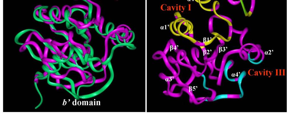
155 137
156 Figure 24 Determination of the E 2 -binding sites in PDIp using computational modeling. A. Protein sequence alignment of human PDI b-b fragment and human PDIp b-b fragment. Positions of α-helices and β-sheets are labeled by green arrows and blue arrows, respectively. α-helices and β-sheets in the b domain are numbered as α1 - α4 and β1 - β5. α-helices and β-sheets in the b domain are numbered as α1 - α4 and β1 - β5. B. Structural superimposition of PDI b-b' fragment (PDB: 2k18; green color) and PDIp b-b' fragment (magenta color). The structure of the PDIp b-b fragment is built using the homology modeling (Insight II) based on the template structure of the PDI b-b' fragment. Protein structures are shown in ribbons. C. The three potential E 2 -binding cavities identified in the PDIp b-b' fragment. Cavity I, cavity II, and cavity III are colored in yellow, green, and blue, respectively. α-helices and β-sheets are labeled as in panel A. 138
157 Next, we used the Affinity module of InsightII to dock E 2 into cavity I. Two candidate E 2 -binding modes are suggested (Figure 25D). In both binding modes, E 2 has a very similar overall orientation and positioning inside the binding pocket, and hydrogen bonds are formed between E 2 and PDIp b-b fragment. In mode I (Figure 25A, 25B, 25C, 25D), one hydrogen bond is formed between the 3-hydroxyl group of E 2 and the nitrogen atom of PDIp-His278. In mode II (Figure 25D), two hydrogen bonds are formed: one between the 3-hydroxyl group of E 2 and PDIp-His278 and the other one between the 17- hydroxyl group of E 2 and PDIp-Gln

158 140
159 Figure 25 Docking analysis of the binding mode of E 2 inside human PDIp b-b fragment. A. Overview of the docking result (mode I) of E 2 binding inside the PDIp b-b fragment. E 2 and H278 are shown in the ball-and-stick format and colored according to atoms. The protein structure is shown in ribbon. The yellow colored region denotes the b domain and magenta colored region denotes the b domain. B. A close-up view of the docking result of the E 2 -PDIp binding model. C. Interaction of E 2 with amino acid residues in the binding pocket (mode I). Labeling of amino acid residues is shown in yellow for the b domain and in magenta for the b domain. E 2 is colored in yellow. Amino acids are shown in the ball-and-stick format and colored according to atoms, i.e., green for carbon, red for oxygen, white for hydrogen, and blue for nitrogen. D. Plots of the docking results of E 2 binding with PDIp b-b fragment in mode I and II. The distance is in angstroms. E 2 is colored in blue and H278 and Q265 are in magenta. 141
160 To experimentally test the binding modes as suggested by our computational docking analysis, we first sought to determine whether the two identified amino acid residues, His278 and Gln265, are indeed involved in the binding interactions between PDIp and E 2 through forming hydrogen bonds. To do so, we introduced point mutations to probe whether changes in these two amino acid residues would alter PDIp s E 2 -binding activity. We first chose to mutate His278 to leucine. The side-chain length of leucine is similar to that of histidine but it does not contain electro-negative atoms that are necessary for the formation of hydrogen bonds. As shown in Figure 26A, the purified H278L b-b fragment completely lost [ 3 H]E 2 -binding activity. Saturation binding assay further confirmed that this mutant fragment did not have an appreciable [ 3 H]E 2 -binding activity even when very high concentrations of [ 3 H]E 2 were present (Figure 26B). Similarly, we also created the H278L mutation in the full-length PDIp, which was overexpressed in mammalian cells and isolated by SEC. Assay of the [ 3 H]E 2 binding activity of each of the SEC fractions showed that the PDIp H278L mutant protein does not have an appreciable [ 3 H]E 2 -binding activity (Figure 26C). Taken together, these data show that PDIp s His278 is indispensable for its binding interaction with E
161 143
162 Figure 26 The H278L mutant protein lacks E 2 -binding activity. A. Total [ 3 H]E 2 binding by purified PDIp b-b fragments (wild-type and H278L mutant proteins, purified from E. coli cells) after incubation with 4.5 nm [ 3 H]E 2 in the absence or presence of excess cold E 2 (10 μm) in 10 mm sodium phosphate buffer. Inset shows the SDS-PAGE analysis of the purified proteins. B. Total [ 3 H]E 2 binding by PDIp b-b fragments (at 20 μg/ml final concentration, wild-type protein and H278L mutant protein) was determined in the presence of increasing concentrations of [ 3 H]E 2 (26 to 420 nm) in 10 mm sodium phosphate buffer (ph 7.4). C. The total [ 3 H]E 2 binding by SEC fractions of cos-7 cell lysates containing the expressed full-length wild-type PDIp or the H278L mutant protein. The final concentration of [ 3 H]E 2 in these incubations was 4.5 nm. Each value is the mean of duplicate determinations. 144
163 Using the same approach, we also mutated Gln265 to leucine. As suggested by the binding mode II (Figure 25D), Gln265 may form a hydrogen bond with the 17- hydroxyl group of E 2. [ 3 H]E 2 binding assays showed that the purified Q265L b-b fragment (over-expressed in E. coli cells, Figure 27A) and the full-length PDIp Q265L mutant protein (over-expressed in cos-7 cells, Figure 27B) each displayed similar [ 3 H]E 2 -binding activity as the corresponding wild-type proteins. The apparent K d value for Q265L b-b fragment was found to be 185 ± 8 nm (Figure 27C), which is nearly the same as that of the wild-type b-b fragment. In addition, we have also prepared the double mutant protein Q265L/H278L for testing its [ 3 H]E 2 -binding activity. As expected, this double mutant protein does not have any [ 3 H]E 2 -binding activity. Collectively, these observations suggest that PDIp s Gln265 may either form a non-essential, weak hydrogen bond with the 17-hydroxyl group of E 2 (as suggested by the binding mode II, Figure 25D), or it may not form a viable hydrogen bond at all (as suggested by the binding mode I, Figure 25D). 145
164 146
165 Figure 27 PDIP Q265L mutant protein retains similar E 2 -binding activity. A. Total [ 3 H]E 2 binding by purified PDIp b-b' fragments (wild-type, Q265L, and Q265L/H278L mutant proteins, purified from E. coli cells) after incubation with 4.5 nm [ 3 H]E 2 in the absence or presence of excess cold E 2 (10 μm) in 10 mm sodium phosphate buffer. B. Total [ 3 H]E 2 binding by SEC fractions of cos-7 cell lysates containing the expressed fulllength PDIp proteins (wild-type, Q265L, or Q265L/H278L mutant), after incubation with 4.5 nm [ 3 H]E 2. C. Total [ 3 H]E 2 binding (TB) by PDIp b-b' Q265L fragment (at 20 μg/ml final concentration) was determined in the presence of increasing concentrations of [ 3 H]E 2 (22 to 660 nm) in 10 mm sodium phosphate buffer (ph 7.4). Non-specific binding (NSB) was determined in the presence of 10 μm excess of cold E 2. Total binding subtracted by the non-specific binding gives rise to specific binding (SB). The binding curve is obtained by curve regression analysis using a hyperbola model in the SigmaPlot software. Each data point is the mean of duplicate determinations. 147
166 To provide further experimental support for the conclusions of the mutagenesis studies, we employed an alternative approach by using a number of E 2 derivatives that share the same core structure as E 2 but with their C-3 or C-17 hydroxyl group selectively modified such that they cannot form the same type of hydrogen bonds with PDIp as does E 2. In these experiments, both purified b-b fragment (expressed in E. coli cells) and purified full-length recombinant human PDIp (expressed in cos-7 cells) were used to assay their relative binding activity by measuring their ability to compete off the binding of [ 3 H]E 2. We found that E 1, E 3, and C2, all of which contain an intact 3-hydroxyl group but differ in their 17-hydroxyl group (structures shown in Figure 28A), could efficiently compete with [ 3 H]E 2 for binding to the b-b fragment or the full-length PDIp (Figure 28B). There is no significant difference between the IC 50 values of E 2, E 1, and C2 (Figure 28C). In contrast, C3, which lacks the 3-hydroxyl group (Figure 28A), displayed no appreciable binding activity (Figure 28B). These observations provide further support for the suggestion that the 3-hydroxyl group of E 2 is essential for the binding interaction with PDIp by forming hydrogen bond(s), whereas the 17-hydroxyl group of E 2 is not important. These results are in complete agreement with the observations made with the point mutation studies described above. 148


167 149
168 Figure 28 Relative binding activity of PDIp for several E 2 derivatives. A. Chemical structures of E 2 and several of its analogs used in this study. B. Relative binding of [ 3 H]E 2 by recombinant PDIp b-b fragment (at 20 μg/ml final concentration, purified from E.coli cells) (see Figure 21B) or by purified recombinant full-length PDIp protein expressed in cos-7 cells (at 20 μg/ml final concentration, see Figure 23). Protein was incubated with 4.5 nm [ 3 H]E 2 in the absence or presence of 10 μm E 2 or its analogs in sodium phosphate buffer (10 mm, ph 7.4). C. Relative [ 3 H]E 2 binding by the PDIp b-b fragment purified from E. coli cells after incubation with 4.5 nm [ 3 H]E 2 in 10 mm sodium phosphate buffer (ph 7.4) in the absence (control, set as 100%) or presence of increasing concentrations of E 2, E 1, and C2. Each value is the mean ± S.D. of triplicate determinations. 150
169 Notably, whereas the two binding modes suggested by the docking studies are very similar in their overall structure and binding interaction, there are also some notable subtle differences. In mode I, the 3-hydroxyl group of E 2 is a hydrogen bond donor and the nitrogen atom of PDIp-His278 is a hydrogen bond acceptor. In mode 2, the 3- hydroxyl group of E 2 serves as a hydrogen bond acceptor and PDIp-His278 as a hydrogen bond donor in the formation of a hydrogen bond. Based on our earlier experience, it is almost certain that only one of the modes is the preferred binding mode. Based on the known difference in the strength of the OH N hydrogen bond (6.9 kcal/mol) vs. the O HN hydrogen bond (1.9 kcal/mol) (Emsley, 1980), it is predicted that binding mode I, which contains the stronger OH N hydrogen bond, would be the preferred binding mode over mode II, which contains the weaker O HN hydrogen bond. To provide experimental evidence for this prediction, we chose to determine the binding activity of 3-methoxyestradiol (structure shown in Figure 29A) for PDIp. Theoretically, the oxygen atom in the 3-methoxy group of 3-methoxyestradiol will only be able to serve as a hydrogen bond acceptor but not a hydrogen bond donor in forming a hydrogen bond (i.e., it can only bind to PDIp in mode I but not in mode II). Computational docking analysis showed that 3-methoxyestradiol can still bind inside the pocket in a similar way as does E 2, and there is enough space around His278 to fully accommodate the methoxy group (Figure 29B). As expected, the model showed that 3-methoxyestradiol (serving as a hydrogen bond acceptor) still can form a hydrogen bond with His278 (serving as a hydrogen bond donor). However, in the binding assay, we found that 3-methoxyestradiol has no appreciable binding activity for the b-b fragment or the full-length human PDIp 151
170 (Figure 29C). This experimental observation suggests that the hydrogen bond formed between 3-methoxyestradiol and His278 likely is either extremely weak, or, more likely, is not formed at all. Taken together, it is concluded that E 2 binds PDIp by forming a hydrogen bond between the 3-hydroxyl group of E 2 and PDIp-His278 according to binding mode I, but not binding mode II (Figure 25D). 152

171 153
172 Figure 29 Docking analysis of the interaction of PDIp with E 2 analog C1. A. Chemical structure of 3-methoxyestradiol (C1). B. Docking analysis of the binding modes of E 2 and C1 in the binding pocket of the PDIp b-b fragment. Protein structure is shown in ribbon and colored in magenta. E 2, C1, and His278 are shown in the ball-andstick format. C1 and His 278 are colored according to atoms and E 2 is colored yellow. Green dashes denote hydrogen bonds. α-helices and β-sheets are labeled as in Figure 24. C. Relative [ 3 H]E 2 binding by recombinant PDIp b-b fragment or by purified recombinant full-length PDIp protein in the absence or presence of 10 μm E 2 or its C1 analog. Each value is the mean ± S.D. of triplicate determinations. 154
173 Notably, it is well known that the 3-hydroxyl group of E 2 also plays an essential role in the binding interaction of E 2 with human ERα and ERβ by forming hydrogen bonds (Brzozowski et al., 1997; Gabbard and Segaloff, 1983; Zhu et al., 2006). The structural model developed in the present study offers a good explanation for the experimental observation that the human PDIp has a much lower E 2 -binding affinity than human ERs, for the following reasons: (i) PDIp forms only one hydrogen bond with the 3-hydroxyl group of E 2, whereas human ERs form two hydrogen bonds with this hydroxyl group. (ii) Whereas the 17β-hydroxyl group of E 2 also plays an important role in its interaction with human ERα or ERβ (Zhu et al., 2006), its role in the interaction with human PDIp appears to be of minimal importance or simply nonexistent. (iii) The E 2 -binding site of PDIp is significantly larger than those of human ERα and ERβ, i.e., it is less compact, which would suggest a relatively looser binding interaction due to less hydrophobic interactions between E 2 and PDIp. (iv) For both ERα and ERβ, almost all amino acid residues in their binding pockets, except those that form hydrogen bonds with E 2, are hydrophobic residues, which provide stronger interactions with the four aliphatic rings of E 2 compared to PDIp. However, in the case of PDIp, some polar residues (Thr253, Thr260, Thr266, Ser225, and Gln265 shown in Figure 25C, 25D) are also present in the binding pocket, which may reduce the hydrophobic interactions with E 2. These notable differences distinguish human ERs from PDIp in their binding interactions with E 2. The results of this study also provide a good explanation for our early observation 155
174 that while PDIp has no appreciable binding affinity for tamoxifen, PDIp still retains considerable binding affinity for ICI-172,780 (Fu and Zhu, 2009) (also see Figure 30B). It is known that tamoxifen does not have a phenolic OH group in its structure (see Figure 30A), and thus it cannot form the necessary hydrogen bond with His278 of PDIp. In comparison, ICI-172,780 has the same core steroid structure as E 2 (see Figure 30D). Our docking analysis showed that ICI-172,780 can still bind inside PDIp s binding pocket in a similar way as E 2 (Figure 30C, 30D), although its long side chain at the C-7 position slightly interferes with the binding, which is consistent with its lower binding affinity (Figure 30B). Lastly, it is of note that observations made in the present study may shed light on understanding the binding interaction of PDIp with its substrate peptides, which was shown to be inhibited by E 2 and its analogs (Klappa et al., 2001; Klappa et al., 1998). Interestingly, an earlier study reported that the specificity of the peptide substrates during interaction with PDIp is associated with the tyrosine and/or tryptophan residues in the peptide substrates (Ruddock et al., 2000). Because these two residues contain free phenolic hydroxyl and amino groups, respectively, it is tempting to suggest that the presence of these amino acid residues may serve as hydrogen bond donors in forming hydrogen bonds with PDIp-His278 during PDIp-peptide interactions, in a similar way as observed for the 3-hydroxyl phenolic group in E 2. This possibility merits further investigation. 156

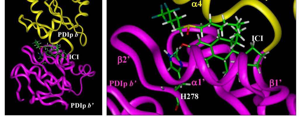
175 157
Design, Synthesis and Biological Screening of Focused Libraries of New Antiestrogens
 Design, Synthesis and Biological Screening of Focused Libraries of New Antiestrogens Presenter: Jelena Janjic, Dipl.Pharm. Graduate student Pharmaceutical Sciences Prof. Dr. Peter Wipf s group Dr. Billy
MECHANISM AND MODE OF HORMONE ACTION. Some definitions. Receptor: Properties of receptors. PRESENTED BY MBUNKUR GLORY NKOSI.
 MECHANISM AND MODE OF HORMONE ACTION. PRESENTED BY MBUNKUR GLORY NKOSI. OUTLINE. Introduction Some definitions Hormone secretion, transport, and clearance from the blood. Feedback control of hormone secretion.
INTERACTION DRUG BODY
 INTERACTION DRUG BODY What the drug does to the body What the body does to the drug Receptors - intracellular receptors - membrane receptors - Channel receptors - G protein-coupled receptors - Tyrosine-kinase
Cell Signaling (part 1)
") 15 Cell Signaling (part 1) Introduction Bacteria and unicellular eukaryotes respond to environmental signals and to signaling molecules secreted by other cells for mating and other communication. In multicellular
15 Cell Signaling (part 1) Introduction Bacteria and unicellular eukaryotes respond to environmental signals and to signaling molecules secreted by other cells for mating and other communication. In multicellular
Close to site of release (at synapse); binds to receptors in
; binds to receptors in") Chapter 18: The Endocrine System Chemical Messengers 1. Neural 2. Endocrine 3. Neuroendocrine 4. Paracrine 5. Autocrine Endocrine System --Endocrine and nervous systems work together --Endocrine vs. Nervous
Chapter 18: The Endocrine System Chemical Messengers 1. Neural 2. Endocrine 3. Neuroendocrine 4. Paracrine 5. Autocrine Endocrine System --Endocrine and nervous systems work together --Endocrine vs. Nervous
Cell Biology Lecture 9 Notes Basic Principles of cell signaling and GPCR system
 Cell Biology Lecture 9 Notes Basic Principles of cell signaling and GPCR system Basic Elements of cell signaling: Signal or signaling molecule (ligand, first messenger) o Small molecules (epinephrine,
HORMONES (Biomedical Importance)
") hormones HORMONES (Biomedical Importance) Hormones are the chemical messengers of the body. They are defined as organic substances secreted into blood stream to control the metabolic and biological activities.
hormones HORMONES (Biomedical Importance) Hormones are the chemical messengers of the body. They are defined as organic substances secreted into blood stream to control the metabolic and biological activities.
Statin inhibition of HMG-CoA reductase: a 3-dimensional view
 Atherosclerosis Supplements 4 (2003) 3/8 www.elsevier.com/locate/atherosclerosis Statin inhibition of HMG-CoA reductase: a 3-dimensional view Eva Istvan * Department of Molecular Microbiology, Howard Hughes
Receptors. Dr. Sanaa Bardaweel
 Receptors Types and Theories Dr. Sanaa Bardaweel Some terms in receptor-drug interactions Agonists: drugs that mimic the natural messengers and activate receptors. Antagonist: drugs that block receptors.
INTRODUCTION TO THE BIOCHEMISTRY OF HORMONES AND THEIR RECPTORS
 INTRODUCTION TO THE BIOCHEMISTRY OF HORMONES AND THEIR RECPTORS 1 Introduction to the Biochemistry of Hormones and their Receptors Lectuctre1 Sunday 17/2/ Objectives: 1. To understand the biochemical nature
Life History of A Drug
 DRUG ACTION & PHARMACODYNAMIC M. Imad Damaj, Ph.D. Associate Professor Pharmacology and Toxicology Smith 652B, 828-1676, mdamaj@hsc.vcu.edu Life History of A Drug Non-Specific Mechanims Drug-Receptor Interaction
This brings me to my point. Disease starts when there is a surplus or deficiency of certain nutritive or non-nutritive substances in the human body.
 WHAT IS DISEASE? A pathological condition of a part of an organ, or system of an organism resulting from various causes, such as infection, genetic defect, or environmental stress, and characterized by
About This Chapter. Hormones The classification of hormones Control of hormone release Hormone interactions Endocrine pathologies Hormone evolution
 About This Chapter Hormones The classification of hormones Control of hormone release Hormone interactions Endocrine pathologies Hormone evolution Hormones: Function Control Rates of enzymatic reactions
Endocrine secretion cells secrete substances into the extracellular fluid
 Animal Hormones Concept 30.1 Hormones Are Chemical Messengers Endocrine secretion cells secrete substances into the extracellular fluid Exocrine secretion cells secrete substances into a duct or a body
Chemical Classification of Hormones
 Steroid Hormones Chemical Classification of Hormones Hormones are chemical messengers that transport signals from one cell to another There are 4 major chemical classes of hormones steroid hormones - i.e.
Lecture 15. Membrane Proteins I
 Lecture 15 Membrane Proteins I Introduction What are membrane proteins and where do they exist? Proteins consist of three main classes which are classified as globular, fibrous and membrane proteins. A
Insulin mrna to Protein Kit
 Insulin mrna to Protein Kit A 3DMD Paper BioInformatics and Mini-Toober Folding Activity Student Handout www.3dmoleculardesigns.com Insulin mrna to Protein Kit Contents Becoming Familiar with the Data...
1st Year MB/BDS Plenary Lecture What is a Hormone?
 1st Year MB/BDS Plenary Lecture What is a Hormone? The term hormone (from the Greek for I arouse to activity or I excite) was first used by Starling in 1905. Hormones are just one type of first (or primary)
Chapter 3. Protein Structure and Function
 Chapter 3 Protein Structure and Function Broad functional classes So Proteins have structure and function... Fine! -Why do we care to know more???? Understanding functional architechture gives us POWER
Receptors Functions and Signal Transduction L1- L2
 Receptors Functions and Signal Transduction L1- L2 Faisal I. Mohammed, MD, PhD University of Jordan 1 Introduction to Physiology (0501110) Summer 2012 Subject Lecture No. Lecturer Pages in the 11 th edition.
Identification of influential proteins in the classical retinoic acid signaling pathway
 Ghaffari and Petzold Theoretical Biology and Medical Modelling (2018) 15:16 https://doi.org/10.1186/s12976-018-0088-7 RESEARCH Open Access Identification of influential proteins in the classical retinoic
The Endocrine System. I. Overview of the Endocrine System. II. Three Families of Hormones. III. Hormone Receptors. IV. Classes of Hormone Receptor
 The Endocrine System I. Overview of the Endocrine System A. Regulates long term metabolic processes B. Releases hormones from endocrine cells 1. Hormones are chemicals 2. Alter metabolism of cells 3. Release
By the name of Allah
 By the name of Allah Receptors function and signal transduction ( Hormones and receptors Types) We were talking about receptors of the neurotransmitters; we have 2 types of receptors: 1- Ionotropic receptors
G-Protein Signaling. Introduction to intracellular signaling. Dr. SARRAY Sameh, Ph.D
 G-Protein Signaling Introduction to intracellular signaling Dr. SARRAY Sameh, Ph.D Cell signaling Cells communicate via extracellular signaling molecules (Hormones, growth factors and neurotransmitters
Hormone. Free Androgen Index. 2-Hydroxyestrone. Reference Range. Hormone. Estrone Ratio. Free Androgen Index
 Hormonal Health PATIENT: Sample Report TEST REF: TST-12345 Hormonal Health 0.61 0.30-1.13 ng/ml DHEA-S 91 35-430 mcg/dl tient: SAMPLE TIENT e: x: N: Sex Binding Globulin 80 18-114 nmol/l Testosterone 0.34
Chapter 11: Enzyme Catalysis
 Chapter 11: Enzyme Catalysis Matching A) high B) deprotonated C) protonated D) least resistance E) motion F) rate-determining G) leaving group H) short peptides I) amino acid J) low K) coenzymes L) concerted
Protein sorting (endoplasmic reticulum) Dr. Diala Abu-Hsasan School of Medicine
 Dr. Diala Abu-Hsasan School of Medicine") Protein sorting (endoplasmic reticulum) Dr. Diala Abu-Hsasan School of Medicine dr.abuhassand@gmail.com An overview of cellular components Endoplasmic reticulum (ER) It is a network of membrane-enclosed
Protein sorting (endoplasmic reticulum) Dr. Diala Abu-Hsasan School of Medicine dr.abuhassand@gmail.com An overview of cellular components Endoplasmic reticulum (ER) It is a network of membrane-enclosed
Ch 11: Endocrine System
 Ch 11: Endocrine System SLOs Describe the chemical nature of hormones and define the terms proand prepro-hormone. Explain mechanism of action of steroid and thyroid hormones Create chart to distinguish
Intracellular Compartments and Protein Sorting
 Intracellular Compartments and Protein Sorting Intracellular Compartments A eukaryotic cell is elaborately subdivided into functionally distinct, membrane-enclosed compartments. Each compartment, or organelle,
Nuclear Receptors. Estrogen and Thyroid Hormone Receptors and their Interaction. Estrogen Hormone Receptor.
 SZENT ISTVÁN UNIVERSITY FACULTY OF VETERINARY MEDICINE Nuclear Receptors Estrogen and Thyroid Hormone Receptors and their Interaction Lior Kerner Budapest, 2012 What are Nuclear Receptors? o Proteins located
P450 CYCLE. All P450s follow the same catalytic cycle of;
 P450 CYCLE All P450s follow the same catalytic cycle of; 1. Initial substrate binding 2. First electron reduction 3. Oxygen binding 4. Second electron transfer 5 and 6. Proton transfer/dioxygen cleavage
Chapter 3. Structure of Enzymes. Enzyme Engineering
 Chapter 3. Structure of Enzymes Enzyme Engineering 3.1 Introduction With purified protein, Determining M r of the protein Determining composition of amino acids and the primary structure Determining the
PAPER No. : 16, Bioorganic and biophysical chemistry MODULE No. : 22, Mechanism of enzyme catalyst reaction (I) Chymotrypsin
 Chymotrypsin") Subject Paper No and Title 16 Bio-organic and Biophysical Module No and Title 22 Mechanism of Enzyme Catalyzed reactions I Module Tag CHE_P16_M22 Chymotrypsin TABLE OF CONTENTS 1. Learning outcomes 2.
Subject Paper No and Title 16 Bio-organic and Biophysical Module No and Title 22 Mechanism of Enzyme Catalyzed reactions I Module Tag CHE_P16_M22 Chymotrypsin TABLE OF CONTENTS 1. Learning outcomes 2.
Reproductive. Estradiol Analyte Information
 Reproductive Estradiol Analyte Information - 1 - Estradiol Introduction Estradiol (E2 or 17β-estradiol) is the major estrogen in humans. Although it is often called the "female" hormone, it is also present
RAS Genes. The ras superfamily of genes encodes small GTP binding proteins that are responsible for the regulation of many cellular processes.
 ۱ RAS Genes The ras superfamily of genes encodes small GTP binding proteins that are responsible for the regulation of many cellular processes. Oncogenic ras genes in human cells include H ras, N ras,
Zool 3200: Cell Biology Exam 4 Part I 2/3/15
 Name: Key Trask Zool 3200: Cell Biology Exam 4 Part I 2/3/15 Answer each of the following questions in the space provided, explaining your answers when asked to do so; circle the correct answer or answers
Synthesis of sex steroids
 Synthesis of sex steroids CH 3 NAD(P)H NAD(P)+ Dehidroepiandroszteron Androszténdion 17- -hidroxiszteroid dehidrogenáz CH 3 dehydroepiandrosterone Dehidroepiandroszteron (DHEA) 3SDH Koleszterin CH 3 aromatase
Effects of Second Messengers
 Effects of Second Messengers Inositol trisphosphate Diacylglycerol Opens Calcium Channels Binding to IP 3 -gated Channel Cooperative binding Activates Protein Kinase C is required Phosphorylation of many
Model Answer. M.Sc. Zoology (First Semester) Examination Paper LZT 103 (Endocrinology)
 Examination Paper LZT 103 (Endocrinology)") Model Answer M.Sc. Zoology (First Semester) Examination-2013 Paper LZT 103 (Endocrinology) Section A 1. (i) d (ii) b (iii) b (iv) c (v) c (vi) a (vii) c (viii) a (ix) d (x) b Section B Q.2 Answer Hormonal
Model Answer M.Sc. Zoology (First Semester) Examination-2013 Paper LZT 103 (Endocrinology) Section A 1. (i) d (ii) b (iii) b (iv) c (v) c (vi) a (vii) c (viii) a (ix) d (x) b Section B Q.2 Answer Hormonal
Receptors Functions and Signal Transduction- L4- L5
 Receptors Functions and Signal Transduction- L4- L5 Faisal I. Mohammed, MD, PhD University of Jordan 1 PKC Phosphorylates many substrates, can activate kinase pathway, gene regulation PLC- signaling pathway
Correlation between estrogen receptor β expression and the curative effect of endocrine therapy in breast cancer patients
 1568 Correlation between estrogen receptor β expression and the curative effect of endocrine therapy in breast cancer patients LIYING GUO 1, YU ZHANG 2, WEI ZHANG 3 and DILIMINA YILAMU 1 1 Department of
Membrane associated receptor transfers the information. Second messengers relay information
 Membrane associated receptor transfers the information Most signals are polar and large Few of the signals are nonpolar Receptors are intrinsic membrane proteins Extracellular and intracellular domains
2013 W. H. Freeman and Company. 12 Signal Transduction
 2013 W. H. Freeman and Company 12 Signal Transduction CHAPTER 12 Signal Transduction Key topics: General features of signal transduction Structure and function of G protein coupled receptors Structure
Receptors Families. Assistant Prof. Dr. Najlaa Saadi PhD Pharmacology Faculty of Pharmacy University of Philadelphia
 Receptors Families Assistant Prof. Dr. Najlaa Saadi PhD Pharmacology Faculty of Pharmacy University of Philadelphia Receptor Families 1. Ligand-gated ion channels 2. G protein coupled receptors 3. Enzyme-linked
LQB383 Testbank. Week 8 Cell Communication and Signaling Mechanisms
 LQB383 Testbank Week 8 Cell Communication and Signaling Mechanisms Terms to learn match the terms to the definitions --------------------------------------------------------------------------------------------------------------------------
BIOLOGY - CLUTCH CH.45 - ENDOCRINE SYSTEM.
 !! www.clutchprep.com Chemical signals allow cells to communicate with each other Pheromones chemical signals released to the environment to communicate with other organisms Autocrine signaling self-signaling,
Signal Transduction Cascades
 Signal Transduction Cascades Contents of this page: Kinases & phosphatases Protein Kinase A (camp-dependent protein kinase) G-protein signal cascade Structure of G-proteins Small GTP-binding proteins,
Part-4. Cell cycle regulatory protein 5 (Cdk5) A novel target of ERK in Carb induced cell death
 A novel target of ERK in Carb induced cell death") Part-4 Cell cycle regulatory protein 5 (Cdk5) A novel target of ERK in Carb induced cell death 95 1. Introduction The process of replicating DNA and dividing cells can be described as a series of coordinated
Part-4 Cell cycle regulatory protein 5 (Cdk5) A novel target of ERK in Carb induced cell death 95 1. Introduction The process of replicating DNA and dividing cells can be described as a series of coordinated
Practice Exam 2 MCBII
 1. Which feature is true for signal sequences and for stop transfer transmembrane domains (4 pts)? A. They are both 20 hydrophobic amino acids long. B. They are both found at the N-terminus of the protein.
Estrogens and progestogens
 Estrogens and progestogens Estradiol and Progesterone hormones produced by the gonads are necessary for: conception embryonic maturation development of primary and secondary sexual characteristics at puberty.
Drug Receptor Interactions and Pharmacodynamics
 Drug Receptor Interactions and Pharmacodynamics Dr. Raz Mohammed MSc Pharmacology School of Pharmacy 22.10.2017 Lec 6 Pharmacodynamics definition Pharmacodynamics describes the actions of a drug on the
Chapter 9. Cellular Signaling
 Chapter 9 Cellular Signaling Cellular Messaging Page 215 Cells can signal to each other and interpret the signals they receive from other cells and the environment Signals are most often chemicals The
Chapter 20. Cell - Cell Signaling: Hormones and Receptors. Three general types of extracellular signaling. endocrine signaling. paracrine signaling
 Chapter 20 Cell - Cell Signaling: Hormones and Receptors Three general types of extracellular signaling endocrine signaling paracrine signaling autocrine signaling Endocrine Signaling - signaling molecules
Chapter 16: Endocrine System 1
 Ch 16 Endocrine System Bi 233 Endocrine system Endocrine System: Overview Body s second great controlling system Influences metabolic activities of cells by means of hormones Slow signaling Endocrine glands
Chapter 10. Regulatory Strategy
 Chapter 10 Regulatory Strategy Regulation of enzymatic activity: 1. Allosteric Control. Allosteric proteins have a regulatory site(s) and multiple functional sites Activity of proteins is regulated by
MODULE No.26: Drug Metabolism
 SUBJECT Paper No. and Title Module No. and Title Module Tag PAPER No. 9: Drugs of Abuse MODULE No. 26: Drug Metabolism FSC_P9_M26 TABLE OF CONTENTS 1. Learning Outcomes 2. Introduction 3. Sites of Drug
CS612 - Algorithms in Bioinformatics
 Spring 2016 Protein Structure February 7, 2016 Introduction to Protein Structure A protein is a linear chain of organic molecular building blocks called amino acids. Introduction to Protein Structure Amine
Molecular Graphics Perspective of Protein Structure and Function
 Molecular Graphics Perspective of Protein Structure and Function VMD Highlights > 20,000 registered Users Platforms: Unix (16 builds) Windows MacOS X Display of large biomolecules and simulation trajectories
H C. C α. Proteins perform a vast array of biological function including: Side chain
 Topics The topics: basic concepts of molecular biology elements on Python overview of the field biological databases and database searching sequence alignments phylogenetic trees microarray data analysis
Chapter 9 - Biological Membranes. Membranes form a semi-permeable boundary between a cell and its environment.
 Chapter 9 - Biological Membranes www.gsbs.utmb.edu/ microbook/ch037.htmmycoplasma Membranes form a semi-permeable boundary between a cell and its environment. Membranes also permit subcellular organization
Lecture 9: Cell Communication I
 02.05.10 Lecture 9: Cell Communication I Multicellular organisms need to coordinate cellular functions in different tissues Cell-to-cell communication is also used by single celled organisms to signal
MCB II MCDB 3451 Exam 1 Spring, minutes, close everything and be concise!
 MCB II MCDB 3451 Exam 1 Spring, 2016 50 minutes, close everything and be concise! Name ID NOTE: QUESTIONS ARE NOT ALL WORTH THE SAME POINTS Total (100) Grade EXAM 1, 2016 MCBII Name 1. Which is UNIQUE
Chapter 17: Functional Organization of the Endocrine System
 Chapter 17: Functional Organization of the Endocrine System AP2 Chapter 17 Pg 586 1 Chapter 17 Outline I. General Characteristics of the Endocrine System II. Chemical structure of hormones III. Control
SAMPLE REPORT. Order Number: PATIENT. Age: 40 Sex: F MRN:
 Patient: Age: 40 Sex: F MRN: SAMPLE PATIENT Order Number: Completed: Received: Collected: SAMPLE REPORT Progesterone ng/ml 0.34 0.95 21.00 DHEA-S mcg/dl Testosterone ng/ml 48 35 0.10 0.54 0.80 430 Sex
Chapter 6 Communication, Integration, and Homeostasis
 Chapter 6 Communication, Integration, and Homeostasis About This Chapter Cell-to-cell communication Signal pathways Novel signal molecules Modulation of signal pathways Homeostatic reflex pathways Cell-to-Cell
Receptors Functions and Signal Transduction L1- L2
 Receptors Functions and Signal Transduction L1- L2 Faisal I. Mohammed, MD, PhD University of Jordan 1 Introduction to Physiology (0501110) Spring 2013 Subject Receptors: types and adaptation - Membrane
Chapter 17: Functional Organization of the Endocrine System
 Chapter 17: Functional Organization of the Endocrine System I. General Characteristics of the Endocrine System A. Terminology 1. What does the term endocrine imply? 2. Endocrine glands secrete 3. A hormone
Porphyrins: Chemistry and Biology
 Porphyrins: Chemistry and Biology 20.109 Lecture 6 24 February, 2011 Goals Explore some essential roles of heme in biology Appreciate how ature has used the same cofactor to achieve diverse functions Gain
Principles of Genetics and Molecular Biology
 Cell signaling Dr. Diala Abu-Hassan, DDS, PhD School of Medicine Dr.abuhassand@gmail.com Principles of Genetics and Molecular Biology www.cs.montana.edu Modes of cell signaling Direct interaction of a
Enzyme-coupled Receptors. Cell-surface receptors 1. Ion-channel-coupled receptors 2. G-protein-coupled receptors 3. Enzyme-coupled receptors
 Enzyme-coupled Receptors Cell-surface receptors 1. Ion-channel-coupled receptors 2. G-protein-coupled receptors 3. Enzyme-coupled receptors Cell-surface receptors allow a flow of ions across the plasma
Molecular understanding of tamoxifen resistance in breast cancer. Renée de Leeuw
 Molecular understanding of tamoxifen resistance in breast cancer Renée de Leeuw Omslag ontworpen door: Theophile Suijkerbuijk (www.theophile.nl) Molecular understanding of tamoxifen resistance in breast
Chp. 17 FUNCTIONAL ORG. Char.of the Endocrine System
 Chp. 17 FUNCTIONAL ORG. Char.of the Endocrine System Glands that secrete chemical signals (hormones) into circulatory system Hormone characteristics Produced in small quantities Secreted into intercellular
Crosstalk between Adiponectin and IGF-IR in breast cancer. Prof. Young Jin Suh Department of Surgery The Catholic University of Korea
 Crosstalk between Adiponectin and IGF-IR in breast cancer Prof. Young Jin Suh Department of Surgery The Catholic University of Korea Obesity Chronic, multifactorial disorder Hypertrophy and hyperplasia
18. PANCREATIC FUNCTION AND METABOLISM. Pancreatic secretions ISLETS OF LANGERHANS. Insulin
 18. PANCREATIC FUNCTION AND METABOLISM ISLETS OF LANGERHANS Some pancreatic functions have already been discussed in the digestion section. In this one, the emphasis will be placed on the endocrine function
(B D) Three views of the final refined 2Fo-Fc electron density map of the Vpr (red)-ung2 (green) interacting region, contoured at 1.4σ.
 Three views of the final refined 2Fo-Fc electron density map of the Vpr (red)-ung2 (green) interacting region, contoured at 1.4σ.") Supplementary Figure 1 Overall structure of the DDB1 DCAF1 Vpr UNG2 complex. (A) The final refined 2Fo-Fc electron density map, contoured at 1.4σ of Vpr, illustrating well-defined side chains. (B D) Three
Supplementary Figure 1 Overall structure of the DDB1 DCAF1 Vpr UNG2 complex. (A) The final refined 2Fo-Fc electron density map, contoured at 1.4σ of Vpr, illustrating well-defined side chains. (B D) Three
Molecular Cell Biology - Problem Drill 19: Cell Signaling Pathways and Gene Expression
 Molecular Cell Biology - Problem Drill 19: Cell Signaling Pathways and Gene Expression Question No. 1 of 10 1. Which statement about cell signaling is correct? Question #1 (A) Cell signaling involves receiving
BIOH111. o Cell Module o Tissue Module o Skeletal system o Muscle system o Nervous system o Endocrine system o Integumentary system
 BIOH111 o Cell Module o Tissue Module o Skeletal system o Muscle system o Nervous system o Endocrine system o Integumentary system Endeavour College of Natural Health endeavour.edu.au 1 Textbook and required/recommended
Reproductive Endocrinology
 Reproductive Endocrinology Reproductive Endocrinology Hypothalamic hormones Gonadotropin releasing hormone (GnRH) - stimulate release of FSH = follicle stimulating hormone LH = luteinizing hormone from
SCHOOL OF MEDICINE AND HEALTH SCIENCES DIVISION OF BASIC MEDICAL SCIENCES DISCIPLINE OF BIOCHEMISTRY & MOLECULAR BIOLOGY
 1 SCHOOL OF MEDICINE AND HEALTH SCIENCES DIVISION OF BASIC MEDICAL SCIENCES DISCIPLINE OF BIOCHEMISTRY & MOLECULAR BIOLOGY PBL SEMINAR: SEX HORMONES PART 1 An Overview What are steroid hormones? Steroid
5. Summary of Data Reported and Evaluation
 168 IARC MONOGRAPHS VOLUME 91 5. Summary of Data Reported and Evaluation 5.1 Exposure data The first oral hormonal contraceptives that were found to inhibit both ovulation and implantation were developed
The Endocrine System. The Endocrine System
 The Endocrine System Like nervous system, endocrine system provides communication and control. Messages are relayed from one cell to another via chemical messengers (hormones). Unlike nervous system which
Chemistry 106: Drugs in Society Lecture 19: How do Drugs Elicit an Effect? Interactions between Drugs and Macromolecular Targets 11/02/17
 Chemistry 106: Drugs in Society Lecture 19: How do Drugs Elicit an Effect? Interactions between Drugs and Macromolecular Targets 11/02/17 Targets for Therapeutic Intervention: A Comparison of Enzymes to
Chapter 41. Lecture 14. Animal Hormones. Dr. Chris Faulkes
 Chapter 41 Lecture 14 Animal Hormones Dr. Chris Faulkes Animal Hormones Aims: To appreciate the variety and roles of hormones in the body To understand the basic types of hormones To understand how hormones
Estrogens & Antiestrogens
 Estrogens & Antiestrogens Menstrual cycle... Changes and hormonal events Natural estrogens: Estadiol >> Estrone > Estriol Ineffective orally Synthesis: From cholesterol ; role of aromatase enzyme in converting
Cell Signaling part 2
 15 Cell Signaling part 2 Functions of Cell Surface Receptors Other cell surface receptors are directly linked to intracellular enzymes. The largest family of these is the receptor protein tyrosine kinases,
GENERAL CHARACTERISTICS OF THE ENDOCRINE SYSTEM FIGURE 17.1
 GENERAL CHARACTERISTICS OF THE ENDOCRINE SYSTEM FIGURE 17.1 1. The endocrine system consists of glands that secrete chemical signals, called hormones, into the blood. In addition, other organs and cells
Sheet #5 Dr. Mamoun Ahram 8/7/2014
 P a g e 1 Protein Structure Quick revision - Levels of protein structure: primary, secondary, tertiary & quaternary. - Primary structure is the sequence of amino acids residues. It determines the other
number Done by Corrected by Doctor
 number 9 Done by Nazek Hyasat Corrected by Bahaa Najjar & mohammed AL-shrouf Doctor Alia Shatnawi HOW DO DRUGS WORK??? You know that receptors are targeted by drugs, the question now is how do these drugs
Lipids and Membranes
 Lipids and Membranes Presented by Dr. Mohammad Saadeh The requirements for the Pharmaceutical Biochemistry I Philadelphia University Faculty of pharmacy Biological membranes are composed of lipid bilayers
PHRM20001 NOTES PART 1 Lecture 1 History of Pharmacology- Key Principles
 PHRM20001 NOTES PART 1 Lecture 1 History of Pharmacology- Key Principles Hippocrates (5 th century BCE):... benefit my patients according to my greatest ability and judgment, and I will do no harm or injustice
Receptor mediated Signal Transduction
 Receptor mediated Signal Transduction G-protein-linked receptors adenylyl cyclase camp PKA Organization of receptor protein-tyrosine kinases From G.M. Cooper, The Cell. A molecular approach, 2004, third
Protein structure. Dr. Mamoun Ahram Summer semester,
 Protein structure Dr. Mamoun Ahram Summer semester, 2017-2018 Overview of proteins Proteins have different structures and some have repeating inner structures, other do not. A protein may have gazillion
Estrogen (ER) receptor biology: Implications for HRT? Dr. Christoph A. Meier Endocrine Unit University Hospital Geneva
 receptor biology: Implications for HRT? Dr. Christoph A. Meier Endocrine Unit University Hospital Geneva") Estrogen (ER) receptor biology: Implications for HRT? Dr. Christoph A. Meier Endocrine Unit University Hospital Geneva Estrogens prevent postmenopausal bone-loss HRT & osteoporosis bip & estrogen bip
Estrogen (ER) receptor biology: Implications for HRT? Dr. Christoph A. Meier Endocrine Unit University Hospital Geneva Estrogens prevent postmenopausal bone-loss HRT & osteoporosis bip & estrogen bip
Chapter 20 Endocrine System
 Chapter 20 Endocrine System The endocrine system consists of glands and tissues that secrete Hormones are chemicals that affect other glands or tissues, many times far away from the site of hormone production
Biochemistry 673 Lecture 2 Jason Kahn, UMCP Introduction to steroid hormone receptor (nuclear receptor) signalling
 signalling") Biochemistry 673 Lecture 2 Jason Kahn, UMCP Introduction to steroid hormone receptor (nuclear receptor) signalling Resources: Latchman Lodish chapter 10, 20 Helmreich, chapter 11 http://www.nursa.org,
Biochemistry 673 Lecture 2 Jason Kahn, UMCP Introduction to steroid hormone receptor (nuclear receptor) signalling Resources: Latchman Lodish chapter 10, 20 Helmreich, chapter 11 http://www.nursa.org,
Cellular Physiology (PHSI3009) Contents:
 Contents:") Cellular Physiology (PHSI3009) Contents: Cell membranes and communication 2 nd messenger systems G-coupled protein signalling Calcium signalling Small G-protein signalling o RAS o MAPK o PI3K RHO GTPases
Cellular Physiology (PHSI3009) Contents: Cell membranes and communication 2 nd messenger systems G-coupled protein signalling Calcium signalling Small G-protein signalling o RAS o MAPK o PI3K RHO GTPases
VII SYNOPSIS. Cancer is a disease, causes due to failure of control of growth and. cell proliferation mechanisms. There are genetic control systems,
 VII SYPSIS Cancer is a disease, causes due to failure of control of growth and cell proliferation mechanisms. There are genetic control systems, regulate the balance between cell birth and death in response
Supplementary Figure 1 (previous page). EM analysis of full-length GCGR. (a) Exemplary tilt pair images of the GCGR mab23 complex acquired for Random
. EM analysis of full-length GCGR. (a) Exemplary tilt pair images of the GCGR mab23 complex acquired for Random") S1 Supplementary Figure 1 (previous page). EM analysis of full-length GCGR. (a) Exemplary tilt pair images of the GCGR mab23 complex acquired for Random Conical Tilt (RCT) reconstruction (left: -50,right:
S1 Supplementary Figure 1 (previous page). EM analysis of full-length GCGR. (a) Exemplary tilt pair images of the GCGR mab23 complex acquired for Random Conical Tilt (RCT) reconstruction (left: -50,right:
The T cell receptor for MHC-associated peptide antigens
 1 The T cell receptor for MHC-associated peptide antigens T lymphocytes have a dual specificity: they recognize polymporphic residues of self MHC molecules, and they also recognize residues of peptide