Exploring the Mechanisms by which Increasing Dietary Protein Induces Iron Transporter Expression and Improves Intestinal Iron Absorption
|
|
|
- Beverley Carson
- 6 years ago
- Views:
Transcription

1 University of Connecticut Doctoral Dissertations University of Connecticut Graduate School Exploring the Mechanisms by which Increasing Dietary Protein Induces Iron Transporter Expression and Improves Intestinal Iron Absorption Carrie Elizabeth Thomas Follow this and additional works at: Recommended Citation Thomas, Carrie Elizabeth, "Exploring the Mechanisms by which Increasing Dietary Protein Induces Iron Transporter Expression and Improves Intestinal Iron Absorption" (2013). Doctoral Dissertations
2 Exploring the Mechanisms by which Increasing Dietary Protein Induces Iron Transporter Expression and Improves Intestinal Iron Absorption Carrie Elizabeth Thomas, Ph.D., R.D. University of Connecticut 2013 Iron deficiency is the most common nutritional deficiency worldwide. Whole body iron balance is primarily controlled through the regulation of intestinal iron absorption. Therefore, the identification of factors that affect iron absorption is important for the development of therapies aimed at improving iron status in deficient individuals. We recently found that increasing dietary protein in rats enhances duodenal iron absorption. This protein-induced increase in iron absorption is associated with augmented transcript levels of key intestinal iron transporters, including the iron importer, DMT1. To determine whether dietary protein directly affects DMT1 expression in vitro, the effect of amino acid supplementation on DMT1 transcript expression and promoter activity was evaluated in Caco-2 cells. Amino acids significantly augmented DMT1 transcript expression by 1.8-fold (P = 0.004) and DMT1 promoter activity by 1.6-fold (P < ) compared to control cells. Since hepcidin is a major regulator of iron absorption and iron transporter expression, the impact of increasing dietary protein/amino acids on hepatic hepcidin expression was evaluated in vivo in rats and in vitro in primary rat hepatocytes. In rats
3 Carrie Elizabeth Thomas University of Connecticut, 2013 consuming a high protein diet, hepatic hepcidin expression was suppressed by 44% compared to those on a medium protein diet (P = ). There was no direct effect of amino acids on hepcidin transcript expression in primary hepatocytes. Finally, to determine whether the effect of dietary protein on iron absorption is relevant to humans, iron status of postmenopausal women who recently completed a double-blind placebo controlled dietary protein supplementation trial was examined. Acutely increasing dietary protein had no significant effect on serum iron status parameters including serum ferritin, hepcidin, and % transferrin saturation. In conclusion, increasing dietary protein in the short-term in rats significantly enhances intestinal iron absorption, which is due at least in part to enhanced duodenal DMT1 transcription and reduced hepatic hepcidin expression. However, acutely increasing protein intake has no significant impact on iron status in iron-replete postmenopausal women. Future work is needed to determine the mechanisms by which increasing dietary protein/amino acids affect duodenal DMT1 transcription and hepatic hepcidin expression and its relevance to the treatment of iron deficiency in humans. ii
4 Exploring the Mechanisms by which Increasing Dietary Protein Induces Iron Transporter Expression and Improves Intestinal Iron Absorption Carrie Elizabeth Thomas B.S., University of Connecticut, 2008 M.S., University of Connecticut, 2010 A Dissertation Submitted in Fulfillment of the Requirements of the Degree of Doctor of Philosophy at the University of Connecticut 2013 i
5 Copyright by Carrie Elizabeth Thomas 2013 ii
6
7 Acknowledgments We are thankful for the support of our funding agencies. This research was funded by a HATCH grant from the College of Agriculture and Natural Resources at the University of Connecticut, a grant from the Diet and Health Initiative at the University of Connecticut, and the Yale Bone Center. This work was very much a collaborative effort that required the assistance and expertise of many people. I am first thankful to my associate advisor, Dr. Hedley Freake, for his support and valuable feedback during the course of my graduate career. His passion for learning and teaching is contagious and helped reinforce my love for education. I am also thankful to Dr. Kimberly O Brien from Cornell University, whose expertise in iron metabolism and assistance with the iron absorption studies were crucial to the success of this project. The vast majority of the work detailed in this dissertation was conducted in close collaboration with my associate advisor, Dr. Karl Insogna. Under Dr. Insogna s direction, I was fortunate to learn and become proficient in many basic laboratory research techniques that I had previously never heard of. From day one, Dr. Insogna was motivating and enthusiastic about research and learning, and instilled in me an inquisitive nature that will help me in all aspects of my life. Most of all, Dr. Insogna taught me by example how to be a wonderful teacher and mentor and gave me the tools necessary to become a great scientist. I am also extremely thankful to my major advisor, Dr. Jane Kerstetter, for providing me with the opportunity to continue my education at UCONN and to attain a Ph.D. degree under her guidance. Since I was an undergraduate student in the iv
8 Dietetics program, I admired Jane s passion for teaching and the seemingly effortless way in which she taught others. Her love for research and discovery along with her love of teaching rubbed off on me, and as a result, I can t imagine doing anything else. Finally, I thank my parents, my husband Garrett, and all of my wonderful friends and family. It would have been very difficult for me to attain this goal without their constant unconditional love and encouragement. I thank them for continuing to support my aspirations and dreams. v
9 Table of Contents Chapter 1: Introduction... 1 Chapter 2: The Physiology of Iron Metabolism 3 Chapter 3: The Effect of Dietary Components on Iron Absorption: a Review of the Literature.. 15 Chapter 4: Increasing Dietary Protein Acutely Augments Intestinal Iron Transporter Expression and Significantly Increases Iron Absorption in Rats...36 Chapter 5: Acutely Increasing Dietary Protein Does Not Impact Iron Status in Iron-Replete Postmenopausal Women Chapter 6: Amino Acids Directly Augment Duodenal DMT1 Expression and DMT1 Promoter Activity In Vitro Chapter 7: Increasing Dietary Protein Suppresses Hepatic Hepcidin Expression In Vivo Chapter 9: Conclusions and Future Study Directions Chapter 9: Significance vi
10 References. 109 vii
11 Chapter 1 Introduction The central hypothesis of this work is that dietary protein regulates the expression of genes vital to iron absorption and metabolism. In particular, this thesis focuses on the effect of increasing dietary protein on the expression of DMT1, a critically important enterocyte iron importer, and hepcidin, the principal regulator of iron absorption. Chapter 2 is a review article that provides an overview of the physiology of iron metabolism. Particular attention is given to the molecular pathways central to iron absorption as well as the regulatory mechanisms controlling iron absorption and metabolism. Chapter 3 reviews our current understanding of nutrient effects on intestinal iron absorption with a focus on dietary protein. This article summarizes both early and recent literature examining the relationships between dietary factors and iron bioavailability and absorption. Underlying molecular mechanisms that may explain these effects are also discussed. Chapter 4 details the preliminary data that led to the present work, which adds to the growing literature addressing whether dietary protein affects intestinal iron absorption. This study, recently published in Faseb J, demonstrates that increasing dietary protein in rats significantly augments iron absorption and the expression of duodenal iron transporters. 1
12 Chapter 5 is new work that examines the impact of a short-term high protein diet on iron status in humans. Overall, no statistically significant association between increasing dietary protein and iron status was observed. The experiments described in Chapter 6 were designed to determine whether amino acids directly affect duodenal DMT1 expression. Exposing Caco-2 cells to amino acids significantly augmented DMT1 mrna levels, due at least in part to an effect on gene transcription. The in vivo and in vitro experiments presented in Chapter 7 sought to determine if dietary protein/amino acids impact hepatic hepcidin expression. In rats, increasing protein intake from a medium to a high level significantly suppressed hepcidin mrna expression. Interestingly, this was associated with alterations in expression of genes involved in the regulation of hepcidin transcription. However, there was no direct effect of amino acids on hepcidin expression in primary rat hepatocytes. Chapter 8 summarizes the experiments detailed in Chapters 5, 6, and 7, and provides possible direction for future research. Chapter 9 discusses the significance and public health implications of the current work. 2
13 Chapter 2 The Physiology of Iron Absorption & Metabolism Introduction Iron is a trace mineral essential for a variety of biological processes. It functions as a key component of hemoglobin, myoglobin, cytochromes, and countless enzymes. Iron exists in several oxidation states, the principal ones being ferrous (Fe 2+ ) and ferric (Fe 3+ ), and has the ability to form bonds with electronegative elements such as oxygen, nitrogen, and sulfur. 1 Consequently, iron has wide-ranging physiologic roles. As an example, the enzyme aconitase, which contains an iron-sulfur cluster, plays an important role in the tricarboxylic acid cycle, a series of chemical reactions used to generate energy from carbohydrates, fats and proteins. 2 Heme, which is a component of the metalloproteins hemoglobin and myoglobin, is another vital iron-containing compound that consists of an iron ion held in a porphyrin ring. The iron within the heme moiety of hemoglobin binds and transports oxygen in erythrocytes, while the heme iron of myoglobin carries oxygen in muscle. 2 Although iron plays a critically important role in a multitude of cellular processes, in excess it is potentially toxic and may cause significant organ damage through the generation of reactive oxygen species. 3 Therefore, the maintenance of iron balance is important for ensuring the availability of sufficient iron for crucial biological processes but also for the prevention of iron toxicity and iron-induced oxidative stress. The purpose of this chapter is to review the pathways involved in iron absorption and metabolism and to describe the major regulatory mechanisms that control these processes. 3
14 Iron homeostasis Iron homeostasis, or the maintenance of iron stores and extracellular iron concentrations, is mainly accomplished through the regulation of dietary iron absorption and iron release from storage pools. Extracellular iron concentrations are largely dependent on the latter, while whole body iron balance is achieved through alterations in intestinal iron absorption. Dietary iron is primarily absorbed in the proximal small intestine, where ~1-2 mg, or roughly 10% of the iron that passes through the GI tract daily, is taken up by duodenal enterocytes. 4 This absorption is balanced by loss of a similar amount of iron through blood loss and the sloughing of skin and mucosal cells. Once dietary iron is absorbed, it is transferred to the circulation and bound to transferrin, which transports it to tissues for storage or utilization. Total body iron ranges between 3-5 g. The majority is found in the erythroid bone marrow and in hemoglobin mature erythrocytes contained within the heme of hemoglobin. Iron used for erythropoiesis is predominantly supplied by reticuloendothelial macrophages, which store and recycle iron from senescent red blood cells (RBCs). From g of total body iron is stored in hepatocytes. 5 Dietary iron absorption is influenced by body iron requirements. For example, states of iron deficiency and excess enhance and reduce duodenal iron uptake, respectively. 6 In iron deficiency, absorption is augmented to 2-4 mg and in iron overload it is reduced to 0.5 mg. 7 Additionally, erythropoietic drive has been shown to augment intestinal iron absorption in order to provide adequate iron for new RBC synthesis. 8 4
15 A particularly striking example of a situation in which iron absorption is altered in response to increasing iron needs is pregnancy. In the 3 rd trimester of gestation, iron requirements increase to ~7-10 mg/d compared to 1 mg/d in the beginning of the 2 nd trimester in an effort to sustain the increase in RBC mass and fetal growth that occur at this time. 9 To compensate for the dramatic rise in iron requirements and maternal-fetal iron transfer that occurs in late pregnancy, maternal iron absorption is concomitantly enhanced in the 2 nd and 3 rd trimesters. 9 While the absorption of iron can be modified in response to changing requirements, there is no efficient physiologically regulated excretion mechanism for iron. For this reason, the regulation of intestinal iron absorption is critical for maintaining appropriate iron balance and for optimizing iron status. Enterocyte iron absorption Dietary iron exists in two forms: heme iron and nonheme iron. Nonheme iron, or inorganic iron, represents the majority (~⅔) of the iron that humans consume. 10 Nonheme iron is present in vegetables and meat, while heme iron, derived from hemoglobin and myoglobin, is found exclusively in animal meat and is more bioavailable than nonheme iron. Both heme and nonheme iron are absorbed by duodenal enterocytes, however, the degree to which they share similar pathways and transport proteins in this process, and whether the same regulatory mechanisms are in place to control their export, is largely unknown. The mechanism of intestinal nonheme iron absorption has been well characterized and involves the coordinated action of several enterocyte iron 5
16 transporters. Duodenal nonheme iron uptake begins with the reduction of Fe 3+ to Fe 2+ at the apical membrane of enterocytes by the brush border ferrireductase, duodenal cytochrome b (Dcytb). 11 Once in the ferrous form, the iron is imported into the cell by the apical transmembrane iron transporter, divalent metal transporter 1 (DMT1). 12,13 In the enterocyte, the absorbed iron can be stored in the cell as ferritin, used for various cellular processes, or exported into the circulation by the basolateral iron exporter, ferroportin 1 (FPN1). 14 However, at this time the process by which inorganic iron is transported through the enterocyte is unknown. Metallochaperones are known to exist for many minerals but a specific iron trafficking protein remains uncharacterized. In order for iron to bind to apo-transferrin in the circulation, the iron must be re-oxidized from the Fe 2+ to the Fe 3+ form, which is accomplished by the basolateral membranebound enterocyte ferroxidase, hephaestin (HEPH), or the circulating ferroxidase, ceruloplasmin (CP). 15 In the bloodstream, transferrin, which can bind up to two iron ions, transports iron to tissues expressing the transferrin receptor (TfR), which take up the transferrin-iron complex via receptor-mediated endocytosis. 16 In the endosome, iron is released from transferrin and TfR by acidification. The free iron is then transported across the endosomal membrane by DMT1, and is either utilized by the cell for the synthesis of iron-containing proteins, or stored in ferritin. Although heme iron absorption has been studied for decades, its absorption pathway is less well understood than that of nonheme iron. In 2008, West and Oates summarized the current hypotheses and controversies regarding heme iron absorption and described two predominant hypotheses in detail. 10 The first postulates that heme is taken up by the enterocyte via receptor-mediated endocytosis and is catabolized within 6
17 the cell in internalized vesicles. Following catabolism, the heme-derived inorganic iron may then join the labile iron pool and undergo similar fates as absorbed nonheme iron (i.e., storage in ferritin, utilization by the cell, or efflux into the circulation via FPN1). This hypothesis is supported by two separate studies, which both utilized mammalian closed duodenal loops. 17,18 In these experiments, the investigators instilled heme or hemoglobin in the intestinal loops, and evaluated the appearance of heme in the duodenal mucosa over time. Both studies showed initial heme localization at the microvillous membrane, followed by its presence within tubulovesicular structures in the apical cytoplasm, and finally in secondary lysosomes. The second hypothesis suggests that heme iron is transported into and through the enterocyte intact by membrane-bound heme import and export proteins. In this model, heme iron is first imported into the cell by a brush border membrane heme importer, the proton-coupled folate transporter/heme carrier protein 1 (PCFT/HCP1), then is transported through the cell intact and exported into the circulation by a basolateral membrane export protein, feline leukemia virus subgroup C receptor-related protein (FLVCR). If exported into the circulation intact through FLVCR, the heme would most likely be transported throughout the bloodstream bound to hemopexin (HPX), which helps prevent heme-mediated oxidative stress and heme-bound iron loss. 19 Alternatively, the heme moiety may be degraded within the enterocyte by the enzyme heme oxygenase 1 (HO-1), which would produce inorganic iron, carbon monoxide (CO), and biliverdin IXα. The free iron released from heme as a result of this catabolic reaction would then join the cytoplasmic nonheme iron pool and either be stored, utilized by the cell, or exported via FPN1. 7
18 While these two different models of heme absorption mechanisms are plausible, neither has been completely validated and important questions remain unanswered. For example, although PCFT/HCP1 was first identified as a heme iron transporter, it is now known that it has a higher affinity for folate, suggesting that folate is its primary substrate. 20,21 Additionally, while FLVCR has been shown to export heme iron in other tissues and is highly expressed in Caco-2 cells, FLVCR-mediated heme export in the intestine has not been demonstrated. A recent study from the Insogna and O Brien laboratories sought to elucidate the mechanisms by which heme iron is absorbed, and to gain insight into whether PCFT/HCP1 and FLVCR1 are involved. 22 In this study, the effect of iron loading on intestinal heme iron absorption and the expression of duodenal heme transporters in rats was investigated. It is known that iron loading significantly reduces enterocyte nonheme iron absorption, in part by reducing basolateral expression levels of FPN1 and apical expression of DMT1 and Dcytb. However, it is not clear whether increasing circulating iron concentrations similarly regulate heme iron uptake. Interestingly, in this study, iron overload suppressed heme iron absorption by 80% compared to controls, which was similar to the reduction observed in nonheme iron absorption. However, this drop in heme absorption could not be explained by changes in the expression levels of PCFT/HCP1 or FLVCR1, as these transporters were not altered in response to iron loading at either the transcript or protein level compared to controls. The considerable reduction in heme iron absorption was however associated with significant decreases in both DMT1 and Dcytb mrna expression (r= 0.8 and r= 0.9, respectively; P < 0.01 for both), and modest non-significant reductions in FPN1 and HEPH. Thus, these data 8
19 may support the hypothesis that heme, if transported into the enterocyte intact, is catabolized within the cell and joins the nonheme iron pool before being exported into the circulation. On the other hand, it is possible that iron loading reduces heme absorption not by affecting expression levels of PCFT/HCP1 and FLVCR1, but by causing the translocation of these transporters from the cell membrane to the cytoplasm. Finally, these findings could point to the existence of as yet unidentified apical and basolateral heme transporters that are responsive to increasing circulating iron concentrations. Therefore, while progress has been made and potential heme transporters are beginning to be characterized, the elucidation of a cohesive heme iron absorption mechanism is still incomplete and warrants further investigation. Regulation of iron absorption and metabolism by hepcidin As mentioned previously, excess body iron is potentially toxic and can lead to oxidative stress and tissue damage. The lack of an efficient iron excretion mechanism makes the regulation of intestinal iron absorption of paramount importance for maintaining iron balance and preventing iron overload. The extent to which iron is absorbed in the intestine is largely controlled by the recently identified iron-regulatory hormone, hepcidin. Hepcidin is an antimicrobial peptide hormone produced in the liver as an 84-amino acid propeptide, which is cleaved to a 25-residue peptide before secretion into the bloodstream. 23 Hepcidin transcription is induced in response to iron overload and inflammation, and is suppressed by hypoxia, erythropoiesis, and iron deficiency Hepcidin directly reduces duodenal nonheme iron absorption by binding 9
20 to FPN1 on the basolateral membrane of enterocytes, which results in hepcidin-fpn1 complex internalization and degradation. 28 This causes inhibition of iron efflux from the enterocyte into the blood and ultimately reduces apical iron uptake. 29 The reduction in brush border iron absorption as a result of hepcidin action on FPN1 may be caused by: 1) cellular iron accumulation, which may inactivate iron regulatory proteins (IRPs) and subsequently destabilize DMT1 mrna; 2) hypoxia-inducible factor-2α (HIF-2α) degradation, which could reduce DMT1 transcription; and/or 3) ubiquitin ligase activation triggered by the binding of hepcidin to ferroportin, which may in turn induce proteasomal degradation of DMT1 and/or other apical membrane transporters Regardless of the mechanism, the ultimate result of increased hepatic hepcidin production and secretion is suppression of duodenal iron absorption. While the hepcidin-induced reduction in iron absorption described above is known to occur for nonheme iron, whether hepcidin plays a key regulatory role in heme iron absorption is less clear. However, if the hypothesis that heme iron is catabolized within the enterocyte before being exported is correct, it is reasonable that hepcidin also affects enterocyte heme iron uptake. And in fact, in the previously mentioned study by the O Brien group, iron overloaded rats exhibited an 80% reduction in intestinal heme iron absorption, which was significantly inversely associated with hepatic hepcidin mrna expression (r = -0.77, P = 0.003), suggesting that hepcidin may indeed also negatively regulate duodenal heme iron uptake. To our knowledge, this is the first in vivo study demonstrating a potential regulatory role of hepcidin on both nonheme and heme iron absorption. 10
21 Besides attenuating intestinal iron absorption, hepcidin also impacts the recycling of iron from senescent RBCs by reticuloendothelial macrophages and the release of iron from stores in hepatocytes. Similar to duodenal enterocytes, macrophages and hepatocytes express the iron exporter FPN1 on their cell surface, which is responsible for iron flux out of the cell and into the circulation. To block macrophage/hepatocyte iron release, hepcidin binds to and causes the internalization and degradation of FPN1, effectively sequestering iron in the cell. 33 The end result is a reduction in circulating iron concentrations. The importance of hepcidin in maintaining optimal iron balance can be appreciated when considering hereditary hemochromatosis (HH), a common autosomal recessive disorder that causes iron overload. The estimated prevalence of HH in the United States is between 1 and 200 and 1 in 500 and primarily affects individuals of northern European descent. 34 Serious complications of uncontrolled HH include cirrhosis, diabetes, cardiomyopathy, hypogonadism, and death. 35 When diagnosed early, HH can be managed and HH-related organ failure prevented by periodic phlebotomy. 36 There are several forms of HH but most are caused by two missense mutations in the hemochromatosis gene HFE, C282Y and H63D leading to loss of function. An estimated 50% to 100% of HH cases are individuals homozygous for the C282Y mutation. 37 These mutations in HFE cause a significant attenuation in hepcidin production leading to inappropriate absorption of dietary iron and liver iron overload. 38 While heterozygosity for the C282Y mutation is often associated with increased levels of transferrin saturation and serum ferritin, it is rarely associated with liver damage. 37 That being said, individuals with an HFE mutation on a single allele may be at increased risk 11
22 for other serious diseases. For example, Shaheen et al. demonstrated that subjects with any HFE gene mutation are significantly more likely to have colon cancer than subjects with no HFE mutation (adjusted OR = 1.40, 95% CI = 1.07 to 1.87). 39 Similarly, the frequency of at least one C282Y allele in breast cancers was higher than frequencies in the general population (36.6% vs. 17%, respectively; P = 0.008). 40 The authors suggest that heterozygosity for an HFE gene mutation might predispose individuals to cancer due to chronic subclinical increases in total body iron stores, which in turn may promote increased oxidative stress and DNA damage. These are just a few examples of the many consequences of hepcidin inadequacy that underscore its importance in the regulation of systemic iron homeostasis and the prevention of iron toxicity. Regulation of iron absorption and metabolism by other factors Although hepcidin is viewed as the most important systemic regulator of iron absorption and metabolism, there are local mechanisms that provide additional protection from cellular iron deficiency and toxicity. One of these is the Iron Responsive Element/Iron Regulatory Protein (IRE/IRP) system. The IRE/IRP system helps to control iron absorption and metabolism through posttranscriptional regulation of key iron-related genes in response to iron availability. IREs are stem loop structural motifs consisting of ~30 nucleotides that are located in the untranslated regions (UTRs) of the mrnas for genes involved in iron transport and storage, including DMT1, ferritin, FPN1, and TfR1. 41 Regulation of gene expression/protein translation is achieved by the binding of IRPs to the IRE of a transcript. In some cases, IRP activation and binding 12
23 inhibits translation, while in other cases it enhances mrna stability. The location of the IRE of a particular gene is generally indicative of its function. For example, when IREs are found in the 5 UTR, IRP binding induced by iron deficiency blocks translation. In times of iron sufficiency, IRPs assemble an iron-sulfur cluster and are rendered inactive and incapable of binding to the 5 IREs, allowing translation to proceed. Conversely, when IREs are present in the 3 UTR, IRP binding as a result of iron scarcity stabilizes mrna expression, while iron repletion or overload causes transcript degradation. Ferritin, FPN1, and HIF-2α have IREs in their 5 UTRs, while DMT1 and TfR1 have IREs in their 3 UTRs. 42 Therefore, in iron-starved cells, IRPs are active, TfR1 and DMT1 mrnas are stabilized, and ferritin translation is inhibited. This allows iron deficient cells to increase their ability to take up transferrin-bound iron and minimize its sequestration into ferritin. Oxygen deprivation (hypoxia) has also been found to impact iron metabolism and the expression of iron-related genes. The regulation of gene expression in response to hypoxia is accomplished by a family of oxygen-related heterodimeric transcription factors known as hypoxia-inducible factors (HIFs). Three HIFs have been characterized thus far: HIF-1α, HIF-2α, and HIF-3α. 43 In normoxia, the HIF regulatory α subunits are hydroxylated by prolyl hydroxylases (PHDs) and subsequently degraded through the ubiquitin-proteasome pathway. 44 However, under hypoxic conditions or iron depletion, PHDs are inhibited, resulting in stabilization of the HIF regulatory subunit, which can then heterodimerize, translocate to the nucleus, and trans-activate HIF target genes. HIFs alter transcription of their targets by binding to hypoxia-responsive elements (HREs) located in their promoter regions. Several iron-related genes possess HREs, 13
24 including hepcidin, DMT1, and erythropoietin (EPO). As an example, HIF-1α binding to the hepcidin HRE causes repression of hepcidin transcription, which may ultimately serve to increase cellular iron export and availability for erythropoiesis. 45 At the same time, stabilization and binding of HIF-2α to HREs in the DMT1 promoter induces DMT1 transcription and intestinal iron absorption. 44 Therefore, even though hepcidin is a crucial regulator of iron homeostasis, other safeguards are in place to further protect the body from iron deficiency and excess. Summary Iron is a mineral that is unique in a number of ways. Although indispensible, iron can be toxic and contribute to the development of serious health problems such as liver disease and diabetes. Even though excess iron is harmful, the human body is unable to excrete consequential amounts of iron. Because of this, several regulatory mechanisms exist to control and adjust dietary nonheme iron absorption in an effort to maintain whole body iron balance, prevent toxicity, and optimize iron status. While our understanding of the metabolic pathways regulating iron absorption and metabolism has greatly improved in recent years, there are still many details left to be uncovered. 14
25 Chapter 3 Effects of Dietary Components on Nonheme Iron Absorption Introduction Disorders affecting iron metabolism are among the most common nutrient disorders worldwide. Some of the most prevalent include iron deficiency, anemia of chronic disease, and hereditary hemochromatosis. These conditions are characterized by either reduced iron stores or iron overload and are often caused by alterations in the amount of dietary iron absorbed by the small intestine. Since improper iron absorption is a major factor in the etiology and exacerbation of these disorders, identifying dietary constituents that affect intestinal iron absorption is important for the development of therapies aimed at improving iron status in individuals with iron imbalance. Several dietary factors have been shown to impact iron absorption, the most well-known being soy, ascorbic acid, minerals such as calcium and zinc, tannins, phytates, and dietary protein. The purpose of this review is to provide an overview of our current understanding of various nutrient effects on iron absorption and the underlying mechanisms involved, with an emphasis on the effect of dietary protein. Since heme iron bioavailability and absorption is for the most part independent of other dietary constituents, the effect of diet on nonheme iron will be the focus of this paper. 46 Dietary Iron Recommendations The recommendations for dietary iron set by the Food and Nutrition Board of the Institute of Medicine in 2001 are based on intakes necessary to maintain minimal levels 15
26 of iron stores, rather than the lower amounts needed to prevent anemia. 47 The recommendations vary according to life stage and gender and take into account iron losses, fetal requirements, increased requirements during growth, and/or increased tissue and storage iron. For example, iron needs are higher for women of childbearing age than for males in order to balance menstrual losses. Thus, the Recommended Dietary Allowance (RDA) for iron is 8 mg/d for adult men and 18 mg/d for adult premenopausal women. The group with the highest iron needs are pregnant women whose RDA is set at 27 mg/d due to blood volume expansion, fetal and placental iron requirements, and blood loss during delivery. Not surprisingly, individuals with higher iron needs are also those most at risk for iron deficiency, which affects 4-5 billion people globally and 9-16% of adolescent and adult females in the U.S. 48,49 In addition to increased iron needs due to menstrual losses, premenopausal women are probably at increased risk for iron deficiency as a result of inadequate dietary iron intake. For example, NHANES data from indicate that approximately 15% of women aged have iron intakes below their Estimated Average Requirement (EAR) of 8.1 mg iron/d, which is the amount of iron expected to satisfy the needs of 50% of individuals in that age group. 50 Similarly, 17% of women aged consume less than the EAR. Therefore, higher iron needs coupled with insufficient dietary intake may increase an individual s susceptibility to iron deficiency. Iron deficiency, which is the most common nutritional deficiency worldwide, is characterized by depleted iron stores resulting in functional tissue iron deficit. Complications of iron deficiency that cause a decline in health and quality of life include progression to iron deficiency anemia, increased risk for preterm delivery and low birth 16
27 weight in pregnant women, delayed infant motor/mental function, fatigue, reduced work capacity, and impaired immune function. 51 According to the World Health Organization, anemia contributes to 20% of all maternal deaths. 52 The current approach to treating iron deficiency is to prescribe iron supplementation. However, iron supplements often cause gastrointestinal side effects that reduce adherence. Since intestinal absorption is a critical regulatory step in the maintenance of iron status, therapies aimed at enhancing intestinal iron absorption may improve iron status in deficient individuals. Further, since many adolescent and adult females do not consume enough iron to meet their needs, the identification of factors that improve the availability and absorption of the iron they do consume is necessary to help prevent iron deficiency in these individuals. Dietary Factors that Inhibit Intestinal Iron Absorption Phytate Phytic acid is a widely distributed natural antioxidant found in grains and vegetables and is the principal storage form of phosphorus in many plants. It has been hypothesized that the inhibitory effect of phytic acid on iron absorption is due to the formation of an insoluble complex between the two in the gastrointestinal tract. 53 Several iron absorption studies in humans support this idea. In one study, reduction of phytic acid content of soy isolate by acid-salt washing and ultrafiltration augmented iron absorption by 2.1-fold compared to the control soy isolate with native phytic acid content. 54 Further, when the phytic acid was added back to the soy isolate, iron absorption returned to its original amount. In a similar study seeking to define the role of phytate in the inhibition of iron absorption, iron absorption was significantly better with 17
28 dephytinized bran than untreated bran. 55 In a separate study, phytic acid was significantly negatively associated with nonheme iron absorption in adult men and women (r = -0.38, P ), and multiple regression analysis revealed that 16.4% of the variance in iron absorption was accounted for by phytic acid (P = ). 56 Soy Soy, a plant in the pea family that originated in China, has been a substantial component of Asian diets for thousands of years. It was introduced to the American colonies in the late 1700s and today accounts for 27% of the total crop area in the country. 57 Soy is used in a multitude of products including infant formulas, flours, protein isolates and concentrates, and textured fibers. Soy-based foods include cheese, drinks, miso, tempeh, tofu, and vegetarian meat substitutes. Many people consume soy for its purported health and nutrition benefits, which have been reported to include lowering plasma cholesterol, preventing cancer, diabetes, and obesity, and protecting against bowel and kidney disease. Vegetarians and vegans favor soy and soy products, as they are good quality, animal product-free protein sources. Aside from being a high-quality protein source, soy contains bioactive proteins and secondary metabolites such as isoflavones and phytic acid, which have been reported to be beneficial for health. Even though processed soy proteins are bioavailable in humans, their nutritional value is lower than that of milk protein, owing in part to the low content of the essential sulfur-containing amino acid methionine. Additionally, while soy can be a useful source of amino acids and other salutary nutrients, it has been shown to inhibit nonheme iron absorption. In 1981, Cook et al. demonstrated that several soy products 18
29 substantially inhibit nonheme iron absorption in humans. 58 Specifically, when egg albumin was substituted for by isolated soy protein, full fat soy flour, or textured soy flour, iron absorption was reduced by 92%, 82%, and 65%, respectively. Substitution of soy products for meat also has deleterious effects on iron absorption. When one-third of the meat in a mixed meal is replaced by soy flour, there is a 62% reduction in nonheme iron absorption. 59 The mechanism by which soy inhibits nonheme iron absorption was first thought to be due to its high phytic acid content. Although the high phytic acid content of soy may partially explain the reduction in iron absorption seen with soy products, there are likely other components of soy that contribute to its inhibitory effect. In support of this, Hurrell et al. showed that removal of nearly all the phytic acid from soy-protein isolates only improved iron absorption from 10-24% to 55% of the absorption from an egg whitecontrol meal, suggesting that soy protein itself suppresses iron absorption. 54 The authors suggest that the inhibitory nature of phytate-free soy isolate may be due to the chelation of iron by insoluble peptides in the duodenum such as carboxylic acid groups. Another report by Lynch et al. corroborates the idea that other factors are involved in the inhibition of iron absorption by soy, and in particular, points to a protein-related moiety. 60 In this study, iron absorption was measured in adult volunteers consuming a series of soybean protein hydrolysates and soy protein isolate. Absorption of iron was greater with enzymatic hydrolysis of soybean protein compared to soy protein isolate, and the improvement in absorption appeared to be linked to the extent of hydrolysis. The investigators also examined the inhibitory role of two specific soy protein fractions, glycinin and conglycinin, which are the major constituents of commercial soy protein 19
30 isolate products. Compared to an egg white control meal, iron absorption in the presence of glycinin was ~78% lower, and 68% lower with conglycinin. However, when phytic acid content was markedly reduced in both fractions, iron absorption only improved with the glycinin fraction, suggesting that conglycinin, independent of phytate content, is partially responsible for impaired iron bioavailability associated with soybean products. In summary, while soy-derived foods are generally good sources of high-quality protein and may provide additional health benefits attributable to their polyphenol content, it is important to keep in mind that constituents of soy hinder iron absorption. This effect appears to be a result of the formation of insoluble, unabsorbable complexes between iron and phytates and/or iron s interaction with conglycinin. Tannins: coffee & tea Like soy, coffee and tea are enjoyed by millions of people worldwide, making them two of the most common beverages consumed. According to NHANES data from , approximately 50% of U.S. adult men and women drink coffee on any given day, while ~30% drink tea. 61 Worldwide, tea is the second most consumed beverage next to water. 62 Common reasons individuals may choose to drink coffee and tea include the high caffeine content as well as for reported health benefits from the phytochemicals present in both. However, as a result of decades of study, it is now known that tea and coffee also prevent iron absorption in the gut. In 1975, Disler et al. showed that drinking tea significantly hindered absorption of iron from both iron salts and food. 63 In a similar study, when tea was substituted for water and consumed with a 20
31 hamburger meal, nonheme iron absorption fell from 3.71% to 1.32%, which represents a 64% reduction in iron absorption (P < 0.001). 64 Coffee also significantly inhibited iron absorption from the hamburger meal, with a mean reduction of 39% compared to water. The inhibitory effect of tea on intestinal iron uptake is thought to be due to tea s high tannic acid content. Tannins are polyphenolic compounds that form complexes with ferric iron in the gut lumen, thus reducing iron solubility and bioavailability. 63 To determine if tannins play a significant part in the inhibition of iron absorption by tea, a recent study directly examined the effect of tannin on iron absorption in rats. 65 Rats were randomized to one of four levels of tannic acid (5, 10, 15 or 20 g tannic acid/kg diet) for 3 weeks and iron absorption assessed on days and of the intervention using the iron balance method. Absorption analysis demonstrated a striking reduction in iron uptake in animals consuming the 10, 15 and 20 g tannic acid groups compared to control animals. Specifically, balance data from days showed a significant 57% reduction in iron absorption in the 15 g tannic acid group and a 53% reduction in the 20 g group compared to controls. Similarly, 3 wks of a high tannic acid diet impaired iron absorption by 53% and 46% (15 g tannic acid/kg diet and 20 g tannic acid/kg diet, respectively), suggesting that tannins may in fact be partially to blame for the suppression in iron absorption that occurs with tea intake. These findings may be applicable to humans since the level of tannic acid given to rats was similar to the usual intake of polyphenols by humans. Coffee is thought to exert its inhibitory effect on intestinal iron uptake by affecting iron solubility in the gut lumen, since studies show marked reductions in iron absorption when coffee is consumed with a meal or 1 h postprandially. 64 Coffee contains much 21
32 less tannic acid than tea so it is less likely to be a major contributor to its effect on iron absorption. Minerals: calcium Several minerals have also been demonstrated to hinder iron absorption including calcium, manganese and zinc. The most extensively studied of these is calcium. In the early 1970s, Monsen and Cook evaluated the effect of calcium and phosphate salts on nonheme iron absorption in healthy men and women. 66 Compared to a control semisynthetic meal, there was a significant 47% reduction in nonheme iron absorption upon addition of calcium and phosphate. Additionally, when calcium and phosphate were added to a semisynthetic meal containing beef, iron absorption decreased from 8.8% to 4.0% (P < 0.001). Interestingly, the single addition of either calcium or phosphate did not significantly lower iron absorption compared to the level observed when neither was added. The authors suggest that this could be explained by the formation of a complex of calcium, phosphate, and iron and that greater amounts of calcium are required to interfere with the absorption of iron on its own. Another study by Cook et al. showed that in healthy adults, 300 mg calcium carbonate significantly reduced iron absorption from a therapeutic dose of ferrous sulfate (37 mg) by 24% when taken with food. 67 Further, in individuals with depleted iron stores, calcium carbonate inhibited iron absorption by 44% from 13% to 7.3%. Surprisingly, calcium supplements only significantly hindered iron absorption when taken with a meal. Thus, when calcium and iron supplements were consumed with water, the inhibitory effect of calcium on iron absorption was absent. These findings 22
33 suggest that adverse effects of calcium supplementation on iron absorption and availability could be avoided if calcium supplements are ingested between meals. Although soy, tannic acid, and calcium have been shown to negatively impact intestinal iron absorption, the likelihood that these nutrient effects on iron assimilation play a major role in the development of iron deficiency in healthy individuals is slim. For example, it is thought that the inhibitory effect of soy may only be an important problem in areas where soy is the major protein source or among individuals who do not consume meat or ascorbic acid. 58 Additionally, many of the abovementioned studies analyzed the impact of dietary factors on iron absorption in the setting of a single meal. It would be more informative and relevant to examine their effect in the context of the whole diet. For example, when nonheme iron absorption was estimated during four 4-d diet periods in which the diets differed in calcium content, no effect of a glass of milk or of an equivalent amount of calcium from fortified foods on iron absorption was observed. 68 Additionally, there was no substantial effect of phytic acid on nonheme iron absorption in this study, which is in disagreement with several prior reports on this subject. Finally, the majority of observational and long-term supplementation studies do not support a detrimental effect of calcium on iron status. 69 Therefore, it appears that although these dietary components are capable of inhibiting iron absorption, it is unlikely that they significantly contribute to the development of iron deficiency in the majority of healthy individuals consuming a varied diet. Nonetheless, it may be beneficial for those at high risk of iron deficiency to consume foods and supplements containing these inhibitory factors separately from those containing appreciable amounts of nonheme iron. 23
34 Dietary Factors that Promote Intestinal Iron Absorption Ascorbic Acid Ascorbic acid, or vitamin C, is one of the single most important dietary factors influencing iron absorption. Specifically, it has been recognized for decades that ascorbic acid, which is found in high concentrations in citrus foods, enhances iron absorption in the small intestine. In a 1977 report by Cook and Monsen, ascorbic acid significantly augmented iron absorption in adult men. 70 When consuming a semisynthetic meal containing no meat, the increase in iron absorption was directly proportional to the amount of ascorbic acid provided. Ascorbic acid consumption also promotes iron absorption and balance in iron-depleted young women. 71 In this study, subjects iron stores were depleted by phlebotomy and by ingesting a low-iron diet until serum ferritin was reduced to <8.5 μg/l. Following depletion, women were supplemented with iron and provided with 1500 mg/d placebo or ascorbic acid for 40 days. Iron balance measurements revealed that women consuming ascorbic acid and those consuming placebo absorbed 38% and 27% of ingested iron, respectively. Additionally, the ascorbic acid group exhibited modestly improved iron status compared to the placebo group, as evidenced by higher levels of hemoglobin, erythrocyte protoporphyrin, zinc protoporphyrin, and serum iron. In a separate study designed to generate a guide for estimating iron availability from meals, ascorbic acid was found to explain 16.4% of the variation in intestinal iron absorption along with phytic acid and meat (P = ). Increasing ascorbic acid content of a meal can also overcome the inhibition of iron absorption caused by tannic acid and phytic acid. 72 Ascorbic acid s enhancing effect on nonheme iron absorption is attributed to its ability to reduce iron 24
35 from the ferric to the ferrous state, thus making it more available for uptake by the brush border iron importer, DMT1. 73,74 This effect of ascorbic acid is similar to that of the enterocyte apical membrane ferrireductase, Dcytb. Therefore, individuals more susceptible to iron insufficiency may consider increasing their vitamin C intake and specifically, consuming ascorbic acid with meals, in order to maximize iron acquisition from otherwise poor sources of bioavailable iron. Dietary Protein As early as 1903, it was suggested that protein breakdown products participate in the absorption of iron. 75 Since then, many investigators have explored the relationship between dietary protein and iron, with a focus on protein s effect on iron bioavailability in the gastrointestinal tract. Experimental data support the conclusion that different sources of protein have divergent effects on iron availability and absorption. This section will outline the evidence supporting a role of dietary protein in intestinal iron absorption, summarize the effects of various sources of protein on intestinal iron uptake, and discuss potential mechanisms underlying these effects. Meat protein and iron absorption For decades, meat has been thought to influence iron bioavailability from food, and specifically, to enhance its availability for absorption by the small intestine. This is the so called meat factor. In 1968, Layrisse et al. showed that iron in black beans and corn is better absorbed when combined with food of animal origin than when given alone. 76 In particular, adult subjects fed corn combined with either veal muscle or fish 25
36 muscle exhibited a significant increase in iron absorption compared to corn alone, such that iron absorption was ~50% greater when corn was given with veal, and ~300% greater when given with fish (P < 0.01 and P < 0.001, respectively). Veal consumed with black beans also significantly augmented iron absorption compared to black beans alone, which amounted to a ~130% increase in absorption (P < 0.001). Several years later, Gordon and Godber tested the hypothesis that beef enhances nonheme iron bioavailability in the rat. 77 Male Sprague Dawley rats were fed a basal iron-deficient diet for one month followed by randomization to four experimental diets. The experimental diets were prepared with soy protein or rice bran, to which was added lactalbumin or freeze-dried washed beef to formulate 4 isoenergetic, isonitrogenous diets of equal iron content. The four diets were provided ad libitum for 11 days. Iron balance experiments demonstrated that substituting washed beef for lactalbumin in diets containing soy protein or rice bran caused a slight but nonsignificant 5% increase in apparent iron absorption. However, bioavailability analyses showed that the relative biological value of iron in rice bran was significantly enhanced from 46% to 75% with the replacement of lactalbumin by washed beef (P 0.05). In humans, Cook and Monsen also observed greater nonheme iron absorption with animal protein compared to ovalbumin. 78 Thus, when beef, pork, lamb, beef liver, chicken, and fish were substituted for ovalbumin in a semisynthetic meal, mean absorption ratios (substituted protein/ovalbumin) were 2.96, 3.21, 3.23, 3.77, 2.43, and 2.11, respectively. In an effort to examine the enhancing effect of muscle tissue on nonheme iron absorption using a complete diet rather than a single meal, Reddy et al. measured nonheme iron absorption in adult men and women during 3 separate 5-d dietary 26
37 periods. 79 The 3 diets consisted of: 1) a freely chosen diet, 2) a vegetarian diet, and 3) a diet in which meat, fish and poultry were increased maximally. Mean absorption modestly increased from 4.81% on the self-selected diet to 6.47% on the high animal meat diet (P = 0.075). Mean absorption during the meat-free diet (5.09%) was similar to the self-selected diet. Using stepwise multiple regression analysis, animal tissue intake was the only factor found to significantly affect nonheme iron absorption. A potential explanation for the rather modest effect of animal meat on iron absorption in this study is the high variability in absorption values between subjects. The percent absorption ranged between %. Additionally, the subjects were all iron replete, which could make it more difficult to capture an enhancing effect of meat if one exists. Since it is known that the concentration of heme iron is high in meat, and that it is highly bioavailable, it was once hypothesized that heme could be the elusive meat factor. To address this possibility, Hurrell et al. investigated the influence of animal tissue and heme on iron absorption in human subjects. 80 Briefly, iron absorption was measured in adult volunteers randomized to consume 4 test meals including one of egg albumin plus 3 meals of either chicken protein fractions or beef protein fractions. The chicken protein fractions consisted of 1) freeze-dried chicken muscle, 2) isolated chicken muscle protein, and 3) heme-free chicken muscle protein, while the beef fractions were 1) freeze-dried beef muscle, 2) isolated beef muscle protein, and 3) heme-free beef muscle protein. Compared to the egg albumin control meal, all chicken and beef protein fractions significantly augmented iron absorption. Interestingly, iron absorption from the heme-free chicken and heme-free beef muscle protein fractions was found to be significantly greater than that from their respective freeze-dried muscle 27
38 protein fractions containing heme (difference= 2.2-fold for chicken, 1.2-fold for beef), suggesting that heme or its degradation products are not responsible for the enhancing effect of meat on nonheme iron absorption. The authors propose instead that the meat factor is protein-related and that partially digested peptides from muscle proteins are involved. Specifically, it was hypothesized that cysteine and histidine residues present in meat peptides released during digestion may bind iron and form complexes that are more soluble and available for absorption. This idea is supported by an earlier clinical trial in which it was found that cysteine plus methionine significantly increased absorption of black bean iron compared to iron absorption from black beans alone. 81 Further experimental evidence supporting an effect of cysteine on iron availability can be found in an in vitro study by Glahn et al., who used Caco-2 cells to define the effects of various amino acids and dipeptides on iron solubility and iron uptake from ferrous and ferric forms of iron. 82 Solubility analyses demonstrated that the addition of cysteine and reduced cysteinyl glycine to an insoluble FeCl 3 solution improved iron solubility from 1.9% to 17% and 9.8%, respectively. Further, when cysteine was added, iron uptake by Caco-2 cells increased 3.2-fold from an FeCl 3 solution and 1.6-fold from an iron nitrilotriacetic acid (Fe 3+ NTA) solution. Reduced cysteinyl glycine also augmented iron uptake from the FeCl 3 and Fe 3+ NTA transport solutions by approximately 2-fold. The enhancing effect observed in this study appears to be due to a combination of an increase in iron solubility, and the ability of cysteine and reduced cysteinyl glycine to reduce iron from the unabsorbable ferric form to the more bioavailable ferrous form. An enhancing effect of cysteine and reduced cysteinyl glycine on solubility cannot be the only mechanism by which these amino acids enhance iron absorption, since Fe 3+ NTA 28
39 is highly soluble on its own (99% soluble). Neither cysteine or reduced cysteinyl glycine changed the solubility of Fe 3+ NTA, but both significantly improved iron uptake from this solution. Therefore, it is more likely that for Fe 3+ NTA, cysteine and reduced cysteinyl glycine most likely served as ferrireductases, converting ferric iron to ferrous iron, thus making it more available for uptake by brush border enterocytes. An earlier study by Kapsokefalou and Miller supports the hypothesis that meat improves nonheme iron absorption in part by altering its valence in the gut lumen. 74 In particular, it was found that under simulated gastrointestinal conditions, meat and hemoglobin increased Fe 2+ production from FeCl 3 by 6-fold, while ascorbic acid and cysteine induced a 12-fold increase in Fe 2+ over the control. Although findings from this study support the hypothesis that hemoglobin may improve iron absorption by reducing it to the ferrous form, it is worth noting that the ferrous iron produced from hemoglobin was only half as dialyzable as that produced from ascorbic acid and cysteine. Dialyzability of iron following in vitro digestion has been reported to reflect iron bioavailability and absorption in humans. 83 Another candidate that may be involved in promoting nonheme iron absorption is a meat fraction known as L-α-glycerophosphocholine, a hydrolytic product of lecithin. 84 After being identified as contributing to a significant increase in iron absorption in Caco- 2 cells, L-α-glycerophosphocholine was added to vegetarian lasagna and administered to adult women with low iron stores. Nonheme iron absorption from the lasagna meal was evaluated using stable iron isotopes and calculated based on RBC incorporation of the isotope 14 days later. Absorption analysis revealed that addition of L-αglycerophosphocholine to vegetarian lasagna increased iron absorption by 1.4% 29
40 compared to control lasagna containing no L-α-glycerophosphocholine (P = 0.023). Interestingly, the effect of L-α-glycerophosphocholine on iron absorption was not significantly different from that observed with ascorbic acid. These findings should be physiologically relevant as the amount of L-α-glycerophosphocholine added to the lasagna meal was equivalent to the amount found in a medium portion of meat. The authors suggest that the mechanism of action may be increased iron bioavailability as a result of chemical binding, since divalent metal ions are able to bind to lecithin and phosphatidylcholine. 85 In summary, it is now known that animal tissue facilitates the absorption of iron from food sources containing otherwise poorly bioavailable nonheme iron. Many reports support that the effect of the meat factor is a luminal improvement in nonheme iron availability, which may be a consequence of 1) the formation of highly soluble complexes of iron and peptides/amino acids, and/or 2) the reduction of ferric iron to the more absorbable ferrous form. The constituent responsible for the meat factor has not been completely characterized, however promising contenders include: cysteine, reduced cysteinyl glycine, and L-α-glycerophosphocholine. Milk protein and iron absorption Unlike meat, it is generally believed that milk protein inhibits iron absorption. For example, as part of the study evaluating the effect of animal tissue on iron absorption, Cook and Monsen observed a marked reduction in iron absorption when milk and cheese were substituted for beef. 78 Whole milk suppressed iron absorption by ~71%, while the suppression by American cheese was ~62%. Several years later, the effect of 30
41 the two major bovine milk protein fractions (whey and casein) on iron dialyzability under simulated gastrointestinal conditions and on the absorption of iron by humans was evaluated. 86 Substitution of casein and whey products resulted in dialyzable iron values that were 6-17% and 26-48% of the value obtained for egg white, respectively. Percent iron absorption in human subjects was reduced by the substitution of casein and whey for egg white, with mean absorption values falling from 6.67% with egg white to 3.65% with casein, and 2.53% with egg white to 0.98% with whey. (Absorption values from egg white were lower in the whey study since several of the participants were male with higher serum ferritin levels compared to those in the casein study). The apparent reduction in iron dialyzability and absorption could be partially mitigated via hydrolysis of the casein and whey proteins. Maximum dialyzable iron was 17.2% with 90% acidhydrolyzed casein and 12.8% with 70% enzyme-hydrolyzed whey protein. Enzymatic hydrolysis of casein also significantly improved iron absorption compared to sodium caseinate by 2-fold, whereas hydrolysis of whey improved absorption 1.7-fold compared to intact whey (P > 0.05 for whey). These findings were of the first to suggest that bovine casein and whey proteins are at least partially responsible for the poor bioavailability of iron in dairy products such as milk, and that hydrolysis of these proteins to smaller peptides and amino acids prior to ingestion may improve iron bioavailability and absorption. Since these findings have significant public health import, due in part to the fact that casein and whey typically serve as protein sources in commercial infant formula, recent investigations have focused on the effect of hydrolyzed casein and whey on nonheme iron absorption. As a result, it is now recognized that hydrolysis of the two 31
42 different cow milk caseins, α s - and β-casein, yield different caseinophosphopeptides (CPPs) that also differ in terms of their impact on iron absorption. In 2005, Kibangou et al. reported results from a study in which the effect of β-casein-derived CPP and α s - casein-derived CPP on in vivo and in vitro duodenal iron uptake was investigated. 87 The in vivo study utilized a rat intestinal loop, which was perfused with either Fe gluconate (control), Fe α s1 -CPP or Fe β-cpp for 2h. Iron uptake from the 3 solutions was evaluated by measuring iron concentrations in the perfusion solute, digestive effluent, and mucosa of the perfused segment. Total uptake and net absorption of Fe from Fe β-cpp were greater than that from Fe gluconate, but these differences were not statistically significant. The differences in total uptake and net absorption between Fe α s1 -CPP and Fe β-cpp were significant, however, with absorption from Fe α s1 - CPP only reaching 50% of the absorption value of Fe β-cpp. For the in vitro study, Caco-2 cells grown on transwell inserts were exposed to Fe gluconate, Fe β-cpp or Fe α s1 -CPP for 2h, after which iron uptake and cellular iron retention was measured. Differences observed between the CPPs in the in vitro study were more pronounced than those of the in vivo perfusion study. For example, total iron uptake from Fe α s1 - CPP was only 28% of the amount seen with Fe β-cpp, and net absorption only reached 40% of that with Fe β-cpp (P < 0.05). These data are in agreement with those from a more recent in vitro study assessing the effect of CPPs on iron availability in Caco-2 cells. 88 Compared to iron sulfate treatment, α s1 -CPP, α s2 -CPP, and β-cpp all significantly augmented cellular ferritin synthesis, with β-cpp being the most effective. It has been suggested that the effect of CPPs on iron absorption results from their ability to bind to divalent metals like iron via a sequence of phosphoserine and 32
43 glutamic acid residues. This complex formation sequesters iron, thus preventing iron chelation by other dietary factors that would render it insoluble. It has also been hypothesized that the iron-cpp complex may be absorbed intact by the enterocyte via endocytosis. 89 Although α s -CPP and β-cpp are similar in sequence and function, their observed differential effects on enterocyte iron uptake may be due to different structure and/or conformation in solution. Additionally, it was proposed that the greater electronegative property of α s1 -CPP arising from additional phosphoserine residues could produce a tight iron-peptide complex and thus, hinder iron availability for absorption compared to β-cpp. In summary, although early studies demonstrated an inhibitory effect of milk protein on nonheme iron absorption, recent data support that the influence is dependent on whether the milk proteins (casein and whey) are intact or hydrolyzed. Hydrolyzed casein/whey may promote iron absorption by binding it and improving iron solubility in the gut, thus making it more readily available for absorption by enterocytes. Dietary protein level and iron absorption Although dietary protein s impact on nonheme iron bioavailability has been studied extensively, less is known about the effect of altering the amount of dietary protein consumed on intestinal iron absorption. To answer this question, Klavins et al. undertook iron absorption studies in rats consuming 5, 10, 15, 20 and 25% protein as casein for 4 wks. In the first experiment, liver iron content at wk 4 was used as a surrogate for iron absorption, while in the second, the iron content of the entire carcass was used. 75 In experiment one, animals consuming diets containing 5 and 10% protein 33
44 exhibited significantly less hepatic iron content compared with their pair-fed controls consuming an 18% protein diet. In contrast, there was no difference in liver iron content between rats on the 15 and 25% protein diets compared to those on the 18% diet. Experiment two showed significant differences in total body iron levels in rats consuming the 5 and 10% protein diets compared to rats consuming 15, 20, and 25% protein. Lower liver and total body iron content seen in the lower protein groups suggests that iron absorption was impaired in these animals and that at least 15% dietary protein is necessary to meet iron absorption needs. These findings confirm those of another study which showed that the amount of iron incorporated into the blood, mucosa of the small intestine, and adrenal glands was greater in rats fed diets containing 20 and 40% casein than in animals fed diets containing 5% casein. 90 Summary and Conclusions Iron is a trace mineral important for a variety of biological functions. The balance of iron within the body is largely dependent on the regulation of duodenal iron absorption, which can be altered in response to changing iron requirements. Although intestinal iron absorption is physiologically controlled through various hormonal and local signals in response to body iron status, iron absorption can also be affected by dietary factors, which either enhance or inhibit iron uptake. Components of the diet that inhibit iron absorption include soy protein, phytates, tannic acid, and calcium while components that improve iron uptake include ascorbic acid and meat protein. The mechanism by which these factors affect nonheme iron absorption is mainly by altering its bioavailability in the GI tract. Elucidation of these nutrient effects on iron absorption 34
45 and the mechanisms involved is important for the development of therapies aimed at improving iron status in deficient (or iron overloaded) individuals. For example, patients with hereditary hemochromatosis should avoid consuming orange juice with meat, as this will likely exacerbate the elevated uptake of dietary iron they already experience. Alternatively, factors that are proven to have an enhancing effect, such as β-cpp, may be exploited and used as nutraceuticals in iron deficient individuals. 35
46 Chapter 4 Increasing Dietary Protein Acutely Augments Intestinal Iron Transporter Expression and Significantly Increases Iron Absorption in Rats Introduction Iron (Fe) deficiency is the most common nutritional deficiency worldwide and the leading cause of anemia in the United States. While essential for a variety of biological functions, excess Fe can be toxic to cells and whole body Fe balance is carefully regulated. No physiologic Fe excretion mechanism exists in mammals, thus regulation of intestinal absorption is crucial for maintaining Fe homeostasis. The pathway of nonheme duodenal Fe absorption has been well characterized. At the brush border membrane of enterocytes, nonheme ferric Fe (Fe 3+ ) is reduced to ferrous Fe (Fe 2+ ) by an apical membrane-bound ferric reductase, duodenal cytochrome b (Dcytb). 11 Once in the ferrous form, the Fe is imported into the enterocyte by the integral membrane protein, divalent metal transporter 1 (DMT1). 12,13 In the cytosol, the Fe may be stored bound to ferritin or exported to the circulation via ferroportin 1 (FPN1), located on the basolateral cell membrane. 14 Reduced Fe is then re-oxidized by the ferric oxidase, hephaestin, to the ferric form and bound to apotransferrin for transport through the circulatory system. 15 The transferrin-bound Fe may then be taken up by tissues expressing the transferrin receptor (TfR) via receptor-mediated endocytosis. 16 The regulation of intestinal nonheme Fe absorption has been extensively studied. One key regulator of Fe absorption is hepcidin, a 25-amino acid peptide hormone 36
47 produced by the liver in response to Fe overload and inflammation. 91 Hepcidin acts by binding to FPN1 on the basolateral membrane of enterocytes and induces its internalization and degradation. 28 This in turn results in reduced basolateral Fe export leading to increased cytosolic Fe in enterocytes. This rise in cellular Fe suppresses DMT1 expression, which reduces intestinal Fe absorption. Additionally, changes in hepcidin expression have recently been found to be associated with changes in Dcytb expression. Specifically, reduced hepcidin expression was associated with significant increases in DMT1 and Dcytb transcript expression in mice. 92 A number of environmental factors have been shown to regulate iron absorption and metabolism. As an example, hypoxia has been shown to affect iron metabolism by transcriptionally regulating DMT1 expression. 44 Dietary factors have also been shown to impact Fe absorption. Ascorbic acid and meat protein enhance Fe absorption by improving its solubility in the gut. 78,93,94 Additionally, milk peptides produced during enzymatic digestion have recently been demonstrated to improve Fe absorption in vitro. 95 In particular, caseinophosphopeptides (CPPs) have been shown to increase Fe absorption ex vivo and in vitro but the mechanisms involved, and the specific CPPs responsible, remain unclear. 89 One hypothesis is that CPPs directly enhance Fe solubility by strongly chelating and protecting Fe from physiological changes associated with decreased Fe absorption. 74,96 To our knowledge there have been no reports of dietary protein modifying the expression of genes involved in Fe metabolism. In fact, it was reported that milk peptides have no effect on the expression of the main Fe transporter DMT1. 97 The specific aims of this study were to re-evaluate the effect of increasing dietary protein on 37
48 Fe absorption in a rat model and to determine if varying levels of dietary protein alter the expression of any genes involved in Fe metabolism using microarray technology. Materials and Methods Experimental animals and diets The diet study protocol and experimental diets used in this work have been previously described by our group. 98 Briefly, g adult female Sprague Dawley (SD) rats (n = 73) were purchased from Charles River Laboratories, Inc. (Wilmington, MA, USA). Animals were housed in the Yale Animal Resource Center and cared for in accordance with institutional animal care use policies. All experiments were approved by the Yale Institutional Animal Care and Use Committee. Upon arrival, rats were placed on standard diet 2018 Teklad Global 18% Protein Rodent Diet (Harlan Teklad, Inc., Madison, WI, USA) for a minimum of 2 weeks to allow for acclimation. For all studies, experimental diets were obtained from Harlan Teklad, Inc. and were nutritionally complete and isocaloric (Table 1). The 5% protein level was chosen to represent a low protein diet because it is the minimum level of protein required to ensure normal growth. 99 The 20% protein diet was used as a normal or moderate protein diet and was representative of the standard rat diet 2018, which contains 24% protein. The 40% protein diet was used as the high protein diet. The 5% and 20% protein diets were made isocaloric to the 40% protein diet by the addition of cornstarch. Lipid from corn oil was held constant in all diets at 14-15% of total calories. The three diets were analyzed for total Fe content using the same method used to determine fecal Fe detailed below. 38
49 Although corn, wheat and soy represent the main protein sources of the standard diet mentioned above (no. 2018; Harlan Teklad, Inc., Madison, WI, USA), casein was chosen as the protein source of the 5%, 20% and 40% experimental diets used in the studies reported here for the following reason. The standard rat chow contains a high concentration of Fe (0.200 mg/g diet) compared to the concentration that is sufficient for growth and achieving maximal hemoglobin concentration (0.035 mg/g diet). 100 Thus, we elected to use casein as the protein source in all our experimental diets because we wished to utilize a purified diet that was sufficient in iron but did not contain excessive amounts that might obscure the effects of dietary protein. Casein was chosen as the principle protein source since that is what Teklad typically uses in formulating experimental diets. Fe balance studies Twenty-four adult female SD rats were randomly assigned to groups of eight with each group receiving one of the three experimental diets, 5%, 20%, or 40% protein, for 7 days ad libitum with free access to tap water. During d 4-7 of the experimental diets, rats were housed individually in metabolic cages, and feces collected over 4 consecutive 24-h periods. Because negligible Fe is excreted in the urine, urine collections and urinary Fe concentrations were not measured. For fecal Fe determination, feces were ashed for 24 h at 600 C. The ash was transferred to 50-ml conical tubes, crushed, and acidified with 0.5 mol/liter HCl. The acidified ash was kept at 4 C with continuous rocking for h. The ash was then centrifuged at room temperature at 450 X g for 6 min using a Gentra GP8R centrifuge (Thermo Scientific, 39
50 Milford, MA, USA). The supernatant was collected and Fe concentration determined by atomic absorption spectrophotometry (PerkinElmer Analyst 800; PerkinElmer Inc, Norwalk, CT, USA). Food intake was determined by weighing the food daily from which Fe intake was calculated using the known Fe content of the diets. The 4 days of fecal Fe measurements were used to calculate Fe absorption by the following equation: % Fe absorption= [(Fe intake-fecal Fe)/(Fe intake)] X 100 Fe stable isotope absorption studies For Fe absorption studies, 22 adult female SD rats were randomly assigned to either a 20% or 40% protein diet for 12 days. The diets were identical to those utilized in the Fe balance study but were formulated to contain an equivalent amount of Fe (~0.036 mg/g diet). Three rats were used as controls and remained on standard rat chow (24% protein) for the duration of the study following the 2 wk acclimation period. Body weights were measured at baseline and on days 7 and 12 of the experimental diets. The Fe isotope ( 57 Fe at 88% enrichment) was purchased as metal from Trace Sciences International (Richmond Hill, Canada) and converted into a sterile, pyrogenfree solution of ferrous sulfate by Anazao Health Corporation (Tampa, FL, USA). The isotopic composition of the tracer solution was validated with the use of a ThermoQuest Triton TI Magnetic Sector Thermal Ionization Mass Spectrometer (ThermoQuest Corporation, Bremen, Germany). On d 7 of the diets, 100 μg 57 FeSO 4 (2.5 μg/μl) was administered through a 16 gauge, 3-inch gavage needle. Following oral gavage, animals were placed in metabolic cages for 5 d to prevent copraphagy. On d 12, rats were anesthetized using 30% V/V isoflurane and blood was collected. Serum was 40
51 separated for biochemical analyses and ml of whole blood was utilized to determine RBC enrichment of 57 Fe by magnetic sector thermal ionization mass spectrometry as described previously. 101 The quantity of 57 Fe incorporated into erythrocytes (mg) was determined using the measured RBC 57 Fe enrichment and an estimation of total circulating Fe based on an assumed blood volume for female SD rats of 78.4 ml/kg 102, the Fe concentration of hemoglobin (3.47 g/kg), and by measuring each animals hemoglobin (in g/l) and weight (in kg) using the following formula: Total circulating Fe =78.4 ml/kg X wt (kg) X Hb (g/dl) X 0.01 X 3.47 (g/kg). The final calculation for Fe absorption was determined based on the assumption that 80% of the absorbed isotope was incorporated into erythrocytes. Blood parameters of iron metabolism To determine if altering dietary protein in the short term results in changes in serum indicators of Fe status, blood and serum were obtained from 6 rats utilized in the stable isotope absorption studies (n = 3 per group) as well as from six additional animals consuming either the 20% or 40% diet for 7 days. Hemoglobin was measured in whole blood using a commercially available colorimetric assay (DIHB-250, BioAssay Systems, Hayward, CA, USA) and the VICTOR3 TM Multilabel Plate Reader (PerkinElmer, Waltham, MA, USA). Serum Fe, total iron-binding capacity (TIBC), and % transferrin saturation were measured in the Yale New Haven Hospital Clinical Chemistry Laboratory using an autoanalyzer. 41
52 Whole genome microarray In a broader effort to understand dietary protein s effects on genes involved in mineral homeostasis, one of us (E.G.-S.) conducted a whole genome microarray screen using RNA isolated from the duodenal mucosa of rats consuming different levels of dietary protein. Eighteen rats were randomized to either a 5% or 40% protein diet for 1 wk. On d 7 of the diets, rats were anesthetized, the abdominal cavity opened and the first 10 cm of the small intestine distal to the pyloric sphincter removed. The intestine was placed on a plastic tray on ice, and mucosal tissue removed by scraping. The mucosa was snap frozen in liquid N and stored at -80 C until analysis. For total RNA extraction, mucosa was ground in liquid N using a mortar and pestle and extracted with TRIzol (Invitrogen, Carlsbad, CA, USA) followed by purification using the RNeasy Mini Kit (QIAGEN, Valencia, CA, USA). The quality of RNA isolated from individual mucosa was assessed by microcapillary gel eletrophoresis and determined to be suitable for microarray analysis (28S:18S = 2:1, OD 260/280 > 1.8, OD 260/230 > 1.8). Four micrograms of RNA from three rats in each diet group were pooled such that there were three pooled RNA samples from each experimental diet with each diet pool analyzed on a single microarray chip. Thus, there were 3 chips for the low protein group and 3 chips for the high protein group, with each chip representing a pool of RNA from 3 animals. The pooled RNA samples were reverse transcribed to cdna and hybridized to the Affymetrix GeneChip Rat Expression Set per company protocol. Raw signal intensity values were imported into GeneSpring X (Agilent Technologies, Santa Clara, CA, USA) and RMA normalization was applied. The normalized signals were then filtered by probe sets for a signal > 100 in at least one sample per condition and then 42
53 analyzed by student s t test (P <0.05). The resulting list of significantly regulated genes was filtered by fold change of at least 1.5. RNA isolation and quantitative real-time PCR To confirm the findings of the gene profiling study, qpcr was performed on the same RNA pools utilized in the microarray. Since the microarray analysis included rats acclimated only to 5% and 40% protein diets, RNA was also prepared from the duodenum of 6 additional animals acclimated to a 20% protein diet (moderate protein diet) for 1 wk. cdna was synthesized using the AffinityScript QPCR cdna Synthesis Kit (Stratagene, La Jolla, CA, USA), and the reverse transcription reaction performed on a PTC-100 PCR machine (MJ Research, Inc., Waltham, MA, USA) using the following protocol: 25 C for 10 min, 43 C for 45 min, and 95 C for 5 min. QPCR was performed using Taqman Gene Expression Assays for the following targets: DMT1 (Rn _m1), Dcytb (Rn _m1), and TfR (Rn _m1) (Applied Biosystems, Foster City, CA, USA) and Brilliant QPCR Master Mix (Stratagene, La Jolla, CA, USA). Intestinal-specific alkaline phosphatase was used as an endogenous reference (Rn _g1, Applied Biosystems, Foster City, CA, USA). The PCR reactions were run using an Opticon2 machine (MJ Research, Inc., Waltham, MA, USA) and the following protocol: 95 C for 10 min, followed by 40 cycles at 95 C for 20 sec and 60 C for 1 min. To determine if dietary protein selectively changed the levels of expression of the DMT1A-IRE and DMT1A-nonIRE isoforms, additional real-time PCR analyses were undertaken using duodenal RNA isolated from 7 rats consuming the 20% protein diet 43
54 and 8 rats consuming the 40% protein diet. cdna was synthesized as described above and qpcr was performed using SYBR Green Master Mix and the following primers: DMT1A-IRE forward primer 5 GGTTAGCGTGGCTTATCTGG 3, reverse primer 5 TGTGCAACGGCACATACTTG 3 ; DMT1A-nonIRE forward primer 5 AAGGCGAAGAAGATCTGGAG 3, reverse primer 5 CCACAGGCCCGCTGTTTG 3. Rat β-actin was used as an endogenous control. The reactions were run using an iq2 Optical System (Bio-Rad, Hercules, CA) and the following protocol: predenaturation at 95 C for 10 min, followed by 40 cycles of 15 sec denaturation at 95 C, annealing at 55 C for 1 min, and final extension at 72 C for 30 sec. Statistical Analysis Statistical analyses were performed using GraphPad Prism version 4.0a (GraphPad Software, Inc., La Jolla, CA, USA). When three groups were studied, results were analyzed using one-way ANOVA. If a significant F statistic was obtained, post hoc analysis was performed using the Kruskal-Wallis test to make pair-wise comparisons between groups. A Student s t test was used to analyze experiments conducted on only two groups. For all statistical analyses, differences with a P value < 0.05 were considered significant. Data are reported as means ± SEM. In the Fe isotopic absorption studies, one animal was excluded from the final analysis because it was considered an outlier since the calculated Fe absorption for that animal was greater than three standard deviations above the mean for the group as a whole. 44
55 Results Fe balance studies Fe balance data from rats consuming the 5%, 20% and 40% protein diets are summarized in Table 2. At baseline, body weights were similar between the three diet groups and mean weight gain was not significantly different among the three groups (14.5 ± 5.90 g in the 5% group, 7.25 ± 2.40 g in the 20% group and ± 2.17 g in the 40% group; P = NS). The Fe content of the three diets did not differ significantly and was mg/g, mg/g and mg/g for the 5%, 20% and 40% diet, respectively. Average food intake (and thus Fe intake) during d 4-7 was also comparable among the groups (Table 2). Mean fecal Fe excretion was significantly higher in the 5% group compared to that in the 20% and 40% groups (4.31 ± mg vs ± and 2.97 ± mg, respectively; P < 0.05). As shown in Figure 1A, iron absorption increased as dietary protein was increased from 5% to 20% to 40% of total calories (7.3 ± ± ± 5.44%, respectively). The difference in mean % Fe absorption between the 40% and 5% protein diets was statistically significant (P < 0.01). However, the difference in absorption between the 5% and 20% as well as that between the 40% and 20% diets was not significant. Fe absorption studies To more closely examine the difference in Fe absorption between the 20% and 40% protein diets using a more sensitive technique, stable isotopic Fe absorption studies were conducted using rats randomized to either the 20% or 40% diet for 7 days. Data from one animal in the 40% protein group was excluded from the final analysis 45
56 because it was considered an outlier since the calculated Fe absorption for that animal was greater than three standard deviations above the mean for the group as a whole. There were no significant differences in baseline weights or weight change during the 12 d experimental period between the two diet groups. However, there was a significant difference in food intake and thus Fe intake between the two diet groups. Animals on the 20% and 40% protein diets consumed an average of 19.2 ± 0.91 g diet per day and 16.8 ± 0.65 g diet per day, respectively (P = 0.047). Fe intake was 0.69 ± 0.03 mg per day in the 20% group and 0.60 ± 0.02 mg per day in the 40% group (P = 0.046). Despite a lower Fe intake, Fe absorption was significantly greater among animals consuming the high-protein diet compared to animals consuming the mediumprotein diet (Figure 1B). Rats on the high-protein diet absorbed an average of 29.5 ± 8.5% of the administered 57 FeSO 4 while those on the medium-protein diet absorbed an average of 17.9 ± 8.0% of the FeSO 4 (P = 0.005). This would mean that on a highprotein diet, rats on average would absorb mg Fe per day, given an intake of mg of Fe, while animals on the medium-protein diet would absorb mg per day, for a net difference of approximately mg per day between the two dietary interventions. To evaluate the possibility that differences in food intake between the high- and medium-protein diets influenced results of the Fe absorption study, we performed a subgroup analysis restricted to animals that consumed more than 16 g of food per day (which represented 90.5% of the group as a whole). Only 2 animals, both from the 40% protein group, consumed less than 16 g per day. When these two animals were eliminated from the analysis, there was no significant difference in food consumption 46
57 between the 20% and 40% groups (P = 0.14), however the difference in Fe absorption remained statistically significant (P = 0.003). These data indicate that the difference in food consumption between the medium- and high-protein diet groups did not contribute to the significant difference observed in Fe absorption. Blood parameters of iron metabolism There were no significant differences in hemoglobin, serum Fe, TIBC, or % transferrin saturation between the 20% and 40% groups (data not shown). Genes differentially regulated by dietary protein A total of 208 genes were found to be differentially expressed in the 40% protein group versus the 5% protein group. Of these 208 genes, 63 were upregulated and 145 were downregulated in the 40% group versus the 5% group. Three of the 63 genes found to be upregulated by the high-protein diet are genes known to be involved in Fe metabolism (DMT1, Dcytb, and TfR), while none of the downregulated genes are known to be related to Fe homeostasis. In the high-protein group compared to the low-protein group, DMT1, Dcytb, and TfR were upregulated by 3.2-fold, 1.8-fold and 1.8-fold, respectively (Table 3). QPCR analysis of the same RNA samples used in the microarray screen confirmed that the expression of these three genes was significantly higher during the high-protein diet compared to the low-protein diet. As summarized in Figure 2, DMT1 was upregulated by 4-fold in the 40% group compared to the 5% group while Dcytb was upregulated by 3.4-fold and TfR was upregulated 1.8-fold. These changes are very comparable to those observed in the microarray screen. Because 47
58 DMT1 and Dcytb were significantly upregulated in the initial microarray screen, additional studies were done to further characterize the impact of dietary protein on their expression levels. Duodenal DMT1 and Dcytb mrna expression Since the microarray study only included animals on low- and high-protein diets, the possibility existed that the difference in the expression of DMT1 and Dcytb between the groups was due to suppression in the 5% group rather than induction in the 40% group. Therefore, we studied 6 additional animals after they had ingested a 20% protein diet for 1 wk and used total RNA isolated from the duodenum of these animals to quantify expression of DMT1 by qpcr. As summarized in Figure 3A, duodenal DMT1 mrna expression was found to be significantly different among the 5%, 20% and 40% diet groups (P < 0.01 by one-way ANOVA). Further, by post-hoc analysis, DMT1 expression was significantly higher in the 40% protein group versus the 20% protein group with a fold difference of 3.8 (P < 0.001). QPCR was also performed to evaluate expression of Dcytb using duodenal RNA isolated from 7 rats consuming the 20% protein diet and 9 rats consuming the 40% protein diet in the Fe absorption study. As shown in Figure 3B, animals consuming the 40% protein diet exhibited significantly greater Dcytb expression compared to those consuming the 20% protein diet (fold change 1.7; P < 0.05). These data indicate that a high-protein diet augments duodenal DMT1 and Dcytb expression compared to both low- and medium-protein diets. 48
59 Dietary protein selectively upregulates the IRE containing isoform of DMT1 Two DMT1 isoforms have been reported to be expressed in the rat duodenum, one containing an iron-responsive element (DMT1A-IRE) and a non-ire isoform (DMT1A-nonIRE). To determine if there was a difference in the expression levels of these two DMT1 isoforms in response to dietary protein, isoform-specific qpcr was undertaken using duodenal RNA isolated from 7 rats consuming the 20% protein diet and 8 rats consuming the 40% protein diet. As shown in Figure 4, DMT1A-IRE was selectively and significantly upregulated in response to increasing dietary protein by 2.8- fold (P < 0.01). There was no significant change in the expression level of the DMT1AnonIRE isoform in response to dietary protein manipulation (data not shown). Discussion We found that in rats, acute changes in dietary protein significantly affected intestinal Fe absorption and the expression of Fe-related genes. Specifically, animals ingesting a high-protein (40%) diet for 1 wk exhibited a two-fold increase in Fe absorption compared to rats on low- and medium-protein diets (5% and 20% protein, respectively). This finding was confirmed using two different in vivo methodologies. The observed augmentation in Fe uptake in the high-protein group was associated with a significantly increased transcript expression for duodenal DMT1, Dcytb and TfR compared to the low-protein group. Furthermore, the high-protein diet was associated with a significant induction in duodenal DMT1 and Dcytb mrna expression compared to a medium-protein diet. These changes were not associated with alterations in 49
60 biochemical markers of Fe status, which is not unexpected given the short duration of the feeding studies and the overall adequacy of Fe content in the experimental diets. To our knowledge, ours is the first study to demonstrate that increasing dietary protein in the form of casein augments intestinal Fe absorption. The effect of protein on Fe absorption has been extensively studied. However, most of these studies employed single meal experiments in humans or have used in situ isolated intestinal loop preparations in experimental animals. 78,103 Several studies examined the impact of various sources of dietary protein on Fe uptake in in vitro cell models, most notably Caco-2 cells. 95 Few studies have altered dietary protein using normal food sources over several days to examine the effect on Fe absorption. Further, as noted in the introduction, the emphasis in studies examining the relationship between dietary protein and Fe economy has primarily been on the ways in which various protein sources alter nonheme Fe bioavailability in the gut. For example, meat protein is well known to enhance nonheme Fe absorption, however the mechanism is still unclear. The effect of casein protein on Fe absorption has also been studied. Compared to meat protein sources, the absorption of nonheme Fe is significantly reduced in the presence of casein. 78,86 Casein has been reported to chelate nonheme Fe and reduce its availability for absorption. This is thought to be the basis for the observation that Fe absorption is acutely reduced by the ingestion of cow s milk. 86 That notwithstanding, recent studies have suggested that peptides produced during digestion of casein (CPPs and hydrolyzed casein) may have divergent effects on nonheme Fe absorption. The two main cow milk caseins are α s -casein and β-casein. Studies have shown that binding of Fe to β-cpp improves Fe absorption compared to binding of Fe to α-cpp, which 50
61 inhibits Fe absorption. 89,103 Our study used a non-hydrolyzed casein-based protein diet. Consequently, the data just reviewed regarding CPPs effects on Fe bioavailability raise the possibility that our findings may in part be explained by the liberation of CPPs from our diets. Prior data regarding CPPs were generated using isolated intestinal loop models, so the relevance of these earlier studies to our in vivo data are uncertain. In addition, if the principal effect of our high-protein diet was to improve Fe bioavailability via a CPP-dependent mechanism, one would predict that this would cause suppression of DMT1 expression. Since we found that DMT1 expression was increased in the highprotein group compared to the medium- and low-protein groups, it is unlikely that CPPdependent changes in Fe bioavailability meaningfully contributed to the observed increase in Fe absorption. Our findings are interesting from a physiologic standpoint. It may be that Fe absorption is enhanced with increasing protein intake since in a nutritionally restricted environment (as was the case evolutionarily) both Fe and protein are necessary for growth. Optimizing absorption of this key mineral would facilitate the necessary expansion of Fe-dependent tissues such as the erythropoietic system. A major finding of this study was that increasing dietary casein intake increased enterocyte DMT1 expression. It is noteworthy that only the expression of the isoform of DMT1 that contained an IRE (DMT1A-IRE) was upregulated by increasing dietary protein. Iron restriction has been previously shown to augment the expression of DMT1A-IRE, but other dietary perturbations that selectively upregulate this isoform to our knowledge have not been reported. DMT1 is known to be critically important for Fe metabolism. Thus, missense mutations in DMT1 in mk mice and Belgrade rats are associated with severe Fe deficiency anemia. 104,105 The mediators of dietary protein- 51
62 induced DMT1 expression in vivo are at present unclear. One reasonable candidate for an in vivo mediator of the changes that we observed is hepcidin. In particular, it is possible that a high-protein diet suppresses hepatic expression of hepcidin. This in turn would result in an increase in DMT1 expression and also explain the observed increase in Dcytb and TfR transcript expression. We did not measure hepatic hepcidin expression in our study so this possibility remains to be explored. The strengths of this study include: 1) Two in vivo approaches were used to confirm an effect of dietary protein on Fe absorption; 2) The microarray analysis was robust given the large number of animals analyzed per chip, which reduced the contribution of random biological variability to our findings; 3) The qpcr data were entirely consistent with the findings in the microarray screen. One limitation of this study is its short duration. The longest period in which rats were consuming experimental diets was 12 days. Therefore, it is unclear whether the observed effects would be sustained with long-term high-protein intake. If the dietary protein-induced increase in Fe absorption was prolonged, it is difficult to predict if this would be beneficial. Thus, studies exploring the effect of long-term consumption of varying levels of dietary protein on Fe absorption and Fe status would help resolve these uncertainties. A second limitation is these studies employed rats and experimental diets, which may not be necessarily generalizable to humans. However, it is worth noting that rats, like humans, regulate Fe metabolism almost entirely by intestinal absorption, with little contribution of urinary excretion. 52
63 In summary, in this short-term animal model, we found that: 1) Increasing dietary protein decreased fecal Fe excretion in balance studies; 2) The decrease in fecal Fe excretion was consistent with an observed increase in intestinal Fe absorption as determined using stable Fe isotopes; 3) Increasing dietary protein increased expression of genes central to Fe absorption and metabolism; 4) The increase in duodenal Fe absorption can be attributed, at least in part, to increased duodenal DMT1 and Dcytb expression. 53
64 FIGURE LEGENDS Figure 1. Iron absorption in response to varying levels of dietary protein. A) Percent iron absorption in rats consuming 5%, 20% and 40% protein diets for 1 wk. Values are M ± SEM, n = 8 per diet group. Means without a common letter differ, P< B) Percent 57 Fe absorption was measured in rats following a 7-day period of a 20% or 40% protein diet. Values are M ± SEM, n = 11 in the 20% group, n = 10 in the 40% group. *P < Figure 2. QPCR confirmation of genes found to be differentially regulated by dietary protein on the microarray screen. Statistical comparison is between the 40% and 5% protein groups for each transcript. Values are M ± SEM, n = 9 per diet group. *P < 0.01, **P < Figure 3. DMT1 and Dcytb expression in response to varying levels of dietary protein. A) Duodenal DMT1 mrna expression in rats ingesting 5%, 20%, and 40% protein diets for 1 wk. Values are M ± SEM, n = 9 in the 5% and 40% groups, n = 6 in the 20% group. Means without a common letter differ, P < B) Duodenal Dcytb mrna expression in rats ingesting 20% and 40% protein diets for 12 d. Values are M ± SEM, n = 7 in the 20% group, n = 9 in the 40% group. *P < Figure 4. Duodenal DMT1A-IRE expression in rats consuming 20% and 40% protein diets for 12 d. Values are M ± SEM, n = 7 in the 20% group, n = 8 in the 40% group. *P <
65 55
66 56
67 57

68 58

69 Figure 3B 59
70 60
71 TABLE 1. Experimental diets utilized in the iron balance and iron absorption studies Protein source 2018 Teklad Global 18% Protein Rodent Diet Corn, wheat & soy 5% protein Iron balance study 20% protein 40% protein Stable isotope absorption study 20% protein 40% protein Casein Casein Casein Casein Casein Kcal/g diet Protein (%)* Carbohydrate (%)* Fat (%)* Ca (%) P (%) Fe (mg/g diet) *For macronutrients, % refers to percent of total kcal of diet. 61
72 TABLE 2. Iron intake, fecal iron, and apparent iron absorption in response to various levels of dietary protein assessed using the iron balance method Dietary protein level 5% (n = 8) 20% (n = 8) 40% (n = 8) 4-d Fe intake (mg) 4.69 ± ± ± d Fecal Fe (mg) 4.31 ± a 3.07 ± b 2.97 ± b Apparent Fe absorption (mg) Apparent Fe absorption (%) 0.39 ± ± ± ±3.18 a 13.9 ± 11.2 a,b 30.8 ± 5.44 b Data are M ± SEM. Means in a row without a common letter differ, P<
73 TABLE 3. Iron-related genes induced following ingestion of a 40% protein diet detected by whole genome microarray screen Unigene Gene Symbol Gene Title Fold Change solute carrier family 11 Rn member 2 Slc11a2 (proton-coupled divalent (DMT1) metal ion transporters), 3.2 Rn TfR transferrin receptor 1.8 Rn Dcytb duodenal cytochrome b
74 Chapter 5 Acutely Increasing Dietary Protein Does Not Impact Iron Status in Iron-Replete Postmenopausal Women Introduction Although the work just summarized indicates that increasing dietary protein augments intestinal iron absorption in experimental animals, whether a short-term high protein diet affects iron absorption and/or parameters of iron metabolism in humans has not been investigated. Therefore, the purpose of this study was to evaluate if increasing dietary protein improves iron status in humans by analyzing samples from a short-term supplemental protein intervention trial recently conducted by our group. It was hypothesized that the 10-day dietary protein intervention during this trial would be associated with an improvement in markers of iron status. Methods Subjects and Design Details of the study design and subject characteristics have been described previously. 106 Briefly, this clinical trial was a randomized, placebo-controlled, doubleblind, cross-over study consisting of two, 10-day dietary interventions of a moderate protein diet to which was added either a whey-protein isolate powder or maltodextrin control powder. The powder was added directly to the subject s food, all of which was prepared in the Yale New Haven Hospital Research Unit s metabolic kitchen. After the first 10-d experimental diet, participants followed a 2-wk washout diet prior to receiving 64
75 the second 10-d experimental diet with the other supplement (protein or placebo). Three timed 24-h urines were obtained between days 0 to 1, 6 to 7, and 9 to 10 of each experimental period for measurement of creatinine and calcium, and fasting blood samples were collected on days 1, 7, and 10 of each experimental period. Ten healthy postmenopausal women participated in this study (mean age= 62 ± 7 years). Exclusion criteria was described previously and included women with dietary protein intakes < 0.6 g/kg/day or > 1.0 g/kg/day, or a body mass index (BMI) > 32 or < The study was approved by Investigational Review Boards at Yale University (New Haven, CT) and the University of Connecticut (Storrs, CT). All participants gave their written informed consent. Additionally, consent was provided for the analysis of serum iron status indicators since this analysis was not part of the original study. Diets Subjects completed a 4-d food record before starting the study to assess their usual nutrient intake; this was used to guide the design of the experimental diets. The experimental diets contained between 0.6 g protein/kg and 1 g protein/kg, and matched the subject s usual intake of protein. The protein sources used for the experimental diets were a typical mix of animal and vegetable sources. All subjects received a multivitamin daily (One-A-Day 50 Plus, Bayer Nutritionals). Since the relationship between dietary protein and urinary calcium excretion was a major outcome of this study, dietary calcium and factors known to affect calcium metabolism were rigorously controlled. For example, dietary calcium was kept in a range of 1,200 to 1,500 mg through diet alone or, when necessary, with the addition of a calcium supplement 65
76 (Tums, GlaxoSmithKline). Other nutrients known to affect calcium metabolism (e.g., sodium, phosphorus) and energy were matched to each subject s usual intake. Fortyfive grams of whey protein (Proven 290, Glanbia Nutritionals, Inc.) or maltodextrin (Maltrin M100 powder, Grain Processing Corporation) was added directly to the subject s foods/beverages each day throughout the experimental diet. Adding 45 g of whey protein isolate to the experimental diet provided subjects with an additional 40 g of protein. The whey protein isolate and maltodextrin (placebo) supplements were formulated to achieve equivalent energy density (160 kcal/45 g) and equivalent content of sodium (73 to 81 mg), potassium (182 mg), phosphorus (112 mg), and calcium (236 to 239 mg) expressed as per 45 g powder. Distilled water was provided ad libitum. Subjects were also allowed to consume one serving of wine or beer per day. Biochemical Assessment Urinary nitrogen and urinary creatinine were measured previously by J.D.B. 106 Serum hepcidin and serum ferritin were measured using commercially available enzyme-linked immunosorbent assays (DRG International, Inc. and Ramco Laboratories, respectively). Serum iron, total iron binding capacity (TIBC), and percent transferrin saturation were measured at the Yale New Haven Hospital Clinical Chemistry Laboratory using an autoanalyzer. To control for variability in the completeness of 24-h urine collections, urinary excretion of nitrogen was expressed as a ratio with creatinine excretion. 66
77 Statistical Analyses Analyses were performed using GraphPad Prism Version 6.0a (GraphPad Software, Inc.). Paired sample t tests and simple linear regression were used to assess differences between baseline and intervention and associations among serum parameters and dietary factors. Non-normally distributed data were transformed prior to linear regression analysis using the natural log. To evaluate associations between serum parameters, Spearman rank correlation for nonparametric data was used. A probability level of P < 0.05 was considered statistically significant. Data are presented as mean ± standard deviation (SD) for normally distributed data and median [SD] for non-normally distributed data. Results Participants Ten subjects completed the study without difficulty. Body weight (71.3 ± 8.4 kg) remained constant in all subjects throughout the study. The mean nutrient contents of the experimental diets (exclusive of the study supplements) were as follows: protein 80.4 ± 19.4 g, iron 16.1 ± 1.1 mg, ascorbic acid 162 ± 20.9 mg, calcium 1,156 ± 124 mg, phosphorus 1,206 ± 240 mg, sodium 2,528 ± 628 mg, magnesium 281 ± 48 mg, potassium 2,800 ± 632 mg, and fiber 19.6 ± 5.5 mg. Within each subject, there were no significant differences in the nutrient contents of the experimental food sources between the two interventions. 67
78 Serum Markers of Iron Metabolism Serum iron status indicators at baseline and d10 of each intervention are shown in Table 1. None of the 10 participants had depleted iron stores at any time during the study, as every subject had a serum ferritin > 20 μg/l. Means for serum ferritin, serum iron, TIBC, and % transferrin saturation at baseline and each intervention were within normal limits. However, 3 subjects had serum ferritin levels greater than 150 μg/l, which is a value associated with high risk of iron overload. 107 As expected, serum values at baseline of each intervention did not differ. Additionally, serum values at d10 of each intervention were not significantly different from each other. There was a trend towards a greater mean TIBC level at d10 of the protein intervention compared to the end of the placebo intervention, but this difference did not reach statistical significance (median TIBC μg/dl and μg/dl, respectively; P = 0.065). As shown in Table 1, within each intervention, there were no significant differences between serum values at baseline and d10 of the experimental diets. Urinary nitrogen and markers of iron status Since urinary nitrogen has been shown to be a valid indicator of protein intake, relationships between urinary nitrogen and markers of iron status were assessed using Spearman rank correlation. Results of this analysis revealed that urinary nitrogen (corrected for creatinine) was negatively associated with serum ferritin (r = ; approximate P = ) and positively associated with TIBC (r = 0.332; approximate P = ). However, linear regression analysis showed that urinary nitrogen was not a 68
79 significant predictor of serum ferritin (Figure 1) or TIBC (not shown). Urinary nitrogen was not significantly associated with the remaining iron status parameters measured (hepcidin, serum iron, % transferrin saturation). Dietary factors and markers of iron status Linear regression analysis was performed to evaluate whether the consumption of various dietary factors was associated with markers of iron status. Iron, meat, and dietary protein intake were not significantly associated with d10 serum ferritin levels. However, dietary ascorbic acid was significantly positively correlated with d10 serum ferritin (R 2 = 0.33; P = ) (Figure 2). Serum hepcidin and markers of iron status Hepcidin is a critical negative regulator of iron absorption and metabolism. Therefore, we tested whether serum hepcidin levels were associated with other markers of iron status. Correlation analysis showed that serum hepcidin was significantly positively correlated with serum ferritin (r = 0.619; approximate P < ) (Figure 3) and negatively correlated with TIBC (r = ; approximate P = ), whereas no significant relationships were observed between serum hepcidin and serum iron or between serum hepcidin and % transferrin saturation. Discussion Increasing dietary protein in the short-term had no effect on iron status in this population of healthy iron-replete postmenopausal women. Thus, serum markers of 69
80 iron status were not altered in response to 10 days of a high protein diet. Further, when urinary nitrogen and dietary protein intake were regressed against serum ferritin, no significant relationship was observed. However, a significant relationship was found between ascorbic acid and iron status, such that vitamin C intake explained 33% of the variability in d10 serum ferritin levels. Additionally, serum hepcidin was positively and significantly correlated with serum ferritin, and negatively correlated with TIBC. In the aggregate, these latter findings indicate that the dietary intervention was capable of inducing some changes in parameters of iron metabolism. The lack of a measurable effect of dietary protein on iron status in this study is in agreement with findings from a recent in vivo rat study conducted by our group. Although we saw an increase in intestinal iron absorption with increasing dietary protein in rats, there was no effect of protein on markers of iron status. 108 This suggests that even though iron absorption may be enhanced in response to higher protein intake, a longer duration study may be required to see a significant impact on iron status. Another limitation of this study is the small sample size. Increasing the number of participants may provide more power to observe a significant effect of dietary protein on iron status. Another possible explanation for the lack of an effect of protein on iron status in this study is that the subjects were postmenopausal women and not iron deficient. Postmenopausal women require less dietary iron than premenopausal women (8 mg/d vs. 18 mg/d, respectively), since postmenopausal women do not need to compensate for the iron losses that occur with menstruation. Therefore, it may be harder to detect an effect of dietary protein in this population particularly if they are already iron replete. 70
81 Thus, it may be more relevant to conduct a protein supplementation trial in iron deficient women of childbearing age. The finding that increasing ascorbic acid intake was positively associated with serum ferritin levels is interesting. It has been known for decades that ascorbic acid significantly enhances intestinal iron absorption in part by reducing nonheme iron from the ferric form to the more bioavailable ferrous form. Therefore, it is not entirely surprising that higher vitamin C consumption is associated with greater iron stores. A previous study evaluating the effect of ascorbic acid on iron status in young women showed that ascorbic acid supplementation augmented intestinal iron absorption but did not affect serum ferritin levels. 71 However, subjects in that study were iron deficient, thus, the iron accumulated in response to ascorbic acid was most likely utilized to replenish hemoglobin and serum iron pools, rather than to replenish storage iron pools associated with serum ferritin. In contrast, participants in our study were not iron depleted; therefore, any additional iron absorbed would most likely be stored in ferritin. The statistically significant relationship between serum hepcidin and serum ferritin observed in this study has been described by others. 101,109 Since hepcidin is a negative regulator of iron absorption, its positive association with serum ferritin is most likely due to feedback responses resulting from elevated systemic iron status. An alternative explanation may be the possible existence of low-grade inflammation among study participants. Inflammatory signals augment both hepcidin and ferritin concentrations in the circulation and inflammation is known to increase with age. Unfortunately, we did not measure the levels of circulating inflammatory cytokines; 71
82 therefore, whether inflammation was a contributing factor to the high levels of serum ferritin observed in this study is unknown. In summary, increasing dietary protein in the short-term does not significantly impact iron status in postmenopausal women. However, longer duration supplementation trials in individuals at high risk for iron deficiency should be performed to more adequately address the effect of dietary protein on human iron metabolism. 72
 and median [SD] (serum hepcidin, ferritin, TIBC, %")
83 Table 1. Baseline and intervention measures of serum markers of iron status Data are means ± SD (serum iron) and median [SD] (serum hepcidin, ferritin, TIBC, % transferrin saturation). Figure 1. Linear regression analysis of serum ferritin and urinary nitrogen corrected for creatinine (R 2 = 0.091, P = 0.059) 73
84 Figure 2. Linear regression analysis of d10 serum ferritin and dietary ascorbic acid (R 2 = 0.33, P = ) Figure 3. Significant correlation between serum ferritin and serum hepcidin (r = 0.619; approximate P < ). 74
85 Chapter 6 Amino Acids Directly Augment Duodenal DMT1 Expression and DMT1 Promoter Activity in Vitro Introduction As described in Chapter 4, increasing dietary protein from a medium (20%) to a high (40%) level in rats significantly augments transcript expression of three genes known to be involved in iron absorption and metabolism. Specifically, in the high protein group compared to the medium protein group, DMT1 was upregulated by 3.8-fold, Dcytb was upregulated by 1.8-fold, and TfR was upregulated by 1.6-fold (P < 0.05 for each). Thus, the observed improvement in iron absorption with increasing dietary protein in this experimental model can be attributed at least in part to increased transcript levels of key duodenal iron transporters. DMT1 was the focus of the following study since it was the most robustly upregulated by the high protein diet and is the primary nonheme iron importer in the enterocyte. The molecular mechanism(s) by which dietary protein induces duodenal DMT1 expression is currently unknown. Therefore, the purpose of this study was to determine if amino acids directly increase DMT1 mrna and/or protein expression, and if so, whether the effect is due to a change in transcriptional activity of the DMT1 promoter. It was hypothesized that Caco-2 cells incubated in media supplemented with amino acids would exhibit an increase in DMT1 mrna and protein expression compared to cells incubated in the absence of added amino acids, and that this would be due to activation of the DMT1 promoter. 75
86 Materials and Methods Cell Culture Caco-2 Bbe cells were kindly provided by Dr. Mark Mooseker (Department of Molecular, Cellular and Developmental Biology, Yale University). Caco-2 cells were grown in T-75 flasks and routinely passaged using Trypsin/0.5% EDTA when 80-90% confluent. Cells were cultured in growth media containing high glucose DMEM, 10% qualified FBS, 1% L-glutamine, 1% non-essential amino acids and 1% penicillin/streptomycin and media was changed every 2-3 days. For real-time PCR analysis, Caco-2 Bbe cells were seeded into 6-well plates at a density of 5 x 10 5 cells/well and studied days post confluent. This time point was chosen based on work published by others showing that by d15 post confluence, Caco-2 cells transdifferentiate into a small intestinal phenotype with brush border microvilli and expression of apical enzymes such as sucrose isomaltase. 110 Once post confluent, Caco-2 Bbe monolayers were exposed to either control media or amino acid-supplemented media for 6 and 24 hours. Specifically, the control media was a physiologic saline solution containing in mm: 25 glucose, 120 NaCl, 4.2 KCl, 1 L-glutamine, 1.1 CaCl 2, 0.3 MgCl 2, 0.4 MgSO 4, 25 NaHCO 3, 0.4 NaH 2 PO 4, 0.5 Na 2 HPO 4, and 1 glycine. The amino acidsupplemented media was prepared by the addition of a mixture of amino acids (containing essential and non-essential amino acids) to the control media at a final concentration twice that of α-mem. Cells were also incubated with 200 μm hydralazine hydrochloride (Sigma Aldrich, St. Louis, MO) in 1% FBS medium for 24h. 76
87 Real-Time PCR Total RNA was isolated and purified from Caco-2 Bbe cells using TRIzol (Invitrogen, Carlsbad, CA) extraction and the RNeasy Mini Kit (QIAGEN, Valencia, CA). Total RNA was reverse transcribed to cdna using the iscript TM cdna Synthesis Kit (Bio-Rad, Hercules, CA) and the reverse transcription reaction performed on a C1000 TM Touch Thermal Cycler System (Bio-Rad, Hercules, CA) using the following protocol: 25 C for 5 min, 42 C for 30 min, and 85 C for 5 min. Real-time PCR analyses were performed using a Taqman Gene Expression Assay for DMT1 (Hs _m1, Applied Biosystems, Foster City, CA) and itaq TM Universal Probes Supermix (Bio-Rad, Hercules, CA). β-actin was used as an endogenous reference (Hs _g1, Applied Biosystems, Foster City, CA). To ensure that the effect of amino acids on DMT1 expression was not due to a generalized effect of adding amino acids, the level of heme carrier protein 1/proton-coupled folate transporter (HCP1/PCFT) transcript expression was also quantified using a Taqman Gene Expression Assay for HCP1 (Hs _m1). PCR reactions were run in an iq2 Optical System (Bio-Rad, Hercules, CA) using the following protocol: 95 C for 10 min, followed by 40 cycles at 95 C for 20 sec and 60 C for 1 min. Relative quantification of target genes was calculated using the comparative C T method. Western Blotting Caco-2 Bbe cells were seeded into 60 mm dishes at a density of 8 x 10 5 cells/dish and studied days post confluent. To examine the direct effect of amino acids on DMT1 protein expression, cells were incubated for 24 hours with the same 77
88 solutions used in the real-time PCR studies (see above). After the 24h treatment period, RIPA lysis buffer (1% Triton-X 100, 1% Na deoxycholate, 0.1% SDS, 158 mm NaCl, 5 mm EDTA, 10 mm Tris ph 7.2) containing protease inhibitors (Roche Diagnostics Corporation, Indianapolis, IN) was added and cells removed using a plastic cell scraper. The cell/lysis buffer mixture was placed on ice for 10 min, and subsequently centrifuged at 4 C for 15 min at maximum speed. The remaining pellet was discarded and the supernatant collected and assayed for total protein content using a BCA TM protein assay (Thermo Scientific, Rockford, IL). SDS-PAGE gel electrophoresis was performed using standard western blot procedures. A rabbit polyclonal antibody to DMT1 that detects a band of approximately 64 kda (sc-30120, Santa Cruz Biotechnology, Inc., Santa Cruz, CA) was used. β-actin was used as a loading control (sc-47778, Santa Cruz Biotechnology, Inc., Santa Cruz, CA) and mouse kidney lysate was used as a positive control for the DMT1 antibody. Cloning Procedures A 616-bp 5 flanking region of the DMT-1A exon was amplified from human genomic DNA and subcloned into the plvx-metluc lentiviral vector by BamHI and EcoRI digestion after PCR (Clontech, Mountainview, CA). The promoter construct was confirmed by sequencing. Lentiviral Infection and Luciferase Assays Lentiviral preparations were generated according to the manufacturer s instructions. Briefly, 80-90% confluent Lenti-X 293T packaging cells were transfected 78
89 with the DMT1 promoter construct using the Lenti-X HTX Packaging System (Clontech, Mountainview, CA). Forty-eight hours after transfection, lentiviral supernatants were harvested and virus production was verified using Lenti-X GoStix TM (Clontech, Mountainview, CA). Twelve to eighteen hours prior to lentiviral transduction, Caco-2 Bbe cells were plated in 12-well plates at a density of 1 x 10 5 cells/well in their complete growth medium. The next day, Caco-2 cells were transduced with viral supernatant for 16 hours using Polybrene as per the manufacturer s protocol. Infected cells were incubated for an additional 48 hours in complete growth medium before the initiation of antibiotic selection using 8 μg/ml puromycin dihydrochloride (Gibco, Carlsbad, CA). Stable transfectants were routinely passaged using Trypsin/0.5% EDTA when 80-90% confluent. Cells were cultured in normal growth media (described above) containing 8 μg/ml puromycin dihydrochloride and media was changed every 2-3 days. For the luciferase assays, Caco-2 Bbe cells stably transduced with the DMT1ppLVX-MetLuc construct were seeded into 12-well plates at a density of 1 x 10 6 cells/well and studied at days post confluent. To examine a direct effect of amino acids on DMT1 promoter activity, cells were incubated for 6 and 24 hours with the same solutions used in the real-time PCR and Western blotting studies described above. Following treatment, conditioned media was collected and Metridia luciferase assays performed using the Lenti-X TM Ready-To-Glow TM Secreted Luciferase Reporter Assay as per the manufacturer s instructions (Clontech, Mountainview, CA). 79
90 Statistical Analyses Statistical analyses were performed using GraphPad Prism version 6.0a (GraphPad Software Inc., La Jolla, CA). A student s t test was used to analyze experiments conducted on only two groups. For all statistical analyses, differences with a P value < 0.05 were considered significant. Data are shown as means ± standard error of the mean (SEM). Results Amino acids increase DMT1 transcript expression Previous data from our laboratory showed that increasing dietary protein augments duodenal DMT1 mrna expression in rats. To investigate whether this effect of protein/amino acids on DMT1 transcript expression is direct, Caco-2 Bbe cells were utilized. The effect of amino acids on DMT1 expression was explored by exposing Caco-2 cell monolayers to control media or amino acid-supplemented media for 6 and 24h and evaluating DMT1 mrna expression by real-time PCR. Cell viability analysis demonstrated that amino acid supplementation had no deleterious effect on cell viability compared to the control treatment (78.1% viable and 77.6% viable, respectively). As shown in Figure 1A, consistent with previous reports by others, incubation of Caco-2 cells with 200 μm hydralazine hydrochloride for 24h significantly increased DMT1 mrna expression by ~3.5-fold compared to regular growth media (P = 0.042). 45 Incubation of post confluent Caco-2 cells with amino acid-supplemented media for 6h had no effect on DMT1 transcript expression (Figure 1B; P = 0.68). However, when Caco-2 cells were supplemented with amino acids for 24h, real-time PCR revealed a 80
91 significant 1.8-fold induction in DMT1 expression compared to control cells (Figure 1C; P = ). Importantly, amino acid supplementation had no stimulatory effect on HCP1 transcript expression (Figure 1D), suggesting that the effect of amino acids on DMT1 was specific. No detectable effect of hydralazine or amino acids on DMT1 protein expression We next determined if the increase in DMT1 transcript expression resulted in a detectable change in DMT1 protein levels. Despite a significant effect of hydralazine hydrochloride on DMT1 mrna expression, there was no significant effect of hydralazine on DMT1 protein expression by Western (Figure 2A and 2B). Likewise, amino acid supplementation had no apparent effect on DMT1 protein expression (Figures 2C and 2D). Amino acids augment DMT1 promoter activity To test whether the observed increase in DMT1 mrna expression in response to amino acid supplementation was due to enhanced DMT1 promoter activity, luciferase assays were conducted in post confluent Caco-2 cells stably expressing DMT1 promoter-driven Metridia luciferase. As shown in Figure 3A, 24-h treatment of Caco-2 cells with 200 μm hydralazine hydrochloride significantly induced the activity of the DMT1 promoter compared to both control (non-infected) and untreated cells (P < 0.01). This is in agreement with other reports and indicates that the DMT1 promoter is active and responsive to known transcriptional activators. 45 DMT1 promoter activity was unaffected by 6-h amino acid treatment (data not shown), which is consistent with the 81
92 real-time PCR findings at this time point. Interestingly, 24-h amino acid treatment significantly augmented DMT1 promoter activity by 1.6-fold compared to control cells (Figure 3B; P < ). Discussion Dietary protein has long been known to affect intestinal iron absorption, but the mechanism has generally been thought to be an effect on iron bioavailability in the GI tract. We previously demonstrated that a high protein diet increases duodenal iron uptake in vivo, due in part to augmented DMT1 transcript expression. In this study, amino acid treatment of Caco-2 cells significantly enhanced DMT1 transcript expression and the activity of the DMT1 promoter. However, the amino acid-induced increase in DMT1 transcript expression was not associated with a detectable change in DMT1 protein expression. Together, these data suggest that the enhancement in DMT1 expression we observed in vivo in response to increasing dietary protein may be due at least in part to a direct effect of amino acids on DMT1 transcription. To our knowledge, this study is the first to identify a direct nutrient effect on DMT1 mrna expression and DMT1 transcriptional activity. While cellular iron status is known to affect DMT1 mrna stability through the IRE/IRP system, little is known about transcriptional regulation of DMT1. However, recently hypoxia has been shown to augment DMT1 transcription via HIF-2α binding to hypoxia-responsive elements (HREs) in the DMT1 promoter. 45 The mechanism(s) by which amino acids affect DMT1 transcription was not examined in this study, however it is possible that amino acid response elements (AAREs) are involved 82
93 in the regulation of DMT1 transcription by amino acid supplementation. There are two examples of amino acid deprivation regulating gene transcription. Leucine deprivation is known to transcriptionally activate the stress response gene CHOP via an AARE in the 5 regulatory region of the gene. 111 A second example is the enzyme asparagine synthetase, which is transcriptionally activated by amino acid deficiency. As is the case for the CHOP gene, an AARE mediates this amino acid effect on asparagine synthetase. 112 Although amino acid deprivation has been shown to regulate gene transcription, we are unaware of any data demonstrating the ability of supplemental amino acids to transcriptionally activate genes. It will be of interest to define the region(s) of the DMT1 promoter responsive to amino acid supplementation by conducting deletional and mutational analyses of the promoter region. Examining the DMT1 promoter sequence for the presence of consensus AAREs would help in evaluating whether AAREs are involved in the regulation of DMT1 transcription by amino acids. Our inability to detect a significant effect of amino acids on DMT1 protein expression should not be surprising, since the induction in DMT1 transcript expression that was observed was fairly modest (1.8 fold). Nutrient effects on gene expression are typically small, therefore Western blotting may not be sensitive enough to detect an effect of amino acids on DMT1 protein expression if one exists. The relative insensitivity of our Western blotting methodology is further supported by the fact that although hydralazine hydrochloride treatment significantly augmented DMT1 transcript levels by 350%, it had no apparent effect on DMT1 protein expression (Figure 2A and 2B). 83
94 In summary, amino acids augment DMT1 mrna expression by directly increasing DMT1 promoter activity in vitro. The molecular mechanisms underlying this effect are not yet known. 84

95 1A. 1B. 85


96 1C. 1D. Figure 1. A. DMT1 mrna expression in response to 24-h treatment with 200 μm hydralazine hydrochloride. B. DMT1 mrna expression in response to 6-h treatment with control media and media supplemented with amino acids. C. DMT1 mrna expression in response to 24-h treatment with control media and media supplemented with amino acids. D. HCP1 mrna expression in response to 24-h treatment with control media and media supplemented with amino acids. *P < 0.05, **P <
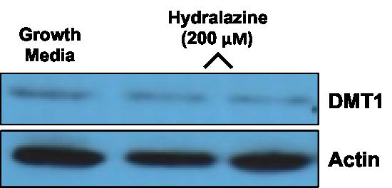
97 A. B. 87

 for")
98 C. D. Figure 2. DMT1 protein expression in Caco-2 cells. A. DMT1 expression by Western blot in Caco-2 cells exposed to growth media and 200 μm hydralazine hydrochloride for 24 h. B. Relative expression of DMT1 in A., normalized to β-actin. C. DMT1 expression by Western blot in Caco-2 cells exposed to control media and 2x amino acids (2x AA) for 24 h. D. Relative expression of DMT1 in C., normalized to β-actin. 88
 200 μm hydralazine hydrochloride for 6h and 24h")
99 A. B. Figure 4. DMT1 promoter activity in response to A) 200 μm hydralazine hydrochloride for 6h and 24h and B) amino acid supplemented media for 24 h. *P < 0.01, **P <
100 Chapter 7 Increasing Dietary Protein Suppresses Hepatic Hepcidin Expression In Vivo Introduction One of the most important systemic hormones regulating iron absorption is hepcidin, a 25-amino acid peptide hormone produced and secreted by the liver in response to iron overload and inflammation. 91 Hepcidin functions by binding to and causing the internalization and degradation of FPN1 on the basolateral membrane of duodenal enterocytes. 28 The consequence is a rise in intracellular iron, which in turn suppresses the expression of DMT1, Dcytb, and ultimately intestinal iron absorption. Hepcidin may also directly affect the expression of DMT1 and Dcytb in enterocytes, since hepcidin treatment was recently found to induce a significant reduction in DMT1 protein expression in Caco-2 cells. 113 The exact mechanism by which hepcidin senses iron has not been fully elucidated but it is well accepted that the bone morphogenic protein (BMP) signaling pathway is intimately involved in this process. In iron replete conditions, BMP6 binds to the BMP cell surface receptor in conjunction with the BMP co-receptor hemojuvelin (HJV), which initiates an intracellular signaling cascade involving the phosphorylation of SMAD1/5/8 and translocation of a SMAD1/5/8-SMAD4 complex to the nucleus, where it binds to enhancers in the hepcidin promoter, activating transcription. 92 Induction of hepcidin transcription also occurs through the binding of holotransferrin to TfR2 in a complex with the hemochromatosis protein, HFE. 42 The intracellular signaling pathways involved in this process have not been clarified but it is 90
101 thought that there is crosstalk with BMP/SMAD signaling. Holotransferrin binding to TfR2 on the cell surface of hepatocytes also causes stabilization of TfR2, further enhancing the interaction with HFE and in turn, hepcidin transcription. During iron depletion however, hepcidin transcription is suppressed through 3 separate mechanisms in the hepatocyte. First, the expression of TfR1 on the cell membrane of hepatocytes increases, causing it to sequester HFE and inhibit HFE binding to TfR2. Additionally, a recently identified hepatocyte membrane serine protease, matriptase-2 (gene name TMPRSS6), has been shown to cleave the membrane-bound BMP coreceptor HJV, thus preventing the binding of BMP6 to its BMP-HJV receptor complex. 114 Finally, iron deficiency increases the release of the soluble form of the BMP co-receptor HJV, which then sequesters BMP6 extracellularly and prevents its interaction with membrane-bound HJV. 115 Thus, several mechanisms are in place to control hepatic hepcidin expression in response to changes in iron status and availability. Recent data from our group showed that increasing dietary protein from a medium to a high level in rats significantly augments intestinal iron absorption and expression of the duodenal iron import proteins, DMT1 and Dcytb, but not the iron export protein FPN. Since hepcidin is a major factor in the regulation of iron absorption, and reduced hepcidin expression has been shown to be associated with significant increases in DMT1 and Dcytb transcript expression, we thought it plausible that hepatic hepcidin expression may also be altered in our model. Therefore, the purpose of this study was to determine if increasing dietary protein affects hepatic hepcidin expression in vivo in rats and in vitro in primary rat hepatocytes. Given that there have been no reports indicating that dietary protein can influence hepcidin production in the liver, it 91
102 was hypothesized that dietary protein would have no effect on hepatic hepcidin expression. Materials and Methods Experimental animals and diets The diet study protocol and experimental diets used in this work have been previously described. 108 Briefly, 250 to 300 g adult female Sprague Dawley rats (n = 28) were purchased from Charles River Laboratories, Inc. (Wilmington, MA). Animals were housed in the Yale Animal Resource Center and cared for in accordance with institutional animal care use policies. All experiments were approved by the Yale and University of Connecticut Institutional Animal Care and Use Committees. On arrival, rats were placed on standard diet 2018, Teklad Global 18% Protein Rodent Diet (Harlan Teklad, Inc., Madison, WI), for a minimum of 2 wk to allow for acclimation. For all studies, experimental diets were obtained from Harlan Teklad and were nutritionally complete and isocaloric (Table 1). The 20% protein diet was used as a normal or moderate protein diet and was representative of the standard rat diet 2018, which contains 24% protein. The 40% protein diet was used as the high protein diet. The 20% protein diet was made isocaloric to the 40% protein diet by the addition of cornstarch. Lipid from corn oil was held constant in both diets at 14-15% of total kcal. Real-time PCR Real-time PCR was performed using RNA isolated from the livers of rats consuming different levels of dietary protein. Twenty-eight rats were randomized to 92
103 either a 20 or 40% protein diet for 12 days. On d 12 of the diets, rats were anesthetized, the abdominal cavity was opened, and sections of the liver removed and snap frozen in liquid N before storage at -70 C. For total RNA extraction, livers were ground in liquid N using a mortar and pestle and extracted with TRIzol (Invitrogen, Carlsbad, CA), followed by purification using the RNeasy Mini Kit (QIAGEN, Valencia, CA). Total RNA was reverse transcribed to cdna using the iscript TM cdna Synthesis Kit (Bio-Rad, Hercules, CA) and the reverse transcription reaction performed on a C1000 TM Touch Thermal Cycler System (Bio-Rad, Hercules, CA) using the following protocol: 25 C for 5 min, 42 C for 30 min, and 85 C for 5 min. Real-time PCR analyses were performed using Taqman Gene Expression Assays for the following targets: hepcidin (Rn _m1), BMP6 (Rn _m1), interleukin-6 (IL-6) (Rn _m1), TfR1 (Rn _m1), and TMPRSS6 (Rn _m1) (Applied Biosystems, Foster City, CA) and itaq TM Universal Probes Supermix (Bio-Rad, Hercules, CA). Hypoxanthine phosphoribosyltransferase 1 (Hprt1) was used as an endogenous reference (Rn _m1, Applied Biosystems, Foster City, CA). PCR reactions were run in an iq2 Optical System (Bio-Rad, Hercules, CA) using the following protocol: 95 C for 10 min, followed by 40 cycles at 95 C for 20 sec and 60 C for 1 min. Relative quantification of target genes was calculated using the comparative C T method. Primary rat hepatocyte culture Freshly isolated primary rat hepatocytes from Sprague Dawley rats were purchased from Invitrogen (Carlsbad, CA). Upon receipt, hepatocytes were 93
104 immediately seeded into collagen-coated 12-well plates at a density of 1 x 10 5 cells/well. To first determine if hepcidin transcript expression could be efficiently altered in this model, hepatocytes were treated for 6 and 24 h with heparin sulfate (400 μg/ml) as described by others. 116 To determine whether amino acids directly affect hepcidin expression, hepatocytes were treated with 2x amino acid media, or control hepatocyte maintenance media (William s E Medium, 0.05% penicillin/streptomycin, 1% ITS+, 1% L-glutamine, 15 mm HEPES, and 0.1 μm dexamethasone) for 6 and 24 h. 2x amino acid media was prepared by adding a mixture of essential and non-essential amino acids to hepatocyte maintenance media, such that the final concentration of amino acids was twice that of the maintenance media with no added amino acids. After treatment for the indicated times, RNA was extracted, cdna synthesized, and QPCR performed as described above. Statistical Analyses Statistical analyses were performed using GraphPad Prism version 6.0a (GraphPad Software Inc., La Jolla, CA). A student s t test was used to analyze experiments conducted on only two groups. One-way ANOVA was used to analyze experiments conducted on 3 groups. For all statistical analyses, differences with a P value < 0.05 were considered significant. Data are shown as means ± SEM. 94
105 Results A high protein diet suppresses hepatic hepcidin expression in vivo As shown in Figure 1A, animals consuming the 40% protein diet exhibited significantly reduced hepatic hepcidin mrna levels compared to those consuming the 20% protein diet. Specifically, the high protein diet suppressed hepcidin transcript expression by 44% compared to the medium protein diet (P = ). To evaluate whether any genes known to be involved in the regulation of hepcidin transcription were altered in response to increasing dietary protein, the mrna levels of BMP6, TMPRSS6, and TfR1 were evaluated using real-time PCR. Interestingly, BMP6 transcript expression was significantly reduced in response to the 40% protein diet compared to the 20% protein diet, such that animals consuming the 40% diet exhibited 20% lower BMP6 levels than those on the 20% protein diet (Figure 1B, P = ). Additionally, TfR1 transcript levels were significantly different between the two diet groups. In particular, TfR1 mrna expression was significantly greater in the 40% group compared to the 20% group, with a fold difference of 1.5 (Figure 1C, P = ). However, dietary protein had no consistent effect on TMPRSS6 mrna levels (Figure 1D). Since inflammation is also known to induce hepcidin transcription, real-time PCR was performed to determine whether increasing dietary protein affects hepatic IL-6 expression. As shown in Figure 2, there was no significant difference in IL-6 mrna expression between the two diet groups, suggesting that there was no change in inflammatory status in response to changes in dietary protein. 95
106 Amino acids have no impact on hepatic hepcidin expression in primary rat hepatocytes To test the hypothesis that dietary protein/amino acids directly affect hepcidin expression, primary rat hepatocytes were utilized. First, to determine whether hepcidin could be efficiently altered in this model, hepatocytes were incubated with 10 ng/ml IL-6 (to induce hepcidin) and 400 μg/ml heparin sulfate (to suppress hepcidin) for 6 and 24h. Neither heparin sulfate nor IL-6 treatment for 6h affected hepcidin expression (data not shown). However, 24-h incubation with IL-6 augmented hepcidin expression by 5.2-fold, while 400 μg/ml heparin sulfate modestly suppressed hepcidin mrna expression by 34% compared to cells incubated with control media (Figure 3A). Next the effect of amino acids on hepcidin expression was examined. As shown in Figure 3B, 24-h treatment with 2x amino acids added to hepatocyte maintenance media enhanced hepcidin transcript levels by ~1.6-fold compared to cells incubated with hepatocyte maintenance media, however this difference was not statistically significant (P = 0.152). Discussion In the present study, we found that acute changes in dietary protein significantly affects hepatic hepcidin expression as well as the expression of genes known to be involved in the regulation of hepcidin transcription in the liver. Specifically, rats consuming a high protein diet (40%) for 12 d exhibited a ~50% suppression in hepatic hepcidin transcript expression compared to rats fed a medium protein diet (20%). This substantial reduction in hepcidin expression with increasing protein intake was 96
107 associated with significant changes in both BMP6 and TfR1 expression in the liver. Increasing dietary protein suppressed BMP6 mrna levels by 20%, whereas TfR1 mrna was augmented by 50%. However, there was no observed difference in TMPRSS6 transcript expression with increasing dietary protein. These findings suggest that suppression of hepatic hepcidin expression may be partially responsible for the concomitant increase in intestinal iron absorption and expression of duodenal iron transporters that we observed with increasing dietary protein. To our knowledge, there are no other reports of dietary protein affecting hepcidin expression in the liver. The only nutrient known to impact hepcidin production is iron. The mechanism by which dietary protein represses hepcidin expression is not known, but the in vitro data presented above imply that amino acids do not directly reduce hepcidin transcript expression. In fact, in our in vitro primary rat hepatocyte model, additional amino acids augmented hepcidin mrna levels, which is in stark contrast to our in vivo results. It is unclear whether this discrepancy is a reflection of a poor in vitro model or an indication that other systemic factors not present in the in vitro setting are involved in the regulation of hepcidin expression by dietary protein. In support of the latter are findings from Sasaki et al., which show that sufficient dietary protein is important for erythropoiesis and that variations in dietary protein levels affect circulating erythropoietin (EPO) concentrations. 117 Interestingly, this report demonstrated that EPO concentrations in rat serum increased as protein content of the diet increased, such that EPO rose from an undetectable level (< 4 mu/ml) with protein deprivation to 35 mu/ml with a 20% casein diet. The authors suggest that certain amino acids may function as a signal to the kidney to stimulate EPO production, 97
108 including secretion of already synthesized EPO into the bloodstream. This is relevant to our findings since EPO and erythropoietic drive have been shown to negatively regulate hepcidin expression. 118 Thus, it is plausible that increasing protein in the diet augments erythropoiesis and EPO production in the kidney, which in turn suppresses hepcidin transcription in the liver. Unfortunately, kidney tissue was not collected in our study therefore this possibility could not be fully explored. It is intriguing that animals on the high protein diet exhibited significantly reduced BMP6 and enhanced TfR1 transcript levels compared to those on the medium protein diet. These changes most likely contributed to the difference observed in hepcidin expression, since BMP6 is a known inducer of hepcidin, and increased TfR1 expression inhibits hepcidin production. The mechanism by which increasing dietary protein affects the hepatic expression of these genes is currently unknown. In summary, these data clearly demonstrate that increasing dietary protein in the short-term significantly reduces hepatic hepcidin mrna expression and that this effect may be attributed at least in part to reduced expression of BMP6 and enhanced expression of TfR1 in the liver. The underlying mechanism responsible for these effects is not yet known but may involve the stimulation of erythropoiesis and circulating EPO levels by protein. 98

109 For macronutrients % refers to % of total kcal. 99

110 1A. 1B. 100
 hepcidin, B) BMP6, C) TfR1,")
 and high (40%) protein diets for 12 d.")
111 1C. 1D. Figure 1. Transcript expression of A) hepcidin, B) BMP6, C) TfR1, and D) TMPRSS6 in the livers of female rats consuming medium (20%) and high (40%) protein diets for 12 d. (N = 14 per group). *P < 0.05, ** P <
NUTRITIONAL CARE IN ANEMIA
 االله الرحمن الرحيم بسم NUTRITIONAL CARE IN ANEMIA Nutrition Departement Faculty of Medicine University of North Sumatera Definition Deficit of circulating RBC associated with diminished oxygen-carrying
For more information about how to cite these materials visit
 Author: John Williams, M.D., Ph.D., 2009 License: Unless otherwise noted, this material is made available under the terms of the Creative Commons Attribution Non-commercial Share Alike 3.0 License: http://creativecommons.org/licenses/by-nc-sa/3.0/
Chapter 13. Trace Minerals
 Chapter 13 Trace Minerals Minerals in the Human Body The Trace Minerals--An Overview Needed in very small quantities in the body. Perform many essential functions important to health. Toxic levels can
Iron Deficiency, Depression, and Other Affective Disorders in Female State Fair Attendees
 Iron Deficiency, Depression, and Other Affective Disorders in Female State Fair Attendees A thesis submitted to the faculty of the University of Minnesota by Kathleen Price In partial fulfillment of the
RELATIONSHIP OF EXCESS IRON TO CHRONIC DISEASE 1. The physiological role of iron and the relationship. of excess iron to chronic disease.
 RELATIONSHIP OF EXCESS IRON TO CHRONIC DISEASE 1 The physiological role of iron and the relationship of excess iron to chronic disease Alicia Cone Metro State University of Denver NUT 3140: Human Nutrition
IRON ABSORTPION IN PREGNANT AND NON-PREGNANT WOMEN AND MECHANISMS OF PLACENTAL IRON TRANSFER
 IRON ABSORTPION IN PREGNANT AND NON-PREGNANT WOMEN AND MECHANISMS OF PLACENTAL IRON TRANSFER A Dissertation Presented to the Faculty of the Graduate School of Cornell University In Partial Fulfillment
YEAR III Pharm.D Dr. V. Chitra
 YEAR III Pharm.D Dr. V. Chitra Anemia can be defined as a reduction in the hemoglobin,hematocrit or red cell number. In physiologic terms an anemia is any disorder in which the patient suffers from tissue
COORDINATION OF IRON AND OXYGEN SIGNALING THROUGH POST- TRANSCRIPTIONAL REGULATION OF HYPOXIA INDUCIBLE FACTOR-2ALPHA MCKALE R.
 COORDINATION OF IRON AND OXYGEN SIGNALING THROUGH POST- TRANSCRIPTIONAL REGULATION OF HYPOXIA INDUCIBLE FACTOR-2ALPHA By MCKALE R. DAVIS Bachelor of Science in Nutrition and Dietetics Texas Christian University
Rama Nada. -Ensherah Mokheemer. 1 P a g e
 - 3 - Rama Nada -Ensherah Mokheemer - 1 P a g e Don t forget to refer to page index wherever you see * Quick revision: In the previous lecture we said that: - your body contains 4-5g of iron (4g in females
Contemporary Nutrition
 Contemporary Nutrition Chapter 6 Proteins Overview of Protein Body is made up of thousands of substances that are made of proteins Each protein has own unique structure and function More than 300 different
Comparison of methods to asses mineral bioavailability (in vitro vs in vivo)
") Comparison of methods to asses mineral bioavailability (in vitro vs in vivo) Ann-Sofie Sandberg Dept of Chemical and Biological Engineering/Food Science Chalmers University of Technology Gothenburg, Sweden
Comparison of methods to asses mineral bioavailability (in vitro vs in vivo) Ann-Sofie Sandberg Dept of Chemical and Biological Engineering/Food Science Chalmers University of Technology Gothenburg, Sweden
Josie Grace C. Castillo, M.D.
 Josie Grace C. Castillo, M.D. 2 types of nutrients Macronutrients Carbohydrate Fats Protein Micronutrients Vitamins Minerals 1 Occur when the quantity or quality of food is not sufficient to meet a persons
Minerals in the Human Body
 Trace Minerals Minerals in the Human Body The Trace Minerals--An Overview Needed in very small quantities in the body. Perform many essential functions important to health. Toxic levels can easily be reached
The Pennsylvania State University. The Graduate School. Department of Nutritional Sciences
 The Pennsylvania State University The Graduate School Department of Nutritional Sciences NEW INSIGHTS INTO INTESTINAL HEME AND NON-HEME IRON ABSORPTION: REGULATION BY DIETARY FACTORS A Dissertation in
Classes of Nutrients A Diet
 Ch. 7 Notes Section 1: What is Nutrition? is the science or study of food and the ways the body uses food. are substances in food that provide energy or help form body tissues and are necessary for life
Introduction 5/2/2013 IRON RELATED GENES AND OXIDATIVE STRESS IN NON- ALCOHOLIC STEATOHEPATITIS. Iron Physiology. Iron Physiology
 // IRON RELATED GENES AND OXIDATIVE STRESS IN NON- ALCOHOLIC STEATOHEPATITIS DIANA MOYA, MD PEDIATRIC GASTROENTEROLOGY FELLOW DIGESTIVE DISEASES & NUTRITION CENTER MAY,. Iron Physiology. /NAFLD. Iron Metabolism
Understanding dietary and fortificant iron: The devil is in the detail
 Understanding dietary and fortificant iron: The devil is in the detail Sylvaine Bruggraber Nutritionist In Industry London, 4 th November 2010 Structure 1. Dietary Iron: 2. Fortificant 3. Bioavailability
The hemoglobin (Hb) can bind a maximum of 220 ml O2 per liter.
 can bind a maximum of 220 ml O2 per liter.") Hemoglobin Hemoglobin The most important function of the red blood cells is totransport (O2) from the lungs into the tissues, and carbon dioxide (CO2) from the tissues back into the lungs. O2 is poorly
Hemoglobin Hemoglobin The most important function of the red blood cells is totransport (O2) from the lungs into the tissues, and carbon dioxide (CO2) from the tissues back into the lungs. O2 is poorly
Nutrition Support of Iron Deficiency
 Sanford Medical Center Aunt Cathy s Guide to: Nutrition Support of Iron Deficiency Cathy Breedon PhD, RD, CSP,FADA Clinical/Metabolic Nutrition Specialist Perinatal/Pediatric Nutrition Specialist Sanford
IRON MANGANESE 6/6/2018. Minerals & Performance : Total Performance, Total Performance Plus
 Minerals & Performance : Total Performance, Total Performance Plus June Educational Webinar 2018 Dr Tania Cubitt Performance Horse Nutrition IRON Heme oxygen carrying capacity Iron deficiency rare Iron
DR. SHAMSUL AZAHARI ZAINAL BADARI DEPARTMENT OF RESOURCES MANAGEMENT AND CONSUMER STUDIES FACULTY OF HUMAN ECOLOGY UPM
 DR. SHAMSUL AZAHARI ZAINAL BADARI DEPARTMENT OF RESOURCES MANAGEMENT AND CONSUMER STUDIES FACULTY OF HUMAN ECOLOGY UPM Mineral nutrients are inorganic elements found in food which the body cannot synthesis.
PDF created with pdffactory trial version
 General Nutrition Concepts Importance of Nutrition Health Appearance Behavior Mood Role of Nutrients in Diet Growth and development Provide energy Regulate metabolism 2 Calories in Food Carbohydrates Protein
There are two groups of minerals: Major salt components: K, Na, Ca, Mg, Cl -, sulfate, phosphate, and HCO
 MINERALS INTRODUCTION 90 elements in the earth s s crust, 25 are known to be essential to life, they are present in living cells, including in food. Food contains additional, non-essential elements. Some
Wellness: Concepts and Applications 8 th Edition Anspaugh, Hamrick, Rosato
 Wellness: Concepts and Applications 8 th Edition Anspaugh, Hamrick, Rosato Forming a Plan for Good Nutrition Chapter 6 Good Nutrition n Nutrition is the study of nutrients and the way the body processes
Iron-Sensing Proteins that Regulate Hepcidin and Enteric Iron Absorption
 Annu. Rev. Nutr. 2010. 30:149 71 First published online as a Review in Advance on April 20, 2010 The Annual Review of Nutrition is online at nutr.annualreviews.org This article s doi: 10.1146/annurev.nutr.012809.104801
Nutrition Basics. Chapter McGraw-Hill Higher Education. All rights reserved.
 Nutrition Basics Chapter 12 1 The Body s Nutritional Requirements Essential nutrients The Six Essential Nutrients: Proteins, Fats, Carbohydrates, Vitamins, Minerals, Water Defined as : Nutrients one must
Zahraa Muneer. Enas Ajarma. Saleem
 2 Zahraa Muneer Enas Ajarma Saleem Blood parameters: 1. RBCs count 2. Haematocrit 3. Haemoglobin content In new-born babies all three parameters of blood are higher than in normal adults. This happens
Laboratory diagnosis of iron deficiency: The interpretation of automated counting parameters. Dr Wayne Thomas Derriford Hospital, Plymouth
 Laboratory diagnosis of iron deficiency: The interpretation of automated counting parameters. Dr Wayne Thomas Derriford Hospital, Plymouth Why does it matter? Over 30% of the Worlds population are anaemic,
Contemporary Nutrition 6 th. th ed. Chapter 9 Minerals
 Contemporary Nutrition 6 th th ed. Chapter 9 Minerals Minerals Various functions in the body Major Minerals Require >100 mg /day Calcium, phosphorus Trace Minerals Require < 100 mg/day Iron, zinc Bioavailability
Vegetarian Eating. Vegetarians consuming a varied and balanced diet will have no problem getting enough protein.
 Vegetarian or plant-based eating can offer many health benefits, including a reduced risk of cancer, diabetes, obesity, high blood pressure and heart disease in fact, many vegetarian communities around
Recent Advances in Erythroid Iron Homeostasis: Implications for Pathophysiology of Microcytic Anemias
 Recent Advances in Erythroid Iron Homeostasis: Implications for Pathophysiology of Microcytic Anemias Prem Ponka Department of Physiology Lady Davis Institute, Jewish General Hospital McGill University,
The Six Essential Nutrient Groups:
 The Six Essential Nutrient Groups: Water: Water transports other nutrients to cells, carries wastes away, aids digestion and more. It makes up more than half your weight. SOURCES: water; juices and
N u r t u r e F o o d s
 Tips on Timing of Diet, Weight Loss, and Exercise 1. Hydrate before you exercise to get the most out of your workout The goal here is to make sure your body has a balanced amount of electrolytes and fluid
Nutrition JMRSO 2017 FOOD SCIENCE
 Nutrition JMRSO 2017 FOOD SCIENCE What is nutrition? Nutrition is the science that interprets the interaction of nutrients and other substances in food in relation to maintenance, growth, reproduction,
EFFECT OF DIETARY IRON DEFICIENCY AND OVERLOAD ON ZIP14 EXPRESSION IN RATS
 EFFECT OF DIETARY IRON DEFICIENCY AND OVERLOAD ON ZIP14 EXPRESSION IN RATS By HYEYOUNG NAM A DISSERTATION PRESENTED TO THE GRADUATE SCHOOL OF THE UNIVERSITY OF FLORIDA IN PARTIAL FULFILLMENT OF THE REQUIREMENTS
Section 5 Feeds and Feeding of Commercial Poultry Notes
 Section 5 Feeds and Feeding of Commercial Poultry Notes Slide 2 Nutrition is a huge component of production cost! The knowledge of nutrient requirements for chickens is astounding. Commercial poultry strains
Hematopoiesis, The hematopoietic machinery requires a constant supply iron, vitamin B 12, and folic acid.
 Hematopoiesis, 200 billion new blood cells per day The hematopoietic machinery requires a constant supply iron, vitamin B 12, and folic acid. hematopoietic growth factors, proteins that regulate the proliferation
Iron depletion in frequently donating whole blood donors. B. Mayer, H. Radtke
 Iron depletion in frequently donating whole blood donors B. Mayer, H. Radtke Iron: relevance oxygen-transporting and storage proteins hemoglobin and myoglobin iron-containing centers in many enzymes mitochondrial
Minerals and water. Minerals 10/23/2017
 Minerals and water MAIN QUESTIONS TO ANSWER: What are some major functions of water in the body? For each mineral, what are the best dietary sources? what are the physiological functions? what are the
6 Essential Minerals for Women's Health. By Dr. Isaac Eliaz
 6 Essential Minerals for Women's Health By Dr. Isaac Eliaz Minerals are essential micronutrients that are required in small amounts for the body to function properly. Untreated mineral deficiencies can
Kazakh Academy of Nutrition
 Kazakh Academy of Nutrition USAID/GAIN MICRONUTRIENT FORTIFICATION PROJECT IN CENTRAL ASIA, AFGHANISTAN AND PAKISTAN ANALYSIS AND JUSTIFICATION THE POSSIBILITY OF HARMONIZING STANDARDS FOR REFINED WHEAT
Calcium Supplement Guidelines
 Supplement Guidelines Item Type text; Book Authors Houtkooper, Linda; Farrell, Vanessa A. Publisher College of Agriculture, University of Arizona (Tucson, AZ) Download date 24/07/2018 09:13:03 Item License
What Are Proteins? Lecture 9: Proteins. Proteins: large complex molecules composed of amino acids. Nutrition 150 Shallin Busch, Ph.D.
 What Are Proteins? Lecture 9: Proteins Nutrition 150 Shallin Busch, Ph.D. Proteins: large complex molecules composed of amino acids. Contain carbon, hydrogen, oxygen, nitrogen Primary source of nitrogen
Models to assess food iron bioavailability
 Graduate Theses and Dissertations Graduate College 2014 Models to assess food iron bioavailability Seth Mensah Armah Iowa State University Follow this and additional works at: http://lib.dr.iastate.edu/etd
FAO/WHO expert consultation on human vitamin and mineral requirements 195
 FAO/WHO expert consultation on human vitamin and mineral requirements 195 Chapter 13 Iron The role of iron in human metabolic processes Iron has several vital functions in the body. It serves as a carrier
Nutrients. Macronutrients. Micronutrients. Others. Carbohydrates Proteins Fats. Vitamins Minerals. Fiber, water
 Nutrition Human Nutrition - The science of food, the nutrients and other substances contained therein, their action, interaction, and balance in relation to health and disease Normal Nutrition - caloric
Chapter 1 & 2 All of the following are macronutrients except Carbohydrates Lipids Protein * Vitamins
 Chapter 1 & 2 All of the following are macronutrients except Carbohydrates Lipids Protein * Vitamins Gram per gram blank provides the most k calories Alcohol Carbohydrates * Lipids Proteins Which of the
Lecture 3. Nutrition
 Lecture 3 Nutrition, Part 1 1 Nutrition 1.Macronutrients a. Water b. Carbohydrates c. Proteins d. Fats 2.Micronutrients a. Minerals b. Vitamins 2 1 Why is Nutrition Important? Your food choices each day
Estimation of Iron Bioavailability from the Diets of an Adolescent Population
 University of Tennessee, Knoxville Trace: Tennessee Research and Creative Exchange Masters Theses Graduate School 6-1984 Estimation of Iron Bioavailability from the Diets of an Adolescent Population Gina
Chapter 2. Planning a Healthy Diet
 Chapter 2 Planning a Healthy Diet Principles and Guidelines Diet Planning Principles Adequacy Sufficient energy Adequate nutrients for healthy people Balance Enough but not too much kcalorie (energy) control
CAYUGA COMMUNITY COLLEGE Division of Computer Science, Mechanical Technology, Electrical Technology, GIS, Math, Nursing, Science
 CAYUGA COMMUNITY COLLEGE Division of Computer Science, Mechanical Technology, Electrical Technology, GIS, Math, Nursing, Science Basic Nutrition - Biol 209 3 Credit Hours CATALOG DESCRIPTION Appropriate
Assessing Iron Deficiency in Adults. Chris Theberge. Iron (Fe) deficiency remains as one of the major global public health problems for
 deficiency remains as one of the major global public health problems for") Assessing Iron Deficiency in Adults Chris Theberge Iron (Fe) deficiency remains as one of the major global public health problems for two reasons. It affects about one fourth of the world s population
Assessing Iron Deficiency in Adults Chris Theberge Iron (Fe) deficiency remains as one of the major global public health problems for two reasons. It affects about one fourth of the world s population
Hemosiderin. Livia Vida 2018
 Hemosiderin Livia Vida 2018 Questions Histochemical caracteristics of the different pigments. Exogenous pigments. Hemoglobinogenic pigments. Causes and forms of jaundice. Hemoglobinogenic pigments. Pathological
PROJECT WOMEN S ANAEMIA. by My HealthWorks. Associate Member. 125A, 2nd Floor, Shahpur Jat, New Delhi ,
 PROJECT WOMEN S ANAEMIA by My HealthWorks Associate Member 125A, 2nd Floor, Shahpur Jat, New Delhi-110049, Whatsapp: 8076964582, Phone No: 011-26496673-(74) info@myhealthworks.in INTRODUCTION Anaemia is
EXSC- STANDARD 14. Nutrients
 SPORTS NUTRITION EXSC- STANDARD 14 Nutrients Standard 14 Gather relevant information from multiple authoritative print and digital sources related to the importance of a balanced diet in the achievement
Hemoglobin and anemia BCH 471
 Hemoglobin and anemia BCH 471 OBJECTIVES Quantitative determination of hemoglobin in a blood sample. Hemoglobin structure Hemoglobin (Hb) is a porphyrin iron (II) protein in RBCs that transport oxygen
Nutrition for Health. Nutrients. Before You Read
 CHAPTER 10 LESSON 2 Nutrition for Health Nutrients BIG Idea Each nutrient in your diet plays a unique and essential role in keeping you healthy. Before You Read Sometimes figuring out what to eat can be
Iron. (2) Microminerals
 Microminerals") Iron (2) Microminerals Iron 1.Background Information 1 1.Definition and Classification Iron is a transition metal element (atomic number: 26, Fe). It is predominantly stored as heme iron which is found
Iron (2) Microminerals Iron 1.Background Information 1 1.Definition and Classification Iron is a transition metal element (atomic number: 26, Fe). It is predominantly stored as heme iron which is found
MOLECULAR MECHANISMS OF IRON UPTAKE BY HEPG2 HEPATOMA CELLS
 MOLECULAR MECHANISMS OF IRON UPTAKE BY HEPG2 HEPATOMA CELLS By LIN ZHANG A THESIS PRESENTED TO THE GRADUATE SCHOOL OF THE UNIVERSITY OF FLORIDA IN PARTIAL FULFILLMENT OF THE REQUIREMENTS FOR THE DEGREE
Study Exercises: 1. What special dietary needs do children <1 yr of age have and why?
 Exam 3 Review: Lectures 21+ Nutrition 150 Winter 2007 Exam: Monday, March 19, 8-10am in regular classroom Strategies for Studying: 1) Go over all lectures including those given by fellow students 2) Read
March 30, ASN Comments and Additions to Select, Proposed Topics and Questions are in Red Below
 March 30, 2018 The American Society for Nutrition (ASN) appreciates the opportunity to comment on the proposed topics and supporting scientific questions that will be considered during the development
Chapter 2 Part 3: Organic and Inorganic Compounds
 Chapter 2 Part 3: Organic and Inorganic Compounds Objectives: 1) List the major groups of inorganic chemicals common in cells. 2) Describe the functions of various types of inorganic chemicals in cells.
Drugs Used in Anemia
 Drugs Used in Anemia Drugs of Anemia Anemia is defined as a below-normal plasma hemoglobin concentration resulting from: a decreased number of circulating red blood cells or an abnormally low total hemoglobin
INTRODUCING DOCTOR DAWN S iron report. Sponsored by
 INTRODUCING DOCTOR DAWN S iron report Sponsored by CONTENTS 2 INTRODUCTION 3 1. GENERAL AWARENESS 4 Fig 1.1 Signs of low iron Fig 1.2 Daily iron requirements 2. WOMEN S HEALTH 6 Fig 2.1 Pregnancy and anaemia
Chapter. The Micronutrients: Vitamins and Minerals. Images shutterstock.com
 Chapter 13 The Micronutrients: Vitamins and Minerals Images shutterstock.com Objectives Differentiate between fat-soluble vitamins and water-soluble vitamins. List functions and sources of major minerals
Around million aged erythrocytes/hour are broken down.
 Anemia Degradation ofheme Around 100 200 million aged erythrocytes/hour are broken down. The degradation process starts in reticuloendothelial cells in the spleen, liver, and bone marrow. [1] The tetrapyrrole
Chapter 2-Nutrition Tools Standards and Guidelines
 Chapter 2-Nutrition Tools Standards and Guidelines MULTIPLE CHOICE 1. Which of the following is an appropriate use for dietary reference intakes (DRI)? a. ensuring that maximum nutrient requirements are
Red meat and iron. Dietary Iron
 Last updated 29 th September 2011 Red meat and iron Iron is essential for the formation of haemoglobin in red blood cells and is also an essential component in many biochemical pathways. For example, it
There are two groups of minerals: Major salt components: K, Na, Ca, Mg, Cl -, sulfate, phosphate, and HCO 3
 MINERALS INTRODUCTION 90 elements in the earth s s crust. 25 are known to be essential to life, they are present in living cells, including in food. Food contains additional, non-essential elements. Some
Nutrition. Chapter 45. Reada Almashagba
 Nutrition Chapter 45 1 Nutrition: - Nutrient are organic substances found in food and are required for body function - No one food provide all essential nutrient Major function of nutrition: providing
Biochemistry of Nutrition. Prof K Syed Department of Biochemistry & Microbiology University of Zululand Room no. 247
 Biochemistry of Nutrition Prof K Syed Department of Biochemistry & Microbiology University of Zululand Room no. 247 SyedK@unizulu.ac.za Class 1 What is Nutritional Biochemistry Importance of food How the
Speaker Tim Wierman. Speaker s Bio
 Speaker Tim Wierman Speaker s Bio President of Nutrition Education Services, Inc Creator of EAT TO COMPETE M.A. Nutrition Education (Immaculata Univ) B.S. (Ohio University) Member of ADA 160 Colleges &
Lesson Two Nutrients and the Body
 Lesson Two Nutrients and the Body Objectives After participating in this lesson, students will Be able to identify key nutrients the body needs and describe their function and importance. Understand that
WHY DO WE NEED FOOD? FOOD AND DIET
 WHY DO WE NEED FOOD? FOOD AND DIET Food gives us the energy required for chemical reaction to keep us alive Energy also required for many activities and to maintain body temperature Food also gives us
OVERVIEW OF DIETARY PROTEINS AND NITROGEN BALANCE
 OVERVIEW OF DIETARY PROTEINS AND NITROGEN BALANCE Proteins and Amino Acids What Are Proteins? Large molecules Made up of chains of amino acids Are found in every cell in the body Are involved in most of
Faculty of Medicine Dr. Tariq Aladily
 Iron deficiency anemia The most common anemia worldwide Only 10% of ingested iron is absorbed Most dietary iron occurs in meat products Absorbed in duodenum Hepcidin By inhibiting ferroportin, hepcidin
Iron deficiency anemia:
 بسم هللا الرمحن الرحمي Before we start: Dr. Malik suggested for the third immunity lecture to be a continuation for the hematology pharmacology. And he will discuss the drugs for treating leukemia and
Lesson 1 Carbohydrates, Fats & Proteins pages
 Lesson 1 Carbohydrates, Fats & Proteins pages 190-201 What are the 3 classes of nutrients that supply your body with energy and how does the body obtain the energy from foods? Describe the roles that carbohydrates,
Nutrition. Nutrition. Contents:
 Nutrition Contents: Nutrition Overview Zinc Iron Protein B-Vitamins Selenium Phosphorous Good Fat vs. Bad Fat Beef and Heart Health Fat & Cholesterol: The Whole Story Lean Beef Lean Beef in Foodservice
Chapter 2 Nutrition Tools Standards and Guidelines
 Chapter 2 Nutrition Tools Standards and Guidelines MULTICHOICE 1. Which of the following statements best describes the recommended dietary allowances (RDA)? (A) they are average nutrient intake goals that
SUMAN PROJECT CONSULTANT (P) LTD. MIXED GRAIN PRODUCTS AND HEALTH BENEFITS
 LTD. MIXED GRAIN PRODUCTS AND HEALTH BENEFITS") SUMAN PROJECT CONSULTANT (P) LTD. MIXED GRAIN PRODUCTS AND HEALTH BENEFITS March 2, 2017 WHAT IS MULTIGRAIN? "Multigrain" simply means that it contains several different types of grains, distinguishing
SUMAN PROJECT CONSULTANT (P) LTD. MIXED GRAIN PRODUCTS AND HEALTH BENEFITS March 2, 2017 WHAT IS MULTIGRAIN? "Multigrain" simply means that it contains several different types of grains, distinguishing
Choosing Healthful Foods
 LESSON 25 Choosing Healthful Foods Before You Read List some of the healthful foods you eat. What makes these foods healthful choices? What You ll Learn the functions and sources of proteins, carbohydrates,
The Importance of Glutamine and Antioxidant Vitamin Supplementation in HIV
 The Importance of Glutamine and Antioxidant Vitamin Supplementation in HIV An Introduction to Glutamine Glutamine is the most abundant amino acid in the human body, and plays extremely important role in
THE NATIONAL ACADEMIES
 THE NATIONAL ACADEMIES NATIONAL ACADEMY OF SCIENCES INSTITUTE OF MEDICINE NATIONAL ACADEMY OF ENGINEERING NATIONAL RESEARCH COUNCIL DIVISION ON EARTH AND LIFE STUDIES BOARD ON AGRICULTURE AND NATURAL RESOURCES
Chapter 4: Nutrition. ACE Personal Trainer Manual Third Edition
 Chapter 4: Nutrition ACE Personal Trainer Manual Third Edition Introduction SCAN group of dieticians who practice in sports and cardiovascular nutrition [SCAN]; locate local SCAN dieticians by contacting
Chapter 1: Food, Nutrition, and Health Test Bank
 Chapter 1: Food, Nutrition, and Health Test Bank MULTIPLE CHOICE 1. Promoting a health care service that improves diabetes management for the elderly in a community would assist in which of the following?
Module 1 An Overview of Nutrition. Module 2. Basics of Nutrition. Main Topics
 Module 1 An Overview of Nutrition Module 2 What is Nutrition? What Are Nutrients? Units of Energy Why we need energy? Maintaining energy balance Daily energy requirements Calorie Requirements for Different
Food Choices. Food Choices. Food Choices. Food Choices. Food Choices. Introduction to Nutrition ALH 1000 Chapter 1 & 2
 Introduction to Nutrition ALH 1000 Chapter 1 & 2 An Overview Of Nutrition And Planning A Healthy Diet Instructor Bonnie Bennett-Campbell, RN MSN Food and Nutrition Play a Significant Role in Life An Individual
Nutrients are: water carbohydrates lipids proteins. minerals vitamins fiber
 Nutrients are: water carbohydrates lipids proteins minerals vitamins fiber WATER Essential nutrient 55-65% body weight Body loses water through evaporation, excretion, and respiration The only nutrient
Supplements That Work
 Supplements That Work Supplements can be confusing and expensive! Do they work? Do you need them? How should you take them? This guide is an introduction to supplements that most people need. Are supplements
LifePak Health Benefits. LifePak is optimum supplementation for:
 LifePak What is LifePak? Our comprehensive nutritional wellness program delivering the optimum amounts of all essential and generally beneficial nutrients for long-term health and well-being. LifePak Health
Tuning Cell Cycle Regulation with an Iron Key
 Cell Cycle ISSN: 1538-4101 (Print) 1551-4005 (Online) Journal homepage: https://www.tandfonline.com/loi/kccy20 Tuning Cell Cycle Regulation with an Iron Key Yu Yu, Zaklina Kovacevic & Des R. Richardson
DIETARY GUIDELINES FOR AMERICANS EIGHTH EDITION
 DIETARY GUIDELINES FOR AMERICANS 2015-2020 EIGHTH EDITION Executive Summary Over the past century, deficiencies of essential nutrients have dramatically decreased, many infectious diseases have been conquered,
Prevent and/or reduce overweight and obesity through improved eating and physical activity.
 A summary of the key recommendations to healthier living for Americans of the 2010 Dietary Guidelines and a weight-loss guide to combat obesity are offered by the May 2011 issue of the Tufts Health & Nutrition
Chapter 06 Lecture Outline. See separate PowerPoint slides for all figures and tables preinserted into PowerPoint without notes.
 Chapter 06 Lecture Outline See separate PowerPoint slides for all figures and tables preinserted into PowerPoint without notes. Copyright 2016 McGraw-Hill Education. 2012 Pearson Permission Education,
?Pairing peanuts with other healthy foods can actually help you. Nutrient Density. Peanuts Improve Nutrient Intake %RDA %RDA
 Peanuts Improve Nutrient Intake snacking on peanuts improves intake of healthy nutrients. One serving of peanuts provides many of the essential nutrients we need each day. These include vitamin E, magnesium,
the only oral supplement so quick and effective:
 The Product the only oral supplement so quick and effective: 30 days to increase ferritinemia and syderemia to +50% [90-120 days with common oral supplementation with iron]. After 1 week the subject will
The human body contains approximately three grams of zinc, the highest concentrations of which are located in the prostate gland and the eye.
 Zinc AT A GLANCE Introduction Zinc (Zn) is found in nearly 100 different enzymes and as such is an essential building block for all life. Zinc is the second most common trace mineral in the body after
Chapter 1. What Is Nutrition? Karen Schuster Florida Community College of Jacksonville. PowerPoint Lecture Slide Presentation created by
 Chapter 1 What Is Nutrition? PowerPoint Lecture Slide Presentation created by Karen Schuster Florida Community College of Jacksonville Copyright 2008 Pearson Education, Inc., publishing as Pearson Benjamin
DR.PAWAN TOSHNIWAL ASSISTANT PROFESSOR BIOCHEMISTRY,
 DR.PAWAN TOSHNIWAL ASSISTANT PROFESSOR BIOCHEMISTRY, CURIOSITY???? WHAT? WHERE? WHY? DISORDES? CURIOSITY = ANSWER TO FIND WHAT ARE VITAMINS AND THERE REQUIREMENTS? WHERE THEY ARE SYNTHESISED, ABSORPED,