grade inflammation associated with increased white adipose tissue mass has been linked to
|
|
|
- Ethelbert Lambert
- 5 years ago
- Views:
Transcription
1 COLLINS, BRIAN, M.S. The Impact of California Table Grapes on Intestinal Inflammation and Microbiota in Mice Fed an American Type Diet. (2015) Directed by Dr. Michael McIntosh. 148 pp. Obesity is a growing health concern in the United States and worldwide. The chronic, low grade inflammation associated with increased white adipose tissue mass has been linked to chronic metabolic disorders such as insulin resistance, hypertension, and hyperlipidemia. Consumption of high fat diets leads to intestinal disorders such as microbial dysbiosis and gut barrier dysfunction that can adversely impact systemic metabolism. One potential dietary strategy to alleviate the high fat-induced chronic inflammation is increased consumption of fruits and vegetables rich in polyphenols due to their antioxidant and anti-inflammatory properties. Notably, several studies have demonstrated that supplementation with grape products or anthocyanins found in grapes reduced inflammation systemically and increased the abundance of beneficial bacteria such as Lactobacillus, and Bifidobacterium in culture. However, the beneficial properties of whole grape powder and one or more of its fractions on intestinal inflammation, microbial populations, and barrier function in mice fed an American type diet rich in saturated fat are unclear. Therefore, the specific aims of this research were to; (i) determine the impact of consuming California table grapes on intestinal health in mice fed an American type diet rich in one type of saturated fat (Aim 1), and (ii) identify a key fraction (i.e., extractable polyphenol (EP) or nonextractable polyphenol (NEP) fractions) of California table grapes that improves markers of intestinal inflammation in mice fed an American type diet rich in four types of saturated fats (Aim 2). In Aim 1, consumption of the powder grape diet; (i) reduced body fat percentage, the total weight of all four fat depots, and inguinal fat depot weight, (ii) increased localization of a tight junction protein linked to improved barrier function, (iii) reduced the abundance of a deleterious sulfidogenic bacteria, and (iv) increased the abundance of beneficial bacteria (e.g., Akkermansia
2 muciniphila, Bifidobacterium, and Lactobacillus), compared to high fat controls. However, the high fat diet did not significantly increase the abundance of inflammatory markers in the intestine nor did the powdered grapes significantly decrease their abundance. Taken together, these data acquired in Aim 1 demonstrate that whole powdered California table grapes improved a marker of gut barrier function and a metabolic profile that was positively correlated with changes in microbiota in mice fed a butter-rich diet. In Aim 2, the polyphenol-rich EP fraction alone or in combination with NEP (EP+NEP), but not unfractioned powdered grapes; (i) decreased body fat percentage, body fat depot weights, and liver triglyceride levels, (ii) improved insulin sensitivity and glucose disposal, (iii) decreased the mrna levels of several inflammatory genes in WAT, and (iv) decreased the expression of the proinflammatory gene Cluster of differentiation 68 in the colon. Taken together, these data demonstrate that the; (i) potential health benefits of consuming grape powder are dependent on the type and amount of fat in the diet, and (ii) extraction of polyphenols from powdered grapes results in improved metabolic profile and decreased systemic inflammation in conjunction with consuming a rich in saturated fats. Collectively, these findings are expected to contribute insight for the development of dietary strategies using table grapes for decreasing obesity and some of its metabolic complications, possibly by altering populations of gut microbes. However, clinical trials are needed to determine the extent to which these findings are applicable to humans.
3 THE IMPACT OF CALIFORNIA TABLE GRAPES ON INTESTINAL INFLAMMATION AND MICROBIOTA IN MICE FED AN AMERICAN TYPE DIET by Brian Collins A Thesis Submitted to the Faculty of the Graduate School at The University of North Carolina at Greensboro in Partial Fulfillment of the Requirements for the Degree of Master of Science Greensboro 2015 Approved by Committee Chair
4 APPROVAL PAGE This thesis written by Brian Collins has been approved by the following committee of the Faculty of The Graduate Scholl at The University of North Carolina at Greensboro. Committee Chair Committee Members Date of Acceptance by Committee Date of Final Oral Examination ii
5 TABLE OF CONTENTS Page LIST OF TABLES... v LIST OF FIGURES... vi CHAPTER I. INTRODUCTION... 1 Overview... 1 Central Hypothesis and Specific Aims... 2 Significance... 3 References... 6 II. REVIEW OF LITERATURE... 7 Obesity and the Consequences of Chronic Increases of Inflammatory Genes... 7 Relationship between Systemic and Intestinal Inflammation... 9 Gut Barrier Function and the Role of Gut Microbiota Association of Gut Microbiota with Chronic Disease States Impact of Diet on Gut Microbiota and the Potential Influence on Host Health Anti-inflammatory Properties of Grapes Potential Prebiotic Properties of Grapes Summary and Gaps in the Literature References III. CALIFORNIA TABLE GRAPE CONSUMPTION REDUCES ADIPOSITY, HEPATIC TRIGLYCERIDES, LIPOGENIC GENE EXPRESSION, AND ABUNDANCE OF SULFIDOGENIC BACTERIA IN MICE FED BUTTER FAT Abstract Introduction Materials and Methods Results Discussion Acknowledgements References iii
6 IV. AN EXTRACTABLE, POLYPHENOL-RICH FRACTION OBTAINED FROM CALIFORNIA TABLE GRAPES DECREASES ADIPOSITY, INSULIN RESISTANCE AND MARKERS OF INFLAMMATION IN HIGH-FAT FED C57BL/6J MICE Abstract Introduction Materials and Methods Results Discussion Acknowledgments References V. EPILOGUE Rationale for Studies 1 and Summary of Results from Studies 1 and Interpretations and Implications for the Results from Study 1 and References iv
7 LIST OF TABLES Page Table 2.1. Association between Gut Microbiota and Chronic Disease States Table 3.1. Diet Formulations Table 3.2. The Effect of Diet Composition on Weight Gain, Consumption, and Utilization Table 3.3. Statistical Analyses of Unifrac Distances Table 3.4. Taxa Associated with Body Fat Table 4.1. Diet Formulation Table 4.2. Phenolic Composition of Grape Powder (GP), Extractable Polyphenol (EP) Fraction and Non-extractable Polyphenol (NEP) Fraction Table 4.3. Anthocyanin Concentration in Grape Powder (GP) and the Extractable Polyphenol (EP) Fraction Table 4.4. Body Weight Gain (BWG), Food Conversion Efficiency (FCE), and Total Calorie Intake per Treatment Group v
8 LIST OF FIGURES Page Figure 1.1. Working Model... 5 Figure 2.1. Impact of Microbial Fermentation of Fiber Figure 2.2. Polyphenol Metabolism and Health Benefits Figure 3.1. Diet Induced Changes to Fat Mass Figure 3.2. Glucose Tolerance Tests Figure 3.3. Liver Gene Expression Figure 3.4. WAT Gene Expression Figure 3.5. Alterations to Intestinal Inflammation and Barrier Function Figure 3.6. Diet Induced Alterations to Sulfidogenic Bacteria and Akkermansia muciniphalia Figure 3.7. Diet Induced Changes to Microbial Species Figure 3.8. PCoA Plots of Unweighted and Weighted UniFrac Distances Figure 3.9. Relative Abundance of Microbial Taxa Figure Principle Component Analysis (PCA) of Cecum Mucosa Figure Relationship between Body Fat and Bacterial Populations Figure 4.1. Proanthocyanin Degree of Polymerization in EP Fraction Figure 4.2. Body Fat Percentages and WAT Depot Weights Figure 4.3. Glucose Tolerance Tests and HOMA-IR Index Figure 4.4. Liver Markers of Inflammation and Lipid Metabolism Figure 4.5. Liver Weight, Triglycerides, and Oil Red O Staining Figure 4.6. Expression of Markers of Inflammation in WAT Figure 4.7. Expression of Markers of Lipid Metabolism in WAT vi
9 Figure 4.8. Expression of Markers of Intestinal Inflammation Figure 4.9. Expression of G-protein Receptors and Glycoproteins in Intestinal Mucosa Figure 5.1. A Model of the Anthropometric and Intestinal Measurements Recorded in Study 1 and Study vii
10 CHAPTER I INTRODUCTION Overview Obesity is a growing health issue, affecting approximately 11% of the world s population and 36% of Americans [1, 2]. Sedentary lifestyle and excessive calorie intake of foods and beverages high in sugars or fats, especially saturated fat, are believed to be the main environmental causes for this disease state [3]. Obesity is of particular concern, because of its comorbidities including type 2 diabetes, hyperlipidemia, and hypertension. Foods containing indigestible carbohydrates commonly referred to as fiber and bioactive components such as phytochemicals have been shown to reduce some of the deleterious conditions associated with obesity. For example, grapes contain numerous phytochemicals including flavonols, anthocyanins, flavan-3-ols, and hydroxycinnammic acids which have been reported to have antioxidant and anti-inflammatory properties that may alleviate oxidative stress and insulin resistance associated with chronic, low grade inflammation [4, 5]. In addition to phytochemicals, grapes contain fiber, an indigestible food component that has been shown to increase butyrate-producing bacteria, which in turn improves intestinal health by enhancing barrier function [reviewed in 6]. However, little is known about the potential benefits of grapes on intestinal microbiota abundance, inflammatory status, or barrier function in mice consuming an American type diet rich in saturated fat. 1
11 Central Hypothesis and Specific Aims The long term goal of our research group is to develop innovative dietary strategies designed to control chronic, low-grade inflammation commonly associated with diet-induced obesity using California table grapes. The central hypothesis for my thesis is that intestinal inflammation and impaired gut barrier function associated with diets high in saturated fats will be attenuated by dietary intervention with California table grapes through the modification of intestinal microbiota and inflammatory status as shown in the working model (Fig. 1.1). Our group is well prepared to carry out this research due to our experience; (i) in laboratory animal husbandry protocols, (ii) live animal testing procedures, and (iii) tissue analyses including RNA isolation, cdna synthesis, qpcr, immunoblotting, and enzyme assays, thereby ensuring minimization of errors in data collection. Our collaborators at the University of Illinois are Dr. H. Rex Gaskins and graduate assistant Patricia Wolfe, experts in microbiota analyses and culturing probiotics. Our collaborators at the North Carolina Research Campus in Kannapolis are Drs. Mary Ann Lila and Mary Grace, experts in fractionating and identifying fruit polyphenols, and Drs. Zhan Zhou and Wei Zong, experts in assessing gut barrier function. Our collaborators at the University of Chicago are Drs. Kristina Martinez, Eugene Chang, and Chase Cockrell, experts in microbiota and short chain fatty acid (SCFA) analyses. To test my hypothesis, I will pursue the following two specific aims: Aim 1. Determine the impact of consuming California table grapes on intestinal health in mice fed an American type diet rich in one type of saturated fat; and Aim 2. Identify a key fraction of California table grapes that improves markers of intestinal inflammation in mice fed an American type diet rich in four types of saturated fats. 2
12 Based on my review of the current literature, the ability of California table grapes, the most abundantly-grown table grapes in the U.S., to suppress intestinal inflammation and improve barrier function in mice fed an American type diet has not yet been examined. Furthermore, the bioactive fraction in table grapes responsible for these potential gut health-promoting actions and their impact on goblet cell function is unknown, and the impact of these grapes on gut microbiota has not been published. Thus, completion of my aims will lead to the following anticipated outcomes; (i) powdered grapes will attenuate gut inflammation in high fat fed mice, as well as increase the number of butyrate-producing bacteria while reducing the number of sulfidogenic bacteria, and (ii) a polyphenol-rich, bioactive fraction (i.e., containing extractable polyphenols) in table grapes will be identified that reduces adiposity and improves glucose tolerance in association with reducing intestinal inflammation. These proposed improvements will be associated with modifications in mucosal microbiota (i.e., decreasing sulfidogenic bacteria and increasing butyrate producing bacteria (i.e., Bifidobacterium) or Akkermansia muciniphila, (a health-promoting microbe), and upregulation of G-protein receptors (i.e., GPR-41,-43, or -119) associated with improved metabolic profiles. Completion of this research will further our understanding of how California table grapes act as prebiotics that improve gut health and subsequent metabolic complications associated with consuming a diet rich in saturated fat. Such knowledge will inform the public about the gut health-promoting properties of California table grapes; a nutritionally and economically important agricultural products in the U.S. Significance Diets high in fats and calories lead to obesity and are associated with chronic, low-grade intestinal inflammation and alterations in the intestinal microbiota [reviewed in 7]. My research will demonstrate that powdered California table grapes will attenuate the deleterious changes 3
13 associated with consuming diets rich in saturated fatty acids. The research proposed in this study is significant, because application of the results and new knowledge gained are expected to lead to new dietary approaches using California table grape products to reduce intestinal inflammation with improvements in intestinal and systemic health. These proposed outcomes have the potential to reduce health care costs and loss of productivity associated with obesity and its comorbidities. 4
 increasing butyrate-producing bacteria and")
14 Figure 1.1. Working Model. Powdered grapes attenuate inflammatory signaling, endotoxemia, and metabolic dysfunction in mice consuming an American diet rich in saturated fats by; (i) increasing butyrate-producing bacteria and Akkermansia muciniphila (A. muciniphila) which enhance goblet cell secretion of mucin and barrier protein synthesis in colonocytes, (ii) reducing intestinal inflammation which will prevent gut permeability, (iii) attenuating the growth of sulfide/sulfate reducing bacteria which produce the cytotoxic gas hydrogen sulfide that increases gut inflammation and permeability. 5
15 References 1. Ogdan CL, Carroll MD, Kit BK, Flegal KM. Prevalence of Childhood and Adult Obesity in the United States, JAMA 2014; 311(8): NIH. Obesity. Last reviewed: May 12, WHO. Obesity. Viewed. January 20, Chuang CC, McIntosh MK. Potential mechanisms by which polyphenol-rich grapes prevent obesity-mediated inflammation and metabolic diseases. Ann Rev Nutr 2011; 31: Felice F, Zambito Y, Di Colo G, D'Onofrio C, Fausto C, et al. Red grape skin and seeds polyphenols: Evidence of their protective effects on endothelial progenitor cells and improvement of their intestinal absorption. Eur J Pharm Biopharm 2012; 80: Scott KP, Duncan SH, Flint HJ. Dietary fibre and the gut microbiota. Nutrition Bulletin 2008; 33: Shen W, Gaskins HR, McIntosh MK. Influence of dietary fat on intestinal microbes, inflammation, barrier function and metabolic outcomes. J Nutr Biochem 2014; 25(3):
16 CHAPTER II REVIEW OF LITERATURE Obesity and the Consequences of Chronic Increases of Inflammatory Genes An estimated 1.4 billion people over the age of 20 are over-weight and an additional 500 million are considered obese worldwide [1]. As of 2010, an estimated 69% of the United States adult population was considered overweight or obese (36%) [2]. Consumption of high calorie diets and sedentary lifestyles are two major causes of obesity. Obesity or excess body fat is defined as having a body mass index (BMI) >30 [3]. The health risks of obesity include high blood pressure, type 2 diabetes, and hyperlipidemia, which have been referred to as the metabolic syndrome [3]. An important linkage between obesity and its associated health risks is chronic, low-grade inflammation. One mediator of inflammation is tumor necrosis factor alpha (TNF-α), which has been shown to cause insulin resistance in muscle and white adipose tissue (WAT) [4, 5]. TNF-α is a cytokine produced by immune cells and adipocytes that increases insulin resistance via inhibition of insulin signaling cascades needed for glucose uptake and utilization [6]. Notably, TNF-α expression is elevated in WAT and muscle of human and animal obesity models [7, 8]. Consistent with these data, TNF-α knockout mice fed high fat diets did not develop insulin resistance [9]. Thus, TNF-α is a pro-inflammatory cytokine associated with increased WAT that contributes to some of the health risks associated with obesity. Obesity is typically accompanied by increased macrophage recruitment within WAT. This recruitment not only increases the secretion of cytokines, inducing further inflammation, but is 7
17 also involved in tissue remodeling [10]. As adipocytes increase in size in order to compensate for increased triglyceride (TG) synthesis from excessive glucose and fatty acid uptake, they become abnormally large, overstressing the collagen matrix and vasculature of the tissue [11]. These changes result in decreased angiogenesis, hypoxia, and adipoapotosis in WAT, leading to the macrophage recruitment designed to clean up necrotic adipocytes in WAT. Increased gene expression and secretion of interleukin-6 (IL-6) and monocyte chemoattractant protein (MCP-1) are further examples of pro-inflammatory cytokines released by expanding adipocytes [12]. The increase in IL-6 results in an increase in c-reactive protein (CRP), which is a cytotoxic, acute phase protein. MCP-1 increases adipocyte inflammation through recruitment of classically-activated macrophages (i.e., M1) which cause the release more pro-inflammatory cytokines including CRP, cluster of differentiation 11c (CDllc), and enzyme inducible nitric oxide synthase (inos). The increased expression of inflammatory peptides or proteins is initiated by an inflammatory kinase cascade. Inflammatory mediators such as inflammatory cytokines and lipids, endoplasmic reticulum (ER) stress, and reactive oxygen species (ROS) activate mitogen-activated protein kinases (MAPK)s, c-jun N-terminal kinase (JNK) and IkB kinase (IKK) [13]. JNK and IKK modulate a phosphorylation signaling cascade resulting in the activation of transcription factors activation protein-1 (AP-1, aka cjun) and nuclear factor-kappa B (NF-κB), respectively. Activation of these transcription factors leads to an increase in the expression of the inflammatory cytokines, exacerbating the inflammatory response. Hyperglycemia and hyperlipidemia due to excess energy consumption increases mitochondrial metabolism leading to an increase in ROS production. ER protein synthesis is also increased in response to mitochondrial need and metabolic stress, resulting in ER stress. Increasing JNK and IKK may also inhibit insulin receptor substrate activation via serine phosphorylation, resulting in insulin resistance [14]. 8
18 Other tissues are also affected by the excessive caloric intake. For example, skeletal and cardiac muscle tissue can experience an increase in free fatty acids (FFA) uptake when blood levels are elevated, resulting in ectopic deposition in these tissues [reviewed in 15]. As in WAT, this leads to an overload of the capacity of ER to metabolize FFAs, leading to an increase in ceramide and diacylglycerol and an inflammatory cascade similar to WAT, resulting in insulin resistance. Increased incorporation of lipids and cyto-chemokine signaling in endothelial cells also leads to increased macrophage accumulation of lipids, resulting in the development of foam cells, compromised blood flow, and cardiovascular disease [16]. Lastly, ectopic accumulation of lipids in pancreas can impair insulin secretion, thereby reducing insulin-stimulated glucose uptake and utilization. Relationship between Systemic and Intestinal Inflammation Although systemic inflammation has been a primary focus in research involving obesity, the effects of increased WAT on intestinal inflammation, and vice versa, have more recently been recognized. Along with instigating chronic, low grade inflammation systemically, diets high in fats also impact intestine health. However, the relationship between WAT and intestinal inflammation due to diet-induced obesity is unclear; i.e., does obesity cause WAT inflammation, which in turn, causes intestinal inflammation and barrier dysfunction, or does intestinal dysbiosis and inflammation due to high fat feeding cause endotoxemia, increased energy harvest, WAT expansion, and inflammation? These questions are currently being debated. Whereas some studies indicate that obesity triggers the release of inflammatory signals from WAT that influence the intestinal tract [17], others indicated that high fat diets trigger intestinal inflammation or endotoxemia, that causes systemic inflammation, possibly due to intestinal barrier dysfunction. For example, activation of the Toll-like receptor 4 (TLR4)/NF-κB signaling pathway by saturated 9
19 fatty acids or lipopolysaccharide (LPS) increases TNF-α and IL-6 expression in intestinal tissue of high fat fed mice [18]. High fat diets have also been shown to modulate the ratio of immune cells produced by intestinal lymphocytes [19]. In particular, mice fed a high fat diet were more susceptible to dextran sulfate sodium (DSS) induced colitis due to an increase of non-cd1drestricted natural killer T (NK T) cells as opposed to regular NK T cells in the colon. Non-CD1drestricted NK T cells produce inflammatory cytokines similar to those expressed in adipocytes leading to an increased susceptibility of colon mucosa to a DSS insult, whereas regulatory T cells would protect against such an insult reducing the occurrence of colitis. Gut Barrier Function and the Role of Gut Microbiota The landscape of the gastrointestinal (GI) tract is one of constant change. Diet composition and microbiota-sensing proteins like TLR4 signal not only for modulation of gene and protein expression, but also for the differentiation of epithelial cells. Crypt cells utilize these signals to differentiate epithelial stem cells into enterocytes, goblet cells, enteroendocrine cells, or antimicrobial peptide secreting cells (e.g., paneth cells), based on the need for increased absorption, physical barrier, chemoreceptors, or microbial regulation, respectively [reviewed in 20]. Despite the constant turnover of epithelial cells, which allows for continuous migration of cell types to areas of need, all of the cell types display tight junction proteins such as claudin-1, occludin-1, or zonula occludens (ZO)-1, forming an effective barrier between cells [21]. Just as the adaptability of epithelial cells and tight junctions between them are meant to be protective mechanisms against the entry of pathogens and deleterious compounds (i.e., bacterial LPS, DNA, or peptidoglycan) into the bloodstream resulting in endotoxemia), so are the secretions of goblet cells. 10
20 Goblet cells form and secrete mucin glycoproteins and mucins, which create a bilayer lining the intestine [22]. The inner layer closest to the enterocytes prevents the passage of pathogens into the enterocyte. The outer layer is more loosely connected creating a homeostatic environment suitable for bacterial attachment and growth [23]. The benefits of mucins have been demonstrated in the development of ulcerative colitis in mucin 2 (MUC2)-depleted mice [24]. Similar to constant epithelial cell turnover, the composition of the microbiota fluidly changes depending on the available dietary substrates consumed and changes in ph resulting from the level of acidic byproducts generated [reviewed in 25]. Thus, the symbiotic relationship between the endothelial cells and microbes can be disrupted by adverse environmental changes such as high fat diets (Table 2.1). Dysfunction of this system is represented by increased plasma, urine, and fecal endotoxin levels, as well as markers of inflammation in the gut including the macrophage recruiter monocyte chemoattractant protein (MCP-1) and TLR4. However, dietary changes to the gut microbiome may be independent of such dysfunctions or obesity. For example, Sprague Dawley rats fed a high fat diet were categorized as obesity-prone or obesityresistant. Regardless of their designation, all high-fat-fed rats exhibited a reduction in total microbial density with an increase in Bacteriodales and Clostridales. However, obesity-prone rats exhibited an increase in intestinal markers of inflammation and reduction of gut barrier function leading to increased levels of plasma LPS. Thus, microbial alterations were independent of increased inflammation and obesity. Of greater concern is the development of endotoxicity. Endotoxicity is represented by an increase in serum levels of lipopolysaccharide (LPS), a cell wall component of gram negative bacteria, demonstrating microbial inflammation and compromised barrier function. This was demonstrated in research involving TLR4-deficient and normal mice [27]. Both normal and TLR4-deficient mice fed a high fat diet showed alterations to the gut microbiome through an increase in the ratio of Firmicutes to Bacteriodetes, primarily 11
21 through an increase in Enterobacteriaceae, a gram-negative bacterial family, which coincided with an increase in intestinal inflammation and impairment of gut barrier function compared to low fat controls [27]. The high-fat-fed mice also had increased systemic inflammatory markers including those related to endotoxicity compared to low-fat-fed mice. The TLR4-deficient mice had a similar increase in body weight compared to the wild type, high-fat-fed mice. However, epididymal tissue weight, and plasma endotoxin levels were significantly decreased in conjunction with the TLR deficiency, demonstrating a correlation between TLR4-mediated inflammation and increased adiposity and endotoxemia. Thus, the impact of diet on gut microbiota has a major effect on systemic inflammation. Association of Gut Microbiota with Chronic Disease States Research has attempted to determine potential mechanisms by which microbiota modulate intestinal inflammation (Table 2.1). Primarily, there are indications that a correlation exists between CD4 + T, Th1, and Th17 cell response to antigens produced by bacteria and chronic intestinal inflammation associated with inflammatory bowel disease (IBD) [reviewed in 28]. These immune response cells are activated by specific stimuli associated with commensal bacteria and their abundance may be increased in healthy GI tracts. The development of diseases associated with chronic inflammation may be the result of alterations in commensal populations as opposed to introduction of a pathogenic strain. Recent research using ulcerative colitis mouse models, which developed intestinal dysbiosis, demonstrated modular increases in Enterobacteraciae, Klebsiella pneumonia, and Proteus mirabalis, that induced colitis when transferred to wild-type mice [29, 30]. IL-10 deficient gnobiotic mice monoassociated with nonpathogenic strains of either Escherichia faecalis or E. coli induced colitis, whereas similar monoassociation with wild type gnobiotic mice did not. 12
22 Interestingly, alterations in the colitis phenotypes were observed to be dependent on the strain. E. faecalis induced slower onset with predominant colitis in the colon whereas E. coli induced a quicker onset predominating in the cecum [31, 32]. Such results may give some indication as to the association between location of site-specific IBD and certain bacterial blooms. Patients with Crohn s disease in particular have notably reduced microbial diversity, yet an increase in epithelial surface colonization and infiltration [33]. Indeed, certain bacteria dominate these colonizations depending on the sight of inflammation as demonstrated by the decrease in Faecalibacterium and Rosburia and increase in Enterobacteriaceae and Ruminococcus gnavus populations associated with Ileal Crohn s Disease. In another study biopsies of intestinal tissue of patients with ulcerative colitis and Crohn s disease compared to healthy controls demonstrated that Proteobacteria and Bacteroidetes were significantly increased by Crohn s disease while Clostridia were decreased compared to healthy and ulcerative colitis patients [34]. These increased bacterial populations associated with IBD are primarily commensal bacteria that normally do not produce inflammation. Thus, it is suspected that a genetic component is also involved. Indeed, the majority of genetic risk factors associated with IBD encode for proteins involved in sensing the microbial environment (e.g., nucleotidebinding oligomerization domain containing 2), host immune response elements (e.g., the IL-12- IL-23R pathway), and gut barrier function [reviewed in 35]. Like IBD, non-alcohol fatty liver disease may be caused by endotoxicity resulting from impaired gut barrier function. A correlation has been found between obesity due to high-fat intake, alterations in gut microbiota, and associated intestinal permeability and non-alcohol fatty liver disease [reviewed in 36]. Although non-alcohol fatty liver is associated with obesity, not all obese individuals develop this condition. This may be due, in part, to individual variability of the host microbiome. Indeed, evidence in animal and human studies suggests that choline dependent 13
23 bacteria may have a great impact on this disease state [37, 38]. In both cases, the increase of choline dependent bacteria such as Gammaproteobacteria and Erysipelotrichi, resulted in conversion of choline to methylamines creating a choline depleted environment. In such cases, liver steatosis increased, providing further evidence to the causal link between microbiota and liver disease. As inflammation progresses, it results in nonalcoholic steatohepatitis and liver cirrhosis. As such, the level of cirrhosis has been shown to be correlated with increased levels of LPS, resulting in activation of nitric oxide modulated enzymes [reviewed in 36]. Furthermore, cirrhosis is evidenced by a depressed endothelial defense system as a result of bacterial translocation. Impact of Diet on Gut Microbiota and the Potential Influence on Host Health Several studies have linked high-fat feeding to remodeling of microbial populations in the GI tract (Table 2.1). The most common outcome of these high-fat-feeding studies is an increase in Firmicute/Bacteroidete ratio in obese subjects [39-41]. This led to an increase in energy harvesting (e.g., increase in short chain fatty acid (SCFA) absorption) and a corresponding increase in adiposity. Germ-free mice inoculated with microbes from obese mice had significantly increased fat accumulation with no dietary modification; further supporting the concept that changes in gut microbes influences the development of obesity [39]. An acute inflammatory response due to the increase in Firmicute/Bacteroidete ratio has also be correlated with a significant increase in the levels of CRP and fecal calprotectin, two proteins secreted in response to pathogenic inflammation [40]. Other indicators of microbiota influence on inflammation in C57BL/6J mice fed high fat, low carbohydrate diets include lower expression levels of intestinal tight junction proteins and increased markers of WAT inflammation and body fat compared to low-fat-fed controls [41-43]. 14
24 Furthermore, several research studies have demonstrated that changing the diet from high fat to low fat has a quick, positive effect on the composition of microbiota and subsequent intestinal inflammation [44, 45]. This demonstrates not only the adaptability of the microbiota to dietary changes, but also that of epithelial integrity and immune response systems. Indeed, dietary changes can induce complete changes in the gut microbiota in as little as 24 hours after dietary change [46]. Emerging research has linked products of microbiota metabolism as a primary cause for changes in intestinal integrity and inflammation. Such metabolic products are influenced by dietary components. For example, hydrogen sulfide is a byproduct of some intestinal microbes such as Bilophila wadsworthia and Desulfovibrionaceae that has emerged as a major deleterious compound associated with high-fat-feeding [47, 48]. Increased hydrogen sulfide resulted in decreased gut barrier function, increased immune response, and reduction of gut-barrier protecting bacteria, like Bifidobacterium [48]. Indeed, multiple studies have shown a positive correlation between increased hydrogen sulfide and development of ulcerative colitis, gut inflammation, IBD, and colon cancer [47-49]. This is due, in part, to the cytotoxic nature of hydrogen sulfide on colonocytes, leading to increased turnover, along with reductions in butyrate oxidation [50-52]. Milk fat, but not lard or polyunsaturated fat, increased Bilophila wadsworthia populations, which was associated with increased incidence of colitis [48]. This was primarily due to higher hepatic taurocholic acid production, a preferred substrate for sulfidogenic bacteria. Thus, the type of fat has a marked effect on gut microbes and their metabolic products, which in turn, impact gut health. This is important to consider when designing dietary strategies to attenuate intestinal and systemic inflammation. 15
25 Dietary compounds that pass through the upper GI unabsorbed become potential substrates for gut microbes. Resistant carbohydrates are a class of potential substrates for fermentation that impact gut microbiota and their metabolic products (Fig. 2.1). Resistant starches, non-starch polysaccharides, and oligosaccharides make up what is commonly referred to as fiber. The resultant fermentation of these substrates leads to production of volatile SCFAs, CO 2, H 2, and formate [53]. The SCFAs most abundantly produced are acetate, propionate, and butyrate. The acidic nature of these products reduces intestinal ph throughout the lower GI, potentially preventing the growth of pathogenic bacteria (i.e., Enterobacteraciae) [54, 55]. This effect on ph may also be a determining factor on which class of fermenters predominate. At more neutral ph (6.5), acetate producers predominate whereas in a more acidic environment (ph 5.5), butyrate producers predominate [56]. Indigestible oligosaccharides facilitate a lower ph allowing butyrate producers to compete for substrates more efficiently than acetate or propionate producers that have slower growth [57]. The majority of SCFAs absorbed by gut epithelial cells are metabolized and the rest enter the hepatic-portal circulation. For example, butyrate is the primary energy source for endothelial cells, whereas propionate is sent to the liver where it is used as a substrate for gluconeogenesis [58]. Aside from being potentially the preferred energy source of enterocytes, butyrate also increases goblet cell gene expression, including MUC2 [59], and inhibits NF-κB signaling thereby reducing inflammation [60, 61]. Acetate can be used by the liver and WAT as a substrate for triglyceride (TG) or cholesterol biosynthesis. Lactate is another fermentation product that is primarily utilized as a substrate for other bacteria [62, 63], or converted to other SCFAs. Increased butyrate production has multiple beneficial effects on intestinal integrity (i.e., increased gut barrier protein synthesis and mucin secretion that improve gut barrier function, reduce endothelial cytokine production, and regulate intestinal inflammation). 16
26 Polyphenols are naturally occurring phytochemicals in plants that have been shown to enhance a number of biological activities, in particular those involved in the suppression of inflammation and oxidative stress and carcinogenesis [reviewed in 64]. Polyphenols are powerful antioxidants, having been shown reduce levels of ROS, reactive nitrogen species, hydrogen peroxide, and nitric oxide. Reducing pro-oxidants, in turn, decreases pro-inflammatory signaling pathways [64]. In addition, polyphenols exhibit apoptosis signaling properties that have been linked to cancer prevention. In vitro studies using breast and pancreatic cancer cell models have shown that resveratrol and epicathicans from red wine promoted apoptosis through several signaling pathways, thus reducing the proliferation of the cancer cells in culture [65, 66]. Polyphenols have also been shown to inhibit inflammatory processes and microbial infiltration into intestinal cells. For example, pomegranate extract inhibited NF-κB activity in human epithelial colorectal adenocarcinoma (Caco2) cells, reducing the level of secreted cytokines [67]. Similarly, geinstein prevented enteric bacteria from entering Caco2 and HT-29 cells, a human colorectal adenocarcinoma cell line [68]. Although polyphenol absorption can be detected almost immediately and peak concentrations occur as quickly as 2 hours post-ingestion [69], the relative abundance in circulation is very low compared to dietary intake and urinary excretion levels [reviewed in 70]. However, known metabolites of polyphenols can be detected in greater concentrations systemically and in feces with different kinetic profiles, indicating that bioaccessibility and bioavailability are complex processes involving multiple stages of liberation, primarily by microbial enzymes prior to their absorption, distribution, and excretion [71]. Over 90% of dietary polyphenols are not absorbed in the upper GI and thus persist in the lower GI tract [reviewed in 72]. This is primarily the result of low concentrations of polyphenols within the food and enzymes necessary to metabolize polyphenols into more readily absorbable forms (Fig. 2.2). Naturally occurring polyphenols require deconjugation in order to diffusion into the 17
27 enterocyte [72]. The brush border of the small intestine contains membrane bound β- glucosidases which facilitate the process for hydrolyzing gluconated polyphenols into absorbable aglycones [73]. Once within the enterocyte, the aglycone will either undergo Phase I (e.g., reduction, oxidation, or hydrolysis) and II (e.g., conjugation) metabolism immediately, converting them into methyl-esters, glucuronides, and sulfates primarily, or be transferred to the liver to undergo similar metabolism [reviewed in 73]. Conjugating aglycones reduces their potential microbial toxicity, while also making them easier to transport as biotransformed polyphenols. Subsequently, they may enter the systemic circulation where they may interact with other tissues, be excreted in the urine, or become bound to bile acids and redistributed back into the intestine following release from the gall bladder [reviewed in 71]. Those polyphenols that are easily absorbed may play some role in the observed health benefits; however, their health benefits are more likely the result of their interaction with gut microbiota. Because the majority of polyphenols persist to the distal small intestine and colon, it is believed that their interactions with the gut microbiota play a greater role in health promotion and disease prevention (Fig. 2.2) [71]. Polyphenols exhibit similar effects on the luminal environment as previously reported with fiber, including reducing ph, thereby impacting the growth of some bacteria while also being a substrate for bacterial fermentation [reviewed in 74]. Research has demonstrated that bacteria, primarily gram-negative anaerobes, are able to metabolize most polyphenols regardless of complexity or conjugation through the use of a variety of enzymes (e.g., esterase, glucosidase, glycosidase, dehydroxylation, decarboxylation, demethylation, and ring fission activity) into generalizable metabolites (e.g., acetate, propionate, butyrate, gallic acid, hippuric acid, ferulic acid) [75-78]. Aside from their influence on ph, polyphenols also exhibit other antibacterial properties (i.e., inhibit quorum sensing [79], disrupt lipid membrane integrity [80] and DNA polymerase activity [81], and alter protein synthesis priority from metabolism to 18
28 protection [82]). As such, polyphenol metabolism to non-toxic byproducts may be a defense mechanism. Nevertheless, the presence of polyphenols and their metabolites may have beneficial effects on the gut microbiome and host health. The ability of some microbes to metabolize polyphenols and antibacterial properties exerted on others demonstrate their prebiotic potential (Fig. 2.2). Caco2 cells in a mix batch culture media treated with quercetin and naringen reduced the adhesion of S. aureus while improving the growth of Lactobacillus [83]. Pomegranate extract supplemented in high-fat-fed Balb/c mice increased Bifidobacterium levels in the cecum, which correlated with decreased inflammation in the colon and visceral adipose tissue [84]. Similarly malvindin-3-glucosides, gallic acid, and a mixture of anthocyanins enhanced the growth of Bifidobacterium and Lactobacillus- Enterococcus bacteria [85]. Indeed human males given a proanthocyanin-rich extract had a dramatic shift in fecal microbial populations from Bacteriodes, Clostridium, and Propionibacterium phyla to Bacteriodes, Lactobacillus, and Bifidobacterium predominance [86]. Taken together these studies strengthen the concept that polyphenol consumption effectively reduces deleterious bacteria while promoting beneficial bacteria. In addition to the observed prebiotic effects, microbial metabolism of polyphenols has a positive influence on host health. As previously mentioned in regards to fiber, polyphenol metabolism may produce byproducts beneficial not only to microbiota homeostasis, but also to host health. Metabolites tested in vitro have been shown to consistently inhibit inos, cycolo-oxygenase-2, IL-1β, IL-6, TNF-α, as well as preform as antioxidant and antiproliferative agents [reviewed in 71]. The ring cleavage of flavonoids into SCFAs similar to fermentation of fiber, has the same beneficial effects on energy intake, metabolism regulation, and improvements to epithelial health and integrity [87]. Other metabolites such as gallic acid, hippuric acid and ferulic acid are less likely to be utilized as a substrate by bacteria and thus are more readily absorbed into the systemic circulation. In vitro 19
29 testing of the degradation of gallic acid compared to anthocyanins in a model representing colonic bacterial cultures showed that anthocyanins were completely degraded within 5 hours while gallic acid remained in measureable concentrations for 24 hours [85]. Human subjects given an anthocyanin supplement had 17 distinguishable metabolites recognized within their serum that have a wide range of kinetic profiles, demonstrating that metabolites of polyphenols can exist long after the initial peak observed when measuring the levels of the polyphenol itself [88]. Taken together, these results as well as previously mentioned results involving polyphenols and fiber suggest that foods particularly berry fruits including grapes, containing bioactive compounds that can have a positive impact on intestinal and systemic health through their interactions and influence on the intestinal microbiome. Anti-inflammatory Properties of Grapes Grapes and their byproducts are commonly consumed throughout the world. Grapes contain nutrients such as water, carbohydrates, fats, vitamins, minerals, and fiber as well as bioactive phytochemicals, particularly polyphenols [reviewed in 89]. The phytochemicals and the fiber components of grapes have been hypothesized to attenuate diet induced, chronic inflammation and pro-oxidants [reviewed in 89]. The phytochemicals in grapes include flavonoids, (pro) anthocyanidins/anthocyanins, and stilbenes [89]. Grapes also contain fiber in the form of cellulose, pectin, and various fructans (e.g., fructo-oligosaccharides, disaccharides, and polysaccharides), which are the main indigestible carbohydrate component of grapes [90, 91]. Anthocyanins are the most abundant polyphenol group in grapes and many other berries [reviewed in 90]. In vitro studies have demonstrated that anthocyanins reduce ROS and insulin resistance in adipocytes exposed to hydrogen peroxide or TNF-α [92]. Consumption of anthocyanin-rich tart cherry powder reduced adiposity, WAT NF-κB activity, and serum TNF-α 20
30 and IL-6 levels in high fat fed rats [93]. Mice fed anthocyanin-rich blueberry powder had reduced levels of inflammation markers, oxidative stress, and insulin resistance compared to controls [94]. Flavanols, primarily quercetin, kampferol, rutin, and isorhamnetin, have also been shown in vitro to reduce oxidative stress in multiple cell types exposed to several inflammatory compounds [95-99]. In vitro testing of liver cells exposed to a cytokine mixture demonstrated that quercetin and kaempferol attenuated an increase in ROS and oxidized glutathione, demonstrating their ability to suppress pro-oxidants [95]. Rutin has been shown to reduce inflammatory gene expression in epithelial sepsis cell models [96]. Similar results were seen in mice treated with quercetin; i.e., quercetin treatment in mice fed a high fat diet lowered mrna levels of pro-inflammatory IL-6, CRP, MCP-1, and acyloxyacyl hydrolase in liver tissue compared to high fat controls [97]. Isorhamnetin, a metabolite of quercetin, reduced LPS- induced oxidative stress in rats [98]. Intestinal colitis was attenuated by concentrated grape juice in Wistar rats, with flavonoids being the proposed facilitators of these beneficial changes in gut health [99]. Potential Prebiotic Properties of Grapes To date, the prebiotic effects of California table grapes have not been explored. However, studies have been conducted on grape products, extracts, and polyphenols including quercetin, fructo-oligosacharrides, and grape juice. Inoculation of Lactobacillus acidophilus and plantarum, two probiotic bacteria, with quercetin plus fructo-oligosacharrides increased their growth compared to controls [100]. Fructo-oligosacharride has also been shown to enhance the growth of butyrate-producing bacteria from Firmicute and Bifidobacterium families [101]. Several grape juice varieties have also demonstrated to have prebiotic effects by increasing the growth of L. acidophilus and L. delbruekii, two probiotic bacteria, while attenuating growth of E. coli in vivo. 21
31 [102]. However, the impact of California table grapes on intestinal health and its association with systemic health in diet-induced obesity has not been reported in the literature. Grape polyphenols may have a major impact on the microflora of the large intestine. Red wine grape polyphenols given to humans for 4 weeks significantly increased the number of Enterococcus, Prevotella, Bacteriodetes, and Bifidobacterium bacteria which were positively correlated to improved blood pressure, and serum concentrations of triglycerides, total cholesterol, and CRP [103]. F344 rats treated with wine polyphenols also had increased numbers of beneficial bacteria namely Bacteriodes, Lactobacillus, and Bifidobacterium [104]. Grape seed extract and wild blueberry supplementation increased Bifidobacterium levels [105, 106]. Further evidence of the prebiotic effects of grape consumption was demonstrated in rats given grape pomace juice, which increased fecal counts of Lactobacillus and Bifidobacterium and consequently resulted in an increase in the concentration of primary bile acids, cholesterol, and cholesterol metabolites while decreasing the concentration of secondary bile acids [107]. This indicates that alteration of the microflora by grape polyphenols may inhibit cholesterol absorption, thereby lowering circulating cholesterol levels. Although these studies were conducted in humans and rats with wine grape, grape seed extract, and blueberries, there is reason to believe that similar results should be demonstrated in mice fed California table grapes. Summary and Gaps in the Literature The rise in obesity is positively correlated with cardiovascular disease, type 2 diabetes, and hypertension via induction of chronic, low-grade inflammation. Overconsumption of calories relative to energy expenditure results in WAT expansion, hypoxia, and adipocyte death with subsequent production of inflammatory cytokines, chemokines, and adipokines. It also causes ectopic deposition of lipids in pancreas, liver, and muscle that further contribute to ER stress, 22
32 insulin resistance, hyperlipidemia, and inflammation. These metabolic outcomes of obesity are associated with macrophage recruitment, which further exasperates inflammatory signaling. Similarly, diet-induced obesity has been linked to intestinal inflammation, which has been associated with changes in populations of gut microbes. Such changes increase the production deleterious bacterial products that together reduce gut barrier function. Barrier dysfunction, in turn, allows translocation of inflammatory signals (e.g., bacterial LPS, DNA, and peptidoglycans) into the blood stream leading to endotoxemia and systemic inflammation. One potential strategy to reduce this inflammatory scenario associated with dietary obesity is to consume foods rich in indigestible carbohydrates and polyphenols that enhance intestinal health. Fructans such as fructo-oligosacharrides have been shown to promote enterocyte health by improving the composition of the microbiota (e.g., increasing butyrate producers and Akkermansia muciniphila and decreasing sulfidogenic bacteria) [81, 82]. Similarly, grapes and other berries have been shown to have anti-oxidant and anti-inflammatory properties due to their polyphenol abundance. Thus, many of the proposed beneficial systemic effects of consuming grapes may be due to their content of indigestible carbohydrates and polyphenols that positively influence intestinal health. However, little is known on whether the concentration of fructans or polyphenols in California table grapes is sufficient to alter gut microbes. Furthermore, the impact of table grapes on intestinal inflammation has not been reported in the literature. In addition, the bioactive fraction of grapes responsible for these potential health benefits is not known. Based on these gaps, I hypothesize that dietary intervention using whole powdered California table grapes will attenuate intestinal inflammation and improve gut barrier function associated with consuming diets high in saturated fat, and that these benefical effects will be associated with modifications of intestinal microbes. This hypothesis will be tested by pursuing the following two specific aims, 23
33 Aim 1. Determine the impact of consuming California table grapes on intestinal health in mice fed an American type diet rich in one type of saturated fat; and Aim 2. Identify a key fraction of California table grapes that improves markers of intestinal inflammation in mice fed an American type diet rich in four types of saturated fat. 24
34 Table 2.1. Association between Gut Microbiota and Chronic Disease States Desulfogenic Bacteria (DSB), hydrogen sulfide (H 2 S), tight junction protein (TJP), interleukin (IL), interferon (IFN), C-reactive protein (CRP), macrophage chemoattractant protein-1 (MCP-1), 25
35 toll-like receptor-4 (TLR4), lipopolysaccharide (LPS), white adipose tissue (WAT), nucleotidebiding oligomerization domain containing 2 (NOD2), autophagy related 16-like 1 (Atg16l1), very low density lipoprotein (VLDL), trimethylamine (TMA), TMA-N-oxide (TMAO). 26
 CO 2, H 2, formate, and short chain fatty acids (SCFA) such as.")
36 Figure 2.1. Impact of Microbial Fermentation of Fiber. Non-digestible poly- and oligosaccharides are (1) depolymerized in the colon forming mono- and oligosaccharides. Anaerobic microbes within the colon digest these residues through fermentation forming (2) CO 2, H 2, formate, and short chain fatty acids (SCFA) such as., lactate, succinate, acetate, propionate, and butyrate. Acetate, propionate, and butyrate may be utilized as; (3) an energy source by other bacteria, (4) an alternative energy source for peripheral tissue, (5) free fatty acid receptor ligands (GPR41/43), (6) to reduce the luminal ph which inhibits the growth of most pathogenic bacteria. Activation of GPR41 and GPR43 on enteroendochrine L cells results in (7) the release of glucagon like peptides (GLP-1, GLP-2) and peptide YY (PYY), or on adipose tissue (GPR41) stimulates the release of leptin. GLP-1 and PYY can (8) increase gut transit time leading to 27
37 increased nutrient absorption and potentially resulting in adipogenesis, or (9) in the presence of lepetin increase satiety thereby reducing nutrient intake. GLP-1 can also (10) stimulate the release of insulin and increase insulin sensitivity resulting in improved glucose uptake in muscle. GLP-2 improves intestinal immunity through (11) increasing gut barrier function and L cell number and improving microbial regulation. Individually, acetate (12) increases triglyceride and cholesterol synthesis, propionate (13) increases gluconeogenesis and cholesterol synthesis in the liver, and butyrate (14) improves gut barrier function and mucin extretion, inhibits nuclear factor-kβ inflammatory cytokine transcription, and it s the prefered endothelial energy source. 28

 levels, while also chelating with unabsorbed metal")
38 Figure 2.2. Polyphenol Metabolism and Health Benefits. Polyphenols are poorly absorbed and thus come in direct contact with gut microbes in the lower GI tract. Although some polyphenols are deconjugated in the small intestine into aglycones, which may passively diffuse into the enterocyte, the majority persist to the distal small intestine and colon. Within the lumen, polyphenols can indirectly influence microbial populations through reducing the ph and hydrogen peroxide (H 2 O 2 ) levels, while also chelating with unabsorbed metal ions, thus negatively influencing pathogenic and some gram-positive bacterial growth. Polyphenols can be toxic to bacterial cells through disrupting normal cell properties, thus there are metabolized to 29
39 reduce the level of toxicity. Some metabolites may be used as energy sources (e.g., SCFAs) promoting beneficial microbial growth, whereas others diffuse through enterocytes or interact within the enterocyte (having beneficial effects). Absorbed polyphenol metabolites and aglycones enter the liver through the portal vein before entering the systemic circulation. Aglycones undergo further metabolism, once again becoming conjugated to make them easier to transport and excrete including transport back into the intestinal lumen via bile acid release from the gall bladder. As polyphenols and metabolites travel through systemic circulation, they may interact with various tissues or be excreted in the urine. 30
40 References 1. WHO. Obesity. Viewed. January 20, Ogdan CL, Carroll MD, Kit BK, Flegal KM. Prevalence of Childhood and Adult Obesity in the United States, JAMA 2014; 311(8): NIH. Obesity. Last reviewed: May 12, Hotamisligil GS, Arner P, Caro JF, Atkinson, RL, Spielgelman BM. Increased adipose expression of tumor necrosis factor-alpha in human obesity and insulin resistance. J Clin Invest 1995; 95: Togashi N, Ura N, Higashiura K, Murakami H, Shimamoto K. The contribution of skeletal muscle tumor necrosis factor-alpha to insulin resistance and hypertension in fructose-fed rats. J Hypertens 2000; 18(11): Steinberg GR, Michell BJ, Van Denderen BJ, Watt MJ, Carey AL, Fam BC, et al. Tumor necrosis factor alpha-induced skeletal muscle insulin resistance involves suppression of AMP-kinase signaling. Cell Metab 2006; 4: Fink LN, Coslford SR, Lee YS, Jensen TE, Bilan PJ, Oberbach A, et al. Pro-Inflammatory macrophages increase in skeletal muscle of high fat-fed mice and correlate with metabolic risk markers in humans. Obesity 2014; 22(3): Fink LN, Coslford SR, Lee YS, Jensen TE, Bilan PJ, et al. Pro-Inflammatory macrophages increase in skeletal muscle of high fat-fed mice and correlate with metabolic risk markers in humans. Obesity 2014; 22(3): Uysal KT, Wiesbrock SM, Marino MW, Hotamisligil GS. Protection from obesityinduced insulin resistance in mice lacking TNF-alpha function. Nature 1997; 389: Strissel KJ, DeFuria J, Shaul ME, Bennett G, Greenberg AS, Obin MS. T-cell recruitment and Th1 polarization in adipose tissue during diet-induced obesity in C57BL/6 mice. Obesity (Silver Spring) 2010; 18(10): Iyer A, Fairlie DP, Prins JB, Hammock BD, Brown L. Inflammatory lipid mediators in adipocyte function and obesity. Nat Rev Endocrin 2010; 6: Weisberg SP, McCann D, Desai M, Rosenbaum R, Leibel L, Ferrante AW. Obesity is associated with macrophage accumulation in adipose tissue. J Clin Invest 2003; 112:
41 13. Wellen K, Hotamisligil G. Inflammation, stress, and diabetes. J Clin Invest 2005; 115: Lee Y, Giraud J, Davis R, White M. c-jun N-terminal Kinase (JNK) Mediates Feedback Inhibition of the Insulin Signaling Cascade. J Bio Chem 2003; 278: Kraegen EW, Cooney GJ. Free fatty acids and skeletal muscle insulin resistance. Curr Opin Lipidol 2008; 19(3): Adamczak M, Wiecek A, Funahashi T, Chudek J, Kokot F, Matsuzawa Y. Decreased plasma adiponectin concentration in patients with essential hypertension. Am. J of Hypert 2003; 16: Gambero A, Marostica M, Saad M, Pedrazzo J. Mesenteric adipose tissue alterations resulting from experimental reactivated colitis. Inflamm Bowel Dis. 2003; 13: Wang N, Wang H, Yao H, Wei Q, Mao XM, Jiang T, et al. Expression and activity of the TLR4/NF-κB signaling pathway in mouse intestine following administration of a shortterm high-fat diet. Exp Ther Med 2013; 6(3): Ma X, Torbenson M, Hamad AR, Soloski MJ, Li Z. High-fat diet modulates non-cd1drestricted natural killer T cells and regulatory T cells in mouse colon and exacerbates experimental colitis. Clin Exp Immunol 2008; 151(1): van der Flier LG, Clevers H. Stem Cells, Self-Renewal, and Differentiation in the Intestinal Epithelium. Ann Rev Phys 2008; 71: Orlando A, Linsalata M, Notarnicola M, Tutino V, Russo F. Lactobacillus GG restoration of the gliadin induced epithelial barrier disruption: the role of cellular polyamines. BMC Microbiol 2014; 31: Johansson ME, Sjövall H, Hansson GC. The gastrointestinal mucus system in health and disease. Nat Rev Gastroenterol Hepatol 2013; 10(6): Johannson ME, Larsson JM, Hansson GC. The two mucus layers of colon are organized by the MUC2 mucin, whereas the outer layer is a legislator of host-microbial interactions. Proc Natl Acad Sci USA 2011; 108: Van der Sluis M, De Koning BA, De Bruijn AC, Velcich A, Meijerink JP, Van Goudoever JB, et al. Muc2-deficient mice spontaneously develop colitis, indicating that MUC2 is critical for colonic protection. Gastroenterology 2006; 131: Scott KP, Duncan SH, Flint HJ. Dietary fibre and the gut microbiota. Nutr Bull 2008; 33:
42 26. de La Serre CB, Ellis CL, Lee J, Hartman AL, Rutledge JC, Raybould HE. Propensity to high-fat diet-induced obesity in rats is associated with changes in the gut microbiota and gut inflammation. Am J Physiol Gastrointest Liver Physiol 2010; 299(2):G Kim KA, Gu W, Lee IA, Joh EH, Kim DH. High fat diet-induced gut microbiota exacerbates inflammation and obesity in mice via the TLR4 signaling pathway. PLoS One 2012; 7(10): e Liu Z, Cao AT, Cong Y. Microbiota regulation of inflammatory bowel disease and colorectal cancer. Seminars in Cancer Biology 2013; 23P: Garrett WS, Lord GM, Punit S, Lugo-Villarino G, Mazmanian SK, Ito S et al. Communicable ulcerative colitis induced by T-bet deficiency in the innate immune system. Cell 2007; 131: Garrett WS, Gallini CA, Yatsunenko T, Michaud M, DuBois A, Delaney ML et al. Enterobacteriaceae act in concert with the gut microbiota to induce spontaneous and maternally transmitted colitis. Cell Host Microbe 2010; 8: Kim SC, Tonkonogy SL, Karrasch T, Jobin C, Sartor RB. Dual-association of gnotobiotic IL-10 / mice with 2 nonpathogenic commensal bacteria induces aggressive pancolitis. Inflamm Bowel Dis 2007; 13: Kim SC, Tonkonogy SL, Albright CA, Tsang J, Balish EJ, Braun J et al. Variable phenotypes of enterocolitis in interleukin 10-deficient mice monoassociated with two different commensal bacteria. Gastroenterology 2005; 128: Flint HJ, Bayer, EA, Rincon MT, Lamed R, White BA. Polysaccharide utilization by gut bacteria: potential for new insights from genomic analysis. Nat Rev Microbiol 2008; 6: Gophna U, Sommerfeld K, Gophna S, Doolittle WF, Veldhuyzen van Zanten SJ. Differences between Tissue-Associated Intestinal Microfloras of Patients with Crohn's Disease and Ulcerative Colitis. J Clin Microbiol 2006; 44(11): Shanahan F. The microbiota in inflammatory bowel disease: friend, bystander, and sometime-villain. Nutr Rev 2012; 70: S31-S Moschen AR, Kaser S, Tilg H. Non-alcoholic steatohepatitis: a microbiota-driven disease. Trends in Endocrinology Metabolism 2013; 24: Spencer MD, Hamp TJ, Reid RW, Fischer LM, Zeisel SH, Fodor AA. Association between composition of the human gastrointestinal microbiome and development of fatty liver with choline deficiency. Gastroenterology 2011; 140:
43 38. Dumas ME, Barton RH, Toye A, Cloarec O, Blancher C, Rothwell A, et al. Metabolic profiling reveals a contribution of gut microbiota to fatty liver phenotype in insulinresistant mice. Proc Natl Acad Sci. USA 2006; 103: Verdam FJ, Fuentes S, de Jonge C, Zoetendal EG, Erbil R, Greve JW, et al. Human intestinal microbiota composition is associated with local and systemic inflammation in obesity. Obesity (Silver Spring) 2013; 21(12):E Million M, Angelakis E, Maraninchi M, Henry M, Giorgi R, Valero R, et al. Correlation between body mass index and gut concentrations of Lactobacillus reuteri, Bifidobacterium animalis, Methanobrevibacter smithii and Escherichia coli. Int J Obes (Lond) 2013; 37(11): Cani PD, Possemiers S, Van de Wiele T, Guiot Y, Everard A, Rottier O, et al. Changes in gut microbiota control inflammation in obese mice through a mechanism involving GLP- 2-driven improvement of gut permeability. Gut 2009; 58: Serino M, Luche E, Gres S, Baylac A, Bergé M, Cenac C, Waget A, Klopp P, et al. Metabolic adaptation to a high-fat diet is associated with a change in the gut microbiota. Gut 2012; 61(4): Lam YY, Ha CW, Campbell CR, Mitchell AJ, Dinudom A, Oscarsson J, et al. Increased gut permeability and microbiota change associate with mesenteric fat inflammation and metabolic dysfunction in diet-induced obese mice. PLoS One 2012; 7(3):e Turnbaugh PJ, Bäckhed F, Fulton L, Gordon JI. Diet-induced obesity is linked to marked but reversible alterations in the mouse distal gut microbiome. Cell Host Microbe 2008; 3: Zhang C, Zhang M, Pang X, Zhao Y, Wang L, Zhao L. Structural resilience of the gut microbiota in adult mice under high-fat dietary perturbations. ISME J 2012; 6(10): Jumpertz R, LE D, Turnbaugh P, Trinidad C, Bogardus C Gordon J, et al. Energy-balance studies reveals associations between gut microbes, caloric load, and nutrient absorption in humans. Am J Clin Nutr 2011; 94: Carbonero F, Benefiel AC, Alizadeh-Ghamsari AH, Gaskins HR. Microbial pathways in colonic sulfur metabolism and links with health and disease. Front Physiol 2012; 3: Devkota S, Wang Y, Musch MW, Leone V, Fehlner-Peach H, Nadimpalli A, et al. Dietary-fat-induced taurocholic acid promotes pathobiont expansion and colitis in Il10-/- mice. Nature 2012; 487(7405): Rowan FE, Docherty NG, Coffey JC, O'Connell PR. Sulphate-reducing bacteria and hydrogen sulphide in the aetiology of ulcerative colitis. Br J Surg 2009; 96(2):
44 50. Roediger WE, Moore J, Babidge W. Colonic sulfide in pathogenesis and treatment of ulcerative colitis. Dig Dis Sci 1997; 42(8): Babidge W, Millard S, Roediger W. Sulfides impair short chain fatty acid beta-oxidation at acyl-coa dehydrogenase level in colonocytes: implications for ulcerative colitis. Mol Cell Biochem 1998; 181(1-2): Christl SU, Eisner HD, Dusel G, Kasper H, Scheppach W. Antagonistic effects of sulfide and butyrate on proliferation of colonic mucosa: a potential role for these agents in the pathogenesis of ulcerative colitis. Dig Dis Sci 1996; 41(12): Giuberti G, Gallo A, Moschini M, Masoero F. In vitro production of short-chain fatty acids from resistant starch by pig faecal inoculum. Animal 2013; 7(9): Roe AJ, O'Byrne C, McLaggan D, Booth IR. Inhibition of Escherichia coli growth by acetic acid: a problem with methionine biosynthesis and homocysteine toxicity. Microbiology 2002; 148(Pt 7): Hirshfield IN, Terzulli S, O'Byrne C. Weak organic acids: a panoply of effects on bacteria. Sci Prog 2003; 86(Pt 4): Walker AW, Duncan SH, McWilliam, Leitch EC, Child MW, Flint HJ. ph and peptide supply can radically alter bacterial populations and short-chain fatty acid ratios within microbial communities from the human colon. Appl Environ Microbiol 2005; 71(7): El Oufir L, Flourié B, Bruley des Varannes S, Barry JL, Cloarec D, Bornet F, et al. Relations between transit time, fermentation products, and hydrogen consuming flora in healthy humans. Gut 1996; 38(6): Russell WR, Hoyles L, Flint HJ, Dumas ME. Colonic bacterial metabolites and human health. Curr Opin Microbiol 2013; 16(3): Hatayama H, Iwashita J, Kuwajima A, Abe T. The short chain fatty acid, butyrate, stimulates MUC2 mucin production in the human colon cancer cell line, LS174T. Biochem Biophys Res Commun 2007; 356(3): Segain JP, Raingeard, de la Blétière D, Bourreille A, Leray V, Gervois N, Rosales C et al. Butyrate inhibits inflammatory responses through NFkappaB inhibition: implications for Crohn's disease. Gut 2000; 47(3): Lührs H, Gerke T, Müller JG, Melcher R, Schauber J, Boxberge F, Scheppach W, Menzel T. Butyrate inhibits NF-kappaB activation in lamina propria macrophages of patients with ulcerative colitis. Scand J Gastroenterol 2002; 37(4):
45 62. Duncan SH, Louis P, Flint HJ. Lactate-utilizing bacteria, isolated from human feces, that produce butyrate as a major fermentation product. Appl. Environ. Microbiol 2004; 70(10): Belenguer A, Holtrop G, Duncan SH, Anderson SE, Calder AG, Flint HJ, Lobley GE. Rates of production and utilization of lactate by microbial communities from the human colon. FEMS Microbiol Ecol 2011; 77(1): Xiuzhen H, Shen T, Hongxiang L. Dietary Polyphenols and Their Biological Significance. Int J Mol Sci. 2007; 8: Sala G, Minutolo F, Macchia M, Sacchi N, Ghidoni R: Resveratrol structure and ceramide-associated growth inhibition in prostate cancer cells. Drugs Exp Clin Res 2003;29: Hakimuddin F, Paliyath G, Meckling K: Selective cytotoxicity of a red grape wine flavonoid fraction against MCF-7 cells. Breast Cancer Res Treat 2004;85: Romier-Crouzet B, Van De WJ, During A, Joly A, Rousseau C, Henry O, et al. Inhibition of inflammatory mediators by polyphenolic plant extracts in human intestinal Caco-2 cells. Food Chem Toxicol , Wells CL, Jechorek RP, Kinneberg KM, Debol SM, Erlandsen SL. The isoflavone genistein inhibits internalization of enteric bacteria by cultured Caco-2 and HT-29 enterocytes. J Nutr 1999;129: Margalef M, Pons Z, Bravo FI, Murguerza B, Arola-Arnal A. Plasma kinetics and microbial biotransformation of grape seed flavanols in rats. J Funct Foods 2015; 12: Mateos AR, Vauzour D, Krueger CG, Shanmuganayagam D, Reed J, Calani L, et al. Bioavailability, bioactivity and impact on health of dietary flavonoids and related compounds: an update. Arch Toxicol 2014; 88: Rein MJ, Renouf M, Cruz-Hernandez C, ctis-goretta L, Thakkar SK, da Silva P M. Bioavailability of bioactive food compounds: A challenging journey to bioefficacy. Br J Clin Pharm 2013; 75: van DJ, Vaughan EE, Jacobs DM, Kemperman RA, van Velzen EJ, Gross G, et al. Metabolic fate of polyphenols in the human superorganism. Proceedings of the Nat Acad Sci USA 2001; 108(Suppl. 1): Chiou YS, Wu JC, Huang Q, Shahidi F, Wang YJ, Ho CT, Pan MH. Metabolic and colonic microbiota transformation may enhance the bioactivities of dietary polyphenols. J Funct Foods 2014; 7:
46 74. Etxeberria U, Fernández-Quintela A, Milagro FI, Aguirre L, Martinez JA, Portillo MP. Impact of polyphenols and polyphenol-rich dietary sources on gut microbiota composition. J Agric Food Chem 2013; 61: Yang J, Qian D, Jiang S, Shang EX, Guo J, Duan JA. Identification of rutin deglycosylated metabolites produced by human intestinal bacteria using UPLC-Q- TOF/MS. J Chrom B, Anal Tech Biomed Life Sci 2012; 898: Labib S, Erb A, Kraus M, Wickert T, Richling E. The pig caecum model: A suitable tool to study the intestinal metabolism of flavonoids. Mol Nutr Food Res 2012; 48: Schroder C, Matthies A, Engst W, Blaut M, Braune A. Identification and expression of genes involved in the conversion of Daidzein and Genistein by the equol-forming Bacterium Slackia isoflavoniconvertens. App Env Micro 2013; 79: Schneider H, Simmering R, Hartmann L, Pforte H, Blaut M. Degradation of quercetin-3- glucoside in gnotobioticrats associated with human intestinal bacteria. J App Micro 2000; 89: González JE, Keshavan ND. Messing with bacterial quorum sensing. Microbiol Mol Biol Rev 2006;70(4): Kemperman RA, Bolca S, Roger LC, Vaughan EE. Novel approaches for analyzing gut microbes and dietary polyphenols: challenges and opportunities. Microbiology 2010;156(11): Cushnie TP, Lamb AJ. Antimicrobial activity of flavonoids. Int J Antimicrob Agents 2005;26(5): Hu L, Wang H, Pei J, Liu Y. Research progress of antitumor effects of resveratrol and its mechanism. Shandong Yiyao 2010;50: Pakar SG, Stevenson DE, Skinner MA. The potential influence of fruit polyphenols on colonic microflora and human gut health. Int J Food Mirco 2008; 124: Neyrinck AM, Van Hée VF, Bindels LB, De Backer F, Cani PD, Delzenne NM. Polyphenol-rich extract of pomegranate peel alleviates tissue inflammation and hypercholesterolaemia in high-fat diet-induced obese mice: potential implication of the gut microbiota. J Nutr 2013; 109: Hidalgo M, Oruna-Concha MJ, Kolida S, Walton GE, Kallithraka S, Spencer JP, et. Metabolism of anthocyanins by human gut microflora and their influence on gut bacterial growth. J Agric Food Chem 2012; 60: Cardona F, Adrés-Lacueva C, Tulipani S, Tinahones FJ, Queipo-Ortuño MI. Benefits of polyphenols on gut microbiota and implications in human health. J Nutr Biochem 2013; 24:
47 87. Czank C, Cassidy A, Zhang Q, Morrison DJ, Preston T, Kroon PA, et al. Human metabolism and elimination of the anthocyanin, cyanidin-3-glucoside: a 13C-tracer study. Am J Clin Nutr 2013; 97: de Ferrars RM, Czanik C, Zhang Q, Botting N, Kroon PA, Cassidy A Kay CD. The pharmacokinetics of anthocyanins and their metabolites in humans. Br J Pharm 2014; 171: Yadav M, Jain S, Bhardwaj A, Nagpal R, Puniya M, et al. Biological and medicinal properties of grapes and their bioactive constituents: an update. J Med Food 2009; 12(3): Chuang CC, McIntosh, MK. Potential mechanisms by which polyphenol-rich grapes prevent obesity-mediated inflammation and metabolic diseases. Ann Rev Nutr 2011; 31: Bravo L, Saura-Calixto. Characterization of dietary fiber and the in vitro indigestible fraction of grape pomace. Am J Enol Vit 1998;49(2): Guo H, Guo J, Jiang X, Li Z, Ling W. Cyanidin-3-O-β-glucoside, a typical anthocyanin, exhibits antilipolytic effects in 3T3-L1 adipocytes during hyperglycemia: involvement of FoxO1-mediated transcription of adipose triglyceride lipase. Food Chem Toxicol 2012; 50(9): Seymour EM, Lewis SK, Urcuyo-Llanes DE, Tanone II, Kirakosyan A, Kaufman PB, Bolling SF. Regular tart cherry intake alters abdominal adiposity, adipose gene transcription, and inflammation in obesity-prone rats fed a high fat diet. J Med Food 2009; 12(5): DeFuria J, Bennett G, Strissel KJ, Perfield JW 2nd, Milbury PE, Greenberg AS, Obin MS. Dietary blueberry attenuates whole-body insulin resistance in high fat-fed mice by reducing adipocyte death and its inflammatory sequelae. J Nutr 2009; 139(8): Crespo I, García-Mediavilla MV, Almar M, González P, Tuñón MJ, Sánchez-Campos S, González-Gallego J. Differential effects of dietary flavonoids on reactive oxygen and nitrogen species generation and changes in antioxidant enzyme expression induced by proinflammatory cytokines in Chang Liver cells. Food Chem Toxicol 2008; 46(5): Ku SK, Lee IC, Han MS, Bae JS. Inhibitory Effects of Rutin on the Endothelial Protein C Receptor Shedding In Vitro and In Vivo. Inflammation 2014 Mar 13. [Epub ahead of print] 97. Boesch-Saadatmandi C, Wagner AE, Wolffram S, Rimbach G. Effect of quercetin on inflammatory gene expression in mice liver in vivo - role of redox factor 1, mirna-122 and mirna-125b. Pharmacol Res 2012; 65(5):
48 98. Seo K, Yang JH, Kim SC, Ku SK, Ki SH, Shin SM. The Antioxidant Effects of Isorhamnetin Contribute to Inhibit COX-2 Expression in Response to Inflammation: A Potential Role of HO-1. Inflammation 2013 Dec 13. [Epub ahead of print] 99. Paiotti AP, Neto RA, Marchi P, Silva RM, Pazine VL, Noguti J, Pastrelo MM, Gollücke AP, Miszputen SJ, Ribeiro DA. The anti-inflammatory potential of phenolic compounds in grape juice concentrate (G8000 ) on 2,4,6-trinitrobenzene sulphonic acid-induced colitis. Br J Nutr 2013; 110(6): Yadav S, Gite S, Nilegaonkar S, Agte V. Effect of supplementation of micronutrients and phytochemicals to fructooligosaccharides on growth response of probiotics and E. coli. Biofactors 2011; 37(1): Scott KP, Martin JC, Duncan SH, Flint HJ. Prebiotic stimulation of human colonic butyrate-producing bacteria and bifidobacteria, in vitro. FEMS Microbiol Ecol 2013; 87(1): Agte V, Khetmalis N, Nilegaonkar S, Karkamkar S, Yadav S. Prebiotic potential of juice grape varieties and some hybrids. Sci Ind Res 2010; 69: Queipo-Ortuño MI, Boto-Ordóñez M, Murri M, Gomez-Zumaquero JM, Clemente- Postigo M, Estruch R, et al. Influence of red wine polyphenols and ethanol on the gut microbiota ecology and biochemical biomarkers. Am J Clin Nutr 2012; 95: Smith AH, Mackie RI. Effect of condensed tannins on bacterial diversity and metabolic activity in the rat gastrointestinal tract. Appl Environ Microbiol 2004; 70: Cueva C, Sánchez-Patán F, Monagas M, Walton GE, Gibson GR, Martín-Álvarez PJ, et al. In vitro fermentation of grape seed flavan-3-ol fractions by human faecal microbiota: changes in microbial groups and phenolic metabolites. FEMS Microbiol Ecol 2012, Vendrame S, Guglielmetti S, Riso P, Arioli S, Klimis-Zacas D, Porrini M. Six-week consumption of a wild blueberry powder drink increases bifidobacteria in the human gut. J Agric Food Chem 2011;59(24): Sembries S, Dongowski G, Mehrlander K, Will F, Dietrich. Physiological effects of extraction juices from apple, grape, and red beet pomaces in rats. J Agric Food Chem 2006; 54:
49 CHAPTER III CALIFORNIA TABLE GRAPE CONSUMPTION REDUCES ADIPOSITY, HEPATIC TRIGLYCERIDES, LIPOGENIC GENE EXPRESSION, AND ABUNDANCE OF SULFIDOGENIC BACTERIA IN MICE FED BUTTER FAT Jessie Baldwin, Brian Collins, Patricia Wolf, Kristina Martinez, Wan Shen, Chia-Chi Chuang, Wei Zhong, Paula Cooney, Chase Cockrell, Eugene Chang, H. Rex Gaskins, and Michael McIntosh Abstract The objective of this study was to examine the extent to which consuming polyphenol-rich, California table grapes reduces adiposity, hepatic steatosis, markers of inflammation or lipid metabolism, or impacts gut microbiota in mice fed a butter-rich diet. Male, C57BL/6J mice were fed a low fat diet or butter-rich diet with or without 3% or 5% grapes for 11 weeks. Total body and inguinal fat content were reduced in mice fed both levels of grapes compared to their highfat, sugar controls. Mice fed 5% grapes had lower liver weights and triglyceride levels, and decreased hepatic expression of lipogenic glycerol-3-phosphate acyltransferase (Gpat1) compared to 5% controls. Mice fed 3% grapes had lower hepatic mrna levels of the lipogenic genes peroxisome proliferator-activated receptor gamma 2, sterol-coa desaturase 1, fatty-acid binding protein 4, and Gpat1 compared to 3% controls. In white adipose tissue (WAT), mice fed 5% grapes had decreased mrna levels of the lipogenic gene acylglycerol-3-phosphate-oacyltransferase 2 compared to controls. Although grape feeding had only a minor impact on markers of inflammation in WAT or intestine, 3% grapes decreased the intestinal abundance of sulfidogenic Desulfobacter spp., and the Bilophila wadsworthia-specific dissimilatory sulfite reductase gene, and tended to increase the abundance of the beneficial bacterium Akkermansia 40
50 muciniphilia compared to controls. In addition, via 16s rrna sequencing of cecum mucosa, Bifidobacterium, Lactobacillus, Allobaculum, and other genera were found to be negatively correlated with body fat percentage and inguinal fat weight. Allobaculum in particular was increased in both the LF and 3% grapes groups. Notably, grape feeding attenuated the high-fat induced impairment in localization of the intestinal tight junction protein zonula occludens. Collectively, these data indicate that some of the adverse health consequences of consuming a diet rich in saturated fat can be attenuated by table grape consumption. Introduction Currently 35.9% of adults and 17% of youth in the United States have been diagnosed as obese [1]. Obesity is also a global health condition affecting more than 10% of adults worldwide [2]. Of growing concern is the association between the rise in obesity and chronic inflammatory conditions such as type 2 diabetes, hypertension, and cardiovascular disease [3]. Physiologically, obesity is the result of an expansion of white adipose tissue (WAT) which typically elicits inflammatory signals involved in the recruitment of macrophages and other immune cells into the WAT. This results in a proliferation of circulating and tissue levels of proinflammatory cytokines and chemokines [4]. While increased tissue levels of these proinflammatory agents perpetuates the inflammatory cycle, those released systemically impair glucose disposal and lipid metabolism and hemostasis that contribute to the development of metabolic diseases. However, the exact mechanisms which initiate WAT inflammation resulting from diet-induced obesity remain unclear. Gut microbes have received much attention due to their potential involvement in the development of obesity [5], chronic inflammation [6-8], and insulin resistance [9]. Diets high in fat, particularly those rich in saturated fatty acids found in milk fat [10], have been implicated in 41
51 the reduction of gut barrier-protecting bacteria as well as increasing the abundance of deleterious bacteria including sulfidogenic bacteria [reviewed in 11]. A correlation exists between the extent of these changes in microbiota and the degree of obesity and insulin resistance in test subjects [12]. Furthermore, the main byproduct of sulfidogenic bacteria like Bilophila wadsworthia and Desulfovibrionaceae spp. is hydrogen sulfide gas, which is genotoxic and cytotoxic and positively correlated with development of ulcerative colitis, gut inflammation, irritable bowel syndrome, and colon cancer [10, 13, 14]. In contrast, changes to the intestinal microbiome can have beneficial effects. Indeed, research models have demonstrated that elimination of gut microbes [5-9, 11, 12, 15] or inoculation with specific pre- (e.g., non-digestible sugars, fiber, or polyphenols) or probiotics (e.g., Lactobacillus acidophilus, Bifidobacterium spp., Akkermansia muciniphila, Clostridium trybutyricum) [reviewed in 11, 16-19] can attenuate obesity or metabolic dysfunction. Polyphenols found in fruits and vegetables [20-22] are of particular interest as they are poorly absorbed in the upper gastrointestinal tract, and thus persist in the distal small intestine, cecum, and colon [23] where they may influence microbiota taxa and their metabolites [24]. In addition, their antiinflammatory, anti-oxidant, or anti-microbial actions have been reported to positively influence gut microbes and inflammation [reviewed in 25]. Grapes are rich in polyphenols including anthocyanins [reviewed in 25], and thus may have beneficial effects on intestinal or systemic inflammation. The anti-inflammatory and anti-oxidant properties of California table grapes have been demonstrated in rats supplemented with 9 human servings (3%, w/w) of powdered table grapes for 18 weeks, resulting in lowered blood pressure, improved cardiac function, and reduced systemic inflammation and oxidative damage [26]. These effects correlated with increased cardiac peroxisome proliferator-activated receptor (PPAR)α/γ activity and decreased nuclear factor kappa B (NF-κB) activity, along with reductions in cytokine 42
52 levels [27]. Similarly, table grape-mediated reductions in atherosclerotic lesion areas were associated with increased serum antioxidant status and decreased macrophage-mediated oxidation of low density lipoproteins (LDL) [28]. Additionally, when women supplemented with powdered California table grapes (36 g/d) their plasma lipid profile improved and plasma (e.g., tumor necrosis factor alpha, TNFα) or urine (e.g., prostaglandin F2 alpha) markers of inflammation and oxidative stress were reduced [29]. Consistent with these data, we demonstrated that C57BL/6J mice fed a high fat diet (60% kcals from lard) supplemented with powdered California table grapes (3%, w/w), improved glucose tolerance at 5 weeks, and decreased markers of inflammation 20-50% in serum and WAT at 18 weeks without affecting body fat levels or food intake [30]. Collectively, these data indicate that; (i) gut microbiota are influenced by diets abundant in saturated fats such as butter fat, in a manner that impacts gut inflammation and barrier function, and systemic inflammation, (ii) non-digestible carbohydrates and polyphenols positively impact adverse outcomes caused by high fat-feeding, and (iii) grape consumption can attenuate inflammation and oxidative stress in several animal models and in humans. However, little is known about how California table grapes decrease chronic inflammation associated with saturated fat-feeding and obesity. Therefore, the objective of this study was to examine the extent to which consuming California table grapes reduces adiposity, hepatic steatosis, markers of inflammation or lipid metabolism, or impacts gut microbiota in mice fed a butter-rich diet. Materials and Methods Animal Four-week old, male C57BL/6J mice were obtained from Jackson Laboratories (Bar Harbor, ME, USA) and acclimated on a standard chow diet for 1 week. Mice were housed in pairs, maintained at a temperature of 22 C with 50% humidity, and exposed to a 12 h light/ 12 h dark 43
53 cycle. Mice received food and water ad libitum and measures of body weight and food intake were conducted once and twice per week, respectively. All experimental procedures were performed under ethical standards and approved by the Institutional Animal Care and Use Committee of the University of North Carolina at Greensboro. Diets Animals were randomly assigned to one of five dietary treatments (n=10 per treatment group) as follows: low fat (LF; 10% of energy from fat), high fat (HF; 34% of energy from fat) plus 3% powdered grapes (w/w; HF-3G), high fat plus 3% sugar (w/w; HF-3S), high fat plus 5% powdered grapes (HF-5G) and high fat plus 5% sugar (HF-5S). The high fat diets consisted of approximately 3% energy from soybean oil and 31% energy from butter. Thus, the HF diets were rich in fat, especially saturated fat, and mimicked the average calories from fat in an Americantype diet. The lyophilized (i.e., powdered) California table grapes, kindly provided by the California Table Grape Commission consisted of a mixture of red, green, and purple seeded and seedless grapes. The 3% and 5% dietary levels of grapes were comparable to 9 and 15 human servings (1 serving is equivalent to 1 cup of whole berries) of grapes, respectively. The HF-3S and HF-5S diets consisted of a mixture of fructose and glucose to control for the natural sugar content of the powdered grape diets. Detailed composition of the diets is illustrated in Table 3.1. Intraperitoneal glucose tolerance tests (GTT) and fasting insulin levels Intraperitoneal (i.p.) GTT were performed on weeks 3, 6, and 9 on non-anesthetized mice. Mice were fasted for 8 h and given an i.p. injection of glucose (i.e., 20% solution at 1 g/kg body weight). Blood from the tail vein collected at baseline and 5, 15, 30, 60, and 120 minutes post-i.p. glucose injection was used to quantify glucose levels using a Bayer Contour blood glucose monitor and strips (Bayer Healthcare, Tarrytown, NY, USA). Plasma insulin levels were detected 44
54 using an ultrasensitive mouse insulin kit (Crystal Chem, Inc, Downers Grove, IL). The homeostasis model assessment method (HOMA) for insulin resistance (IR) was used employing the following formula: [fasting insulin concentration (ng/ml) x 24 x fasting glucose concentration (mg/dl)] / 405 [16]. Body fat measurements via Dual X-Ray Absorptiometry (DEXA) Percent body fat was measured using DEXA on a GE Lunar Prodigy Advanced System (GE Healthcare, Milwaukee, WI) at weeks 5 and 10. During the measurement, mice were lightly anesthetized with isoflurane using a SomnoSuite Small Animal Anesthesia System with Integrated Digital Vaporizer isoflurane system. Measurements were taken in duplicate to reduce the possibility of error and values expressed are an average of the two measurements. Tissue collection After 11 weeks of dietary intervention, mice were fasted for 8 h and euthanized via isofluraneinduced anesthesia followed by decapitation. Plasma was collected at time of harvest. Four white adipose tissue (WAT) depots were collected; epididymal, mesenteric, inguinal, and retroperitoneal. Additionally, livers were harvested and intestinal mucosa and digesta were collected from the duodenum, jejunum, ileum, cecum, and proximal and distal colon. Weights of the WAT depots and liver were recorded and all collected samples were immediately snap-frozen in liquid nitrogen and stored at -80 o C until analysis. Liver and serum triglyceride (TG) levels Liver TG content was measured as previously described [31]. Plasma TG content was determined using a commercial assay from Thermo Scientific and was conducted following the 45
55 manufacturer s protocol (Infinity TG assay #TR22421 and TG standards #TG22923; Norcross, GA). RNA extraction and qpcr Adipose and intestinal samples were homogenized in QIAsol reagent and total RNA was extracted using QIAgen mini lipid kit obtained from Qiagen (Valencia, CA). For hepatic samples, a QIAgen mini universal kit from Qiagen was used. The quality and concentration of RNA were examined using absorbance at 260 nm and integrity determined using the absorbance ratio of 260/280 on a Nanodrop ND-1000 Spectrophotometer (Thermo Scientific, Wilmington, DE). Complementary DNA was created by reverse transcription using 1 ug of RNA and a high capacity cdna Archive Kit (Applied Biosystems, Foster City, CA) according to manufacturer s protocols. qpcr was performed in a 7500 FAST Real Time PCR system (Applied Biosystems). The expression of different genes related to inflammation, lipogenesis, and lipolysis in WAT depots and liver were measured using Taqman Gene expression assays purchased from Applied Biosystems. TATA-binding protein (Tbp) was the endogenous reference gene utilized for all assays and fold differences in gene expression were calculated as 2 ΔΔCt. Immunoblotting Immunoblotting was conducted as previously described [31] using primary antibodies for sterol-coa desaturase-1 (#2283S; SCD1, Cell Signaling), carnitine palmitoyltransferase 1A (#12252S; CPT1a, Cell Signaling), peroxisome proliferator-activated receptor gamma (#2443S; PPARγ Cell Signaling), β-actin (#4967; Cell Signaling), proliferator-activated receptor alpha (#sc9000; PPARα, Santa Cruz Biotechnology Inc., Santa Cruz, CA), glycerol-3-phosphate acyltransferase (#sc382257; GPAM, Santa Cruz), and sterol regulatory element-binding protein 1C (#sc366; SREBP1c, Santa Cruz) all at dilutions of 1:1000. Adipocyte fatty acid binding 46
56 protein (ap2) was kindly provided by Dr. David Bernlohr (U. of Minnesota) and used at a 1:10,000 dilution. Horseradish peroxidase-conjugated secondary antibodies were probed for 2 h at room temperate at 1:1000 dilutions. Blots were exposed to a chemiluminescence reagent and X-ray films were developed using a SRX-101A Konica Minolta film developer. Barrier function in ileum The localization of the tight junction protein zonula occludens-1 (ZO-1) was determined in ileum samples that had been embedded in Tissue-Tek Cyro-OCT compound, sliced in 5 mm sections, fixed in cold methanol, and incubated with a polyclonal rabbit anti-zo-1 antibody as previously described [32]. PCR amplification of 16S rrna and functional gene targets Real-time quantitative PCR (qpcr) was performed with a 7900HT Fast Real-Time PCR System (Applied Biosystems, Foster City, CA). 16S rrna gene-specific primers were used to target specific bacterial genera; Akkermansia muciniphila, Bilophila wadsworthia (F- AAGTCCTTCGGGGCGAGTAA) (R-ATCCTCTCAGACCGGCTAC), Desulfovibrio spp. (DSV), Desulfobulbus spp. (DBB), Desulfobacter spp. (DSB), and Desulfotomaculum spp. (DFM) [4]. The B. wadsworthia-specific dissimilatory sulfite reductase (dsra-bw) [20], which encodes an enzyme that catalyzes a step in the reduction of inorganic sulfate to hydrogen sulfide, was targeted to measure the abundance of this member of the Desulfovibrionaceae family. Four functional genes utilized by Fusobacterium nucleatum in the fermentation of cysteine were measured; i.e., two homologues of L-cysteine desulfhydrase (FN0625 and FN1220), Cystathionine-β synthase (FN1055), and L-methionine-γ-lyase (FN1419). The abundance of butyrate-producing bacteria was also measured using the degenerate primer Butyrl-CoA transferase. Standard curves were constructed using cloned 16S rrna and functional genes or 47
57 amplified PCR product. Genomic DNA extraction and PCR experiments were conducted as previously described [4]. Sequencing of 16s rrna gene using Illumina Mi-Seq platform To assess bacterial community structure, primers specific for 16S rrna V4-V5 region (Forward: 338F: 5 -GTGCCAGCMGCCGCGGTAA-3 and Reverse: 806R: 5 - GGACTACHVGGGTWTCTAAT-3 ) that contained Illumina 3' adapter sequences as well as a 12-bp barcode were used. Sequences were generated by an Illumina MiSeq DNA platform at Argonne National Laboratory and analyzed by the program Quantitative Insights Into Microbial Ecology (QIIME) [33]. Operational Taxonomic Units (OTUs) were picked at 97% sequence identity using open reference OTU picking against the Greengenes database (05/13 release) [34]. OTUs were quality filtered based on default parameters set in the open-reference OTU command in QIIME and sequences were rarified to an equal sampling depth of reads per sample. Representative sequences were aligned via PyNAST [33], taxonomy assigned using the RDP Classifier [35], and a phylogenetic tree was built using FastTree [36]. Beta Diversity is represented by measuring UniFrac distances calculated using both weighted and unweighted algorithms and visualized with PCoA plots generated in Emperor. Prior to statistical analyses, OTUs occurring in less than 50% of samples were filtered from the OTU table. Significant changes in OTU abundance were assessed using Kruskal Wallis test (FDR correction p 0.05). Multivariate statistical tests include ADONIS, ANOSIM, and PERMANOVA tests [33]. Spearman correlations and Principal Component Analysis (PCR) were run using MATLAB software. 48
58 Statistical Analysis Data were analyzed using a one-way ANOVA and Student s t test to compute individual pairwise comparisons of means (p<0.05). We also used Bonferroni s posthoc test to perform specific comparisons where appropriate. Analyses were conducted using the JMP software program version 10.0 for Windows (SAS, Cary, NC). Relative abundances generated from 16s rrna sequencing were analyzed in GraphPad Prism Version 6 using ANOVA followed by Dunn s test for multiple comparisons. Data are expressed as means + S.E.M. Results Grape consumption decreases body fat Body weight gains and energy intakes were greater in all HF-fed mice compared to the LF control mice (Table 3.2). Mice fed powdered grapes (HF-3G, HF-5G) had similar body weights and energy intakes compared to their HF-sugar controls (HF-3S, HF-5S; Table 3.2). Mice fed the HF-sugar control diets had greater body fat percentage and total WAT depot weights compared to the LF controls (Fig. 3.1). Mice fed the HF-3G diet had lower percent body fat at week 5 and mice fed powdered grapes at both levels had lower body fat percentages and inguinal fat depot weights at week 10 compared to their respective HF-sugar controls (Fig. 3.1). The HF-3G diet had lower total WAT depot weights compared to HF-3S controls (Fig. 3.1). GTT, fasting glucose, insulin, and TG levels, and HOMA-IR scores unaffected by grapes To assess the impact of grape consumption on insulin resistance and glucose sensitivity, GTT were conducted at weeks 3, 6, and 10, and fasting plasma glucose and insulin and HOMA-IR were measured at week 11. Mice consuming the HF-sugar control diets had impaired GTT at all three time points compared to the LF controls (Fig. 3.2). However, consuming grapes did not significantly improve GTT, fasting insulin, glucose, serum TG levels, or HOMA-IR scores. 49
59 Grape consumption lowers hepatic TG levels and the expression of several lipogenic genes Liver tissue was analyzed to assess the impact of grape consumption on liver TG levels and markers of lipogenesis and fatty acid oxidation. Mice consuming the HF-sugar control diets had greater liver weights, TG levels (HF-5S only), and mrna levels of the lipogenic genes Pparγ2, Scd1 (HF-5S only), Srebp1c, cluster of differentiation 36 (Cd36; HF-5S only), and glycerol-3- phosphate acyltransferase 2 (Gpat2; HF-3S only) compared to the LF-fed mice (Fig. 3.3). Mice fed the HF-3G diet had decreased mrna levels of Pparγ2, Scd1, fatty acid binding protein 4 (Fabp4), and Gpat1 compared to the HF-3S controls. Mice fed the HF-5G diet had decreased TG levels and mrna levels of Gpat1 compared to the HF-5S controls. None of the genes associated with fatty acid oxidation (e.g., Pparα, Cpt1a, acyl-coa oxidase 1 (Acox1)) were impacted by grape feeding. Consuming grapes had no impact on the protein levels of Pparγ, Scd1, Pparα, Fabp, Cpt1a, or Gpam (data not shown). Grape consumption differentially impacts WAT genes associated with inflammation and lipid metabolism To determine whether insulin resistance caused by HF-feeding was due to WAT inflammation, inguinal (subcutaneous), epididymal (visceral), and mesenteric (visceral) WAT mrna levels for several proinflammatory genes (i.e., cluster of differentiation 11c (Cd11c), epidermal growth factor-like module containing mucin-like hormone receptor 1 (Erm1; F4/80 human orthologue), monocyte chemoattractant protein 1 (Mcp1), Tnfα, Toll-like receptor 4 (Tlr4), and interleukin 6 (Il6) were measured. In epididymal WAT, mice fed the HF-sugar controls had increased mrna levels of Tnfα (HF-5S), Cd11c (HF-5S), Il6, and Mcp1 (HF-5S) compared to the LF controls (Fig. 3.4A). Grape feeding had no impact on these genes. In inguinal depot WAT, only mice fed the HF-5S diet had increased inflammatory gene expression (i.e., Mcp1), which was decreased in 50
60 mice fed the HF-5G diet (Fig. 3.4A). In mesenteric WAT, inflammatory gene expression was not increased by HF-feeding. To determine if the reduction in adiposity by grape consumption was due to alterations in the expression of genes associated with fat synthesis or oxidation, epididymal and inguinal WAT were analyzed for mrna markers of; (i) lipogenesis (i.e., Pparγ2, Srebp1c, Scd1, acylglycerol-3- phosphate-o-acyltransferase 2 (Agpat2), fatty acid synthase (Fas), acetyl-coa carboxylase (Acc), perilipin 1 (Plin1), Gpat1), (ii) lipolysis (i.e., adipose TG lipase (Atgl); and (iii) beta-oxidation (i.e., Acox1, Cpt1b, Pparα). In epididymal WAT, the mrna levels of the lipogenic genes Pparγ2, Agpat2, and Scd1 (HF-5S) were higher in the HF-sugar controls compared to the LFcontrols, and similar to the HF-grape groups (Fig. 3.4B). In inguinal WAT, the mrna levels of the lipogenic genes Srebp1c (HF-3S), Scd1, Agpat2 (HF-5S), Fas (HF-5S), and Gpat2 (HF-5S) were higher in the HF-sugar controls compared to the LF-controls (Fig. 3.4C). Mice fed the HF- 5G diets had lower mrna levels of Agpat2 compared to HF-5S control. The mrna levels of the fatty-acid oxidizing genes Pparα and Cpt1b, and the lipolytic gene Atgl (HF-3S) were lower in the HF-sugar controls compared to the LF control, and similar to the HF-grape groups. Minimal influence of HF-feeding and grape consumption on markers of intestinal inflammation and barrier function Given the reported adverse effects of consuming saturated fats [reviewed in 11], particularly from milk fat [10], on intestinal health, and potential prebiotic impact of grapes, we measured the effects of our diets on markers of intestinal inflammation and barrier function. For inflammatory status, the mrna levels of Cd11c, Erm1, Mcp1, Tnfα, Tlr4, and Il6 in ileum and distal colon mucosa and the activity of duodenal alkaline phosphatase and ileal myeloperoxidase were measured. Surprisingly, the only proinflammatory gene increased in the ileal mucosa of HF-sugar 51
61 fed mice was Tnfα, which was similar to the HF-5G group (Fig. 3.5A). None of the proinflammatory genes measured in the colonic mucosa where increased by HF-sugar feeding or grape consumption. Similarly, the activities of alkaline phosphatase and myeloperoxidase were not influenced by HF-sugar feeding or grape consumption (data not shown). To assess intestinal barrier function, the mrna levels of the tight junction proteins zonula occludens (Zo1), claudin-1, and occludin-1 and the localization of ZO-1 at the apical surface of the ileal epithelium were measured. Although the ileum mucosal gene expression of these tight junction proteins was not impacted by the diets, the localization of ZO-1 was impaired in the HFsugar control diets compared to the LF controls, and improved by grape feeding (Fig. 3.5B). 16s rrna sequencing of the gut microbiota reveals genera associated with body fat percentage and inguinal fat pad weight Given the reported adverse effects of consuming saturated fats [reviewed in 11], particularly from milk fat [10], on intestinal microbes, we measured the effects of our diets on the abundance of several mucosal sulfidogenic bacteria (i.e., DBB, DSB, dsra, DFM, DSV, and B. wadsworthia) or their gene products (i.e., B. wadsworthia specific dsra-bw, Fusobacterium nucleatum functional genes including two L-cysteine desulfyhdrases (FN0625 and FN1220) cystathionine-β synthase (FN1055), and L-methionine-γ-lyase (FN1419)). Although HF-feeding did not increase the expression of any of these genes associated with sulfur metabolism in ileum or colon mucosa compared to LF control mice (data not shown), mice consuming the HF-3G diet had decreased mrna levels of DSB and dsra-bw expression in ileum mucosa compared to mice consuming the HF-3S diet (Fig. 3.6A). We also measured the expression of a functional gene (i.e., butyrl-coa transferase) of health- promoting, butyrate-producing bacteria and on the abundance of Akkermansia muciniphila, a 52
62 mucin-degrading bacteria associated with prebiotic-mediated reduction in obesity [37]. Although not statistically significant, mice consuming grapes had increased levels of Akkermansia muciniphila in the cecum digesta (HF-3G), colon digesta (HF-3G), and proximal colon mucosa (HF-5G) compared to their HF-sugar controls (Fig. 3.6B). Sequencing of the 16s rrna gene reveals alterations in microbial structure and relationships In order to better understand the impact of grape feeding on gut bacterial community structure, an untargeted approach was used by sequencing the 16s rrna gene in cecum mucosal samples. Sequencing was performed on an Illumina MiSeq platform using primers targeting the V4-V5 region of the 16s rrna gene. Data were analyzed using QIIME 1.8 Software (33-Caporaso et al. 2010). Alpha Diversity analyses revealed that the HF groups containing 5% grapes or 5% sugar resulted in reduced observed species compared to 3% grapes (p = ) or 3% sugar (p< ), respectively (Fig 3.7). Notably, HF diet containing 3% sugar reduced observed species compared to 3% grapes (p=0.0026), but there was no difference in observed species in 5% grape and sugar groups. These results suggest that elevated sugar content may result in decreased alpha diversity or membership of the gut microbiota. Principal Coordinate Analysis (PCoA) of weighted UniFrac distances did not reveal obvious differences in beta diversity or community structure across diet groups but there was clear separation between the LF group and HF groups based on unweighted UniFrac distances (Fig. 3.8). To further interrogate differences between diet groups, multivariate statistical tests including ADONIS, ANOSIM, and PERMANOVA were conducted. Significant differences were found among both unweighted and weighted UniFrac distances across diet groups (Table 3.3). Subsequently, OTUs that were not represented in up to 50% of the samples were removed to reduce noise from low abundance OTUs. Next, a Kruskal Wallis test was performed to determine significant differences in relative abundance of taxa 53
63 between groups. Here, it was found that several bacterial taxa were significantly altered based on diet (Fig. 3.9). While many genera were reduced in the HF diets compared to the LF diet, some were selectively increased in HF-3G group such as Ruminoccocus and Anaeroplasma in the Firmicutes and Tenericutes phylum, respectively (Fig. 3.9). To assess differences between specific diet groups, Principal Component Analysis (PCA) was conducted and clear clustering was evident between the LF and HF-3G group (Fig. 3.10A) and between the HF-3G and HF-3S group (Fig 3.10B). Principal Component Analysis between the LF and HF-3G group revealed that ~60% of the variance in relative bacterial abundance between the two groups is explained with Principal Component 1 (PC1). PC1 exhibits strong correlations with two strains of bacteria: S24-7 within the Bacteroides phylum and Clostridiales in the Firmicutes phylum (Fig. 3.10A). This indicates that the variance explained by PC1 is primarily due to the relative abundances of these two bacterial strains. Principal component 2 (PC2) explained an additional 20% of the variance between groups and was primarily correlated with Akkermansia. PCA between the HF-3G and HF-3S groups revealed that ~60% of the variance in relative microbial abundance between the two groups was explained by PC1. PC1 was once again strongly correlated with S24-7 and Clostridiales (Fig. 3.10B). PC2 explained ~20% of the variance and exhibited a strong correlation with Akkermansia, however this was positively associated with the HF-3S group. These data suggest that most variance between the LF and HF- 3G was explained by the abundance of S24-7 in the LF group. Interestingly, S24-7 also explained the variance between the HF-3G and HF-3S groups, as it was positively correlated with HF-3G compared to HF-3S. These data may suggest that S24-7 is associated with improved metabolic profile of the LF and HF-3G groups. Lastly, Spearman Correlation analysis revealed significant negative correlations between body fat percentage and several genera (Fig. 3.11, Table 3.4) with the most profound being 54
64 Bifidobacterium (p = ), a butyrate producer, and others including Lactobacillus (p = ) and Allobaculum (p = 0.034; Fig 3.11, Table 3.4). These particular genera were similarly correlated with inguinal fat pad weight. Clostridiales of the Firmicutes phylum was positively correlated with body fat percentage (p = ). Interestingly, a heatmap made from the taxa that were significantly correlated with body fat percentage, reveals that Allobaculum belonging to the Tenericutes phylum was increased in both the LF and HF3G group (Fig. 3.11). Little is known regarding the functional role of Allobaculum in the gut ecosystem and is currently being investigated for its potential association with a lean phenotype and improved metabolic health. Discussion Our data demonstrate that consuming table grapes (i.e., 3-5%, w/w; equivalent to 9-15 human servings) attenuates the accumulation of body and liver fat in mice fed a diet rich in butter compared to control mice. However, these lipid-lowering effects of grapes were not associated with improvements in glucose tolerance or markers of inflammation in intestinal mucosa or WAT. Notably, the impaired localization of the intestinal tight junction protein ZO-1 in high fatfed mice was improved by grape consumption. Populations of the deleterious sulfidogenic bacteria Desulfobacter spp, and the Bilophila wadsworthia-specific dissimilatory sulfite reductase gene were decreased by grape consumption (HF-3G), and populations of the beneficial bacterium Akkermansia muciniphila tended to be higher the colonic mucosa (HF-5G) or digesta (HF-3G) of grape fed mice compared to their respective controls. Additionally, via 16s rrna sequencing analysis, we found significant differences across groups in the relative abundance of S24-7 and Akkermansia which were found to be negatively correlated with HF diet compared to the LF diet. Intriguingly, correlation analysis showed a strong negative correlation between Bifidobacterium and body fat percentage and inguinal fat pad weight. Taken together, these data indicate that 55
65 consumption of table grapes attenuates adiposity and steatosis that are positively correlated with a marker of intestinal integrity and changes in several species of gut microbes in mice fed a butterrich diet. Influence of dietary polyphenols on adiposity, steatosis, and glucose tolerance Many studies have demonstrated that consuming diets rich in calories from fat, particularly saturated fat, and sugars promote obesity and its metabolic complications [reviewed in 11]. The ability for foods rich in polyphenols, including grapes, to prevent obesity-mediated inflammation or related disorders has been demonstrated [reviewed in 25]. For example, grape seed procyanidin extract supplementation reduces body weight gain and adipose tissue mass in hamsters fed a HF diet [38]. Additionally, mice fed a HF diet supplemented with muscadine grape phytochemicals rich in anthocyanins had decreased body weights, less lipid accumulation in the liver, and improved glucose tolerance compared to high fat controls [39]. While several studies demonstrate anti-inflammatory and anti-obesity effects of grapes or their extracts, the mechanisms by which these effects occur are less clear. Anthocyanins are one of the most abundant phytochemicals in grapes [31] and therefore may be responsible for mediating the reductions in adiposity and steatosis. For example, anthocyanins purified from purple sweet potatoes attenuated hepatic lipid accumulation via the activation of adenosine monophosphateactivated protein kinase and decreased expression of SREBP1 and its downstream target genes in mice fed a HF diet [40]. Our study found that powdered grape supplementation at 3% and 5% reduced body weight and fat gain and hepatic lipid accumulation, but had no significant effects on the expression of Srebp1c and Fas. One explanation for the differential effects of extracted anthocyanins cited above [40] and our anthocyanin-rich grapes may be due to the binding of anthocyanins to fibers or proteins in the intact grapes, making them less bioavailable to the host. 56
66 Although we discovered that the expression of several hepatic genes associated with lipogenesis (e.g., Pparγ, Scd1, Fabp4, Gpat1) in liver were lower in mice consuming the HF-3G compared to the HF-3S controls, the protein levels of associated with these genes were not lowered by grape feeding. However, we did not measure the activity of these proteins, and therefore do not know they were impacted by grape consumption. Relationship between dietary fat, sulfite-reducing bacteria, barrier function, and inflammation Devkota et al. [10] demonstrated that feeding a milk-fat-based diet similar in amount and composition to the western diet (i.e., 37% kcals from fat) to IL-10 knockout mice decreased gut barrier-protecting bacteria and increased sulfate-reducing bacteria and increased intestinal markers of inflammation consistent with inflammatory bowel disease [10]. Mice consuming the milk fat diet had increased abundance of B. wadsworthia, which was associated with a proinflammatory T helper type immune response and increased colitis. Notably, taurine-conjugated bile salts caused these inflammatory responses, in part, due to their high sulfur content, which stimulates sulfite-reducing bacteria like B. wadsworthia. In a parallel 3 week study, these authors fed C57BL/6J mice the same high fat diet [10]. Consistent with the IL-10 knockout mice, C57BL/6J mice fed the milk fat diet had an increased bloom of the sulfidogenic bacteria B. wadsworthia and the B. wadsworthia-specific dissimilatory sulfite reductase gene dsra-bw. Milk fat-fed C57/6J mice also had a higher abundance of Bacteriodetes and a lower abundance of Firmicutes compared to the low fat-fed mice [10]. However, milk fat-fed C57BL/6J mice did not present overt colitis as did the IL-10 knockout mice [10]. In the present study, we did not observe any increases in the expression of sulfidogenic bacteria-related genes in the high fat-fed controls compared to the LF fed mice, although the HF-3G had lower expression levels of dsra-bw and DSB in the ileum mucosa compared to the HF-3S control group (Fig. 3.6A). This decreased 57
67 abundance of sulfidogenic bacteria correlated with improved localization of the ileal tight junction protein ZO-1 (Fig. 3.5). Although no markers of inflammation in the intestinal samples were significantly increased by the milk fat based diet or decreased by grape feeding, the expression levels of several inflammatory genes in visceral epididymal WAT (i.e., Tnfα, Cd11c, Mcp1) and subcutaneous inguinal WAT (i.e., Mcp1) were increased by HF feeding (Fig. 3.4A), and HF-5G consumption reduced Mcp1expression in inguinal WAT compared to its control HF- 5S (Fig. 3.4A). These results are comparable to data showing that HF feeding selectively increases the abundance of a specific type of bacteria that are associated with intestinal inflammation [41]. Apoa-I knockout mice, which present impaired glucose tolerance and increased adiposity, and wild type mice were fed a very HF diet (i.e., ~60% kcals from lard) for 25 weeks. HF fed mice had increased abundance of Desulfovibrionaceae, a family of sulfate/sulfite reducing bacteria that produce hydrogen sulfide, a genotoxic gas that causes barrier dysfunction and endotoxemia [42]. Consistent with these data, we showed that C57BL/6J mice fed a very HF diet (i.e., ~60% kcals from lard) for 20 weeks increased three types of sulfidogenic bacteria in colonic mucosa, impaired localization of ZO-1, and mrna levels of markers of macrophage infiltration in intestinal mucosa and WAT compared to LF fed mice [32]. Collectively, these data demonstrate that consuming diets enriched with lard or butter, two fat sources containing high levels of saturated fatty acids, increases the abundance or markers of sulfate-reducing bacteria associated with impairment of intestinal barrier function and contribute to systemic inflammation. Relationship between dietary fat, Akkermansia muciniphila, barrier function, and inflammation Akkermansia muciniphila, a commensal, mucin-degrading bacteria, plays a role in preventing the development of diet-induced obesity [37]. Under normal conditions, these bacteria represent 58
68 3-5% of the gut microbial population in humans. However, HF feeding reduces the colonic abundance of these bacteria in mice 100-fold, and oligofructose prebiotic treatment prevents this loss. Oligofructose-mediated increase in A. muciniphila was linked to decreased metabolic endotoxemia and markers of inflammation in WAT. Furthermore, supplementation of C57BL/6J mice fed a HF diet with A. muciniphila for 4 weeks lead to a decrease in adiposity, gut barrier dysfunction, metabolic endotoxemia, glucose intolerance, insulin resistance, and Cd11c expression in WAT, and improved mucus thickness along the epithelium compared to control mice [37]. Consistent with these data demonstrating that oligofructose prebiotic treatment attenuates a HF diet-induced reduction of A. muciniphila, our data indicate that supplementing high fat, butter-rich diets with 5% California table grapes increases the abundance of A. muciniphila in proximal colon mucosa compared to the HF-sugar controls (Fig. 3.6B). This increase in A. muciniphila was positively associated with improved localization of ZO-1 in the apical area of the ileal epithelium compared to HF control mice (Fig. 3.5). Furthermore, based on our 16s rrna sequencing data of the cecum mucosa, the genus Akkermansia was positively associated with mice fed a LF diet, further supporting a relationship with a lean phenotype. Limitations and unanswered questions The current study did not achieve the anticipated increases in intestinal and systemic inflammation in young C57Bl/6J mice fed a high fat diet rich in milk-fat as demonstrated in IL-10 knockout mice fed milk-fat [10] or in Apoa-I knockout mice fed lard [40]. Perhaps feeding mice extremely HF diets (e.g., 60% kcals from lard) is necessary to instigate intestinal and systemic inflammation [11, 30]. Our current study sought to provide a more physiological approach to achieve diet-induced obesity accompanied by systemic and intestinal inflammation. The use of this more physiological model for the time span of 11 weeks may not have been aggressive 59
69 enough in this model to elicit a robust inflammatory response, as the percentage of calories from fat (34% of kcals) or the composition of fat (primarily milk fat) may not have been adequate, to induce a severe case of obesity and inflammation. Future studies should examine the effect of different polyphenols or fractions present in grapes on obesity and systemic and intestinal inflammation using a diet induced obesity mouse model fed a diet that is more representative of the fat composition of the typical American diet (i.e., a mixture of animal and vegetable fats consisting of beef tallow, lard, milk fat, shortening, and vegetable oils [43].) Acknowledgements We would like to acknowledge the contributions of everyone involved in collecting data for this study. Brian Collins and Jessie Baldwin equally conducted all stages of the animal study, including caring, feeding, and weighing of the mice, the glucose tolerance testing, and the measurement of body fat using DEXA. Brian Collins additionally measured markers of ileal and colonic gene and protein expression associated with inflammation, ileal activity of myeloperoxidase, and duodenal activity alkaline phosphatase. Jessie Baldwin additionally measured markers of inguinal and epididymal gene and protein expression associated with inflammation, fat depot and liver gene expression of markers of lipolysis and lipogenesis, and serum triglyceride levels. Robin Hopkins measured plasma insulin using an ELISA assay. Chia- Chi Chuang measured liver triglyceride levels. H.R. Gaskins and Patricia Wolfe together measured the PCR of 16s rrna for sulfidogenic bacteria and A. muciniphalia markers. Kristina Martinez, Eugene Chang measured the sequencing of 16s rrna using the Illumina Mi-seq platform and Chase Cockrell conducted the analyses of data shown in Figures Wei Zhong measured the ileal localization of the tight junction protein ZO-1. Paula Cooney measured the body fat mass using DEXA (with the assistance of Brian Collins and Jessie Baldwin). 60
70 Table 3.1. Diet Formulations LF, low fat control; HF-3S, high fat- 3% sugar control; HF-3G, high fat- 3% grapes; HF-5S, high fat-5% sugar control; HF-3G, high fat-5% grapes 61
71 Table 3.2. The Effect of Diet Composition on Weight Gain, Consumption, and Utilization Mean ± SEM without a common lower case letter in a column differ (p<0.050 using one-way ANOVA and Student s t test. BWG = total body weight gain (g). FI= total food intake per cage (g). FCE= food conversion efficiency per cage. Kcal= total caloric intake per cage (kcals). 62

72 Table 3.3. Statistical Analyses of Unifrac Distances 63

73 Table 3.4. Taxa Associated with Body Fat 64
74 Figure 3.1. Diet Induced Changes to Fat Mass. Adiposity indices of C57BL/6J mice fed a low fat diet or butter-rich, high fat diets with or without 3% or 5% powdered grapes for 10 weeks. (A) Body fat percentages were measured at week 5 and week 10 using dual energy x-ray absorptiometry (DEXA). (B) At week 11, epididymal, inguinal, retroperitoneal, and mesenteric white adipose tissue (WAT) depots were excised and weighed. The weights of the epididymal, inguinal, retroperitoneal, and mesenteric depots were measured, and their sum labelled total WAT. Means ± SEM without a common lowercase letter differ (p<0.05) using one-way ANOVA and Student s t test. Means ± SEM (n=9-10) sharing the symbol * differ using the Bonferroni s adjustment (p<0.01).; LF, low fat; 3S, high fat diet containing 3% sugar; 3G high fat diet 65
75 containing 3% grapes; 5S, high fat diet containing 5% sugar; 5G, high fat diet containing 5% grapes. 66
76 Figure 3.2. Glucose Tolerance Tests. Glucose tolerance tests (GTT) s of C57BL/6J mice fed a low fat diet or butter-rich, high fat diets with or without 3% or 5% powdered grapes for 10 weeks. At weeks 3, 6, and 9, GTTs were conducted on mice fasted for 8 h and injected i.p. with a 20% glucose solution. Data are expressed as total area under the curve (AUC) for the GTTs. Means ± SEM (n=9-10) without a common lowercase letter differ (p<0.05) using one-way ANOVA and Student s t test. LF, low fat; 3S, high fat diet containing 3% sugar; 3G high fat diet containing 3% grapes; 5S, high fat diet containing 5% sugar; 5G, high fat diet containing 5% grapes. 67
77 Figure 3.3. Liver Gene Expression. Liver weights, liver triglyceride levels, and the expression of several lipogenic genes in liver of C57BL/6J mice fed a low fat diet or butter-rich, high fat diets with or without 3% or 5% powdered grapes for 10 weeks. qpcr was conducted to measure mrna abundance of genes associated with hepatic lipogenesis. Means ± SEM (n=9-10) without a common lowercase letter differ (p<0.05) using one-way ANOVA and Student s t test. LF, low fat; 3S, high fat diet containing 3% sugar; 3G high fat diet containing 3% grapes; 5S, high fat diet containing 5% sugar; 5G, high fat diet containing 5% grapes. 68
78 69
79 Figure 3.4. WAT Gene Expression. The expression of markers of inflammation and lipid metabolism in epididymal and inguinal WAT of C57BL/6J mice fed a low fat diet or butter-rich, high fat diets with or without 3% or 5% powdered grapes for 10 weeks. qpcr was conducted to measure mrna abundance of genes associated with (A) inflammation and lipid metabolism in (B) epididymal (EPI; visceral) and (C) subcutaneous (ING; subcutaneous) WAT depots. Means ± SEM (n=9-10) without a common lowercase letter differ (p<0.05) using one-way ANOVA and Student s t test. LF, low fat; 3S, high fat diet containing 3% sugar; 3G high fat diet containing 3% grapes; 5S, high fat diet containing 5% sugar; 5G, high fat diet containing 5% grapes. 70
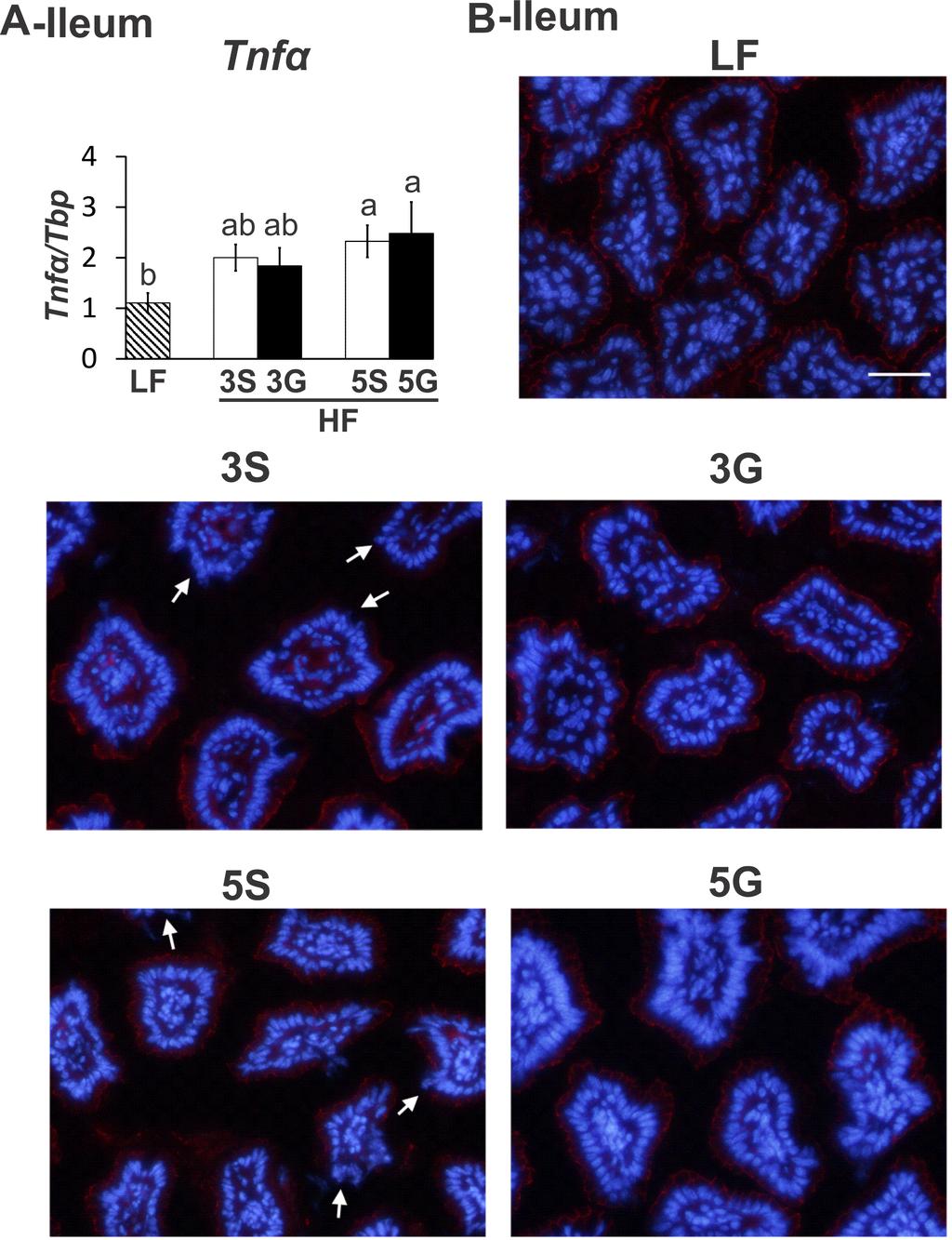
80 71
81 Figure 3.5. Alterations to Intestinal Inflammation and Barrier Function. (A) Ileum mucosa expression of TNFα and (B) the localization of the tight junction protein ZO-1 at the apical area of the ileum epithelium in C57BL/6J mice fed a low fat diet or butter-rich, high fat diets with or without 3% or 5% powdered grapes for 10 weeks. qpcr was conducted to measure mrna abundance of inflammatory genes. Localization of ZO-1 was visualized by immunostaining of ileum samples (n=5). Means ± SEM (n=9-10 for TNFα) without a common lowercase letter differ (p<0.05) using one-way ANOVA and Student s t test. LF, low fat; 3S, high fat diet containing 3% sugar; 3G high fat diet containing 3% grapes; 5S, high fat diet containing 5% sugar; 5G, high fat diet containing 5% grapes. 72
82 Figure 3.6. Diet Induced Alterations to Sulfidogenic Bacteria and Akkermansia muciniphalia. Abundance of sulfidogenic bacteria and Akkermansia muciniphilia in the intestinal mucosa or digesta of C57BL/6J mice fed a low fat diet or butter-rich, high fat diets with or without 3% or 5% powdered grapes for 10 weeks. (A) Abundance of the sulfidogenic, Bilophila wadsworthia-specific, functional gene target dissimilatory sulfate reductase (dsra-bw) and the targeted sulfidogenic bacterial genera Desulfobacter (DSB) species in the ileum mucosa. (B) The abundance of the probiotic Akkermansia muciniphilia in colon mucosa and digesta and in cecum digesta. qpcr was conducted to measure mrna abundance of the functional genes and bacterial genera. Means ± SEM (n=9-10) without a common lowercase letter differ (p<0.05) using oneway ANOVA and Student s t test. LF, low fat; 3S, high fat diet containing 3% sugar; 3G high fat 73
83 diet containing 3% grapes; 5S, high fat diet containing 5% sugar; 5G, high fat diet containing 5% grapes. Means ± SEM (n=9-10) without a common lowercase letter differ (p<0.05). 74

 Above - Area Under the Curve (AUC) of rarefaction curves shown for each diet group. Below Observed species at 15,000 sequencing reads for each diet group.")
84 Figure 3.7. Diet Induced Changes to Microbial Species. Observed bacterial species in cecum mucosa of C57BL/6J mice fed a low fat diet or butter-rich, high fat diets with or without 3% or 5% powdered grapes for 10 weeks. (A) Rarefaction curves of observed species are shown. Samples were rarified to 15,000 sequencing reads per sample. (B) Above - Area Under the Curve (AUC) of rarefaction curves shown for each diet group. Below Observed species at 15,000 sequencing reads for each diet group. LF, low fat; 3S, high fat diet containing 3% sugar; 3G high fat diet containing 3% grapes; 5S, high fat diet containing 5% sugar; 5G, high fat diet containing 75
85 5% grapes. Data are presented as Means ± SEM (n=9-10) using one-way ANOVA and Dunn s test for multiple comparisons. 76

86 Figure 3.8. PCoA Plots of Unweighted and Weighted UniFrac Distances. LF, low fat; 3S, high fat diet containing 3% sugar; 3G high fat diet containing 3% grapes; 5S, high fat diet containing 5% sugar; 5G, high fat diet containing 5% grapes. 77

87 Figure 3.9. Relative Abundance of Microbial Taxa. Significantly altered relative abundances of microbial taxa (i.e., A- Firmicutes, B- Actinobacteria, and C- Tenericutes) found across C57BL/6J mice fed a low fat diet or butter-rich, high fat diets with or without 3% or 5% powdered grapes for 10 weeks. Taxa shown were significantly altered based on Kruskal Wallis 78
88 test run using QIIME software following filtering of OTUs that were not present in 50% of samples. FDR corrected p values based on this analysis are shown. To conduct multiple comparisons relative abundances were analyzed via ANOVA followed by Dunn s test for multiple comparisons. LF, low fat; 3S, high fat diet containing 3% sugar; 3G high fat diet containing 3% grapes; 5S, high fat diet containing 5% sugar; 5G, high fat diet containing 5% grapes. Data are presented as Means ± SEM (n=9-10). Unless otherwise indicated, asterisks show significant differences of HF diets with or without grapes compared to LF control. 79
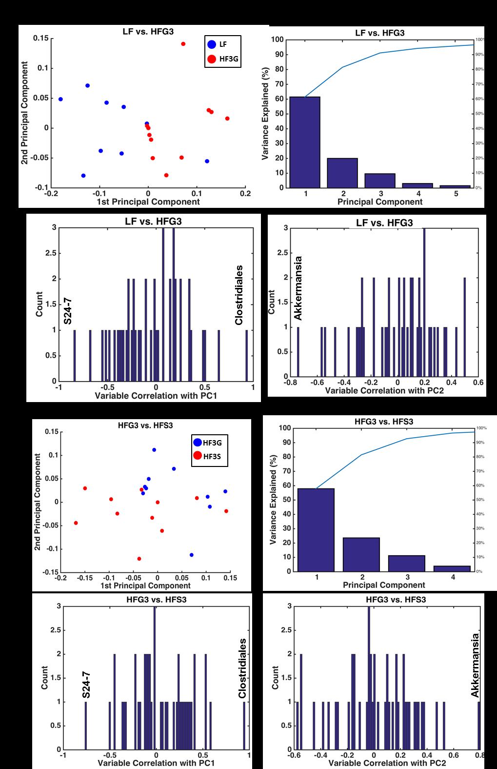
89 80
90 Figure Principle Component Analysis (PCA) of Cecum Mucosa. The microbial relative abundances from C57BL/6J mice fed a low fat diet or butter-rich, high fat diets with or without 3% or 5% powdered grapes for 10 weeks. (A) Left - PCA plot between low fat (LF) and high fat + 3% grapes (HF3G) are shown. Middle Pareto plot showing percentage of variance explained by principal components. Right Histogram showing individual genera that were negatively or positively correlated with LF and/or HF-3G diets. (B) Same as A but showing PCA of HF-3G vs high fat + 3% sugar (HF-3S) groups. 81
 Bifidobacterium relative abundance is negatively correlated with body fat percentage (r = -0.53, p = 0.")
 Heatmap showing relative abundances of taxa that were significantly correlated with body fat percentage.")
91 Figure Relationship between Body Fat and Bacterial Populations. (A) Bifidobacterium relative abundance is negatively correlated with body fat percentage (r = -0.53, p = 0.001) and inguinal fat pad weight (r = -0.48, p = 0.004) as determined by Spearman Correlation Analysis. (B) Heatmap showing relative abundances of taxa that were significantly correlated with body fat percentage. LF, low fat; 3S, high fat diet containing 3% sugar; 3G high fat diet containing 3% grapes; 5S, high fat diet containing 5% sugar; 5G, high fat diet containing 5% grapes. 82
92 References 1. Ogdan CL, Carroll MD, Kit BK, Flegal KM. Prevalence of Childhood and Adult Obesity in the United States, JAMA 2014;311(8): WHO. Obesity. Viewed. Jan. 20, NIH. Obesity. Viewed Jan. 20, Strissel KJ, DeFuria J, Shaul ME, Bennett G, Greenberg AS, Obin MS. T-cell recruitment and Th1 polarization in adipose tissue during diet-induced obesity in C57BL/6 mice. Obesity (Silver Spring) 2010;18(10): Ding S, Chi M, Scull M, Rigby R, Schwerbrock N, Magness S, Jobin C, Lund P. High fat diet: bacterial interactions promote intestinal inflammation which precedes and correlates with obesity and insulin resistance in mice. PLOS One 2010;5(8): e Garrett WS, Lord GM, Punit S, Lugo-Villarino G, Mazmanian SK, Ito S, et al. Communicable ulcerative colitis induced by T-bet deficiency in the innate immune system. Cell 2007;131(1): Cani P, Amar J, Iglesias M, Poggi M, Knauf C, Bastelica D, Neyrinck N, et al. Metabolic endotoxemia initiates obesity and insulin resistance. Diabetes 2007;56: Cani P, Bibiloni R, Knauf C, Waget A, Neyrinck A, Delzenne N, Barcelona R. Changes in gut microbiota control metabolic endotoxemia-induced inflammation in high-fat dietinduced obesity and diabetes in mice. Diabetes 2008;57: Delzenne N, Cani P. Gut microbiota and pathogenesis of insulin resistance. Cur Dab Rep 2011;11: Devkota S, Wang Y, Musch M, Leone V, Fehlner-Peach Hl, Nadimpalli A, et al. Dietaryfat-induced taurocholic acid promotes pathobiont expansion and colitis in IL10-/- mice. Nature 2012;487: Shen W, Gaskins HR, McIntosh M. Influence of dietary fat on intestinal microbes, inflammation, barrier function, and metabolic outcomes. J Nutr Biochem 2014;25: Zhang C, Zhang M, Wang S, Han R, Cao Y, Hua W, et al. Interactions between gut microbiota, host genetics, and diet relevant to development of metabolic syndromes in mice. ISME J 2010;4:
93 13. Carbonero F, Benefiel AC, Alizadeh-Ghamsari AH, Gaskins HR. Microbial pathways in colonic sulfur metabolism and links with health and disease. Front Physiol 2012;3: Rowan FE, Docherty NG, Coffey JC, O Connell PR. Sulphate-reducing bacteria and hydrogen sulphide in the aetiology of ulcerative colitis. Br J Surg 2009;96(2): Backhed F, Manchester J, Semenkovich C, Gordon J. Mechanisms underlying the resistance to diet-induced obesity in germ-free mice. Proc Nat Acad Sci 2007;104: Delzenne N, Neyrinck A, Backhed F, Cani P. Targeting gut microbiota in obesity: effects of prebiotics and probiotics. Nat Rev Endocrin 2011;7: Delzenne N, and Cani P. Interaction between obesity and gut microbiota: relevance to nutrition. Ann Rev Nutr 2011b;31: Delzenne N, Neyrinck A, Cain P. Modulation of the gut microbiota by nutrients with prebiotic properties: consequences for host health in the context of obesity and metabolic syndrome. Micro Cell Fact 2011; 10: S Cani P, Neyrinck N, Fava F, Knauf C, Burcelin R, Tuophy K, et al. Selective increases of bifidobacteria in the gut microbiota improve high-fat-diet induced diabetes in mice through a mechanisms involving endotoxemia. Diabetolgia 2007;50: Keperman R, Bolca S, Roger L, Vaughan E. Novel approaches for analyzing gut microbes and dietary polyphenols: challenges and opportunities. Microbiol 2010;156: Neyrinck A, Van Hee V, Bindels L, Possemiers S, de Backer F, Cani P, et al. Polyphenolrich extract of pomegranate peel alleviates tissue inflammation and hypercholesterolemia in high-fat-induced obese mice: potential implication of the gut microbiota. Br J Nutr 2013;109: Benn T, Kim B, Park YK, Wegner C, Harness E, Nam TG, et al. Polyphenol-rich blackcurrant extract prevents inflammation in diet-induced obese mice. J Nutr Biochem 2014;25: Van Duynhoven J, Vaughan E, Jacobs D, Kemperman R, van Velzen E, Gross G, et al. Metabolic fate of polyphenols in the human superorganism. Proc Nat Acad Sci 2011;108: Selma M, Espin J, Tomas-Barberan F. Interactions between phenolics and gut microbiota: role in human health. J Agric Food Chem 2009;57: Chuang CC, and McIntosh MK. Potential mechanisms by which polyphenol-rich grapes prevent obesity-mediated inflammation and metabolic diseases. Ann Rev Nutr 2011;31:
94 26. Seymour E, Singer A, Bennink M, Parikh R, Kirakosyan A, Kaufman P, Broiling S. Chronic intake of phytochemical-enriched diet reduces cardiac fibrosis and diastolic dysfunction caused by prolonged salt-sensitive hypertension. J Gerontol A Biol Sci Med Sci 2008; 63: Seymour EM, Bennink MR, Watts SW, Bolling SF. Whole grape intake impacts cardiac peroxisome proliferator-activated receptor and nuclear factor kappab activity and cytokine expression in rats with diastolic dysfunction. Hypertension 2010;55: Fuhrman B, Volkova N, Coleman R, Aviram M. Grape powder polyphenols attenuate atherosclerosis development in apolipoprotein E deficient mice and reduce macrophage atherogenicity. J Nutr 2005; 135: Zern T, Wood R, Greene C, West K, Liu Y, Aggarwal, et al. Grape polyphenols exert a cardioprotective effect in pre- and postmenopausal women by lowering plasma lipids and reducing oxidative stress. J. Nutr 2005; 135: Chuang CC, Shen W, Chen H, Xie G, Jia W, Chung S, McIntosh MK. Differential effects of grape powder and its extract on glucose tolerance and chronic inflammation in high fatfed obese mice. J Agric Food Chem 2012; 60: Shen W, Chuang CC, Martinez K, Reid T, Brown JM, Xi L, et al. Conjugated Linoleic Acid Reduces Adiposity and Increases Markers of Browning and Inflammation in White Adipose Tissue of Mice. J Lipid Res 2013; 54: Shen W, Wolf PG, Carbonero F, Zhong W, Reid T, Gaskins HR, McIntosh MK. Intestinal and systemic inflammatory responses are positively associated with sulfidogenic bacteria abundance in high-fat-fed male C57BL/6J mice. J Nutr 2014; 144: Caporaso JG, Kuczynski J, Stombaugh J, Bittinger K, Frederic BD, Costello EK, et al. QIIME allows analysis of high-throughput community sequencing data. Nat Methods 2010;7: doi: /nmeth.f McDonald D, Price MN, Goodrich J, Nawrocki EP, DeSantis TZ, Probst A, et al. An improved Greengenes taxonomy with explicit ranks for ecological and evolutionary analyses of bacteria and archaea. ISME J 2012;6: Wang Q, Garrity GM, Tiedje JM, Cole JR. Naïve Bayesian Classifier for Rapid Assignment of rrna Sequences into the New Bacterial Taxonomy. Appl Envir Microbiol 2007;73: Price MN, Dehal PS, Arkin AP. FastTree 2 Approximately Maximum-Likelihood Trees for Large Alignments. PLoS One 2010; 5, e
95 37. Everard A, Belzer C, Geurts L, Ouwerkerk JP, Druart C, Bindels LB, Guiot Y, Derrien M, Muccioli GG, Delzenne NM, de Vos WM, Cani PD. Cross-talk between Akkermansia muciniphila and intestinal epithelium controls diet-induced obesity. Proc Nat Acad Sci 2013; 110(20): Camari A, del Bas JM, Crescenti A, Arola L. Low doses of grape seed procyanidins reduce adiposity and improve the plasma lipid profile in hamsters. Int J Obes (Lond) 2013;37(4): Gourineni V, Shay NF, Chung S, Sandhu AK, Gu L. Muscadine Grape (Vitis rotundifolia) and Wine Phytochemicals Prevented Obesity-Associated Metabolic Complications in C57BL/6J Mice. J Agric Food Chem 2012;60(31): Hwang YP, Choi JH, Han EH, Kim HG, Wee JH, Jung KO, Jung KH, Kwon KI., Jeong TC, Chung YC, Jeong HG. Purple sweet potato anthocyanins attenuate hepatic lipid accumulation through activating adenosine monophosphate-activated protein kinase in human HepG2 cells and obese mice. Nutr Resour 2011;31(12): Zhang C, Zhang M, Wang S, Han R, Cao Y, Hua W, et al. Interactions between gut microbiota, host genetics, and diet relevant to development of metabolic syndromes in mice. ISME J 2010;4: Carbonero F, Benefiel AC, Alizadeh-Ghamsari AH, Gaskins HR. Microbial pathways in colonic sulfur metabolism and links with health and disease. Front Physiol 2012;3: Wells HF, Buzby JC. Dietary Assessment of Major Trends in U.S. Food Consumption, US Dept Agric Econ Resour 2008;
96 CHAPTER IV AN EXTRACTABLE, POLYPHENOL-RICH FRACTION OBTAINED FROM CALIFORNIA TABLE GRAPES DECREASES ADIPOSITY, INSULIN RESISTANCE, AND MARKERS OF INFLAMMATION IN HIGH-FAT FED C57BL/6J MICE Brian Collins, Jessie Baldwin, Chia-Chi Chuang, Paula Cooney, Robin Hopkins, and Michael McIntosh Abstract The objective of this study was to determine the extent to which consuming two methanolextractable fractions of California table grapes reduced adiposity, hepatic steatosis, or markers of inflammation or lipid metabolism in mice fed a high-fat, American-type diet. Male C57BL/6J mice were fed a low fat diet, a high fat (HF) diet, or a HF diet containing California table grapes (GP; 5% w/w), an extractable polyphenol (HF-EP) fraction from GP, a non-extractable polyphenol (HF-NEP) fraction from GP, or an equal combination of both fractions (HF-EP+NEP) for 16 weeks. Mice fed the HF-EP and HF-EP+NEP-containing diets had lower percentages of body fat and amounts of total white adipose tissue (WAT) and improved glucose tolerance compared to the HF controls. In epididymal WAT, the mrna levels of the inflammatory genes cluster of differentiation 11c, monocyte chemoattractant protein 1, epidermal growth factor-like module-containing mucin-like hormone receptor 1, tumor necrosis factor alpha, and the lipogenic gene, acylglycerol-3-phosphate-o-acyltransferase 2 were lower in mice fed the HF-EP- and HF- EP+NEP-containing diets compared to HF controls. Mice fed HF-NEP and HF-EP+NEP diets had reduced liver weights and mice fed HF-EP+NEP diets had lower liver triglyceride levels compared to the HF controls. Mice fed the HF-EP+NEP diets had higher hepatic mrna levels of 87
97 hormone sensitive lipase and adipose triglyceride lipase, and decreased expression of C-reactive protein compared to the HF controls. Surprising, HF-GP feeding did not decreased adiposity, hepatic steatosis, or inflammation nor did the HF or grape diets alter markers of intestinal inflammation. Taken together, these data demonstrate that the polyphenol-rich EP fraction from California table grapes attenuates many of the adverse health consequences associated with consuming a HF diet, independent of influencing intestinal inflammatory status. Introduction Obesity has been on the rise in the U.S. since the 1960 s and affects approximately one third of the adult population [1]. The incidence of obesity is also increasing globally, affecting approximately 300 million adults worldwide [2]. Obesity is intimately associated with chronic inflammatory conditions that contribute to the metabolic syndrome (e.g., type 2 diabetes, hypertension, and cardiovascular disease) [3]. Over consumption of calories coinciding with a lack of physical activity are the major risk factors for obesity development resulting in expansion of white adipose tissue (WAT). As WAT grows beyond the capacity of the vasculature system to provide it with nutrients and oxygen, an increase in inflammatory signals recruit macrophages and other immune cells into the WAT. As the immune response progresses, the abundance of circulating and tissue proinflammatory cytokines and chemokines increases [4]. This inflammatory scenario disrupts metabolic processes which results in impaired glucose and fatty acid uptake and metabolism, and hemostasis, thereby contributing to the development of metabolic diseases. Although there is a general consensus on the metabolic consequences of obesity-mediated inflammation, the exact mechanisms that initiate these events are not clearly understood. Recently, the role that gut microbes play in the development of the metabolic syndrome has received attention due to their sensitivity to environmental changes that can trigger obesity [5], 88
98 chronic inflammation [6-8], and insulin resistance [9]. Diets high in fat have been implicated in the reduction of microbial diversity, in particular gut barrier-protecting bacteria as well as increasing the abundance of deleterious bacteria [reviewed in 10]. For example, an increase in the ratio of Firmicutes to Bacteriodetes is positively correlated with the development of obesity and insulin resistance [11]. Diets rich in saturated fat [11, 12], particularly from milk [13], increase the abundance of sulfidogenic bacteria like Bilophila wadsworthia and Desulfovibrionaceae spp. which generate hydrogen sulfide gas. Hydrogen sulfide is geno- and cytotoxic and positively correlated with development of ulcerative colitis, gut inflammation, irritable bowel syndrome, and colon cancer [13-15]. Notably, the effects of high fat (HF) diet on body weight gain are repressed in microbiota-free mouse models as well as fecal microbial transplants from healthy donors into obese subjects [16]. Therefore, interventions directed towards modifying the microbial composition of the gastrointestinal (GI) tract may alleviate obesity-mediated outcomes. The use of non-digestible carbohydrates, fiber, or polyphenols as prebiotics shows promise as potential interventions for the metabolic consequences of obesity [reviewed in 13, 17-19]. Prebiotics are agents that selectively stimulate the growth or activities of specific microbial populations in the gut which translates into health benefits for the host [20]. Fiber, in particular inulin-type fructans, has been shown to increase the abundance of Bifidobacteria which was positively correlated with decreased hyperglycemia, endotoxemia, and systemic cytokine levels [21, 22]. Similar effects have been demonstrated in obese subjects with short term supplementation of gluco-oligosaccharides [23]. It is believed that the primary benefit of fiber fermentation by intestinal microbes is through increased productions of short chain fatty acids (SCFA), which have be shown to regulate the synthesis of GI peptides that influence energy intake (i.e., glucagon-like peptide (Glp)-1 and 2, peptide YY (Pyy), and ghrelin) as well as energy 89
99 storage and metabolism through interactions with G-protein receptors (Gpr) 41 and 43 [reviewed in 20]. Although much focus has been placed on the beneficial properties of fiber, polyphenols found in fruits and vegetables may also improve GI health [24-27]. Absorption of polyphenols is poor in the upper gastrointestinal tract, leading to increased availability in the lower GI tract [27]. Thus, polyphenols may have a significant influence on microbiota taxa and their metabolites [28]. Indeed, the anti-inflammatory, anti-oxidant, and anti-microbial actions of polyphenols have been reported to positively influence gut microbes and host inflammation [reviewed in 29]. As such, one potential mechanism to improve the outcomes associated with diet-induced obesity is increased consumption of poorly digestible food components including polyphenols. Grapes and other berries are rich in polyphenols including anthocyanins [reviewed in 29], which have known anti-inflammatory and anti-oxidant effects [30]. These beneficial effects of grapes have been associated with reduced cytokine levels via suppression of nuclear factor kappa B (NFκB) and increased peroxisome proliferator-activated receptor (PPAR) α/γ [31]. Consistent with these data, we demonstrated that C57BL/6J mice consuming a HF diet (i.e., 60% kcals from lard) supplemented with whole powdered California table grapes (3%, w/w) had improved glucose tolerance after 5 weeks and decreased markers of inflammation ~20-50% in serum and WAT after 18 weeks [32]. We also demonstrated that consuming a moderate fat diet (i.e., 34% kcals from milk fat) supplemented with whole powdered grapes (3% or 5% w/w) reduced adiposity, improved liver triglyceride (TG) levels, modestly reduced WAT inflammatory gene expression, and lowered the cecum levels of sulfidogenic bacteria, while tending to increase the abundance of Akkermansia muciniphila and Allobaculum in the proximal colon and cecum, respectively (Chapter III). 90
100 However, the identities of the bioactive fractions in whole powdered table grapes and the role that gut microbiota play in improvements in diet-induced obesity in mice fed grapes are unknown. Therefore, the objective of this study was to determine the extent to which consuming methanol-extractable fractions of table grapes reduced adiposity, hepatic steatosis, or markers of inflammation or lipid metabolism in mice fed a high-fat, American-type diet. We separated lyophilized powder from whole grapes into two methanol-extractable fractions; i.e., an extractable polyphenol (EP) fraction and a non-extractable polyphenol (NEP) fraction. These fractions were incorporated into a HF, American-type diet (i.e., similar in fat amount and type to the 75th percentile of American diets [33]). This HF diet was fed alone or in combination with the EP fraction, the NEP fraction, both fractions (EP+NEP), or whole grape powder (GP; 5%, w/w) for 16 weeks. Body composition and glucose tolerance were measured at 5 week intervals. At week 16, markers of intestinal and systemic inflammation, insulin resistance, and lipid metabolism were measured. Materials and Methods Reagents and materials Reference compounds procyanidin A2 (PAC-A2), procyanidin B2 (PAC-B2), catechin and epicatechin were purchased from Chromadex (Irvine, CA). 4-dimethylaminocinnamaldehyde (DMAC), Folin-Ciocalteu reagent, and trifluoroacetic acid (TFA) were purchased from Sigma- Aldrich Inc. (St. Louis, MO). All organic solvents were HPLC grade and obtained from VWR International (Suwanee, GA). Extraction and polyphenol enrichment Freeze-dried grape powder (200 g x 5 batches) was blended each with 1 L 50% acidified methanol (0.1% TFA). The mixture was centrifuged (Sorvall RC-6 plus, Asheville, NC) at
101 rpm for 10 min, and the supernatant was collected. A sample from the combined supernatants (2 L) was evaporated and freeze-dried to afford the crude extract. The rest of aq-methnol extract was evaporated to remove the organic solvent, and then loaded to Amberlite XDA-7 resin, stirred for 20 minutes, and the supernatant was discarded. The resin was washed with water to get rid of all free sugars and organic acids. The polyphenols were eluted from the resin with 100% methanol, organic solvent was evaporated under vacuum, and the remaining aqueous extract was freezedried to afford the EP fraction. The pelleted material (plant debris after extraction) was put in a vacuum oven (45 ºC) to get rid of the organic solvent before freeze-drying to afford the NEP fraction. Alkaline hydrolysis of NEP fraction Alkaline hydrolysis of NEP fraction was performed according to Yang et al. [34] with some modifications. In 15 ml centrifuge tube, 2.0 ml of 4 M NaOH were added to 0.5 g NEP, flushed with nitrogen, closed and incubated for 1 hour at room temperature. The mixture was adjusted to ph 7 with drops of concentrated hydrochloric acid, than loaded onto a column packed with celite at a ratio 1:2 v/w. The hydrolyzed polyphenols were eluted with 30 ml methanol-ethyl acetate (20:80 v/v), and evaporated to dryness. Determination of total phenolics, anthocyanins and proanthocyanidins Total phenolics (TP) were determined in EP and NEP fraction hydrolysates with Folin- Ciocalteu reagent [35]. Concentrations were expressed as mg/l gallic acid equivalents. Total monomeric anthocyanin (ANC) content was measured in EP using the ph differential spectrophotometric method [36], and expressed as cyanidin glucoside equivalent. Total proanthocyanidin content (PAC) was determined in EP using the DMAC method as previously described [37], and quantified as procyanidin A2 equivalent. 92
102 HPLC profile analyses of anthocyanins and proanthocyanidins HPLC analyses were conducted according to the previously reported protocols [38]. Animals Four-week old, male C57BL/6J mice were obtained from Jackson Laboratories (Bar Harbor, ME, USA) and acclimated on a standard chow diet for 1 week. Mice were housed in pairs, maintained at a temperature of 22 C with 50% humidity, and exposed to a 12 h light/ 12 h dark cycle. Mice received food and water ad libitum and measures of body weight and food intake were conducted once and twice per week, respectively. All experimental procedures were performed under ethical standards and approved by the Institutional Animal Care and Use Committee of the University of North Carolina at Greensboro. Diets Animals were randomly assigned to one of six dietary treatments (n=13 per treatment group) as follows: low fat (LF; 10% of energy from fat), HF (45% of energy from fat), HF plus extractable polyphenol fraction (HF-EP), HF plus non-extractable polyphenol fraction (HF-NEP), HF plus extractable and non-extractable polyphenol fraction (HF-EP+NEP) and HF plus 5% powdered grapes (HF-GP) (Table 4.1). The HF diets consisted of approximately 10.7% energy from soybean oil, 9.8% of energy from butter, 7.8% energy from lard, 12.2% energy from shortening, and 4.5% energy from beef tallow [33, 39]. Thus, the HF diets were rich in fat with the proportion of the sources of fat mimicking an American-type diet [33]. The amounts of EP and NEP fractions that were added to the HF diets were equal to their relatively amounts in the in the 5% powdered whole grape diet (i.e., grape powder contains 2.3% and 6.9% EP and NEP, respectively). The lyophilized (i.e., powdered) California table grapes were kindly provided by the California Table Grape Commission and consisted of a mixture of red, green, and purple 93
103 seeded and seedless grapes. The 5% dietary level of grapes is comparable to 15 human servings of grapes. Because the extraction process removes the sugar fraction in grapes, a mixture of fructose and glucose was added to the HF, HF-EP, HF-NEP, and HF-EP+NEP diets to control for the sugar content of the powdered grape diet. Detailed composition of the diets is illustrated in Table 4.1. Intraperitoneal glucose tolerance tests (GTT)s and fasting insulin levels Intraperitoneal (i.p.) GTTs were performed on weeks 7, 12, and 16 on non-anesthetized mice. Mice were fasted for 8 h and given an i.p. injection of glucose (i.e., 20% solution at 1 g/kg body weight). Blood from the tail vein collected at baseline and 5, 15, 30, 60, and 120 minutes post-i.p. glucose injection was used to quantify glucose levels using a Bayer Contour blood glucose monitor and strips (Bayer Healthcare, Tarrytown, NY, USA). Plasma insulin levels were detected using an ultrasensitive mouse insulin kit (Crystal Chem, Inc, Downers Grove, IL). The homeostasis model assessment method (HOMA) for insulin resistance (IR) was used employing the following formula: [fasting insulin concentration (ng/ml) x 24 x fasting glucose concentration (mg/dl)] / 405 [32]. Body fat measurements via Dual X-Ray Absorptiometry (DEXA) Percent body fat was measured using DEXA on a GE Lunar Prodigy Advanced System (GE Healthcare, Milwaukee, WI) at weeks 6, 11, and 15. During the measurement, mice were lightly anesthetized with isoflurane using a SomnoSuite Small Animal Anesthesia System with Integrated Digital Vaporizer isoflurane system. Measurements were taken in duplicate or triplicate to reduce the possibility of error and values expressed are an average of the two measurements. 94
104 Tissue collection After 16 weeks of dietary intervention, mice were fasted for 8 h and euthanized via isofluraneinduced anesthesia followed by cardiac puncture. Plasma was collected at the time of harvest. Four WAT depots were collected; epididymal, mesenteric, inguinal, and retroperitoneal. Additionally, livers were harvested and intestinal mucosa and digesta were collected from the duodenum, jejunum, ileum, cecum, or proximal or distal colon. Weights of the WAT depots and liver were recorded and all collected samples were immediately snap-frozen in liquid nitrogen and stored at -80 o C until analysis. Liver and plasma TG levels Liver TG content was measured as previously described [40]. Plasma TG content was determined using a commercial assay from Thermo Scientific and was conducted following the manufacturer s protocol (Infinity TG assay #TR22421 and TG standards #TG22923; Norcross, GA). Liver Oil-Red-O Staining Liver tissues were frozen in OCT compounds, cut at 5 μm, and mounted on slides. The sections were fixed with 10% formalin for 10 minutes and then the slides were washed with deionized water for 5 minutes. Fixed tissues were then rinsed with 60% isopropanol for 5 minutes. To prepare Oil red O stock, 0.5 g of Oil red O (Sigma-Aldrich) was mixed with 100 ml of isopropanol. To prepare Oil red O working solution, 30 ml of Oil red O stock was mixed with 20 ml of distilled water and filtered using a 0.24 µm vacuum filter. Samples were submerged in the working solution for 15 minutes, briefly rinsed with 60% isopropanol, and then rinsed with deionized water for 30 seconds before imaging. 95
105 RNA extraction and qpcr Adipose and intestinal samples were homogenized in QIAsol reagent and total RNA was extracted using QIAgen mini lipid kit obtained from Qiagen (Valencia, CA). For hepatic samples, a QIAgen mini universal kit from Qiagen was used. The quality and concentration of RNA were examined using absorbance at 260 nm and integrity determined using the absorbance ratio of 260/280 on a Nanodrop ND-1000 Spectrophotometer (Thermo Scientific, Wilmington, DE). Complementary DNA was created by reverse transcription using 1 ug of RNA and a high capacity cdna Archive Kit (Applied Biosystems, Foster City, CA) according to manufacturer s protocols. qpcr was performed in a 7500 FAST Real Time PCR system (Applied Biosystems). The expression of different genes related to inflammation, lipogenesis, and lipolysis in WAT depots and liver were measured using Taqman Gene expression assays purchased from Applied Biosystems. TATA-binding protein (Tbp) was the endogenous reference gene utilized for all assays and fold differences in gene expression were calculated as 2 ΔΔCt. Statistical Analysis Data were analyzed using a one-way ANOVA and Student s t tests to compute individual pairwise comparisons of means (p<0.05). Bonferroni s posthoc tests were also performed for specific comparisons where appropriate. Analyses were conducted using the JMP software program version 10.0 for Windows (SAS, Cary, NC). Data are expressed as means + S.E.M. Results Polyphenol content and profile of EP, NEP, and GP Polyphenol analysis revealed that the weight percentages of polyphenols in the EP and NEP fractions were 2.26% and 6.91% respectively. The EP fraction contained mg/g of total phenolics and the NEP fraction contained 10.5 mg/g of total phenolics. Total anthocyanin and 96
106 proanthocyanidin content of the EP fraction was 37.8 mg/g and mg/g, respectively, whereas these polyphenols were not detectable in the NEP fraction (Table 4.2). Individual anthocyanin analysis revealed that the most abundant anthocyanins present in the EP fraction were peonidin-3- O-glucoside (36.6 u/mg), malvidin-3-o-glucoside (30.8 u/mg), and peondin-3-o-cis (6 -O-pcoumaryl)-glucoside (28.5 u/mg) (Table 4.3). The profile of the degree of polymerization of the proanthocyanidins in the EP fraction is shown in (Fig. 4.1). EP fraction with or without NEP fraction lowers body fat gain Body weight gains and energy intakes were greater in all HF-fed mice compared to the LF control mice (Table 4.4). There was no difference in the energy intake between all of the HF-fed mice, with the exception of the HF-EP+NEP group that consumed approximately 10% fewer calories than the HF controls (Table 4.4). Mice fed the HF-EP and HF-EP+NEP diets had lower body weight gains (Table 4.4; ~28% and 40%, respectively) and body fat percentages than the HF controls (Fig. 4.2; 27% and 37%, respectively). Mice fed the HF-EP, HF-NEP and HF-EP+NEP diets had lower total WAT depot weights compared to HF controls (Fig.4.2). Paradoxically, mice fed the HF-GP diet did not have improvements in body fat percentage, body weight, and total WAT depot weights compared to the HF controls. EP fraction improves glucose disposal, insulin resistance, and hypertriglyceridemia To assess the impact of grape consumption on glucose disposal and insulin resistance, GTTs were conducted at weeks 7, 12, and 16, and fasting plasma insulin and HOMA-IR were measured at week 16, respectively. Mice consuming the HF control diet had impaired GTTs at all three time points compared to the LF controls (Fig. 4.3A). This HF diet impairment was attenuated at all three time points in mice fed the HF-EP diet. Mice fed the HF-NEP and HF-EP+NEP diets had improved GTTs at weeks 12 and 16 compared to the HF controls. HOMA-IR index calculations 97
107 at week 16 indicated that mice consuming the HF-EP+NEP fraction had improved insulin sensitivity compared to the HF controls (Fig. 4.3B). Surprisingly, consuming grapes did not significantly improve GTT, fasting plasma insulin, and HOMA-IR measurements. Mice fed the HF-EP and the HF-EP+NEP diets had lower plasma TG levels compared to the HF-fed controls (Fig. 4.3C). Grape fraction consumption lowers hepatic TG levels and alters the expression of genes associated with lipid metabolism and inflammation Liver tissue was analyzed to assess the impact of the diets on liver TG levels and markers of lipogenesis, lipolysis, and fatty acid oxidation. Mice consuming the HF control diet had greater liver weights, TG content, and mrna levels of the lipogenic genes Pparγ and Fas compared to the LF-fed mice (Fig. 4.4). Compared to LF controls, HF feeding also increased gene expression of steoryl regulatory element binding protein 1 (Srebp-1c), and stearoyl-coa desaturase 1 (Scd1), which was not effected by whole grape or grape fraction feeding (Figure 4.4). Mice consuming the HF-NEP and HF-EP+NEP diets had lower liver weights and mice consuming the HF- EP+NEP diet had lower liver TG content compared to HF controls. Mice fed the HF-EP and HF- EP+NEP diets had greater mrna levels of hormone sensitive lipase (Hsl), while mice fed the HF-EP+NEP diet had increased mrna levels of adipose triglyceride lipase (Atgl) compared to HF controls (Figure 4.4). HF feeding increased the hepatic gene expression of the inflammatory marker C-reactive protein 1 (Crp1), which was attenuated by HF-EP and HF-EP+NEP feeding (Figure 4.4). Interestingly, all mice consuming HF diets had ~60% lower mrna levels of phosphoenolpyruvate carboxykinase (Pck1), a marker of gluconeogenesis. No treatment differences in the expression of carnitine palmitoyltransferase 1-a (Cpt1a) were detected (data not shown). Hepatic oil-red-o staining indicated greater lipid content in the livers of mice fed the HF 98
108 control diet compared to the LF controls, which was attenuated by HF-EP, HF-NEP, and HF- EP+NEP consumption (Fig. 4.5). EP fraction with or without NEP fraction improves the expression of WAT genes associated with inflammation and lipid metabolism To determine if the negative metabolic effects caused by HF-feeding were associated with inflammation in WAT, mrna levels of several proinflammatory genes were measured in inguinal (subcutaneous) and epididymal (visceral) WAT. In inguinal WAT, mice fed the HF control diet had greater mrna levels of cluster of differentiation 11c (Cd11c) and monocyte chemoattractant protein 1 (Mcp1) compared to LF controls (Fig.4.6A). This HF-induced increase was attenuated in mice fed the HF-EP and HF-EP+NEP diets. In epididymal WAT, HF-feeding increased mrna levels of Cd11c, Mcp1, tumor necrosis factor alpha (TNFα), and epidermal growth factor-like module containing mucin-like hormone receptor 1 (Erm1; F4/80 human orthologue) compared to LF controls (Fig. 4.6B). Consistent with the inguinal data, mice fed the HF-EP and HF-EP+NEP diets had lower mrna levels of these genes compared to the HF controls. To determine whether the reduction in adiposity observed with feeding of HF-EP, HF-NEP, and HF-EP+NEP diets was associated in alterations in lipid metabolism, inguinal and epididymal WAT were examined for markers of; (i) lipogenesis (i.e., Pparγ, Srebp1c, Scd1, acylglycerol-3- phosphate-o-acyltransferase 2 (Agpat2), fatty acid synthase (Fas), glycerol-3-phosphate acyltransferase2 (Gpat2)), (ii) lipolysis (i.e., Atgl (iii) beta-oxidation (i.e., carnitine palmitoyltransferase 1-b (Cpt1b), Pparα), (iv) SCFA receptors (i.e. GPR43), and (v) plasminogen activator inhibitor-1 (Pai-1). In inguinal WAT, mice fed the HF diet had greater expression levels of Agpat2, which were lower in the HF-EP and HF-EP+NEP groups (Fig. 4.7A). Cpt1b gene 99
109 expression was lower in mice fed the HF-EP, HF-EP+NEP diets compared to the HF-fed mice. No treatment differences in gene expression were observed for Srebp1c, Scd1, Fas, Gpat2, Atgl, and Pparα. In epididymal WAT, mice consuming the HF diet had higher mrna levels of Agpat2, a marker of TG biosynthesis, compared to LF controls (Fig. 4.7B). Feeding HF-EP and HF- EP+NEP attenuated this HF-induced increase in Agpat2 expression. Additionally, mice fed the HF diet-had lower levels of Pparγ compared to LF controls. The expression of Pparα, a marker of beta oxidation, was reduced in mice fed the HF control and the GP diets compared to LF controls. Surprisingly, HF feeding increase the mrna levels of Cpt1b, beta-oxidation marker, which was reduced by HF-EP and HF-EP+NEP feeding. Additionally, mice fed the HF-EP+NEP diet had lower mrna levels of GPR43, a SCFA-activated receptor associated with increased energy harvest, compared to HF- and LF-fed controls. No differences in gene expression in epididymal WAT were observed for Srebp-1c, Scd1, Fas, Gpat2, and Atgl (data not shown). Minimal influence of HF-feeding and grape fraction consumption on markers of intestinal inflammation Given the reported adverse effects of consuming saturated fats [reviewed in 10] on intestinal health, and potential prebiotic impact of grapes, we measured the effects of the diets on markers of intestinal inflammation and barrier function. For inflammatory status, the mrna levels of Cd11c, Cd68, Erm1, Mcp1, Tnfα, toll-like receptor 4 (Tlr4), and interleukin (Il)1b in ileum and proximal colon mucosa and the activity of ileal myeloperoxidase were measured. Unexpectedly, the mrna levels of proinflammatory genes were not increased in the ileal mucosa of HF- fed mice compared to the LF controls. Equally surprising, mice consuming the HF-GP diets had greater mrna levels of Tlr4 and Cd68 compared to the HF-fed mice (Fig. 4.8A). Although the 100
110 activity of myeloperoxidase, an enzyme expressed neutrophils and indicative of neutrophil infiltration, was ~50% lower in the ileal mucosa of mice consuming the HF-EP and HF-EF+NEP diets compared to the HF-fed mice, these differences were not statistically significant (Fig. 4.8A). Similarly, the expression of Tlr4 was greater in the colonic mucosa of HF- GP fed mice compared to the HF controls (Fig. 4.8B). Two of the proinflammatory genes measured in the colonic mucosa where increased by HF feeding (i.e., Cd11c, Cd68) and only Cd68 was attenuated in mice consuming the HF-EP+NEP diet (Fig. 4.8B). Influence of grape fractions on intestinal L-cell gene expression Intestinal microbial metabolites impact local and systemic health, in part, by influencing fermentation products (e.g., SCFA), energy harvest, and the release of endocrine signals from L- cells located in the mucosa epithelium. Locally, these metabolites influence the integrity of the mucosa barrier and systemically impact energy balance and carbohydrate and lipid metabolism [20]. Therefore, we determined the influence of the dietary treatments on the induction of genes associated with SCFA receptors (e.g., Gpr41, -43, and -119) and two of their downstream targets (e.g., Glp and Pyy) in the ileum and proximal colon. Mice fed the HF-EP, HF-NEP, and HF- EP+NEP diets had greater mrna levels Gpr43, but not Gpr41, in the ileum mucosa compared to the HF controls (Fig. 4.9A). Mice fed the HF-EP+NEP and the HF-GP diets had lower mrna levels of Gpr41 in the ileum mucosa compared to the HF controls. Mice fed the HF-GP diet has the highest levels of Glp compared to all other treatments (Fig.4.9A). In contrast, mice fed the HF diet had higher mrna levels of Gpr43 in the proximal colon compared to the HF-NEP, HF- EP+NEP, and HF-GP groups (Fig. 4.9B). Lastly, HF-feeding increased Pyy mrna levels in the proximal colon, which were lower in mice fed the HF-EP+NEP diet. 101
111 Discussion Rationale and significance of this study We previously demonstrated that feeding whole GP in conjunction with a HF diet (i.e., 34% kcals from fat; 3% from soybean oil and 31% from milk fat, which was similar to the amount of fat consumed by Americans in the 50 th percentile [33] for 10 weeks modestly reduced adiposity (3% and 5% GP), hepatic steatosis (5% GP), and the ileal mucosa abundance of the deleterious sulfidogenic bacterium species DSB and a sulfidogenic gene drsa-bw (3% GP), and increased the proximal colon mucosa abundance of the beneficial bacterium Akkermansia muciniphila (5% GP) and the localization of the tight junction protein ZO-1 on the apical epithelial surface of the ileum (3% and 5% GP). However, consuming the HF diet for 10 weeks only modestly increased adiposity, insulin resistance, and several markers of inflammation in epididymal (visceral) WAT, and had no effects on markers of intestinal inflammation. Therefore, we conducted the current 16 week study using a higher level of dietary fat (i.e., 45% kcals) and a fatty acid profile that was similar to Americans in the 75 th percentile (i.e., soybean oil, butter, lard, shortening, and beef tallow [33] to enhance the metabolic consequences of diet-induced obesity. We also wanted to know which fraction of GP was responsible for reducing adiposity and its potential metabolic complications. Therefore, we examined the effects of whole GP (i.e., 5%) and two isolated fractions of grapes (i.e. methanol derived EP and NEP fractions which were fed at amounts equal to those in 5% GP) on the development of obesity, steatosis, glucose intolerance, and WAT inflammation in C57BL/6J mice fed a HF diet similar to Americans. The EP fraction contained ~ 16 times more polyphenols, particularly anthocyanins (i.e., glucosides of anthocyanidins; Table 4.3) and proanthocyanidins (i.e., polymers of anthocyanidins formed by the condensation of flavans; Table 4.2), than the NEP fraction. The results indicate that in the context of HF feeding, consumption of the polyphenol-rich, EP fraction, alone or in combination with the NEP fraction, 102
112 attenuated diet-induced obesity, insulin resistance, steatosis, and chronic inflammation in WAT. However, consuming whole GP had no beneficial impact on the outcomes measured, and reasons for this lack of an effect are unclear. Potential mechanisms by which anthocyanin-rich EP fraction suppresses adiposity and inflammation and improves insulin sensitivity Anthocyanins and proanthocyanidins are groups of flavonoids present in relatively large amounts in table grapes. Anthocyanins give these berries their unique colors. Our phenolic analysis indicated that the most abundant anthocyanins found in California table grapes are malvidin-3-o-glucoside, peonidin-3-o-glucoside, and cyanidin-3-o-glucoside. While studies have demonstrated anti-obesity and anti-diabetic effects of anthocyanins [reviewed in 41], studies focusing on the effects of feeding whole grapes and grape fractions feeding in subjects consuming a diet similar in fat amount and types to the American diet are lacking. Consistent with our anthocyanidin-rich EP data, mice consuming a HF diet (i.e., 60% kcals from fat, primarily lard) supplemented with anthocyanin-rich muscadine grape or wine extracts for 15 weeks had lower body weights, plasma glucose and TG levels, insulin resistance, and CRP levels compared to controls (42). Also, dietary supplementation of grape seed procyanidins has been demonstrated to prevent body weight gain, reduces WAT inflammatory markers (i.e. Tnfα, Il-6, Crp) in highfat-fed rats (43). Similar effects were observed with grape seed extract supplementation including reductions in weight gain, WAT weights and blood lipid levels in HF fed mice (44), and improvements insulin sensitivity and reductions in oxidative stress in high-fructose fed rats (45). These findings are consistent with our data demonstrating that EP-fed mice had less body weight gain, reduced WAT weights, lower plasma TG, improved insulin sensitivity, and reduced markers of inflammation in WAT (i.e. Cd11c, Mcp1, F4/80). 103
113 While research is lacking on the effects of grape anthocyanins specifically, anthocyanins from other dietary sources have been well documented in the literature. Anthocyanins have been demonstrated to consistently exert anti-inflammatory and anti-obesity effects in vitro, and in some instances in vivo. For example, anthocyanins from black soybeans reversed weight gain, reduced the levels of serum TG and cholesterol, and increased the levels of high-density lipoprotein (HDL) (46). These findings are consistent with our data demonstrating that mice fed the anthocyanin-rich EP fraction had reduced body weight gain and plasma TG levels compared to the HF controls. Also, anthocyanins from purple corn prevented HF diet induced increases in body weight and adipose tissue weights in mice (47). Similarly, in HF (60% kcals from lard) fed mice, purified anthocyanins from blueberries prevented body weight gain and fat gain compared to controls (48). Additionally, tart cherries, similar to grapes with respect to anthocyanin profile, reduced fat mass and WAT markers of inflammation including Il-6 and Tnfα. (49). These data are consistent with our data demonstrating that the anthocyanin-rich EP fraction decreased body weight gain, fat mass, plasma TG levels, and WAT expression of inflammatory markers including Tnfα, Mcp1, Cd11c, F4/80. Additionally, 3T3-L1 preadipocytes treated with anthocyanins had decreased TG accumulation and gene and protein expression of Pparγ and Fas (50). While we did not observe alterations in expression of Pparγ and Fas within the WAT, we did observe that mice fed the EP fraction had reduced expression of the lipogenic gene Agpat2. We also demonstrated in this study that mice fed the anthocyanin-rich EP fraction had improved glucose tolerance and insulin sensitivity compared to the HF controls. Similar effects of improvements in glucose tolerance were observed with supplementation of anthocyanins from Maqui Berry in HF fed mice (51) and also with supplementation of anthocyanins from Cornelian cherry in HF fed mice It has been reported that cyanidin-3-o-glucoside supplementation improves insulin sensitivity in diabetic mice via down-regulation of retinol binding protein and 104
114 up-regulation of GLUT4 gene expression (53). This could be a potential mechanism by which the EP fraction improved glucose tolerance and insulin sensitivity. One novel finding of our study was that the anthocyanin-rich EP fraction increased expression of Hsl, a gene associated with lipolysis, and reduced expression the lipogenic-related gene, Pparγ within the liver. This indicates that the EP fraction may reduce steatosis through up-regulation of lipolytic pathways and suppression of lipogenic pathways. Further research examining the effect of the EP fraction on these pathways would be beneficial to better understand the mechanisms by which the EP fraction reduces adiposity or steatosis. Relationship of intestinal inflammation to systemic inflammation High fat feeding is known to increase body fat mass and has been shown to consistently induce chronic, low grade inflammation systemically [reviewed in [54]. However, the link between diet and intestinal inflammation has not been as extensively studied. In the current study, HF feeding did not increase markers of inflammation in the ileum or proximal colon after 16 weeks, nor was there any beneficial effect of GP or grape fraction consumption. However, other research has shown that HF diets can trigger intestinal inflammation or endotoxemia that may increase systemic inflammation, possibly due to intestinal dysbiosis and barrier dysfunction [7-13]. For example, mice fed a HF diet had increased levels of saturated fatty acids and lipopolysaccharide (LPS) within intestinal tissue samples resulting in increased Tnf-α and Il-6 expression through activation of the toll-like receptor Tlr4/Nf-κB signaling pathway [18]. Mice fed a HF diet were also more susceptible to dextran sulfate sodium (DSS) induced colitis [19]. As a result the immune response was reduced which was associated with an increase in non- CD1d-restricted natural killer T (NK T) cells, which produce inflammatory cytokines similar to those secreted by adipocytes, decreasing the number of regular NK T cells normally found in the 105
115 colon. In addition, dietary supplementation with bioactive food components such as grape seed extract and red wine extracts has been shown to improve the intestinal health of subjects fed HF diets. For example, chronic consumption of grape seed extract increases tight junction protein and reduces fecal calprotectin; a neutrophil protein used as a marker of intestinal inflammation [55], and attenuates inflammatory signaling in chemically-induced colitis [56]. Furthermore, in Wistar rats treated with concentrated grape juice, induced colitis was attenuated [57]. In the current study, consumption of the EP and EP+NEP fractions notably attenuated several proinflammatory markers in the WAT, but only EP+NEP decreased the expression of the macrophage markers Cd68 in the proximal colon. Although the activity of the neutrophil enzyme myeloperoxidase was decreased by 40-50% in the EP and EP+NEP groups compared to the HF controls, these differences were not statistically significant. However, measuring the protein levels of inflammatory markers and the activity of myeloperoxidase and alkaline phosphates in the colon may provide more insight. Alternatively, the intestinal linkage to systemic effects of HF and grape fraction feeding may be due to changes in gut microbiota, as indicated in our first study (Chapter III). The intestinal microbiota is highly influenced by environmental changes which have been linked to alterations in host health and energy intake. Research has shown that alteration to the gut microbiota by HF feeding can result in increased adiposity, insulin resistance and increased inflammatory cytokines and chemokines [58-60]. This has been linked to impaired gut barrier function (i.e., the inhibition of tight junction protein production or reduction of mucin secretion from goblet cells) leading to endotoxemia [61,62] and impaired regulation of energy intake and metabolism through activation TLR-dependent enteroendocrine signaling pathways resulting in impaired neural response to leptin [reviewed in 63]. Conversely, microbial consumption of dietary fiber and polyphenols such as those found in grapes, may reduce intestinal and systemic 106
116 inflammation through regulation of energy intake and improved intestinal barrier function [reviewed in 64]. Grape polyphenols in particular may have a major impact on the microflora of the large intestine. For example, red wine grape polyphenols given to humans for 4 weeks significantly increased the number of Enterococcus, Prevotella, Bacteriodetes, and Bifidobacterium bacteria which correlated to improved blood pressure, and serum levels of TG, total cholesterol, and CRP [65]. Further evidence of the prebiotic effects of grape consumption can be seen in rats given grape pomace juice which increased fecal counts of Lactobacillus and Bifidobacterium which consequently also resulted in an increase in the concentration of primary bile acids, cholesterol, and cholesterol metabolites while decreasing the concentration of secondary bile acids [66]. This suggests that alteration of the microflora by grape polyphenols may inhibit cholesterol absorption thus improving circulating levels. Indeed, fermentation of fiber and polyphenols by colonic microbes into SCFA has been shown to regulate energy intake and metabolism through stimulating the release of glycoproteins (e.g., Glp-1, Glp-2, and Pyy) by activating Gpr43 and Gpr41 on enteroendocrine cells [reviewed in 67]. Butyrate in particular is also beneficial to intestinal homeostasis as an endothelial energy source as well as through increasing tight junction protein synthesis and mucin secretions [68, 69]. In the current study, the expression of several markers of enteroendocrine cell secretions associated with SCFA activation were upregulated. However, these results were conflicting between the ileum (i.e., Gpr43 mrna levels increased by the EP, NEP, EP+NEP diets) and the proximal colon (i.e., Gpr43 mrna levels increased by HF diet only). Future analysis of the intestinal microbiota composition, protein levels of Gprs, Pyy, and Glp1/2 levels, and gut barrier function should provide more insight about the extent to consuming HP, GP, and grape fractions influences these intestinal biomarkers. 107
117 Limitations and unanswered questions The beneficial systemic effects of consuming the EP grape fraction may be due to its rich anthocyanidin content. However, the whole GP diet did not have the same positive outcomes as the EP or NEP diets. There were several differences in the design of our previous (Chapter III) and current study that may have contributed to these conflicting results. The previous research was shorter (10 weeks) compared to the current study (16 weeks) and the diet composition with regards to fat (i.e., primarily butter fat at 34% kcals versus a combination of butter, lard, shortening, and beef tallow at 45% kcals from fat, respectively) and fiber (50 g versus 40 g cellulose, respectively). The additional 6 weeks in length of the current study may have caused any potential health benefits of the whole grape diet to have diminished. Yet, by week 10 of the second study, there were no differences between the HF and GP group in regards to body weight, body fat percentage, and glucose tolerance. Furthermore, there may have been a different interaction between the GP and the four types of fats in the current study versus the sole fat source (butter) in the first study (i.e., the polyphenols in the whole GP may interact with butter differently than with the other fat sources, thereby affecting their bioaccessibility and bioavailability). Indeed, the fat content of the diet can differentially impact polyphenol digestion and absorption [70]. Such an interaction may have enabled the polyphenols to inhibit lipase activity, possibly affecting the rate of fat absorption. The increase in calories in the current study may have been too excessive for the beneficial effects of the whole GP to have a positive impact on lipid absorption as well. In order to increase the percentage of fat in the current study, the amount of cellulose and corn starch were reduced compared to the previous study. The alteration in carbohydrate and fiber content may have resulted more time for increased absorption of macronutrients. Such a scenario would result in greater energy harvest, adipogenesis, and triglyceride synthesis in the liver and WAT, independent of any effects by the grape powder. 108
118 Furthermore, the extraction process of the anthocyanin rich fraction may have increased the bioaccessibility and subsequent bioavailability of the polyphenols within the powder by liberating them from interactions within the food matrix, making them more susceptible to interactions with brush boarder enzymes potential enhancing their absorption in the small intestine. Similarly, liberation of the polyphenols may have altered the luminal environment, thereby improving the confluence of beneficial bacteria. Another possible impact of the reduced fiber may have been on the microbiota of the large intestine, as fiber is a primary energy source for a vast majority of microbes in this environment. As such, the reduced fiber may impact the production of SCFAs as well as reduce the diversity of the microbiota, which typically results in intestinal inflammation, impaired barrier function, or adverse systemic effects. Interestingly, although the GP diet had no positive health impact in the second study, the extractable polyphenol fraction did have a positive effect on reducing HFmediated adiposity and systemic inflammation. Future studies should focus on feeding single anthocyanins and there combinations (i.e., candidates in Table 4.3) in conjunction to a HF diet to see which may be the most effective. Acknowledgements We would like to acknowledge the contributions of everyone involved in collecting data for this study. Brian Collins and Jessie Baldwin equally conducted all stages of the animal study, including caring, feeding, and weighing of the mice, the glucose tolerance testing, and the measurement of body fat using DEXA. Brian Collins additionally measured markers of ileal and colonic gene and protein expression associated with inflammation, ileal activity of myeloperoxidase. Jessie Baldwin additionally measured markers of inguinal and epididymal gene and protein expression associated with inflammation, fat depot and liver gene expression of markers of lipolysis and lipogenesis, and serum triglyceride levels. Robin Hopkins measured 109
119 plasma insulin using an ELISA assay. Mary-Ann Lila and Mary Grace together performed the methylated extraction process to produce the EP and NEP fraction and measured the concentration and composition of the polyphenols present. Chia-Chi Chuang measured liver triglyceride levels and Oil-Red-O liver staining. Paula Cooney measured the body fat mass using DEXA (with the assistance of Brian Collins and Jessie Baldwin). 110
120 Table 4.1. Diet Formulation Ingredients (gram) LF HF GP EP NEP EP+NEP Casein L-Cystine Corn Starch Maltodextrin Fructose Dextrose, Monohydrate Sucrose Cellulose Soybean Oil Butter Lard Shortening Beef Tallow Mineral Mix DiCalcium Phosphate Calcium Carbonate Potassium Citrate Vitamin Mix Choline Bitartrate NEP Grape Fraction EP Grape Fraction Grape Powder Total *LF, low fat control; HF, high fat sugar control; GP, grape powder; EP, extractable polyphenol fraction; NEP, non-extractable polyphenol fraction. 111
121 Table 4.2. Phenolic Composition of Grape Powder (GP), Extractable Polyphenol (EP) Fraction and Non-extractable Polyphenol (NEP) Fraction Assay GP EP NEP Weight percentage (%) Total phenolics ± ± 1.02 Total anthocyanins ± 1.8 ND Total proanthocyanidins 3 (mg/g) ± 7.0 ND Total phenolics, mg/g DM as gallic acid equivalent (Folin Ciocaleau assay) 2 Total anthocyanins, mg/g DM as cyaniding glucoside equivalent (ph differential assay) 3 Total proanthocyanidins, mg/g DM as cyaniding glucoside equivalent (DMAC assay) 112
122 Table 4.3. Anthocyanin Concentration in Grape Powder (GP) and the Extractable Polyphenol (EP) Fraction Peak # Identification GP Concentrations (mg/kg) 1 Delphinidin-3-O-glucoside Pyanidin-3-O-glucoside Petunidin-3-O-glucoside Peonidin-3-O-glucoside Malvidin-3-O-glucoside Malvidin-3-O-(6"-O-acetyl-glucoside) Petunidin-3-O-cis-(6"-p-coumaryl-glucoside) Cyanidin-3-O-(6"-O-p-coumaryl-glucoside) Petunidin-3-O-trans-(6"-O-p-coumarylglucoside) Peonidin-3-O-cis-(6"-p-coumaryl-glucoside Malvidin-3-O-cis(6"-p-coumaryl-glucoside Peonidin-3-O-cis-(6"-O-p-coumaryl-glucoside) Malvidin-3-O-trans-(6"-O-p-coumarylglucoside) 4.93 Total anthocyanins EP Concentrations (µ/mg) 113
123 Table 4.4. Body Weight Gain (BWG), Food Conversion Efficiency (FCE), and Total Calorie Intake per Treatment Group.* Diets BWG (g) FCE (per cage, g food/g BW) Kcal Intake (per cage) LF 11.4±0.5 d 28.74±1.24 a ±82.01 c HF 24.8±1.0 a 13.71±0.35 cd ±99.43 a EP 17.8±0.9 b 18.43±0.46 b ± ab NEP 20.2±0.9 b 15.98±0.63 c ± ab EP+NEP 15.0±0.9 c 20.71±1.21 b ±63.30 b GP 24.9±1.0 a 13.26±0.31 d ±97.74 a *LF, low fat control; HF, high fat sugar control; GP, grape powder; EP, extractable polyphenol fraction; NEP, non-extractable polyphenol fraction. 114
124 DP 6 DP 5 DP 2 DP 3 DP 4 Monomors DP > 8 units Figure 4.1. Proanthocyanin Degree of Polymerization in EP Fraction. NP-HPLC-FLD chromatogram (excitation 230 nm, emission 320 nm) for proanthocyanidins showing degree of polymerization (DP) in extractable polyphenol (EP) from grapes. 115
125 Figure 4.2. Body Fat Percentages and WAT Depot Weights. Adiposity indices of C57BL/6J mice fed a low fat (LF),high fat (HF) diet, or a HF diet containing whole powdered table grapes (GP), the extractable polyphenol (EP) fraction from grapes, the non-extractable polyphenol (NEP) fraction from grapes, or an equal combination of the EP and NEP fractions (EP+NEP) for 16 weeks. Body fat percentages were measured at weeks 6, 11, and 15 using dual energy x-ray absorptiometry (DEXA). At week 16, the inguinal, retroperitoneal, epididymal, and mesenteric white adipose tissue (WAT) depots were excised and weighed. The weights of the inguinal, retroperitoneal, epididymal, and mesenteric depots were measured, and their sum labelled total 116
126 WAT. Means ± SEM (n = 9-10) without a common lowercase letter differ (p<0.05) using oneway ANOVA and Student s test. 117
127 Figure 4.3. Glucose Tolerance Tests and HOMA-IR Index. (A)Glucose tolerance tests (GTT)s, (B) the homeostatic model assessment of insulin resistance (HOMA-IR), and (C)plasma TG levels of C57BL/6J mice fed a low fat (LF),high fat (HF) diet, or a HF diet containing whole powdered table grapes (GP), the extractable polyphenol (EP) fraction from grapes, the nonextractable polyphenol (NEP) fraction from grapes, or an equal combination of the EP and NEP fractions (EP+NEP) for 16 weeks. At weeks 7, 12, and 16, GTTs were conducted on mice fasted for 8 h and injected i.p. with a 20% glucose solution. Fasting plasma insulin was collected at week 16 and used to calculate the HOMA-IR index. Data are expressed as total area under the curve (AUC) for the GTTs. Means ± SEM (n=9-10) without a common lowercase letter differ (p<0.05) using one-way ANOVA and Student s test. 118
128 Figure 4.4. Liver Markers of Inflammation and Lipid Metabolism. Liver gene expression of markers of inflammation, lipolysis, and lipogenesis in liver of C57BL/6J mice fed a low fat (LF), high fat (HF) diet, or a HF diet containing whole powdered table grapes (GP), the extractable polyphenol (EP) fraction from grapes, the non-extractable polyphenol (NEP) fraction from grapes, or an equal combination of the EP and NEP fractions (EP+NEP) for 16 weeks. qpcr was conducted to measure mrna abundance of genes associated with hepatic inflammation, lipolysis, or lipogenesis. Means ± SEM (n=9-10) without a common lowercase letter differ (p<0.05) using one-way ANOVA and Student s test. 119
 Liver weights, (B) liver triglyceride content, and (C) Oil red O staining of liver tissue of C57BL/6J mice fed a low fat (LF),high fat (HF) diet, or a HF diet containing whole powdered table")
129 Figure 4.5. Liver Weight, Triglycerides, and Oil Red O Staining. (A) Liver weights, (B) liver triglyceride content, and (C) Oil red O staining of liver tissue of C57BL/6J mice fed a low fat (LF),high fat (HF) diet, or a HF diet containing whole powdered table grapes (GP), the extractable polyphenol (EP) fraction from grapes, the non-extractable polyphenol (NEP) fraction from grapes, or an equal combination of the EP and NEP fractions (EP+NEP) for 16 weeks. At week 16, liver tissues were excised and frozen in OCT compounds, cut at 5 μm, mounted on slides, and stained with Oil red O solution. Means ± SEM (n=9-10) without a common lowercase letter differ (p<0.05) using one-way ANOVA and Student s test. 120
130 Figure 4.6. Expression of Markers of Inflammation in WAT. The expression of markers of inflammation in (A) inguinal and (B)epididymal WAT of C57BL/6J mice fed a low fat (LF),high fat (HF) diet, or a HF diet containing whole powdered table grapes (GP), the extractable polyphenol (EP) fraction from grapes, the non-extractable polyphenol (NEP) fraction from grapes, or an equal combination of the EP and NEP fractions (EP+NEP) for qpcr was conducted to measure mrna abundance of genes associated with inflammation in inguinal (A; subcutaneous) and epididymal (B; visceral) WAT depots. Means ± SEM (n=9-10) without a common lowercase letter differ (p<0.05) using one-way ANOVA and Student s test. 121
131 Figure 4.7. Expression of Markers of Lipid Metabolism in WAT. The expression of markers of lipogenesis, lipolysis, or fatty-acid oxidation in (A) inguinal and (B) epididymal WAT of C57BL/6J mice fed a low fat (LF),high fat (HF) diet, or a HF diet containing whole powdered table grapes (GP), the extractable polyphenol (EP) fraction from grapes, the non-extractable polyphenol (NEP) fraction from grapes, or an equal combination of the EP and NEP fractions (EP+NEP) for 16 weeks. qpcr was conducted to measure mrna abundance of genes associated with lipogenesis, lipolysis, and fatty-acid oxidation in inguinal (A; subcutaneous) and epididymal 122
132 (B; visceral) WAT depots. Means ± SEM (n=9-10) without a common lowercase letter differ (p<0.05) using one-way ANOVA and Student s test. 123
133 Figure 4.8. Expression of Markers of Intestinal Inflammation. The expression of markers of inflammation in the (A) ileum, (B) proximal colon, and the (A) activity of myeloperoxidase in the mucosa of C57BL/6J mice fed a low fat (LF),high fat (HF) diet, or a HF diet containing whole powdered table grapes (GP), the extractable polyphenol (EP) fraction from grapes, the nonextractable polyphenol (NEP) fraction from grapes, or an equal combination of the EP and NEP fractions (EP+NEP) for 16 weeks. qpcr was conducted to measure mrna abundance of genes associated with inflammation in ileum mucosa and proximal colon mucosa. Means ± SEM (n=9-10) without a common lowercase letter differ (p<0.05) using one-way ANOVA and Student s t test. 124
134 Figure 4.9. Expression of G-protein Receptors and Glycoproteins in Intestinal Mucosa. The expression of G-protein receptors (Gpr) 41 and 43, peptide YY (Pyy), and glucagon like protein (Glp) were measured as markers of SCFA regulation in the (A) ileum and (B) proximal colon mucosa of C57BL/6J mice fed a low fat (LF),high fat (HF) diet, or a HF diet containing whole powdered table grapes (GP), the extractable polyphenol (EP) fraction from grapes, the nonextractable polyphenol (NEP) fraction from grapes, or an equal combination of the EP and NEP fractions (EP+NEP) for 16 weeks. qpcr was conducted to measure mrna abundance of genes associated with regulation of energy intake in ileum mucosa and proximal colon mucosa. Means 125
135 ± SEM (n=9-10) without a common lowercase letter differ (p<0.05) using one-way ANOVA and Student s test. 126
136 References 1. Ogdan CL, Carroll MD, Kit BK, Flegal KM. Prevalence of Childhood and Adult Obesity in the United States, JAMA 2014;311(8): doi: /jama WHO. Obesity. Viewed. April 4, NIH. Obesity. Last reviewed: May 12, Strissel KJ, DeFuria J, Shaul ME, Bennett G, Greenberg AS, Obin MS. T-cell recruitment and Th1 polarization in adipose tissue during diet-induced obesity in C57BL/6 mice. Obesity (Silver Spring) 2010; 18(10): Ding S, Chi M, Scull M, Rigby R, Schwerbrock N, Magness S, et al. High fat diet: bacterial interactions promote intestinal inflammation which precedes and correlates with obesity and insulin resistance in mice. PLOS One 2010; 5: e Garrett WS, Lord GM, Punit S, Lugo-Villarino G, Mazmanian SK, Ito S, et al. Communicable ulcerative colitis induced by T-bet deficiency in the innate immune system. Cell 2007; 131(1): Cani P, Amar J, Iglesias M, Poggi M, Knauf C, Bastelica D, Neyrinck N, et al. Metabolic endotoxemia initiates obesity and insulin resistance. Diabetes 2007; 56: Cani P, Bibiloni R, Knauf C, Waget A, Neyrinck A, Delzenne N, Burcelin R. Changes in gut microbiota control metabolic endotoxemia-induced inflammation in high-fat dietinduced obesity and diabetes in mice. Diabetes 2008; 57: Delzenne N, Cani P. Gut microbiota and pathogenesis of insulin resistance. Curr Diab Rep 2011; 11: Shen W, Gaskins HR, McIntosh M. Influence of dietary fat on intestinal microbes, inflammation, barrier function, and metabolic outcomes. J Nutr Biochem 2014; 25: Zhang C, Zhang M, Wang S, Han R, Cao Y, Hua W, et. Interactions between gut microbiota, host genetics, and diet relevant to development of metabolic syndromes in mice. The ISME Journal 2010; 4: Wan S, Wolf PG, Carbonero F, Wei Z, Reid T, Gaskins HR, McIntosh M K. Intestinal and Systemic Inflammatory Responses Are Positively Associated with Sulfidogenic Bacteria Abundance in High-Fat-Fed Male C57BL/6J Mice. J Nutr 2014; 144(8):
137 13. Devkota S, Wang Y, Musch M, Leone V, Fehlner-Peach Hl, Nadimpalli A, et al. Dietaryfat-induced taurocholic acid promotes pathobiont expansion and colitis in IL10-/- mice. Nature 2012; 487: Cabonero F, Benefiel AC, Alizadeh-Ghamsari AH, Gaskins HR. Microbial pathways in colonic sulfur metabolism and links with health and disease. Front Physiol 2012; 3:448. doi: /fphys Rowan FE, Docherty NG, Coffey JC, O Connell PR. Sulphate-reducing bacteria and hydrogen sulphide in the aetiology of ulcerative colitis. Br J Surg 2009; 96(2): Backhed F, Manchester J, Semenkovich C, Gordon J. Mechanisms underlying the resistance to diet-induced obesity in germ-free mice. Proc Nat Acad Sci 2007; 104: Delzenne N, Neyrinck A, Backhed F, Cani P. Targeting gut microbiota in obesity: effects of prebiotics and probiotics. Nat Rev Endocrin 2011; 7: Delzenne N, and Cani P. Interaction between obesity and gut microbiota: relevance to nutrition. Ann Rev Nutr 2011b;31: Delzenne N, Neyrinck A, Cani P. Modulation of the gut microbiota by nutrients with prebiotic properties: consequences for host health in the context of obesity and metabolic syndrome. Microbial Cell Fact 2011; 10: S Geurts L, Neyrnick A, Delzenne N, Knaug C, Cani P. Gut microbiota controls adipose tissue expansion, gut barrier and glucose metabolism: novel insights into molecular targets and interventions using prebiotics. Beneficial Microbes 2014; 5: Cani P, Neyrinck N, Fava F, Knauf C, Burcelin R, Tuophy K, et al. Selective increases of bifidobacteria in the gut microbiota improve high-fat-diet induced diabetes in mice through a mechanisms involving endotoxemia. Diabetolgia 2007; 50: Dewulf EM, Cani PD, Neyrinck AM, Possemiers S, Holle AV, Muccioli GG, et al. Inulintype fructans with prebiotic properties counteract GPR43 overexpression and PPARgammarelated adipogenesis in the white adipose tissue of high-fat diet-fed mice. J Nut Biochem 2011; 22: Serino M, Luche E, Gres S, Baylac A, Berge M, Cenac C, et al. Metabolic adaptation to a high-fat diet is associated with a change in the gut microbiota. Gut 2012; 61: Keperman R, Bolca S, Roger L, Vaughan E. Novel approaches for analyzing gut microbes and dietary polyphenols: challenges and opportunities. Microbiology 2010; 156:
138 25 Neyrinck A, Van Hee V, Bindels L, Possemiers S, de Backer F, Cani P, et al. Polyphenolrich extract of pomegranate peel alleviates tissue inflammation and hypercholesterolemia in high-fat-induced obese mice: potential implication of the gut microbiota. Br J Nutr 2013; 109: Benn T, Kim B, Park YK, Wegner C, Harness E, Nam TG, et al. Polyphenol-rich blackcurrant extract prevents inflammation in diet-induced obese mice. J Nutr Biochem 2014; 25: Van Duynhoven J, Vaughan E, Jacobs D, Kemperman R, van Velzen E, Gross G, et al. Metabolic fate of polyphenols in the human superorganism. Proc Nat Acad Sci 2011;108: Selma M, Espin J, Tomas-Barberan F. Interactions between phenolics and gut microbiota: role in human health. J Agric Food Chem 2009; 57: Chuang CC, and McIntosh MK. Potential mechanisms by which polyphenol-rich grapes prevent obesity-mediated inflammation and metabolic diseases. Ann Rev Nutr 2011; 31: Seymour E, Singer A, Bennink M, Parikh R, Kirakosyan A, Kaufman P, Broiling S. Chronic intake of phytochemical-enriched diet reduces cardiac fibrosis and diastolic dysfunction caused by prolonged salt-sensitive hypertension. J Gerontol A Biol Sci Med Sci 2008; 63: Seymour EM, Bennink MR, Watts SW, Bolling SF. Whole grape intake impacts cardiac peroxisome proliferator-activated receptor and nuclear factor kappab activity and cytokine expression in rats with diastolic dysfunction. Hypertension 2010; 55: Chuang CC, Shen W, Chen H, Xie G, Jia W, Chung S, McIntosh M. Differential effects of grape powder and its extract on glucose tolerance and chronic inflammation in high fat-fed obese mice. J Agric Food Chem 2012; (in press/on line Dec. 2012). 33. U.S. Department of Agriculture; Agricultural Research Service; Beltsville Human Nutrition Research Center; Food Surveys Research Group (Beltsville MD) and; U.S. Department of Health and Human Services; Centers for Disease Control and Prevention; National Center for Health Statistics (Hyattsville MD). What We Eat in America, NHANES Yang J, Hai Liu R, Halim L. Antioxidant and antiproliferative activities of common edible nut seeds. LWT - Food Sci and Tech 2009; 42: Singleton VL, Orthofer R, Lamuela-Raventos RM. Analysis of total phenols and other oxidation substrates and antioxidants by means of Folin-Ciocalteu reagent. Methods Enzymol 1999; 299:
139 36. Lee J, Durst RW, Wrolstad RE. Determination of total monomeric anthocyanin pigment content of fruit juices, beverages, natural colorants, and wines by the ph differential method: collaborative study. J AOAC Int 2005; 88: Prior RL, Fan E, Ji H, Howell A, Nio C, Payne MJ, Reed J. Multi-laboratory validation of a standard method for quantifying proanthocyanidins in cranberry powders. J. Sci. Food Agric 2010; 90: Grace MH,Esposito D, Dunlap K, Lila MA. Comparative analysis of phenolic content and profile, antioxidant capacity, and anti-inflammatory bioactivity in wild Alaskan and commercial Vaccinium berries. J of Agric Food Chem 2014; 62: Hintze KJ, Benninghoff AD, Ward RE. Formulation of the Total Western Diet (TWD) as a Basal Diet for Rodent Cancer Studies. J Agric Food Chem 2012; 60: Shen W, Chuang CC, Martinez K, Reid T, Brown JM, Xi L, et al. Conjugated Linoleic Acid Reduces Adiposity and Increases Markers of Browning and Inflammation in White Adipose Tissue of Mice. J Lipid Research 2013; 54: He J, and Giusti M. Anthocyanins: natural colorants with health-promoting properties Ann Rev Food Sci Technol 2010; 1: Gourineni V, Shay NF, Chung S, Sandhu AK, Gu L. Muscadine grape (Vitis rotundifolia) and wine phytochemicals prevented obesity-associated metabolic complications in C57BL/6J mice. J Agric Food Chem 2012; 60(31): Terra X, Pallares V, Ardèvol A, Cinta Bladré, Fernández-Larrea J, Pujadas G, et al. Modulatory effect of grape-seed procyanidins on local and systemic inflammation in dietinduced obesity in rats. J Nutr Biochem 2010;22(4): Park SH, Park TS, Cha YS. Grape seed extract (Vitis vinifera) partially reverses high fat diet-induced obesity in C57BL/6J mice. Nutr Res Pract 2008; 2(4): Suwannaphet W, Meeprom A, Yibchok-Anun S, Adisakwattana S. Preventive effect of grape seed extract against high-fructose diet-induced insulin resistance and oxidative stress in rats. Food Chem Toxicol 2010; 48(7): Kwon SH, Ahn IS, Kim SO, Kong CS, Chung HY, Do MS, Park KY. Anti-obesity and hypolipidemic effects of black soybean anthocyanins. J Med Food 2007; 10(3): Tsuda T, Horio F, Uchida K, Aoki H, Osawa T. Dietary cyaniding 3-O-beta-D-glucosiderich purple corn color prevents obesity and ameliorates hyperglycemia in mice. J Nutr 2003; 133(7): Prior RL, Wu X, Gu L, Hager TJ, Hager A, Howard LR. Whole berries versus berry anthocyanins: interactions with dietary fat levels in the C57BL/6J mouse model of obesity. J Agric Food Chem 2008; 56(3):
140 49. Seymour EM, Lewis SK, Urcuyo-Llanes DE, Tanone II, Kirakosyan A, Kaufman PB, Bolling SF. Regular tart cherry intake alters abdominal adiposity, adipose gene transcription, and inflammation in obesity-prone rats fed a high fat diet. J Med Food 2009; 12(5): Lee B, Lee M, Lefevre M, Kim HR. Anthocyanins inhibit lipogenesis during adipocyte differentiation of 3T3-L1 preadipocytes. Plant Foods Hum Nutr 2014; 69(2): Rojo LE, Ribnicky D, Logendra S, Poulev A, Rojas-Silva P, Kuhn P, et al. In vitro and in vivo anti-diabetic effects of anthocyanins from Maqui Berry (Aristotelia chilensis). Food Chem 2011; 131(2): Jayaprakasam B, Olson LK, Schutzki RE, Tai MH, Nair MG. Amelioration of obesity and glucose intolerance in high fat fed C57BL/6 mice by anthocyanins and ursolic acid in Cornelian cherry (Cornus mas). J Agric Food Chem 2006; 54(1): Sasaki R, Nisimura N, Hoshino H, Isa Y, Kadowaki M, Ichi T, et al. Cyanidin 3-glucoside ameliorates hyperglycemia and insulin sensitivity due to downregulation of retinol binding protein 4 expression in diabetic mice. Biochem Pharmacol 2007; 74(11): Shen W, Gaskins HR, McIntosh M. Influence of dietary fat on intestinal microbes, inflammation, barrier function, and metabolic outcomes. J Nutr Biochem 2014;25: Goodrich KM, Fundaro G, Griffin LE, Grant A, Hulver MW, Ponder MA, Neilson AP. Chronic admistration of dietary grape seed extract increases colonic expression of gut tight junction protein occludin and reduces fecal calprotectin: a secondary analysis of healthy Wistar Furth rats. Nutr Res 2012; 32: Wang YH, Ge B, Yang XL, Zhai J, Yang LN, Wang XX, et al. Pranthocyanidins from grape seed modulates the nuclear factor-kappa B signal transduction pathways in rats with TNBS-induced recurrent ulcerative colitis. Int Immunopharm 2011; 11: Paiotti AP, Neto RA, Marchi P, Silva RM, Pazine VL, Noguti J, et al. The antiinflammatory potential of phenolic compounds in grape juice concentrate (G8000 ) on 2,4,6-trinitrobenzene sulphonic acid-induced colitis. Br J Nutr 2013; 110(6): Turnbaugh PJ, Bäckhed F, Fulton L, Gordon JI. Diet-induced obesity is linked to marked but reversible alterations in the mouse distal gut microbiome. Cell Host Microbe 2008; 3: Serino M, Luche E, Gres S, Baylac A, Bergé M, Cenac C, Waget A, Klopp P, et al. Metabolic adaptation to a high-fat diet is associated with a change in the gut microbiota. Gut 2012; 61(4):
141 60. Jumpertz R, LE D, Turnbaugh P, Trinidad C, Bogardus C Gordon J, et al. Energy-balance studies reveals associations between gut microbes, caloric load, and nutrient absorption in humans. Am J Clin Nutr 2011; 94: Lam YY, Ha CW, Campbell CR, Mitchell AJ, Dinudom A, Oscarsson J, et al. Increased gut permeability and microbiota change associate with mesenteric fat inflammation and metabolic dysfunction in diet-induced obese mice. PLoS One 2012; 7(3):e Cani PD, Possemiers S, Van de Wiele T, Guiot Y, Everard A, Rottier O, et al. Changes in gut microbiota control inflammation in obese mice through a mechanism involving GLP-2- driven improvement of gut permeability. Gut 2009; 58: Reybould HE. Gut microbiota, epithelial function and derangements in obesity. J Physiol 2012; 590(3): Chuang CC, McIntosh, MK. Potential mechanisms by which polyphenol-rich grapes prevent obesity-mediated inflammation and metabolic diseases. Ann Rev Nutr 2011; 31: Queipo-Ortuño MI, Boto-Ordóñez M, Murri M, Gomez-Zumaquero JM, Clemente-Postigo M, Estruch R, et al. Influence of red wine polyphenols and ethanol on the gut microbiota ecology and biochemical biomarkers. Am J Clin Nutr 2012; 95: Sembries S, Dongowski G, Mehrlander K, Will F, Dietrich H. Physiological effects of extraction juices from apple, grape, and red beet pomaces in rats. J Agric Food Chem 2006; 54: Bleau C, Karelis AD, St-Pierre DH, Lamontagne L. Crosstalk between intestinal microbiota, adipose tissue and skeletal muscle as an early event in systemic low-grade inflammation and the development of obesity and diabetes. Diabetes Metab Res Rev 2014; [E-pub] doi: /dmrr Hatayama H, Iwashita J, Kuwajima A, Abe T. The short chain fatty acid, butyrate, stimulates MUC2 mucin production in the human colon cancer cell line, LS174T. Biochem Biophys Res Commun 2007; 356(3): Segain JP, Raingeard, de la Blétière D, Bourreille A, Leray V, Gervois N, Rosales C et al. Butyrate inhibits inflammatory responses through NFkappaB inhibition: implications for Crohn's disease. Gut 2000; 47(3): Ribnicky D, Roopchand D, Oren A, Grace M, Pouleve A, Lila MA, et al. Effects of a high fat meal matrix and protein complexation on the bioaccessibility of blueberry anthocyanins using TNO gastrointestinal model (TIM-1). Food Chem 2014; 142:
142 CHAPTER V EPILOGUE Rationale for Studies 1 and 2 Obesity is a rapidly growing health issue in the United States [1]. Of particular concern is its positive correlation with chronic diseases associated with the metabolic syndrome; i.e., type 2 diabetes, hypertension, and hyperlipidemia [2]. Studies have shown that consumption of high fat, high sugar diets over an extended period of time results in; (i) increases in cytokines such as tumor necrosis (TNF)α, interleukin (IL)-6, and monocyte chemoattractant protein (MCP)-1 released from white adipose tissue (WAT) [3-5], (ii) recruitment of macrophages to clean up necrotic adipocytes due to overstressing of the collagen matrix and vascular tissue [6,7], (iii) cytokine-induced reduction of insulin sensitivity leading to hyperglycemia [8], and (iv) ectopic fat deposition due hyperlipidemia from the diet and WAT remodeling [reviewed in 9]. Research has also demonstrated a correlation between diet-induced obesity and alterations to the intestinal environment such as increased intestinal inflammation, decreased gut barrier function, and alterations to the gut microbiota [10-12]. Current strategies for suppressing the deleterious inflammation association with WAT include reducing calorie intake, increasing physical activity levels, pharmacological interventions, and surgery [13]. However, long-term lifestyle changes associated with these strategies are often difficult to maintain, thus negating their beneficial effects on weight reduction. Therefore, it is important to find alternatives for combating obesityrelated symptoms that may prove more maintainable and sustainable. One potential dietary strategy to reduce pro-inflammatory and pro-oxidant effects of diets rich in fat is the consumption 133
143 of grapes, grape products, or supplements containing grape powder or extracts, given their inherent abundance of polyphenols, which have reported anti-inflammatory and anti-oxidant properties. Currently, research on grapes and grape products (e.g. grape juice, raisins, wine, grape extracts, grape powder) has demonstrated that the polyphenols in these products (e.g., anthocyanins, quercetin, and resveratrol) possess antioxidant and anti-inflammatory properties that are beneficial in protecting the cardiovascular system and attenuating some diet-induced proinflammatory markers [14-18]. However, the mechanism by which polyphenol-rich foods such as grapes attenuate obesity-mediated inflammation is still unclear. Reducing the deleterious effects of a high fat (HF) diet on intestine health may be one such potential mechanism. In addition to the polyphenols in grapes, they also contain fiber which is known to improve gut barrier function and influence commensal bacterial composition [19-22]. Therefore, I investigated the following aims; Aim 1. Determine the impact of consuming California table grapes on intestinal health in mice fed an American type diet rich in one type of saturated fat (Chapter III); and Aim 2. Identify a key fraction of California table grapes that improves markers of intestinal inflammation in mice fed an American type diet rich in four types of saturated fats (Chapter IV). Summary of Results from Studies 1 and 2 The results from these two studies varied. In the first study consuming one or both levels of powdered whole grapes (i.e., 3 or 5%, w/w) in conjunction with the HF diet (i.e., 34% kcals from fat) rich in butter fat (Chapter III) reduced body fat percentage, the total weight of all four fat 134
144 depots, or inguinal fat depot weight after 10 wk of treatment. However, these beneficial effects of grape consumption did not equate to improved glucose tolerance. There was an improvement in a marker of gut barrier function (i.e., increased localization of the tight junction protein zonula occluden (ZO)-1 on the apical surface of the ileum epithelium) in the ileum by powder grapes when compared to their HF fed controls. However, the HF diet did not significantly increase the m RNA levels of several proinflammatory genes (e.g., tumor necrosis factor-α (Tnfα), cluster differentiation (Cd)11c, mucin-like hormone receptor 1 (Erm1), monocyte chemoattractant protein 1 (Mcp1), and toll-like receptor 4 (Tlr4)) or proinflammatory enzymes (e.g., myeloperoxidase and alkaline phosphatase) in intestinal mucosawhen compared to low fat controls. High-fat-fed mice consuming grapes had a lower abundance of deleterious sulfidogenic bacteria (e.g., decreased mrna levels of Desulfobacter spp and dsra genes) when compared to controls in the cecum mucosa, and a trend for greater abundance of the beneficial bacteria Akkermansia muciniphila in the cecum digesta, colon digesta, or colon mucosa. High fat feeding decreased the relative abundance and variance of bacterial populations compared to low fat controls. Consumption of 3% grape powdersimproved variance compared to its respective controls. For example, 3% grape supplementation increased the abundance of Ruminoccocus and Anaeroplasma in the Firmicutes and Tenericutes phylum, respectively, along with S24-7 of the Bacteriodes phylum compared to the 3% HF sugar controls. Relatively no differences were observed between the 5% grape supplementation and its respective controls. Lastly, certain beneficial bacteria populations (Bifidobacterium, Lactobacillus, Allobaculum) were inversely correlated with body fat mass and inguinal fat depot weights In the second study (Chapter IV), mice were fed a higher level of fat (i.e., 45% kcals from fat) containing a mixture of saturated fat sources (i.e., lard, beef tallow, shortening, and butter) that more closely represented the American diet (i.e., 75 th percentile according to [23]). This higher 135
145 level of fat was given for 16 weeks in order to determine if it would increase intestinal and systemic inflammation, because such pro-inflammatory effects were not observed in the first study after 10 wk of feeding a HF diet (35% kcal) primarily composed of butter. These HF fed mice were given one of three fractions; i.e., a methanol-extractable, polyphenol-rich fraction (EP), a non-extractable, polyphenol-poor, fiber rich fraction (NEP), and the combination of both fractions (EP + NEP). The levels of EP and NEP given were similar to those found in the 5% grape diet using in the first study. A 5% grape diet was also fed to mice, based on the outcomes of the first study. Notably, the EP fraction alone or in combination with NEP (EP+NEP) reduced body fat percentage, total body fat depot weights, and inguinal, mesenteric, and epididymal fat depot weights when compared to the HF controls. Rates of glucose disposal and the HOMA-IR score were also improved in the EP and EP+NEP groups compared to the HF controls. Lastly, the mrna levels of several inflammatory genes in WAT were lower in the EP and EP+NEP group compared to the HF controls. Surprisingly, the 5% grape diet did not reduce adiposity, as in Study 1, and it actually impaired glucose disposal compared to the mice fed the HF diet alone. Similar to the first study, no markers of inflammation were upregulated in the ileum by HF diet alone; however grape powder did significantly increase the expression of Tlr4 and Cd68, but not others (e.g., Tnfα, Cd11c, Mcp1, and inter-leukin (Il)1-β). In the proximal colon, HF diet increased the expression of Cd11c and Cd68, with only EP+NEP attenuating the expression of Cd68. Grape powder once again increased the expression of Tlr4. However, G-protein receptor (Gpr) 43 expression was up regulated by the grape fraction diets (EP, NEP, and EP+NEP), indicating that ligands for this receptor (e.g., short chain fatty acids (SCFA)) may have been increased in the ileum. However, data on SCFA abundance, ZO-1 localization, and microbiota profiles are not currently available. 136
146 Interpretations and Implications for the Results from Study 1 and 2 Potential reasons for the differential effects of the whole grape diets Taken together, these results indicate that the beneficial systemic effects of consuming grapes in first study (Chapter III) and the EP fraction in the second study (Chapter IV) may be due to their rich polyphenol content. The results of the second study, however, did not demonstrate the same beneficial systemic effects from consuming the powdered grapes as were seen in the first study. There were several differences in the design of the two studies that may have contributed to these conflicting results; (1) the length of the study- 10 weeks versus 16 weeks, (2) the fat composition- primarily from butter versus from butter, lard, shortening, and beef tallow, (3) the percentage of kilocalories from fat- 34% kcals versus 45% kcals, and (4) the amount of fiber (50 g and 40 g cellulose respectively). The length of study 2 was 6 weeks longer than study 1, which may have caused any potential health benefits of the whole grape diet to have diminished. Yet, by week 10 of study 2, there were no differences between the HF and grape powder group in regards to body weight, body fat percentage, and glucose tolerance. As previously mentioned, the first study focused on a single saturated fat type (i.e., butter) while the second study was a mixed saturated fat diet (i.e., butter, lard, shortening, and beef tallow) more representative in composition to the American diet. As a result, there may have been a different interaction between the powdered grapes and the four types of fats in the second study 2; (i.e., the polyphenols in the powder grapes may interact with milk fat differently than with the other fat sources). Such an interaction may have enabled the polyphenols to inhibit lipase activity, possibly affecting the rate of fat absorption. The second study contained a higher percentage of fat (45% kcals) then the first study (34% kcals). It is likely that the increase in calories was too excessive for the beneficial effects of the whole food to have a positive impact on lipid absorption. In order to increase the percentage of fat in the second study, the amount of fiber was 137
147 reduced compared to the first study (i.e., cellulose was reduced by 10 g and corn starch was reduced by 120 g).the alteration in carbohydrate and fiber content may have resulted in more time in transit, allowing more time for increased absorption of macronutrients. Such a scenario would result in greater energy harvest, adipogenesis, and triglyceride synthesis in the liver and WAT, independent of any effects by the grape powder. Another possible impact of the reduced fiber may have been on the microbiota of the large intestine, as fiber is a primary energy source for a vast majority of microbes in this environment. As such, the reduced fiber may impact the production of SCFAs as well as reduce the diversity of the microbiota which typically results in intestinal inflammation or adverse systemic effects. Interestingly, although the powdered grapes had no positive health impact in the second study, the extractable polyphenol fraction did have a positive effect on reducing high-fat-mediated adiposity and systemic inflammation. Potential reasons for the beneficial effects of the EP fraction I speculate that the acidified-methanol used to extract the polyphenols in the second study may have improved the bioaccessibility and thus bioavailibity of the polyphenols in grapes. This may have increased the beneficial effects of the liberated polyphenols in several ways that would not have been as effective as part of the whole grape. The EP fraction, which is rich in anthocyanins, may have impacted lipid absorption by interacting with lipid droplets, increasing the size of the hydroscopic outer layer of the droplet yet reducing the surface area for interaction with pancreatic lipases. This would directly impact the intestinal absorption of fat, thus reducing the amount of free fatty acids available for triglyceride synthesis, chylomicron packaging, and subsequent storage in WAT. Lipids may also bind to polyphenols and protect them from metabolism in the intestine, thus improving their bioavailability. As the polyphenols in the EP fraction are not 138
148 bound to other components as they are in the whole grape, they would be able to bind to the lipids and thus increase their concentrations systemically. The antioxidant properties of the polyphenols in the EP fractions may also have been enhanced following extraction; i.e., once they were separated from the matrices within the whole grape by the acidified methanol. This may have aided in reducing any deleterious effects of potential lipid peroxidation produced during the digestive process. As polyphenols are poorly absorbed (e.g., 1-5%), these changes may have been a result of colonic microbial interaction with the extracted polyphenols. This may have led to alterations in the intestinal microbiota that could have improved the concentration of beneficial bacteria (i.e., Bacteriodetes or Lactobacillus) and reduce that of deleterious bacteria (i.e., Firmicutes or Clostridium) [24-28]. The increase in products of the beneficial bacteria, namely the production of SCFAs, may have resulted in improved insulin sensitivity and satiety through interaction with G-protein receptors located on enteroendocrine cells. Consistent with this hypothesis, Gpr43 mrna levels were higher in the EP, NEP, and EP+NEP groups compared to the HF controls. However, the contribution of gut microbes to these systemic health benefits is still speculative, because of the minimal impact of the HF diets and grapes on markers of intestinal health. Similarly, it is unclear whether or not powdered grapes or their extracts improve intestinal health, and if such changes translate into improvements observed in weight, body fat percentage, and systemic markers of inflammation and lipid metabolism assessed by my collaborator Jessie Baldwin. Research questions based on results Based on these findings, I have developed the following research questions; (i) What effects would grape consumption have on intestinal inflammation and microbiota in a model of induced colitis using a chemical agent (e.g., dextran sulfate sodium) or in a genetic model (e.g., IL10 139
149 knockout) mouse models?, (ii) What effects would grape consumption have on intestinal inflammation and microbiota of C57BL/6J mice fed 60% kcals from fat?, (iii) What effect would grape consumption have in concert with a probiotic on the intestinal health, microbiome composition, and SCFA production in the intestine of mice fed a HF diet?, (iv) What effects would the EP fraction compared to whole grapes have in the previous three models mentioned above?, (v) What impact would EP fraction have on the growth, differentiation, or mucin secretion of goblet cells (endothelial mucin secreting cells) and HT29 cells (human colonic adenocarcinoma cells that are able to differentiate into mature intestinal cells)?, (vi) What impact would the EP fraction have on the growth of beneficial bacteria like Lactobacillus acidophilus, L. plantarum, L. delbruekii, or Akkermansia muciniphila in culture?, (vii) Would culturing the microbiota from the cecum of mice fed extractable polyphenols with known prebiotics/probiotics produce an exponential increase in beneficial products compared the cecum microbiota of mice fed a control diet and similarly treated?, (viii) Would individual anthocyanins, alone or in combination, that are abundant in the EP fraction have a greater effect on intestinal and systemic inflammation in mice then the EP itself?, and (ix) What would be the potential mechanisms for the attenuation of systemic inflammation and insulin resistance in mice fed a HF diet supplemented with the EP fraction or candidate polyphenols? Future analysis for the second study will involve exploration of the potential anti-oxidant effects of powdered grapes and the EP fraction. This will be done by measuring the mrna or activity levels of enzymes associated with anti-oxidant related transcription factor NF-E2-related factor-2 (e.g., superoxide dismutase 2, glutathione peroxidase 2, heme oxygenase 1, and glutamate-cysteine ligase, catalytic subunit) which are known to combat oxidative stress through reduction of reactive oxygen species. These results along with future research studies conducted to answer my proposed questions could be critical in providing a better understanding of the 140
150 interaction between microbiota, polyphenols, and intestinal and systemic inflammation. In so doing, it is possible that a new area of dietary intervention could be established in regards to chronic inflammatory diseases. My research has demonstrated that grapes and their extracted polyphenols may improve health outcomes, depending on the level and type of fat calories. As such, implementing effective dietary strategies using grapes or their extracted polyphenols would have the benefit of reducing health care cost and loss of productivity associated with obesity and subsequent comorbidities. 141
-1, grape powder (GP), homeostasis model assessment-estimated insulin resistance (HOMA-IR), extractable polyphenol fraction (EP), nonextractable polyphenol fraction (NEP),")
151 Figure 5.1. A Model of the Anthropometric and Intestinal Measurements Recorded in Study 1 and Study 2. zonula occludin (ZO)-1, grape powder (GP), homeostasis model assessment-estimated insulin resistance (HOMA-IR), extractable polyphenol fraction (EP), nonextractable polyphenol fraction (NEP), G-Protein receptor (GPR), toll-like receptor (TLR)4, cluster of differentiation (CD), proximal (P.). 142
Dysbiosis & Inflammation
 MASTERING THE MICROBIOME: Dysbiosis & Inflammation 2017 Tom Fabian, PhD It is reasonable to propose that the composition of the microbiome and its activities are involved in most, if not all, of the biological
The role of intestinal microbiota in metabolic disease-a novel therapeutic target.
 Michael Connolly Department of Food Biosciences, The University of Reading The role of intestinal microbiota in metabolic disease-a novel therapeutic target. University of Reading 2008 www.reading.ac.uk
Gut Microbiota and IBD. Vahedi. H M.D Associate Professor of Medicine DDRI
 Gut Microbiota and IBD Vahedi. H M.D Associate Professor of Medicine DDRI 1393.3.1 2 GUT MICROBIOTA 100 Trillion Microbes - 10 times more than cells in our body Collective weight of about 1kg in human
Intestinal Microbiota in Health and Disease
 Intestinal Microbiota in Health and Disease February 27, 2015 Master s Course in Gastroenterology Prof. Kathy McCoy 1 Overview Overview of Gut Microbiota Microbiota in Health Microbiota in Disease 2 Gut
9/25/2018 COPE WEBINAR SERIES FOR HEALTH PROFESSIONALS DID YOU USE YOUR PHONE TO ACCESS THE WEBINAR?
 COPE WEBINAR SERIES FOR HEALTH PROFESSIONALS DID YOU USE YOUR PHONE TO ACCESS THE WEBINAR? September 26, 2018 The Gut Microbiome-Diabetes Connection Moderator: Lisa Diewald MS, RD, LDN Program Manager
HOW THE MICROBIOME AFFECTS OUR HEALTH
 HOW THE MICROBIOME AFFECTS OUR HEALTH THE INTESTINAL BARRIER AND INTESTINAL PERMEABILITY Intestinal Barrier: a functional body Defense from translocation of dietary antigens, bacteria or bacterial endotoxins
MICROBIOM AND OBESITY HEINZ GYAKY 2018 BUDAPEST
 MICROBIOM AND OBESITY HEINZ GYAKY 2018 BUDAPEST HUMAN MICROBIOM 10 Billion bacterias are building a 1,5 2 kg heavy human microbiom It is located mainly in the human gut There is a intestinal controlled
The Gut Microbiota: Evidence For Gut Microbes as Contributors to Weight Gain
 The Gut Microbiota: Evidence For Gut Microbes as Contributors to Weight Gain Michael T. Bailey, Ph.D. Center for Microbial Pathogenesis The Research Institute, Nationwide Children s Hospital Department
ROLE OF THE GUT BACTERIA
 ROLE OF THE GUT BACTERIA Our Good Bacteria In a perfect world, we would all have a proper ratio of good bacteria And what could this proper ratio do for us? The knowledge of the connections between our
Dietary supplementation in treating non-alcoholic fatty liver disease Dr. Ahmad Saedi Associate Professor School of Nutritional Sciences and
 Dietary supplementation in treating non-alcoholic fatty liver disease Dr. Ahmad Saedi Associate Professor School of Nutritional Sciences and Dietetics Tehran University of Medical Sciences Honorary Academic
Overview of the Microbiome in Health and Disease Cindy D. Davis
 Overview of the Microbiome in Health and Disease Cindy D. Davis davisci@mail.nih.gov OFFICE OF DIETARY SUPPLEMENTS 1 Outline 1.What is the microbiome? 2.What is the evidence that diet can influence the
Health Benefits of Prebiotic Dietary Fiber
 Health Benefits of Prebiotic Dietary Fiber JENNIFER ERICKSON, PhD, RD Objectives Provide some background on dietary fiber To define the term "prebiotic dietary fiber" To discuss potential health effects
Dietary Fibres Soluble Fibres: can be.. Insoluble Fibres : can be..
 Dietary Fibres The fraction of edible parts of plants or analogous carbohydrates that are: Resistant to digestion and absorption in the human small intestine with.. Complete or partial fermentation in
OBESITY AND THE CONNECTION TO THE GUT
 OBESITY AND THE CONNECTION TO THE GUT Weight Loss Most weight loss programs are based on calorie in = calorie out Does not matter how they are dressed up Most of these plans being healthier foods to people
Diet, Microbiome and Health Cindy D. Davis
 Diet, Microbiome and Health Cindy D. Davis davisci@mail.nih.gov OFFICE OF DIETARY SUPPLEMENTS 1 Outline 1.What is the microbiome? 2.How does it vary over the lifespan? 3.What is the evidence that diet
Department of Nutrition and Food Science Texas A&M University
 PREVENTION OF OBESITY RELATED DISEASES THROUGH CHERRY CONSUMPTION: WHAT WE CAN SAY SO FAR? Giuliana Noratto, Ph.D. Department of Nutrition and Food Science Texas A&M University OUTLINE CHERRIES FOR PREVENTION
Necrotizing Enterocolitis: The Role of the Immune System
 Necrotizing Enterocolitis: The Role of the Immune System Patricia Denning, M.D. Associate Professor in Pediatrics Division of Neonatology Emory University School of Medicine What is NEC? What is NEC? Necrotizing
Module Outline. 1. Microbiome overview: getting a sense of the microbiome, research, what we know
 Module Outline 1. Microbiome overview: getting a sense of the microbiome, research, what we know 2. Bacteria: features, functions, communities & taxonomy 3. Other microbes: archaea, fungi, viruses, parasites
Innate Immunity. Chapter 3. Connection Between Innate and Adaptive Immunity. Know Differences and Provide Examples. Antimicrobial peptide psoriasin
 Chapter Know Differences and Provide Examples Innate Immunity kin and Epithelial Barriers Antimicrobial peptide psoriasin -Activity against Gram (-) E. coli Connection Between Innate and Adaptive Immunity
GUT MICROBIOME WHAT IS IT? WHY IS IT IMPORTANT FOR HUMAN HEALTH?
 GUT MICROBIOME WHAT IS IT? WHY IS IT IMPORTANT FOR HUMAN HEALTH? Corrie Whisner, PhD School of Nutrition and Health Promotion Arizona State University Center for Research on Ingredient Safety Annual Meeting
Increasing barrier function with multispecies probiotics
 Increasing barrier function with multispecies probiotics Elsbeth Pekelharing, MSc Science Liaison We strive for the most effective microbiome management solutions with our evidence-based & indication-specific
The enteric microbiota: Implications for IBD. Eugene B. Chang, M.D. University of Chicago
 The enteric microbiota: Implications for IBD Eugene B. Chang, M.D. University of Chicago On a per cell basis, humans are mostly prokaryote 100 90 80 70 60 50 40 30 20 10 0 EuK ProK The microbial flora
Diverticular Disease: Looking beyond fiber
 Binghamton University The Open Repository @ Binghamton (The ORB) Health & Wellness Studies Faculty Scholarship Decker School of Nursing Fall 10-28-2015 Diverticular Disease: Looking beyond fiber Lina Begdache
Studies on probiotics effects on innate immune functions in the gastrointestinal tract of broiler chicks (SUMMARY)
") Doctoral Thesis Studies on probiotics effects on innate immune functions in the gastrointestinal tract of broiler chicks (SUMMARY) ELSAYED SEDDEK IBRAHEM MOHAMMED Department of Bioresource Science Graduate
Doctoral Thesis Studies on probiotics effects on innate immune functions in the gastrointestinal tract of broiler chicks (SUMMARY) ELSAYED SEDDEK IBRAHEM MOHAMMED Department of Bioresource Science Graduate
4/17/2019 DISCLOSURES OBJECTIVES GI MICROBIOME & HEALTH: A REVIEW. Nancy C. Kois, MD, FCAP Contemporary Pathology Services. There are no disclosures
 GI MICROBIOME & HEALTH: A REVIEW Nancy C. Kois, MD, FCAP Contemporary Pathology Services DISCLOSURES There are no disclosures OBJECTIVES Definitions: GI microbiota, GI microbiome, probiotic, prebiotic
The two different types of fibers are soluble and insoluble fibers
 FIBER FACTS Fiber is the indigestible remnants of plant cells found in fruits, vegetables, whole grains, nuts, seeds, and beans, act through the digestive tract. It cannot be digested by enzymes present
The Gut Microbiome: 101 Justin Carlson University of Minnesota
 The Gut Microbiome: 101 Justin Carlson University of Minnesota Where are we now? 360 B.C. 2003 Human Gut Microbes Associated With Obesity Ley et al., Nature. 2006. Consumer Driven Science For Better of
Bacteriology. Mycology. Patient: SAMPLE PATIENT DOB: Sex: MRN: Rare. Rare. Positive. Brown. Negative *NG. Negative
 Patient: SAMPLE PATIENT DOB: Sex: MRN: 3.2 0.9-26.8 U/g 1.2 0.2-3.3 mg/g 2.2 1.3-8.6 micromol/g 1.1 1.3-23.7 mg/g 1.1 0.2-3.5 mg/g Rare 1.0 0.2-8.8 mg/g Rare 4.4 2.6-32.4 mg/g 64.6 >= 13.6 micromol/g Bacteriology
9/8/17. Reducing Inflammation + Supporting the Immune System. Module #2 Fighting Inflammation
 Reducing Inflammation + Supporting the Immune System 1 Table of Contents Module 1: What is Inflammation? Inflammatory markers Signs and symptoms Chronic disease risk Module 2: Fighting Inflammation Acidity/alkalinity
INTESTINAL MICROBIOTA EXAMPLES OF INDIVIDUAL ANALYSES
 EXAMPLES OF INDIVIDUAL ANALYSES INTESTINAL MICROBIOTA Microbiota in the animal or human intestine has evolved together with the host. Consequently, the gastrointestinal tract could be considered a metacommunity,
Bibliografia Microbiota
 Bibliografia Microbiota Systematic Review: Gut Microbiota in Fecal Samples and Detection of Colorectal Neoplasms. The role of the intestinal microbiome in ocular inflammatory disease. The gut microbiome
Gut Lung Axis Implication of the Gut Microbiota beyond its niche
 Gut Lung Axis Implication of the Gut Microbiota beyond its niche Reema Subramanian PhD Candidate (4 th year) Supervisor: Prof. Margaret Ip Department of Microbiology, CUHK Joint Graduate Student Seminar
Gut microbiota, metabolic syndrome, obesity and the nutrient sensor pathways
 Gut microbiota, metabolic syndrome, obesity and the nutrient sensor pathways Department of Gastroenterology, Endocrinology & Metabolism Medical University Innsbruck Herbert Tilg Nothing to disclose Fig.
ulcer healing role 118 Bicarbonate, prostaglandins in duodenal cytoprotection 235, 236
 Subject Index Actin cellular forms 48, 49 epidermal growth factor, cytoskeletal change induction in mucosal repair 22, 23 wound repair 64, 65 polyamine effects on cytoskeleton 49 51 S-Adenosylmethionine
The number of microorganisms residing in our intestines is 10 times the number of our somatic and germ cells.
 The number of microorganisms residing in our intestines is 10 times the number of our somatic and germ cells. The number of microorganisms residing in our intestines is 10 times the number of our somatic
Slide 1. Slide 2 Learning outcomes. Slide 3. Year 1 MBChB Lecture 15 Introduction to the Gut Microbiota. The importance of microbiota
 Slide 1 Year 1 MBChB Lecture 15 Introduction to the Gut Microbiota Professor Barry Campbell Gastroenterology Research Unit Cellular & Molecular Physiology, Institute of Translational Medicine bjcampbl@liv.ac.uk
Is there an anti-inflammatory diet in IBD?
 CCFA North Texas Chapter IBD education symposium December 2, 2017, Dallas, TX Is there an anti-inflammatory diet in IBD? Themos Dassopoulos, MD Director, Baylor Scott and White Center for IBD Baylor University
VITAMINS, MINERALS AND THE GUT
 VITAMINS, MINERALS AND THE GUT Nutrients Looking at individual nutrients that are involved with gut health can be misleading This is not about taking individual nutrients It supports more a whole food
The impact of the microbiome on brain and cognitive development
 The Gut-Brain Axis The impact of the microbiome on brain and cognitive development Diane Stadler, PhD, RD Oregon Health & Sciences University, Portland, Oregon Lao-American Nutrition Institute With acknowledgements
Fecal Microbiota Transplantation for Severe sepsis and Diarrhea : a Case Report
 Fecal Microbiota Transplantation for Severe sepsis and Diarrhea : a Case Report Qiurong Li Institute of General Surgery, Jinling Hospital Nanjing Univeristy Gut Microbiota 100 trillion cells 10-fold of
PREBIOTIC MECHANISMS OF ACTION
 PREBIOTIC MECHANISMS OF ACTION Seema Hooda, Kelly S. Swanson, George C. Fahey, Jr. Department t of Animal Sciences Division of Nutritional Sciences University of Illinois at Urbana-Champaign Institute
Resistant Starch A Comparative Nutrition Review. James M. Lattimer Ph.D., PAS Assistant Professor Animal Sciences & Industry
 Resistant Starch A Comparative Nutrition Review James M. Lattimer Ph.D., PAS Assistant Professor Animal Sciences & Industry Personal Background Education B.S. Kansas State University Animal Sciences &
The role of nutrition in optimum gastrointestinal health
 The role of nutrition in optimum gastrointestinal health Kelly A. Tappenden, Ph.D., R.D., FASPEN Kraft Foods Human Nutrition Endowed Professor University Distinguished Teacher-Scholar University of Illinois
XV Jornada de Avances en Hepatología Málaga 20 y 21 de Mayo de Barrera intestinal en la cirrosis. Agustín Albillos
 XV Jornada de Avances en Hepatología Málaga 20 y 21 de Mayo de 2016 Barrera intestinal en la cirrosis Agustín Albillos Hospital Universitario Ramón y Cajal Universidad de Alcalá, Ciberehd Madrid, Spain
Novel method of probiotic administration
 Novel method of probiotic administration Multifactorial Prematurity Low birth weight Enteral feeds Altered intestinal microflora microflora Infants that develop NEC have changes in microbial community:
What Have We Learned About the Microbiome in Pediatric IBD?
 What Have We Learned About the Microbiome in Pediatric IBD? Ted Denson, MD Cincinnati Children s Hospital Medical Center and the University of Cincinnati College of Medicine Disclosures: NIH, CCF, AbbVie
What is the evidence that dietary components can act on the microbiome and influence health?
 What is the evidence that dietary components can act on the microbiome and influence health? Kristin Verbeke Translational Research in Gastrointestinal Disorders KU Leuven, Leuven, Belgium Diet? health
GROUP 5. Jerrold R. Turner Nathalie Delzenne Wenke Feng Reuben Wong Thierry Piche Yehuda Ringel Irina Kirpich Brant Johnson
 GROUP 5 Jerrold R. Turner Nathalie Delzenne Wenke Feng Reuben Wong Thierry Piche Yehuda Ringel Irina Kirpich Brant Johnson Todd Klaenhammer Eamonn Quigley A. The Intestinal Epithelial Cell Barrier B. IEC
Ever wonder what s really happening on the inside?
 For Practitioners Ever wonder what s really happening on the inside? Are your patients suffering from diarrhea, constipation, bloating, gas or indigestion? Rocky Mountain Analytical is now offering Gut-Well
Clinically proven to quickly relieve symptoms of common gastrointestinal disorders. TERRAGASTRO - Good health starts in the gut
 Clinically proven to quickly relieve symptoms of common gastrointestinal disorders GASTROINTESTINAL DISEASE Referred to as gastrointestinal diseases, they are common disorders which affect the esophagus,
Innate Immunity. Connection Between Innate and Adaptive Immunity. Know Differences and Provide Examples Chapter 3. Antimicrobial peptide psoriasin
 Know Differences and Provide Examples Chapter * Innate Immunity * kin and Epithelial Barriers * Antimicrobial peptide psoriasin -Activity against Gram (-) E. coli Connection Between Innate and Adaptive
Adipose Tissue as an Endocrine Organ. Abdel Moniem Ibrahim, MD Professor of Physiology Cairo University
 Adipose Tissue as an Endocrine Organ Abdel Moniem Ibrahim, MD Professor of Physiology Cairo University Functions of Adipose Tissue Adipose tissue expresses and secretes a variety of bioactive peptides,
Biacid: A EU approved natural growth promoter for Broilers
 Biacid is a blend of calcium salts of organic acids and essential oils. Through the optimal combination of calcium salts of organic acids and essential oils, it enhances broiler microflora within the gut
Gut Reaction. Mary ET Boyle, Ph. D. Department of Cognitive Science UCSD
 Gut Reaction Mary ET Boyle, Ph. D. Department of Cognitive Science UCSD Ley, R. et al (2005) PNAS vol. 102 no. 31 Bacterial diversity in the distal gut (ceca) of C57BL6 mice. (A) Phylogenetic tree of
Faecalibacterium prausnitzii
 Faecalibacterium prausnitzii is an anti-inflammatory commensal bacterium identified by gut microbiota analysis of Crohn disease patients PNAS 105(43): 16731-16736, 2008. Speaker: Ming-Cheng Chen Advisor:
Resistant Starch for a Healthy Gut
 Resistant Starch for a Healthy Gut Experience Life Resistant Starch for a Healthy Gut Heather Rogers March 2016 Long underappreciated, resistant starch is a friendly carb component that delivers healthy
Microbiome GI Disorders
 Microbiome GI Disorders Prof. Ram Dickman Neurogastroenterology Unit Rabin Medical Center Israel 1 Key Points Our gut microbiota Were to find them? Symbiosis or Why do we need them? Dysbiosis or when things
The use of molecular nutrition and nutrigenomics research to understand metabolic plasticity and health
 The use of molecular nutrition and nutrigenomics research to understand metabolic plasticity and health Michael Müller Professor of Nutrigenomics & Systems Nutrition @nutrigenomics You are what you eat,
Health Benefits of Turmeric/Curcumin
 Health Benefits of Turmeric/Curcumin Shobha Ghosh, PhD, FAHA Professor of Medicine and Physiology Department of Internal Medicine Target Disease Clinical Trials with Curcumin # Dose of Curcumin Findings
Gut Microbiome Essentials
 CORE COMPONENTS I: Gut Microbiome Essentials 2016 Tom Fabian, PhD Module Outline 1. Microbiome overview: getting a sense of the microbiome, research, what we know 2. Bacteria: features, functions, communities
Like every other part of the body gut bacteria can affect the thyroid and how it functions The information is preliminary Much of it is linking
 THYROID AND THE GUT Like every other part of the body gut bacteria can affect the thyroid and how it functions The information is preliminary Much of it is linking thyroid issues with abnormal gut bacteria
Microbiome in You: Optimizing Gut Bacteria for Better IBD Management
 Microbiome in You: Optimizing Gut Bacteria for Better IBD Management KT Park, M.D., M.S. Assistant Professor Co-Director, Stanford Children s Inflammatory Bowel Disease Center Stanford University School
Slide 1 THYROID AND THE GUT. Slide 2. Slide 3 The Gut Affecting The Thyroid
 Slide 1 THYROID AND THE GUT Slide 2 Like every other part of the body gut bacteria can affect the thyroid and how it functions The information is preliminary Much of it is linking thyroid issues with abnormal
Basic Nutritional Science
 WTCS Repository 10-806-172 Basic Nutritional Science Course Outcome Summary Course Information Description Total Credits 3.00 This course provides an introduction into the science of nutrition. Basics
FIBER HEALTH BENEFITS
 FIBER FIBER HEALTH BENEFITS Normal Laxation: Increase stool weight from fiber, water retained by fiber and bacterial mass Eases defecation and prevents or relieves constipation Cereal fibers are best;
Immunology, Faculty of Medicine University of Bari, Bari (Italy) Proprietà benefiche di latte, vino e olio extra vergine di oliva
 Proprietà benefiche di latte, vino e olio extra vergine di oliva") Immunology, Faculty of Medicine University of Bari, Bari (Italy) Proprietà benefiche di latte, vino e olio extra vergine di oliva Professor Emilio Jirillo basketball court (about 400 m 2 ) mucosa-associated
Immunology, Faculty of Medicine University of Bari, Bari (Italy) Proprietà benefiche di latte, vino e olio extra vergine di oliva Professor Emilio Jirillo basketball court (about 400 m 2 ) mucosa-associated
Health benefits of mango supplementation as it relates to weight loss, body composition, and inflammation: a pilot study
 Title of Study: Health benefits of mango supplementation as it relates to weight loss, body composition, and inflammation: a pilot study Principal Investigator: Dr. Edralin A. Lucas Nutritional Sciences
The role of gut microbiome in IBS
 The role of gut microbiome in IBS Chung Owyang, MD H. Marvin Pollard Professor of Internal Medicine Professor of Molecular and Integrative Physiology Chief, Division of Gastroenterology Director, Pollard
IMMUNE TOLERANCE, ALLERGIES & THE GUT
 IMMUNE TOLERANCE, ALLERGIES & THE GUT Foods and Sensitivity The food is not the issue No food is more susceptible to being an allergy or sensitivity than any other food There are three factors: 1.Frequency
FOR OPTIMAL GUT HEALTH KEMIN.COM/GUTHEALTH
 FOR OPTIMAL GUT HEALTH KEMIN.COM/GUTHEALTH ALETA A SOURCE OF 1,3-BETA GLUCANS Aleta is highly bioavailable, offering a concentration greater than 5% of 1,3-beta glucans. Aleta provides a consistent response
Slide 1 IMMUNE TOLERANCE, ALLERGIES & THE GUT. Slide 2 Foods and Sensitivity. Slide 3
 Slide 1 IMMUNE TOLERANCE, ALLERGIES & THE GUT Slide 2 Foods and Sensitivity The food is not the issue No food is more susceptible to being an allergy or sensitivity than any other food There are three
Beta Glucan Synbiotic Formula Brief
 Beta Glucan Synbiotic Formula Brief The Philosophy Complex chronic diseases are a mounting problem worldwide with many factors giving rise to a pandemic concern. Pollution, life style choices, stress levels,
Bacteriology. Mycology. Genova Diagnostics SAMPLE REPORT. Rare. Rare. Negative. Brown. Negative *NG. Negative
 Completed: November 2010 Genova Diagnostics eceived: October 2010 Collected: October 2010 oute Number:7 4.2 0.9-26.8 U/g 0.9 0.2-3.3 mg/g 0.8 1.3-8.6 micromol/g 42.7 1.3-23.7 mg/g 1.7 0.2-3.5 mg/g are
1. Digestion of foods and absorption of nutrients takes place in stomach and small bowel in only 2-3 h.
 1. Digestion of foods and absorption of nutrients takes place in stomach and small bowel in only 2-3 h. 2. The waste is then stored for 2 days in the large bowel. 3. Do modern humans need a large bowel?
Butyrate induces cell apoptosis through activation of JNK MAP kinase pathway in human colon cancer RKO cells.
 Chem Biol Interact. 2010 May 14;185(3):174-81. Epub 2010 Mar 25. Butyrate induces cell apoptosis through activation of JNK MAP kinase pathway in human colon cancer RKO cells. Zhang Y, Zhou L, Bao YL, Wu
Stable Isotope Probing of gut bacteria RNA. Wayne Young. Identifying gut bacteria that can use sialic acid using an RNA-SIP approach
 Stable Isotope Probing of gut bacteria RNA Identifying gut bacteria that can use sialic acid using an RNA-SIP approach Wayne Young Food Nutrition & Health Team Food & Bio-based Products Group AgResearch
FIBOFIT IS Water soluble fiber
 FIBOFIT IS Water soluble fiber Table of nutritional value Per Serving 8 g (1Sachet) 100 g %W/W Wheat Dextrin 8g 100 g 100 % Energy 0.096 kcal Fats & Its Derinatives 0.008 g 0.1 g Protein 0.008 g 0.1 g
The Bioavailability of Dietary Flavonoids & Related Phenolic Compounds. Dietary phenolics. Feeding Studies. Stomach. Tissues. bile.
 The Bioavailability of Dietary Flavonoids & Related Phenolic Compounds Dietary phenolics Stomach Tissues Possible Routes for Consumed Dietary Phenolics in Humans bile General circulation Small intestine
Flavonoids and Inflammation
 Flavonoids and Inflammation David Heber MD,PHD Professor of Medicine and Public Health Director, UCLA Center for Human Nutrition David Geffen School of Medicine, UCLA Phytonutrient Classes Carotenoids
What Guts Have To Do With It
 What Guts Have To Do With It The Microbiome, Health & Chronic Disease Presented by: Sara Kuykendall, MBA, RD, LD 1 Objectives Provide a brief introduction to the gut microbiome Describe association of
Lavanya Nutankalva,MD Consultant: Infectious Diseases
 Lavanya Nutankalva,MD Consultant: Infectious Diseases Introduction The word Probiotic was derived from the Greek phrase meaning for life." was first coined in the 1960s by Lilly and Stillwell. Probiotics
Mark Manary MD. International Symposium on Understanding Moderate Malnutrition in Children for Effective Interventions
 Possible role of the microbiome in the development of acute malnutrition and implications for food-based strategies to prevent and treat acute malnutrition International Symposium on Understanding Moderate
Commensal Bacteria, Toll-like Receptors and Intestinal Injury. Journal Club December 16, 2004
 Commensal Bacteria, Toll-like Receptors and Intestinal Injury Journal Club December 16, 2004 Gut-Commensal Interactions Nutrient metabolism Tissue development Resistance to colonization with pathogens
10/27/2016. Processing in the Large Intestine. The colon of the large intestine is connected to the small intestine
 The hepatic portal vein carries nutrient-rich blood from the capillaries of the villi to the liver, then to the heart The liver regulates nutrient distribution, interconverts many organic molecules, and
BUTYRATE PRODUCING BACTERIA AS PROBIOTIC TREATMENT IN IBD
 INTERNATIONAL SYMPOSIUM ON IBD RESEARCH FUNDED BY PATIENTS' ORGANISATIONS BUTYRATE PRODUCING BACTERIA AS PROBIOTIC TREATMENT IN IBD Annelies Geirnaert Laboratory of Microbial Ecology and Technology (LabMET),
Fructose in Insulin Resistance- Focused on Diabetes 순천향대학교부천병원 내분비내과 정찬희
 Fructose in Insulin Resistance- Focused on Diabetes 순천향대학교부천병원 내분비내과 정찬희 Introduction Unique characteristics of Fructose Metabolism Mechanism for Fructose-Induced Insulin Resistance Epidemiological Studies
New Insights on the Structure of the Human Gut Microbiota. Chaysavanh Manichanh, PhD Vall d Hebron Research Institute Barcelona
 New Insights on the Structure of the Human Gut Microbiota Chaysavanh Manichanh, PhD Vall d Hebron Research Institute Barcelona Sessio Societat Catalana Malalties Infecciosas i Microbiologia March 20th,
Welke rol speelt het microbioom bij malaborptie? - Microbiome and malabsorption -
 Welke rol speelt het microbioom bij malaborptie? - Microbiome and malabsorption - Daisy Jonkers Division Gastroenterology-Hepatology Maastricht University Medical Center+ 20 april 2017 d.jonkers@maastrichtuniversity.nl
SCFA in gut health. Kristin Verbeke. On behalf of the Prebiotic Task Force of ILSI Europe
 SCFA in gut health Kristin Verbeke Translational Research in Gastrointestinal Disorders KU Leuven On behalf of the Prebiotic Task Force of ILSI Europe Acetic acid Major anions in the large intestine Propionic
Enhancing Bioavailability of Nutrients
 Round Table Enhancing Bioavailability of Nutrients Yrjö H. Roos 1 April 2013 ESPCA/São Paulo School of Advanced Science Advances in Molecular Structuring of Food Materials Faculty of Animal Science and
THE LIVER AND THE GUT
 THE LIVER AND THE GUT Liver & Gut A quintessential relationship One does not work well without the other There is a continuous bidirectional communication between these organs through the bile, hormones,
HUMAN GUT MICROBIOTA
 HUMAN GUT MICROBIOTA Patrizia Brigidi Department of Pharmaceutical Sciences, University of Bologna, Italy patrizia.brigididi@unibo.it The Gut-Liver axis: a bidirectional relation in health and disease
1. Which nutrient is so vital to health that you wouldn't live more than a few days without it? A) vitamins B) water C) minerals D) protein
 vitamins B) water C) minerals D) protein") Nutrition & You, 4e (Blake) Chapter 1 What Is Nutrition? Legend: For sentences / statements that DO NOT HAVE answer choices, Answer A if TRUE Answer B IF False 1. Which nutrient is so vital to health that
Nutrition & You, 4e (Blake) Chapter 1 What Is Nutrition? Legend: For sentences / statements that DO NOT HAVE answer choices, Answer A if TRUE Answer B IF False 1. Which nutrient is so vital to health that
Slide 1 THE LIVER AND THE GUT. Slide 2 Liver & Gut. Slide 3 Gut Health For Detoxing
 Slide 1 THE LIVER AND THE GUT Slide 2 Liver & Gut A quintessential relationship One does not work well without the other There is a continuous bidirectional communication between these organs through the
Pro and Cons of Intermittent Fasting
 Pro and Cons of Intermittent Fasting Agenda What is intermittent fasting? 2 main types Difference between men and women IF and the gut How to balance all the information for clients Definition Term used
Metabolic Syndrome. DOPE amines COGS 163
 Metabolic Syndrome DOPE amines COGS 163 Overview - M etabolic Syndrome - General definition and criteria - Importance of diagnosis - Glucose Homeostasis - Type 2 Diabetes Mellitus - Insulin Resistance
Exosomes as a. Novel Therapeutic Approach to Gastrointestinal Diseases Rebecca Murray APRN, FNP, CDE
 Exosomes as a Novel Therapeutic Approach to Gastrointestinal Diseases Rebecca Murray APRN, FNP, CDE Endocrine Nurse Practitioner Institute for Hormonal Balance Orlando, FL Medical Director Ward-Murray
INTRODUCING YOUR GUT BACTERIA
 INTRODUCING YOUR GUT BACTERIA Microflora Intestinal flora 1.5 kg We would die with 5 years of birth if we did not have them as we would not develop a proper immune system 1000 species and 5000 strains
17 th International Conference of the Inflammation Research Association: Wednesday AM (Day 4) September 9-13, 2012
 September 9-13, 2012") 17 th International Conference of the Inflammation Research Association: Wednesday AM (Day 4) September 9-13, 2012 The Sagamore Resort, Bolton Landing, NY, USA The following report includes highlights
17 th International Conference of the Inflammation Research Association: Wednesday AM (Day 4) September 9-13, 2012 The Sagamore Resort, Bolton Landing, NY, USA The following report includes highlights