THE KNEE Michael McMurray, PT, DPT, OCS, FAAOMPT Orthopaedic Manual Physical Therapy Series Charlottesville
|
|
|
- Ada Hunter
- 5 years ago
- Views:
Transcription


1 THE KNEE Michael McMurray, PT, DPT, OCS, FAAOMPT Orthopaedic Manual Physical Therapy Series Charlottesville Prior to the Exam Health History Questionnaire Reproduction Without Consent 1

2 Prior to the Exam Patient Profile Age Occupation/Rec. activities Family history Previous injuries/symptoms Medications Prior to the Exam Body Chart Functional Questionnaires Reproduction Without Consent 2
3 Questionnaire Indication MCID Comments Knee Injury and Osteoarthritis Outcome Score (KOOS) Hip and Knee Pain: 22.39; Planning the Subjective OA/post TKA Stiffness: 29.12; Physical Function: 13.11; Other: 14 Functional Knee Questionnaires Ligament International Knee Documentation Committee Questionnaire Lysholm Knee Score Cincinnati Knee Rating System Injury Ligament and Meniscal Injuries Nonspecific Knee Conditions Extension of the WOMAC 11.5 Combination of self report and examination findings 10 Evidence for usefulness inconclusive Pain: 2.45; Swelling: 2.86; Partial Giving Way: 2.82; Full Giving Way: 2.30 Combination of self report and examination findings Knee Outcome Survey (KOS) Lower Extremity Function Scale (LEFS) Nonspecific Knee Conditions All Lower Extremity Conditions 8.87 Reliable/Valid/Responsive for fxnal limitations for the knee 9 Valid for all lower ext conditions, excellent test retest reliability Excellent test-retest reliability Excellent responsiveness Minimal Detectable Change=6 points True Change Minimal Clinically Important Difference=9 points Clinically Meaningful Change Reproduction Without Consent 3
4 Subjective History of Current Complaint Injury Mechanism Direction of force Area/Severity of immediate pain Swelling site and onset Fast» Hemarthrosis» Intracapsular Injury (ACL, PCL, Capsule) Slow» Intrasynovial or Extra-Capsular» Menisci, collaterals, quad/patellar tendon, patellar subluxation Feeling of tearing or popping Subjective Gradual/Insidious Area first affected Related factors New or altered activities (new job, new gym workout) Contributing factors Previous knee surgery Current hip pathology Hypermobility (dancer/gymnast) Current/Previous foot issues Reproduction Without Consent 4
5 Subjective Progression of Symptoms Direction Localized vs non specific Presence of crepitus, deformity, instability Rate/Amount of recovery since onset Past History History of referred symptoms (ie lumbar radic) Previous trauma, surgery Treatment received and effect Subjective Current Symptoms Area of Symptoms Knee pathology is typically local, suspect referral if in a vague pattern Possible referral from SIJ, hip Anterior knee may be L2,3,4 Posterior knee may be L5-S2 Tibiofemoral Joint Typically deep Pain may spread distally, rarely proximally Ligament, tendons, and menisci typically hurt locally OA hurts at joint line, deep posteriorly, infrapatellar, or over fat pads Plica hurts at medial knee Reproduction Without Consent 5
6 Subjective Anterior Knee Supra or Infrapatellar fat pad Quad/Patellar Tendon Patellofemoral joint Posterior Knee Soft Tissue Baker s Cyst DJD Meniscus Lateral Knee Lateral patellar facet ITB Ligamentous Superior Tib/Fib Meniscus Medial Knee Meniscus Soft Tissue Plica Ligamentous Medial patellar facet Medial compartment of tibiofemoral joint Subjective Behavior of Symptoms Relate restricted activities to mechanics involved Will help to plan objective exam and expectations for findings Routine activities Walking» Surface, incline/decline, distance prior to onset Stairs» Ascending/Descending Squatting Kneeling Running/Jumping/Hopping Sit to stand transfers Prolonged sitting/standing Reproduction Without Consent 6
7 Subjective Special Questions Locking/Catching Differentiate true locking vs pain inhibition» Consistent mechanism?» Meniscal/Loose Body» Patellofemoral Giving Way/Buckling Establish position or movement» Straight plane walking: Patellar Instability» Cutting Movements: ACL, PCL, Capsule» Descending Stairs: Quad Inhibition May be due to ligamentous instability, meniscal injury, patellofemoral tracking disorder or neurological Crepitus/Clicking Location Consistent position Painful vs nonpainful Subjective Swelling Location Pattern Easing Factors Stationary vs movement» Arthritic: Increased symptoms with prolonged positions, also with too much activity Brace or support Daily Pattern Daily pattern of symptoms Reproduction Without Consent 7
8 Imaging PT Decisions and Imaging Reveal type and extent of injuries and/or pathology Correlation of pathology to patient presentation Requires extensive physical exam Facilitates clinical decision making Helps to limit uncertainty Not an absolute Reproduction Without Consent 8

")

9 Do We Need It? Comparison of PTs, GP s and orthopedists for diagnostic accuracy vs MRI Diagnostic accuracy = between PTs and orthopedist and significantly greater than non orthopedic providers (~80%) Do We Need It? Patients were independently evaluated and triaged by a PT and an ortho or sports med MD High diagnostic agreement and triage concordance between PT and MD Reproduction Without Consent 9
")
 Prevalence of")
 groups Reproduction")
10 Prevalence of any abnormality was 89% Osteophytes most common abnormality (74%) Followed by cartilage damage (69%) and bone marrow lesions (52%) Prevalence of any abnormality high in painful (97%) and non painful (88%) groups Reproduction Without Consent 10

11 Imaging in Asymptomatic Knees Bone Marrow Edema 25%/41% Patellar Tendon Signal 39%/41% Articular Cartilage 100%/35% Joint Effusion 28%/35% Meniscal Pathology 10%/12% Key Principals of Diagnostic Imaging Do No Harm XR exposes pt to radiation Iodine affects kidney Use imaging only when positive findings will alter the intervention Images are a small component of the greater patient examination Images are special tests and therefore need the context of the rest of the examination -Gail Deyle 2015 Reproduction Without Consent 11

12 Diagnostic Imaging Revels Pathology The Clinical Examination Provides Relevance -Gail Deyle Reproduction Without Consent 12


13 Meniscus Imaging Normal Meniscus Horizontal Meniscal Fissure Reproduction Without Consent 13


14 ACL Normal ACL ACL Reproduction Without Consent 14


15 MCL Normal MCL Normal MRI Resource Reproduction Without Consent 15


16 Orthopaedic Manual Physical Therapy Series Charlottesville Reproduction Without Consent 16


17 Reproduction Without Consent 17

18 Reproduction Without Consent 18
19 Knee Imaging Rules Pittsburgh Rules more specific (60% vs 27%) and better interobserver agreement Equal Sensitivity (99%) Pittsburgh Rules can be used for all ages, Ottawa rules not designed for patients under 13. Ottawa rules better validated across a wider sample of adult patients Differential Diagnosis Referral Knee pain can be referred from lumbar spine, SIJ or hip Differential Diagnosis Lumbar radiculopathy/ddd SIJ dysfunction Slipped femoral capital epiphysis Femoral Neck Stress Fx: medial knee pain Osteochondritis dessicans Legg-Calve-Perthes Dz Osgood-Schlatters Reproduction Without Consent 19

20 Medial Knee Pain Vastus Medialis Lateral Knee Pain Vastus Lateralis Reproduction Without Consent 20

21 Posterior Knee Pain Popliteus Plantaris Hamstring Osteochondritis Dissecans Separation of articular cartilage from subchondral bone Presentation Age Male > Female Femoral Condyles 75% of cases Cause not totally understood Possibly due to strenuous, repetitive stress Genetic Endocrine Disorders Ischemia Reproduction Without Consent 21
 surgery is indicated Physical therapy for lesser stages (stable) Joint protection interventions/rom/flexibility/open chain therex initially x 4-6 weeks")
22 Osteochondritis Dissecans Symptoms Gradual worsening, starts as a mild ache at knee Commonly swollen and painful to the touch Difficulty with weightbearing/gait/prolonged standing Treatment Based on stage of disorder More progressed (unstable) surgery is indicated Physical therapy for lesser stages (stable) Joint protection interventions/rom/flexibility/open chain therex initially x 4-6 weeks Progess to closed chain and functional therex as lesion heals Reproduction Without Consent 22


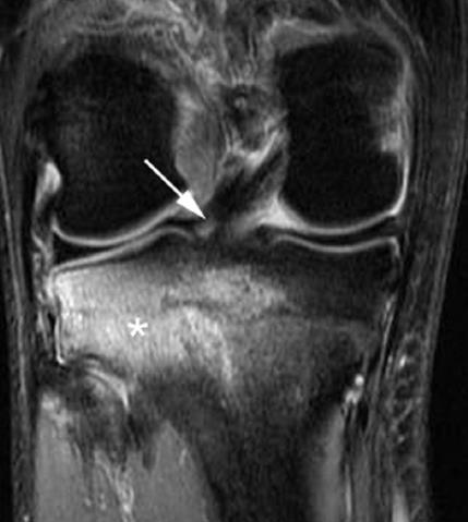
23 T2 Weighted Image of 15 year old with unstable OCD Solid Line: focal defect Dashed Arrow: Fragment Healed stable OCD treated with conservative treatment 6 mo after diagnosis Reproduction Without Consent 23
 Bilateral in 20-30% of cases Osgood-Schlatter")
24 Osgood-Schlatter Painful irritation to anterior tibial tubercle Age of Presentation Boys age Girls age 8-12 Boys>Girls Symptoms Painful swelling at anterior tibial tubercle Mild and intermittent initially Severe and constant in acute phase Leg pain or knee pain Worsens with running, jumping, stairs or direct contact (kneeling) Bilateral in 20-30% of cases Osgood-Schlatter Findings Tenderness and prominence in area of tibial tuberosity Reproduced with resisted knee extension Anterior mass may be only finding following resolution of acute phase Treatment Ice Reduced activity NSAID s Physical Therapy Strengthening/flexibility of quads, hamstrings, ITB, gastroc/soleus Quadriceps strengthening progression low intensity-high intensity Prognosis Full recovery in 90% of patients without surgery Symptoms may continued intermittently for months Reproduction Without Consent 24

25 Pre-Objective Exam Establish hypothesis and differential diagnoses to guide objective exam Red Flags or Yellow Flags? Prioritize Structures to be examined Clearing exams of adjacent joints Neuro exam? Begin to determine extent of objective exam based on SINS Standing Observation Knee/Hip angles Feet position Scars/deformities Atrophy/bruising Objective Reproduction Without Consent 25
26 Functional Testing Gait Analysis Walking/Running Squatting Single Leg Double Leg Trunk Rotation Heel Raises Double/Single leg hop tests Step down test Swing Test Functional Testing Reproduction Without Consent 26


27 Squatting Single Leg Double Leg 5 Trials All requirements met for 4/5 to be Good Reproduction Without Consent 27


28 Single Leg Squat A: Good B: Poor C: Poor Hip/Pelvis D: Poor Hip/Knee SFMA FN Functional Non Painful FP Functional Painful DP Dysfunctional Painful DN Dysfunctional Non Painful Reproduction Without Consent 28


29 SFMA Multi Segmental Rotation Feet together, arms at sides Rotate as far as possible without moving feet Pelvis must rotate more than 50deg Shoulders must rotate more than 50deg No loss of height Reproduction Without Consent 29


 Knees should remain in neutral Reproduction Without")
30 Single Limb Stance Feet together, arms by sides Lift one leg to 90deg flexion Hold position for 10sec Repeat with eyes closed Look for loss of height or arms to flail Some increased sway with eyes closed is normal Overhead Deep Squat Feet shoulder width apart and straight Extend arms overhead Patient descends as deeply as possible into squat Heels remain on floor, head and chest facing forward and arms overhead Hands should remain the same width (anterior view) and should stay behind toes (lateral view) Knees should remain in neutral Reproduction Without Consent 30
31 Objective Lumbar Clearing AROM/Quadrant Special Testing (as needed) Meniscus Thessaley Ege s Objective Sitting Myotomal/Reflex/Sensation exam If warranted Slump Test If warranted Reproduction Without Consent 31
32 Objective Supine Palpation Superior tib/fib Patellar poles Infrapatellar/Suprapatellar bursae Medial/Lateral Joint Lines Hip Clearing PROM/AROM all planes FABER FADIR SLR test if warranted Tibiofemoral Joint AROM/AROM with overpressure Flexion Extension End Feels Objective Passive Physiological Motion Flexion, flexion with abduction, flexion with adduction Extension, extension with abduction, extension with adduction Tibial IR/ER at 90 deg flexion End Feels Passive Accessory Motions A-P Medial/Lateral Rotation Superior Tib-Fib Joint Passive Accessory Motion A-P P-A Reproduction Without Consent 32


33 Passive Physiological/Accessory Motion Goal Reproduce concordant sign Localize dysfunction through different planes of testing Can use prolonged holds or repeated movements Be aware of end feels and guarding Passive Physiological Motion Flexion/Flexion with abd/flexion with add Pt supine, support lateral femur against chest Passively flex knee to end range Take out to ~10-20deg short of available range Firmly stabilize femur with one hand, other on distal tibia Flex again while directing heel toward greater trochanter Repeat again while directing heel toward groin Reproduction Without Consent 33


34 Passive Physiological Motion Extension/Extension with abd/extension with add Pt supine, support ankle with one hand, other hand interthenar eminence at tibial tubercle Extend knee by sidebending trunk Move interthenar eminence to lateral tibia, and support ankle at lateral aspect Extend knee again causing a extension/abduction movement Move proximal hand to medial tibia, distal hand to medial malleoli and repeat causing extension/adduction movement Passive Physiological Motion Tibial IR/ER at 90deg Pt supine, knee flexed to approx 90deg Passively internally rotate tibia Repeat for external rotation Reproduction Without Consent 34


35 Passive Accessory Motion Anterior-Posterior Pt supine, knee in open packed position on a bolster Place both thumbs on tibial tubercle and wrap hands around proximal tibia Direct force posteriorly moving tibia on femur Passive Accessory Motion Posterior-Anterior Pt supine, knee flexed to 60-80deg of flexion Grasp around proximal tibia with thumbs on tibial tubercle while sitting on foot to stabilize Move tibia in anterior direction on femur Reproduction Without Consent 35


36 Passive Accessory Motion Medial-Lateral Shear Pt supine, knee flexed 10-20deg on bolster Medial Grasp medial aspect of distal femur and lateral aspect of proximal tibia Stabilize femur while applying medially directed movement of tibia on femur Lateral Grasp lateral aspect of distal femur and medial aspect of proximal tibia Stabilize femur while applying laterally directed movement of tibia on femur Passive Accessory Motion Rotation Pt supine, knee flexed to approx 90deg, foot stabilized by sitting on it Grasp lateral half of tibia with one hand, stabilize femur with other Apply an anterior and laterally directed movement of tibia on femur Repeat by applying posterior and medially directed movement with same hand holds Repeat on other side for anterior/lateral and posterior/medial Reproduction Without Consent 36


37 Passive Accessory Motion Superior Tib-Fib A-P/P-A Pt sidelying with involved side up, knees bent and pillow between knees Stand behind pt and place thumbs on posterior aspect of head of the fibula Produce posterior to anterior movement of fibula on tibia Move to in front of pt, repeat by placing thumbs on anterior aspect of head of fibula and produce anterior to posterior movement Patellar Assessment Intra-rater reliability is good Inter-rater reliability is variable Validity is good to moderate Reproduction Without Consent 37

38 Strong validity and intrarater reliability Patellar Assessment Position Assessment Tilt Rotation Shift Reproduction Without Consent 38

39 Patellar Mobility Assessment Superior/Inferior Pt supine, knee in open packed position Place apex of patella in interthenar eminence Align forearm with shaft of femur Apply inferior glide of patella Repeat for superior glide Medial/Lateral Pt supine, knee in open packed position Stand on lateral side of knee Grasp patella and move in a lateral direction Repeat for medial glide Patellar Glide Test Normal= excursion of ½ patella Supine cont d Muscle Length Testing Hamstrings Gastroc/Soleus Hip external rotators Special Testing ACL Lachman Anterior Drawer Pivot Shift PCL Posterior Drawer Posterior Sag Sign Quadriceps Active Test Objective Reproduction Without Consent 39


40 Pivot Shift ACL Lachman Pivot Shift Pt supine with knee extended One hand holds ankle, other hand applies medial rotation force at tibia Slowly flex knee maintaining rotation As reach about 20deg flexion the tibial plateau will relocate Positive test is a thud or clunk of lateral tibia posteriorly Reproduction Without Consent 40


41 Pivot Shift Sens.24 Spec.98 +LR 8.5 -LR.9 Rule in ACL Tear Lachman s Test Pt supine with knee flexed to 15deg Stabilize at distal femur with one hand, grasp behind proximal tibia with other hand Apply anterior tibial force to prox tibia Positive if greater anterior displacement of tibia compared to other side or empty end feel Reproduction Without Consent 41

42 Lachman Test Sens.85 Spec.94 +LR 1.2 -LR.2 Helps rule out the presence of a torn ACL Anterior Drawer Test Pt supine, knee flexed to approx 90deg with foot flat PT sits on pt s foot, grasp behind prox tibia with thumbs palpating at tibial tuberosities Apply anterior tibial force Positive if greater anterior translation compared to other side or empty end feel Reproduction Without Consent 42

43 Anterior Drawer Sens.55 Spec.92 +LR 7.3 -LR.5 Rule in ACL Tear PCL Quadriceps Active Test Posterior Drawer Posterior Sag Sign Reproduction Without Consent 43
44 Quadriceps Active Test Pt supine with knee flexed to 90 Pt s thigh should be relaxed, PT stabilizing foot Have pt slide foot gently down table to initiate quadriceps Will see anterior displacement of tibia Quadriceps Active Test Sens: 98% Spec: 99% +LR: 98 -LR:.04 Most specific test for PCL rupture Reproduction Without Consent 44

45 Posterior Drawer Test Pt supine, knee flexed to approx 90deg with foot flat PT sits on pt s foot, grasp behind prox tibia with thumbs palpating at tibial tuberosities Apply posterior tibial force Positive if greater posterior translation compared to other side Sens: 90% Spec: 99% +LR: 90 -LR:.1 Posterior Drawer Test Helps rule out the presence of a torn PCL Reproduction Without Consent 45

46 Posterior Sag Sign Pt supine with knee flexed to 90deg and hip flexed to 90deg Make sure pt is relaxed in the position Possible false negative with increased muscle tone Positive if tibia is positioned posterior Possible false negatives with hx of Osgood Schlatters Posterior Sag Test Sens: 79% Spec: 100% +LR: LR:.21 Uninvolved Rule in presence of a PCL tear Involved Reproduction Without Consent 46
47 Posterolateral Corner Prone ER ER Recurvatum Posterior Drawer with ER Assessment Cluster Posterior drawer test in ER Prone ER test ER Recurvatum test Reliability and specificity not tested Reproduction Without Consent 47
48 Posterior Drawer with ER at 30/90 ER tibia and apply posterior force If normal at 90 but excess at 30 suspect PLC injury Positive if tibia rotates excessively compared to other side If rotates and subluxes posteriorly or excess motion at 30 and 90 suspect PCL injury External Rotation Recurvatum Test Pt supine in a relaxed position Pick up pt s leg by great toe Watch for hyperextension and tibial ER compared to other side Reproduction Without Consent 48
49 Prone ER Test at 30 and 90 Pt prone, clinician grasps distal leg, flexes knee and ER tibia + if ER exceeds 10deg of other leg + at 30 but not at 90= isolated PLC injury + at both = concomitant PCL Sidelying Strength Testing Objective Glut strength testing Muscle Length Testing Hip flexor Ober s Superior Tib-Fib Passive Accessory Motion Neurodynamics Modified slump if needed Reproduction Without Consent 49
50 Objective Prone Strength Testing Quad Hamstrings Hip IR/ER Muscle Length Testing Quad Hip Flexor Neurodynamic Testing Prone Knee Bend Clearing Exam Lumbar pa (central and upa) Lumbar palpation Special Test PLC Prone ER Test Rule In Rule Out Best Test Meniscus Thessaly Apley s Compression Test McMurray's Joint Line Tenderness Thessaly Cluster of Tests ACL Pivot Shift Anterior Drawer Lachman Lachman with empty endfeel PCL Quadriceps Active Test Posterior Sag Posterior Drawer Posterior Sag Quadriceps Active Test MCL Valgus at 30deg Valgus at 30deg Valgus at 30deg LCL Varus at 30deg Varus at 30deg Varus at 30deg Post Drawer with ER at 30deg Post Drawer with ER at 30deg PLC Prone ER at 30deg Prone ER at 30deg ER Recurvatum Test ER Recurvatum Test Cluster of Tests Reproduction Without Consent 50
RN(EC) ENC(C) GNC(C) MN ACNP *** MECHANISM OF INJURY.. MOST IMPORTANT *** - Useful in determining mechanism of injury / overuse
 ENC(C) GNC(C) MN ACNP *** MECHANISM OF INJURY.. MOST IMPORTANT *** - Useful in determining mechanism of injury / overuse") HISTORY *** MECHANISM OF INJURY.. MOST IMPORTANT *** Age of patient Sport / Occupation - Certain conditions are more prevalent in particular age groups (Osgood Schlaters in youth / Degenerative Joint Disease
HISTORY *** MECHANISM OF INJURY.. MOST IMPORTANT *** Age of patient Sport / Occupation - Certain conditions are more prevalent in particular age groups (Osgood Schlaters in youth / Degenerative Joint Disease
Physical Examination of the Knee
 History: Pain Traumatic vs. atraumatic? Acute vs Chronic Previous procedures done on the knee? Swelling, catching, instability General Setup Examine standing, sitting and supine Evaluate gait Examine hip
Physical Examination of the Knee
 History: Pain Traumatic vs. atraumatic Acute vs Chronic Mechanism of injury Swelling, catching, instability Previous evaluation and treatment General Setup Examine standing, sitting and supine Evaluate
PRIMARY CARE EXAMINATION OF KEY JOINTS. Thomas M. Howard, MD, FACSM FFPC Sports Medicine
 PRIMARY CARE EXAMINATION OF KEY JOINTS Thomas M. Howard, MD, FACSM FFPC Sports Medicine General exam principles: Expose entire joint and opposite limb for comparison Have a Differential Diagnosis Exam
Knee Injury Assessment
 Knee Injury Assessment Clinical Anatomy p. 186 Femur Medial condyle Lateral condyle Femoral trochlea Tibia Intercondylar notch Tibial tuberosity Tibial plateau Fibula Fibular head Patella Clinical Anatomy
The Knee. Two Joints: Tibiofemoral. Patellofemoral
 Evaluating the Knee The Knee Two Joints: Tibiofemoral Patellofemoral HISTORY Remember the questions from lecture #2? Girth OBSERVATION TibioFemoral Alignment What are the consequences of faulty alignment?
Musculoskeletal Examination Benchmarks
 Musculoskeletal Examination Benchmarks _ The approach to examining the musculoskeletal system is the same no matter what joint or limb is being examined. The affected and contralateral region should both
Checklist for Physical Examination of the Knee Muscuoskeletal Block -- Chris McGrew MD, Andrew Ashbaugh DO
 Checklist for Physical Examination of the Knee Muscuoskeletal Block -- Chris McGrew MD, Andrew Ashbaugh DO This handout is for use as a rough guide and study aid. Your instructor may perform certain maneuvers
Examination of the Knee
 Examination of the Knee Wash your hands & Introduce the exam to the patient Positioning & Draping With the patient supine, make sure both legs are exposed in order to compare each side be sure to use draping
Arthritic history is similar to that of the hip. Add history of give way and locking, swelling
 KNEE VASU PAI Arthritic history is similar to that of the hip. Add history of give way and locking, swelling INJURY MECHANISM When How Sequence Progress Disability IKDC Activity I - Strenuous activity
Knee Joint Assessment and General View
 Knee Joint Assessment and General View Done by; Mshari S. Alghadier BSc Physical Therapy RHPT 366 m.alghadier@sau.edu.sa http://faculty.sau.edu.sa/m.alghadier/ Functional anatomy The knee is the largest
Prevention and Treatment of Injuries. Anatomy. Anatomy. Chapter 20 The Knee Westfield High School Houston, Texas
 Prevention and Treatment of Injuries Chapter 20 The Knee Westfield High School Houston, Texas Anatomy MCL, Medial Collateral Ligament LCL, Lateral Collateral Ligament PCL, Posterior Cruciate Ligament ACL,
Ligamentous and Meniscal Injuries: Diagnosis and Management
 Ligamentous and Meniscal Injuries: Diagnosis and Management Daniel K Williams, MD Franciscan Physician Network Orthopedic Specialists September 29, 2017 No Financial Disclosures INTRODUCTION Overview of
Goals &Objectives. 1. Review the anatomy of the knee 2. Practice your hands-on skills 3. By the end of the workshop:
 Clinical Knee Exam Goals &Objectives 1. Review the anatomy of the knee 2. Practice your hands-on skills 3. By the end of the workshop: Be able to categorize knee injuries Understand the significance of
Anterior Knee Pain in Children. Joseph Chorley, MD Associate Professor, Pediatrics Baylor College of Medicine
 Anterior Knee Pain in Children Joseph Chorley, MD Associate Professor, Pediatrics Baylor College of Medicine Goals and Objectives To learn how to care for patients with chronic knee pain To be able to
On Field Assessment and Management of Acute Knee Injuries: A Physiotherapist s Perspective
 On Field Assessment and Management of Acute Knee Injuries: A Physiotherapist s Perspective Jessica Condliffe Physiotherapist / Clinic Manager TBI Health Wellington Presentation Outline Knee anatomy review
Evaluation of the Knee and Shoulder
 Evaluation of the Knee and Shoulder Karen J. Boselli, MD Northeast Regional Nurse Practitioner Conference May 2018 Knee Overview History Examination Top 5 diagnoses When to image When to refer Pain most
W. Dilworth Cannon, M.D. Professor of Clinical Orthopaedic Surgery University of California San Francisco
 Knee Pain And Injuries In Adults W. Dilworth Cannon, M.D. Professor of Clinical Orthopaedic Surgery University of California San Francisco Pain Control Overview Narcotics rarely necessary after 1 st 1-2
9/24/2012. Greg Bennett, PT, DSc Excel Physical Therapy Marymount University
 Greg Bennett, PT, DSc Excel Physical Therapy Marymount University Hx often diagnostic Least to most threatening Sx trump exam Develop consistent routine Don t inflame inflamed tissue 1 1. ESTABLISH OR
KNEE EXAMINATION. Tips & Tricks from an Emergency Physician Perspective. EM Physicians Less Exposed to MSK Medicine
 KNEE EXAMINATION Tips & Tricks from an Emergency Physician Perspective Dr P O CONNOR Emergency Medicine Physician EUSEM 10/09/2018 EM Physicians Less Exposed to MSK Medicine Musculoskeletal Medicine becoming
Anterior Cruciate Ligament (ACL)
") Anterior Cruciate Ligament (ACL) The anterior cruciate ligament (ACL) is one of the 4 major ligament stabilizers of the knee. ACL tears are among the most common major knee injuries in active people of
Anterior Cruciate Ligament (ACL) The anterior cruciate ligament (ACL) is one of the 4 major ligament stabilizers of the knee. ACL tears are among the most common major knee injuries in active people of
The examination of the painful knee. Maja K Artandi, MD, FACP Clinical Associate Professor of Medicine Stanford University
 The examination of the painful knee Maja K Artandi, MD, FACP Clinical Associate Professor of Medicine Stanford University Objectives of the talk By the end of this talk you will know The important anatomy
Overview Ligament Injuries. Anatomy. Epidemiology Very commonly injured joint. ACL Injury 20/06/2016. Meniscus Tears. Patellofemoral Problems
 Overview Ligament Injuries Meniscus Tears Pankaj Sharma MBBS, FRCS (Tr & Orth) Consultant Orthopaedic Surgeon Manchester Royal Infirmary Patellofemoral Problems Knee Examination Anatomy Epidemiology Very
Exam of the Knee and Ankle I HAVE NO FINANCIAL DISCLOSURES RELEVANT TO THIS PRESENTATION
 Exam of the Knee and Ankle I HAVE NO FINANCIAL DISCLOSURES RELEVANT TO THIS PRESENTATION Disclosures I have no relevant financial relationships with the manufacturers of any commercial products and or
BATES VISUAL GUIDE TO PHYSICAL EXAMINATION. OSCE 4: Knee Pain
 BATES VISUAL GUIDE TO PHYSICAL EXAMINATION OSCE 4: Knee Pain This video format is designed to help you prepare for objective structured clinical examinations, or OSCEs. You are going to observe and participate
PRINCIPLES OF EXAMNINIG THE KNEE
 Welcome! Pignon, Haiti IS IT. GOOD MORNING LORD! OR GOOD LORD, MORNING! PRINCIPLES OF EXAMNINIG THE KNEE Greg Bennett, PT, DSc Excel Physical Therapy Marymount University Rules Hx often diagnostic Least
American College of Physicians 2013 Ohio Chapter Scientific Meeting Columbus, OH October 11, 2013
 American College of Physicians 2013 Ohio Chapter Scientific Meeting Columbus, OH October 11, 2013 Paul J. Gubanich, MD, MPH Assistant Professor of Internal Medicine/Sports Medicine Team Physician, Ohio
Anatomy. ACL PCL MCL LCL Meniscus. Medial Lateral
 Skis for Knees Anatomy ACL PCL MCL LCL Meniscus Medial Lateral Knee Anatomy THE KNEE HISTORY Pain (PQRST) Contact vs noncontact Effusions Mechanical symptoms Locking Instability (falls) Initial treatment
Rehabilitation Guidelines for Anterior Cruciate Ligament (ACL) Reconstruction
 Reconstruction") Rehabilitation Guidelines for Anterior Cruciate Ligament (ACL) Reconstruction The knee is the body's largest joint, and the place where the femur, tibia, and patella meet to form a hinge-like joint. These
Rehabilitation Guidelines for Anterior Cruciate Ligament (ACL) Reconstruction The knee is the body's largest joint, and the place where the femur, tibia, and patella meet to form a hinge-like joint. These
Balanced Body Movement Principles
 Balanced Body Movement Principles How the Body Works and How to Train it. Module 3: Lower Body Strength and Power Developing Strength, Endurance and Power The lower body is our primary source of strength,
Functional Movement Screen (Cook, 2001)
") Functional Movement Screen (Cook, 2001) TEST 1 DEEP SQUAT Purpose - The Deep Squat is used to assess bilateral, symmetrical, mobility of the hips, knees, and ankles. The dowel held overhead assesses bilateral,
Functional Movement Screen (Cook, 2001) TEST 1 DEEP SQUAT Purpose - The Deep Squat is used to assess bilateral, symmetrical, mobility of the hips, knees, and ankles. The dowel held overhead assesses bilateral,
7/20/14. Patella Instability. Alignment. PF contact areas. Tissue Restraints. Pain. Acute Blunt force trauma Disorders of the Patellafemoral Joint
 Patella Instability Acute Blunt force trauma Disorders of the Patellafemoral Joint Evan G. Meeks, M.D. Orthopaedic Surgery Sports Medicine The University of Texas - Houston Pivoting action Large effusion
I have nothing to disclose
 Management of Common Knee Disorders: What You Knee d to Know UCSF Essentials of Women s Health July 8, 2015 Carlin Senter, M.D. I have nothing to disclose Learning objectives: in 1 hour you will be able
The Knee. Prof. Oluwadiya Kehinde
 The Knee Prof. Oluwadiya Kehinde www.oluwadiya.sitesled.com The Knee: Introduction 3 bones: femur, tibia and patella 2 separate joints: tibiofemoral and patellofemoral. Function: i. Primarily a hinge joint,
Knee Injuries. PSK 4U Mr. S. Kelly North Grenville DHS. Medial Collateral Ligament Sprain
 Knee Injuries PSK 4U Mr. S. Kelly North Grenville DHS Medial Collateral Ligament Sprain Result from either a direct blow from the lateral side in a medial direction or a severe outward twist Greater injury
RN(EC) ENC(C) GNC(C) MN ACNP *** MECHANISM OF INJURY.. MOST IMPORTANT ***
 ENC(C) GNC(C) MN ACNP *** MECHANISM OF INJURY.. MOST IMPORTANT ***") HISTORY *** MECHANISM OF INJURY.. MOST IMPORTANT *** Age of patient - Certain conditions are more prevalent in particular age groups (Hip pain in children may refer to the knee from Legg-Calve-Perthes
HISTORY *** MECHANISM OF INJURY.. MOST IMPORTANT *** Age of patient - Certain conditions are more prevalent in particular age groups (Hip pain in children may refer to the knee from Legg-Calve-Perthes
Muscle Testing of Knee Extensors. Yasser Moh. Aneis, PhD, MSc., PT. Lecturer of Physical Therapy Basic Sciences Department
 Muscle Testing of Knee Extensors Yasser Moh. Aneis, PhD, MSc., PT. Lecturer of Physical Therapy Basic Sciences Department Muscle Testing of Knee Extensors othe Primary muscle Quadriceps Femoris -Rectus
FUNCTIONAL ANATOMY: Knee and Leg
 ACSM Team Physician Course San Antonio Feb 2015 FUNCTIONAL ANATOMY: Knee and Leg Marlene DeMaio, MD Professor, Orthopaedic Surgery Marshall University VAMC Huntington, WV Mary Lloyd Ireland, MD Professor
Knee Contusions and Stress Injuries. Laura W. Bancroft, M.D.
 Knee Contusions and Stress Injuries Laura W. Bancroft, M.D. Objectives Review 5 types of contusion patterns Pivot shift Dashboard Hyperextension Clip Lateral patellar dislocation Demonstrate various stress
IFAST Assessment. Name: Date: Sport: Review Health Risk Assessment on initial consult form. List Client Goals (what brings you here?
 IFAST Assessment Name: Date: Sport: Review Health Risk Assessment on initial consult form List Client Goals (what brings you here?) Cardiovascular Measurements Blood Pressure Resting Heart Rate Body Composition
AAP Boot Camp KNEE AND ANKLE EXAM
 AAP Boot Camp KNEE AND ANKLE EXAM Disclosures I have no relevant financial relationships with the manufacturers of any commercial products and or providers of commercial services discussed in this CME
ACL AND PCL INJURIES OF THE KNEE JOINT
 ACL AND PCL INJURIES OF THE KNEE JOINT Dr.KN Subramanian M.Ch Orth., FRCS (Tr & Orth), CCT Orth(UK) Consultant Orthopaedic Surgeon, Special interest: Orthopaedic Sports Injury, Shoulder and Knee Surgery,
Recognizing common injuries to the lower extremity
 Recognizing common injuries to the lower extremity Bones Femur Patella Tibia Tibial Tuberosity Medial Malleolus Fibula Lateral Malleolus Bones Tarsals Talus Calcaneus Metatarsals Phalanges Joints - Knee
emoryhealthcare.org/ortho
 COMMON SOCCER INJURIES Oluseun A. Olufade, MD Assistant Professor, Department of Orthopedics and PM&R 1/7/18 GOALS Discuss top soccer injuries and treatment strategies Simplify hip and groin injuries in
THE LOWER EXTREMITY EXAM FOR THE FAMILY PRACTITIONER
 THE LOWER EXTREMITY EXAM FOR THE FAMILY PRACTITIONER Melinda A. Scott, D.O. Orthopedic Associates of Dayton Board Certified in Primary Care Sports Medicine GOALS Identify landmarks necessary for exam of
What is arthroscopy? Normal knee anatomy
 What is arthroscopy? Arthroscopy is a common surgical procedure for examining and repairing the inside of your knee. It is a minimally invasive surgical procedure which uses an Arthroscope and other specialized
ACL Athletic Career. ACL Rupture - Warning Features Intensive pain Immediate swelling Locking Feel a Pop Dead leg Cannot continue to play
 FIMS Ambassador Tour to Eastern Europe, 2004 Belgrade, Serbia Montenegro Acute Knee Injuries - Controversies and Challenges Professor KM Chan OBE, JP President of FIMS Belgrade ACL Athletic Career ACL
Mastering the Musculoskeletal Exam UCSF Essentials of Women s Health July 7, 2016 Carlin Senter, M.D. Henry Crevensten, M.D.
 Mastering the Musculoskeletal Exam UCSF Essentials of Women s Health July 7, 2016 Carlin Senter, M.D. Henry Crevensten, M.D. I have nothing to disclose Outline Knee exam Shoulder exam Knee Anatomy The
Non Surgical Management Of Hip And Knee Osteoarthritis Toolkit. Evaluation and Diagnosis of Osteoarthritis in Primary Care
 Non Surgical Management Of Hip And Knee Osteoarthritis Toolkit Evaluation and Diagnosis of Osteoarthritis in Primary Care OA-HxPE-716.indd 1 TABLE OF CONTENTS HISTORY TAKING... 3 EVALUATION OF SUSPECTED
SOFT TISSUE KNEE INJURIES
 SOFT TISSUE KNEE INJURIES Soft tissue injuries of the knee commonly occur in all sports or in any activity that requires sudden changes in activity or movement. The knee is a complex joint and any injury
PHASE ONE: THE FIRST SIX WEEKS AFTER INJURY
 Exercises After Injury to the Anterior Cruciate Ligament (ACL) of the Knee Dr. Abigail R. Hamilton, M.D. PHASE ONE: THE FIRST SIX WEEKS AFTER INJURY Initially, the knee needs to be protected-use the knee
TREATMENT GUIDELINES FOR GRADE 3 PCL TEAR
 GENERAL CONSIDERATIONS Posterior cruciate ligament (PCL) injuries occur less frequently than anterior cruciate ligament (ACL) injuries, but are much more common than previously thought. The PCL is usually
Functional Movement Test. Deep Squat
 Functional Movement Test Put simply, the FMS is a ranking and grading system that documents movement patterns that are key to normal function. By screening these patterns, the FMS readily identifies functional
Objectives. The BIG Joint. Case 1. Boney Architecture. Presenter Disclosure Information. Common Knee Problems
 3:30 4:15 pm Common Knee Problems SPEAKER Christopher J. Visco, MD Presenter Disclosure Information The following relationships exist related to this presentation: Christopher J. Visco, MD: Speaker s Bureau
Diagnosis and Management of Knee Conditions. Jenny Love / Lynn Robertson AFLAR Oct 2009
 Diagnosis and Management of Knee Conditions Jenny Love / Lynn Robertson AFLAR Oct 2009 AIMS Review 4 common Knee Conditions: Anterior knee pain Meniscal Injuries Ligament injuries ACL Osteoarthritis Discuss
Please differentiate an internal derangement from an external knee injury.
 Knee Orthopaedic Tests Sports and Knee Injuries James J. Lehman, DC, MBA, DABCO University of Bridgeport College of Chiropractic Knee Injury Strain, Sprain, Internal Derangement Anatomy of the Knee Please
CHAPTER 8: THE BIOMECHANICS OF THE HUMAN LOWER EXTREMITY
 CHAPTER 8: THE BIOMECHANICS OF THE HUMAN LOWER EXTREMITY _ 1. The hip joint is the articulation between the and the. A. femur, acetabulum B. femur, spine C. femur, tibia _ 2. Which of the following is
Clinical Evaluation and Imaging of the Patellofemoral Joint Common clinical syndromes
 Clinical Evaluation and Imaging of the Patellofemoral Joint Common clinical syndromes A. Panagopoulos Lecturer in Orthopaedics Medical School, Patras University Objectives Anatomy of patellofemoral joint
The Knee. Clarification of Terms. Osteology of the Knee 7/28/2013. The knee consists of: The tibiofemoral joint Patellofemoral joint
 The Knee Clarification of Terms The knee consists of: The tibiofemoral joint Patellofemoral joint Mansfield, p273 Osteology of the Knee Distal Femur Proximal tibia and fibula Patella 1 Osteology of the
Differential Diagnosis
 Case 31yo M who sustained an injury to L knee while playing Basketball approximately 2 weeks ago. He describes pivoting and hyperextending his knee, which swelled over the next few days. He now presents
Lateral knee injuries
 Created as a free resource by Clinical Edge Based on Physio Edge podcast episode 051 with Matt Konopinski Get your free trial of online Physio education at Orthopaedic timeframes Traditionally Orthopaedic
In the name of god. Knee. By: Tofigh Bahraminia Graduate Student of the Pathology Sports and corrective actions. Heat: Dr. Babakhani. Nov.
 In the name of god Knee By: Tofigh Bahraminia Graduate Student of the Pathology Sports and corrective actions Heat: Dr. Babakhani Nov. 2014 1 Anatomy-Bones Bones Femur Medial/lateral femoral condyles articulate
The Knee. Tibio-Femoral
 The Knee Tibio-Femoral Osteology Distal Femur with Proximal Tibia Largest Joint Cavity in the Body A modified hinge joint with significant passive rotation Technically, one degree of freedom (Flexion/Extension)
Disclosures. Knee Anatomy. Objective. Five Common Knee and Ankle Conditions You Will See in Office Practice 8/11/2016
 ESSENTIALS OF PRIMARY CARE: A Core Curriculum for Ambulatory Practice August 7-12, 2016 Five Common Knee and Ankle Conditions You Will See in Office Practice I have nothing to disclose Disclosures Cindy
Brennen Lucas, M.D. Advanced Orthopaedic Associates
 Brennen Lucas, M.D. Advanced Orthopaedic Associates 2778 N. Webb Rd. Wichita, KS 67226 316-631-1600 Fax: (316) 631-1674 1 (800) 362-0591 GUIDELINES FOR REHABILITATION FOLLOWING SURGICAL RECONSTRUCTION
Quads (machines) Cable Lunge
 Cable Lunge") Cable Lunge Cable Lunge 1) Stand with feet hip width apart and a cable attached around your waist. Take left leg and step back approximately 2 feet standing on the ball of the foot. 2) Start position:
Cable Lunge Cable Lunge 1) Stand with feet hip width apart and a cable attached around your waist. Take left leg and step back approximately 2 feet standing on the ball of the foot. 2) Start position:
SMALL GROUP SESSION 16 January 8 th or 10 th Shoulder pain case/ Touch workshop/ Upper and Lower Extremity Examination
 SMALL GROUP SESSION 16 January 8 th or 10 th Shoulder pain case/ Touch workshop/ Upper and Lower Extremity Examination Suggested Readings: Opatrny L. The Healing Touch. Ann Int Med 2002; 137:1003. http://www.annals.org/cgi/reprint/137/12/1003.pdf
Jennifer L. Cook, MD
 Jennifer L. Cook, MD Florida Joint Replacement and Sports Medicine Center 5243 Hanff Lane New Port Richey, FL 34652 Phone: (727)848-4249 Fax: (727) 841-8934 ANTERIOR CRUCIATE LIGAMENT RECONSTRUCTION POST-OPERATIVE
King Khalid University Hospital
 King Khalid University Hospital Rehabilitation Department Ortho Group Rehabilitation Protocol: PCL RECONSTRUCTION +/- ACL / MCL / LCL / POSTEROLATERAL CORNER 1. General Guidelines: Time lines in this rehabilitation
Musculoskeletal Examination
 Musculoskeletal Examination Statement of Goals Know how to perform a complete musculoskeletal examination. Learning Objectives A. Describe the anatomy of the musculoskeletal system including the bony structures,
Evaluation of Knee Problems
 Evaluation of Knee Problems OBJECTIVES Review knee anatomy Explain tests to look for pathology Briefly introduce knee problems Only by a thorough knowledge of anatomy and functional testing can one make
BIOMECHANICAL EXAMINATION OF THE PEDIATRIC LOWER EXTREMITY
 BIOMECHANICAL EXAMINATION OF THE PEDIATRIC LOWER EXTREMITY B.Resseque, D.P.M. ARCH HEIGHT OFF WEIGHTBEARING Evaluate arch height by placing a ruler from the heel to the first metatarsal head Compare arch
Physiotherapy Information following Anterior Cruciate Ligament (ACL) Reconstruction
 Reconstruction") Physiotherapy Information following Anterior Cruciate Ligament (ACL) Reconstruction Name:... Surgery Date:... Graft:... Orthopaedic Outpatient Appointment Date: Time: Location: Contact Number: Contacting
Physiotherapy Information following Anterior Cruciate Ligament (ACL) Reconstruction Name:... Surgery Date:... Graft:... Orthopaedic Outpatient Appointment Date: Time: Location: Contact Number: Contacting
42 nd Annual Symposium on Sports Medicine. Knee Injuries In The Pediatric Athlete. Disclosure
 42 nd Annual Symposium on Sports Medicine Travis Murray, MD Assistant Professor University of Texas Health Science Center San Antonio January 23, 2015 Knee Injuries In The Pediatric Athlete Disclosure
Warm-Up and Stretching Exercises
 Warm-Up and Stretching Exercises Most athletes (swimmers included) use a combination of controlled movement exercises and specific joint/muscle stretching to improve performance potential. The proposed
This presentation is the intellectual property of the author. Contact them for permission to reprint and/or distribute.
 43 rd Annual Symposium on Sports Medicine UT Health Science Center San Antonio School of Medicine January 22-23, 2016 Intra-articular / Extra-synovial 38 mm length / 13 mm width Fan-shaped structure narrowest-midportion
Knee Capsular Disorder. ICD-9-CM: Stiffness in joint of lower leg, not elsewhere classified
 1 Knee Capsular Disorder "Knee Capsulitis" ICD-9-CM: 719.56 Stiffness in joint of lower leg, not elsewhere classified Diagnostic Criteria History: Physical Exam: Stiffness Aching with prolonged weight
ASSESSMENT AND MANAGEMENT OF THE KNEE AND LOWER LIMB.
 ASSESSMENT AND MANAGEMENT OF THE KNEE AND LOWER LIMB www.fisiokinesiterapia.biz Overview History Examination X-rays Fractures and Dislocations. Soft Tissue Injuries Other Knee/Lower limb Problems Anatomy
ANTERIOR KNEE PAIN. Explanation. Causes. Symptoms
 ANTERIOR KNEE PAIN Explanation Anterior knee pain is most commonly caused by irritation and inflammation of the patellofemoral joint of the knee (where the patella/kneecap connects to the femur/thigh bone).
Rehabilitation for Patellar Tendinitis (jumpers knee) and Patellofemoral Syndrome (chondromalacia patella)
 and Patellofemoral Syndrome (chondromalacia patella)") Rehabilitation for Patellar Tendinitis (jumpers knee) and Patellofemoral Syndrome (chondromalacia patella) Patellar Tendinitis The most common tendinitis about the knee is irritation of the patellar tendon.
Rehabilitation for Patellar Tendinitis (jumpers knee) and Patellofemoral Syndrome (chondromalacia patella) Patellar Tendinitis The most common tendinitis about the knee is irritation of the patellar tendon.
Chapter 20 The knee and related structures
 Chapter 20 The knee and related structures Athletic Training Spring 2014 Jihong Park Bones & joints Femur, tibia, fibula, & patella Femur & tibia Weight bearing & muscle attachment Patella functions Anterior
Anterior knee pain.
 Anterior knee pain What are the symptoms? Anterior knee pain is very common amongst active adolescents and athletes participating in contact sports. It is one of the most common problems/injuries seen
Exercise Report For: Augusta James
 Exercise Report For: Optimizing Sport Performance Provided By: Greg Redman BScPT, BScKin, Wave Physiotherapy Phone: 250-763-9283 Fax:, www.wavephysio.ca Page: 1 Stretch hip flexor kneel w/ball Stretch
Physical & Occupational Therapy
 In this section you will find our recommendations for exercises and everyday activities around your home. We hope that by following our guidelines your healing process will go faster and there will be
ACL Patient Assessment and Progress Sheet. Patient Sticker
 Patient Sticker Thank you for taking the time to answer these questions which should only take a few minutes. The answers you give are very useful as they will help us assess your progress following your
Rehabilitation Guidelines for Meniscal Repair
 Rehabilitation Guidelines for Meniscal Repair The knee is the body's largest joint, and the place where the femur, tibia, and patella meet to form a hinge-like joint. These bones are supported by a large
Patellofemoral Pain Syndrome
 What is patellofemoral pain syndrome? Patellofemoral Pain Syndrome Patellofemoral pain syndrome is pain behind the kneecap. It has been given many names, including patellofemoral disorder, patellar malalignment,
APTA Intro to Identity. The Movement System The Kinesiopathologic Model Movement System Impairment Syndromes of the Knee THE HUMAN MOVEMENT SYSTEM
 The Movement System The Kinesiopathologic Model Movement System Impairment Syndromes of the Knee Shirley Sahrmann, PT, PhD, FAPTA Professor Emerita Statement of Privacy To protect the privacy of the subjects
SOFT TISSUE INJURIES OF THE KNEE: Primary Care and Orthopaedic Management
 SOFT TISSUE INJURIES OF THE KNEE: Primary Care and Orthopaedic Management Gauguin Gamboa Australia has always been a nation where emphasis on health and fitness has resulted in an active population engaged
VIRGINIA ORTHOPEDIC MANUAL PHYSICAL THERAPY INSTITUTE TECHNIQUE MANUAL
 VIRGINIA ORTHOPEDIC MANUAL PHYSICAL THERAPY INSTITUTE TECHNIQUE MANUAL Lumbar and Thoracic Spine Lumbar AROM Assessment -Patient Positioning: Standing, appropriately undressed so that the lumbar and thoracic
APPLICATION OF THE MOVEMENT SYSTEMS MODEL TO THE MANAGEMENT COMMON HIP PATHOLOGIES
 APPLICATION OF THE MOVEMENT SYSTEMS MODEL TO THE MANAGEMENT COMMON HIP PATHOLOGIES Tracy Porter, PT, DPT Des Moines University Department of Physical Therapy Objectives Review current literature related
MEDIAL PATELLOFEMORAL LIGAMENT REPAIR & TIBIAL TUBERCLE OSTEOTOMY
 MEDIAL PATELLOFEMORAL LIGAMENT REPAIR & TIBIAL TUBERCLE OSTEOTOMY Revised SEP 2013 SPECIAL PRECAUTIONS/ LIMITATIONS: 1) CRUTCHES/ WEIGHT BEARING: Partial weight bearing at day 1 in brace locked at 0 extension
REHABILITATION FOLLOWING ANTERIOR CRUCIATE LIGAMENT RECONSTRUCTION (using Hamstring Graft)
") REHABILITATION FOLLOWING ANTERIOR CRUCIATE LIGAMENT RECONSTRUCTION (using Hamstring Graft) PHASE 1: (0-3 WEEKS) Goal: Protect graft, manage pain, decrease swelling and improve range of movement. To optimise
REHABILITATION FOLLOWING ANTERIOR CRUCIATE LIGAMENT RECONSTRUCTION (using Hamstring Graft) PHASE 1: (0-3 WEEKS) Goal: Protect graft, manage pain, decrease swelling and improve range of movement. To optimise
40 th Annual Symposium on Sports Medicine. Knee Injuries In The Pediatric Athlete. Disclosure
 40 th Annual Symposium on Sports Medicine Travis Murray, MD Assistant Professor University of Texas Health Science Center San Antonio Knee Injuries In The Pediatric Athlete Disclosure Dr. Travis Murray
GENERAL EXERCISES KNEE BMW MANUFACTURING CO. PZ-AM-G-US I July 2017
 GENERAL EXERCISES KNEE BMW MANUFACTURING CO. PZ-AM-G-US I July 2017 Disclosure: The exercises, stretches, and mobilizations provided in this presentation are for educational purposes only are not to be
The causes of OA of the knee are multiple and include aging (wear and tear), obesity, and previous knee trauma or surgery. OA affects usually the
, obesity, and previous knee trauma or surgery. OA affects usually the") The Arthritic Knee The causes of OA of the knee are multiple and include aging (wear and tear), obesity, and previous knee trauma or surgery. OA affects usually the medial compartment of the knee, and
The Arthritic Knee The causes of OA of the knee are multiple and include aging (wear and tear), obesity, and previous knee trauma or surgery. OA affects usually the medial compartment of the knee, and
Mark Adickes, M.D. Orthopedics and Sports Medicine 7200 Cambridge St. #10A Houston, Texas Phone: Fax:
 Mark Adickes, M.D. Orthopedics and Sports Medicine 7200 Cambridge St. #10A Houston, Texas 77030 Phone: 713-986-6016 Fax: 713-986-5411 MENISCAL REPAIR PROTOCOL Longitudinal Meniscal Repair This rehabilitation
Strengthening the ACL and Knee Health Warm-up Program
 Strengthening the ACL and Knee Health Warm-up Program Researched and compiled by: Jordan Holtz B.S., NCSF- PT Workouts and Exercises recommended and proven by: Dr. Laura Inverarity, P.T., D.O. Elizabeth
What is Medial Plica Syndrome?
 What is Medial Plica Syndrome? It is a congenital disorder in which the thin wall of fibrous tissue extends from the synovial capsule of the knee. Pain usually occurs when the synovial capsule becomes
Medial Patellofemoral Ligament Reconstruction Guidelines Brian Grawe Protocol
 Medial Patellofemoral Ligament Reconstruction Guidelines Brian Grawe Protocol Progression is based on healing constraints, functional progression specific to the patient. Phases and time frames are designed