LOCKING TEP LOCKING TITANIUM ELASTIC PIN INTRAMEDULLARY NAIL
|
|
|
- Arlene Blankenship
- 5 years ago
- Views:
Transcription


1 LOCKING TEP LOCKING TITANIUM ELASTIC PIN INTRAMEDULLARY NAIL
2 ... Index Introduction Features Indicatiıons Surgical Technique Femoral Surgical Technique Tibial Surgical Technique Ulna Radius Surgical Technique Humeral Surgical Technique Removing of the Implant Implants & Instruments mm.0-.0 mm Radiographic Cases Introductions Locking TEP (titanium elastic pin) has been developed to take advantage of more than the titanium elastic pin by giving slope to the front the elastic intramedullary nail system to improve stability and providing easy of application. Axial and rotational stability are increased by locking with the grooved hill of nail located in the fixing hole in the end part. Minimally invasive surgical approach is preferred in the treatment of long bone fractures in pediatric orthopaedics. This system is designed for the treatment of long bone fractures seen in children and young people. This system can also be used in the treatment of humerus and upper extremity diaphyseal fractures in adults and in the femur and tibia fractures for small-statured patients. L-TEP has significant advantage for fixation of diaphyseal fractures of narrow medullary canal. There are elastic nails flat, flexible, having different diameters and lengths also corrective, generating distraction and reduction forces beside this curved nails are available increasing application and stability. Front slope of these nails are formed at the production stage appropriately with the surgical technique. L-TEP is an intramedullary nailing system locking on the bone cortex with its end to provide rotational stabilization. It is based on the symmetrical bracing action of two elastic nails inserted into the metahaphysis, each of which bears against the inner bone at three points. This produces the following four properties: flexural stability, axial stability, translation stability and rotational stability. All four are essential for achieving optimal results ISO 900:008 ISO 38:003
3 Features L-TEP is produced with Ti6Al7Nb alloy suitable with ASTM F 9/ISO 83- standard and it has flexible capabilities reverted back after bending for different degrees. The system is available in two different sets of nails located; mm a set of small nails, major ones.0-.0 mm. Straight and front slope nails are placed in the set having lengths of and 00 mm. It is designed inclined both end parts of the front beveled to provide easy application and comfortable detection interior through broken line in order to improve stability. It is formed as bayonet type by giving 3 mm radius to its cm end of the beveled nails for metaphyseal fixation slope is given at the. cm section of the nail placed at the end. In the final part of the nails 0 angled screw holes are available to lock screws. As two locking screw holes place in the major nails (.0 ve.0 mm) as the others ( mm) have just one hole for the locking screw. These two screw holes placed at the end of the major nails are designed 0 to each other to provide fixation of screws. Fixation of diaphyseal fractures including narrow medullary canal, L-TEP provides advantages according to flexibility in comparison to the other nails. L-TEP has similar application methods like classic elastic nails. L-TEPs provide intramedullary fixation with three point principle like classic elastic nails. Besides it provides fixation with locking screw at the end of the nails as it is defined in the name of the product. The significant feature of the L-TEP is resolving/minimising of migration and irritation problems occurring frequently in the TEN applications with increasing stability.
4 Another feature offered by the system is no need to form the nails during the operation with the previously shaped nails. This feature significantly shortens the operation time. It is an advantage that there is also straight form of nails in the set container allowing the user bending or curving the L-TEP according to operation requirements. There are two different types of this special curved L-TEP as lateral and medial As known elastic nails are generally applied with in pairs. Lateral nail placed in the set container is used in the double nailing application by fixing the nails opposite direction (medial & lateral) to each other. As an example; all the end of nails has to direct to the side of application in the place of metaphyseal fixation (final position) during the opposite to each other proximal tibia or distal femur double nailing application. In other words, after the application, the end of the nail entered from the lateral has to direct to lateral cortex and the end of the nail entered from the medial have to direct to medial cortex. There may always not be the opportunity of fixation of the implant by opposite to each other (medial & lateral) during the double nailing application. For example, both of the nails have to be applied from lateral in the application of humerus or femur proximal entrances. Lateral Nail Medial Nail Position of lateral nails in opposite directions applied distal of the femur Medial nails are designed with diameters of 3. mm and.0 mm according to these requirements. Fixation direction of end point in the metaphysis is designed to reach to medial cortex despite the entrance from lateral of the other nail as the first one entered from the lateral reaches to lateral cortex. To summarize; L-TEPs are designed suitable for minimal invasive surgical approach and they can be double applied. For this purpose, L-TEPs have the feature of applicability of both of nails from the lateral entrance or as one of from medial entrance the other one from the lateral. L-TEPs can be implanted with the antegrade or retrograde techniques providing protection of epiphyseal line. Indications L-TEP is used for intramedullary fixation of diaphyseal and metaphyseal fractures of long bones in children. Indication range of L-TEP varies according to the patient's age, fracture zone and type of fracture. L-TEP can be used at the long bones of upper and lower extremity (transverse, oblique, spiral short diaphyseal fractures and metaphyseal fractures) for children aged between 3- years with intramedullary nailing system. Position of the Lateral and Medial nails applied lateral of the Proximal Humerus Also it can be used for adults to fix humerus and forearm diaphyseal fractures and metaphyseal fractures. 3

5 Surgical Techique Pre-operative planning must be done carefully. Input point of nail and final location of the end part have to be placed out of the articular capsule for the protection of epiphysis and epiphyseal plate and can vary according to the anatomical region to be applied Example surgical technique: Standard technique applied for femoral transverse shaft fractures can be based as a beginning. Femoral Surgical Technique NAIL ENTRY POINT AND OPENING OF THE MEDULLARY CANAL: The narrowest part of the medullary canal is measured and nail diameter has to be one-third of it. Reduction is made. Full rotation control is made according to the intact limb. The entry point of the locking TEP; it should be. cm proximal to the distal physis one another as opposite, medial and lateral. Take care not to damage growth plate. It is important for symmetrical connection to open medullary space concordant from both parts. The length of the nails should be adjusted in the pre-operative period. The length of the nail should be short cm from the distance between proximal and distal physis of the fractured bone. L-TEP is entered into the bone by passing the cortex with Awl and reached the medulla by using Curved Awl with to the shaft axis. Entry hole has to be mm greater than the nail diameter. Same process has to be repeated for the opposite entry hole.
6 INSERTING THE NAIL: Under favour of the Nail Inserter last part of nail can be implemented and providing the right position of the nail without the necessity of fluoroscopy. The nail is inserted into the open medullary canal as close as possible to the fracture line. Second nail is inserted like the first one. The first cross encounter of the nails is provided on the metaphyseal region. First nail is transmitted by hand or Hammer to the fracture line. Impactor-Extractor Rod can be used during the process. Second nail is transmitted like the first nail. Fracture reduction can be done by turning the nails. End of nail is sent a few centimetres forward to the fracture to provide fracture fragments to hold on safely. The nails are sent through the fracture line in sequence by making last reduction. Second cross encounter of the nail is provided after passing the fracture zone and end points of nails not to damage to femoral calcar.
7 3 THE FINAL POSITION OF THE NAIL: Final stabilization is made in that ways cm distal to proximal epiphys line with the help of fluoroscopy control of nail position. At this step, the Nail Inserter is taken out. Provide the nail dive in cortex with the help of Final Impractor with the slight strikes of hammer. THE IMPLEMENTATION OF THE LOCKING SCREW OF THE NAIL: Drill with Bone Drill with constant angle by installing the Threaded Drill Sleeve to lock the hole of the nail. 6

8 Length of screw is determined with the Depth Gauge. The appropriate locking screw is inserted through the nails locking hole with a Screwdriver to fix the nails. The operation is terminated by the suture of the incision area. 7
9 LATERAL AND MEDIAL NAILING TECHNIQUE: Monolateral application method is preferred in fracture of femoral diaphysis close to distal and fracture of distal metaphysis. This technique is different from the shaft fracture fixation technique. Entrance area is opened approximately. cm from the antero-lateral of the subthrocanteric area longitudinally for the first nail. Second nail is inserted cm lateral to the first nail s entry point with the same principle as the first one. Medial nail is used to provide support in the medullary canal at the level of fracture. First and second nail are inserted to fracture line on the base of three-points principle. The nails are positioned furthering with TEP Final Impactor after the nails alignment in order to provide support against each other for reduction and detection. Fracture fixation is completed with inserting the nail locking screws. Tibial Surgical Technique It is always fixed in tibia antegradly not to damage entry point of tendon at distal, so it is contraindicated in the fractures of proximal tibia. Plane of the nails is settled diagonally because of triangular anatomical structure of the tibia and in this case, it is tent to recurvate tibia by causing tension on the posterior. The entry point is determined as antero-lateral and antero-medial in the - cm distal of the proxymal physics. The first nail is transmitted by passing through the fracture line with Inserter and anatomic reduction is provided before proceeding adhesion in the distal metaphysis. After all, detection is completed by furthering second nail to the fulcrum. 8
10 Ulna Radius Surgical Technique Just one nail is inserted individually in the radius and ulna intramedullary applications because both bones generate one unit. A single nail is used on both radius & ulna to fill 60% of the medullary canal. It is recommended that the nail should always be placed in a retrograde approach on the radius to avoid the risk of damage to the radial nerve which is close to elbow joint Entry point is determined at the proximal of radial steloid or on the lister tubercul (from approximately cm proximal of physis) for radius diaphysis fractures. Incision is made in distal cm in length toward dorso-radial by saving radial nerve. Ulna entry point is determined from posterior of olecranon antegradly or cm from proximal metaphysis to olecranon apophysis. Humeral Surgical Technique Monolateral antegrad technique is used to treat fractures of the distal humerus. In this technique, entry point is determined at the level of lateral humerus deltoid muscle connection point. The nail entry points are opened to nails approximately located on. cm distal and cm lateral from each other. Fracture fixation is made suitable via determined principles. Monolateral retrograd technique is used on posterior implanting zone to treat fractures of the proximal humerus and humerus shaft. In this technique, entry point is located exterior postero-lateral of the lateral supracondylar hill for each nails. Nails are applied for humerus with same standard techniques used for tibia and forearm fractures. 9

11 Removing of the Implant Locking screw is removed with a screw driver compatible with locking screw of the nail. Screw is pulled to the surface of the bone outwardly locking nail is inserting of extract hook with the screw hole. It can be used as shown in figure above with Impractor-Extractor Rod, the rear portion of the hook in the meantime. All of the nail is pushed out of the bone with Impractor-Extractor Rod and slightly strikes of Hammer, Nail Inserter is inserted the section once taken out of the bone. cm section of the end of nails. 0
12 Implants & Instruments mm Locking TEP NO CATALOG NO UBB NO DESCRIPTION QTY DESIGN TRAY LOCKING TI ELASTIC PIN (M-TEP) 3.X00 MM LOCKING TI ELASTIC PIN (M-TEP) 3.X0 MM LOCKING TI ELASTIC PIN (M-TEP) 3.X300 MM LOCKING TI ELASTIC PIN (M-TEP) 3.X30 MM LOCKING TI ELASTIC PIN (M-TEP) 3.X00 MM LOCKING TI ELASTIC PIN (L-TEP) 3.X00 MM LOCKING TI ELASTIC PIN (L-TEP) 3.X0 MM LOCKING TI ELASTIC PIN (L-TEP) 3.X300 MM LOCKING TI ELASTIC PIN (L-TEP) 3.X30 MM LOCKING TI ELASTIC PIN (L-TEP) 3.X00 MM LOCKING TI ELASTIC PIN (STRAIGHT) 3.X00 MM 3
13 NO CATALOG NO UBB NO DESCRIPTION QTY DESIGN TRAY LOCKING TI ELASTIC PIN (L-TEP) X00 MM LOCKING TI ELASTIC PIN (L-TEP) X0 MM LOCKING TI ELASTIC PIN (L-TEP) X300 MM LOCKING TI ELASTIC PIN (STRAIGHT).0X300 MM LOCKING TI ELASTIC PIN (L-TEP).X00 MM LOCKING TI ELASTIC PIN (L-TEP).X0 MM LOCKING TI ELASTIC PIN (L-TEP).X300 MM LOCKING TI ELASTIC PIN (STRAIGHT).X300 MM LOCKING TI ELASTIC PIN (L-TEP) 3X00 MM LOCKING TI ELASTIC PIN (L-TEP) 3X0 MM LOCKING TI ELASTIC PIN (L-TEP) 3X300 MM LOCKING TI ELASTIC PIN (L-TEP) 3X30 MM LOCKING TI ELASTIC PIN (L-TEP) 3X00 MM LOCKING TI ELASTIC PIN (STRAIGHT) 3X00 MM

14 NO CATALOG NO UBB NO DESCRIPTION QTY DESIGN TRAY LOCKING TEP BENDER Ø MM Ø 3. MM LOCKING TEP INSERTER Ø MM- Ø 3. MM LOCKING TEP IMPACTOR-EXTRACTOR ROD LOCKING TEP FINAL IMPACTOR AWL MM CURVED AWL MM BONE HAMMER SMALL WITH SLOTTED END LOCK. NAIL EXTRACTOR HOOK BONE DRILL.0X30 MM THREADED DRILL SLEEVE Ø FOR SMALL HEAD SCREWS DEPTH GAUGE (0-0 MM) SCREW DRIVER QUICK TIP HEXAGONAL.7XØX MM SCREW DRIVER HEX.QUICK TIP WITH HOLDER.7XØXMM SOFT SCREW DRIVER QUICK SMALL PIN CUTTER UP TO Ø L-TEP SCREW TRAY Ø -3. MM NAIL FIXATION SCREW.7 X 0 MM NAIL FIXATION SCREW.7 X MM NAIL FIXATION SCREW.7 X MM NAIL FIXATION SCREW.7 X 6 MM NAIL FIXATION SCREW.7 X 8 MM NAIL FIXATION SCREW.7 X 0 MM NAIL FIXATION SCREW.7 X MM NAIL FIXATION SCREW.7 X MM NAIL FIXATION SCREW.7 X 6 MM NAIL FIXATION SCREW.7 X 8 MM NAIL FIXATION SCREW.7 X 30 MM NAIL FIXATION SCREW.7 X 3 MM NAIL FIXATION SCREW.7 X 3 MM NAIL FIXATION SCREW.7 X 36 MM NAIL FIXATION SCREW.7 X 38 MM NAIL FIXATION SCREW.7 X 0 MM CONTAINER 60X70X0 MM

15 Implants & Instruments.0-.0 mm Locking TEP NO CATALOG NO UBB NO DESCRIPTION QTY DESIGN TRAY LOCKING TI ELASTIC PIN (M-TEP) X00 MM LOCKING TI ELASTIC PIN (M-TEP) X0 MM LOCKING TI ELASTIC PIN (M-TEP) X300 MM LOCKING TI ELASTIC PIN (M-TEP) X30 MM LOCKING TI ELASTIC PIN (M-TEP) X00 MM LOCKING TI ELASTIC PIN (L-TEP) X00 MM LOCKING TI ELASTIC PIN (L-TEP) X0 MM LOCKING TI ELASTIC PIN (L-TEP) X300 MM LOCKING TI ELASTIC PIN (L-TEP) X30 MM LOCKING TI ELASTIC PIN (L-TEP) X00 MM LOCKING TI ELASTIC PIN (STRAIGHT) X00 MM LOCKING TI ELASTIC PIN (STRAIGHT) X00 MM 3
16 Implants & Instruments.0-.0 mm Locking TEP NO CATALOG NO UBB NO DESCRIPTION QTY DESIGN TRAY LOCKING TI ELASTIC PIN (L-TEP) X00 MM LOCKING TI ELASTIC PIN (L-TEP) X0 MM LOCKING TI ELASTIC PIN (L-TEP) X300 MM LOCKING TI ELASTIC PIN (L-TEP) X30 MM LOCKING TI ELASTIC PIN (L-TEP) X00 MM AWL.MM CURVED AWL.MM LOCKING TEP BENDER MM-MM LOCKING TEP INSERTER MM-MM LOCKING TEP IMPACTOR-EXTRACTOR ROD LOCKING TEP FINAL IMPACTOR BONE HAMMER SMALL WITH SLOTTED END LOCK. NAIL EXTRACTOR HOOK BONE DRILL,7 X 00 MM THREADED DRILL SLEEVE Ø.7 (0 MM) SCREW DRIVER QUICK TIP.XØX0 MM SOFT SCREW DRIVER QUICK SMALL DEPTH GAUGE - LOWER EXTREMITY PLATES L-TEP SCREW TRAY Ø - MM CANCEL. LOCK. SCREW TI X MM CANCEL. LOCK. SCREW TI X 6 MM CANCEL. LOCK. SCREW TI X 8 MM CANCEL. LOCK. SCREW TI X 0 MM CANCEL. LOCK. SCREW TI X MM CANCEL. LOCK. SCREW TI X MM CANCEL. LOCK. SCREW TI X 6 MM CANCEL. LOCK. SCREW TI X 8 MM CANCEL. LOCK. SCREW TI X 30 MM CANCEL. LOCK. SCREW TI X 3 MM CANCEL. LOCK. SCREW TI X 3 MM CANCEL. LOCK. SCREW TI X 36 MM CANCEL. LOCK. SCREW TI X 38 MM CANCEL. LOCK. SCREW TI X 0 MM CANCEL. LOCK. SCREW TI X MM CANCEL. LOCK. SCREW TI X MM CANCEL. LOCK. SCREW TI X 6 MM CANCEL. LOCK. SCREW TI X 8 MM CANCEL. LOCK. SCREW TI X 0 MM CANCEL. LOCK. SCREW TI X MM CANCEL. LOCK. SCREW TI X 60 MM CONTAINER 60X70X MM

17
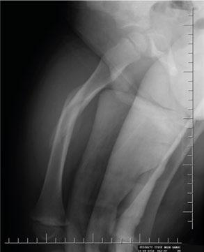
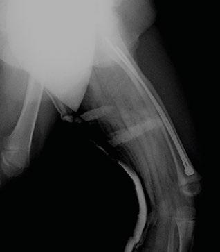

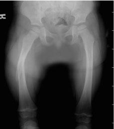
18 RADIOGRAPHIC CASES Pre-op AP Pre-op Lateral Post-op AP Post-op Lateral 7
19 Notes
20 Rev 0/0..06
humerus InSafeLOCK Nail
 humerus InSafeLOCK Nail Introduction Content Humerus InSafeLOCK Nail is an innovative intramedullary nailing system, developed for humerus problems. Humerus fractures have 5-6 % incidence of all bone fractures.
THE NANCY NAIL. The End Caps ADVANTAGES OF NANCY NAIL
 NANCY NAIL THE NANCY NAIL Nancy nails are manufactured from a specific titanyum alloy with proprietary surface treatment, which provides increased fatigue resistance. Six nail diameters (1.5 mm 2.0 mm
PediFlex Advanced Elastic Stable Intramedullary Nails SURGICAL TECHNIQUE
 PediFlex Advanced Elastic Stable Intramedullary Nails SURGICAL TECHNIQUE 2 2 TABLE OF CONTENTS System Overview Indications... 4 Implant Selection... 4 Surgical Technique Product Information Inserter Assembly
Chapter 4: Forearm 4.3 Forearm shaft fractures, transverse (12-D/4)
") AO Manual of ESIN in children s fractures Chapter 4: Forearm 4.3 Forearm shaft fractures, transverse (12-D/4) Title AO Manual of ESIN in children Subtitle Elastic stable intramedullary nailing (ESIN) Author
AO Manual of ESIN in children s fractures Chapter 4: Forearm 4.3 Forearm shaft fractures, transverse (12-D/4) Title AO Manual of ESIN in children Subtitle Elastic stable intramedullary nailing (ESIN) Author
Orthopedic Bone Nail System - Distal Femoral Nail Surgical Technique Manual
 Orthopedic Bone Nail System - Distal Femoral Nail Surgical Technique Manual Note: The surgical procedures should be performed under the guidance of qualified skilled orthopedic surgeons, and this surgical
Double Engine Orthopedic Bone Nail System Universal Humeral Nail
 Double Engine Orthopedic Bone Nail System ----------- Universal Humeral Nail Surgical Technique Manual Note: The surgical procedures should be performed under the guidance of qualified skilled orthopedic
3. PATIENT POSITIONING & FRACTURE REDUCTION 3 8. DISTAL GUIDED LOCKING FOR PROXIMAL NAIL PROXIMAL LOCKING FOR LONG NAIL 13
 Contents IMPLANT FEATURES 2 1. INDICATIONS 3 2. PRE-OPERATIVE PLANNING 3 3. PATIENT POSITIONING & FRACTURE REDUCTION 3 4. INCISION 4 5. ENTRY POINT 4-6 6. PROXIMAL NAIL INSERTION 6-7 7. PROXIMAL LOCKING
INTRAMEDULLARY. Medical Devices. Femur Intramedullary Nail
 FEMUR INTRAMEDULLARY Femur Intramedullary Nail Multi-functional Standard and Recon Antegrade and Retrograde Application Different Multi-Planner Locking Choices Medical Devices Introductions The new, multifunctional
TEN Titanium ElasticNail Surgical Technique
 Surgical Technique Original Instruments and Implants of the Association for the Study of Internal Fixation AO/ASIF Table of contents System description 2 Indications/Contraindications 3 Standard surgical
System. Humeral Nail. Surgical Technique
 System Humeral Nail Surgical Technique Contents IMPLANT FEATURES 2 1. INDICATIONS 3 2. PRE-OPERATIVE PLANNING 3 3. PATIENT POSITIONING & FRACTURE REDUCTION 3 4. INCISION 4 5. ENTRY POINT 4-6 6. PROXIMAL
Surgical Technique. Intramedullary locked Nailing With Screws for Humerus Fractures Solid/Cannulated. Humeral Interlocking Nail.
 Screws for Humerus Fractures Surgical Technique Humeral Interlocking Nail Approved by Humerus Nail Kit Code 08050001 Contents Introduction Implant design Indications Pre-operative planning Patient positioning
A Locking IM Rod that won't back out. Simple and straight to the point! SURGICAL TECHNIQUE
 A Locking IM Rod that won't back out. Simple and straight to the point! SURGICAL TECHNIQUE The SLIM (Simple Locking IntraMedullary) System is a new generation of pediatric orthopedic nails specifically
NeoGen Tibia Nail System
 NeoGen Tibia Nail System LESS IS MORE TE-2070-03 Surgical Technique BLE OF CONTENT Preface Surgical Technique Appendix Products Information Patient Preparation Entry Portal Fracture Reduction Canal Preparation
Medical Devices HUMERUS. WRIStX
 Medical Devices DDDAF HUMERUS WRIStX DAF HUMERUS FIXATOR It is a single plan external fixation tool and used for the treatment of the fractures of humerus, radius and ulna for adults. It is also used at
Surgical Technique. Targeter Systems Overview
 Surgical Technique Targeter Systems Overview PERI-LOC Locked Plating System Targeter Systems Overview Table of contents Product overview... 2 Introduction... 2 Indications... 2 Design features and benefits...
PATENTED A-PFN. Antirotator Proximal Femoral Nail. Medical Devices
 PATENTED A-PFN Antirotator Proximal Femoral Nail Medical Devices Introductions Intertrochanteric femoral fractures constitute 0% of all the bone fractures. They are frequently seen in elderly patients
A locking plate system that expands a surgeon s options in trauma surgery. Zimmer NCB Plating System
 A locking plate system that expands a surgeon s options in trauma surgery Zimmer NCB Plating System The Power of Choice The power of having true intraoperative options is at your fingertips. Using standard
Surgical Technique. Proximal Humerus Locking Plate
 Surgical Technique Proximal Humerus Locking Plate PERI-LOC Upper Extremity Locked Plating System 3.5mm & 4.5mm Proximal Humerus Locking PlatesCatalog Infor Table of Contents Introduction.........................................................2
pediatric orthopedic implants fixator
 pediatric orthopedic implants DDDAF fixator DAF HUMERUS FIXATOR It is a single plan external fixation tool and used for the treatment of the fractures of humerus, radius and ulna for adults. It is also
AcUMEDr. FoREARM ROD SYSTEM
 AcUMEDr FoREARM ROD SYSTEM FoREARM ROD SYSTEM Since 1988 Acumed has been designing solutions to the demanding situations facing orthopedic surgeons, hospitals and their patients. Our strategy has been
Table of contents. Introduction... 3 Indications... 3 Contraindications... 3 TRIGEN Humeral Nail Specifications... 6
 Surgical Technique Table of contents Introduction... 3 Indications... 3 Contraindications... 3 TRIGEN Humeral Nail Specifications... 6 Surgical Technique Patient positioning... 7 Establish the incision
Olecranon Osteotomy Nail. For simple fractures and osteotomies of the olecranon.
 Olecranon Osteotomy Nail. For simple fractures and osteotomies of the olecranon. Technique Guide Discontinued June 2016; AVAILABLE FOR IMPLANT REMOVAL PURPOSES ONLY DSEM/TRM/0517/0843 Table of Contents
LCP Medial Distal Tibia Plate, without Tab. The Low Profile Anatomic Fixation System with Angular Stability and Optimal Screw Orientation.
 LCP Medial Distal Tibia Plate, without Tab. The Low Profile Anatomic Fixation System with Angular Stability and Optimal Screw Orientation. Technique Guide LCP Small Fragment System Table of Contents Introduction
Surgical Technique. Anterolateral and Medial Distal Tibia Locking Plates
 Surgical Technique Anterolateral and Medial Distal Tibia Locking Plates PERI-LOC Periarticular Locked Plating System Anterolateral and Medial Distal Tibia Locking Plates Surgical Technique Contents Product
NeoGen Femoral Nail System
 NeoGen Femoral Nail System LESS IS MORE TE-2070-04 Surgical Technique BLE OF CONTENT Preface Standard Femoral Mode Recon Mode Post-Operative Management Appendix Products Information Indication Patient
LCP Distal Tibia Plate
 Surgical Technique LCP Locking Compression Plate Original Instruments and Implants of the Association for the Study of Internal Fixation AO/ASIF Table of contents Indications 3 Implants/Instruments 5 Surgical
TRAUMATOLOGY. Trochanter
 TRAUMATOLOGY Trochanter 1 References Prof. Dr. András Sárváry Head of Department Fővárosi Önkormányzat Péterfy Sándor úti Kórház Rendelőintézet és Baleseti Központ Budapest The following surgical description
Technique Guide. 3.5 mm LCP Olecranon Plates. Part of the Synthes locking compression plate (LCP) system.
 system.") Technique Guide 3.5 mm LCP Olecranon Plates. Part of the Synthes locking compression plate (LCP) system. Table of Contents Introduction 3.5 mm LCP Olecranon Plates 2 AO Principles 3 Indications 3 Clinical
Technique Guide 3.5 mm LCP Olecranon Plates. Part of the Synthes locking compression plate (LCP) system. Table of Contents Introduction 3.5 mm LCP Olecranon Plates 2 AO Principles 3 Indications 3 Clinical
Technique Guide. 3.5 mm LCP Low Bend Medial Distal Tibia Plates. Part of the Synthes locking compression plate (LCP) system.
 system.") Technique Guide 3.5 mm LCP Low Bend Medial Distal Tibia Plates. Part of the Synthes locking compression plate (LCP) system. Table of Contents Introduction 3.5 mm LCP Low Bend Medial Distal Tibia Plates
Technique Guide 3.5 mm LCP Low Bend Medial Distal Tibia Plates. Part of the Synthes locking compression plate (LCP) system. Table of Contents Introduction 3.5 mm LCP Low Bend Medial Distal Tibia Plates
Long Volar Plates for Diaphyseal-Metaphyseal Radius Fractures LCP. Dia-Meta Volar Distal Radius Plates. Surgical Technique
 Long Volar Plates for Diaphyseal-Metaphyseal Radius Fractures LCP Dia-Meta Volar Distal Radius Plates Surgical Technique Table of Contents Introduction LCP Dia-Meta Volar Distal Radius Plates 2 AO Principles
PediLoc 3.5mm and 4.5mm Contour Femur Plate Surgical Technique
 PediLoc 3.5mm and 4.5mm Contour Femur Plate Surgical Technique Surgical Technique Contour Femur Plate The technique description herein is made available to the healthcare professional to illustrate the
Surgical Technique. Olecranon Locking Plate
 Surgical Technique Olecranon Locking Plate PERI-LOC Locked Plating System Olecranon Locking Plate Surgical Techniquealog Infor Table of Contents Introduction...2 Indications...3 Plate Features...3 Patient
Locking Radial Head Plates
 Locking Radial Head Plates Locking Radial Head Plates Since 1988, Acumed has been designing solutions to the demanding situations facing orthopaedic surgeons, hospitals and their patients. Our strategy
Surgical Technique. Forearm Fracture Solutions
 Surgical Technique Forearm Fracture Solutions Acumed is a global leader of innovative orthopaedic and medical solutions. We are dedicated to developing products, service methods, and approaches that improve
Surgical Technique. 3.5mm and 4.5mm Lateral Proximal Tibia Locking Plates
 Surgical Technique 3.5mm and 4.5mm Lateral Proximal Tibia Locking Plates PERI-LOC Periarticular Locked Plating System 3.5mm and 4.5mm Lateral Proximal Tibia Locking Plate Surgical Technique Contents Product
Surgical Technique. Distal Humerus Locking Plate
 Surgical Technique Distal Humerus Locking Plate PERI-LOC Locked Plating System Distal Humerus Locking Plate Surgical Technique Table of Contents Introduction...2 Indications...3 Plate Features...3 Patient
LCP Distal Humerus Plates
 The anatomic fixation system for the distal humerus with angular stability Surgical technique LCP Locking Compression Plate Contents Indications and contraindications 2 Implants 3 Instruments 5 Preparation
A locking plate system that expands a surgeon s options in trauma surgery. Zimmer NCB Plating System
 A locking plate system that expands a surgeon s options in trauma surgery Zimmer NCB Plating System The Power of Choice The power of having true intraoperative options is at your fingertips. Using standard
2.7 mm/3.5 mm Variable Angle LCP Elbow System DJ9257-B 1
 2.7 mm/3.5 mm Variable Angle LCP Elbow System DJ9257-B 1 System overview Simply complete: A comprehensive system, consisting of five (5) distal humerus plates and three (3) types of olecranon plates Implant
TIPMED EXTERNAL FIXATION SYSTEMS
 TIPMED EXTERNAL FIXATION SYSTEMS ANATOMICAL LOCATIONS FOR EXTERNAL FIXATION SYSTEMS Humeral Dynamic Axial Fixator Elbow Fixator Pelvic Dynamic Axial Fixator Pennig Wrist Fixator Hand Fixator Finger Fixator
Small Fragment Plating System. Securing optimal fixation through locked and compression plating technology
 Small Fragment Plating System Securing optimal fixation through locked and compression plating technology Contents Design Rationale Introduction Interfragmentary Fixation Insertion of a 3.5 mm Cortical
TIBIAL NAILING SYSTEM OPTIONS MADE EASY
 S U R G I C A L T E C H N I Q U E TIBIAL NAILING SYSTEM OPTIONS MADE EASY TABLE OF CONTENTS DESIGN SUMMARY INSTRUMENT OVERVIEW AND JIG OPTIONS 1 2 ENTRY AND CANAL PREP NAIL INSERTION LOCKING NAIL REMOVAL
OPERATING MANUAL AND TECHNIQUE GUIDE FOR TITANIUM FEMORAL AND TIBIAL NAILING SYSTEMS
 OPERATING MANUAL AND TECHNIQUE GUIDE FOR TITANIUM FEMORAL AND TIBIAL NAILING SYSTEMS ORTHO-MEDICAL GMBH TITANIUM FEMORAL NAIL OPERATIVE TECHNIQUE Introduction: Why a new type of femoral nail? The latest
Conventus CAGE PH Surgical Techniques
 Conventus CAGE PH Surgical Techniques Conventus Orthopaedics The Conventus CAGE PH (PH Cage) is a permanent implant comprised of an expandable scaffold, made from nitinol and titanium, which is deployed
MJ-FLEX The New Metaizeau Nail TM. Operative Technique
 MJ-FLEX The New Metaizeau Nail TM Operative Technique General Description 3 Intended Use 3 Indication For Use 3 Features And Benefits 4 Equipment Required 5 Implant Principles 6 Nail Selection 6 Prior
GREENS SURGICALS. Redefining Excellence INSTRUMENT SYSTEM PREPARED BY: DR. VINAY KUMAR
 GREENS SURGICALS Redefining Excellence TIBIA AND FEMUR INSTRUMENT SYSTEM PREPARED BY: DR. VINAY KUMAR OPERATIVE TECHNIQUES INDEX SR.NO CONTENTS 1 LIST OF INSTRUMENT FOR TIBIA AND FEMUR. 2 RADIO GRAPH OF
Surgical Technique. Cannulated Angled Blade Plate 3.5 and 4.5, 90
 Surgical Technique Cannulated Angled Blade Plate 3.5 and 4.5, 90 Cannulated Angled Blade Plate 3.5 and 4.5, 90 Table of contents Indications/Contraindications 2 Implants 3 Surgical technique 5 Implant
Titanium Solid Humeral Nail System
 For Antegrade or Retrograde Insertion With Spiral Blade, Conventional and Compression Locking Titanium Solid Humeral Nail System Surgical Technique Table of Contents INTRODUCTION Foreword.... 2 Indications....
Technique Guide. 3.5 mm LCP Periarticular Proximal Humerus Plate. Part of the Synthes locking compression plate (LCP) system.
 system.") Technique Guide 3.5 mm LCP Periarticular Proximal Humerus Plate. Part of the Synthes locking compression plate (LCP) system. Table of Contents Introduction 3.5 mm LCP Proximal Humerus Plate 2 AO Principles
Technique Guide 3.5 mm LCP Periarticular Proximal Humerus Plate. Part of the Synthes locking compression plate (LCP) system. Table of Contents Introduction 3.5 mm LCP Proximal Humerus Plate 2 AO Principles
TRAUMATOLOGY. Humerus
 TRAUMATOLOGY Humerus 1 References Dr. Zoltán DETRE Lead Surgeon Szent János Kórház, Budapest Prof. Dr. András SÁRVÁRY Head of Department, Director Főv. Önk. Péterfy Sándor úti Kórház Rendelőintézet és
3.5 mm Locking Attachment Plate
 For Treatment of Periprosthetic Fractures 3.5 mm Locking Attachment Plate Surgical Technique Table of Contents Introduction 3.5 mm Locking Attachment Plate 2 Indications 4 Surgical Technique Preparation
Small Fragment Plating System
 Small Fragment Plating System Securing optimal fixation through locked and compression plating technology SURGICAL TECHNIQUE RECOVERY FUNCTION SURVIVORSHIP DePuy believes in an approach to trauma surgery
PediNail Pediatric Femoral Nail
 PediNail Pediatric Femoral Nail Surgical Technique Table of Contents Indications...3 Patient Positioning...3 Approach...4 Reaming...5 Nail Placement...6 Proximal Interlocking...7 Distal Interlocking...8
Surgical Technique. CONQUEST FN Femoral Neck Fracture System
 Surgical Technique CONQUEST FN Femoral Neck Fracture System Table of Contents Introduction... 3 Indications... 3 Product Overview... 4 Surgical Technique... 5 Patient Positioning... 5 Reduce the Fracture...
LCP Proximal Radius Plates 2.4. Plates for radial head rim and for radial head neck address individual fracture patterns of the proximal radius.
 Technique Guide LCP Proximal Radius Plates 2.4. Plates for radial head rim and for radial head neck address individual fracture patterns of the proximal radius. Table of Contents Introduction LCP Proximal
NCB Distal Femur System. Surgical Technique
 NCB Distal Femur System Surgical Technique NCB Distal Femur System Surgical Technique 3 Surgical Technique NCB Distal Femur System Table of Contents Introduction 4 Indications 8 Preoperative Planning
Biomet Pediatric Locking Nail System
 Innovation Meets Evolution...EBI Trauma is now: Biomet Pediatric Locking Nail System Surgical Technique Contents Introduction...................... Page 1 Design Rationale................... Page 2 Patient
Technique Guide. 3.5 mm LCP Low Bend Medial Distal Tibia Plate Aiming Instruments. Part of the 3.5 mm LCP Percutaneous Instrument System.
 Technique Guide 3.5 mm LCP Low Bend Medial Distal Tibia Plate Aiming Instruments. Part of the 3.5 mm LCP Percutaneous Instrument System. Table of Contents Introduction 3.5 mm LCP Low Bend Medial Distal
Surgical Technique. Fibula Rod System
 Surgical Technique Fibula Rod System Acumed is a global leader of innovative orthopaedic and medical solutions. We are dedicated to developing products, service methods, and approaches that improve patient
Zimmer Natural Nail System
 Zimmer Natural Nail System Antegrade Femoral Nail Surgical Technique (Piriformis Fossa & Greater Trochanteric Approaches) Zimmer Natural Nail System Antegrade Femoral Surgical Technique 1 Zimmer Natural
Aesculap Targon TX. Intramedullary Nail for Tibial Fractures...going to X-tremes. Aesculap Orthopaedics
 Aesculap Targon TX Intramedullary Nail for Tibial Fractures...going to X-tremes Aesculap Orthopaedics Surgical Technique Preoperative Planning KH483 X-ray template Targon TX (KH483200 : 375 mm-420 mm KH48320
WINSTA-R. Distal Radius System
 Distal Radius System Table of Contents Introduction WINSTA-R System 2 Indication 2 Surgical Technique Palmar Access for Radius Plate 3 Dorsal Access for Radius Plate 3 Positioning of the Radius Plate
For Distal Femur Fractures. 95º Condylar Plate. Quick Reference Chart
 For Distal Femur Fractures 95º Condylar Plate Quick Reference Chart 95 Condylar Plate. Quick reference chart for distal femur fractures. Insert guide wires Fix condylar fragments with 6.5 mm cancellous
LCP Medial Proximal Tibial Plate 4.5/5.0. Part of the Synthes LCP periarticular plating system.
 LCP Medial Proximal Tibial Plate 4.5/5.0. Part of the Synthes LCP periarticular plating system. Technique Guide This publication is not intended for distribution in the USA. Instruments and implants approved
U2 PSA. Revision Knee. Surgical Protocol
 U2 PSA TM Revision Knee Surgical Protocol Table of Contents 1 Component Removal... 1 2 Tibial Preparation... 1 2.1 Tibial Canal Preparation... 1 2.2 Proximal Tibial Resection... 2 2.3 Non Offset Tibial
LCP Low Bend Medial Distal Tibia Plates 3.5 mm. Anatomic plates with low profile head for intra- and extraarticular fractures.
 LCP Low Bend Medial Distal Tibia Plates 3.5 mm. Anatomic plates with low profile head for intra- and extraarticular fractures. Surgical Technique This publication is not intended for distribution in the
Open reduction; plate fixation 1 Principles
 Executive Editor: Peter Trafton Authors: Martin Jaeger, Frankie Leung, Wilson Li Proximal humerus 11-A2 Open reduction, plate fixation Search search... Shortcuts All Preparations All Approaches All Reductions
Types of Plates 1. New Dynamic Compression Plate: Diaphyseal fracture: Radius, Ulna, Humerus, Rarely tibia
 Types of Plates 1. New Dynamic Compression Plate: DCP Diaphyseal fracture: Radius, Ulna, Humerus, Rarely tibia 1. Undercut adjacent to the holes low contact: less stress shield 2. Undercut at the undersurface
PediLoc 3.5mm and 4.5mm Bowed Femur Plate Surgical Technique
 PediLoc 3.5mm and 4.5mm Bowed Femur Plate Surgical Technique 2957 Bow Broch_REV_B.indd 1 2/10/11 12:47 PM Surgical Technique Bowed Femur Plate The technique description herein is made available to the
Operasjonsteknikk. Tibia
 Operasjonsteknikk Tibia TRIGEN META-NAIL Tibial Nail System Surgical Technique Table of Contents Indications...2 Implant Specifications...3 Surgical Technique Patient Positioning...4 Incision and Entry
The study of distal ¼ diaphyseal extra articular fractures of humerus treated with antegrade intramedullary interlocking nailing
 2018; 4(4): 46-50 ISSN: 2395-1958 IJOS 2018; 4(4): 46-50 2018 IJOS www.orthopaper.com Received: 01-08-2018 Accepted: 03-09-2018 Dr. Ankur Parikh Orthopaedics, Jehangir Hospital, Sassoon road, Pune, Dr.
Clinical Evaluation Surgical Technique
 Clinical Evaluation Surgical Technique Table of Contents EMPERION Specifications 3 EMPERION Surgical Technique 9 EMPERION Catalog 18 Nota Bene: This technique description herein is made available to the
Technique Guide. TomoFix Osteotomy System. A comprehensive plating system for stable fixation of osteotomies around the knee.
 Technique Guide TomoFix Osteotomy System. A comprehensive plating system for stable fixation of osteotomies around the knee. Table of Contents Introduction TomoFix Osteotomy System 2 AO Principles 4 Indications
Surgical Technique.
 Surgical Technique www.biomet.co.uk INTRODUCTION design principals Recent advances in imaging technology have enabled orthopaedic surgeons to extend closed treatment of femoral fractures to include more
4/28/2010. Fractures. Normal Bone and Normal Ossification Bone Terms. Epiphysis Epiphyseal Plate (physis) Metaphysis
 Metaphysis") Fractures Normal Bone and Normal Ossification Bone Terms Epiphysis Epiphyseal Plate (physis) Metaphysis Diaphysis 1 Fracture Classifications A. Longitudinal B. Transverse C. Oblique D. Spiral E. Incomplete
Fractures Normal Bone and Normal Ossification Bone Terms Epiphysis Epiphyseal Plate (physis) Metaphysis Diaphysis 1 Fracture Classifications A. Longitudinal B. Transverse C. Oblique D. Spiral E. Incomplete
Fibula Rod System. Lateral Malleolus Fracture Indications:
 Fibula Rod System Fibula Rod System Since 1988, Acumed has been designing solutions for the demanding situations facing orthopaedic surgeons, hospitals and their patients. Our strategy has been to know
QUICK REFERENCE GUIDE. The PreFix Fixator (92000 Series) ALWAYS INNOVATING
 ALWAYS INNOVATING") 21 The PreFix Fixator (92000 Series) ALWAYS INNOVATING INTRODUCTION The PreFix fixator is designed to provide temporary external fixation. This may be needed when local facilities or the condition of the
21 The PreFix Fixator (92000 Series) ALWAYS INNOVATING INTRODUCTION The PreFix fixator is designed to provide temporary external fixation. This may be needed when local facilities or the condition of the
Titanium Cannulated Lateral Entry Femoral Recon Nail
 Expert Nailing System Titanium Cannulated Lateral Entry Femoral Recon Nail Surgical Technique Table of Contents Introduction Titanium Cannulated Lateral Entry 2 Femoral Recon Nail Expert System AO Principles
Low Bend Distal Tibia Plates
 Part of the DePuy Synthes Locking Compression Plate (LCP ) System 3.5 mm LCP Low Bend Medial Distal Tibia Plates Surgical Technique Table of Contents Introduction 3.5 mm LCP Low Bend Medial Distal Tibia
Technique Guide. 4.5 mm LCP Proximal Tibia Plates. Part of the Synthes LCP Periarticular Plating System.
 Technique Guide 4.5 mm LCP Proximal Tibia Plates. Part of the Synthes LCP Periarticular Plating System. Table of Contents Introduction 4.5 mm LCP Proximal Tibia Plates 2 AO Principles 4 Indications 5 Surgical
AcUMEDr. Locking Proximal Humeral Plate. PoLARUSr PHPt
 AcUMEDr Locking Proximal Humeral Plate PoLARUSr PHPt PoLARUSr PHPt LOCKING PROXIMAL HUMERAL PLATE Since 1988 Acumed has been designing solutions to the demanding situations facing orthopedic surgeons,
Humeral Universal. Product Rationale & Surgical Technique
 TM Humeral Universal Product Rationale & Surgical Technique Contents Design Summary 3 Implant Overview 4 Precautions 7 Antegrade Entry and Canal Preparation 8 Antegrade Nail Insertion 14 Antegrade Locking
Table of contents. Introduction Indications Contraindications TRIGEN META-NAIL Tibial Nail Specifications... 5
 Surgical Technique Table of contents Introduction... 3 Indications... 4 Contraindications... 4 TRIGEN META-NAIL Tibial Nail Specifications... 5 Surgical Technique... 6 Patient positioning... 6 Prepare
PROXIMAL HUMERAL NAILING SYSTEM OPERATIVE TECHNIQUE
 PROXIMAL HUMERAL NAILING SYSTEM OPERATIVE TECHNIQUE PROXIMAL HUMERAL NAILING SYSTEM Contributing Surgeons: Rupert Beikert, M.D. Senior Trauma Surgeon, Murnau Trauma Center Murnau, Germany Rosemary Buckle,
The Titanium Tibial Nail System
 The Titanium Tibial Nail System Solid Tibial Nails (UTN) and Cannulated Tibial Nails (CTN) Surgical Technique This publication is not intended for distribution in the USA. Instruments and implants approved
Technique Guide. SureLock Distal Targeting Device. C-arm guided targeting for trochanteric fixation nail.
 Technique Guide SureLock Distal Targeting Device. C-arm guided targeting for trochanteric fixation nail. Table of Contents Introduction SureLock Distal Targeting Device 2 Surgical Technique Preoperative
Aesculap Targon FN. Head Preserving Solution for Medial Femoral Neck Fractures. Aesculap Orthopaedics
 Aesculap Targon FN Head Preserving Solution for Medial Femoral Neck Fractures Aesculap Orthopaedics Targon FN Operating Technique Indications for Targon FN AO 3 B. AO 3 B.2 AO 3 B.3 Undisplaced intracapsular
S U R G I C A L T E C H N I Q U E David A. McQueen, MD Return to Menu
 S U R G I C A L T E C H N I Q U E David A. McQueen, MD TOTAL KNEE INSTRUMENTS Wichita Fusion Nail Introduction...1 Preoperative Planning...2 Surgical Technique...3-8 Wichita Fusion Nail Surgical Technique
LCP Anterolateral Distal Tibia Plate 3.5. The low profile anatomic fixation system with optimal plate placement and angular stability.
 LCP Anterolateral Distal Tibia Plate 3.5. The low profile anatomic fixation system with optimal plate placement and angular stability. Technique Guide LCP Small Fragment System Table of Contents Introduction
Phoenix Tibial Nail System
 Surgical Technique Phoenix Tibial Nail System Featuring CoreLock Technology Each nail features CoreLock Technology, a preassembled, embedded locking mechanism for locking all proximal oblique screws, which
Cannulated Angled Blade Plate 3.5 and 4.5, 90.
 Cannulated Angled Blade Plate 3.5 and 4.5, 90. Technique Guide This publication is not intended for distribution in the USA. Instruments and implants approved by the AO Foundation. Table of Contents Introduction
Principles of intramedullary nailing. Management for ORP
 Principles of intramedullary nailing Eakachit Sikarinklul,MD Basic Principles of Fracture Management for ORP Bangkok Medical Center Bangkok, 22-24 July 2016 Learning outcomes At the end of this lecture
Distal Cut First Femoral Preparation
 Surgical Technique Distal Cut First Femoral Preparation Primary Total Knee Arthroplasty LEGION Total Knee System Femoral preparation Contents Introduction...3 DCF femoral highlights...4 Preoperative planning...6
Titanium Cannulated Adolescent Lateral Entry Femoral Nail. Expert Nailing System.
 Titanium Cannulated Adolescent Lateral Entry Femoral Nail. Expert Nailing System. Technique Guide EXPERT Nailing System Table of Contents Introduction Titanium Cannulated Adolescent Lateral Entry 2 Femoral
Wrist Fixation System
 Wrist Fixation System Anatomy / Fracture Implant EXTRA & SIMPLE ARTICULAR Volar Radius Volar Fixed Angle Plate Volar Bearing Plate Radial Peg Plate Volar Hook Plate Volar Buttress Pin Volar Shear Plate
OBSOLETED. LCP Medial Distal Tibia Plate, without Tab. The Low Profile Anatomic Fixation System with Angular Stability and Optimal Screw Orientation.
 LCP Medial Distal Tibia Plate, without Tab. The Low Profile Anatomic Fixation System with Angular Stability and Optimal Screw Orientation. Surgical Technique LCP Small Fragment System This publication
Surgical Technique 1
 Surgical Technique 1 TRIGEN META-NAIL Semi-extended Instrument Set Surgical Technique Table of Contents Indications... 2... TRIGEN META-NAIL Tibial Nail Specifications... 3 Instruments for opening the
MICRONAIL. Intramedullary Distal Radius System SURGICAL TECHNIQUE
 MICRONAIL II Intramedullary Distal Radius System SURGICAL TECHNIQUE Contents Introduction 3 4 Chapter 1 5 Chapter 2 6 Appendix A 18 Appendix B 19 Surgeon Design Team Introduction Product Information Surgical