TRAUMATOLOGY. Trochanter
|
|
|
- Marshall Curtis
- 5 years ago
- Views:
Transcription


1 TRAUMATOLOGY Trochanter 1
2 References Prof. Dr. András Sárváry Head of Department Fővárosi Önkormányzat Péterfy Sándor úti Kórház Rendelőintézet és Baleseti Központ Budapest The following surgical description contains general outlines for intramedullary nailings performed on the femur with SpectruM Trochanter system. However, the operating surgeon shall adapt the content to the patient, fracture type and all other relevant factors that may have influence on the outcome of the surgery. Therefore, Sanatmetal Ltd. strongly recommends participation on workshops and trainings prior to the initial operation.
3 Contents 1. Introduction 4 4. Implant list The implant SpectruM Trochanter nail, short The instrumentarium SpectruM Trochanter nail, long Indications Trochanter spiral Implant sizes Support screw Closing cap Surgical description Locking screw Patient positioning Incision Preparation of the intramedullary Reaming Instrument list Filled up tray Instruments Assembly of the targeting arm and the 7 implant, determinig nail length (long nail) 3.6 Introduction of the nail Guide wire insertion I Guide wire insertion II Measuring the length of the trochanter 9 screw 3.10 Proximal locking I Proximal locking II Proximal locking III Supporting screw insertion Distal locking I. (short nail) Distal locking II. (short nail) Distal locking I. (long nail) Distal locking II. (long nail) Distal locking III. (long nail) Removal of the targeting arm End cap insertion Implant removal 14


 Canullated technique reposition keeping during operation Optimal targeting arm shape for more comfortable insertion point Colour coded")
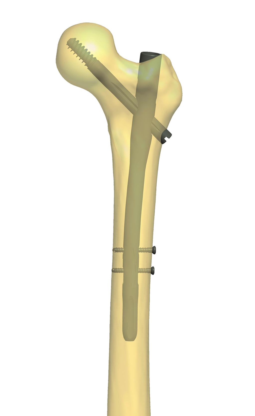
4 1 Introduction The SpectruM Trochanter implant and instrument system combines the advantages of the previously distributed and successfully applied systems furthermore the advantages of the radiolucent targeting arm. The system based on a well-considered development process, where the main aims were the simplicity and the striving for the minimal time duration of the operation. 1.1 The implant 1.2 The instrument set Cannulated nail Short nail trochanter fractrures Long, anatomic nail combined fractures 125, 130, 135 angled nails Dynamic locking possibility Usable with Trochanter Spiral as well The supporting screw is protected against loosening Sliding assured by support screw Asymmetric slots on the screw shaft limited blacksliding Special thread profile for easier screw insertion Raw materials:, Titanium alloy Special distal hole for the distal targeting device Different length end caps Radiolucent targeting arm Universal targeting arm (125, 130, 135 ) Canullated technique reposition keeping during operation Optimal targeting arm shape for more comfortable insertion point Colour coded instrument set for obvious surgical steps Instrument are protected against fall out 1.3 Indications Intertrochanteric fractures Pertrochanteric fractures Pseudarthrosis Subtrochanteric fractures (long nail) Pertrochanteric and diaphysis fractures (long nail) Pathological fracture trochanter and diaphysis (long nail) 4
 CCD (angle) 125 125 130 130 135 135 Proximal diameter (mm) 16 16 Distal diameter (mm) 10 10 11 11 12")

5 Implant range SpectruM Trochaner nail Short Long (left, right) CCD (angle) Proximal diameter (mm) Distal diameter (mm) Tochanter screw Ø10.5 mm Raw material Anodised Titanium 2.3 Locking screw Ø4.8 mm Raw material Anodised Titanium Raw material Anodised Titanium 2.5 Closing cap 2.4 Support screw 0-25 Raw material Anodised Titanium Raw material Anodised Titanium The support screw and the nail are in the same package. 5


6 3 Surgical description 3.1 Patient positioning In supine position on extension surgical table. 3.2 Incision Between the greater trochanter and the wing of the iliac bone. The proper place depends on the quantity of the soft tissues. 3.3 Preparation of the intramedullary Drill in the Kirschner wire (Ø3.2 mm, NOT threaded) through the tip of the greater trochanter under image intensifier control. Drive the awl to the wire and open the intramedullary canal. Remove the Kirschner wire. 3.4 Reaming Introduce the 4/3x900 mm olive tip guide rod through the awl to the intramedullary canal with the T-handle. Remove the awl. Place the soft tissue protector to the trochanter tip through the guide rod and ream the proximal place of the nail with the cannulated conical reamer. While using the soft tissue protector and the conical reamer the position of the awl made gap can be slightly modified. Remove the conical reamer and the soft tissue protector. Ream the intramedullary canal if needed, then remove the reamer. In case of problems remove the reamer head with the olive tip guide rod. 6


7 3.5 Assembly of the targeting arm the implant, determining nail length (long nail) In case of long nails measure the length of the guide wire part outside of the femur with a measuring rod. Subtract this value from the 900 mm length of the olive tip guide wire. The result is the required nail length. To make measurement easier the 500 mm is marked on the guide wire. Assemble the chosen Spectrum Trochanter nail onto the targeting arm with the fixing screw. Tighten the screw with the 8 mm screwdriver. Attention Always check the assembly before insertion. Place the soft tissue protector into the red slot. Push the stepped drill through the tissue protector. In case of optimal assembly the sleeve drives the drill to the proper hole. 3.6 Introduction of the nail Insert the nail into the intramedullary canal with rotating movements. The proximal end of the nail is indicated by the Kirschner wire led through the targeting arm as per the image. There are 3 slots on the metal part of the targeting arm connecting to the nail. This can help the identification of the nail tip under image intensifier. The nail tip is located 5 mms away from the most distal slot. Determine the rotational position of the nail with a Kirschner wire under image intensifier as seen on the image. Check the optimal sinking of the nail with the indicator. Place the indicator into the targeting arm to the corresponding angulation slots in such a way that its sleeves for Kirschner wires shall facet he anterior. Place a Kirschner wire into the distal of the two sleeves and under image intensifier check the depth of the nail. In optimal case the Kirschner wire s centerline is about 8 mm from the Adam arch. 7


 on the targeting arm and hit the bone surface. 3.")

8 3 Surgical description If the nail insertion is blocked, and the surgeon decide that the guide wire is removable, after removal of the guide wire, connect the 8 mm screwdriver to the connecting screw and guide the nail to its final position with light mallet blows. In case the guide wire is irremovable, remove the nail, ream again the intramedullary canal, and introduce the nail again into the canal. Remove the olive tip guide rod. Important Do not hit the targeting arm. 3.7 Guide wire insertion I. Push the red marked soft tissue protector (assembled with the drill sleeve) to the appropriate angulation hole (125, 130, 135 ) on the targeting arm and hit the bone surface. 3.8 Guide wire insertion II. Through the red sleeve open the cortical with a 3.2 mm spiral drill (1). Drill the 3.2 mm Kirschner wire to 5 mm far from the subchondralis surface through the distal hole of the red sleeve under image intensifier control (2). Repeat the process in the proximal holes (3,4)


 then drill through the Remove the")


9 3.9 Measuring the length of the trochanter screw Remove the 3.2 mm sleeve and push the length gauge to the Kirschner wire. Read the necessary screw length at the end of the Kirschner wire. Inbetween values should be rounded up Proximal locking I. Set the bumper of the stepped drill to the value of the previous step (the value is shown in the window) then drill through the Kirschner wire. Remove the stepped drill Proximal locking II. Assemble the chosen trochanter screw with the threaded retaining stem to the screw insertion device and fix with a hand (in case it can be secured by a 3.5 mm screwdriver). The compression nut on the insertion device is set into upper position. 9


10 3 Surgical description 3.12 Proximal locking III. The trochanter screw is driven in through the soft tissue protector that the end of the screw should be 5-8mm far from the subchondralis surface. Check the position under an image intensifier. After performing this operation the T-wrench of the screw insertion device should be perpendicular or parallel with the targeting arm. This way the supporting screw will slide to the slot on the trochanter screw shaft. Turn the compression nut on the retaining stem to the direction of the arrow to achieve necessary compression. (On occasion wrench can be used.) Remove the reposition maintaining Kirschner wire in the proximal hole Supporting screw insertion Put the supporting screw to the 3.5 mm flexible screwdriver and drive it into the nail. Loosen up the support screw with a quarter turn so that the sliding effect may prevail. Move the T-handle with slight rotating movements to check the stability against rotation of the supporting screw. Dismount the screw insertion device and remove the Kirschner wire and the red sleeve. In case of long nail continue with 3.16 step. 10
 Place the assembled green soft tissue")


 into the chosen S marked hole of the targeting")
 Remove the drill sleeve, the length of the")


11 3.14 Distal locking I. (short nail) Place the assembled green soft tissue protector (for dynamic hole - sign ) and the sleeve into the green marked distal targeting arm. Then drill with a 4.2 mm spiral drill. In case of only static locking use the soft tissue protector and drill sleeve (only for static holes - sign) into the chosen S marked hole of the targeting arm. D S S 3.15 Distal locking II. (short nail) Remove the drill sleeve, the length of the screw should be measured through the green soft tissue protector, push the measurer up to the bone surface. The value shown at the soft tissue protector directly determines the necessary length of the interlocking screw. Important Continue with 3.18 step. Alternative length gauging method: put the drill stop on the drillbit, just above the spiral part. Perform drilling. Read the necessary screw length at bottom side of the stop. The selected screw should be driven in with a 3.5 mm screwdriver through the soft tissue protector. 11
 Check the position of the hole under an image intensifier and make skin")


 Put the distal targeting device into the hole on the bone (the double")
.")


12 3 Surgical description 3.16 Distal locking I. (long nail) Check the position of the hole under an image intensifier and make skin incision on the given place. Put the free-hand target device onto the bone and find the accurate place of the locking hole with the help of the image intensifier. Drill the closer corticalis with a 6 mm spiral drill through the soft tissue protector. Make drilling carefully. As soon as the drill passes through the corticalis and reaches the nail stop the movement. Remove the free-hand target device Distal locking II. (long nail) Put the distal targeting device into the hole on the bone (the double hole should face to distal direction) then click it into the special slot on the nail (1). After skin incision put the tissue protector and the drill sleeve into the chosen hole of the distal targeting device, push them to the bone surface and perform drilling (2). Hold the distal targeting device continuously on the bone surface during drilling. Remove the drill sleeve. 1 Measure the required screw length through the soft tissue protector, push the measurer up to the bone surface (3). Alternative length gauging method: put the drill stop on the drillbit, just above the spiral part. Perform drilling. Read the necessary screw length at bottom side of the stop. 2 Drive the screw in (4). 3 D Special S 4 12


13 3.18 Distal locking III. (long nail) Remove the manual aiming device and put back the free hand targeting device and the drill sleeve on the special hole. Drill through the ventral wall of the cortical with the 4.2 mm drillbit through the soft tissue protector and drill sleeve. After length measurement introduce the screw. Mind that the screw will hold only in the farther cortical. Do not tighten with excessive force to prevent sinking the screw head. Warning Do not use the scaled drillbit for length gauging! 3.19 Removal of the targeting arm Loosen the connecting screw with the 8 mm screwdriver and dismantle the targeting arm from the nail End cap insertion Drive in the proper size end cap with the 3.5 mm screwdriver or the 3.5 mm adapter assembled to the flexible shaft or the 3.5 mm flexible screwdriver. 13


14 3 Surgical description 3.21 Implant removal Remove the distal 4.8 mm locking screws with the 3.5 mm screwdriver. After opening up the nail end take the end cap out with the 3.5 mm screwdriver. Loosen the supporting screw with the 3.5 mm screwdriver or the 3.5 mm adapter assembled to the flexible shaft. Connect the nail impactor dowel to the end of the Spectrum trochanter nail. Open the soft tissues and assemble the impactor with T-handle with the threaded stem to the end of the trochanter screw. Take the trochanter screw out. Hit the nail out of the bone with the forked hammer. 14
15 15
16 4 Implant list 4.1 SpectruM Trochanter nail, short CCD=125 Distal diameter Anodised Titanium CCD=130 Distal diameter Anodised Titanium CCD=135 Distal diameter Anodised Titanium
17 4.2 SpectruM Trochanter nail, long CCD=125 Distal diameter Left Right Left Right Left Right CCD=130 Distal diameter Left Right Left Right Left Right CCD=135 Distal diameter Left Right Left Right Left Right
18 4 Implant list CCD=125 Anodised Titanium Distal diameter Left Right Left Right Left Right CCD=130 Anodised Titanium Distal diameter Left Right Left Right Left Right Anodised Titanium CCD=135 Distal diameter Left Right Left Right Left Right
19 4.3 Trochanter screw Ø10.5 mm 4.6 Locking screw Ø4.8 mm 4.4 Support screw 4.5 Closing cap Anodised Titanium Anodised Titanium Anodised Titanium Anodised Titanium
20 5 Instrument list 5.1 Filled-up tray Surgical description Description Size Quantity Cat. no. Linear gauge for locking screws T-wrench with compression nut Threaded Retaining Stem Depth gauge for locking screws 4.8 mm Stepped drill 10.5/6.7 can. 3.2 mm Target device SpectruM Trochanter Awl Free-hand target device SpectruM Trochanter Nail Extractor Dowel Flexible shaft T-handle Distal targeting device SpectruM Trochanter Spiral drill 4.2x250 mm Spiral drill 4.2x280 mm Conical reamer 16 mm Soft tissue protector 16 mm Measuring rod 500 mm Spiral drill with quick-conn. end 3.2x310 mm Kirschner wire 3.2x375 mm Threaded kirschner wire 3.2x375x10 mm Fork Hammer Screwdriver 3.5 mm Spiral drill 6 mm Guide Rod with olive Tip 4/3x900 mm Screwdriver 8 mm Wrench Drill stop 4.2 mm Indicator (optional Double wire (optional) Tray (empty) SpectruM Trochanter Filled-up tray (SpectruM Trochanter)


 258900016 Stepped drill (10.5/6.")
 254915009 Target device")

21 5.2 Instruments Linear gauge for locking screws T-wrench with compression nut Threaded Retaining Stem Linear gauge for locking screws (4.8 mm) Stepped drill (10.5/6.7 can. 3.2 mm) Target device SpectruM Trochanter Awl Free-hand target device SpectruM Trochanter Nail Extractor Dowel


 258900011 Conical")

 939999072")

22 5 Instrument list Flexible shaft T-handle Distal targeting device-sm Trochanter Spiral drill (4.2x250 mm) Spiral drill (4.2x280 mm) Conical reamer (16 mm) Soft tissue protector (16 mm) Measuring rod (500 mm) Spiral drill with quick-connecting end (3.2x310 mm)
 256732375")


 210510002 Spiral")


 210510242")
23 Kirschner wire (3.2x375 mm) Threaded kirschner wire (3.2x375x10 mm) Fork Hammer Screwdriver (3.5 mm) Spiral drill (6 mm) Guide Rod with Olive Tip (4/3x900 mm) Screwdriver (8 mm) Wrench Drill stop (4.2 mm) Indicator (optional) Double wire (optional)


24 Product family TRAUMATOLOGY 1.1. Intramedullary nails Humerus nails Ulna-radius nails Trochanter nails Femur nails Tibia nails Fibula nails Sanat PIN 1.2. Plates 1.3. Screws 1.4. Fixateur externe 1.5. Other ORTHOPEADICS DENTAL SPINE Contact address: 5, Faiskola st Eger, Hungary phone: fax: TRAUMA REV_C 16/09/2014
TRAUMATOLOGY. Humerus
 TRAUMATOLOGY Humerus 1 References Dr. Zoltán DETRE Lead Surgeon Szent János Kórház, Budapest Prof. Dr. András SÁRVÁRY Head of Department, Director Főv. Önk. Péterfy Sándor úti Kórház Rendelőintézet és
TRAUMATOLOGY. Vortex. Distal Tibia plate
 TRAUMATOLOGY Vortex Distal Tibia plate 1 References Dr. László Vámhidy PhD. Head of Department, Clinical Director MSI Department of Traumatology and Hand Surgery Pécs, Hungary Prof. Dr. Endre Varga Professor
TRAUMATOLOGY. Vortex. Clavicle plate ±15
 Vortex TRAUMATOLOGY Clavicle plate ±15 1 References Dr. Ferenc Tóth Chief Medical Director 1. 1.1 1.2 1.3 Introduction The implant The instruments Indications 4 4 4 4 Péterfy Sándor úti Kórház Rendelőintézet
Orthopedic Bone Nail System - Distal Femoral Nail Surgical Technique Manual
 Orthopedic Bone Nail System - Distal Femoral Nail Surgical Technique Manual Note: The surgical procedures should be performed under the guidance of qualified skilled orthopedic surgeons, and this surgical
Double Engine Orthopedic Bone Nail System Universal Humeral Nail
 Double Engine Orthopedic Bone Nail System ----------- Universal Humeral Nail Surgical Technique Manual Note: The surgical procedures should be performed under the guidance of qualified skilled orthopedic
Zimmer ITST Intertrochanteric/ Subtrochanteric Fixation System. Abbreviated Surgical Technique
 Zimmer ITST Intertrochanteric/ Subtrochanteric Fixation System Abbreviated Surgical Technique ITST System Abbreviated Surgical Technique Indications The ITST Intramedullary Nail is indicated for use in
NeoGen Tibia Nail System
 NeoGen Tibia Nail System LESS IS MORE TE-2070-03 Surgical Technique BLE OF CONTENT Preface Surgical Technique Appendix Products Information Patient Preparation Entry Portal Fracture Reduction Canal Preparation
INTRAMEDULLARY NAILING TIBIA AND FEMUR 1.3 TIBIAL AND FEMORAL NAILING FEMORAL NAILING WITH RETROGRADE INSERTION IMPLANTS AND OPERATING MANUAL
 1.3 TIBIA AND FEMORA NAIING FEMORA NAIING WITH RETROGRADE INSERTION IMPANTS AND OPERATING MANUA INTRAMEDUARY NAIING TIBIA AND FEMUR Medical Products Manufacturing and Trading td. General informations
Sirus Antegrade Femoral Nail System Surgical Technique
 Sirus Antegrade Femoral Nail System Surgical Technique The Cannulated Titanium Nail with Anatomical Shape and Lateral Entry Point Disclaimer This document is intended exclusively for experts in the field,
System. Humeral Nail. Surgical Technique
 System Humeral Nail Surgical Technique Contents IMPLANT FEATURES 2 1. INDICATIONS 3 2. PRE-OPERATIVE PLANNING 3 3. PATIENT POSITIONING & FRACTURE REDUCTION 3 4. INCISION 4 5. ENTRY POINT 4-6 6. PROXIMAL
3. PATIENT POSITIONING & FRACTURE REDUCTION 3 8. DISTAL GUIDED LOCKING FOR PROXIMAL NAIL PROXIMAL LOCKING FOR LONG NAIL 13
 Contents IMPLANT FEATURES 2 1. INDICATIONS 3 2. PRE-OPERATIVE PLANNING 3 3. PATIENT POSITIONING & FRACTURE REDUCTION 3 4. INCISION 4 5. ENTRY POINT 4-6 6. PROXIMAL NAIL INSERTION 6-7 7. PROXIMAL LOCKING
INTRAMEDULLARY. Medical Devices. Femur Intramedullary Nail
 FEMUR INTRAMEDULLARY Femur Intramedullary Nail Multi-functional Standard and Recon Antegrade and Retrograde Application Different Multi-Planner Locking Choices Medical Devices Introductions The new, multifunctional
OPERATING MANUAL AND TECHNIQUE GUIDE FOR TITANIUM FEMORAL AND TIBIAL NAILING SYSTEMS
 OPERATING MANUAL AND TECHNIQUE GUIDE FOR TITANIUM FEMORAL AND TIBIAL NAILING SYSTEMS ORTHO-MEDICAL GMBH TITANIUM FEMORAL NAIL OPERATIVE TECHNIQUE Introduction: Why a new type of femoral nail? The latest
humerus InSafeLOCK Nail
 humerus InSafeLOCK Nail Introduction Content Humerus InSafeLOCK Nail is an innovative intramedullary nailing system, developed for humerus problems. Humerus fractures have 5-6 % incidence of all bone fractures.
3. Insert Tocar Sleeves Insert the NCB tissue protection sleeve assembly 1.6 to 10mm through a skin incision (Fig. 38).
.") NCB Proximal Humerus Plating System Surgical Technique 19 2. Temporary Plate Fixation The plate can be temporary fixed to the bone with 1.6mm K-wire through the proximal cannulated fixation screw of the
NCB Proximal Humerus Plating System Surgical Technique 19 2. Temporary Plate Fixation The plate can be temporary fixed to the bone with 1.6mm K-wire through the proximal cannulated fixation screw of the
NeoGen Femoral Nail System
 NeoGen Femoral Nail System LESS IS MORE TE-2070-04 Surgical Technique BLE OF CONTENT Preface Standard Femoral Mode Recon Mode Post-Operative Management Appendix Products Information Indication Patient
Pre-Operative Planning. Positioning of the Patient
 Surgical Technique Pre-Operative Planning Decide upon the size and angle of the barrel plate to be used from measuring the x-rays. To maximise the sliding action when using shorter lag screws, the Short
PATENTED A-PFN. Antirotator Proximal Femoral Nail. Medical Devices
 PATENTED A-PFN Antirotator Proximal Femoral Nail Medical Devices Introductions Intertrochanteric femoral fractures constitute 0% of all the bone fractures. They are frequently seen in elderly patients
Technique Guide. SureLock Distal Targeting Device. C-arm guided targeting for trochanteric fixation nail.
 Technique Guide SureLock Distal Targeting Device. C-arm guided targeting for trochanteric fixation nail. Table of Contents Introduction SureLock Distal Targeting Device 2 Surgical Technique Preoperative
Technique Guide. DHS Blade. For osteoporotic bone.
 Technique Guide DHS Blade. For osteoporotic bone. Table of Contents Introduction Features and Benefits 2 Indications and Contraindications 4 Clinical Cases 5 Surgical Technique Implantation 6 Implant
A locking plate system that expands a surgeon s options in trauma surgery. Zimmer NCB Plating System
 A locking plate system that expands a surgeon s options in trauma surgery Zimmer NCB Plating System The Power of Choice The power of having true intraoperative options is at your fingertips. Using standard
Technique Guide. 3.5 mm LCP Low Bend Medial Distal Tibia Plate Aiming Instruments. Part of the 3.5 mm LCP Percutaneous Instrument System.
 Technique Guide 3.5 mm LCP Low Bend Medial Distal Tibia Plate Aiming Instruments. Part of the 3.5 mm LCP Percutaneous Instrument System. Table of Contents Introduction 3.5 mm LCP Low Bend Medial Distal
PFN. Proximal Femoral Nail Standard/Short, PFN Long PFN
 PFN. Proximal Femoral Nail Standard/Short, PFN Long PFN Surgical Technique This publication is not intended for distribution in the USA. Instruments and implants approved by the AO Foundation. 357.001
Arthrodesis of the Ankle
 TM Arthrodesis of the Ankle Plantar View of the Foot 1. Make a lateral incision to expose the tibia-talus joint and the talo-calcaneal joint. Prepare the arthrodesis sites surgically with a rasp or an
Antegrade Femoral Nail (AFN)
") Antegrade Femoral Nail (AFN) Surgical Technique This publication is not intended for distribution in the USA. Instruments and implants approved by the AO Foundation. Image intensifier control Warning This
Antegrade Femoral Nail (AFN) Surgical Technique This publication is not intended for distribution in the USA. Instruments and implants approved by the AO Foundation. Image intensifier control Warning This
LCP Distal Tibia Plate
 Surgical Technique LCP Locking Compression Plate Original Instruments and Implants of the Association for the Study of Internal Fixation AO/ASIF Table of contents Indications 3 Implants/Instruments 5 Surgical
The Titanium Tibial Nail System
 The Titanium Tibial Nail System Solid Tibial Nails (UTN) and Cannulated Tibial Nails (CTN) Surgical Technique This publication is not intended for distribution in the USA. Instruments and implants approved
Surgical Technique. Fibula Rod System
 Surgical Technique Fibula Rod System Acumed is a global leader of innovative orthopaedic and medical solutions. We are dedicated to developing products, service methods, and approaches that improve patient
Technique Guide. 3.5 mm LCP Low Bend Medial Distal Tibia Plates. Part of the Synthes locking compression plate (LCP) system.
 system.") Technique Guide 3.5 mm LCP Low Bend Medial Distal Tibia Plates. Part of the Synthes locking compression plate (LCP) system. Table of Contents Introduction 3.5 mm LCP Low Bend Medial Distal Tibia Plates
Technique Guide 3.5 mm LCP Low Bend Medial Distal Tibia Plates. Part of the Synthes locking compression plate (LCP) system. Table of Contents Introduction 3.5 mm LCP Low Bend Medial Distal Tibia Plates
A locking plate system that expands a surgeon s options in trauma surgery. Zimmer NCB Plating System
 A locking plate system that expands a surgeon s options in trauma surgery Zimmer NCB Plating System The Power of Choice The power of having true intraoperative options is at your fingertips. Using standard
TITANIUM TIBIAL NAIL SySTEM
 TITANIUM TIBIAL NAIL SySTEM Solid and Cannulated Nails SURGICAL TEChNIqUE Table of contents Introduction Indications 2 Preoperative Implant Selection 6 Surgical Technique Instruments for Opening the Tibia
3.5 mm Locking Attachment Plate
 For Treatment of Periprosthetic Fractures 3.5 mm Locking Attachment Plate Surgical Technique Table of Contents Introduction 3.5 mm Locking Attachment Plate 2 Indications 4 Surgical Technique Preparation
Zimmer Natural Nail System
 Zimmer Natural Nail System Antegrade Femoral Nail Surgical Technique (Piriformis Fossa & Greater Trochanteric Approaches) Zimmer Natural Nail System Antegrade Femoral Surgical Technique 1 Zimmer Natural
LOCKING TEP LOCKING TITANIUM ELASTIC PIN INTRAMEDULLARY NAIL
 LOCKING TEP LOCKING TITANIUM ELASTIC PIN INTRAMEDULLARY NAIL ... Index -3 3-8 8 9 9 0 7 Introduction Features Indicatiıons Surgical Technique Femoral Surgical Technique Tibial Surgical Technique Ulna Radius
The Vilex FUZETM. Dual Thread Screw & Intramedullary Nail in One Implant. The Ultimate TTC Arthrodesis Internal Fixator
 The Vilex FUZETM Dual Thread Screw & Intramedullary Nail in One Implant The Ultimate TTC Arthrodesis Internal Fixator Introduction The Vilex FUZE TM TTC Arthrodesis Compression Nail combines the attributes
LCP Medial Distal Tibia Plate, without Tab. The Low Profile Anatomic Fixation System with Angular Stability and Optimal Screw Orientation.
 LCP Medial Distal Tibia Plate, without Tab. The Low Profile Anatomic Fixation System with Angular Stability and Optimal Screw Orientation. Technique Guide LCP Small Fragment System Table of Contents Introduction
PFNA-II. Proximal Femoral Nail Antirotation.
 PFNA-II. Proximal Femoral Nail Antirotation. Technique Guide This publication is not intended for distribution in the USA. Instruments and implants approved by the AO Foundation. Image intensifier control
Intra Medullary Interlocking Nailing System
 Intra Medullary Interlocking Nailing System www.orthotraumaint.co.uk Interlocking Intra Medullary Nails for lower extremity fractures INDICATIONS: Stable and unstable Femoral Shaft fractures Femur Nailing
titanium cannulated adolescent lateral entry femoral nail
 titanium cannulated adolescent lateral entry femoral nail Expert Nailing System with Radiolucent Instrumentation SurgIcal technique Table of Contents Introduction Titanium Cannulated Adolescent Lateral
Expert A2FN. Designed for small statured patients.
 Expert A2FN. Designed for small statured patients. Technique Guide Expert Nailing System This publication is not intended for distribution in the USA. Instruments and implants approved by the AO Foundation
For intramedullary fixation of proximal femoral fractures SURGICAL TECHNIQUE
 For intramedullary fixation of proximal femoral fractures SURGICAL TECHNIQUE TABLE OF CONTENTS INTRODUCTION Clinical Cases 2 AO Principles 4 Indications and Precautions 5 SURGICAL TECHNIQUE Preparation
Surgical Technique. Cannulated Angled Blade Plate 3.5 and 4.5, 90
 Surgical Technique Cannulated Angled Blade Plate 3.5 and 4.5, 90 Cannulated Angled Blade Plate 3.5 and 4.5, 90 Table of contents Indications/Contraindications 2 Implants 3 Surgical technique 5 Implant
Principles of intramedullary nailing. Management for ORP
 Principles of intramedullary nailing Eakachit Sikarinklul,MD Basic Principles of Fracture Management for ORP Bangkok Medical Center Bangkok, 22-24 July 2016 Learning outcomes At the end of this lecture
TIBIAL NAILING SYSTEM OPTIONS MADE EASY
 S U R G I C A L T E C H N I Q U E TIBIAL NAILING SYSTEM OPTIONS MADE EASY TABLE OF CONTENTS DESIGN SUMMARY INSTRUMENT OVERVIEW AND JIG OPTIONS 1 2 ENTRY AND CANAL PREP NAIL INSERTION LOCKING NAIL REMOVAL
Technique Guide. 3.5 mm LCP Olecranon Plates. Part of the Synthes locking compression plate (LCP) system.
 system.") Technique Guide 3.5 mm LCP Olecranon Plates. Part of the Synthes locking compression plate (LCP) system. Table of Contents Introduction 3.5 mm LCP Olecranon Plates 2 AO Principles 3 Indications 3 Clinical
Technique Guide 3.5 mm LCP Olecranon Plates. Part of the Synthes locking compression plate (LCP) system. Table of Contents Introduction 3.5 mm LCP Olecranon Plates 2 AO Principles 3 Indications 3 Clinical
Titanium Cannulated Lateral Entry Femoral Recon Nail
 Expert Nailing System Titanium Cannulated Lateral Entry Femoral Recon Nail Surgical Technique Table of Contents Introduction Titanium Cannulated Lateral Entry 2 Femoral Recon Nail Expert System AO Principles
TIPMED EXTERNAL FIXATION SYSTEMS
 TIPMED EXTERNAL FIXATION SYSTEMS ANATOMICAL LOCATIONS FOR EXTERNAL FIXATION SYSTEMS Humeral Dynamic Axial Fixator Elbow Fixator Pelvic Dynamic Axial Fixator Pennig Wrist Fixator Hand Fixator Finger Fixator
Anatomical Shoulder System
 Anatomical Shoulder System Adjustable. Convertible. Compatible. Anatomical Shoulder System by Zimmer This unique system is like an individually produced prosthesis specifically designed for each patient.
Titanium Cannulated Adolescent Lateral Entry Femoral Nail. Expert Nailing System.
 Titanium Cannulated Adolescent Lateral Entry Femoral Nail. Expert Nailing System. Technique Guide EXPERT Nailing System Table of Contents Introduction Titanium Cannulated Adolescent Lateral Entry 2 Femoral
Antegrade Femoral Nail (AFN)
") Antegrade Femoral Nail (AFN) Surgical Technique This publication is not intended for distribution in the USA. Instruments and implants approved by the AO Foundation. Contents Indications/contraindications
Antegrade Femoral Nail (AFN) Surgical Technique This publication is not intended for distribution in the USA. Instruments and implants approved by the AO Foundation. Contents Indications/contraindications
U2 PSA. Revision Knee. Surgical Protocol
 U2 PSA TM Revision Knee Surgical Protocol Table of Contents 1 Component Removal... 1 2 Tibial Preparation... 1 2.1 Tibial Canal Preparation... 1 2.2 Proximal Tibial Resection... 2 2.3 Non Offset Tibial
Intramedullary Nail Systems
 Intramedullary Nail Systems TianJin ZhengTian Medical Instrument Co., td. A Company of NATON Medical Group Facility Add: East of Guihuazhi oad, West of Jingyi oad, Airport Economic Zone, Tianjin, 0008,
Expert A2FN. Designed for small statured patients.
 Expert A2FN. Designed for small statured patients. Surgical Technique Expert Nailing System This publication is not intended for distribution in the USA. Instruments and implants approved by the AO Foundation.
Mini External Fixator.
 Mini External Fixator. Assembly and Surgical Technique This publication is not intended for distribution in the USA. Instruments and implants approved by the AO Foundation. Image intensifier control Warning
Olecranon Osteotomy Nail. For simple fractures and osteotomies of the olecranon.
 Olecranon Osteotomy Nail. For simple fractures and osteotomies of the olecranon. Technique Guide Discontinued June 2016; AVAILABLE FOR IMPLANT REMOVAL PURPOSES ONLY DSEM/TRM/0517/0843 Table of Contents
Expert R/AFN. Retrograde/Antegrade Femoral Nail.
 Expert R/AFN. Retrograde/Antegrade Femoral Nail. Surgical Technique EXPERT Nailing System This publication is not intended for distribution in the USA. Instruments and implants approved by the AO Foundation
Triathlon TS Knee System. Surgical Protocol
 Triathlon TS Knee System Surgical Protocol Triathlon TS Knee System Surgical Protocol Table of Contents Acknowledgments..........................................................2 Exposure...................................................................4
Technique Guide. Locking Attachment Plate. For treatment of periprosthetic fractures.
 Technique Guide Locking Attachment Plate. For treatment of periprosthetic fractures. Table of Contents Introduction Locking Attachment Plate 2 Indications 4 Surgical Technique Patient Positioning 5 Preparation
Femoral Recon Nail System FRN
 Greater Trochanter Piriformis Fossa Approaches For Intramedullary Fixation of Femoral Shaft Fractures Femoral Recon Nail System FRN Surgical Technique Table of Contents AO Principles 2 Indications and
GREENS SURGICALS. Redefining Excellence INSTRUMENT SYSTEM PREPARED BY: DR. VINAY KUMAR
 GREENS SURGICALS Redefining Excellence TIBIA AND FEMUR INSTRUMENT SYSTEM PREPARED BY: DR. VINAY KUMAR OPERATIVE TECHNIQUES INDEX SR.NO CONTENTS 1 LIST OF INSTRUMENT FOR TIBIA AND FEMUR. 2 RADIO GRAPH OF
Titanium Cannulated Retrograde/ Antegrade Femoral Nail
 Expert Nailing System With Radiolucent Instrumentation Titanium Cannulated Retrograde/ Antegrade Femoral Nail Surgical Technique Table of Contents Introduction Titanium Cannulated Retrograde/Antegrade
DHS Plate 135º. DHS Platte 135º
 Section of hip fixation plate compression system DHS Plate 3º DHS Platte 3º Plaque de DHS 3º Indication : The compression hip plate is primarly indicated for intertrochanteric fractures. However, it can
PROXIMAL FEMORAL NAIL REMOVAL SET
 PROXIMAL FEMORAL NAIL REMOVAL SET for PFN, TFN and PFNA/PFNA-II Instruments and Implants approved by the AO Foundation. This publication is not intended for distribution in the USA. SURGICAL TECHNIQUE
Titanium Solid Humeral Nail System
 For Antegrade or Retrograde Insertion With Spiral Blade, Conventional and Compression Locking Titanium Solid Humeral Nail System Surgical Technique Table of Contents INTRODUCTION Foreword.... 2 Indications....
AcUMEDr. FoREARM ROD SYSTEM
 AcUMEDr FoREARM ROD SYSTEM FoREARM ROD SYSTEM Since 1988 Acumed has been designing solutions to the demanding situations facing orthopedic surgeons, hospitals and their patients. Our strategy has been
Expert ALFN. Adolescent Lateral Femoral Nail
 Expert ALFN. Adolescent Lateral Femoral Nail Surgical Technique EXPERT Nailing System This publication is not intended for distribution in the USA. Instruments and implants approved by the AO Foundation
Biomet Pediatric Locking Nail System
 Innovation Meets Evolution...EBI Trauma is now: Biomet Pediatric Locking Nail System Surgical Technique Contents Introduction...................... Page 1 Design Rationale................... Page 2 Patient
designed to advance the treatment of hip fractures.
 designed to advance the treatment of hip fractures. introducing the tfn-advanced proximal femoral nailing system (tfna). the tfna system is a new system designed to solve a wide range of unmet needs for
Technique Guide. DHS/DCS System. Including LCP DHS and DHS Blade.
 Technique Guide DHS/DCS System. Including LCP DHS and DHS Blade. Table of Contents Introduction System Overview 2 Features and Benefits 4 Indications and Contraindications 6 Clinical Cases 8 Surgical
Surgical Technique.
 Surgical Technique www.biomet.co.uk INTRODUCTION design principals Recent advances in imaging technology have enabled orthopaedic surgeons to extend closed treatment of femoral fractures to include more
Periarticular Aiming Arm Instruments for LCP Proximal Tibial Plate 4.5/5.0. Part of the LCP Periarticular Aiming Arm Instrument System (large).
.") Technique Guide Periarticular Aiming Arm Instruments for LCP Proximal Tibial Plate 4.5/5.0. Part of the LCP Periarticular Aiming Arm Instrument System (large). Image intensifier control Warning This description
Technique Guide Periarticular Aiming Arm Instruments for LCP Proximal Tibial Plate 4.5/5.0. Part of the LCP Periarticular Aiming Arm Instrument System (large). Image intensifier control Warning This description
Surgical Technique. Intramedullary locked Nailing With Screws for Humerus Fractures Solid/Cannulated. Humeral Interlocking Nail.
 Screws for Humerus Fractures Surgical Technique Humeral Interlocking Nail Approved by Humerus Nail Kit Code 08050001 Contents Introduction Implant design Indications Pre-operative planning Patient positioning
Femoral Recon Nail System FRN
 Greater Trochanter Piriformis Fossa Approaches For Intramedullary Fixation of Femoral Shaft Fractures Femoral Recon Nail System FRN Surgical Technique Image intensifier control This description alone does
Surgical technique. UHN/PHN Humeral Nailing System.
 Surgical technique UHN/PHN Humeral Nailing System. 358.590 Radiographic Ruler for UHN 292.260 Kirschner Wire 2.5 mm with trocar tip, length 280 mm, Stainless Steel 351.120 Awl with T-Handle, cannulated,
Surgical Technique. Anterolateral and Medial Distal Tibia Locking Plates
 Surgical Technique Anterolateral and Medial Distal Tibia Locking Plates PERI-LOC Periarticular Locked Plating System Anterolateral and Medial Distal Tibia Locking Plates Surgical Technique Contents Product
QUICK REFERENCE GUIDE. The Orthofix Femoral Nailing System. By Prof. Dr. D. Pennig
 QUICK REFERENCE GUIDE The Orthofix Femoral Nailing System By Prof. Dr. D. Pennig Whenever possible, femoral fractures should be stabilized within the first 24 hours following injury, provided the patient
Arcos Interlocking Distal Stem. Surgical Technique Addendum to the Arcos Modular Femoral Revision System
 Arcos Interlocking Distal Stem Surgical Technique Addendum to the Arcos Modular Femoral Revision System One Surgeon. One Patient. Over 1 million times per year, Biomet helps one surgeon provide personalized
Surgical Technique. CONQUEST FN Femoral Neck Fracture System
 Surgical Technique CONQUEST FN Femoral Neck Fracture System Table of Contents Introduction... 3 Indications... 3 Product Overview... 4 Surgical Technique... 5 Patient Positioning... 5 Reduce the Fracture...
Expert R /AFN Retrograde /Antegrade Femoral Nail.
 Expert R /AFN Retrograde /Antegrade Femoral Nail. Technique Guide Expert Nailing System Table of Contents Introduction Features 2 AO/ASIF principles of internal fixation 7 Indications 9 Cases 10 Retrograde
The Titanium Cannulated Tibial Nail with Proximal Bend. Expert nailing system.
 The Titanium Cannulated Tibial Nail with Proximal Bend. Expert nailing system. Technique Guide Expert Nailing System Table of Contents Introduction Titanium Cannulated Tibial Nail with 2 Proximal Bend
Fibula Rod System. Lateral Malleolus Fracture Indications:
 Fibula Rod System Fibula Rod System Since 1988, Acumed has been designing solutions for the demanding situations facing orthopaedic surgeons, hospitals and their patients. Our strategy has been to know
PediNail Pediatric Femoral Nail
 PediNail Pediatric Femoral Nail Surgical Technique Table of Contents Indications...3 Patient Positioning...3 Approach...4 Reaming...5 Nail Placement...6 Proximal Interlocking...7 Distal Interlocking...8
Titanium Distal Femoral Nail System
 For Retrograde Insertion Titanium Distal Femoral Nail System Surgical Technique Table of Contents Introduction Titanium Distal Femoral Nail System 2 AO Principles 4 Indications 5 Clinical Cases 6 Surgical
TELEFIX SURGICAL TECHNIQUE. Implant system for the anterior stabilization of the thoracolumbar spine
 TELEFIX Implant system for the anterior stabilization of the thoracolumbar spine Instruments and implants approved by the AO Foundation. This publication is not intended for distribution in the USA. SURGICAL
Cannulated Angled Blade Plate 3.5 and 4.5, 90.
 Cannulated Angled Blade Plate 3.5 and 4.5, 90. Technique Guide This publication is not intended for distribution in the USA. Instruments and implants approved by the AO Foundation. Table of Contents Introduction
SWEMAC CHS. Compression Hip Screw System
 SWEMAC CHS Compression Hip Screw System Swemac CHS Compression Hip Screw System This system provides a simple and easy-to-use solution for all surgeons facing hip fractures. Offering a wide choice of hip
UFN Unreamed Femoral Nail CFN Cannulated Femoral Nail
 UFN Unreamed Femoral Nail CFN Cannulated Femoral Nail Surgical Technique This publication is not intended for distribution in the USA. Instruments and implants approved by the AO Foundation. UFN/CFN Unreamed/Cannulated
WINSTA-R. Distal Radius System
 Distal Radius System Table of Contents Introduction WINSTA-R System 2 Indication 2 Surgical Technique Palmar Access for Radius Plate 3 Dorsal Access for Radius Plate 3 Positioning of the Radius Plate
Suprapatellar Instrumentation for Titanium Cannulated Tibial Nail. Expert nailing system.
 Suprapatellar Instrumentation for Titanium Cannulated Tibial Nail. Expert nailing system. Technique Guide Expert Nailing System Table of Contents Introduction Suprapatellar Instrumentation for Titanium
Zimmer NexGen MIS Tibial Component. Cemented Surgical Technique IMAGE TO COME
 Zimmer NexGen MIS Tibial Component Cemented Surgical Technique IMAGE TO COME Zimmer NexGen MIS Tibial Component Cemented Surgical Technique 1 Zimmer NexGen MIS Tibial Component Cemented Surgical Technique
3.5 MM VA-LCP PROXIMAL TIBIA PLATE SYSTEM
 3.5 MM VA-LCP PROXIMAL TIBIA PLATE SYSTEM Part of the DePuy Synthes Variable Angle Periarticular Plating System SURGICAL TECHNIQUE TABLE OF CONTENTS INTRODUCTION 3.5 mm VA-LCP Proximal Tibial Plate 2 AO
Technique Guide. LCP Proximal Femoral Hook Plate 4.5/5.0. Part of the LCP Periarticular Plating System.
 Technique Guide LCP Proximal Femoral Hook Plate 4.5/5.0. Part of the LCP Periarticular Plating System. Table of Contents Introduction Features and Benefits 2 AO ASIF Principles 4 Indications 5 Surgical
Biomet Large Cannulated Screw System
 Biomet Large Cannulated Screw System s u r g i c a l t e c h n i q u e A Complete System for Simplified Fracture Fixation 6.5mm & 7.3mm The Titanium, Self-drilling, Self-tapping Large Cannulated Screw
The new removable head option. Anatomical Shoulder System
 The new removable head option Anatomical Shoulder System Anatomical Shoulder System by Zimmer This unique system offers the advantages of a custom-made prosthesis. The modularity and design of the Anatomical
Surgical Technique 1
 Surgical Technique 1 TRIGEN META-NAIL Semi-extended Instrument Set Surgical Technique Table of Contents Indications... 2... TRIGEN META-NAIL Tibial Nail Specifications... 3 Instruments for opening the
Distal Lateral Femur Plate PP. Surgical Technique
 1 Disclaimer This surgical technique is exclusively intended for medical professionals, especially physicians, and therefore may not be regarded as a source of information for non-medical persons. The
M/DN Femoral Interlocking & Recon Nail Intramedullary Fixation Surgical Technique
 M/DN Femoral Interlocking & Recon Nail Intramedullary Fixation Surgical Technique M/DN Femoral Interlocking & Recon Nail Intramedullary Fixation 1 Surgical Techniques for Fixation of Femoral Fractures
Expert TN. Tibial Nail.
 Expert TN. Tibial Nail. Surgical Technique Expert Nailing System This publication is not intended for distribution in the USA. Instruments and implants approved by the AO Foundation. Image intensifier
Cannulated Pediatric Osteotomy System (CAPOS). A single system of osteotomy blade plates and cannulated instrumentation.
. A single system of osteotomy blade plates and cannulated instrumentation.") Cannulated Pediatric Osteotomy System (CAPOS). A single system of osteotomy blade plates and cannulated instrumentation. Technique Guide This publication is not intended for distribution in the USA. Instruments
Cannulated Pediatric Osteotomy System (CAPOS). A single system of osteotomy blade plates and cannulated instrumentation. Technique Guide This publication is not intended for distribution in the USA. Instruments
Surgical Technique. Forearm Fracture Solutions
 Surgical Technique Forearm Fracture Solutions Acumed is a global leader of innovative orthopaedic and medical solutions. We are dedicated to developing products, service methods, and approaches that improve