Overview. Acceptance criteria for all protocols
|
|
|
- Neal Dickerson
- 6 years ago
- Views:
Transcription

1 X-Ray protocol
2
3 Overview The Smith & Nephew VISIONAIRE X-Ray protocol is essentially an AP leg length image. The images are preferred to be done erect, but can be done supine if necessary due to the type of equipment available or patient safety issues. The protocols below describe multiple ways in which this imaging can be performed. Acceptance criteria for all protocols 1 The correct name identifier and date of birth must be present on or associated with all images. 2 The correct letter markers are required to be applied to all images. 3 To ensure that all anatomy is adequately exposed from the hip joint to the ankle joint. Three exposures are ideal, but four exposures are acceptable. Adequate overlap is required to allow stitching of images. (No overlap markers are required for X-Rays done with single exposure films.) 4 No overpenetrated/underpenetrated images, hip to ankle. All anatomy must be visible to achieve accurate measurements. 5 Unilateral X-Rays are preferred, but not required. 6 All joint spaces must be visible: hip, knee and ankle. Images should not stitch/overlap at the knee joint. 7 If possible, the ankle must be in a true AP position. The patient s hips should be squared with, or parallel to, the Bucky. The patient s foot should be perpendicular to the Bucky. If a true AP cannot be attained while standing due to joint deformity or patient instability, the supine X-Ray procedure should be used. 8 No patient movement between any of the images that make a complete image set. 9 CT localizers can no longer be accepted. CT localizers, surviews or scouts are not accepted. 10 All X-Rays acquired on the whole leg standing exam should be sent even if your software autostitches images together. (Stitched image set plus original set of images must be sent). 11 X-Ray images must be saved and uploaded in full DICOM format only. No viewer is required. If your PACS system also produces JPEG images with the DICOM set, this is acceptable. No lossy or lossless DICOM files can be accepted.


4 Protocol #1 Multiple overlapping images (erect) Patient not touching 1 Patient is standing in true anatomical position in front of a wall Bucky or X-Ray table with a foot board that can be placed in vertical position with feet forward and legs straight. Do not place feet/ankles together. If deformity of patient anatomy will not allow entire leg to be in true AP position, it is most important to have the foot/ankle in a true AP position. See Figure 1. 2 Four radiopaque markers are placed on the patient so as to be seen on overlapping images. These can be anything radiopaque (ie, pennies, small coins). These markers are used by Smith & Nephew to stitch multiple images together as though taken as one. See Figure 1. Figure 1 Markers on patient Figure 2 3 The stitched long leg X-Ray is used to identify the patient s mechanical axis, which is one of the most critical measurement used by the VISIONAIRE Engineer when determining correct alignment. Advise patient of importance of not moving between exposures. The only things that should move between exposures are the image receptor and X-Ray tube, and this should only be a vertical motion. To be sure that moving the image receptor will not cause patient to adjust position, be sure that the patient is not touching or leaning against wall Bucky. See Figure 2. If using vertical X-Ray table, patient can lean against table because patient will be able to maintain same position when table or image receptor are moved. Verify acceptable patient movement between exposures by checking to see if at least 50% of the fibula lines up. See examples of resulting images. 4 Leg centered to cassette. 5 SID can be any distance as long as it is the same on all exposures. 6 Exposure 1 shows hip joint and most of the femur. See Figure 2. The entire femoral head must be visible as this is critical to establish the mechanical axis of the femur. 7 Exposure 2 shows the knee joint and overlaps exposure 1. See Figure 3. There should not be a break in the knee joint, any stitching should be on the shaft. Note: Center knee as close as possible to the center of the image receptor. 8 Exposure 3 shows lower leg/ankle and overlaps exposure 2. See Figure 4. The entire ankle must be visible as this is critical to establish the mechanical axis of the tibia. This is also used as an indication of tibial rotation. Figure 3 Figure 4 Examples of resulting images Femoral head clearly visible Markers on patient Markers on patient Markers on patient 10% overlap of the femur 10% overlap of the tibia and fibula


5 Protocol #2 Multiple overlapping images (supine) 1 Patient is positioned supine with legs positioned in true anatomical position to appear as though they are standing. Do not place feet/ankles together and be sure that the foot of interest is dorsi-flexed. Ensure that the ankle remains in a true AP position throughout the exam. If concerned that patient may not be able to hold position through multiple exposures, immobilize the leg before taking first exposure. If deformity of patient anatomy will not allow entire leg to be in true AP position, it is most important to have the foot/ankle in a true AP position. See Figures Four radiopaque markers are placed on the patient so as to be seen on overlapping images. These can be anything radiopaque (ie pennies, small coins). These markers are used by Smith & Nephew to stitch multiple images together as though taken as one. See Figure 5. 3 Advise patient of importance of not moving between exposures. The only things that should move between exposures are the image receptor and X-Ray tube, or the table top. 4 SID can be any distance as long as it is the same on all exposures. 5 Exposure 1 shows hip joint and most of the femur. See Figure 5. 6 Exposure 2 shows the knee joint and overlaps exposure 1. See Figure 6. 7 Exposure 3 shows lower leg/ankle and overlaps exposure 2. See Figure 7. The resulting images should appear the same as Protocol #1. Figure 5 Figure 6 Figure 7 Markers on patient Exposure 1 covers hip joint to below top marker

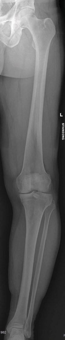
6 Protocol #3 Digital leg length X-Ray 1 Patient is standing in true anatomical position in front of image receptor with feet forward and legs straight. Do not place feet/ankles together. If deformity of patient anatomy will not allow entire leg to be in true AP position, it is most important to have the foot/ankle in a true AP position. 2 Expose entire leg from hip to ankle according to machine manufacturer s recommendation ensuring that patient does not move between exposures. 3 Figure 8 shows an acceptable digital image. Patella projected midline Figure 8


7 Quality X-Rays are imperative to the VISIONAIRE process. Rotation in the full leg X-Ray can alter the measured mechanical axis of a patient. Rotation between images that require stitching can cause the same outcome. Also not being able to identify all of the required anatomy makes defining the correct mechanical axis difficult. Below is an example of how much variation can be seen between acceptable and unacceptable X-Rays. Looking at these images side by side, it is obvious that since the accepted images correctly represent the patient s anatomy, that using the rejected X-Rays would have caused much of the alignment to be inaccurate. Femur alignment The rejected radiograph appears to have a bigger femur bow angle and is severely more externally rotated than the accepted images. This would result in an alignment with the incorrect femur valgus angle. Tibia alignment As can be observed in the tibia, the rejected image would not have provided the correct tibia mechanical or anatomical axis value. The tibia is externally rotated, which would have caused a much larger tibia bow angle to be measured than what is reality. With this information being incorrect, it would cause the tibia coronal alignment to also be inaccurate. Overall alignment With the tibia and femur images being as poor as they are in the rejected image, the measured full leg deformity would be wrong as well. Knowing the magnitude of the full leg deformity affects how Smith & Nephew approaches aligning the case. Without proper X-Rays, it is not possible to align a case accurately. Smith & Nephew aligning a case based off of the rejected X-Rays on the left would not be confident in the alignment or the blocks that are provided to the surgeon. Unacceptable X-Ray Acceptable X-Ray
8 Smith & Nephew, Inc Brooks Road Memphis, TN USA Telephone: Information: Orders and Inquiries: Trademark of Smith & Nephew Smith & Nephew V2 04/17
Overview. Acceptance criteria for all protocols
 X-Ray protocol Overview The Smith & Nephew VISIONAIRE X-Ray protocol is essentially an AP leg length image. The images are preferred to be done erect, but can be done supine if necessary due to the type
Proteus XR/f Patient positioning guide
 Proteus XR/f Patient positioning guide PROTEUS XR/F Now a single digital x-ray room accommodates nearly all your radiographic studies. With extended tube coverage and wireless detectors, Proteus XR/f gives
Surgical Technique. VISIONAIRE FastPak Instruments for the LEGION Total Knee System
 Surgical Technique VISIONAIRE FastPak Instruments for the LEGION Total Knee System VISIONAIRE FastPak for LEGION Instrument Technique* Nota Bene The technique description herein is made available to the
Extramedullary Tibial Preparation
 Surgical Technique Extramedullary Tibial Preparation Primary Total Knee Arthroplasty LEGION Total Knee System Extramedullary tibial preparation Contents Introduction...2 EM tibial highlights...3 Preoperative
A better knee replacement begins with you
 A better knee replacement begins with you Why your scans are so important VISIONAIRE Patient Matched Instrumentation is a patient specific product that requires MRI and an X-Ray to make the product. The
Surgical Technique. VISIONAIRE Disposable Instruments for the LEGION Total Knee System
 Surgical Technique VISIONAIRE Disposable Instruments for the LEGION Total Knee System VISIONAIRE and LEGION Disposable instrument technique* Note: All disposable instruments are interchangeable with the
Radiographic Positioning Summary (Basic Projections RAD 222)
") Lower Extremity Radiographic Positioning Summary (Basic Projections RAD 222) AP Pelvis AP Hip (Unilateral) (L or R) AP Femur Mid and distal AP Knee Lateral Knee Pt lies supine on table Align MSP to Center
Lower Extremity Radiographic Positioning Summary (Basic Projections RAD 222) AP Pelvis AP Hip (Unilateral) (L or R) AP Femur Mid and distal AP Knee Lateral Knee Pt lies supine on table Align MSP to Center
Imaging assessment of Unicomp candidates!
 7th Advanced Course on Knee Surgery - 2018: Imaging assessment of Unicomp candidates! Presenter: Anders Troelsen, MD, ph.d., dr.med., Professor Distribution of the basic primary OA patterns Medial FT:
Distal Cut First Femoral Preparation
 Surgical Technique Distal Cut First Femoral Preparation Primary Total Knee Arthroplasty LEGION Total Knee System Femoral preparation Contents Introduction...3 DCF femoral highlights...4 Preoperative planning...6
KNEE ALIGNMENT SYSTEM (KAS) MRI Protocol
 MRI Protocol") KNEE ALIGNMENT SYSTEM (KAS) MRI Protocol Sample referral sticker Referral Sticker Insert here Corin 17 Bridge Street Pymble NSW Australia 2073 P: +61 (0)2 9497 7400 F: +61 (0)2 9497 7498 E: KAS.customerservice@coringroup.com
KNEE ALIGNMENT SYSTEM (KAS) MRI Protocol Sample referral sticker Referral Sticker Insert here Corin 17 Bridge Street Pymble NSW Australia 2073 P: +61 (0)2 9497 7400 F: +61 (0)2 9497 7498 E: KAS.customerservice@coringroup.com
Functional Movement Test. Deep Squat
 Functional Movement Test Put simply, the FMS is a ranking and grading system that documents movement patterns that are key to normal function. By screening these patterns, the FMS readily identifies functional
Alignment Rod. For intraoperatively confirming correction of the mechanical leg axis.
 Alignment Rod. For intraoperatively confirming correction of the mechanical leg axis. Easy to use Accuracy of surgery Reduces X-ray exposure Table of Contents Introduction Alignment Rod 2 Handling Technique
Intramedullary Tibial Preparation
 Surgical Technique Intramedullary Tibial Preparation Primary Total Knee Arthroplasty LEGION Total Knee System Intramedullary tibial preparation Contents Introduction...2 IM tibial highlights...3 Preoperative
Country Health SA Medical Imaging
 Country Health SA Medical Imaging REMOTE OPERATORS POSITIONING GUIDE Contents Image Evaluation Page 4 Positioning Guides Section 1 - THORAX 1.1 Chest Page 5 1.2 Bedside Chest Page 7 1.3 Ribs Page 8 Section
PROPHECY. Preoperative Navigation Guides ANKLE CT SCAN PROTOCOL
 PROPHECY Preoperative Navigation Guides ANKLE CT SCAN PROTOCOL 90 FIGURE 1 Examples FIGURE 1 Examples of neutral ankle positioning. PROPHECY Ankle CT Scan Protocol PROPHECY INBONE and PROPHECY INFINITY
BIOMECHANICAL EXAMINATION OF THE PEDIATRIC LOWER EXTREMITY 2017
 BIOMECHANICAL EXAMINATION OF THE PEDIATRIC LOWER EXTREMITY 2017 B. RESSEQUE, D.P.M., D.A.B.P.O. Professor, N.Y. College of Podiatric Medicine ARCH HEIGHT OFF WEIGHTBEARING Evaluate arch height by placing
BIOMECHANICAL EXAMINATION OF THE PEDIATRIC LOWER EXTREMITY
 BIOMECHANICAL EXAMINATION OF THE PEDIATRIC LOWER EXTREMITY B.Resseque, D.P.M. ARCH HEIGHT OFF WEIGHTBEARING Evaluate arch height by placing a ruler from the heel to the first metatarsal head Compare arch
Clinical Evaluation Surgical Technique
 Clinical Evaluation Surgical Technique Table of Contents EMPERION Specifications 3 EMPERION Surgical Technique 9 EMPERION Catalog 18 Nota Bene: This technique description herein is made available to the
OrthoMap Express Knee Product Guide. OrthoMap Express Knee Navigation Software 2.0
 OrthoMap Express Knee Product Guide OrthoMap Express Knee Navigation Software 2.0 Product Guide 1 Introduction Introduction The Stryker OrthoMap Express Knee Navigation System is providing surgeons with
Knee Surgical Technique
 Knee Surgical Technique COMPASS Universal Hinge by Jimmy Tucker, M.D. Orthopaedic Surgeon Director, Arkansas Sports Medicine, P.A. Little Rock, Arkansas Table of contents Design features 3 Indications
BOW LEGS (GENU VARUM)
") BOW LEGS (GENU VARUM) By Dr John Ebnezar INTRODUCTION Have you noticed how your knees look like? If you observe carefully you will see that both your knees are not parallel but deviated slightly outwards
BOW LEGS (GENU VARUM) By Dr John Ebnezar INTRODUCTION Have you noticed how your knees look like? If you observe carefully you will see that both your knees are not parallel but deviated slightly outwards
Quads (medicine ball)
") Saggital Front Reach Saggital Front Reach 1) Start position: Stand with feet hip width apart. Hold medicine ball or dumbbell at waist. 2) Step forward 2-3 feet with the heel striking first and lean torso
Saggital Front Reach Saggital Front Reach 1) Start position: Stand with feet hip width apart. Hold medicine ball or dumbbell at waist. 2) Step forward 2-3 feet with the heel striking first and lean torso
Innovations 2017 & 2018
 Innovations 2017 & 2018 medicad 5.0 Hip 3D Spine 3D Knee 3D Shoulder 3D The Orthopedic Solution medicad Version 5.0 CHECK OUT WHAT'S NEW: Hip Automatic measuring of femoral or acetabular offset Automated
2017 Resident Advanced Trauma Techniques Course COMPLICATIONS / CHALLENGES MALUNIONS/DEFORMITY
 2017 Resident Advanced Trauma Techniques Course COMPLICATIONS / CHALLENGES MALUNIONS/DEFORMITY What is a Malunion? Definition: a fracture that has healed in a nonanatomic (i.e. deformed) position Must
PROPHECY INBONE. Preoperative Navigation Guides
 PROPHECY INBONE Preoperative Navigation Guides Simple. Fast. Accurate. Simple. Fast. Accurate. Prophecy Envision the Results PROPHECY Preoperative Navigation Guides have ushered in a new era of total ankle
Knee spanning solutions
 Knee spanning solutions System features Indications Intended to be used on adults or pediatric patients as required for fracture fixation (open or closed); post-traumatic joint contracture which has resulted
Institutional review board approval was obtained prior to the start of this study.
 Lower Limb Alignment and Length Measurements - Comparison of Computed Tomography, Upright Full-Length Conventional Radiography and Upright Biplanar Linear-Low Dose X-ray Scanner Poster No.: C-1382 Congress:
RADIOGRAPHY OF THE KNEE, PATELLA, and FEMUR
 RADIOGRAPHY OF THE KNEE, PATELLA, and FEMUR KNEE AP Projection Patient Position: Part Position: Leg in Center Femoral condyles Central Ray: - Asthenic patient - if ASIS to tabletop is < 19 cm Sthenic patient
Pocket Guide. Version 4.1: Fracture reduction and deformity correction software
 Pocket Guide www.spatialframe.com Version 4.1: Fracture reduction and deformity correction software Shoulder bolt Master tab Strut 5 Strut 1 Strut 6 Strut 4 Strut 2 Strut 3 ID band Figure 1 Frame assembly
Radiology Positioning Practical Test #2 Table (By Jung Park):
:") Radiology Positioning Practical Test #2 Table (By Jung Park): (Lower Extremity): patient is fully gowned / no artifacts / properly shielded (exposure for femur and below : hold still, don t move ) (exposure
Radiology Positioning Practical Test #2 Table (By Jung Park): (Lower Extremity): patient is fully gowned / no artifacts / properly shielded (exposure for femur and below : hold still, don t move ) (exposure
Balanced Body Movement Principles
 Balanced Body Movement Principles How the Body Works and How to Train it. Module 3: Lower Body Strength and Power Developing Strength, Endurance and Power The lower body is our primary source of strength,
Cybex Leg Extension. Legs
 Cybex Leg Extension Legs Set Up Procedure Seat Back Pad Adjust pad by pulling out yellow pin in back of the seat and make it so when seated knees align with machines axis of rotation (Yellow Arrows) Ankle
... ON THE JOB. Holmblad Variations
 Holmblad Variations Dan L. Hobbs, M.S.R.S., R.T.(R)(CT)(MR), is an associate professor in the department of radiographic science at Idaho State University in Pocatello. In Chicago during the summer of
TKA Gap Planning. Supporting healthcare professionals
 TKA Gap Planning The NAVIO TKA Gap Planning stage helps you adjust the plan based on gap information between the femur and tibia implants. Supporting healthcare professionals Interactive Views Four interactive
 17.2 A-P Lower Leg Measure: A-P at mid-lower leg Protection: Apron draped over pelvis SID: 40 Table top No Tube Angle Film: 7 x17 I.D. down or diagonal 14 x 17 www.fisiokinesiterapia.biz A-P Lower Leg
.org. Tibia (Shinbone) Shaft Fractures. Anatomy. Types of Tibial Shaft Fractures
 Shaft Fractures. Anatomy. Types of Tibial Shaft Fractures") Tibia (Shinbone) Shaft Fractures Page ( 1 ) The tibia, or shinbone, is the most common fractured long bone in your body. The long bones include the femur, humerus, tibia, and fibula. A tibial shaft fracture
Tibia (Shinbone) Shaft Fractures Page ( 1 ) The tibia, or shinbone, is the most common fractured long bone in your body. The long bones include the femur, humerus, tibia, and fibula. A tibial shaft fracture
Ligament Balancing. Instructional Course. Preliminary programme. Invited Chairman: Professor Henrik Schroeder-Boersch (DE)
") Ligament Balancing Instructional Course Preliminary programme Invited Chairman: Professor Henrik Schroeder-Boersch (DE) Expert Connect Centre (ECC), Croxley Green, United Kingdom 18-19 May 2017 Supporting
Ligament Balancing Instructional Course Preliminary programme Invited Chairman: Professor Henrik Schroeder-Boersch (DE) Expert Connect Centre (ECC), Croxley Green, United Kingdom 18-19 May 2017 Supporting
FUNCTIONAL TESTING GUIDELINES FOR ACL RECONSTRUCTION TESTING INSTRUCTIONS FOR CLINICIANS
 FUNCTIONAL TESTING GUIDELINES FOR ACL RECONSTRUCTION TESTING INSTRUCTIONS FOR CLINICIANS A number of criteria should be met before advanced functional testing of ACL reconstruction or ACL deficient knees
RADIOGRAPHY OF THE ANKLE and LOWER LEG
 RADIOGRAPHY OF THE ANKLE and LOWER LEG Patient Position: ANKLE AP Projection Part Position: True Slight to place foot s long axis Center to Central Ray: to IR Midway Note: Ankle joint is to tips of malleoli
LESSON ASSIGNMENT. Positioning for Exams of the Spine. After completing this lesson, you should be able to identify:
 LESSON ASSIGNMENT LESSON 4 Positioning for Exams of the Spine. LESSON ASSIGNMENT Paragraphs 4-1 through 4-15. LESSON OBJECTIVES After completing this lesson, you should be able to identify: 4-1. Identify
Functional Movement Screen (Cook, 2001)
") Functional Movement Screen (Cook, 2001) TEST 1 DEEP SQUAT Purpose - The Deep Squat is used to assess bilateral, symmetrical, mobility of the hips, knees, and ankles. The dowel held overhead assesses bilateral,
Functional Movement Screen (Cook, 2001) TEST 1 DEEP SQUAT Purpose - The Deep Squat is used to assess bilateral, symmetrical, mobility of the hips, knees, and ankles. The dowel held overhead assesses bilateral,
PRE & POST OPERATIVE RADIOLOGICAL ASSESSMENT IN TOTAL KNEE REPLACEMENT. Dr. Divya Rani K 2 nd Year Resident Dept. of Radiology
 PRE & POST OPERATIVE RADIOLOGICAL ASSESSMENT IN TOTAL KNEE REPLACEMENT Dr. Divya Rani K 2 nd Year Resident Dept. of Radiology PRE OPERATIVE ASSESSMENT RADIOGRAPHS Radiographs are used for assessment and
ORTHOSCAN MOBILE DI POSITIONING GUIDE
 ORTHOSCAN MOBILE DI POSITIONING GUIDE Table of Contents SHOULDER A/P of Shoulder... 4 Tangential (Y-View) of Shoulder... 5 Lateral of Proximal Humerus... 6 ELBOW A/P of Elbow... 7 Extended Elbow... 8 Lateral
Resurfacing Patellar Preparation
 Surgical Technique Resurfacing Patellar Preparation Primary Total Knee Arthroplasty LEGION Total Knee System Resurfacing Patellar preparation Contents Introduction...2 Instrument assembly...3 Resurfacing
Total Knee Original System Primary Surgical Technique
 Surgical Procedure Total Knee Original System Primary Surgical Technique Where as a total hip replacement is primarily a bony operation, a total knee replacement is primarily a soft tissue operation. Excellent
POSTERIOR 1. situated behind: situated at or toward the hind part of the body :
 ANATOMICAL LOCATION Anatomy is a difficult subject with a large component of memorization. There is just no way around that, but we have made every effort to make this course diverse and fun. The first
POSTURE ANALYSIS. What is good posture?
 POSTURE ANALYSIS What is good posture? Posture is the position in which you hold your body upright against gravity while standing or sitting. Good posture involves training your body to stand, walk, sit
EVOS MINI with IM Nailing
 Case Series Dr. John A. Scolaro EVOS MINI with IM Nailing A series of studies Introduction Intramedullary nailing has become the standard for many long bone fractures. Fracture reduction prior to nail
A Patient s Guide to Rotational Deformities in Children
 A Patient s Guide to Rotational Deformities in Children 2350 Royal Boulevard Suite 200 Elgin, IL 60123 Phone: 847.931.5300 Fax: 847.931.9072 DISCLAIMER: The information in this booklet is compiled from
SESSION #207 UNDERSTANDING FUNCTION FROM THE GROUND UP Greg Roskopf, MA Owner/developer of Muscle Activation Techniques
 SESSION #207 UNDERSTANDING FUNCTION FROM THE GROUND UP Greg Roskopf, MA Owner/developer of Muscle Activation Techniques PRESCRIBING EXERCISE AS A COMPONENT OF HEALTH: PEOPLE ARE COMING TO US TO GET HEALTHY!
SCALING Radiographic Technique
 SCALING Radiographic Technique SCALING FOR DIGITAL X-RAYS As images become filmless. Current planning practices with acetate sheets become difficult or obsolete. When images are printed to film sometimes
Comprehensive Solutions
 Comprehensive Solutions Supporting healthcare professionals Flexibility Today s knee implant patients present increasingly diverse scenarios. The versatility of the JOURNEY II Total allows surgeons to
ANATOMIC. Navigated Surgical Technique 4 in 1 TO.G.GB.016/1.0
 ANATOMIC Navigated Surgical Technique 4 in 1 TO.G.GB.016/1.0 SCREEN LAYOUT Take screenshot Surgical step Dynamic navigation zone Information area and buttons 2 SCREEN LAYOUT Indicates action when yellow
Surgical Technique. CONQUEST FN Femoral Neck Fracture System
 Surgical Technique CONQUEST FN Femoral Neck Fracture System Table of Contents Introduction... 3 Indications... 3 Product Overview... 4 Surgical Technique... 5 Patient Positioning... 5 Reduce the Fracture...
INTERTAN Nails Geared for Stability
 Geared for stability The TRIGEN INTERTAN nail brings advanced TRIGEN nail technology to hip fractures. With a unique integrated, interlocking screw construct, TRIGEN INTERTAN nail provides all the benefits
9/26/2012. Basic Terminology. Basic Terminology continued. Kinesiology Terminology. Kinesiology = The study of movement
 Kinesiology Terminology Basic Terminology Kinesiology = The study of movement This definition is so broad. What other fields of study come together to create kinesiology? Yes!! And it relates them all
Routine Guide EXAMINATION PROJECTION CASSETTE SIZE NOTES PRINT ORIENTATION. 14x17 CW* 14x17LW 14x17LW. 14x17LW 14x17LW 14x17LW
 EXAMINATION PROJECTION CASSETTE SIZE NOTES PRINT ORIENTATION A-C Joints without weights with weights 14x17 CW* One 14x17 divided; both shoulders on one exposure. *If part does not fit, do 10x12s CW. Both
KILLER #8. Workout Summary REALITY FITNESS THE WORKOUTS KILLER #8 1. Don t forget to warm up and cool down! Take a 1 minute break in between each set.
 KILLER #8 Workout Summary Don t forget to warm up and cool down! Take a 1 minute break in between each set. SERIES ONE Complete 3 sets of series one and then 3 sets of series two. EXERCISE 1 - Alternating
CONTRIBUTING SURGEON. Barry Waldman, MD Director, Center for Joint Preservation and Replacement Sinai Hospital of Baltimore Baltimore, MD
 CONTRIBUTING SURGEON Barry Waldman, MD Director, Center for Joint Preservation and Replacement Sinai Hospital of Baltimore Baltimore, MD System Overview The EPIK Uni is designed to ease the use of the
KNEE AND FULL LIMB RADIOGRAPHY
 MOST Operations Manual page 1 TABLE OF CONTENTS KNEE AND FULL LIMB RADIOGRAPHY 1. Introduction... 3 2. Background and rationale... 4 3. Equipment and supplies... 4 4. Inclusion/exclusion criteria and safety...
Radiographic Positioning for Dogs
 Radiographic Positioning for Dogs Elbow Radiographs: Lateral View A routine elbow exam consists of a lateral, flexed lateral and craniocaudal view. When performing elbow radiographs, a quality control
Increasing surgical freedom Restoring patient function
 Increasing surgical freedom Restoring patient function Fracture specific plating solutions for the most common tibia and fibula fractures Frequency of fracture occurrences* 66% 61% 36% 36% 28% 14% 20%
THE KNEE SOCIETY VIRTUAL FELLOWSHIP
 THE KNEE SOCIETY VIRTUAL FELLOWSHIP CHAPTER 2: RADIOGRAPHIC EVALUATION OF THE KNEE Radiographic Evaluation of the Knee Presented by: R. Michael Meneghini, MD COPYRIGHT 2016 THE KNEE SOCIETY Disclosures
Trauma Patient Medical Record
 http://www.edheads.org/activities/trauma/swf/index.html Summarize patient information when entering Emergency Department (ED): Name: Date: p: Trauma Patient Medical Record Glasgow Coma Scale Vital Signs
AAP Boot Camp KNEE AND ANKLE EXAM
 AAP Boot Camp KNEE AND ANKLE EXAM Disclosures I have no relevant financial relationships with the manufacturers of any commercial products and or providers of commercial services discussed in this CME
Exam of the Knee and Ankle I HAVE NO FINANCIAL DISCLOSURES RELEVANT TO THIS PRESENTATION
 Exam of the Knee and Ankle I HAVE NO FINANCIAL DISCLOSURES RELEVANT TO THIS PRESENTATION Disclosures I have no relevant financial relationships with the manufacturers of any commercial products and or
JUMPTRAINER. (1) Secure the Jump Trainer tubes to the belt and ankle cuffs. (2) Start with feet shoulder width apart, in an athletic stance.
 Secure the Jump Trainer tubes to the belt and ankle cuffs. (2) Start with feet shoulder width apart, in an athletic stance.") JUMPTRAINER DRILL1: SQUAT JUMP DRILL 1 SQUAT JUMP (2) Start with feet shoulder width apart, in an athletic stance. (3) Perform a squat by bending at the knees and pushing hips back and down. (4) Explode
JUMPTRAINER DRILL1: SQUAT JUMP DRILL 1 SQUAT JUMP (2) Start with feet shoulder width apart, in an athletic stance. (3) Perform a squat by bending at the knees and pushing hips back and down. (4) Explode
Administrative - Master Syllabus COVER SHEET
 Administrative - Master Syllabus COVER SHEET Purpose: It is the intention of this to provide a general description of the course, outline the required elements of the course and to lay the foundation for
The TRIGEN SURESHOT system has my back.
 Because control means everything to me. The TRIGEN SURESHOT system has my back. Introducing the TRIGEN SURESHOT Distal Targeting System, the revolutionary virtual imaging system that does so much more.
Introduction to Biomechanical Analysis
 Introduction to Biomechanical Analysis LEARNING OBJECTIVES: At the end of this laboratory exercise the student will be able to: Identify forces used during activities Identify moments used during activities
Chapter 10: Flexibility
 Chapter 10: Flexibility Lesson 10.1: Flexibility Facts Self-Assessment 10: Arm, Leg, and Trunk Flexibility Lesson Objectives: Describe the characteristics of flexibility. Explain how you benefit from good
Instructional Course Lecture 2011
 Instructional Course Lecture 2011 Yoon Hae Kwak Dept. of Orthopaedic Surgery Hallym University Sacred Heart Hospital Hallym University Medical Center Rotational and Angular variations of the lower extremities
Trauma fixation choices chart your fracture
 Trauma fixation choices chart your fracture This brochure is intended for informational and educational purposes only. It is the responsibility of operating physicians to determine and utilize the appropriate
Surgical Technique. Hinge Disassembly and Rebuild Technique
 Surgical Technique Hinge Disassembly and Rebuild Technique Revision Knee Arthroplasty Surgical Technique LEGION HK Hinge Knee System Introduction The LEGION HK Hinge Knee System has been designed as an
Quads (machines) Cable Lunge
 Cable Lunge") Cable Lunge Cable Lunge 1) Stand with feet hip width apart and a cable attached around your waist. Take left leg and step back approximately 2 feet standing on the ball of the foot. 2) Start position:
Cable Lunge Cable Lunge 1) Stand with feet hip width apart and a cable attached around your waist. Take left leg and step back approximately 2 feet standing on the ball of the foot. 2) Start position:
The challenge. Knee replacement patients report lower levels of satisfaction versus hip replacement patients. Return to sports activity post surgery
 The challenge Knee replacement patients report lower levels of satisfaction versus hip replacement patients. Return to sports activity post surgery THA 52% Post-op TKA 42% Preop 36% Preop 34% Post-op Total
ASSESSMENT OF FLEXIBILITY
 Name: Date ASSESSMENT OF FLEXIBILITY Objective The purpose of this lab is to gain an assessment of the participant s flexibility. A number of key joints and movement patterns will be assessed to gain an
The Causes of Early Hip Extension in the Golf Swing
 The Causes of Early Hip Extension in the Golf Swing Hypothesis: Our hypothesis for this research is when a golfer fails any of Leg Lowering, Toe Touch, or Overhead Deep Squat tests early hip extension
BORGinsole Measurement devices
 BORGinsole Measurement devices BORGinsole Angle-Finder Dorsal Flexion of the first Metatarsophalangeal joint - P. is sitting up on the examination table, with legs straight. - T. is sitting at the end
Proper Bike Position (Fitting)
") Proper Bike Position (Fitting) COMFORT + EFFICIENCY = PERFORMANCE (The Golden Rule) Three reasons why bike set up is very important for everyone: 1/ Optimum Performance. 2/ Injury Prevention 3/ Your body
Proper Bike Position (Fitting) COMFORT + EFFICIENCY = PERFORMANCE (The Golden Rule) Three reasons why bike set up is very important for everyone: 1/ Optimum Performance. 2/ Injury Prevention 3/ Your body
NDIPE Dictionary of Visual Terms
 NDIPE Dictionary of Visual Terms One of the challenges of the indoor activity is knowing terms used by the staff and fellow members. As a potential member of the NDIPE, you are expected to have a firm
Technique Guide. SureLock Distal Targeting Device. C-arm guided targeting for trochanteric fixation nail.
 Technique Guide SureLock Distal Targeting Device. C-arm guided targeting for trochanteric fixation nail. Table of Contents Introduction SureLock Distal Targeting Device 2 Surgical Technique Preoperative
JOINT RULER. Surgical Technique For Knee Joint JRReplacement
 JR JOINT RULER Surgical Technique For Knee Joint JRReplacement INTRODUCTION The Joint Ruler * is designed to help reduce the incidence of flexion, extension, and patellofemoral joint problems by allowing
Core (machines) Medicine Ball Back Extension
 Medicine Ball Back Extension") Medicine Ball Back Extension Medicine Ball Back Extension 1. Position body face down on apparatus placing hips and ankles on respective pads. 2. Place hips (and not stomach) on pad. Place lower leg or
Medicine Ball Back Extension Medicine Ball Back Extension 1. Position body face down on apparatus placing hips and ankles on respective pads. 2. Place hips (and not stomach) on pad. Place lower leg or
CHAPTER 8: THE BIOMECHANICS OF THE HUMAN LOWER EXTREMITY
 CHAPTER 8: THE BIOMECHANICS OF THE HUMAN LOWER EXTREMITY _ 1. The hip joint is the articulation between the and the. A. femur, acetabulum B. femur, spine C. femur, tibia _ 2. Which of the following is
Patient Guide. Intramedullary Skeletal Kinetic Distractor For Tibial and Femoral Lengthening
 Patient Guide Intramedullary Skeletal Kinetic Distractor For Tibial and Femoral Lengthening Introduction You have decided to have a limb lengthening operation. The surgery you have chosen uses a device
MSK CT Extremities: Positioning and Reformations
 MSK CT Extremities: Positioning and Reformations Hand: Patient lying in prone position, with affected arm extended above head. Place body off centered in effort to set affected hand in isocenter. Hand
ROTC Physical Assessment, the Army Physical Fitness Test and Required Height/Weight Information
 ROTC Physical Assessment, the Army Physical Fitness Test and Required Height/Weight Information ROTC Physical Assessment Scholarship Applicants are required to complete a Presidential Challenge Physical
Most forms of dance use turnout. It has been
 Turnout in Dancers A Comprehensive Overview of Active and Passive Turnout Gayanne Grossman, P.T., Ed.M., B.F.A., F.I.A.D.M.S. Department of Theatre and Dance, Muhlenberg College, Allentown, PA Most forms
Assessment of the Feet Handbook
 Assessment of the Feet Handbook The content of this handbook has been adapted from the assessment methods designed by Simon J. Wikler, Doctor of Surgical Chiropody Step Forward to Better Health Reflexology
Definition of Anatomy. Anatomy is the science of the structure of the body and the relation of its parts.
 Definition of Anatomy Anatomy is the science of the structure of the body and the relation of its parts. Basic Anatomical Terms Anatomical terms for describing positions: Anatomical position: Supine position:
Evaluation of Gait Mechanics Using Computerized Plantar Surface Pressure Analysis and it s Relation to Common Musculoskeletal Problems
 Evaluation of Gait Mechanics Using Computerized Plantar Surface Pressure Analysis and it s Relation to Common Musculoskeletal Problems Laws of Physics effecting gait Ground Reaction Forces Friction Stored
CORRECTIVE OSTEOTOMY BRINGING THE PLAN TO THE BONE (TRIGONOMETERY, GUIDE WIRES, SLA MODELING AND ART)
") CORRECTIVE OSTEOTOMY BRINGING THE PLAN TO THE BONE (TRIGONOMETERY, GUIDE WIRES, SLA MODELING AND ART) Randy J. Boudrieau, DVM, DACVS, DECVS Cummings School of Veterinary Medicine at Tufts University, North
CORRECTIVE OSTEOTOMY BRINGING THE PLAN TO THE BONE (TRIGONOMETERY, GUIDE WIRES, SLA MODELING AND ART) Randy J. Boudrieau, DVM, DACVS, DECVS Cummings School of Veterinary Medicine at Tufts University, North
DEEP SQUAT. Upper torso is parallel with tibia or toward vertical Femur below horizontal Knees are aligned over feet Dowel aligned over feet
 APPENDIX 9 SCORING CRITERIA DEEP SQUAT Upper torso is parallel with tibia or toward vertical Femur below horizontal Knees are aligned over feet Dowel aligned over feet Upper torso is parallel with tibia
Pre - Operative Rehabilitation Program for Patellar Instability
 Pre - Operative Rehabilitation Program for Patellar Instability This protocol is designed to assist you with your preparation for surgery and should be followed under the direction of a physiotherapist
INVISION Total Ankle Replacement System with PROPHECY Preoperative Navigation Revision of a Failed Agility Total Ankle Replacement
 016625 REVISION R INVISION Total Ankle Replacement System with PROPHECY Preoperative Navigation Revision of a Failed Agility Total Ankle Replacement CASE STUDY Patient History The patient was a 65-year-old
Stretching - At the Workstation Why is stretching important?
 Stretching - At the Workstation Why is stretching important? No matter how well a workstation is designed, problems may arise if attention is not paid to the way the work is done. Working at a computer
PediLoc Extension Osteotomy Plate (PLEO)
") PediLoc Extension Osteotomy Plate (PLEO) Left PLEO Plates Sizes: 6, 8 and 10 hole plates Right PLEO Plates Sizes: 6, 8 and 10 hole plates PediLoc Extension Osteotomy Plate The technique description herein
PediLoc Extension Osteotomy Plate (PLEO) Left PLEO Plates Sizes: 6, 8 and 10 hole plates Right PLEO Plates Sizes: 6, 8 and 10 hole plates PediLoc Extension Osteotomy Plate The technique description herein
The Language of Anatomy. (Anatomical Terminology)
") The Language of Anatomy (Anatomical Terminology) Terms of Position The anatomical position is a fixed position of the body (cadaver) taken as if the body is standing (erect) looking forward with the upper
The Language of Anatomy (Anatomical Terminology) Terms of Position The anatomical position is a fixed position of the body (cadaver) taken as if the body is standing (erect) looking forward with the upper
Total Knee Replacement
 Total Knee Replacement A total knee replacement, also known as total knee arthroplasty, involves removing damaged portions of the knee, and capping the bony surfaces with man-made prosthetic implants.