RADIOGRAPHY OF THE ANKLE and LOWER LEG
|
|
|
- Jasmine McKinney
- 5 years ago
- Views:
Transcription
1 RADIOGRAPHY OF THE ANKLE and LOWER LEG

2 Patient Position: ANKLE AP Projection Part Position: True Slight to place foot s long axis Center to
3 Central Ray: to IR Midway Note: Ankle joint is to tips of malleoli
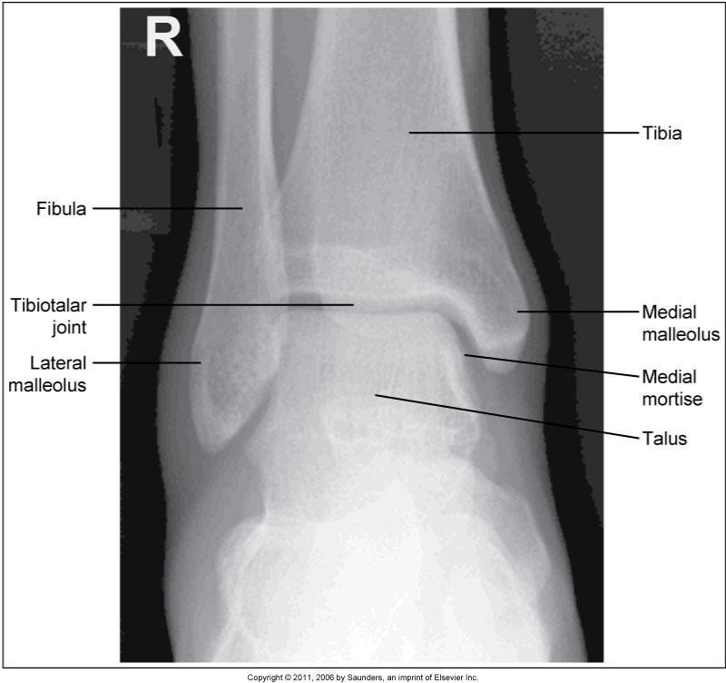
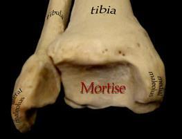
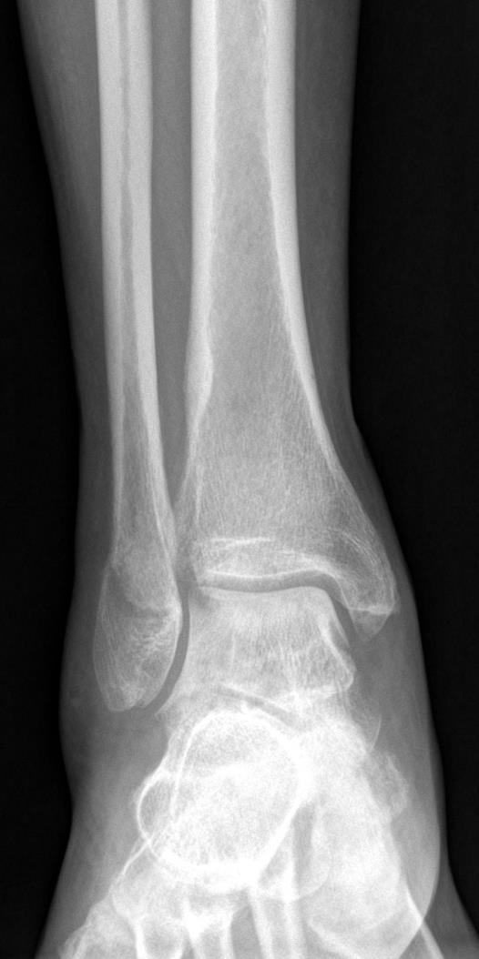
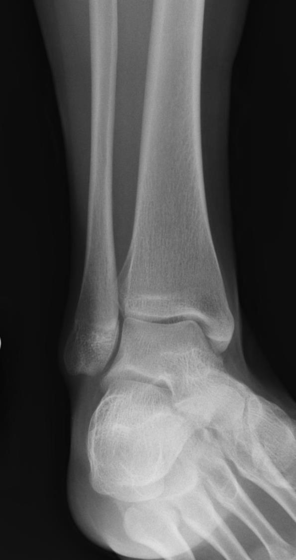
4 Structures Shown

5 AP Ankle Criteria for Evaluation The long axis of the leg should be in alignment. The tibia should be slightly joint should be open. Medial mortise should. Talus should be slightly The foot should be

6 AP ankle Error ankle, too much of the is superimposed on the and the tibiotalar joint. The leg is slightly rotated. Correction: rotate slightly to place the leg in.


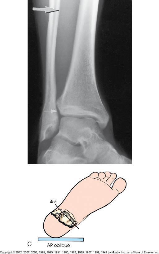
7 ANKLE AP Mortise Oblique Projection Medial Rotation Patient Position: Part Position: Foot Leg and foot rotated Intermalleolar plane to IR Center to

8 Central Ray: to IR Midway Structures Shown:
9 Oblique Mortise Ankle Criteria for Evaluation The foot should be. The distal fibula should be demonstrated. Lateral mortise ( joint) should be joint should be open. Medial mortise. The lateral and medial malleoli should be demonstrated. The tibia should be very slightly superimposed.

10 Oblique Mortise ankle Error the leg is this causes the talofibular joint enough and the medial and lateral malleoli are not from the IR. Correction - Rotate the lower leg ( ) until the malleoli are from the IR.


11 Oblique Mortise ankle Error the foot is flexed this will cause the to be too close to the and will obscure the joint. Correction: the foot. Good image Calcaneus Fx. patient unable to dorsiflex foot

")
12 ANKLE AP Oblique (45º) Projection Medial Rotation Patient Position: Central Ray: to IR Midway Part Position: Foot Leg and foot rotated Center to (midway between )
13 Structures Shown:
14 Oblique 45 degree ankle Criteria for Evaluation The medial mortise should be, the lateral mortise is. The fibula should be demonstrated. The should be demonstrated distal to the lateral mortise and fibula. The should be slightly seen.


15 Oblique 45 degree ankle Error - rotation the fibula will be superimposed on the and the will be completely open. Good image Correction - foot and decrease rotation of leg to degrees

16 Oblique 45 degree ankle Error - rotation and foot is, the sinus tarsi is, and the fibula is superimosed on the. Correction: foot and rotation of leg to degrees Good image

17 Comparison AP Mortise 45 Oblique
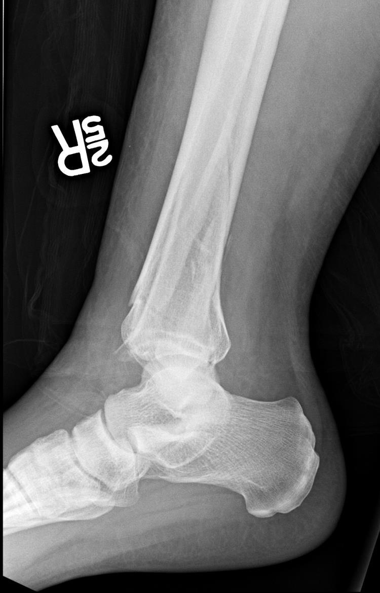
18 ANKLE Lateral (Mediolateral) Projection Patient Position: Part Position: Leg and foot in of ankle on IR foot to with leg Center to
")
19 Central Ray: to IR Alternate ( ) projection
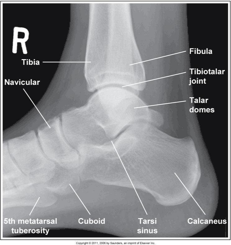

20 Structures shown:
21 Lateral ankle Criteria for Evaluation The long axis of the foot is The talofibular joint The distal fibula is superimposed over the The domes of the talus The is seen

22 Lateral ankle Error; leg is rotated, the fibula is too far, the talus is. Correction rotate leg.

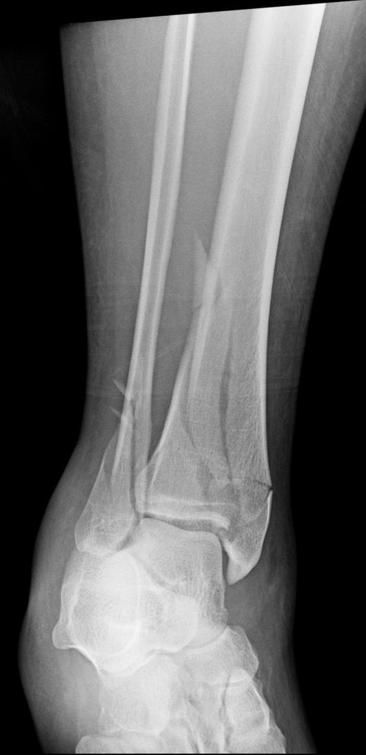
23 Trimalleolar Fracture includes:


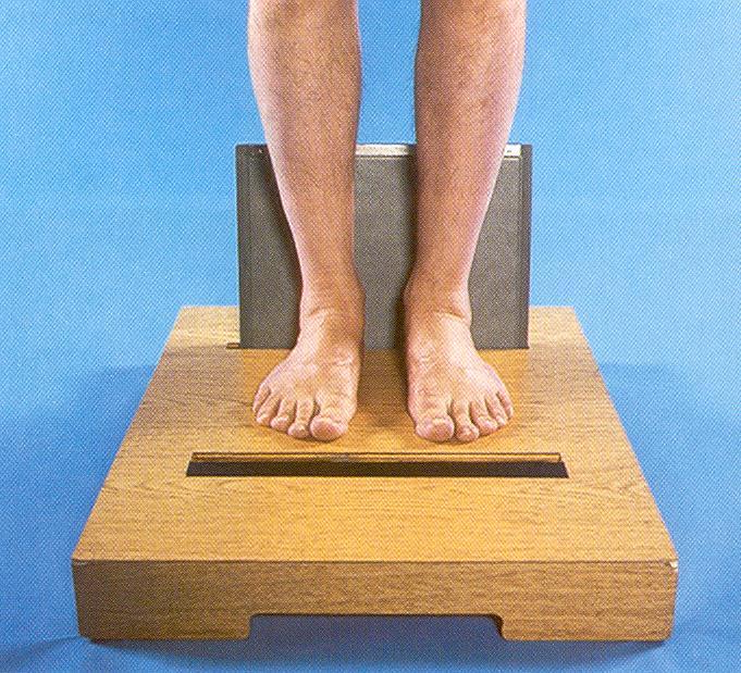
24 STRESS METHODS Stress views demonstrate ruptures of ligament is seen as a Stress Inversion Shows ligament


25 Stress Eversion Shows ligament


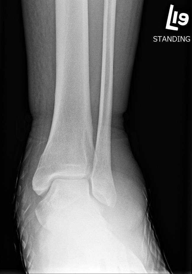
26 Alternative Method AP Ankle & Lateral


27 Patient Position: LEG AP Projection Part Position: Leg in Foot is to º Femoral condyles to IR Central Ray: to IR of leg

28 Structures Shown:

29 Criteria for Evaluation: The long axis of the leg should be in alignment Ankle and knee joint Distal fibula superimposed over. Slight overlap of over tibia

30 Patient Position: LEG Lateral Projection Part Position: Turned Patella to IR Femoral condyles superimposed Knee flexed º

31 Central Ray: to IR of leg Note Cross-table lateral with horizontal beam is obtained if patient is unable to

32 Structures Shown: Criteria for Evaluation: The of the leg should be in alignment with the long axis of the IR. Ankle and knee joint Distal fibula lying over. Slight overlap

33 Lateral Leg error: Leg is ; head of fibula is from the tibia and the distal. Correction: roll patella to IR ( rotation of leg)

34 Lateral Leg error: Leg is ; head of fibula is and the distal fibula is. Correction: roll from the IR ( rotation of leg)
35 Situation: A radiograph of an AP ankle projection reveals that the lateral joint space is not open (lateral malleolus is partially superimposed by the talus). The superior and medial joint spaces are open. Is a repeat exposure necessary? Solution:
36 Situation: A radiograph of a AP mortise projection of the ankle reveals that the lateral malleolus is slightly superimposed over the talus and the lateral joint space is not open. Solution: Insufficient rotation of the foot and ankle was most likely the cause for this radiographic outcome.
37 Situation: A patient enters radiology with a possible ligament tear to the lateral aspect of the ankle. Initial ankle radiographs are negative for fracture or dislocation. Because the clinic is in a rural setting, the patient cannot have an MRI performed to evaluate the ligaments of the ankle. Solution: AP projections may provide an assessment of the soft tissue structures of the ankle.
 17.2 A-P Lower Leg Measure: A-P at mid-lower leg Protection: Apron draped over pelvis SID: 40 Table top No Tube Angle Film: 7 x17 I.D. down or diagonal 14 x 17 www.fisiokinesiterapia.biz A-P Lower Leg
RADIOGRAPHY OF THE ELBOW & HUMERUS
 RADIOGRAPHY OF THE ELBOW & HUMERUS Patient Position: ELBOW AP Projection in same plane Part Position: Hand in ; patient Centered to Humeral epicondyles Central Ray: Structures Shown: AP Elbow Criteria
Radiographic Positioning Summary (Basic Projections RAD 222)
") Lower Extremity Radiographic Positioning Summary (Basic Projections RAD 222) AP Pelvis AP Hip (Unilateral) (L or R) AP Femur Mid and distal AP Knee Lateral Knee Pt lies supine on table Align MSP to Center
Lower Extremity Radiographic Positioning Summary (Basic Projections RAD 222) AP Pelvis AP Hip (Unilateral) (L or R) AP Femur Mid and distal AP Knee Lateral Knee Pt lies supine on table Align MSP to Center
X-Ray Rounds: (Plain) Radiographic Evaluation of the Ankle.
 Radiographic Evaluation of the Ankle.") X-Ray Rounds: (Plain) Radiographic Evaluation of the Ankle www.fisiokinesiterapia.biz Anatomy Complex hinge joint Articulations among: Fibula Tibia Talus Tibial plafond Distal tibial articular surface
X-Ray Rounds: (Plain) Radiographic Evaluation of the Ankle www.fisiokinesiterapia.biz Anatomy Complex hinge joint Articulations among: Fibula Tibia Talus Tibial plafond Distal tibial articular surface
BIOMECHANICS OF ANKLE FRACTURES
 BIOMECHANICS OF ANKLE FRACTURES William R Reinus, MD MBA FACR Significance of Ankle Fractures Most common weight-bearing Fx 70% of all Fxs Incidence is increasing Bimodal distribution Men 15-24 Women over
radiologymasterclass.co.uk
 http://radiologymasterclass.co.uk Hip X-ray anatomy - Normal AP (anterior-posterior) Shenton's line is formed by the medial edge of the femoral neck and the inferior edge of the superior pubic ramus Loss
ORTHOSCAN MOBILE DI POSITIONING GUIDE
 ORTHOSCAN MOBILE DI POSITIONING GUIDE Table of Contents SHOULDER A/P of Shoulder... 4 Tangential (Y-View) of Shoulder... 5 Lateral of Proximal Humerus... 6 ELBOW A/P of Elbow... 7 Extended Elbow... 8 Lateral
RADIOGRAPHY OF THE KNEE, PATELLA, and FEMUR
 RADIOGRAPHY OF THE KNEE, PATELLA, and FEMUR KNEE AP Projection Patient Position: Part Position: Leg in Center Femoral condyles Central Ray: - Asthenic patient - if ASIS to tabletop is < 19 cm Sthenic patient
Radiology Positioning Practical Test #2 Table (By Jung Park):
:") Radiology Positioning Practical Test #2 Table (By Jung Park): (Lower Extremity): patient is fully gowned / no artifacts / properly shielded (exposure for femur and below : hold still, don t move ) (exposure
Radiology Positioning Practical Test #2 Table (By Jung Park): (Lower Extremity): patient is fully gowned / no artifacts / properly shielded (exposure for femur and below : hold still, don t move ) (exposure
The Lower Limb VII: The Ankle & Foot. Anatomy RHS 241 Lecture 7 Dr. Einas Al-Eisa
 The Lower Limb VII: The Ankle & Foot Anatomy RHS 241 Lecture 7 Dr. Einas Al-Eisa Ankle joint Synovial, hinge joint Allow movement of the foot in the sagittal plane only (1 degree of freedom): dorsiflexion:
CHAPTER 8: THE BIOMECHANICS OF THE HUMAN LOWER EXTREMITY
 CHAPTER 8: THE BIOMECHANICS OF THE HUMAN LOWER EXTREMITY _ 1. The hip joint is the articulation between the and the. A. femur, acetabulum B. femur, spine C. femur, tibia _ 2. Which of the following is
FIBULAR & SYNDESMOSIS MALUNIONS
 FIBULAR & SYNDESMOSIS MALUNIONS MICHAEL P. CLARE, MD FLORIDA ORTHOPAEDIC INSTITUTE TAMPA, FL USA MORTISE INHERENTLY UNSTABLE LATERAL MALLEOLUS ACTS AS BUTTRESS / POST RESIST LATERAL TRANSLATION OF TALUS
The radiologist and the raiders of the lost image
 The radiologist and the raiders of the lost image Poster No.: P-0072 Congress: ESSR 2014 Type: Educational Poster Authors: M. J. Ereño Ealo, E. Montejo Rodrigo, B. Sancho, E. Pastor; Galdakao/ES Keywords:
Introduction. The primary function of the ankle and foot is to absorb shock and impart thrust to the body during walking.
 The ankle 1 Introduction The primary function of the ankle and foot is to absorb shock and impart thrust to the body during walking. OSTEOLOGRY The term ankle refers primarily to the talocrural joint,
Copyright 2004, Yoshiyuki Shiratori. All right reserved.
 Ankle and Leg Evaluation 1. History Chief Complaint: A. What happened? B. Is it a sharp or dull pain? C. How long have you had the pain? D. Can you pinpoint the pain? E. Do you have any numbness or tingling?
11/2/17. Lateral Collateral Complex Medial Collateral Complex Distal Tibiofibular Syndesmosis Spring Ligament
 Andrew J Grainger Leeds, UK Lateral Collateral Complex ial Collateral Complex Distal Tibiofibular Syndesmosis Spring Ligament Brief anatomy review Scan tips and tricks Pathological appearances andrewgrainger@nhs.net
Disclosures. Syndesmosis Injury. Syndesmosis Ligaments. Objectives. Mark M. Casillas, M.D.
 Disclosures Syndesmosis Injury No relevant disclosures Mark M. Casillas, M.D. 1 Objectives Syndesmosis Ligaments Understand the syndesmosis anatomy and function Classify syndesmosis injuries Describe treatment
RADIOGRAPHY OF THE WRIST
 RADIOGRAPHY OF THE WRIST Patient Position: WRIST PA Projection, elbow in same plane Part Position: Hand ; fingers centered to IR Central Ray: Structures Shown: NOTE: Optional AP projection best demonstrates
Anterior Impingement
 Anterior Impingement Ziali Sivardeen BMedSci, (MRCS), AFRCS, FRCS (Tr & Orth) Consultant Trauma and Orthopaedic Surgeon (Shoulder, Knee and Sports Injuries) Aims Causes of Anterior Ankle Pain Ankle Impingement
Acute Ankle Injuries, Part 1: Office Evaluation and Management
 t June 08, 2009 Obesity [1] Each acute ankle injury commonly seen in the office has associated with it a mechanism by which it can be injured, trademark symptoms that the patient experiences during the
Country Health SA Medical Imaging
 Country Health SA Medical Imaging REMOTE OPERATORS POSITIONING GUIDE Contents Image Evaluation Page 4 Positioning Guides Section 1 - THORAX 1.1 Chest Page 5 1.2 Bedside Chest Page 7 1.3 Ribs Page 8 Section
CLINICAL PRESENTATION AND RADIOLOGY QUIZ QUESTION
 Donald L. Renfrew, MD Radiology Associates of the Fox Valley, 333 N. Commercial Street, Suite 100, Neenah, WI 54956 12/08/2012 Radiology Quiz of the Week # 102 Page 1 CLINICAL PRESENTATION AND RADIOLOGY
To describe he knee joint, ligaments, structure & To list the main features of other lower limb joints
 To describe he knee joint, ligaments, structure & neurovascular supply To demonstrate the ankle joint anatomy To list the main features of other lower limb joints To list main groups of lymph nodes in
Ankle Tendons in Athletes. Laura W. Bancroft, M.D.
 Ankle Tendons in Athletes Laura W. Bancroft, M.D. Outline Protocols Normal Anatomy Tendinopathy, partial and complete tears Posterior tibial, Flexor Hallucis Longus, Achilles, Peroneal and Anterior Tibial
Surgery-Ortho. Fractures of the tibia and fibula. Management. Treatment of low energy fractures. Fifth stage. Lec-6 د.
 Fifth stage Lec-6 د. مثنى Surgery-Ortho 28/4/2016 Indirect force: (low energy) Fractures of the tibia and fibula Twisting: spiral fractures of both bones Angulatory: oblique fractures with butterfly segment.
RADIOGRAPHY OF THE HAND, FINGERS & THUMB
 RADIOGRAPHY OF THE HAND, FINGERS & THUMB FINGERS (2nd 5th) - PA Projection Patient Position: Seated; hand ; elbow on IR table top Part Position: Fingers centered to IR unless protocol is Central Ray: Perpendicular
Lower Extremity Dislocations: Management and Triage on the Field
 Lower Extremity Dislocations: Management and Triage on the Field Scott J Tarantino, MD Towson Orthopaedic Associates, Towson, MD None Disclsures Purpose To provide you with knowledge which may guide you
The Leg. Prof. Oluwadiya KS
 The Leg Prof. Oluwadiya KS www.oluwadiya.sitesled.com Compartments of the leg 4 Four Compartments: 1. Anterior compartment Deep fibular nerve Dorsiflexes the foot and toes 2. Lateral Compartment Superficial
Injuries to the ankle are common
 Diagnosis and Treatment Of Ankle Fractures ANNE M SCOTT, BSRS, R.T.(R) Ankle fractures are common among all populations, although incidence increases in the elderly. They are most often the result of simple
Clarification of Terms
 Clarification of Terms The plantar aspect of the foot refers to the role or its bottom The dorsal aspect refers to the top or its superior portion The ankle and foot perform three main functions: 1. shock
.org. Ankle Fractures (Broken Ankle) Anatomy
 Anatomy") Ankle Fractures (Broken Ankle) Page ( 1 ) A broken ankle is also known as an ankle fracture. This means that one or more of the bones that make up the ankle joint are broken. A fractured ankle can range
Ankle Fractures (Broken Ankle) Page ( 1 ) A broken ankle is also known as an ankle fracture. This means that one or more of the bones that make up the ankle joint are broken. A fractured ankle can range
AAP Boot Camp KNEE AND ANKLE EXAM
 AAP Boot Camp KNEE AND ANKLE EXAM Disclosures I have no relevant financial relationships with the manufacturers of any commercial products and or providers of commercial services discussed in this CME
Journal reading. Introduction. Introduction. Ottawa Ankle Rules. Method
 Journal reading Presenter: PGY 林聖傑 Supervisor: Dr. 林俊龍 102.12.23 The accuracy of ultrasound evaluation in foot and ankle trauma Salih Ekinci, MD American Journal of Emergency Medicine 31 (2013) 1551 1555
Basic Principles of Fractures & Easily Missed Fractures. Mr Irfan Merchant Trauma & Orthopaedic Registrar Bedford Hospital, East of England
 Basic Principles of Fractures & Easily Missed Fractures Mr Irfan Merchant Trauma & Orthopaedic Registrar Bedford Hospital, East of England Objectives Types Fracture Patterns Fracture Healing Assessing
BLUE SKY SCHOOL OF PROFESSIONAL MASSAGE AND THERAPEUTIC BODYWORK Musculoskeletal Anatomy & Kinesiology KNEE & ANKLE MUSCLES
 BLUE SKY SCHOOL OF PROFESSIONAL MASSAGE AND THERAPEUTIC BODYWORK Musculoskeletal Anatomy & Kinesiology KNEE & ANKLE MUSCLES MSAK201-I Session 3 1) REVIEW a) THIGH, LEG, ANKLE & FOOT i) Tibia Medial Malleolus
Basic Radiographic Principles Part II
 Basic Radiographic Principles Part II Kristopher Avant, D.O. October 19 th, 2016 I have no disclosures relevant to the material presented in this discussion. Good Stuff!!! 1 Really? Really! Musculoskeletal
Extraarticular Lateral Ankle Impingement
 Extraarticular Lateral Ankle Impingement Poster No.: C-1282 Congress: ECR 2016 Type: Educational Exhibit Authors: C. Cevikol; Keywords: Trauma, Diagnostic procedure, MR, CT, Musculoskeletal system, Musculoskeletal
Commonly Missed Injuries of the Extremities
 Commonly Missed Injuries of the Extremities Dr. Tudor H. Hughes M.D., FRCR Department of Radiology University of California School of Medicine San Diego, California 1. Base of skull 2. Odontoid process
Physical Examination of the Foot & Ankle
 Inspection Standing, feet straight forward facing toward examiner Swelling Deformity Flatfoot (pes planus and hindfoot valgus) High arch (pes cavus and hindfoot varus) Peek-a-boo heel Varus Too many toes
Deltoid and Syndesmosis Ligament Injury of the Ankle Without Fracture
 Deltoid and Syndesmosis Ligament Injury of the Ankle Without Fracture Chris D. Miller, MD, Walter R. Shelton,* MD, Gene R. Barrett, MD, F. H. Savoie, MD, and Andrea D. Dukes, MS From the Mississippi Sports
SURGICAL AND APPLIED ANATOMY
 Página 1 de 9 Copyright 2001 Lippincott Williams & Wilkins Bucholz, Robert W., Heckman, James D. Rockwood & Green's Fractures in Adults, 5th Edition SURGICAL AND APPLIED ANATOMY Part of "47 - ANKLE FRACTURES"
Fibular Malalignment in Subjects with Chronic Ankle Instability
 Fibular Malalignment in Subjects with Chronic Ankle Instability Takumi Kobayashi 1,2, Eiichi Suzuki 3, Naohito Yamazaki 3, Makoto Suzukawa 4, Atsushi Akaike 4, Kuniaki Shimizu 4, Kazuyoshi Gamada 1. 1
Ankle Injuries: Anatomical and Biomechanical Considerations Necessary for the Development of an Injury Prevention Program
 0196-6011 /80/0103-0171$02.00/0 THE JOURNAL OF ORTHOPAEDIC AND SPORTS PHYSICAL THERAPY Copyright O 1980 by The Orthopaedic and Sports Medicine Sections of the American Physical Therapy Association Ankle
Joints of the Lower Limb II
 Joints of the Lower Limb II Lecture Objectives Describe the components of the knee and ankle joint. List the ligaments associated with these joints and their attachments. List the muscles acting on these
Evidence-Based Examination of the Foot Presented by Alexis Wright, PT, PhD, DPT, FAAOMPT Practice Sessions/Skill Check-offs
 Evidence-Based Examination of the Foot Presented by Alexis Wright, PT, PhD, DPT, FAAOMPT Practice Sessions/Skill Check-offs Module Five: Movement Assessment of the Foot/Ankle (1 hour CEU Time) Skilled
Musculoskeletal Ultrasound Technical Guidelines. VI. Ankle
 European Society of MusculoSkeletal Radiology Musculoskeletal Ultrasound Technical Guidelines VI. Ankle Ian Beggs, UK Stefano Bianchi, Switzerland Angel Bueno, Spain Michel Cohen, France Michel Court-Payen,
Influence of bone morphology and injured ligament of the ankle on ankle stress radiographs
 Influence of bone morphology and injured ligament of the ankle on ankle stress radiographs Gye Wang Lee, MD, Chin Youb Chung, MD, Moon Seok Park, MD Seung Yeol Lee, MD, Myung Ki Chung, MD, Byung Chae Jo,
HANDS ON: Knee Evaluation J. Scott Delaney MD, FRCPC, FACEP, CSPQ
 HANDS ON: Knee Evaluation J. Scott Delaney MD, FRCPC, FACEP, CSPQ FACULTY DISCLOSURE Dr. Delaney has no affiliation with the manufacturer of any commercial product or provider of any commercial service
ANKLE JOINT ANATOMY 3. TALRSALS = (FOOT BONES) Fibula. Frances Daly MSc 1 CALCANEUS 2. TALUS 3. NAVICULAR 4. CUBOID 5.
 Fibula. Frances Daly MSc 1 CALCANEUS 2. TALUS 3. NAVICULAR 4. CUBOID 5.") ANKLE JOINT ANATOMY The ankle joint is a synovial joint of the hinge type. The joint is formed by the distal end of the tibia and medial malleolus, the fibula and lateral malleolus and talus bone. It is
ANKLE JOINT ANATOMY The ankle joint is a synovial joint of the hinge type. The joint is formed by the distal end of the tibia and medial malleolus, the fibula and lateral malleolus and talus bone. It is
The Dance Hall by Vincent van Gogh,1888
 The Dance Hall by Vincent van Gogh,1888 Articulations of the pelvic girdle Lumbosacral joints, sacroiliac joints & pubic symphysis The remaining joints of the lower limb Hip joint Knee joint Tibiofibular
Exercise Science Section 4: Joint Mechanics and Joint Injuries
 Exercise Science Section 4: Joint Mechanics and Joint Injuries An Introduction to Health and Physical Education Ted Temertzoglou Paul Challen ISBN 1-55077-132-9 Types of Joints Fibrous joint Cartilaginous
My Technique for Adjusting the Excessively Pronated Foot
 My Technique for Adjusting the Excessively Pronated Foot by Mark N. Charrette, DC One can think of Chiropractic in terms of science, art, and philosophy. The art or application of Chiropractic technique
Prevention and Treatment of Injuries. Anatomy. Anatomy. Tibia: the second longest bone in the body
 Prevention and Treatment of Injuries The Ankle and Lower Leg Westfield High School Houston, Texas Anatomy Tibia: the second longest bone in the body Serves as the principle weight-bearing bone of the leg.
Pre-operative evaluation
 Pre-operative evaluation Andrea Meyer-Lindenberg Clinic of Small Animal Surgery and eproduction Ludwig-Maximilians-University Munich Importance of pre-operative planning Evaluate patient before selecting
ANKLE PLANTAR FLEXION
 ANKLE PLANTAR FLEXION Evaluation and Measurements By Isabelle Devreux 1 Ankle Plantar Flexion: Gastrocnemius and Soleus ROM: 0 to 40-45 A. Soleus: Origin: Posterior of head of fibula and proximal1/3 of
2017 Resident Advanced Trauma Techniques Course COMPLICATIONS / CHALLENGES MALUNIONS/DEFORMITY
 2017 Resident Advanced Trauma Techniques Course COMPLICATIONS / CHALLENGES MALUNIONS/DEFORMITY What is a Malunion? Definition: a fracture that has healed in a nonanatomic (i.e. deformed) position Must
Recognizing common injuries to the lower extremity
 Recognizing common injuries to the lower extremity Bones Femur Patella Tibia Tibial Tuberosity Medial Malleolus Fibula Lateral Malleolus Bones Tarsals Talus Calcaneus Metatarsals Phalanges Joints - Knee
Outline. Ankle/Foot Anatomy Ankle Sprains Ottawa Ankle Rules DDx: The Sprain That Wasn t
 Ankle Injuries Outline Ankle/Foot Anatomy Ankle Sprains Ottawa Ankle Rules DDx: The Sprain That Wasn t Anatomy: Ankle Mortise Bony Anatomy Lateral Ligament Complex Medial Ligament Complex Ankle Sprains
Extramedullary Tibial Preparation
 Surgical Technique Extramedullary Tibial Preparation Primary Total Knee Arthroplasty LEGION Total Knee System Extramedullary tibial preparation Contents Introduction...2 EM tibial highlights...3 Preoperative
Section Three: The Leg, Ankle, and Foot Lecture: Review of Clinical Anatomy, Patterns of Dysfunction and Injury, and
 Section Three: The Leg, Ankle, and Foot Lecture: Review of Clinical Anatomy, Patterns of Dysfunction and Injury, and Treatment Implications for the Leg, Ankle, and Foot Levels I and II Demonstration and
OTM Lecture Gait and Somatic Dysfunction of the Lower Extremity
 OTM Lecture Gait and Somatic Dysfunction of the Lower Extremity Somatic Dysfunction Tenderness Asymmetry Range of Motion Tissue Texture Changes Any one of which must be present to diagnosis somatic dysfunction.
Copyright 2012 by The McGraw-Hill Companies, Inc. All rights reserved. McGraw-Hill/Irwin
 CHAPTER 8: THE LOWER EXTREMITY: KNEE, ANKLE, AND FOOT KINESIOLOGY Scientific Basis of Human Motion, 12 th edition Hamilton, Weimar & Luttgens Presentation Created by TK Koesterer, Ph.D., ATC Humboldt State
Module Three: Interventions of the Foot/Ankle
 Evidence-Based Treatment of the Foot Presented by Alexis Wright, PT, PhD, DPT, FAAOMPT Practice Sessions/Skill Check-offs Module Three: Interventions of the Foot/Ankle (75 minutes) Skilled Process a rearfoot
Ankle Ligament Injury: Don t Worry- It s Only a Sprain Wes Jackson MD Orthopaedic Foot & Ankle
 Ankle Ligament Injury: Don t Worry- It s Only a Sprain Wes Jackson MD Orthopaedic Foot & Ankle Outline I. Epidemiology II. Classification and Types of Sprains III. Anatomy IV. Clinical Assessment and Imaging
CLINICAL PRESENTATION AND RADIOLOGY QUIZ QUESTION
 Donald L. Renfrew, MD Radiology Associates of the Fox Valley, 333 N. Commercial Street, Suite 100, Neenah, WI 54956 11/24/2012 Radiology Quiz of the Week # 100 Page 1 CLINICAL PRESENTATION AND RADIOLOGY
THE LOWER EXTREMITY EXAM FOR THE FAMILY PRACTITIONER
 THE LOWER EXTREMITY EXAM FOR THE FAMILY PRACTITIONER Melinda A. Scott, D.O. Orthopedic Associates of Dayton Board Certified in Primary Care Sports Medicine GOALS Identify landmarks necessary for exam of
CURRENT TREATMENT OPTIONS
 CURRENT TREATMENT OPTIONS Fix single column or both: Always fix both. A study by Svend-Hansen corroborated the poor results associated with isolated medial malleolar fixation in bimalleolar ankle fractures.
Routine Guide EXAMINATION PROJECTION CASSETTE SIZE NOTES PRINT ORIENTATION. 14x17 CW* 14x17LW 14x17LW. 14x17LW 14x17LW 14x17LW
 EXAMINATION PROJECTION CASSETTE SIZE NOTES PRINT ORIENTATION A-C Joints without weights with weights 14x17 CW* One 14x17 divided; both shoulders on one exposure. *If part does not fit, do 10x12s CW. Both
Knee Contusions and Stress Injuries. Laura W. Bancroft, M.D.
 Knee Contusions and Stress Injuries Laura W. Bancroft, M.D. Objectives Review 5 types of contusion patterns Pivot shift Dashboard Hyperextension Clip Lateral patellar dislocation Demonstrate various stress
Anatomy and evaluation of the ankle.
 Anatomy and evaluation of the ankle www.fisiokinesiterapia.biz Ankle Anatomical Structures Tibia Fibular Talus Tibia This is the strongest largest bone of the lower leg. It bears weight and the bone creates
Grade III injuries of the lateral ligaments of the ankle: the incidence and
 Archives of Emergency Medicine, 1986, 3, 247-251 Grade III injuries of the lateral ligaments of the ankle: the incidence and a simple stress test C. L. MUWANGA, MARGARET HELLIER, D. N. QUINTON, J. P. SLOAN
~, /' ~::'~ EXTENSOR HALLUCIS LONGUS. Leg-anterolateral :.:~ / ~\,
 TIBIALIS ANTERIOR Lateral condyle of tibia, upper half of lateral surface of tibia, interosseous membrane Medial side and plantar surface of medial cuneiform bone, and base of first metatarsal bone Dorsiflexes
Biology 325 Fall 2003
 Name: pre-lab exercise due at beginning of your lab session Matching a. fibrous joints b. cartilaginous joints c. synovial joints 1. exhibit a joint cavity 2. types are sutures and syndesmoses 3. bones
Therapeutic Foot Care Certificate Program Part I: Online Home Study Program
 Therapeutic Foot Care Certificate Program Part I: Online Home Study Program 1 Anatomy And Terminology Of The Lower Extremity Joan E. Edelstein, MA, PT, FISPO Associate Professor of Clinical Physical Therapy
Biokinesiology of the Ankle Complex
 Rehabilitation Considerations Following Ankle Fracture: Impact on Gait & Closed Kinetic Chain Function Disclosures David Nolan, PT, DPT, MS, OCS, SCS, CSCS I have no actual or potential conflict of interest
An Introduction to Radiographic Views & Anatomy
 An Introduction to Radiographic Views & Anatomy Morey J. Kolber, PT, PhD, OCS, Cert MDT, CSCS*D An Introduction to Radiographic Views & Anatomy M.S.P.T. 1995-University of Miami Nova Southeastern University
Imaging assessment of Unicomp candidates!
 7th Advanced Course on Knee Surgery - 2018: Imaging assessment of Unicomp candidates! Presenter: Anders Troelsen, MD, ph.d., dr.med., Professor Distribution of the basic primary OA patterns Medial FT:
QUICK REFERENCE GUIDE. Arthrodiatasis. Articulated Joint Distraction
 4 Arthrodiatasis Articulated Joint Distraction ARTHRODIATASIS OF THE HIP To prepare the assembly, remove the female component and replace it with the ProCallus articulated body for the hip. Remove cam
Hip joint Type: Articulating bones:
 Ana (242 ) Hip joint Type: Synovial, ball & socket Articulating bones: Formed between head of femur and lunate surface of acetabulum of hip bone. Capsule: it is a strong fibrous sleeve connecting the articulating
Trimalleolar fracture of right ankle icd 10
 P ford residence southampton, ny Trimalleolar fracture of right ankle icd 10 2015/16 ICD-10-CM S82.856A Nondisplaced trimalleolar fracture of unspecified lower leg, initial encounter for closed fracture.
Ankle Injuries. Ankle Sprain. Range of Motion. The most likely diagnosis is lateral ligament sprain. Dorsiflexion Plantarflexion Inversion
 Ankle Injuries Dr Peter Brukner, OAM Sports Physician Associate Professor Centre for Sports Medicine Research & Education The University of Melbourne Adjunct Professor School of Human Movement Studies
UvA-DARE (Digital Academic Repository) Treatment of osteochondral defects of the talus van Bergen, C.J.A. Link to publication
 Treatment of osteochondral defects of the talus van Bergen, C.J.A. Link to publication") UvA-DARE (Digital Academic Repository) Treatment of osteochondral defects of the talus van Bergen, C.J.A. Link to publication Citation for published version (APA): van Bergen, C. J. A. (2014). Treatment
UvA-DARE (Digital Academic Repository) Treatment of osteochondral defects of the talus van Bergen, C.J.A. Link to publication Citation for published version (APA): van Bergen, C. J. A. (2014). Treatment
PRONATION-ABDUCTION FRACTURES
 C H A P T E R 1 2 PRONATION-ABDUCTION FRACTURES George S. Gumann, DPM (The opinions of the author should not be considered as reflecting official policy of the US Army Medical Department.) Pronation-abduction
Knee, Ankle, and Foot: Normal and Abnormal Features with MRI and Ultrasound Correlation. Disclosures. Outline. Joint Effusion. Suprapatellar recess
 Knee, Ankle, and Foot: Normal and Abnormal Features with MRI and Ultrasound Correlation Jon A. Jacobson, M.D. Professor of Radiology Director, Division of Musculoskeletal Radiology University of Michigan
Ankle Sprains and Their Imitators
 Ankle Sprains and Their Imitators Mark Halstead, MD Dr. Mark Halstead is the Associate Professor of the Departments of Orthopedics and Pediatrics at Washington University School of Medicine; Director of
The Muscular System. Chapter 10 Part D. PowerPoint Lecture Slides prepared by Karen Dunbar Kareiva Ivy Tech Community College
 Chapter 10 Part D The Muscular System Annie Leibovitz/Contact Press Images PowerPoint Lecture Slides prepared by Karen Dunbar Kareiva Ivy Tech Community College Table 10.14: Muscles Crossing the Hip and
4/28/2010. Fractures. Normal Bone and Normal Ossification Bone Terms. Epiphysis Epiphyseal Plate (physis) Metaphysis
 Metaphysis") Fractures Normal Bone and Normal Ossification Bone Terms Epiphysis Epiphyseal Plate (physis) Metaphysis Diaphysis 1 Fracture Classifications A. Longitudinal B. Transverse C. Oblique D. Spiral E. Incomplete
Fractures Normal Bone and Normal Ossification Bone Terms Epiphysis Epiphyseal Plate (physis) Metaphysis Diaphysis 1 Fracture Classifications A. Longitudinal B. Transverse C. Oblique D. Spiral E. Incomplete
Scar Engorged veins. Size of the foot [In clubfoot, small foot]
![Scar Engorged veins. Size of the foot [In clubfoot, small foot]](/thumbs/78/77722241.jpg "Scar Engorged veins. Size of the foot [In clubfoot, small foot]") 6. FOOT HISTORY Pain: Walking, Running Foot wear problem Swelling; tingly feeling Deformity Stiffness Disability: At work; recreation; night; walk; ADL, Sports Previous Rx Comorbidities Smoke, Sugar, Steroid
6. FOOT HISTORY Pain: Walking, Running Foot wear problem Swelling; tingly feeling Deformity Stiffness Disability: At work; recreation; night; walk; ADL, Sports Previous Rx Comorbidities Smoke, Sugar, Steroid
The Lower Limb II. Anatomy RHS 241 Lecture 3 Dr. Einas Al-Eisa
 The Lower Limb II Anatomy RHS 241 Lecture 3 Dr. Einas Al-Eisa Tibia The larger & medial bone of the leg Functions: Attachment of muscles Transfer of weight from femur to skeleton of the foot Articulations
Multiapical Deformities p. 97 Osteotomy Concepts and Frontal Plane Realignment p. 99 Angulation Correction Axis (ACA) p. 99 Bisector Lines p.
 p. 99 Bisector Lines p.") Normal Lower Limb Alignment and Joint Orientation p. 1 Mechanical and Anatomic Bone Axes p. 1 Joint Center Points p. 5 Joint Orientation Lines p. 5 Ankle p. 5 Knee p. 5 Hip p. 8 Joint Orientation Angles
Normal Lower Limb Alignment and Joint Orientation p. 1 Mechanical and Anatomic Bone Axes p. 1 Joint Center Points p. 5 Joint Orientation Lines p. 5 Ankle p. 5 Knee p. 5 Hip p. 8 Joint Orientation Angles
Proteus XR/f Patient positioning guide
 Proteus XR/f Patient positioning guide PROTEUS XR/F Now a single digital x-ray room accommodates nearly all your radiographic studies. With extended tube coverage and wireless detectors, Proteus XR/f gives
5 COMMON INJURIES IN THE FOOT & ANKLE
 5 COMMON INJURIES IN THE FOOT & ANKLE MICHAEL P. CLARE, MD FLORIDA ORTHOPAEDIC INSTITUTE TAMPA, FL USA MECHANISM OF INJURY HOW DID IT HAPPEN? HIGH ENERGY VS LOW ENERGY DIRECTION OF FORCES INVOLVED LIVING
Impingement Syndromes of the Ankle. Noaman W Siddiqi MD 5/4/2006
 Impingement Syndromes of the Ankle Noaman W Siddiqi MD 5/4/2006 Ankle Impingement Overview Clinical DX Increasingly recognized cause of chronic ankle pain Etiology can be soft tissue or osseous Professional
Title Management of Ankle and Lower Limb Injuries Protocol in MIUs and WICs
 Document Control Title Management of Ankle and Lower Limb Injuries Protocol in MIUs and WICs Author Author s job title Professional Lead, Minor Injuries Unit Directorate Emergency Services, Logistics and
BIOMECHANICAL EXAMINATION OF THE PEDIATRIC LOWER EXTREMITY
 BIOMECHANICAL EXAMINATION OF THE PEDIATRIC LOWER EXTREMITY B.Resseque, D.P.M. ARCH HEIGHT OFF WEIGHTBEARING Evaluate arch height by placing a ruler from the heel to the first metatarsal head Compare arch
CASE REPORT RARE CASE OF DELTOID LIGAMENT AVULSION WITH MEDIAL MALLEOLUS FRACTURE OF ANKLE JOINT: CASE REPORT
 RARE CASE OF DELTOID LIGAMENT AVULSION WITH MEDIAL MALLEOLUS FRACTURE OF ANKLE JOINT: CASE REPORT Maruthi C.V 1, Roshan Pais 2 HOW TO CITE THIS ARTICLE: Maruthi CV, Roshan Pais. Rare case of deltoid ligament
Bledsoe Extender Brace Application Instructions CP Rev D 03/07
 Bledsoe Extender Brace Application Instructions CP020193 Rev D 03/07 Council Directive 93/2/EEC of 1 June 1993 concerning Medical devices Fast adjustment- Fast Hinge- Fast Straps- Fast Application This
5.1 Identify, describe the attachments of and deduce the actions of the muscles of the thigh:
 5.1 Identify, describe the attachments of and deduce the actions of the muscles of the thigh: Anterior group Proximal attachment Distal attachment Sartorius ASIS» Upper part of shaft tibia (middle surface)»
THE FOOT S CONNECTED TOO... Evaluation Procedures for Orthotic Therapy Prescription 2005
 THE FOOT S CONNECTED TOO... Evaluation Procedures for Orthotic Therapy Prescription 2005 Unpublished Copyright Biomechanical Services, Inc. 2003 Biomechanical Services, Inc. 1050 Central Ave., Suite D
High Ankle Sprains: Diagnosis & Treatment
 High Ankle Sprains: Diagnosis & Treatment Mark J. Mendeszoon, DPM, FACFAS, FACFAOM Precision Orthopaedic Specialties University Regional Hospitals Advanced Foot & Ankle Fellowship- Director It Is Only