ORTHOSCAN MOBILE DI POSITIONING GUIDE
|
|
|
- Sophie Wilkerson
- 6 years ago
- Views:
Transcription

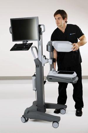
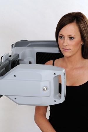
1 ORTHOSCAN MOBILE DI POSITIONING GUIDE

2 Table of Contents SHOULDER A/P of Shoulder... 4 Tangential (Y-View) of Shoulder... 5 Lateral of Proximal Humerus... 6 ELBOW A/P of Elbow... 7 Extended Elbow... 8 Lateral of Elbow in Flexion... 9 Lateral of Elbow in Flexion Lateral of Elbow in Flexion...11 HAND/WRIST Lateral of Wrist A/P of Wrist Lateral of Hand Oblique View of Hand/Wrist A/P of Hand KNEE Weight Bearing A/P of Knee Weight Bearing Lateral of Knee Oblique Sunrise of Patello-Femoral Articulation Non-Weight Bearing A/P of Knee Non-Weight Bearing Lateral of Knee Non-Weight Bearing Lateral of Knee in Flexion... 22
3 Table of Contents TIBIA/FIBULA A/P of Distal Third of Tibia/Fibula Oblique View of Tibia/Fibula FOOT/ANKLE Stressed View of Tibiotalar Joint A/P of Ankle Manually Stressed Non-Weight Bearing A/P of Hind Foot Non-Weight Bearing Lateral of Hind Foot Non-Weight Bearing Lateral of Hind Foot Oblique View of Foot Non-Weight Bearing A/P View of Foot A/P of Foot Lateral of Fore Foot Weight Bearing A/P of Foot Weight Bearing A/P of Foot Weight Bearing A/P of Ankle Weight Bearing Lateral of Ankle REFERENCE X-Ray CPT Code Reference Fluoroscopy CPT Code Reference... 40

4 A/P of Shoulder Patient is seated with back against the detector. Patient arm rests at the side. Slight external rotation or supination of hand will help better visualize the greater tuberosity. 4


5 Tangential (Y-View) of Shoulder Anterior portion of affected shoulder is placed flat against detector. Opposite shoulder is then rotated out degrees. Scapula should form y shape projection. Provides view for dislocations of humeral head relative to glenoid. 5


6 Lateral of Proximal Humerus Patient is seated with arm abducted and fully extended. Hand is pronated to face floor and arm obliquely rests on the flat detector. 6


7 A/P of Elbow Patient is seated with arm fully extended and abducted to 90 degrees. Hand is supinated while dorsal elbow lies flat on detector at an oblique angle to maximize field of view. 7


8 Extended Elbow Patient is seated with humerus abducted 90 degrees. Arm is fully extended with hand perpendicular with the floor. Elbow rests on detector obliquely oriented to optimize field of view. Internal/external rotation of arm may be required to optimize image orientation. 8


9 Lateral of Elbow in Flexion Patient is seated with humerus abducted 90 degrees. Elbow is flexed 90 degrees and hand is pronated. Elbow rests flat on detector. Extremity runs parallel to detector. Internal/external rotation and flexion/extension can be introduced to study kinematics of the joint. 9

10 Lateral of Elbow in Flexion Patient is seated with humerus abducted 90 degrees. Elbow is flexed 90 degrees and hand is straight with thumb pointing up. Elbow rests flat on detector. Internal/external rotation of the hand can be introduced to scan joint and interosseous space of radius and ulna. 10


11 Lateral of Elbow in Flexion Patient is seated with humerus abducted 90 degrees. Elbow is flexed 90 degrees and hand is pronated. Elbow rests flat on detector. Internal/external rotation of hand can be introduced to study kinematics of joint. 11


12 Lateral of Wrist Fifth ray is placed flat on the flat detector with no rotation, flexion, or extension of the wrist. Thumb is oriented to be parallel with the beam. Internal and external rotation of wrist can be introduced to visualize articular surfaces. 12


13 A/P of Wrist Patient is seated. Arm is fully extended. Hand rests on detector palmar side down. Medial and lateral stress may be introduced to assess joint stability. Internal/external rotation under fluoroscopy will help visualize articular surfaces. 13

14 Lateral of Hand Place hand laterally with fingers straight and thumb pointing up 60 degrees to the imaging source. Slight palmar bias of thumb may aide in visualization. 14


15 Oblique View of Hand/Wrist Place hand flat on detector and externally rotate 45 degrees. Fanning of fingers may allow better visualization of individual rays. Introducing inclination/declination of wrist will provide perspective of carpal bones. 15


16 A/P of Hand Hand is positioned flat on the detector with fingers spread, palmar side down. Ulnar and radial deviation may be introduced to assess joint space and articulation. 16

17 Weight Bearing A/P of Knee Mobile DI is lowered and turned parallel to the floor. Patient stands with back of knee against flat panel. Be sure the foot is not internally/externally rotated. Slight caudal/cephalad orientation of beam may be introduced to obtain preferred view. 17

18 Weight Bearing Lateral of Knee View of distal femur, patella, proximal tibia and fibula. Flat detector lowered and turned parallel to the floor. Flexion may be introduced to assess articulations. Medial aspect of knee is kept flat on the detector. 18


19 Oblique Sunrise of Patello-Femoral Articulation Patient stands and flexes knee 90 degrees while flexing hip. Lifted foot may be rested on stool to help stabilize. This may also be done from a seated position and with deeper flexion if possible. Alternatively, patient may be positioned perpendicular to Mobile DI in same pose to allow for variable orientation of the beam. 19

20 Non-Weight Bearing A/P of Knee Patient positioned supine on table with knee fully extended. True A/P may require 3-5 degrees of internal rotation. Bump may be placed under heel to achieve full extension. 20

21 Non-Weight Bearing Lateral of Knee Patient positioned on their side with c-arm perpendicular to joint. Knee is fully extended and bump placed under medial side of foot for support. Slight internal/external rotation deviations will help visualize posterior femoral condyles and posterior tibia. 21


22 Non-Weight Bearing Lateral of Knee in Flexion Patient is positioned seated with knee flexed to 90 degrees. Placing a bump under lower thigh of affected knee will raise to keep contra-lateral limb out of field. This is useful to assess tracking of patella, tibia slope/plateau, and distal femoral condyles. Slight external rotation of detector may aid view of posterior femoral condyles. 22
23 A/P of Distal Third of Tibia/Fibula Patient is placed supine on the table with c-arm oriented obliquely or perpendicular to extremity. Knee can be fully extended or slightly flexed, support under the knee is optional. Dorsi-flexion of foot may be preferred if tibiotalar joint is included. Foot should be oriented neutral with little internal/external rotation to best visualize interosseous space. 23

24 Oblique View of Tibia/Fibula Patient is positioned supine on table with knee fully extended and internally rotated 45 degrees. C-arm can be oriented obliquely or perpendicular to extremity. 24


25 Stressed View of Tibiotalar Joint Patient is supine on table with knee fully extended. Heel rests flat on detector. Distal tibia is firmly supported while lateral stress internally orients distal foot. Stress may be applied to posterior aspect of foot if desired. This provides an optimal fluoroscopic image of tibiotalar stability as well as a clear shot of the distal fibula and lateral malleolus. 25

 will achieve mortise view of")
26 A/P of Ankle Patient s leg is fully extended with heel resting on detector with foot dorsi-flexed. 15 degrees of internal rotation (lesser toe should be centered on heel) will achieve mortise view of malleoli. 26


27 Manually Stressed Non-Weight Bearing A/P of Hind Foot Patient placed supine on table either flat or seated. Knee is fully extended. Tech supports posterior tibia to elevation of 25 degrees and manually dorsi-flexes distal foot. More degrees of elevation and flexion may provide better visualization of dorsal/posterior calcaneus. 27


28 Non-Weight Bearing Lateral of Hind Foot Patient is seated with affected leg extended. External rotation allows foot to lay flat on lateral edge. This view is optimal for view of the plantar aspect of the calcaneus and tarsals. Introduction of dorsi-flexion may facilitate imaging. True lateral of calcaneus will position lateral malleolus 1 cm posterior to medial malleolus. 28


29 Non-Weight Bearing Lateral of Hind Foot Patient supine on table with affected heel on flat detector. Contra-lateral leg may be draped off side of table. Knee and hip are flexed with external rotation introduced to lay lateral aspect of foot flat on detector. 29


30 Oblique View of Foot Patient is positioned supine on table with hip and knee flexed to 45 degrees. Medial aspect of foot is placed flat on the detector. Knee, ankle, and foot are internally rotated degrees. 30


31 Non-Weight Bearing A/P View of Foot Patient is placed supine on table with hip and knee flexed to 45 degrees. Plantar surface of foot is laid flat on detector. Optimal view of the metatarsal, tarsal bones, and anterior tibia. 31


32 A/P of Foot Knee is flexed to patient s comfort. Forefoot is placed flat on detector, plantar side down. 32


33 Lateral of Fore Foot Mobile DI turned 90 degree to be parallel with the floor. Patient is seated with affected leg free floating. Foot is positioned against flat detector with dorsi-flexion. 33


34 Weight Bearing A/P of Foot Patient stands on radiolucent foot bench with toes facing the Mobile DI accessory cart. Plantar surface of foot is positioned flat on top of bench over detector. Optimal weight-bearing view of the tarsal and tarsal bones. 34


35 Weight Bearing A/P of Foot Patient stands on radiolucent foot bench with knee abutting the x-ray source tube. Plantar surface of foot is laid flat on top of bench, perpendicular to the detector. Optimal weight-bearing view of the metatarsal and tarsal bones. 35


36 Weight Bearing A/P of Ankle Patient stands on top of radiolucent foot bench with height adjusted platform. Heel is positioned as close as possible to flat detector receptor. Internally rotating the leg degrees may aid in obtaining desired Mortise view optimal for viewing weight-bearing tibiotalar joint, talar-tilt, distal fibula, and malleoli. 36


37 Weight Bearing Lateral of Ankle Patient stands on radiolucent foot bench with height adjusted platform. Lateral aspect of ankle is positioned as close as possible to the flat detector receptor. 37
38 X-Ray CPT Code Reference Code Description Clavicle, complete Scapula, complete Shoulder, 1 view Shoulder, complete, minimum of 2 views Acromioclavicular joints, bilateral, with or without weighted distraction Humerus, minimum of 2 views Elbow, 2 views Elbow, complete, minimum of 3 views Forearm, 2 views Wrist, 2 views Wrist, complete, minimum of 3 views Hand, 2 views Hand, minimum of 3 views Finger or fingers, minimum of 2 views 38
39 X-Ray CPT Code Reference Code Description Femur, 2 views Knee, 1 or 2 views Knee, complete, 4 or more views Tibia and fibula, 2 views Tibia and fibula, lower extremity, infant, minimum of 2 views Ankle, 2 views Ankle, complete, minimum of 3 views Foot, 2 views Foot, complete, minimum of 3 views Calcaneus, minimum of 2 views Toe(s), minimum of 2 views 39
40 Fluoroscopy CPT Code Reference Code Description Fluoroscopy (separate procedure), up to one hour physician time, other than or Fluoroscopy, physician time more than one hour, assisting a non-radiologic physician Fluoroscopic guidance for needle placement Manual application of stress performed by physician for joint radiography, including contralateral joint if indicated Joint survey, single view, two or more joints (specify) Shoulder, radiologic examination, arthrography, radiological supervision, and interpretation Elbow, radiologic examination, arthrography, radiological supervision, and interpretation Wrist, radiologic examination, arthrography, radiological supervision, and interpretation Knee, radiologic examination, arthrography, radiological supervision, and interpretation Wrist, radiologic examination, arthrography, radiological supervision, and interpretation 40
41 Rev D 3/2/2017
42
Radiographic Positioning Summary (Basic Projections RAD 222)
") Lower Extremity Radiographic Positioning Summary (Basic Projections RAD 222) AP Pelvis AP Hip (Unilateral) (L or R) AP Femur Mid and distal AP Knee Lateral Knee Pt lies supine on table Align MSP to Center
Lower Extremity Radiographic Positioning Summary (Basic Projections RAD 222) AP Pelvis AP Hip (Unilateral) (L or R) AP Femur Mid and distal AP Knee Lateral Knee Pt lies supine on table Align MSP to Center
Radiology Positioning Practical Test #2 Table (By Jung Park):
:") Radiology Positioning Practical Test #2 Table (By Jung Park): (Lower Extremity): patient is fully gowned / no artifacts / properly shielded (exposure for femur and below : hold still, don t move ) (exposure
Radiology Positioning Practical Test #2 Table (By Jung Park): (Lower Extremity): patient is fully gowned / no artifacts / properly shielded (exposure for femur and below : hold still, don t move ) (exposure
Country Health SA Medical Imaging
 Country Health SA Medical Imaging REMOTE OPERATORS POSITIONING GUIDE Contents Image Evaluation Page 4 Positioning Guides Section 1 - THORAX 1.1 Chest Page 5 1.2 Bedside Chest Page 7 1.3 Ribs Page 8 Section
Multiple Choice Identify the letter of the choice that best completes the statement or answers the question.
 RA202 positioning class three- EXM Multiple Choice Identify the letter of the choice that best completes the statement or answers the question. 1. Which of the following hand projections would be used
The Skeletal System THE APPENDICULAR SKELETON
 The Skeletal System THE APPENDICULAR SKELETON The appendicular skeleton consists of the girdles and the skeleton of the limbs. The upper (anterior) limbs are attached to the pectoral (shoulder) girdle
Evidence-Based Examination of the Foot Presented by Alexis Wright, PT, PhD, DPT, FAAOMPT Practice Sessions/Skill Check-offs
 Evidence-Based Examination of the Foot Presented by Alexis Wright, PT, PhD, DPT, FAAOMPT Practice Sessions/Skill Check-offs Module Five: Movement Assessment of the Foot/Ankle (1 hour CEU Time) Skilled
Hands PA; Obl. Lat.; Norgaard s Thumb AP; Lat. PA. PA; Lat.: Obls.; Elongated PA with ulnar deviation
 Projections Region Basic projections Additional / Modified projections Upper Limbs Hands PA; Obl. Lat.; Norgaard s Thumb ; Lat. PA Fingers PA; Lat. Wrist PA; Lat. Obls. Scaphoid Lunate Trapezium Triquetral
Exercise 11. The Appendicular Skeleton
 Exercise 11 The Appendicular Skeleton The Appendicular Skeleton The appendicular skeleton contains 126 bones. Consists of the upper and lower limbs, the pectoral girdles, and the pelvic girdles. The pectoral
Proteus XR/f Patient positioning guide
 Proteus XR/f Patient positioning guide PROTEUS XR/F Now a single digital x-ray room accommodates nearly all your radiographic studies. With extended tube coverage and wireless detectors, Proteus XR/f gives
10/12/2010. Upper Extremity. Pectoral (Shoulder) Girdle. Clavicle (collarbone) Skeletal System: Appendicular Skeleton
 Girdle. Clavicle (collarbone) Skeletal System: Appendicular Skeleton") Skeletal System: Appendicular Skeleton Pectoral girdle Pelvic girdle Upper limbs Lower limbs 8-1 Pectoral (Shoulder) Girdle Consists of scapula and clavicle Clavicle articulates with sternum (Sternoclavicular
Skeletal System: Appendicular Skeleton Pectoral girdle Pelvic girdle Upper limbs Lower limbs 8-1 Pectoral (Shoulder) Girdle Consists of scapula and clavicle Clavicle articulates with sternum (Sternoclavicular
Types of Body Movements
 Types of Body Movements Bởi: OpenStaxCollege Synovial joints allow the body a tremendous range of movements. Each movement at a synovial joint results from the contraction or relaxation of the muscles
BIOMECHANICAL EXAMINATION OF THE PEDIATRIC LOWER EXTREMITY
 BIOMECHANICAL EXAMINATION OF THE PEDIATRIC LOWER EXTREMITY B.Resseque, D.P.M. ARCH HEIGHT OFF WEIGHTBEARING Evaluate arch height by placing a ruler from the heel to the first metatarsal head Compare arch
RADIOGRAPHY OF THE ANKLE and LOWER LEG
 RADIOGRAPHY OF THE ANKLE and LOWER LEG Patient Position: ANKLE AP Projection Part Position: True Slight to place foot s long axis Center to Central Ray: to IR Midway Note: Ankle joint is to tips of malleoli
 17.2 A-P Lower Leg Measure: A-P at mid-lower leg Protection: Apron draped over pelvis SID: 40 Table top No Tube Angle Film: 7 x17 I.D. down or diagonal 14 x 17 www.fisiokinesiterapia.biz A-P Lower Leg
Goniometry. Wrist Flexion: Pt seated with forearm resting on table (use olecranon process & midline of ulna as reference for stationary arm)
") Goniometry Wrist Flexion: Pt seated with forearm resting on table (use olecranon process & midline of ulna as reference for stationary arm) Wrist Extension: Pt seated with forearm resting on table (Goniometer
Goniometry Wrist Flexion: Pt seated with forearm resting on table (use olecranon process & midline of ulna as reference for stationary arm) Wrist Extension: Pt seated with forearm resting on table (Goniometer
Chapter 8 The Skeletal System: The Appendicular Skeleton. Copyright 2009 John Wiley & Sons, Inc.
 Chapter 8 The Skeletal System: The Appendicular Skeleton Appendicular Skeleton It includes bones of the upper and lower limbs Girdles attach the limbs to the axial skeleton The pectoral girdle consists
Anatomy and Physiology 2016
 Anatomy and Physiology 2016 O = Temporal line I = coronoid process (Mandible) A = elevates mandible (chewing) O = galea aponeurotica (layer of dense fibrous tissue which covers the upper part of the cranium)
Radiography Protocols
 Radiography Protocols Upper Limb Second through Fifth Digits (Standard 3 views) First Digit (Thumb) (Standard 3 views) Hand (Standard 3 views) Wrist (Standard 4 views) Forearm (Standard 2 views) Elbow
Maximal isokinetic and isometric muscle strength of major muscle groups related to age, body weight, height, and sex in 178 healthy subjects
 Maximal isokinetic and isometric muscle strength of major muscle groups related to age, body weight, height, and sex in 178 healthy subjects Test protocol Muscle test procedures. Prior to each test participants
The Appendicular Skeleton
 8 The Appendicular Skeleton PowerPoint Lecture Presentations prepared by Jason LaPres Lone Star College North Harris 8-1 The Pectoral Girdle The Pectoral Girdle Also called shoulder girdle Connects the
RADIOGRAPHY OF THE ELBOW & HUMERUS
 RADIOGRAPHY OF THE ELBOW & HUMERUS Patient Position: ELBOW AP Projection in same plane Part Position: Hand in ; patient Centered to Humeral epicondyles Central Ray: Structures Shown: AP Elbow Criteria
The Language of Anatomy. (Anatomical Terminology)
") The Language of Anatomy (Anatomical Terminology) Terms of Position The anatomical position is a fixed position of the body (cadaver) taken as if the body is standing (erect) looking forward with the upper
The Language of Anatomy (Anatomical Terminology) Terms of Position The anatomical position is a fixed position of the body (cadaver) taken as if the body is standing (erect) looking forward with the upper
Functional Movement Test. Deep Squat
 Functional Movement Test Put simply, the FMS is a ranking and grading system that documents movement patterns that are key to normal function. By screening these patterns, the FMS readily identifies functional
Pectoral (Shoulder) Girdle
 Girdle") Chapter 8 Skeletal System: Appendicular Skeleton Pectoral girdle Pelvic girdle Upper limbs Lower limbs 8-1 Pectoral (Shoulder) Girdle Consists of scapula and clavicle Clavicle articulates with sternum
Chapter 8 Skeletal System: Appendicular Skeleton Pectoral girdle Pelvic girdle Upper limbs Lower limbs 8-1 Pectoral (Shoulder) Girdle Consists of scapula and clavicle Clavicle articulates with sternum
In which arm muscle are intramuscular injections most often given? (not in text)
") AP1 Lab 9 - Muscles of the Arms and Legs Locate the following muscles on the models and on yourself. Recall anatomical position. Directional terms such as anterior, posterior, lateral, etc. all assume
AP1 Lab 9 - Muscles of the Arms and Legs Locate the following muscles on the models and on yourself. Recall anatomical position. Directional terms such as anterior, posterior, lateral, etc. all assume
Dr.Israa H. Mohsen. Lecture 5. The vertebral column
 Anatomy Lecture 5 Dr.Israa H. Mohsen The vertebral column The vertebral column a flexible structure consisting of 33 vertebrae holds the head and torso upright, serves as an attachment point for the legs,
Functional Movement Screen (Cook, 2001)
") Functional Movement Screen (Cook, 2001) TEST 1 DEEP SQUAT Purpose - The Deep Squat is used to assess bilateral, symmetrical, mobility of the hips, knees, and ankles. The dowel held overhead assesses bilateral,
Functional Movement Screen (Cook, 2001) TEST 1 DEEP SQUAT Purpose - The Deep Squat is used to assess bilateral, symmetrical, mobility of the hips, knees, and ankles. The dowel held overhead assesses bilateral,
MLT Muscle(s) Patient Position Therapist position Stabilization Limb Position Picture Put biceps on slack by bending elbow.
 Patient Position Therapist position Stabilization Limb Position Picture Put biceps on slack by bending elbow.") MLT Muscle(s) Patient Position Therapist position Stabilization Limb Position Picture Put biceps on slack by bending elbow. Pectoralis Minor Supine, arm at side, elbows extended, supinated Head of Table
MLT Muscle(s) Patient Position Therapist position Stabilization Limb Position Picture Put biceps on slack by bending elbow. Pectoralis Minor Supine, arm at side, elbows extended, supinated Head of Table
MSK CT Extremities: Positioning and Reformations
 MSK CT Extremities: Positioning and Reformations Hand: Patient lying in prone position, with affected arm extended above head. Place body off centered in effort to set affected hand in isocenter. Hand
A. Incorrect! The appendicular skeleton includes bones of the shoulder, arm, hand, pelvis, leg and foot.
 Anatomy and Physiology - Problem Drill 08: The Skeletal System III No. 1 of 10 1. Which of the following statements about the appendicular skeleton is correct? A. The appendicular skeleton includes bones
BIOMECHANICAL EXAMINATION OF THE PEDIATRIC LOWER EXTREMITY 2017
 BIOMECHANICAL EXAMINATION OF THE PEDIATRIC LOWER EXTREMITY 2017 B. RESSEQUE, D.P.M., D.A.B.P.O. Professor, N.Y. College of Podiatric Medicine ARCH HEIGHT OFF WEIGHTBEARING Evaluate arch height by placing
Joints. Vi Michelle Austin
 Joints Vi Michelle Austin Joints Overview A joint, otherwise known as an articulation, is a point at which points connect. They are constructed to allow movement (except for skull bones) and provide mechanical
radiologymasterclass.co.uk
 http://radiologymasterclass.co.uk Hip X-ray anatomy - Normal AP (anterior-posterior) Shenton's line is formed by the medial edge of the femoral neck and the inferior edge of the superior pubic ramus Loss
RADIOGRAPHY OF THE WRIST
 RADIOGRAPHY OF THE WRIST Patient Position: WRIST PA Projection, elbow in same plane Part Position: Hand ; fingers centered to IR Central Ray: Structures Shown: NOTE: Optional AP projection best demonstrates
Chapter 8B. The Skeletal System: Appendicular Skeleton. The Appendicular Skeleton. Clavicle. Pectoral (Shoulder) Girdle
 Girdle") The Appendicular Skeleton Chapter 8B The Skeletal System: Appendicular Skeleton 126 bones Pectoral (shoulder) girdle Pelvic (hip) girdle Upper limbs Lower limbs Functions primarily to facilitate movement
The Appendicular Skeleton Chapter 8B The Skeletal System: Appendicular Skeleton 126 bones Pectoral (shoulder) girdle Pelvic (hip) girdle Upper limbs Lower limbs Functions primarily to facilitate movement
Balanced Body Movement Principles
 Balanced Body Movement Principles How the Body Works and How to Train it. Module 3: Lower Body Strength and Power Developing Strength, Endurance and Power The lower body is our primary source of strength,
Terms of Movements by Prof. Dr. Muhammad Imran Qureshi
 Terms of Movements by Prof. Dr. Muhammad Imran Qureshi Three systems of the body work in coordination to perform various movements of the body. These are: A System of Bones (Osteology), A System of Muscles
Anatomy. Anatomy deals with the structure of the human body, and includes a precise language on body positions and relationships between body parts.
 Anatomy deals with the structure of the human body, and includes a precise language on body positions and relationships between body parts. Proper instruction on safe and efficient exercise technique requires
Prime movers provide the major force for producing a specific movement Antagonists oppose or reverse a particular movement Synergists
 Dr. Gary Mumaugh Prime movers provide the major force for producing a specific movement Antagonists oppose or reverse a particular movement Synergists Add force to a movement Reduce undesirable or unnecessary
SMALL GROUP SESSION 16 January 8 th or 10 th Shoulder pain case/ Touch workshop/ Upper and Lower Extremity Examination
 SMALL GROUP SESSION 16 January 8 th or 10 th Shoulder pain case/ Touch workshop/ Upper and Lower Extremity Examination Suggested Readings: Opatrny L. The Healing Touch. Ann Int Med 2002; 137:1003. http://www.annals.org/cgi/reprint/137/12/1003.pdf
WEEKEND 2 Elbow. Elbow Range of Motion Assessment
 Virginia Orthopedic Manual Physical Therapy Institute - 2016 Technique Manual WEEKEND 2 Elbow Elbow Range of Motion Assessment - Patient Positioning: Sitting or supine towards the edge of the bed - Indications:
Evidence-Based Examination of the Elbow, Wrist, and Hand
 Evidence-Based Examination of the Elbow, Wrist, and Hand Presented by Chad Cook, PT, PhD, MBA, FAAOMPT Practice Sessions/Skill Check-offs Chapter Five: Movement Examination of the Elbow, Wrist, and Hand
BORGinsole Measurement devices
 BORGinsole Measurement devices BORGinsole Angle-Finder Dorsal Flexion of the first Metatarsophalangeal joint - P. is sitting up on the examination table, with legs straight. - T. is sitting at the end
Introduction to Human Osteology Chapter 3: Hands and Feet
 Introduction to Human Osteology Chapter 3: Hands and Feet Roberta Hall Kenneth Beals Holm Neumann Georg Neumann Gwyn Madden Revised in 1978, 1984, and 2008 Bones of the Hand Eight carpal bones, in two
Movement Terminology. The language of movement is designed to allow us to describe how the body moves through space.
 Movement Terminology The language of movement is designed to allow us to describe how the body moves through space. In exercise it allows us to communicate with other movement professionals so we can describe
Shoulder Position: Supine arm in the neutral position. Collateral arm above head Indication: fracture humerus, fracture scapula
 Shoulder Position: Supine arm in the neutral position. Collateral arm above head Indication: fracture humerus, fracture scapula No instrumentation With metal or cast KV/ Effective mas/rotation time 140/300/1.0
P V S MEMORIAL HOSPITAL LTD.
 SHOULDER XRAYS Instability Series o True AP (Grashey s) o Axillary o Stryker Notch view o True AP in Internal rotation o Scapular Y view o West Point view for Bony Bankart ( looks like modif axillary view)
9/26/2012. Basic Terminology. Basic Terminology continued. Kinesiology Terminology. Kinesiology = The study of movement
 Kinesiology Terminology Basic Terminology Kinesiology = The study of movement This definition is so broad. What other fields of study come together to create kinesiology? Yes!! And it relates them all
Lab Activity 9. Appendicular Skeleton Martini Chapter 8. Portland Community College BI 231
 Lab Activity 9 Appendicular Skeleton Martini Chapter 8 Portland Community College BI 231 Appendicular Skeleton Upper & Lower extremities Shoulder Girdle Pelvic Girdle 2 Humerus 3 Humerus: Proximal End
RADIOGRAPHY OF THE KNEE, PATELLA, and FEMUR
 RADIOGRAPHY OF THE KNEE, PATELLA, and FEMUR KNEE AP Projection Patient Position: Part Position: Leg in Center Femoral condyles Central Ray: - Asthenic patient - if ASIS to tabletop is < 19 cm Sthenic patient
Chapter 6 part 2. Skeletal Muscles of the Body
 Chapter 6 part 2 Skeletal Muscles of the Body Basic Principles 600 + muscles in the human body (you are required to learn 45, lucky kids)! Skeletal Muscles pull on bones Origin of a muscle = point of attachment
POSTERIOR 1. situated behind: situated at or toward the hind part of the body :
 ANATOMICAL LOCATION Anatomy is a difficult subject with a large component of memorization. There is just no way around that, but we have made every effort to make this course diverse and fun. The first
31b Passive Stretches:! Technique Demo and Practice - Lower Body
 31b Passive Stretches:! Technique Demo and Practice - Lower Body 31b Passive Stretches:! Technique Demo and Practice - Lower Body! Class Outline" 5 minutes" "Attendance, Breath of Arrival, and Reminders
32b Passive Stretches: Guided Full Body
 32b Passive Stretches: Guided Full Body 32b Passive Stretches: Guided Full Body! Class Outline" 5 minutes" "Attendance, Breath of Arrival, and Reminders " 10 minutes "Lecture:" 25 minutes "Lecture:" 15
TRAINING LAB SKELETAL REMAINS: IDENTIFYING BONES NAME
 TRAINING LAB SKELETAL REMAINS: IDENTIFYING BONES NAME Background: Skeletal remains are important pieces of evidence. The flesh, muscle, and organs of a victim rapidly decompose; however, the victim s skeleton
Practical 2 Worksheet
 Practical 2 Worksheet Upper Extremity BONES 1. Which end of the clavicle is on the lateral side (acromial or sternal)? 2. Describe the difference in the appearance of the acromial and sternal ends of the
Routine Guide EXAMINATION PROJECTION CASSETTE SIZE NOTES PRINT ORIENTATION. 14x17 CW* 14x17LW 14x17LW. 14x17LW 14x17LW 14x17LW
 EXAMINATION PROJECTION CASSETTE SIZE NOTES PRINT ORIENTATION A-C Joints without weights with weights 14x17 CW* One 14x17 divided; both shoulders on one exposure. *If part does not fit, do 10x12s CW. Both
General Procedure and Rules
 General Procedure and Rules PROCEDURE Description: This assessment is a measure of upper extremity (UE) and lower extremity (LE) motor and sensory impairment. Equipment: A chair, bedside table, reflex
YOGA ANATOMY. Part Three - Bones. Yoga Teacher Training Robin Bennett 200 RYT
 YOGA ANATOMY Yoga Teacher Training Part Three - Bones 2015 Robin Bennett 200 RYT THE HUMAN SKELETON BONE COMPOSITION A femur head with a cortex of compact bone and medulla of trabecular (spongy) bone OSTEOBLASTS
Principles of Anatomy and Physiology
 Principles of Anatomy and Physiology 14 th Edition CHAPTER 8 The Skeletal System: The Appendicular Skeleton The Appendicular Skeleton The 126 bones of the appendicular skeleton are primarily concerned
Musculoskeletal Examination
 Musculoskeletal Examination Statement of Goals Know how to perform a complete musculoskeletal examination. Learning Objectives A. Describe the anatomy of the musculoskeletal system including the bony structures,
Chapter 8. The Appendicular Skeleton. Lecture Presentation by Lee Ann Frederick University of Texas at Arlington Pearson Education, Inc.
 Chapter 8 The Appendicular Skeleton Lecture Presentation by Lee Ann Frederick University of Texas at Arlington An Introduction to the Appendicular Skeleton The Appendicular Skeleton 126 bones Allows us
National Boards Part 4 Technique. Exam Format 5 stations (1 doctor and 1 patient). 2 setups per station (5 minutes) cervical
. 2 setups per station (5 minutes) cervical") 1 National Boards Part 4 Technique Exam Format 5 stations (1 doctor and 1 patient). 2 setups per station (5 minutes) cervical thoracic lumbar pelvic extremity Expect examiner interaction Graded on a Scantron
1 National Boards Part 4 Technique Exam Format 5 stations (1 doctor and 1 patient). 2 setups per station (5 minutes) cervical thoracic lumbar pelvic extremity Expect examiner interaction Graded on a Scantron
CHAPTER 8 LECTURE OUTLINE
 CHAPTER 8 LECTURE OUTLINE I. INTRODUCTION A. The appendicular skeleton includes the bones of the upper and lower extremities and the shoulder and hip girdles. B. The appendicular skeleton functions primarily
RADIOGRAPHY OF THE HAND, FINGERS & THUMB
 RADIOGRAPHY OF THE HAND, FINGERS & THUMB FINGERS (2nd 5th) - PA Projection Patient Position: Seated; hand ; elbow on IR table top Part Position: Fingers centered to IR unless protocol is Central Ray: Perpendicular
Evidence- Based Examination of the Shoulder Presented by Eric Hegedus, PT, DPT, MHSC, OCS, CSCS Practice Sessions/Skill Check- offs
 Evidence- Based Examination of the Shoulder Practice Session & Skills Check- offs Evidence- Based Examination of the Shoulder Presented by Eric Hegedus, PT, DPT, MHSC, OCS, CSCS Practice Sessions/Skill
PRE-LAB EXERCISES. Before we get started, look up the definitions of these common bone marking terms: Canal: Condyle: Facet: Fissure:
 1 PRE-LAB EXERCISES When studying the skeletal system, the bones are often sorted into two broad categories: the axial skeleton and the appendicular skeleton. This lab focuses on the appendicular skeleton,
Bone Composition. Bone is very strong for its relatively light weight The major components of bone are:
 Human Bones Bone Composition Bone is very strong for its relatively light weight The major components of bone are: Calcium carbonate Calcium phosphate Collagen Water Cortical Bone Spongy Bone Medullary
Amy Warenda Czura, Ph.D. 1 SCCC BIO130 Lab 7 Appendicular Skeleton & Articulations
 The Skeletal System II: Appendicular Skeleton and Articulations Exercises 11, 13 (begins: page 145 in 9 th and 10 th editions) Exercises 10, 11 (begins: page 147 in 11 th edition, page 149 in 12 th edition)
THROWER S TEN EXERCISE PROGRAM David Andrew Parker, MD
 THROWER S TEN EXERCISE PROGRAM David Andrew Parker, MD The thrower s ten exercise program has been designed to exercise the major muscles necessary to return to throwing. The program s goal is to be an
Exercise 13. Articulations and Body Movements
 Exercise 13 Articulations and Body Movements Articulations Articulations, or joints, are points where a bone is connected to one or more other bones. Articulations hold the skeleton together. Articulations
Biology 218 Human Anatomy. Adapted from Martini Human Anatomy 7th ed. Chapter 7 The Skeletal System Appendicular Division
 Adapted from Martini Human Anatomy 7th ed. Chapter 7 The Skeletal System Appendicular Division Introduction The appendicular skeleton includes: Pectoral girdle Shoulder bones Upper limbs Pelvic girdle
ANKLE PLANTAR FLEXION
 ANKLE PLANTAR FLEXION Evaluation and Measurements By Isabelle Devreux 1 Ankle Plantar Flexion: Gastrocnemius and Soleus ROM: 0 to 40-45 A. Soleus: Origin: Posterior of head of fibula and proximal1/3 of
Biology 152 Appendicular Skeleton Anatomy Objectives
 Biology 152 Appendicular Skeleton Anatomy Objectives We will learn proper bone names, left/right/medial, and the parts of bones in this exercise. Start by learning the names of the bones. As you gain comfort
SKELETAL SYSTEM 206. AXIAL SKELETON 80 APPENDICULAR SKELETON 126 (see Figure 6.1) Clavicle. Clavicle. Pectoral girdles. Scapula. Scapula.
 Clavicle. Clavicle. Pectoral girdles. Scapula. Scapula.") SKELETAL SYSTEM 206 AXIAL SKELETON 80 APPENDICULAR SKELETON 126 (see Figure 6.1) Pectoral girdles 4 Clavicle Scapula 2 2 Clavicle Scapula Humerus 2 Humerus Upper limbs 60 Radius 2 Ulna Carpal bones Metacarpal
SKELETAL SYSTEM 206 AXIAL SKELETON 80 APPENDICULAR SKELETON 126 (see Figure 6.1) Pectoral girdles 4 Clavicle Scapula 2 2 Clavicle Scapula Humerus 2 Humerus Upper limbs 60 Radius 2 Ulna Carpal bones Metacarpal
Understand the skeletal system:
 Understand the skeletal system: Including axial and appendicular skeleton All joints in the body All major bones Development of bones & bone growth Training effects on the skeletal system All movements
Connects arm to thorax 3 joints. Glenohumeral joint Acromioclavicular joint Sternoclavicular joint
 Connects arm to thorax 3 joints Glenohumeral joint Acromioclavicular joint Sternoclavicular joint Scapula Elevation Depression Protraction (abduction) Retraction (adduction) Downward Rotation Upward Rotation
Lever system. Rigid bar. Fulcrum. Force (effort) Resistance (load)
 Resistance (load)") Lever system lever is any elongated, rigid (bar) object that move or rotates around a fixed point called the fulcrum when force is applied to overcome resistance. Force (effort) Resistance (load) R Rigid
Lever system lever is any elongated, rigid (bar) object that move or rotates around a fixed point called the fulcrum when force is applied to overcome resistance. Force (effort) Resistance (load) R Rigid
Human Anatomy, First Edition McKinley & O'Loughlin
 Human Anatomy, First Edition McKinley & O'Loughlin Chapter 8 : Appendicular Skeleton 8-1 Appendicular Skeleton Includes the bones of the upper and lower limbs. The girdles of bones that attach the upper
Surgical Care at the District Hospital. EMERGENCY & ESSENTIAL SURGICAL CARE
 Surgical Care at the District Hospital 1 18 Orthopedic Trauma Key Points 2 18.1 Upper Extremity Injuries Clavicle Fractures Diagnose fractures from the history and by physical examination Treat with a
Basic Radiographic Principles Part II
 Basic Radiographic Principles Part II Kristopher Avant, D.O. October 19 th, 2016 I have no disclosures relevant to the material presented in this discussion. Good Stuff!!! 1 Really? Really! Musculoskeletal
OMT Without An OMT Table Workshop. Dennis Dowling, DO FAAO Ann Habenicht, DO FAAO FACOFP
 OMT Without An OMT Table Workshop Dennis Dowling, DO FAAO Ann Habenicht, DO FAAO FACOFP Cervical Somatic Dysfunction (C5 SR RR) - Seated 1. Patient position: seated. 2. Physician position: standing facing
Active-Assisted Stretches
 1 Active-Assisted Stretches Adequate flexibility is fundamental to a functional musculoskeletal system which represents the foundation of movement efficiency. Therefore a commitment toward appropriate
Module Three: Interventions of the Foot/Ankle
 Evidence-Based Treatment of the Foot Presented by Alexis Wright, PT, PhD, DPT, FAAOMPT Practice Sessions/Skill Check-offs Module Three: Interventions of the Foot/Ankle (75 minutes) Skilled Process a rearfoot
Overview of Anatomy and Physiology Crash Course Questions (2 pts/piece 30 In Class Points on 1 st grad in 2 nd quarter)
") Overview of Anatomy and Physiology Crash Course Questions (2 pts/piece 30 In Class Points on 1 st grad in 2 nd quarter) Objective: Review material from unit #1-3 and preview unit #5 on the skeletal system
Overview of Anatomy and Physiology Crash Course Questions (2 pts/piece 30 In Class Points on 1 st grad in 2 nd quarter) Objective: Review material from unit #1-3 and preview unit #5 on the skeletal system
SIDE KARATE KICK CHARLENE S TOP 10 AQUA AB EXERCISES: PHOTO SERIES OF SELECTED EXERCISES. Pumping Side Karate Kick. Photo # A1:
 SIDE KARATE KICK CHARLENE S TOP 10 AQUA AB EXERCISES: PHOTO SERIES OF SELECTED EXERCISES Pumping Side Karate Kick Photo # A1: a Start with inner unit activation and a powerful stance with global muscles
Copyright 2003 Pearson Education, Inc. publishing as Benjamin Cummings. Dr. Nabil khouri
 Dr. Nabil khouri Appendicular Skeleton The appendicular skeleton is made up of the bones of the upper and lower limbs and their girdles Two girdles: Pectoral girdles attach the upper limbs to the body
Elbow Anatomy, Growth and Physical Exam. Donna M. Pacicca, MD Section of Sports Medicine Division of Orthopaedic Surgery Children s Mercy Hospital
 Elbow Anatomy, Growth and Physical Exam Donna M. Pacicca, MD Section of Sports Medicine Division of Orthopaedic Surgery Children s Mercy Hospital Contributing Factors to Elbow Injury The elbow is affected
Leonid Solomin, Elena Schepkina, Pavel Kulesh, Viktor Vilensky, Konstantin Korchagin, Peter Skomoroshko Reference Lines and Angles
 Leonid Solomin, Elena Schepkina, Pavel Kulesh, Viktor Vilensky, Konstantin Korchagin, Peter Skomoroshko Reference Lines and Angles 2 For each of bones reference lines are offered. The angles at which these
Location Terms. Anterior and posterior. Proximal and Distal The term proximal (Latin proximus; nearest) describes where the appendage joins the body.
 describes where the appendage joins the body.") HUMAN ANAT OMY Location Terms Anterior and posterior In human anatomical usage, anterior refers to the front of the individual. Similarly, posterior refers to the back of the subject. In standard anatomical
HUMAN ANAT OMY Location Terms Anterior and posterior In human anatomical usage, anterior refers to the front of the individual. Similarly, posterior refers to the back of the subject. In standard anatomical
BLUE SKY SCHOOL OF PROFESSIONAL MASSAGE AND THERAPEUTIC BODYWORK. Musculoskeletal Anatomy & Kinesiology MUSCLES, MOVEMENTS & BIOMECHANICS
 BLUE SKY SCHOOL OF PROFESSIONAL MASSAGE AND THERAPEUTIC BODYWORK Musculoskeletal Anatomy & Kinesiology MUSCLES, MOVEMENTS & BIOMECHANICS MSAK101-I Session 7 Learning Objectives: 1. List the three types
Biology 325 Fall 2003
 Name: pre-lab exercise due at beginning of your lab session Matching a. fibrous joints b. cartilaginous joints c. synovial joints 1. exhibit a joint cavity 2. types are sutures and syndesmoses 3. bones
Dr Nabil khouri MD. MSc. Ph.D
 Dr Nabil khouri MD. MSc. Ph.D Foot Anatomy The foot consists of 26 bones: 14 phalangeal, 5 metatarsal, and 7 tarsal. Toes are used to balance the body. Metatarsal Bones gives elasticity to the foot in
The Elbow and Radioulnar Joints Kinesiology. Dr Cüneyt Mirzanli Istanbul Gelisim University
 The Elbow and Radioulnar Joints Kinesiology Dr Cüneyt Mirzanli Istanbul Gelisim University 1 The Elbow & Radioulnar Joints Most upper extremity movements involve the elbow & radioulnar joints. Usually
SMALL GROUP SESSION 16 January 8 th or 10 th. Shoulder pain case/ Touch workshop/ Upper and Lower Extremity Examination
 SMALL GROUP SESSION 16 January 8 th or 10 th Shoulder pain case/ Touch workshop/ Upper and Lower Extremity Examination Suggested Readings: Opatrny L. The Healing Touch. Ann Int Med 2002; 137:1003. http://www.annals.org/cgi/reprint/137/12/1003.pdf
Medical Terminology. Anatomical Position, Directional Terms and Movements
 Medical Terminology Anatomical Position, Directional Terms and Movements What we will cover... Content Objectives Students will be able to gain a better understanding and application of medical terminology
Stretch Packet. Stretch Packet
 Stretch Packet Stretch Packet Stretching is a form of physical exercise in which a specific muscle or tendon is deliberately flexed or stretched in order to improve the muscle's felt elasticity and achieve
Flexibility. STRETCH: Kneeling gastrocnemius. STRETCH: Standing gastrocnemius. STRETCH: Standing soleus. Adopt a press up position
 STRETCH: Kneeling gastrocnemius Adopt a press up position Rest one knee on mat with the opposite leg straight Maintain a neutral spine position Push through arms to lever ankle into increased dorsiflexion
Practical 1 Worksheet
 Practical 1 Worksheet ANATOMICAL TERMS 1. Use the word bank to fill in the missing words. reference side stand body arms palms anatomical forward All anatomical terms have a(n) point which is called the
Appendicular Skeleton. Dr. Carmen E. Rexach Anatomy 35 Mt. San Antonio College
 Appendicular Skeleton Dr. Carmen E. Rexach Anatomy 35 Mt. San Antonio College Pectoral girdle clavicle scapula Upper limb brachium antebrachium carpus manus Pelvic girdle oscoxae Lower limb femoral region