Disclosures. What not to miss. Musculoskeletal Diagnoses Not To Be Missed. I have a horrible deep sea fishing addiction. My golf handicap is my swing
|
|
|
- Roxanne George
- 6 years ago
- Views:
Transcription
1 Musculoskeletal Diagnoses Not To Be Missed Bradford H. Stiles, MD, FAAFP Disclosures I have a horrible deep sea fishing addiction My golf handicap is my swing No financial disclosures What not to miss Scaphoid fracture Scapholunate dissociation Skier s thumb Jersey finger Central slip rupture Little Leaguer s elbow Distal biceps rupture Pectoralis major tear Complete rotator cuff tear SCFE Legg-Calve-Perthes Patella/Quad tendon tear Osteochondritis dessicans Achilles tendon rupture Lisfranc injury Jones fracture Cauda equina syndrome
 3 zones: distal pole, waist,")


2 Scaphoid fractures Most commonly fractured carpal bone MOI: FOOSH injury (also assoc w/distal radius, lunate and radial head fxs) 3 zones: distal pole, waist, proximal pole Waist: 80%, proximal pole: 15%, distal pole: 5% Tenuous blood supply from distal end only High incidence of non-union & AVN in more proximal fractures Presentation/exam Classic history May not have any swelling Tenderness at anatomic snuffbox Positive axial load test of 1 st MC Pain with flexion/extension


3 Normal radiographs Radiographs PA, lateral, oblique (standard) and scaphoid views Negative initial x-ray doesn t rule out a fx Rule of thumb: if positive hx/moi and snuffbox TTP, treat as fracture, reimage in 7-10 days Treatment Nondisplaced middle/proximal third fxsimmobilized in long arm thumb spicacast; distal third ND fxsmay be immobilized in short arm thumb spica cast Distal 1/3 fxs 4-6 weeks Middle 1/3 and waist fxs weeks (may change to short arm thumb spica cast after 6 weeks) Proximal 1/3 fxs weeks May consider ORIF for proximal 1/3 fractures All displaced fractures need Ortho referral Risk of proximal AVN & severe radiocarpal OA if not treated properly


 Radiographs: standard AP may be normal; need clenched fist")
; may have ring sign Refer to Ortho Hand surgeon;")
4 Scapholunate dissociation Most common ligament injury of the wrist Commonly missed; long term complication is severe: radiocarpal OA MOI = FOOSH Exam: tender over S-L area; positive scaphoid shift (Watson s) test (palpable clunk during palpation of scaphoid tubercle while patient goes from flexion/ulnar deviation to extension/radial deviation) Radiographs: standard AP may be normal; need clenched fist view; > 3mm S-L gap is positive ( Terry Thomas, Leon Spinks, David Letterman sign); may have ring sign Refer to Ortho Hand surgeon; generally require reconstruction


5 Ring sign and scapholunate angle Skier s Thumb Sprain/tear of the UCL of the first MCP joint AKA Gamekeeper s thumb May have bony avulsion Get x-rays if possible before testing


6 Pain and swelling at site of UCL Stress testing of joint in 30 of flexion May need MRI to look for Stener lesion Stener lesion Proximal end of the UCL becomes trapped superficial to the adductor pollicis aponeurosis Requires surgical repair Treatment If Stener lesion not present, thumb spica splint for 4-6 weeks If Stener lesion is present, refer to Hand Surgery Long term complication from improper treatment is osteoarthritis


7 Jersey finger Flexor digitorum profundus tendon rupture/avulsion FDP flexes the DIP joint MOI: forced extension with active DIP flexion (e.g., grabbing a jersey) Ring finger most common Inability to flex DIP joint Tendon may retract into palm All require urgent surgical management Central slip avulsion of the extensor tendon MOI: forced flexion of extended PIP joint Swelling of PIP joint, tender on dorsal side Unable to extend PIP joint against resistance X-ray to assess for large avulsion Splint PIP joint in full extension (leave DIP/MCP joints free) for 6-8 weeks


8 Boutonniere deformity Late complication of central slip injury Lateral bands drift volar to rotation axis becoming PIP flexors with DIP hyperextension (proximal phalanx buttonholes dorsally Deformity presents after several weeks Usually requires surgery Biceps Tendon Rupture 90+% are proximal Distal ruptures require early diagnosis Majority require surgical repair due to significant loss of supination strength Usually acute injury DDx: anterior capsule strain, coronoid process fracture, lateral antebrachial cutaneous n. entrapment PE: biceps tendon not palpable in antecubital fossa


9 Distal Biceps Tendon Rupture X-rays to r/o avulsion fractures MRI to confirm complete tear Early surgical repair due to retraction/scarring Pectoralis Major Injury Internal rotator and flexor of humerus Two heads: sternal and clavicular Tear can occur at bony insertion medially, M-T junction laterally or in muscle belly (rare) MOI: usually acute pull/snap from heavy lifting Rupture of inferior sternal head easy to miss May see defect in power prayer position MRI useful if diagnosis is unclear Surgical repair is dependent on strength requirements for sport/occupation Surgical outcomes better with earlier repair, but late reconstruction is possible
 Diagnostic injection can help to differentiate tear from tendonitis/bursitis Untreated complete tears increase risk of GH")
 than females (mean age 11 years) Associated with period of rapid growth, obesity Highest risk group is African-American boys Present with painful limp; pain usually in groin but may be in")
10 Complete Rotator Cuff Tear Complete tears will often retract and scar down, making surgical repair difficult if delayed More common in adults > 40 years old Often diagnosed as tendonitis Obvious weakness with RC testing (empty-can, drop-arm or lift-off tests) Diagnostic injection can help to differentiate tear from tendonitis/bursitis Untreated complete tears increase risk of GH osteoarthritis Slipped capital femoral epiphysis (SCFE) Femoral head slips inferior and posterior to femoral neck Incidence 2/100,000 children; may be bilateral (20-40%) More common in boys (mean age 13 years) than females (mean age 11 years) Associated with period of rapid growth, obesity Highest risk group is African-American boys Present with painful limp; pain usually in groin but may be in anterior thigh or knee (referred pain) Radiographic diagnosis Klein s line, ice cream off the cone Surgery required


11 Legg-Calve-Perthes Avascular necrosis of femoral head leading to collapse and flattening of femoral head Etiology unknown Males >> females Most common in boys 4 10 years old Often painless, but will develop limp; easily diagnosed on x-ray Goal of treatment is containment of femoral head in acetabulum Bracing Physical therapy Blood supply generally returns over several months, leading to new bone growth In children under age 6 years old with appropriate treatment, greater chance of ending up with normal hip joint


12 Femoral neck stress fracture Often misdiagnosed or missed Extreme risk of displacement Result of overuse/repetitive stress Common in athletes, military recruits History of recent increased activity (frequency or intensity) Tension vs. Compression side Must get x-rays if suspicious; may take 2-4 weeks for x-rays to be positive Usually present with groin pain or anterior thigh pain with any weight-bearing activity


13 Further work-up required if x-rays negative but suspicious history Bone scan can be positive within 24 hours of injury MRI extremely sensitive Treatment is dependent on location, compression vs. tension side Nondisplacedcompression side stress fractures treated conservatively with NWB until fracture is healed (6-8 weeks); serial radiographs essential to monitor for any worsening All tension side stress fractures are treated surgically


 and urgent Ortho referral Need to be repaired in")
14 Patellar/Quad Tendon Rupture Most common in 3 rd & 4 th decades of life Increased risk after ACL reconstruction using patellar tendon graft Risk factors: trauma, steroid use, quinolone use, DM, RA/SLE, chronic tendonitis Inability to fully extend leg; focal deformity often present X-rays may show avulsion off patella or patellar migration (inferior/superior); consider contralateral films for comparison Knee immobilizer (straight leg) and urgent Ortho referral Need to be repaired in first few weeks


15 Osteochondritis dissecans(ocd) Most common in the knee, but can also be seen in ankle, elbow or any large joint Repetitive microtrauma/overuse leads to subchondralbone death and subsequent articular cartilage fragmentation 20%-30% have bilateral involvement (knees) Radiographic diagnosis; MRI is the gold standard and used for classification
 Stage IV: dislocation of fragment into the joint space OCD Treatment Stage I: non-operatively with restricted weightbearing x 6-8 weeks Stage II: Non-operative if growth plates still")
16 OCD MRI Classification 4 stages based on MRI Stage I: low signal intensity Stage II: hypointense rim (no separation of lesion) Stage III: high signal intensity with underlying cystic changes (instability) Stage IV: dislocation of fragment into the joint space OCD Treatment Stage I: non-operatively with restricted weightbearing x 6-8 weeks Stage II: Non-operative if growth plates still open; in adults it depends on the size of the lesion Stage III-IV: surgery Missed diagnosis can lead to permanent damage and early degenerative joint disease ACHILLES TENDON RUPTURE Classic patient: y/o male playing basketball (or tennis) Classic history: pushing off, felt like I was shot in the back of my lower leg ; loud snap often heard across the court Rupture occurs in watershed area 2-6 cm above insertion

17 Palpable defect Swelling/ecchymosis possible Positive Thompson s test Imaging studies obtained to r/o avulsion fracture Diagnosis Thompson Test Treatment Casting vs. surgery Can treat non-operatively with serial casting starting in equinus; long-leg cast required for the first month Surgical repair allows for quicker weightbearing 6 months recovery time
")


18 LISFRANC INJURY Classic forced plantar flexion injury (trip down stairs) Midfoot injury Easily missed Beware of the burrito foot WB films required Radiographic findings can be very subtle Low threshold for MRI if exam suspicious Refer to Ortho foot/ankle specialist Midfoot DJD and chronic foot pain if missed


19 JONES FRACTURE Proximal 5 th MT diaphyseal fracture 0.5cm 1.5cm from the proximal tip of the 5 th MT Distinguish between tuberosity fractures and Jones fracture Jonesland is area of very poor vascular supply; high rate of non-union

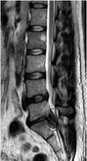
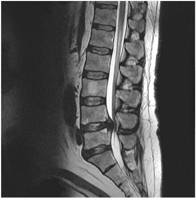
20 Treatment Tuberosity avulsion fractures/stress fractures treated in walking boot or hard-sole shoe True Jones fracture requires either prolonged NWB cast immobilization (8-12 weeks) or surgical ORIF Missed diagnosis leads to nonunion and chronic foot pain Cauda Equina Syndrome Terminal end of the spinal cord is at L1-2 Below this spinal canal is filled with the L2-S4 nerve roots, or cauda equina( horse tail ) Syndrome results from sudden reduction in volume of lumbar spinal canal Central disc herniation Epidural abscess or hematoma Trauma/fracture with retropulsion Onset may be sudden or over hours Symptoms Pain out of proportion; radicularpain/numbness bilaterally but worse on one side Saddle anesthesia Lower extremity weakness/paralysis Urinary or bowel incontinence or urinary retention Surgical emergency STAT MRI STAT spine surgery evaluation Missed diagnosis can lead to permanent nerve damage and paralysis
21 Questions?
Hand & Wrist Casey G. Batten MD Assistant Clinical Professor UCSF Sports Medicine
 Hand & Wrist Casey G. Batten MD Assistant Clinical Professor UCSF Sports Medicine Topics: Scaphoid Fracture Scapholunate Separation TFCC Injury Thumb Ulnar Collateral Lig (UCL) Injury Extensor Injury /
Office Orthopedics. No conflict of interest No financial disclosures 1/31/2018
 Office Orthopedics Amin Afsari DO Orthopedic Hand and Upper Extremity Surgery Orthopedic Institute of Wisconsin Midwest Orthopedic Specialty Hospital 1 No conflict of interest No financial disclosures
Acute Wrist Injuries OUCH!
 Acute Wrist Injuries OUCH! Case the athlete FOOSH from sporting event 2 days ago C/O wrist swelling, pain, worse with movement Hmmm Wrist pain Exam of the wrist - basics Appearance Swelling, bruising,
Kristin Kelley, DPT, OCS, FAAOMPT Orthopaedic Manual Physical Therapy Series Charlottesville Trauma/Fractures
 WRIST/HAND PATHOLOGY Kristin Kelley, DPT, OCS, FAAOMPT Orthopaedic Manual Physical Therapy Series Charlottesville 2017-2018 Trauma/Fractures Hook of Hamate Fractures Triangular Fibrocartilage Complex (TFCC)
Trauma/Fractures WRIST/HAND PATHOLOGY. TFCC Injury. Hook of Hamate Fracture. Property of VOMPTI, LLC
 WRIST/HAND PATHOLOGY Kristin Kelley, DPT, OCS, FAAOMPT Orthopaedic Manual Physical Therapy Series Charlottesville 2017-2018 Trauma/Fractures Hook of Hamate Fractures Triangular Fibrocartilage Complex (TFCC)
MR: Finger and Thumb Injuries
 MR: Finger and Thumb Injuries Laura W. Bancroft, M.D. Professor of Radiology University of Central Florida Florida State University Outline Normal anatomy of the fingers and thumb MR imaging protocols
CASE ONE CASE ONE. RADIAL HEAD FRACTURE Mason Classification. RADIAL HEAD FRACTURE Mechanism of Injury. RADIAL HEAD FRACTURE Imaging
 CASE ONE An eighteen year old female falls during a basketball game, striking her elbow on the court. She presents to your office that day with a painful, swollen elbow that she is unable to flex or extend
Hand and wrist emergencies
 Chapter1 Hand and wrist emergencies Carl A. Germann Distal radius and ulnar injuries PEARL: Fractures of the distal radius and ulna are the most common type of fractures in patients younger than 75 years.
The Forearm, Wrist, Hand and Fingers. Contusion Injuries to the Forearm. Forearm Fractures 12/11/2017. Oak Ridge High School Conroe, Texas
 The Forearm, Wrist, Hand and Fingers Oak Ridge High School Conroe, Texas Contusion Injuries to the Forearm The forearm is constantly exposed to bruising and contusions in contact sports. The ulna receives
COMMON CARPAL INJURIES IN ATHLETES Nicholas A. Bontempo, MD Orthopedic Associates of Hartford I HAVE NO CONFLICTS OR DISCLOSURES TO REPORT OUTLINE
 COMMON CARPAL INJURIES IN ATHLETES Nicholas A. Bontempo, MD Orthopedic Associates of Hartford I HAVE NO CONFLICTS OR DISCLOSURES TO REPORT OUTLINE The carpus Scaphoid fracture Scapholunate ligament tear
Hand & Wrist Injuries. DR MA Manjra
 Hand & Wrist Injuries DR MA Manjra 1 Background Up to 25% of all athletic injuries General population Sport people Sport specific Position specific Multifaceted Time of season Level of athlete Parents
FINGER INJURIES. Chapter 24, pgs ,
 FINGER INJURIES Chapter 24, pgs 727 730, 741 743 1. Demonstrate mastery of anatomical references to the hand and fingers. 2. Compare and contrast Mallet Finger, Swan Neck Deformity and Boutonnière Deformity.
Sports Medicine in your office: What not to miss!
 Sports Medicine in your office: What not to miss! 2018 Primary Care Approach to Treating the Injured Athlete May 4, 2018 John H. Wilckens, MD Associate Professor, Dept of Orthopaedic Surgery Disclosures
What you don t want to miss
 March 25, 2009 Vishal Michael Shah, M.D. What you don t want to miss Spectrum of Injuries Contusions Sprains Dislocations Fractures Lacerations Tendon Avulsions Ligament Tears Overuse Injuries FINGER
Episode 52 Commonly Missed Uncommon Orthopedic Injuries. Lisfranc Injuries. Drs. Ivy Cheng & Hossein Medhian. Prepared by Dr. Keerat Grewal, Oct 2014
 Prepared by Dr. Keerat Grewal, Oct 2014 Episode 52 Commonly Missed Uncommon Orthopedic Injuries Drs. Ivy Cheng & Hossein Medhian Lisfranc Injuries Q: What is a Lisfranc injury? Lisfranc injuries are a
Wrist and Hand Complaints
 Wrist and Hand Complaints Charles S. Day, M.D., M.B.A. Chief, Hand & Upper Extremity Surgery St. Elizabeth s Medical Center Tufts University School of Medicine Primary Care Internal Medicine 2018 Outline
Elbow/Wrist/Hand Pointers
 Elbow/Wrist/Hand Pointers Elbow Injuries -break elbow into 4 quadrants -Lateral -Medial -Posterior -Anterior 1. Lateral Epicondylosis (Tennis Elbow) a. extensor supinator tendinopathy b. repetitive gripping/wrist
Top 10 Ortho Urgent Care Injuries. J.C. Clark, M.D. ORA Orthopedics
 Top 10 Ortho Urgent Care Injuries J.C. Clark, M.D. ORA Orthopedics 10. Proximal Humerus Fractures Treatment Simple sling ICE, pain meds Button-down shirts Recliner to sleep in It will be up to the surgeon
Scaphoid Fractures. Mohammed Alasmari. Orthopaedic Surgery Demonstrator Majmaah University
 Scaphoid Fractures Mohammed Alasmari Orthopaedic Surgery Demonstrator Majmaah University 1 2 Scaphoid Fractures Introduction Anatomy History Clinical examination Radiographic evaluation Classification
Urgent Cases and Foreign Bodies
 Urgent Cases and Foreign Bodies Catherine J. Brandon, MD, MS University of Michigan Ann Arbor, MI, USA Introduction: Patients added on to the schedule from the emergency department or as urgent add-on
Goals. Initial management skeletal trauma. Physical Exam ABC OF PRIMARY CARE MEDICINE FRACTURE MANAGEMENT 12/4/2010
 ABC OF PRIMARY CARE MEDICINE FRACTURE MANAGEMENT Brian Feeley, MD UCSF Sports Medicine and Shoulder Surgery Goals Discuss common fractures and initial management, treatment guidelines Let your patients
Common. Common Hand Problems in Elite Athletes
 Common Hand Problems in Elite Athletes Fred Corley M.D. Dept. of Orthopaedic Surgery UTHSCSA I have no disclosures concerning this talk. The University of Texas Health Science Center @ San Antonio - Orthopaedics
11/5/14. I will try to make this painless. Great, a Fracture, Now What? Objectives. Basics for Fracture Workup. Basics for Fracture Workup
 Great, a Fracture, Now What? I will try to make this painless Mary Greve MS, PA-C Department of Orthopedic Surgery Trauma Team University of Iowa Hospitals and Clinics Mary-Greve@uiowa.edu Pager 2121 Objectives
Physical therapy of the wrist and hand
 Physical therapy of the wrist and hand Functional anatomy wrist and hand The wrist includes distal radius, scaphoid, lunate, triquetrum, pisiform, trapezium, trapezoid, capitate, and hamate. The hand includes
Pediatric Fractures. Objectives. Epiphyseal Complex. Anatomy and Physiology. Ligaments. Bony matrix
 1 Pediatric Fractures Nicholas White, MD Assistant Professor of Pediatrics Eastern Virginia Medical School Attending, Pediatric Emergency Department Children s Hospital of The King s Daughters Objectives
SPORTS INJURIES IN HAND
 Grundkurs SGSM-SSMS Sion 2015 SPORTS INJURIES IN HAND Dr S. KŠmpfen EPIDEMIOLOGY Incidence of hand, finger and wrist injuries in sports : 3% Ð 9 % RADIAL-SIDED WRIST PAIN 1)! Distal Radius Fractures 2)!
Surgical Care at the District Hospital. EMERGENCY & ESSENTIAL SURGICAL CARE
 Surgical Care at the District Hospital 1 18 Orthopedic Trauma Key Points 2 18.1 Upper Extremity Injuries Clavicle Fractures Diagnose fractures from the history and by physical examination Treat with a
Orthopedic X-Rays most commonly missed
 Orthopedic X-Rays most commonly missed Vukiet Tran, MD, MHSc, MBA University Health Network Toronto, Canada 1 COI Disclosure I am the current Medical Director for Best Doctors Canada. Presenter: Dr. Vu
Physical Examination of the Knee
 History: Pain Traumatic vs. atraumatic? Acute vs Chronic Previous procedures done on the knee? Swelling, catching, instability General Setup Examine standing, sitting and supine Evaluate gait Examine hip
SCAHPO-LUNATE DISSOCIATION
 SCAHPO-LUNATE DISSOCIATION Introduction Scapho-lunate dissociation is the most common significant ligamentous injury of the wrist. The condition is also sometimes referred to as rotary subluxation of the
Physical Examination of the Knee
 History: Pain Traumatic vs. atraumatic Acute vs Chronic Mechanism of injury Swelling, catching, instability Previous evaluation and treatment General Setup Examine standing, sitting and supine Evaluate
Sick Call Screener Course
 Sick Call Screener Course Musculoskeletal System Upper Extremities (2.7) 2.7-2-1 Enabling Objectives 1.46 Utilize the knowledge of musculoskeletal system anatomy while assessing a patient with a musculoskeletal
Ligaments of Elbow hinge: sagittal plane so need lateral and medial ligaments
 Ligaments of Elbow hinge: sagittal plane so need lateral and medial ligaments Ulnar Collateral ligament on medial side; arising from medial epicondyle and stops excess valgus movement (lateral movement)
Wrist & Hand Assessment and General View
 Wrist & Hand Assessment and General View Done by; Mshari S. Alghadier BSc Physical Therapy RHPT 366 m.alghadier@sau.edu.sa http://faculty.sau.edu.sa/m.alghadier/ Functional anatomy The hand can be divided
AAP Boot Camp KNEE AND ANKLE EXAM
 AAP Boot Camp KNEE AND ANKLE EXAM Disclosures I have no relevant financial relationships with the manufacturers of any commercial products and or providers of commercial services discussed in this CME
ACUTE HAND INJURIES FOR THE PRIMARY CARE PHYSICIAN
 Vincent Shaw, MD, FAAFP Program Director Baton Rouge General Family Medicine Residency Program Baton Rouge General Sports Medicine Fellowship ACUTE HAND INJURIES FOR THE PRIMARY CARE PHYSICIAN Disclosures
Index. Note: Page numbers of article titles are in boldface type.
 Index Note: Page numbers of article titles are in boldface type. A Achilles tendonitis, criteria for full competition in, 164 165 description of, 164 patient education in, 165 prophylactic support in,
Case. 5 year old with 2 weeks leg pain and now refusing to walk + Fevers, lower leg swelling, warmth Denies and history of trauma or wounds
 Case 5 year old with 2 weeks leg pain and now refusing to walk + Fevers, lower leg swelling, warmth Denies and history of trauma or wounds Exam I: Swelling over entire tibia extending to foot P: Tenderness
Index. Note: Page numbers of article titles are in boldface type.
 Note: Page numbers of article titles are in boldface type. A Abscess, epidural, 822 824 Achilles tendon rupture, 894 895, 981 982 Acromioclavicular separations, shoulder pain in, 751 753 Adhesive capsulitis,
Pediatric Upper Extremity Injuries. Andrew Westbrook, DO
 Pediatric Upper Extremity Injuries Andrew Westbrook, DO Case #1 12 yo male who presents to sports medicine clinic due to right shoulder pain Pain started 3 days ago during a baseball game when he was playing
Hand injuries. The metacarpal bones may fracture through the base, shaft or the neck.
 Hand injuries Metacarpal injuries The metacarpal bones may fracture through the base, shaft or the neck. Shaft fractures; these are caused by direct trauma which may cause transverse # of one or more metacarpal
Fractures and dislocations around elbow in adult
 Lec: 3 Fractures and dislocations around elbow in adult These include fractures of distal humerus, fracture of the capitulum, fracture of the radial head, fracture of the olecranon & dislocation of the
FOOSH It sounded like a fun thing at the time!
 FOOSH It sounded like a fun thing at the time! Evaluating acute hand and wrist injuries Larry Collins, MPAS, PA-C, ATC, DFAAPA Assistant Professor, Physician Assistant Program Assistant Professor, Department
FOOSH It sounded like a fun thing at the time!
 FOOSH It sounded like a fun thing at the time! Evaluating acute hand and wrist injuries Larry Collins, MPAS, PA-C, ATC, DFAAPA Assistant Professor, Physician Assistant Program Assistant Professor, Department
KNEE EXAMINATION. Tips & Tricks from an Emergency Physician Perspective. EM Physicians Less Exposed to MSK Medicine
 KNEE EXAMINATION Tips & Tricks from an Emergency Physician Perspective Dr P O CONNOR Emergency Medicine Physician EUSEM 10/09/2018 EM Physicians Less Exposed to MSK Medicine Musculoskeletal Medicine becoming
Carpal rows injuries!
 Carpal rows injuries! Michael Papaloïzos! Center for Hand Surgery and Therapy Geneva, Switzerland no conflict of interest to declare Fractures of carpal bones! The fractured scaphoid! Fracture-dislocations
Evaluation of the Hip and Knee
 Evaluation of the Hip and Knee Causes of hip pain RA Osteoarthritis Psoriatic arthritis Septic arthritis Bursitis Hip fx Labral tear Tendinitis Referred back pain Cancer AVN Legg-Calve-Perthes Paget's
The Kienböck disease and scaphoid fractures. Mariusz Bonczar
 The Kienböck disease and scaphoid fractures Mariusz Bonczar The Kienböck disease and scaphoid fractures Mariusz Bonczar Kienböck disease personal experience My special interest for almost 25 years Thesis
The Painful Elbow, Wrist, and Hand. Jennifer R Marks, MD
 The Painful Elbow, Wrist, and Hand Jennifer R Marks, MD The Painful Elbow A 44 yo M presents to clinic complaining of a sore elbow What further questions do you have for this patient? What is on your differential
PEM GUIDE CHILDHOOD FRACTURES
 PEM GUIDE CHILDHOOD FRACTURES INTRODUCTION Skeletal injuries account for 10-15% of all injuries in children; 20% of those are fractures, 3 out of 4 fractures affect the physis or growth plate. Always consider
Nerves of Upper limb. Dr. Brijendra Singh Professor & Head Department of Anatomy AIIMS Rishikesh
 Nerves of Upper limb Dr. Brijendra Singh Professor & Head Department of Anatomy AIIMS Rishikesh 1 Objectives Origin, course & relation of median & ulnar nerves. Motor & sensory distribution Carpal tunnel
Clinical Orthopaedic Rehabilitation Volume 1 and 2
 Clinical Orthopaedic Rehabilitation Volume 1 and 2 COURSE DESCRIPTION This program is a practical, clinical guide that provides guidance on the evaluation, differential diagnosis, treatment, and rehabilitation
THE HIP. Cooler than cool, the pinnacle of what is "it". Beyond all trends and conventional coolness.
 THE HIP Cooler than cool, the pinnacle of what is "it". Beyond all trends and conventional coolness. Objectives Hip anatomy Causes of hip pain Hip exam Anatomy Bones Ilium Anterior Superior Iliac Spine
5/8/2017. Finger Injuries in Football. Tendon Injuries of the Hand and Wrist in Football Steve Kronlage, MD Andrews Institute Gulf Breeze, Florida
 Finger Injuries in Football Tendon Injuries of the Hand and Wrist in Football Steve Kronlage, MD Andrews Institute Gulf Breeze, Florida A jammed finger is an injury (at very least a torn ligament) A swollen
Common Elbow Problems
 Common Elbow Problems Duncan Ferguson FRACS Knee and Shoulder Specialist Elbow Instability Common 10-25% of elbow injuries Median age 30 yrs Most simple dislocations are stable after reduction recurrence
7/1/2012. Repetitive valgus stresses cause microfractures in the apophyseal cartilage (weak link) Common in year olds
 Common in year olds") 1 2 3 4 5 6 7 When growing pains are not growing pains David W. Gray,M.D. Medical Director Orthopedics Differential Diagnosis Fracture Ligament Injury Disloclation Cartilage Injury Apophysitis Inflammation
1 2 3 4 5 6 7 When growing pains are not growing pains David W. Gray,M.D. Medical Director Orthopedics Differential Diagnosis Fracture Ligament Injury Disloclation Cartilage Injury Apophysitis Inflammation
Division of Student Affairs A Primer on the Musculoskeletal Examination Technique and Commonly Missed Injuries in Student Health
 Division of Student Affairs A Primer on the Musculoskeletal Examination Technique and Commonly Missed Injuries in Student Health C.S. Nasin, MD, CAQ Sports Medicine Medical Director Head Team Physician
Kinesiology of The Wrist and Hand. Cuneyt Mirzanli Istanbul Gelisim University
 Kinesiology of The Wrist and Hand Cuneyt Mirzanli Istanbul Gelisim University Bones The wrist and hand contain 29 bones including the radius and ulna. There are eight carpal bones in two rows of four to
Lower Extremity Sports Injuries
 Lower Extremity Sports Injuries AAP Musculoskeletal Boot Camp Sigrid F. Wolf, MD Pediatric Sports Medicine Fellow Northwestern University Lurie Children s Hospital Disclosure I have no relevant financial
CLINICAL PRESENTATION AND RADIOLOGY QUIZ QUESTION
 Donald L. Renfrew, MD Radiology Associates of the Fox Valley, 333 N. Commercial Street, Suite 100, Neenah, WI 54956 10/13/2012 Radiology Quiz of the Week # 94 Page 1 CLINICAL PRESENTATION AND RADIOLOGY
Ouch, That s Gotta Hurt! Pediatric Fractures & Injuries
 Ouch, That s Gotta Hurt! Pediatric Fractures & Injuries Greg Canty, MD Medical Director, Sports Medicine Center Attending Physician, Emergency Medicine Children s Mercy Kansas City 2011 Children s Mercy
Resolving the Top Three Boot Camp Injuries. Ryan Matthiesen DO
 Resolving the Top Three Boot Camp Injuries Ryan Matthiesen DO About Me Oklahoma State College of Osteopathic Medicine Family Medicine Residency Plaza Medical Center Sports Medicine Fellowship Texas Tech
Anatomy and Physiology II. Review Shoulder Girdle New Material Upper Extremities - Bones
 Anatomy and Physiology II Review Shoulder Girdle New Material Upper Extremities - Bones Anatomy and Physiology II Shoulder Girdle Review Questions From Last Lecture Can you identify the following muscles?
Shaun P. Garff, DO Physician of Sports Medicine
 Shaun P. Garff, DO Physician of Sports Medicine Speaker Disclosure I have no actual or potential conflicts of interest to disclose as it relates to this presentation. A little about me Born and raised
Basic Care of Common Fractures Utku Kandemir, MD
 Basic Care of Common Fractures Utku Kandemir, MD Assistant Clinical Professor Trauma & Sports Medicine Dept. of Orthopaedic Surgery UCSF / SFGH History Physical Exam Radiology Treatment History Acute trauma
Trauma & Orthopaedic Undergraduate Syllabus
 Trauma & Orthopaedic Undergraduate Syllabus Introduction The purpose of this document is to provide a recommended syllabus for medical students in Trauma & Orthopaedics (T&0). It should help students on
Hand Injuries in Baseball
 Hand Injuries in Baseball Steven S. Shin, MD, MMSc Director of Hand Surgery, Kerlan-Jobe Orthopaedic Clinic Co-Director of Hand Surgery, Cedars-Sinai Health System Los Angeles, California Hand Consultant
EMERGENCY PITFALLS IN ORTHOPAEDIC TRAUMA. Thierry E. Benaroch, MD, FRCS MCH Trauma Rounds February 9, 2009
 EMERGENCY PITFALLS IN ORTHOPAEDIC TRAUMA Thierry E. Benaroch, MD, FRCS MCH Trauma Rounds February 9, 2009 MORAL OF THE STORY Fracture distal radius and intact ulna W/O radius fracture will most likely
Fractures and dislocations of the fingers
 Chapter 1 Fractures and dislocations of the fingers Felix S. Chew, M.D., and Catherine Maldjian, M.D. Case 1 1 Phalangeal tuft avulsion fracture 31-year-old woman injured in a ground-level fall. Lateral
Wrist and Hand Anatomy
 Wrist and Hand Anatomy Bone Anatomy Scapoid Lunate Triquetrium Pisiform Trapeziod Trapezium Capitate Hamate Wrist Articulations Radiocarpal Joint Proximal portion Distal portion Most surface contact found
UPPER EXTREMITY INJURIES. Recognizing common injuries to the upper extremity
 UPPER EXTREMITY INJURIES Recognizing common injuries to the upper extremity ANATOMY BONES Clavicle Scapula Spine of the scapula Acromion process Glenoid fossa/cavity Humerus Epicondyles ANATOMY BONES Ulna
5 COMMON INJURIES IN THE FOOT & ANKLE
 5 COMMON INJURIES IN THE FOOT & ANKLE MICHAEL P. CLARE, MD FLORIDA ORTHOPAEDIC INSTITUTE TAMPA, FL USA MECHANISM OF INJURY HOW DID IT HAPPEN? HIGH ENERGY VS LOW ENERGY DIRECTION OF FORCES INVOLVED LIVING
SMF PCP Treatment & Referral Guideline Orthopedics Developed February 1, 2003 Revised: October, 2011
 SUTTER MEDICAL FOUNDATION (SMF) 2800 L Street, 7 th Floor Sacramento, CA 95816 SMF PCP Treatment & Referral Guideline Orthopedics Developed February 1, 2003 Revised: October, 2011 I. Shoulder Pain...Page
Orthopedic Emergencies. Peter Gutierrez, MD Pediatric Emergency Medicine Children s Healthcare of Atlanta
 Orthopedic Emergencies Peter Gutierrez, MD Pediatric Emergency Medicine Children s Healthcare of Atlanta Disclosures I have no relevant financial relationships to disclose I do not intend to discuss unapproved
Disclosures Head to Toe: Common Sports Injuries in Kids
 Disclosures Head to Toe: Common Sports Injuries in Kids None R. Jay Lee MD Director Pediatric Orthopaedic Fellowship Assistant Professor Pediatric Orthopaedics Johns Hopkins / Bloomberg Children s Objectives
NORTH BAY SYMPOSIUM SATURDAY JANUARY 20 TH 2018
 NORTH BAY SYMPOSIUM SATURDAY JANUARY 20 TH 2018 ROBERT HAIMSON, M.D. ORTHOPEDIC SURGEON SMGR FELLOW IN AAOS DIPLOMATE IN ABOS COMMON MUSCULOSKELETAL CONDITIONS COMMON MUSCULOSKELETAL CONDITIONS: WHAT
Chpter 2 Nonoperative Management of Non-displaced Acute Scaphoid Fracture
 Chpter 2 Nonoperative Management of Non-displaced Acute Scaphoid Fracture Megan Tomaino and Thomas B. Hughes Case Presentation The patient is a 15-year-old male with a history of left wrist pain following
Anatomy of the Musculoskeletal System
 Anatomy of the Musculoskeletal System Kyle E. Rarey, Ph.D. Department of Anatomy & Cell Biology and Otolaryngology University of Florida College of Medicine Outline of Presentation Vertebral Column Upper
Musculoskeletal Examination Benchmarks
 Musculoskeletal Examination Benchmarks _ The approach to examining the musculoskeletal system is the same no matter what joint or limb is being examined. The affected and contralateral region should both
Trapezium is by the thumb, Trapezoid is inside
 Trapezium is by the thumb, Trapezoid is inside Intercarpal Jt Radiocarpal Jt Distal Middle Proximal DIP PIP Interphalangeal Jts Metacarpalphalangeal (MCP) Jt Metacarpal Carpometacarpal (CMC) Jt Trapezium
SHOULDER Highly mobile, so less stable. Abnormalities cloaked within extensive musculature, dx can be difficult Bony abnormalities less common than li
 SPORTS MEDICINE CASES A quick tour of some local joints Featuring gco common o and unusual problems SHOULDER Highly mobile, so less stable. Abnormalities cloaked within extensive musculature, dx can be
Management of Wrist and Hand Injuries
 Sunday General Session Management of Wrist and Hand Injuries Shaun Garff, DO Sports Medicine Physician Methodist Health System Dallas, Texas Educational Objectives By the end of this educational activity,
The Upper Limb. Elbow Rotation 4/25/18. Dr Peter Friis
 The Upper Limb Dr Peter Friis Elbow Rotation Depending upon the sport, the elbow moves through an arc of approximately 75⁰ to 100⁰ in about 20 to 35 msec. The resultant angular velocity is between 1185
8 Recovering From HAND FRACTURE SURGERY
 8 Recovering From HAND FRACTURE SURGERY Hand fractures are caused by trauma and result in breaking (fracturing) the phalanges or metacarpals. Surgery involves achieving acceptable alignment and providing
Index. radiologic.theclinics.com. Note: Page numbers of article titles are in boldface type.
 Index Note: Page numbers of article titles are in boldface type. A Acromioclavicular joint injuries in football players, 318, 319 ALPSA. See Anterior labroligamentous periosteal sleeve avulsion. Anterior
ANTERIOR CRUCIATE LIGAMENT INJURY
 ANTERIOR CRUCIATE LIGAMENT INJURY WHAT IS THE ANTERIOR CRUCIATE LIGAMENT? The anterior cruciate ligament (ACL) is one of four major ligaments that stabilizes the knee joint. A ligament is a tough band
GET HIP! CAPA 2015 Annual Conference WHAT IS HIP? HIP JOINT. Bradford H. Stiles, M.D., FAAFP
 GET HIP! Bradford H. Stiles, M.D., FAAFP WHAT IS HIP? HIP JOINT Synovial ball-and-socket joint Articulation between femoral head and acetabulum Acetabulum formed by the confluence of pelvis bones (ilium,
EXAMINATION OF THE WRIST BEYOND THE BASICS OMA SPORT MED Janice Harvey MD CCFP CFFP Dip. Sp Med.
 EXAMINATION OF THE WRIST BEYOND THE BASICS OMA SPORT MED 2019 Janice Harvey MD CCFP CFFP Dip. Sp Med. CFPC CoI Templates: Slide 1 used in Faculty presentation only. FACULTY/PRESENTER DISCLOSURE Faculty:
Sports Medicine Unit 16 Elbow
 Sports Medicine Unit 16 Elbow I. Bones a. b. c. II. What movements does the elbow perform? a. Flexion b. c. Pronation d. III. Muscles in motion a. FLEXION (supinated) i Brachialis (pronated) ii (neutral)
Evaluation of the Knee and Shoulder
 Evaluation of the Knee and Shoulder Karen J. Boselli, MD Northeast Regional Nurse Practitioner Conference May 2018 Knee Overview History Examination Top 5 diagnoses When to image When to refer Pain most
Post test for O&P 2 Hrs CE. The Exam
 Post test for O&P 2 Hrs CE The Exam This examination is taken in "open book" format. That means you are free to answer the questions after research or discussion with your fellow workers. We feel this
STRUCTURAL BASIS OF MEDICAL PRACTICE EXAMINATION 5. September 30, 2011
 STRUCTURAL BASIS OF MEDICAL PRACTICE EXAMINATION 5 September 30, 2011 PART l. Answer in the space provided. (12 pts) 1. Identify the structures. (2 pts) EXAM NUMBER A. Suprascapular nerve B. Axillary nerve
Salisbury Foundation Trust Radiology Department Referral Guidelines for Primary Care: Musculoskeletal Imaging
 Salisbury Foundation Trust Radiology Department Referral Guidelines for Primary Care: Musculoskeletal Imaging These guidelines have been issued in conjunction with the Royal College of Radiology referral
Index. orthopedic.theclinics.com. Note: Page numbers of article titles are in boldface type.
 Index Note: Page numbers of article titles are in boldface type. A Acetabular fractures thromboembolic disease after, 341 Achilles tendon rupture ACL. See Anterior cruciate ligament (ACL) Adolescent idiopathic
Elbow. Chapter 2 LISTEN. Mechanism of Injury (If Applicable) Pain
 Pain") Chapter 2 Elbow LISTEN Mechanism of Injury (If Applicable) Patient usually remembers their position at the time of injury Certain mechanisms of injury result in characteristic patterns Fall on outstretched
Chapter 2 Elbow LISTEN Mechanism of Injury (If Applicable) Patient usually remembers their position at the time of injury Certain mechanisms of injury result in characteristic patterns Fall on outstretched
1. A 40-year-old male has dislocated his right 2 nd MCP. You have pulled as hard as you can but cannot reduce the dislocation. The problem is likely:
 CHAPTER 50 HAND 2 OCTOBER 2013 1. A 40-year-old male has dislocated his right 2 nd MCP. You have pulled as hard as you can but cannot reduce the dislocation. The problem is likely: A. He is a gamer and
Peggers Super Summaries: Foot Injuries
 Lisfranc Injury ANATOMY Roman arch with recessed 2 nd MT base AP medial side of intermediate cuneiform to 2 nd MT base Oblique medial side of lateral cuneiform with 3 rd MT base and 4 th with medial boarder
The Shoulder. Jennifer R Marks, MD
 The Shoulder Jennifer R Marks, MD Shoulder Anatomy Skeletal & ligamentous components: The joint is comprised of a confluence of Scapula Clavicle Humerus https://www.shoulderdoc.co.uk/article/ http/ www.shoulderdoc.co.uk/article/117777
MCQWeek2. All arise from the common flexor origin. The posterior aspect of the medial epicondyle is the common flexor origin.
 MCQWeek2. 1. Regarding superficial muscles of anterior compartment of the forearm: All arise from the common flexor origin. The posterior aspect of the medial epicondyle is the common flexor origin. Flexor