RADIOLOGY (SURGERY) BY MARYAM MALIK Rawalpindi Medical College
|
|
|
- Francine Simpson
- 6 years ago
- Views:
Transcription
1 RADIOLOGY (SURGERY) BY MARYAM MALIK Rawalpindi Medical College
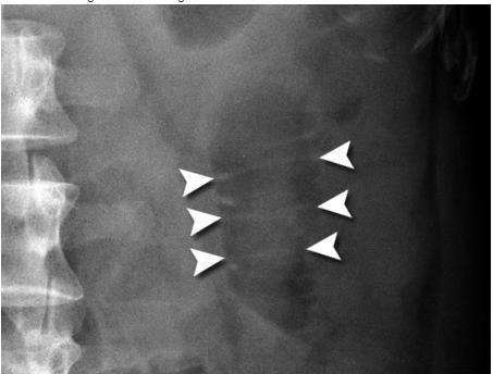
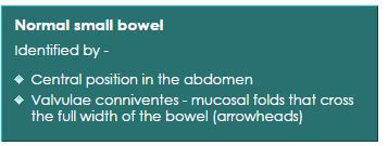
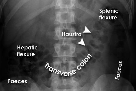
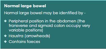
2 NORMAL BOWEL GAS PATTERN Any part of the bowel may be visible if it contains gas/air within the lumen. Gas/air is of low density and forms a natural contrast against surrounding denser soft tissues. It is often difficult to differentiate between normal small and large bowel, but this often becomes easier when the bowel is abnormally distended. The upper limit of normal diameter of the bowel is generally accepted as 3cm for the small bowel, 6cm for the colon and 9cm for the caecum (proximal colon) (3/6/9 rule). Sigmoid= 5cm
3
4
5

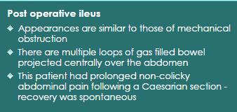
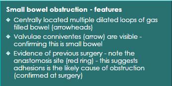
6 POST-OP ILEUS
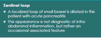
7 Sentinel loop Intra-abdominal inflammation, such as with pancreatitis, can lead to a localized ileus. This may appear as a single loop of dilated bowel known as a 'sentinel loop.'
8
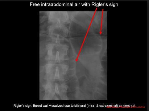
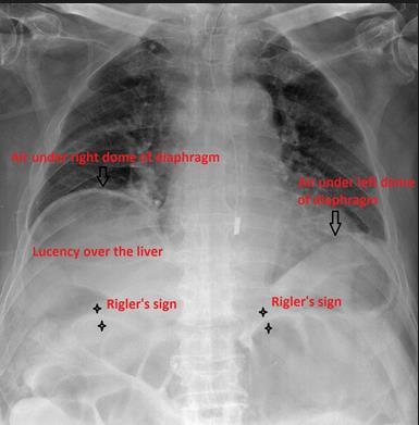
9 Rigler sign The Rigler sign, also known as the double wall sign, is seen on an x-ray of the abdomen when air is present on both sides of the intestine, i.e. when there is air on both the luminal and peritoneal side of the bowel wall. Pneumoperitoneum may be a result of perforation or, from recent instrumentation or surgery. A false double wall sign can result from two loops of bowel being in contact with one another.
10
11
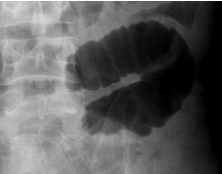
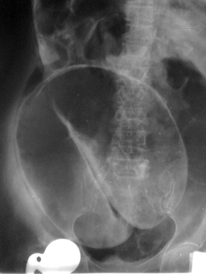
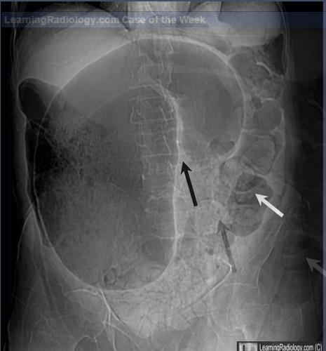
12 SIGMOID VOLVULUS Sigmoid volvulus is a cause of large bowel obstruction and occurs when the sigmoid colon twists on the sigmoid mesocolon. COFFEE BEAN SIGN on X-ray
13
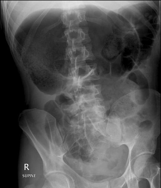
14 CAECAL VOLVULUS Caecal volvulus describes torsion of the caecum around its own mesentery which often results in obstruction. If unrecognised can result in bowel perforation and faecal peritonitis.
15
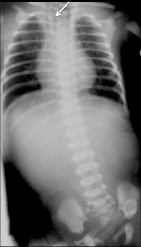
16 Esophageal atresia may show a dilated pharyngeal pouch the presence of air in the stomach and bowel in the setting of oesophageal atresia implies that there is a distal fistula. if an oesophago-gastric (feeding) tube insertion has been attempted this may show the tube blind looping and turning back at the upper thoracic part of the oesophagus or heading into the trachea and/or bronchial tree.
17
18 Oesophageal atresia without fistula. The Replogle tube (arrow) is coiled in the dilated blind ending oesophageal pouch. AXR demonstrates absence of bowel gas indicating that there is no distal tracheoesophageal fistula
")
19 Double bubble sign (duedenal atresia)

20 Perforated duedenal ulcer

21 Foreign body rectum

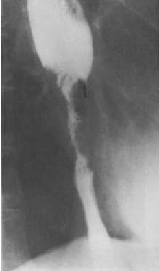
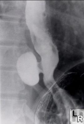
22 Achalasia : bird s beak appearance on braium swallow

23 ULCERATIVE COLITIS: barium enema showing a lead pipe colon Lead pipe appearance of colon is the classical barium enema finding in chroniculcerative colitis. There is complete loss of haustral markings in the diseased section of colon, and the organ appears smooth walled and cylindrical.

24 Carcinoma esophagus
25 Epiphrenic diverticula

26 Gastric ulcer

27 Gastric ulcer


28 Pyloric obstruction The shoulder sign, the impression of the hypertrophied pyloric muscle on the distended gastric antrum
29 MRCP Magnetic Resonance Cholangiopancreatography
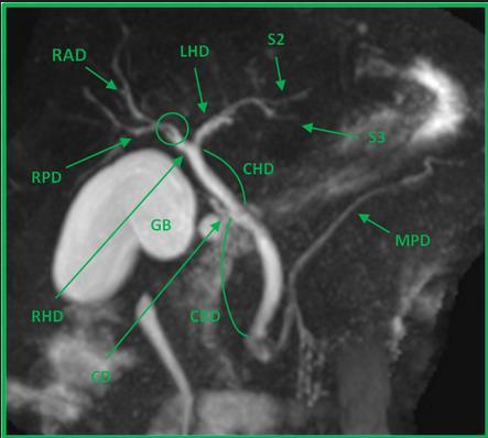
30 "Normal hepatic ductal anatomy". Coronal oblique MIP reformat image reveals the confluence (circle) between the right posterior duct (RPD) and the right anterior duct (RAD), originating the right hepatic duct (RHD). Note that the RPD has a more horizontal route while the RAD is more vertical. By its turn the RHD joins the left hepatic duct (LHD), originating the common hepatic duct. The LHD results from the confluence of the ducts of the left hepatic lobe segments, here only represented by segments II (S2) and III (S3). Cystic duct (CD), common bile duct (CBD), main pancreatic duct (MPD), gallbladder (GB).
31
 crossing the whole pancreas and continuing as the duct of Wirsung (DW) at the pancreatic head.")
32 Normal pancreatic ductal anatomy. Coronal oblique reformat shows the main pancreatic duct (MPD) crossing the whole pancreas and continuing as the duct of Wirsung (DW) at the pancreatic head. At its distal portion the DW joins the common bile duct (CBD), draining into the major papilla (pointing triangle). The duct of Santorini is not demonstrated

33
 in its distal")
34 MRCP showing stone in lower part of MRCP image shows a dilated bile duct with a dark stone (arrow) in its distal end. CBD
35 ERCP Endoscopic Retrograde Cholangiopancreatography

36 ERCP showing normal CBD & pancreatic duct

37 These two fluorospot images taken during an ERCP demonstrates stones in the common bile duct on the left radiograph, and cystic duct on the right radiograph
 : It is a radiologic technique used to visualize the anatomy of the")
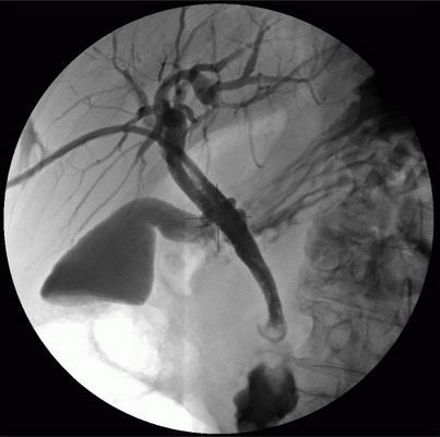
38 PTC (Percutaneous transhepatic cholangiography) PTC (Percutaneous Transhepatic Cholangiography) : It is a radiologic technique used to visualize the anatomy of the biliary tract.
39


40 T-tube cholangiograms T-tube cholangiograms are a fluroscopic study performed in the setting of hepatobiliary disease.

41 Peroperative cholangiogram during cholecystectomy

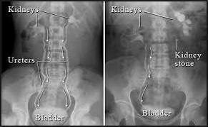
42 XRAY KUB showing radio-opaque shadows consistent with renal stone
43
")
44 XRAY KUB showing staghorn (phosphate) stones

45 XRAY KUB showing bladder stone A plain KUB showing a large smooth oval radio-opaque mass suggesting a calcified bladder stone

46 Normal intravenous urograms

47 IVU showing bilateral double ureters

48 Ascending cystography showing urinary bladder diverticuli A cystogram showing marked diverticulation with a large superior diverticulum.

49 Retrograde Urethrogram showing urethral stricture

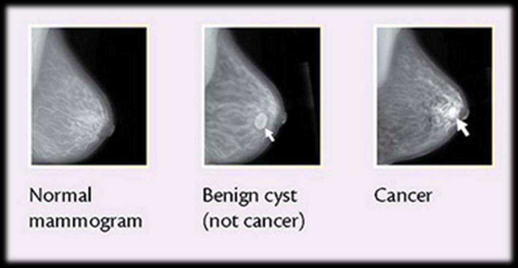
50 Normal mammogram
51


52 Mammographic images showing wellcircumscribed masses with features of fibroadenoma/cyst

53 Mammography showing cancer

54 Xray showing right clavicular fracture

55 Xray showing right shoulder dislocation

56 Xray showing mid shaft fracture of humerus

57 Xray showing posterior elbow dislocation

58 Xray showing the fracture of lower end of radius & ulna

59 Xray showing Undisplaced Inter- Trochanteric Femoral Fracture

60 Xray showing neck of left femur fracture

61 Xray showing left hip dislocation

62 Xray showing Austin Moore Prosthesis for femur neck fracture

63 Xray showing total hip replacement

64 Xray showing dynamic hip screw transfixing an inter-trochanteric fracture of right femur

65 Xray showing knee dislocation

66 Xray showing fracture of lower part of tibia & fibula
67 Xray showing lateral malleolar fracture
 and the tibia (2) are broken and the malleolar fragments (arrow: medial malleolus, arrowhead: lateral malleolus) are displaced.")
68 Xray showing bi-malleolar fracture (pott fracture) with distortion of ankle joint Bimalleolar fracture and right ankle dislocation on X-ray (anteroposterior). Both the end of the fibula (1) and the tibia (2) are broken and the malleolar fragments (arrow: medial malleolus, arrowhead: lateral malleolus) are displaced.

69 Xray showing fracture of 1 st metatarsal bone Preoperative radiographs showing the dorsal dislocation at the MTP joint and fracture of the base of the metatarsal bone.

70 CT Scan showing multiple metastasis in brain

71 NORMAL CT-SCAN BRAIN

72 CT Scan showing subdural hematoma

73 CT Scan showing epidural hematoma

74 CT Scan showing hydrocephalus

75 CT Scan showing multiple fractures in frontal part of skull

76 MRI showing intrameduallary tumor of spinal cord
 showing normal anatomy")
77 Magnetic resonance angiogram (MRA) showing normal anatomy

78 CT angiogram of abdomen & lower limb showing normal vasculature

79 Angiogram showing abdominal aortic aneurysm

80 This magnetic resonance angiogram (MRA) of the lower extremities was obtained by using the bolus-chase technique. A short-segment high-grade stenosis is present in the middle of the left superficial femoral artery. Note the collateral arterial supply.

81 Magnetic resonance angiogram showing narrowing of left popliteal artery

82 CT scan showing tumor in head of pancreas The arrow indicates the superior mesenteric artery.

83 CT scan showing tumor in tail of pancreas

84 CT scan showing tumor in right kidney

85 CT SCAN showing bilateral kidney cysts

86 CT scan showing normal liver

87 Liver metastasis Multiple hypodense lesions seen in the liver with no significant contrast enhancement.

88 LIVER CYST Oval, well defined Imperceptible or thin wall Water density

89 CT scan showing tumor in right lobe of liver (arrowheads)
Lab Monitor Images Dissection of the Abdominal Vasculature + Lower Digestive System
 Lab Monitor Images Dissection of the Abdominal Vasculature + Lower Digestive System Stomach & Duodenum Frontal (AP) View Nasogastric tube 2 1 3 4 Stomach Pylorus Duodenum 1 Duodenum 2 Duodenum 3 Duodenum
Plain abdomen The standard films are supine & erect AP views (alternative to erect, lateral decubitus film is used in ill patients).
.") Plain abdomen The standard films are supine & erect AP views (alternative to erect, lateral decubitus film is used in ill patients). The stomach can be readily identified by its location, gastric rugae
Plain abdomen The standard films are supine & erect AP views (alternative to erect, lateral decubitus film is used in ill patients). The stomach can be readily identified by its location, gastric rugae
Abdominal radiology 腹部放射線學
 Abdominal radiology 腹部放射線學 台北醫學大學 - 市立萬芳醫院 留偉順 laowilson@hotmail.com The Normal Abdominal Series Chest Supine abdomen Erect abdomen Left lateral decubitus abdomen Learning objectives Understanding normal
Introduction and Definitions
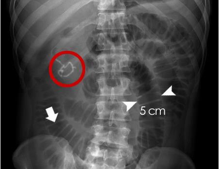
 Bowel obstruction Introduction and Definitions Accounts for 5% of all acute surgical admissions Patients are often extremely ill requiring prompt assessment, resuscitation and intensive monitoring Obstruction
ASSESSING THE PLAIN ABDOMINAL RADIOGRAPH M A A M E F O S U A A M P O F O
 ASSESSING THE PLAIN ABDOMINAL RADIOGRAPH M A A M E F O S U A A M P O F O Introduction The abdomen (less formally called the belly, stomach, is that part of the body between the thorax (chest) and pelvis,
Nasogastric tube. Stomach. Pylorus. Duodenum 1. Duodenum 2. Duodenum 3. Duodenum 4
 Esophagus Barium Swallow Stomach and Duodenum 4 year old Upper GI Nasogastric tube Stomach and Duodenum 4 year old Upper GI Nasogastric tube Stomach Pylorus Duodenum 1 Duodenum 2 Duodenum 3 Duodenum 4
PROFESSIONAL SKILLS 1 3RD YEAR SEMESTER 6 RADIOGRAPHY. THE URINARY SYSTEM Uz. Fatema shmus aldeen Tel
 PROFESSIONAL SKILLS 1 3RD YEAR SEMESTER 6 RADIOGRAPHY THE URINARY SYSTEM Uz. Fatema shmus aldeen Tel. 0925111552 Professional skills-2 THE URINARY SYSTEM The urinary system (review anatomy and physiology)
Radiology of the abdomen Lecture -1-
 Radiology of the abdomen Lecture -1- Objectives To know radiology modalities used in abdomen imaging mainly GI tract. To know advantages and disadvantages of each modality. To know indications and contraindications
UNDERSTANDING X-RAYS: ABDOMINAL IMAGING THE ABDOMEN
 UNDERSTANDING X-RAYS: ABDOMINAL IMAGING THE ABDOMEN Radiology Enterprises radiologyenterprises@gmail.com www.radiologyenterprises.com STOMACH AND SMALL BOWEL STOMACH AND SMALL BOWEL Swallowed air is a
Cholangiocarcinoma (Bile Duct Cancer)
") Cholangiocarcinoma (Bile Duct Cancer) The Bile Duct System (Biliary Tract) A network of bile ducts (tubes) connects the liver and the gallbladder to the small intestine. This network begins in the liver
Cholangiocarcinoma (Bile Duct Cancer) The Bile Duct System (Biliary Tract) A network of bile ducts (tubes) connects the liver and the gallbladder to the small intestine. This network begins in the liver
INVESTIGATIONS OF GASTROINTESTINAL DISEAS
 INVESTIGATIONS OF GASTROINTESTINAL DISEAS Lecture 1 and 2 دز اسماعيل داود فرع الطب كلية طب الموصل Radiological tests of structure (imaging) Plain X-ray: May shows soft tissue outlines like liver, spleen,
Development of pancreas and Small Intestine. ANATOMY DEPARTMENT DR.SANAA AL-AlSHAARAWY DR.ESSAM Eldin Salama
 Development of pancreas and Small Intestine ANATOMY DEPARTMENT DR.SANAA AL-AlSHAARAWY DR.ESSAM Eldin Salama OBJECTIVES At the end of the lecture, the students should be able to : Describe the development
Normal anatomy and anatomic variants of the biliary tree and pancreatic ductal system at MRCP - what the clinicians want to know
 Normal anatomy and anatomic variants of the biliary tree and pancreatic ductal system at MRCP - what the clinicians want to know Poster No.: C-1696 Congress: ECR 2014 Type: Educational Exhibit Authors:
Abdominal ultrasound:
 Abdominal ultrasound: Non-traumatic acute abdomen Wittanee Na-ChiangMai, MD Department of Radiology ChiangMai University 26/04/2017 Contents Technique of examination Normal anatomy Emergency conditions
1 Right & left Hepatic ducts Gastric Impression of spleen
 Pancreatic Model 1 Right & left Hepatic ducts 14 Gastric Impression of spleen 2 Common hepatic duct 15 Renal Impression of spleen 3 Cystic Duct 16 Colic Impression of spleen 4 Common Bile Duct 17 Splenic
Original Policy Date 12:2013
 MP 6.01.30 Magnetic Resonance Cholangiopancreatography Medical Policy Section Radiology Is12:2013sue 3:2005 Original Policy Date 12:2013 Last Review Status/Date 12:2013 Return to Medical Policy Index Disclaimer
GIT RADIOLOGY. Water-soluble contrast media (e.g. gastrograffin) are the other available agents.which doesn t cause inflammatory peritonitis..
 are the other available agents.which doesn t cause inflammatory peritonitis..") GIT RADIOLOGY Imaging techniques-general principles: Contrast examinations: Barium sulphate is the best contrast for GIT (with good mucosal coating & excellent opacification & being inert); but is contraindicated
GIT RADIOLOGY Imaging techniques-general principles: Contrast examinations: Barium sulphate is the best contrast for GIT (with good mucosal coating & excellent opacification & being inert); but is contraindicated
My Patient Has Abdominal Pain PoCUS of the Biliary Tract and the Urinary Tract
 My Patient Has Abdominal Pain PoCUS of the Biliary Tract and the Urinary Tract Objectives PoCUS for Biliary Disease PoCUS for Renal Colic PoCUS for Urinary Retention Biliary Disease A patient presents
This lab activity is aligned with Visible Body s Human Anatomy Atlas app. Learn more at visiblebody.com/professors
 1 This lab activity is aligned with Visible Body s Human Anatomy Atlas app. Learn more at visiblebody.com/professors 2 A. Digestive System Overview To Start: Go to the Views menu and scroll down to the
In any operation. Indications. Anaesthesia. Position of the patient. Incision. Steps of the operation. Complications.
 In any operation Indications. Anaesthesia. Position of the patient. Incision. Steps of the operation. Complications. Abdominal operation I position for operation Supine Abdominal operation I position for
A patient with an unusual congenital anomaly of the pancreaticobiliary tree
 A patient with an unusual congenital anomaly of the pancreaticobiliary tree Thomas Hocker, HMS IV BIDMC Core Radiology Case Presentation September 17, 2007 Review of Normal Pancreaticobiliary Tract Anatomy
3 Circulatory Pathways
 40 Chapter 3 Circulatory Pathways Systemic Arteries -Arteries carry blood away from the heart to the various organs of the body. -The aorta is the longest artery in the body; it branches to give rise to
Development of the Digestive System. W.S. O The University of Hong Kong
 Development of the Digestive System W.S. O The University of Hong Kong Plan for the GI system Then GI system in the abdomen first develops as a tube suspended by dorsal and ventral mesenteries. Blood
4/28/2010. Fractures. Normal Bone and Normal Ossification Bone Terms. Epiphysis Epiphyseal Plate (physis) Metaphysis
 Metaphysis") Fractures Normal Bone and Normal Ossification Bone Terms Epiphysis Epiphyseal Plate (physis) Metaphysis Diaphysis 1 Fracture Classifications A. Longitudinal B. Transverse C. Oblique D. Spiral E. Incomplete
Fractures Normal Bone and Normal Ossification Bone Terms Epiphysis Epiphyseal Plate (physis) Metaphysis Diaphysis 1 Fracture Classifications A. Longitudinal B. Transverse C. Oblique D. Spiral E. Incomplete
Development of the Digestive System. W.S. O School of Biomedical Sciences, University of Hong Kong.
 Development of the Digestive System W.S. O School of Biomedical Sciences, University of Hong Kong. Organization of the GI tract: Foregut (abdominal part) supplied by coeliac trunk; derivatives include
Anatomy: Know Your Abdomen
 Anatomy: Know Your Abdomen Glossary Abdomen - part of the body below the thorax (chest cavity); separated by the diaphragm. Anterior - towards the front of the body. For example, the umbilicus is anterior
Bile Duct Injury during Lap Chole. Bile Duct Injury during cholecystectomy TOPICS. 1. Prevalence, mechanisms, prevention and diagnosis
 Bile Duct Injury during cholecystectomy Catherine HUBERT Jean-Fran François GIGOT Benoît t NAVEZ Division of Hepato-Biliary Biliary-Pancreatic Surgery Department of Abdominal Surgery and Transplantation
Ex. 1 :Language of Anatomy
 Collin College BIOL 2401 : Human Anatomy & Physiology Ex. 1 :Language of Anatomy The Anatomical Position Used as a reference point when referring to specific areas of the human body Body erect Head and
Pancreas & Biliary System. Dr. Vohra & Dr. Jamila
 Pancreas & Biliary System Dr. Vohra & Dr. Jamila 1 Objectives At the end of the lecture, the student should be able to describe the: Location, surface anatomy, parts, relations & peritoneal reflection
TOPICS FOR PRACTICAL LESSONS, DISCIPLINE RADIOLOGY For the IIIrd year students Faculty of Medicine, university year
 TOPICS FOR PRACTICAL LESSONS, DISCIPLINE RADIOLOGY For the IIIrd year students Faculty of Medicine, university year 2018-2019 I. Evolution of radiology. Notion of Radiophysics. 1. Medical imaging definition.
Imaging of liver and pancreas
 Imaging of liver and pancreas.. Disease of the liver Focal liver disease Diffusion liver disease Focal liver disease Benign Cyst Abscess Hemangioma FNH Hepatic adenoma HCC Malignant Fibrolamellar carcinoma
Contrast Radiography
 Contrast Radiography Type of contrast materials [A] Barium sulphate: for evaluation of the gastrointestinal tract [B] Water soluble contrast materials Oral use : Gastrographin IV injection : Urographin,Telebrix
The Human Body. Lesson Goal. Lesson Objectives 9/10/2012. Provide a brief overview of body systems, anatomy, physiology, and topographic anatomy
 The Human Body Lesson Goal Provide a brief overview of body systems, anatomy, physiology, and topographic anatomy Medial Lateral Proximal Distal Superior Inferior Anterior Lesson Objectives Explain the
Pathology of Intestinal Obstruction. Dr. M. Madhavan, MBBS., MD., MIAC, Professor of Pathology Saveetha Medical College
 Pathology of Intestinal Obstruction Dr. M. Madhavan, MBBS., MD., MIAC, Professor of Pathology Saveetha Medical College Pathology of Intestinal Obstruction Objectives list the causes of intestinal obstruction
Tools of the Gastroenterologist: Introduction to GI Endoscopy
 Tools of the Gastroenterologist: Introduction to GI Endoscopy Objectives Endoscopy Upper endoscopy Colonoscopy Endoscopic retrograde cholangiopancreatography (ERCP) Endoscopic ultrasound (EUS) Endoscopic
Abdomen and Pelvis CT (1) By the end of the lecture students should be able to:
 By the end of the lecture students should be able to:") RAD 451 Abdomen and Pelvis CT (1) By the end of the lecture students should be able to: State the common indications for Abdomen and pelvis CT exams Identify possible contra indications for Abdomen and
RAD 451 Abdomen and Pelvis CT (1) By the end of the lecture students should be able to: State the common indications for Abdomen and pelvis CT exams Identify possible contra indications for Abdomen and
COMPETENCY REQUIREMENTS for the CERTIFICATION EXAMINATION
 COMPETENCY REQUIREMENTS for the 10/2013 CERTIFICATION BOARD FOR RADIOLOGY PRACTITIONER ASSISTANTS CERTIFICATION EXAMINATION Note: The competency requirements contained in this document will be in effect
Appropriate Imaging Tests Lead to Meaningful Results. Dr. Richard Wasley May 2011
 Appropriate Imaging Tests Lead to Meaningful Results Dr. Richard Wasley May 2011 Summarize the advantages and limitations of specific imaging tests and why clinical information is so important to radiologists
CT abdomen and pelvis
 CT abdomen and pelvis General indications: Assessment of vague abdominal symptoms (pain, colics,distenstion,...) Varifecation of a lesion discovered by other diagnostic modalities as US, barium,ivp, Staging
Pancreatic Cancer. What is pancreatic cancer?
 Scan for mobile link. Pancreatic Cancer Pancreatic cancer is a tumor of the pancreas, an organ that is located behind the stomach in the abdomen. Pancreatic cancer does not always cause symptoms until
- Tamara Wahbeh. - Fareed Khdair. 0 P a g e
 -1 - Tamara Wahbeh - - Fareed Khdair 0 P a g e GI Embryology Note: I included everything in the records and slides; anything in the slide not included in this sheet was not mentioned by the doctor during
Role of radiology and imaging in the daignosis of acute abdominal conditions
 Role of radiology and imaging in the daignosis of acute abdominal conditions Miah MAY Introduction In our day to day practice we have to face many of the acute abdominal conditions. As we know acute abdomen
Emergency radiology of the large-bowel: What radiologists should know
 Emergency radiology of the large-bowel: What radiologists should know Poster No.: C-1659 Congress: ECR 2016 Type: Educational Exhibit Authors: A. Falkowski, D. Boll; Basle/CH Keywords: Colon, Emergency,
OUTLINE ANATOMY, RADIOGRAPHY,
 16 ABDOMEN R OUTLINE SUMMARY OF PROJECTIONS, 84 ANATOMY, 85 Abdominopelvic cavity, 85 SUMMARY OF ANATOMY, 86 SUMMARY OF PATHOLOGY, 86 EXPOSURE TECHNIQUE CHART, 87 ABBREVIATIONS, 87 RADIOGRAPHY, 88 Abdominal
Fareed Khdair, MD Assistant Professor Chief, Section of Pediatric Gastroenterology, Hepatology, and Nutrition University of Jordan School of Medicine
 Fareed Khdair, MD Assistant Professor Chief, Section of Pediatric Gastroenterology, Hepatology, and Nutrition University of Jordan School of Medicine Outline Lecture one : Gut formation Foregut: esophagus,
Basics of Interventional Radiology Coding 2018
 Basics of Interventional Radiology Coding 2018 Prepared and Published By: MedLearn Publishing A Division of MedLearn Media, Inc. 445 Minnesota Street, Suite 514 St. Paul, MN 55101 1-800-252-1578 medlearnmedia.com
Anatomical Considerations for Lab Practical II
 Anatomical Considerations for Lab Practical II For each of the following please be prepared to provide: Identification System Organ(s) or ducts to Function(s) location which it is attached Use your lecture
Introduction to The Human Body
 1 Introduction to The Human Body FOCUS: The human organism is often examined at seven structural levels: chemical, organelle, cell, tissue, organ, organ system, and the organism. Anatomy examines the structure
Basics of Interventional Radiology Coding 2017
 Basics of Interventional Radiology Coding 2017 Prepared and Published By: MedLearn Publishing A Division of Panacea Healthcare Solutions, Inc. 287 East Sixth Street, Suite 400 St. Paul, MN 55101 1-800-252-1578
Spleen indications of splenectomy complications OPSI
 Intestinal obstruction Differences between adynamic ileus and mechanical obstruction Aetiology Pathophysiology (Cluster contractions- bowel proximal to the obstruction dilate- wall of obstructed gut is
No Disclosures. Approach to Abdominal Radiographs
 Approach to Abdominal Radiographs Tapas K. Tejura, M.D. Assistant Professor of Clinical Radiology Keck Medical Center of USC tapas.tejura@med.usc.edu No Disclosures 34-year-old male with acute abdominal
Back to Basics: What Imaging Test should I order? Jeanne G. Hill, M.D. Pediatric Radiology Medical University of South Carolina
 Back to Basics: What Imaging Test should I order? Jeanne G. Hill, M.D. Pediatric Radiology Medical University of South Carolina Disclosure Neither I nor any member of my immediate family has a relevant
Radiology of GI system diseases
 GI Cycle - Lecture 12 436 Teams Radiology of GI system diseases Objectives 1. 2. 3. To know common GIT Pathologies presentation. To understand step wise approach in requesting GIT Radiology Investigations.
Uroradiology Tutorial For Medical Students
 Uroradiology Tutorial For Medical Students Lesson 3: Cystography & Urethrography Part 1 American Urological Association Introduction Conventional radiography of the urinary tract includes several diagnostic
CPT CODES. Ph: (307) Fax: (307) CATSCAN IV Contrast: 87.00
 Fax: (307) CATSCAN IV Contrast: 87.00") Ph: (307) 382-4282 Fax: (307) 382-4291 CPT CODES CATSCAN IV Contrast: 87.00 74150 Abdomen w/o contrast $ 809.00 74160 Abdomen w/ contrast $1175.00 w/ contrast: $1262.00 74170 Abdomen w_w/o contrast $1324.00
Ph: (307) 382-4282 Fax: (307) 382-4291 CPT CODES CATSCAN IV Contrast: 87.00 74150 Abdomen w/o contrast $ 809.00 74160 Abdomen w/ contrast $1175.00 w/ contrast: $1262.00 74170 Abdomen w_w/o contrast $1324.00
Appendix 9: Endoscopic Ultrasound in Gastroenterology
 Appendix 9: Endoscopic Ultrasound in Gastroenterology This curriculum is intended for clinicians who perform endoscopic ultrasonography (EUS) in gastroenterology. It includes standards for theoretical
USMLE Step 1 Problem Drill 17: Gastrointestinal System
 USMLE Step 1 Problem Drill 17: Gastrointestinal System Question No. 1 of 10 1. A surgeon is planning to remove a patient s gallbladder endoscopically. During the procedure, the endoscope will traverse
Release Notes and Installation Instructions. Medtech32. ACC Subsidy Updates. For Radiology
 Release Notes and Installation Instructions Medtech32 ACC Subsidy Updates For Radiology (April 2014) These Release Notes contain important information for all Medtech32 users. Please ensure that they are
Anatomy and Physiology Unit 1 Review Sheet
 Anatomy and Physiology Unit 1 Review Sheet Chapter 1 Name Date Hour 1. investigates the body's structure, whereas investigates the processes or functions of living things. A. Physiology, cytology B. Physiology,
Radiology of hepatobiliary diseases
 GI cycle - Lecture 14 436 Teams Radiology of hepatobiliary diseases Objectives 1. To Interpret plan x-ray radiograph of abdomen with common pathologies. 2. To know the common pathologies presentation.
Artery 1 Head and Thoracic Arteries. Arrange the parts in the order blood flows through them.
 Artery 1 Head and Thoracic Arteries 1. Given the following parts of the aorta: 1. abdominal aorta 2. aortic arch 3. ascending aorta 4. thoracic aorta Arrange the parts in the order blood flows through
Medical application of transabdominal ultrasound in gastrointestinal diseases
 Medical application of transabdominal ultrasound in gastrointestinal diseases Hsiu-Po Wang Department of Emergency Medicine National Taiwan University Hospital Real-time ultrasound has become a standard
LOOKING FOR AIR IN ALL THE WRONG PLACES Richard M. Gore, MD North Shore University Health System University of Chicago Evanston, IL
 SIGNIFICANCE OF EXTRALUMINAL ABDOMINAL GAS: LOOKING FOR AIR IN ALL THE WRONG PLACES Richard M. Gore, MD North Shore University Health System University of Chicago Evanston, IL SCBT/MR 2012 October 26,
UNIVERSITY DEVELOPMENT CENTER. Course Specification 2015/2016 For the Anatomy (first year) Medicine Anatomy and Embryology Department 29/12/2015
 Medicine Anatomy and Embryology Department 29/12/2015") Course Specification 2015/2016 For the Anatomy (first year) Faculty : Department : Medicine Anatomy and Embryology Department Course Specification: Programme (s) on which the course is given : M.B.B.Ch
Course Specification 2015/2016 For the Anatomy (first year) Faculty : Department : Medicine Anatomy and Embryology Department Course Specification: Programme (s) on which the course is given : M.B.B.Ch
General Imaging. Imaging modalities. Incremental CT. Multislice CT Multislice CT [ MDCT ]
![General Imaging. Imaging modalities. Incremental CT. Multislice CT Multislice CT [ MDCT ]](/thumbs/76/74079340.jpg "General Imaging. Imaging modalities. Incremental CT. Multislice CT Multislice CT [ MDCT ]") General Imaging Imaging modalities Conventional X-rays Ultrasonography [ US ] Computed tomography [ CT ] Radionuclide imaging Magnetic resonance imaging [ MRI ] Angiography conventional, CT,MRI Interventional
General Imaging Imaging modalities Conventional X-rays Ultrasonography [ US ] Computed tomography [ CT ] Radionuclide imaging Magnetic resonance imaging [ MRI ] Angiography conventional, CT,MRI Interventional
Gastrointestinal Pathology. August 2007
 Gastrointestinal Pathology August 2007 Case 1 Dysphagia and halitosis Case 1 Dilatation of the oesophagus with a smooth narrowing of its lower end. The large volume of contained fluid indicates delayed
Appendix 5. EFSUMB Newsletter. Gastroenterological Ultrasound
 EFSUMB Newsletter 87 Examinations should encompass the full range of pathological conditions listed below A log book listing the types of examinations undertaken should be kept Training should usually
MAKING CONNECTIONS. Los Angeles Medical Center
 MAKING CONNECTIONS Los Angeles Medical Center Resident: Chris Molloy, MD Fellow: Christian Coroian, MD, MBA Attending: Tina Hardley, MD Program/Dept(s): Los Angeles Medical Center CHIEF COMPLAINT & HPI
Surgical Workload, Outcome and Research Database: V1.1
 Technical Guidance for Surgical Workload, Outcome and Research Database: V1.1 Contents 1. Standard Indicators... 5 1.1. Activity Volume... 5 1.2. Average Length of Stay (Days)... 5 1.3. 2/7/30 day Re-admission
Post-operative complications following hepatobiliary surgery: imaging findings and current radiological treatment options
 Post-operative complications following hepatobiliary surgery: imaging findings and current radiological treatment options Poster No.: C-1501 Congress: ECR 2015 Type: Educational Exhibit Authors: A. Hadjivassiliou,
The abdominal Esophagus, Stomach and the Duodenum. Prof. Oluwadiya KS
 The abdominal Esophagus, Stomach and the Duodenum Prof. Oluwadiya KS www.oluwadiya.com Viscera of the abdomen Abdominal esophagus: Terminal part of the esophagus The stomach Intestines: Small and Large
Anatomy of the Large Intestine
 Large intestine Anatomy of the Large Intestine 2 Large Intestine Extends from ileocecal valve to anus Length = 1.5-2.5m = 5 feet Regions Cecum = 2.5-3 inch Appendix= 3-5 inch Colon Ascending= 5 inch Transverse=
Surgical Management of CBD Injury Jin Seok Heo
 Surgical Management of CBD Injury Jin Seok Heo Department of Surgery, Samsung Medical Center Sungkyunkwan University School of Medicine, Seoul, Republic of Korea Bile duct injury (BDI) Introduction Incidence
Body Regions Review. Anatomical Position. Anatomical Planes. Supine versus Prone 9/9/2009
 Body Regions Review The fundamental divisions of the human body Christine Sparks Anatomy / Physiology I Sept. 9, 2009 Anatomical Position Universal terms are used to describe the body accurately and result
Arteriovenostomy for renal dialysis 39.27, 39.42
 Surgery categories NHSN Surgery codes (Reference: NHSN Operative Procedure Category Mappings to ICD-9-CM Codes, October 2010 www.cdc.gov/nhsn/pdfs/pscmanual/9pscssicurrent.pdf) Operative aortic aneurysm
73725x2 MRA Pelvis Runoff (to ankle) CTA Abdomen with & without CTA Cardiac Brain without 70551
 CTA Abdomen with & without CTA Cardiac Brain without 70551") CT CT Myelogram MRI Abdomen without 74150 Cervical 62302 Abdomen / MRCP 74181 Abdomen with 74160 Thoracic 62303 Abdomen / MRCP with & without 74183 Abdomen with & without 74170 Lumbar 62304 Abdomen / Pelvis
CT CT Myelogram MRI Abdomen without 74150 Cervical 62302 Abdomen / MRCP 74181 Abdomen with 74160 Thoracic 62303 Abdomen / MRCP with & without 74183 Abdomen with & without 74170 Lumbar 62304 Abdomen / Pelvis
YOU MUST BRING GLOVES FOR THIS ACTIVITY
 ACTIVITY 10: VESSELS AND CIRCULATION OBJECTIVES: 1) How to get ready: Read Chapter 23, McKinley et al., Human Anatomy, 5e. All text references are for this textbook. 2) Observe and sketch histology slide
Honors Biology: Rat Dissection ONLINE ASSIGNMENT
 Name: Honors Biology: Rat Dissection ONLINE ASSIGNMENT You and your group members will use the Honors Biology WIKI to create an online dissection manual. The point of this assignment is to illustrate what
Exploring Anatomy: the Human Abdomen
 Exploring Anatomy: the Human Abdomen PERITONEUM AND PERITONEAL CAVITY PERITONEUM The peritoneum is a thin serous membrane that lines the abdominal cavity and covers, in variable amounts, the viscera within
Biliary tree dilation - and now what?
 Biliary tree dilation - and now what? Poster No.: C-1767 Congress: ECR 2012 Type: Educational Exhibit Authors: I. Ferreira, A. B. Ramos, S. Magalhães, M. Certo; Porto/PT Keywords: Pathology, Diagnostic
Suspected Foreign Body Ingestion
 Teresa Liang Suspected Foreign Body Ingestion 1. General Presentation Background: Of more than 100,000 cases of foreign body ingestion reported each year in the United States, 80% occur in children, with
RADIOLOGY (Management)
") ULTRASOUND BETA SCAN/ U/S ORBITAL 1600 Daily U/S WHOLE ABDOMEN (Abd + Pelvis) 1200 Daily U/S PELVIS 1200 Daily U/S ABDOMEN 1200 Daily U/S BREAST 1800 Daily U/S FOLLICULAR STUDY 3000 Daily U/S FOLLICULAR
ULTRASOUND BETA SCAN/ U/S ORBITAL 1600 Daily U/S WHOLE ABDOMEN (Abd + Pelvis) 1200 Daily U/S PELVIS 1200 Daily U/S ABDOMEN 1200 Daily U/S BREAST 1800 Daily U/S FOLLICULAR STUDY 3000 Daily U/S FOLLICULAR
Diagnostic Imaging
 www.fisiokinesiterapia.biz Diagnostic Imaging Diagnostic Imaging is no longer limited to radiography. Major technological advancements have lead to the use of new and improved imaging technologies. The
is the study of the body s structure. a. Histology b. Anatomy c. Embryology d. Physiology
 is the study of the body s structure. a. Histology b. Anatomy c. Embryology d. Physiology is the study of the body s function. a. Histology b. Anatomy c. Embryology d. Physiology When the anatomy of a
Intestinal Obstruction Clinical Presentation & Causes
 Intestinal Obstruction Clinical Presentation & Causes V Chidambaram-Nathan Consultant Transplant and General Surgeon Sheffield Kidney Institute Northern General Hospital Intestinal Obstruction One of the
Bone Composition. Bone is very strong for its relatively light weight The major components of bone are:
 Human Bones Bone Composition Bone is very strong for its relatively light weight The major components of bone are: Calcium carbonate Calcium phosphate Collagen Water Cortical Bone Spongy Bone Medullary
To describe the liver. To list main structures in porta hepatis.
 GI anatomy Lecture: 6 د. عصام طارق Objectives: To describe the liver. To list main structures in porta hepatis. To define portal system & portosystemic anastomosis. To list parts of biliary system. To
STREAM. Human Body Project Pages Website QR Code body project/
 STREAM Human Body Project Pages 1 16 Website QR Code https://sites.google.com/a/wyckoffschools.org/human body project/ Project Checklist Did you include Head a brain that can open to show the inside as
Radiological Investigations of Abdominal Trauma
 76 77 Investigations of Abdominal Trauma Introduction: Trauma to abdominal organs is a common cause of patient morbidity and mortality among trauma patients. Causes of abdominal trauma include blunt injuries,
FOLLICULAR / OVULATION STUDY USG HIP JOINT (LEFT) USG HIP JOINT (RIGHT) USG KNEE JOINT (LEFT) USG KNEE JOINT (RIGHT) USG KUB USG MUSKULOSKELETAL USG
 USG HIP JOINT (RIGHT) USG KNEE JOINT (LEFT) USG KNEE JOINT (RIGHT) USG KUB USG MUSKULOSKELETAL USG") RADIOLOGY TESTS SONOGRAPHY,COLOR DOPPLER 3D/4D ANAMOLY SCAN 3D/4D ANAMOLY SCAN TWINS 3D/4D USG PELVIS ABDOMEN & PELVIS USG ABDOMEN UPPER USG ANKLE JOINT (LEFT) USG ANKLE JOINT (RIGHT) USG B SCAN BREAST
RADIOLOGY TESTS SONOGRAPHY,COLOR DOPPLER 3D/4D ANAMOLY SCAN 3D/4D ANAMOLY SCAN TWINS 3D/4D USG PELVIS ABDOMEN & PELVIS USG ABDOMEN UPPER USG ANKLE JOINT (LEFT) USG ANKLE JOINT (RIGHT) USG B SCAN BREAST
A novel plain abdominal radiograph sign to diagnose malrotation with volvulus
 A novel plain abdominal radiograph sign to diagnose malrotation with volvulus Nataraja RM 1, Mahomed AA 1* 1. Department of Paediatric Surgery, Royal Alexandra Hospital for Sick Children, Brighton,UK *
Small Plicae Circularis. Short Closely packed together. Sparse, completely absent at distal part Lymphoid Nodule
 Intestines Differences Between Jejunum and Ileum Types Jejunum Ileum Color Deeper red Paler pink Calibre Bigger Smaller Thickness of wall Thick and Heavy Thin and Lighter Vascularity Highly vascularised
Uroradiology For Medical Students
 Uroradiology For Medical Students Lesson 4: Cystography & Urethrography - Part 2 American Urological Association Review Cystography is useful in evaluating the bladder, the urethra and the competence of
Evidence Process for Abdominal Pain Guideline Research 11/16/2017. Guideline Review using ADAPTE method and AGREE II instrument 11/16/2017
 Evidence Process for Abdominal Pain Guideline Research Guideline Review using ADAPTE method and AGREE II instrument Approximately 139 Potentially relevant guidelines identified in various resources* 59
To describe normal anatomy and common variants in biliary tree anatomy including the prevalence of these.
 Up for debate: Should 3.0T magnetic resonance pancreatography (MRCP) become a routine part of preoperative planning for patients undergoing laparoscopic cholecystectomy? Poster No.: C-0196 Congress: ECR
Jhia Anjela D. Rivera 1 1. BS Biology, Department of Biology, College of Science, Polytechnic University of the Philippines
 DIGESTIVE SYSTEM Jhia Anjela D. Rivera 1 1 BS Biology, Department of Biology, College of Science, Polytechnic University of the Philippines DIGESTIVE SYSTEM Consists of the digestive tract (gastrointestinal
Basic Abdominal and Pelvic Imaging Concepts. David L. Smith, MD Assistant Professor of Radiology
 Basic Abdominal and Pelvic Imaging Concepts David L. Smith, MD Assistant Professor of Radiology Basic Imaging Concepts Contrast Resolution vs Spacial Resolution Spacial Resolution......refers to the ability
Chapter Overview. Chapter 1. Anatomy. Physiology
 Chapter Overview Chapter 1 An Introduction to the Human Body Define Anatomy and Physiology Levels of Organization Characteristics of Living Things Homeostasis Anatomical Terminology 1 2 Anatomy Describes
LECTURE 11 & 12: ABDOMINAL VISCERA ABDOMINAL CONTENTS DIVISION. The location of abdominal viscera is divided into 4 quadrants:
 LECTURE 11 & 12: ABDOMINAL VISCERA ABDOMINAL CONTENTS DIVISION The location of abdominal viscera is divided into 4 quadrants: - horizontal line across the umbilicus divides the upper quadrants from the
CARDIOVASCULAR DANIL HAMMOUDI.MD
 CARDIOVASCULAR DANIL HAMMOUDI.MD 18 Systemic Circulation Figure 19.19 Pulmonary Circulation Figure 19.18b 1. Thyroid gland 2. Trachea 3. Brachiocephalic 4. Common carotid 5. Internal jugular 6. Superior