Cavus Foot: Subtle and Not-So-Subtle AOFAS Resident Review Course September 28, 2013
|
|
|
- Brett McDonald
- 6 years ago
- Views:
Transcription


1 Cavus Foot: Subtle and Not-So-Subtle Course September 28, 2013 Matthew M. Roberts, MD Associate Professor of Clinical Orthopaedic Surgery Co-Chief, Foot and Ankle Service Hospital for Special Surgery
2 Disclosure Nothing to disclose

3 What is a Cavus Foot? Image: Foot and Ankle International, April 2002;23:

4 What is a Cavus Foot?

5 Cavus vs. Planus

6 Valgus vs. Varus
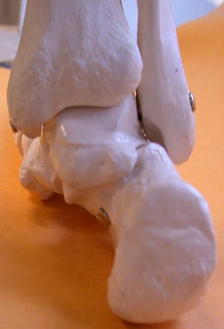
7 Cavovarus
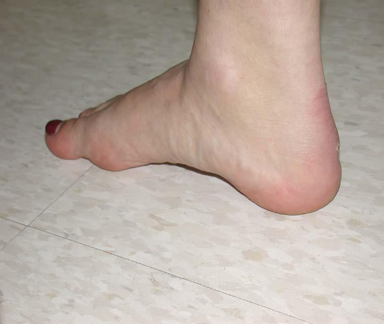
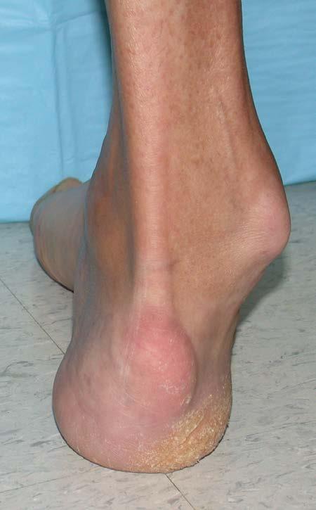
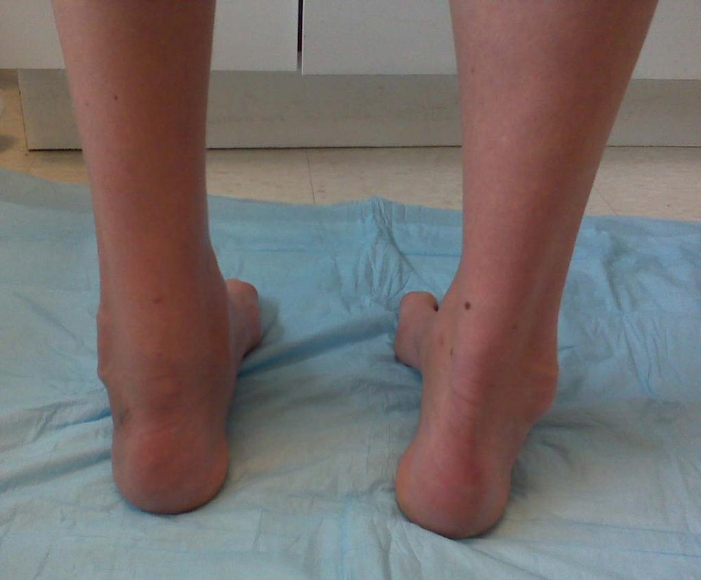
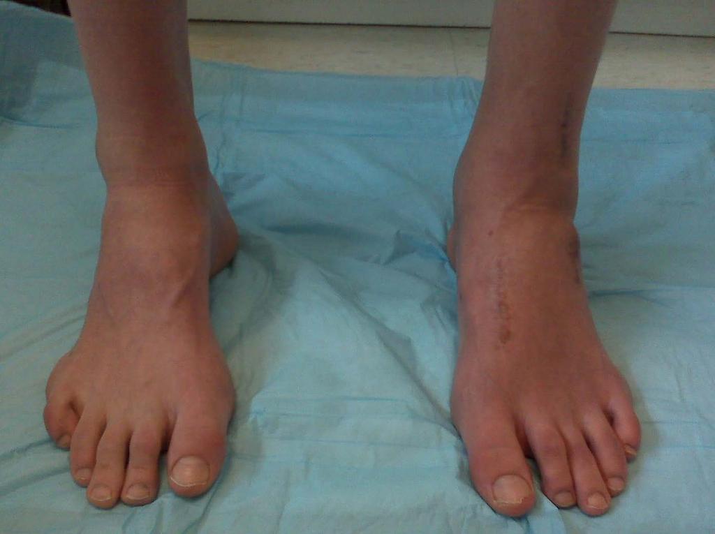
8 Physical Exam High arches Peek-a-boo heels from the front Varus heels from behind Coleman block test

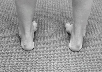
9 Images: Foot and Ankle International, March 2005;26:
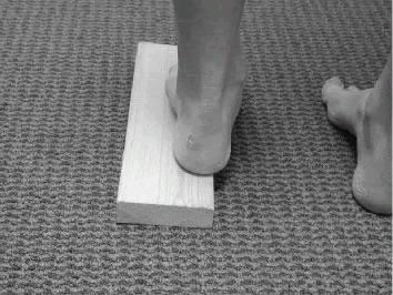
10 Coleman Block Test Varus Heel Stand on block with 1 st ray dropping over the edge Does this correct the hindfoot varus? If it does, then it is: Forefoot Driven Hindfoot Varus

11 Coleman Block
12 Forefoot-Driven Varus Plantarflexed 1 st Ray Supple hindfoot If you correct the first ray, then you correct the hindfoot varus, which improves foot mechanics With varus, there is no eversion to allow for shock absorption at heel strike which transfers stresses above.
13 Plantarflexed 1 st Metatarsal One leg of the tripod is too high High sesamoid pressures Foot tipping Instability
14 Problems Peroneal tendon tendonitis/subluxation/tears 5 th Metatarsal fractures Stress, Jones, Nonunions Sesamoiditis 1 st /5 th Metatarsalgia/Calluses Recurrent ankle sprains Ankle arthritis
15

16 Severe Cavovarus from Polio
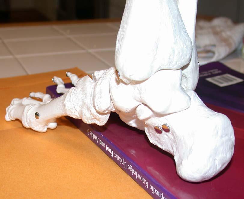
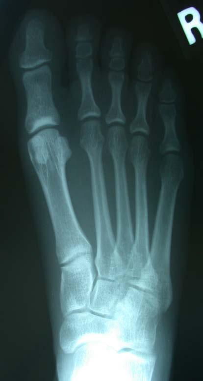
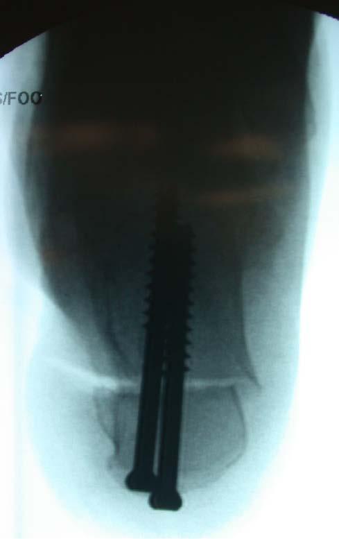
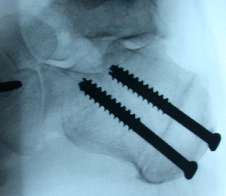
17 Cavovarus Radiographs High arch, plantarflexed 1 st ray Hindfoot supinated Open sinus tarsi Fibula is posterior Double talar dome High calcaneal pitch
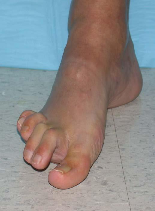
18 Severe Cavovarus

19 Severe Cavovarus

20 Varus Ankle Arthritis

21 Moderate Cavus

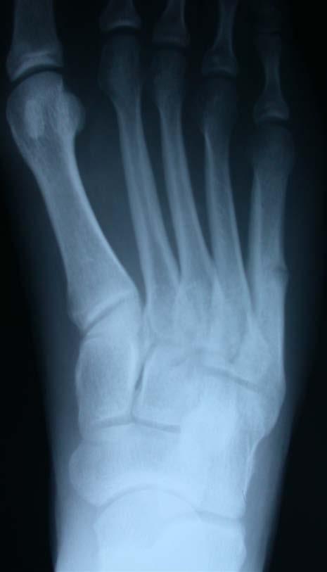
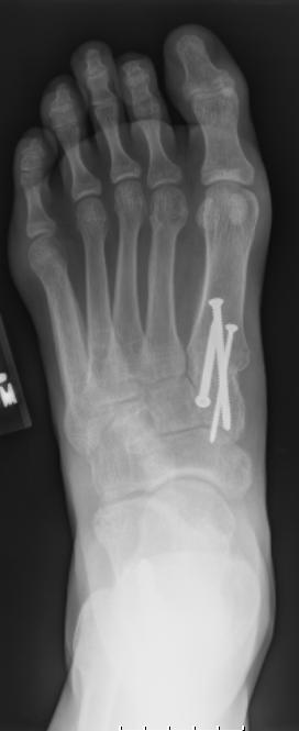
22 5 th Metatarsal Stress Fracture

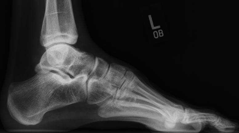
23 Subtle Cavus

24 Subtle Cavus

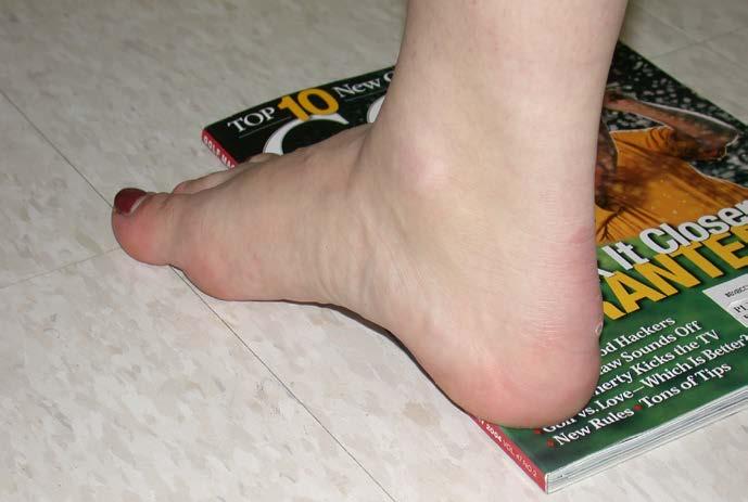
25 Sports Coleman Block


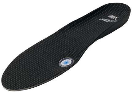
26 Coleman Block Orthotics Archrival by Donjoy
27 Etiology CNS Cerebral Palsy, Friedrich s Ataxia Spine Myelodysplasia, Syringomyelia, Polio, Tumors, Intrathecal Lipoma, Tethered Cord *(May have unilateral or progressive cavovarus)* Peripheral Nerves Hereditary Sensorimotor Neuropathy Muscular- Deep posterior compartment syndrome Idiopathic
28 Charcot-Marie-Tooth Most common hereditary motor-sensory neuropathy Results from abnormal myelination affecting peripheral nerves from distal to proximal 94% of patients have a foot deformity Pathology: Onion Bulb formation from segmental demyelination and remyelination PMP-22 (peripheral myelin protein) gene defect in 80% of most common CMT-1 (demyelinating) type which is autosomal dominant
29 CMT Pathophysiology Weakness and atrophy of foot intrinsics, tibialis anterior, and peroneus brevis; while the posterior muscles and peroneus longus retain their strength. As the muscle imbalances become longer standing, the more rigid the deformities become.
30 Deformities Equinus Tib Ant overpowered by Gastroc/Soleus Cavus Peroneus Longus overpowers Tib Ant Hindfoot Varus Posterior Tibial overpowers Brevis Claw Toes Loss of intrinsic function. Extensors recruited to help dorsiflexion, while flexors overpull
31 Extensor Recruitment
32 Achilles Contracture
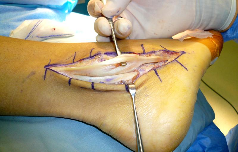
33 Soft Tissue Procedures for Charcot-Marie-Tooth Plantar Fascia release Achilles tendon lengthening for Equinus Extensor tendon lengthening for Hammering Toes EHL to 1 st Metatarsal Neck for Clawed Hallux Peroneus Longus to Brevis for Weak or Torn Brevis Posterior Tibial Tendon Transfer to Dorsum for Foot Drop Girdlestones for Flexible Hammertoes
34 Bony Procedures for CMT IP Fusions for Rigid Hammertoes Dorsiflexion MT Osteotomies for Plantarflexed Mets Dorsiflexing Tarsometatarsal Fusions for PF Mets Lateral closing wedge Calcaneal Osteotomies for Varus Triple Arthrodesis for Rigid/Arthritic Deformities
35
36
37

38
39
40 Cavus Foot Ask yourself, why does this patient have a cavus foot? Early recognition is important to prevent or treat lateral sided or ankle pathology Subtle cavus can be treated with orthotics Early stages can be treated with tendon transfers Later, more rigid cases require the addition of bony procedures
41 Summary A cavus foot can lead to lateral foot and ankle overload and injury. When evaluating a patient with ankle instability, ankle arthritis, 5 th metatarsal fracture/nonunion, sesamoiditis, and/or peroneal tendon dysfunction, assess the hindfoot/forefoot alignment. Define the origin of the hindfoot varus using the Coleman Block. Correct underlying deformity, not just the tip of the iceberg.
42 Image Sources Wülker N, Hurschler C. Cavus foot correction in adults by dorsal closing wedge osteotomy. Foot Ankle Int. April 2002;23: Manoli, A II, Graham, B. The subtle cavus foot, the underpronator, a review. Foot Ankle Int. March 2005;26:
SUBTLE CAVUS IN SPORTS INJURIES
 SUBTLE CAVUS IN SPORTS INJURIES MICHAEL P. CLARE, MD FLORIDA ORTHOPAEDIC INSTITUTE TAMPA, FL USA NON-NEUROMUSCULAR NORMAL VARIANT: 20-25% INCIDENCE LEDOUX, ET AL. FAI 24, 2003 FOREFOOT-DRIVEN / MORE SUBTLE
TENDON TRANSFER IN CAVUS FOOT
 TENDON TRANSFER IN CAVUS FOOT Cavovarus deformity is defined by fixed equinus of the forefoot on the hindfoot, resulting in a pathologic elevation of the longitudinal arch, with either a fixed or flexible
Rod Hammett Consultant Orthopaedic Surgeon Musgrove Park Hospital
 Rod Hammett Consultant Orthopaedic Surgeon Musgrove Park Hospital What patients does the surgeon want to see? What patients does the neurologist want the surgeon to see?? What does the surgeon need
Physical Examination of the Foot & Ankle
 Inspection Standing, feet straight forward facing toward examiner Swelling Deformity Flatfoot (pes planus and hindfoot valgus) High arch (pes cavus and hindfoot varus) Peek-a-boo heel Varus Too many toes
Columbia/NYOH FOOT and ANKLE ROTATION-SPECIFIC OBJECTIVES
 Updated 2/8/10 Columbia/NYOH FOOT and ANKLE ROTATION-SPECIFIC OBJECTIVES INTERPERSONAL AND COMMUNICATION SKILLS Resident will at all times demonstrate behavior that is beyond reproach. Residents must be
Pediatric Orthopaedic Surgery and the HMSNs
 Reviewed and accepted by the 2011-2012 Neuromuscular Committee of the American Association of Neuromuscular & Electrodiagnostic Medicine Certified for CME credit 10/2011 05/2020 Reviewed 10/2017 by the
Scar Engorged veins. Size of the foot [In clubfoot, small foot]
![Scar Engorged veins. Size of the foot [In clubfoot, small foot]](/thumbs/78/77722241.jpg "Scar Engorged veins. Size of the foot [In clubfoot, small foot]") 6. FOOT HISTORY Pain: Walking, Running Foot wear problem Swelling; tingly feeling Deformity Stiffness Disability: At work; recreation; night; walk; ADL, Sports Previous Rx Comorbidities Smoke, Sugar, Steroid
6. FOOT HISTORY Pain: Walking, Running Foot wear problem Swelling; tingly feeling Deformity Stiffness Disability: At work; recreation; night; walk; ADL, Sports Previous Rx Comorbidities Smoke, Sugar, Steroid
Section Three: The Leg, Ankle, and Foot Lecture: Review of Clinical Anatomy, Patterns of Dysfunction and Injury, and
 Section Three: The Leg, Ankle, and Foot Lecture: Review of Clinical Anatomy, Patterns of Dysfunction and Injury, and Treatment Implications for the Leg, Ankle, and Foot Levels I and II Demonstration and
Index. Note: Page numbers of article titles are in boldface type.
 Index Note: Page numbers of article titles are in boldface type. A Abductor hallucis (ABH) tendon transfer, for hallux varus, technique for, 458 462 bone tunnels and course diagram in, 458, 460 capsulotomy
Foot & Ankle Disorders
 Foot & Ankle Disorders Hillingdon PGMC 6-7-2013 Htwe Zaw FRCS (Tr&Orth) Consultant Foot & Ankle and Trauma Surgeon Hillingdon Hospitals NHS Foundation Trust Overview Anatomy: hindfoot-midfoot coupling
Copyright 2004, Yoshiyuki Shiratori. All right reserved.
 Ankle and Leg Evaluation 1. History Chief Complaint: A. What happened? B. Is it a sharp or dull pain? C. How long have you had the pain? D. Can you pinpoint the pain? E. Do you have any numbness or tingling?
Therapeutic Foot Care Certificate Program Part I: Online Home Study Program
 Therapeutic Foot Care Certificate Program Part I: Online Home Study Program 1 Anatomy And Terminology Of The Lower Extremity Joan E. Edelstein, MA, PT, FISPO Associate Professor of Clinical Physical Therapy
The Leg. Prof. Oluwadiya KS
 The Leg Prof. Oluwadiya KS www.oluwadiya.sitesled.com Compartments of the leg 4 Four Compartments: 1. Anterior compartment Deep fibular nerve Dorsiflexes the foot and toes 2. Lateral Compartment Superficial
The Lower Limb VI: The Leg. Anatomy RHS 241 Lecture 6 Dr. Einas Al-Eisa
 The Lower Limb VI: The Leg Anatomy RHS 241 Lecture 6 Dr. Einas Al-Eisa Muscles of the leg Posterior compartment (superficial & deep): primary plantar flexors of the foot flexors of the toes Anterior compartment:
The Valgus Foot in Cerebral Palsy Equinovalgus not Plano-Valgus. Alfred D. Grant, M.D. David Feldman, M.D.
 The Valgus Foot in Cerebral Palsy Equinovalgus not Plano-Valgus Alfred D. Grant, M.D. David Feldman, M.D. Norman Otsuka, MD M.D. THE PURPOSE OF THIS PRESENTATION IS TO STATE CLEARLY THAT THE VALGUS FOOT
17/10/2017. Foot and Ankle
 17/10/2017 Alicia M. Yochum RN, DC, DACBR, RMSK Foot and Ankle Plantar Fasciitis Hallux Valgus Deformity Achilles Tendinosis Posterior Tibialis Tendon tendinopathy Stress Fracture Ligamentous tearing Turf
FACTS 1. Most need only Gastro aponeurotic release [in positive Silverskiold test]
![FACTS 1. Most need only Gastro aponeurotic release [in positive Silverskiold test]](/thumbs/83/88335212.jpg "FACTS 1. Most need only Gastro aponeurotic release [in positive Silverskiold test]") FOOT IN CEREBRAL PALSY GAIT IN CEREBRAL PALSY I True Equinus II Jump gait III Apparent Equinus IV Crouch gait Group I True Equinus Extended hip and knee Equinus at ankle II Jump Gait [commonest] Equinus
FOOT IN CEREBRAL PALSY GAIT IN CEREBRAL PALSY I True Equinus II Jump gait III Apparent Equinus IV Crouch gait Group I True Equinus Extended hip and knee Equinus at ankle II Jump Gait [commonest] Equinus
What Happens to the Paediatric Flat Foot? Peter J Briggs Freeman Hospital Newcastle upon Tyne
 What Happens to the Paediatric Flat Foot? Peter J Briggs Freeman Hospital Newcastle upon Tyne We don t know!! Population Studies 2300 children aged 4-13 years Shoe wearers Flat foot 8.6% Non-shoe wearers
بسم هللا الرحمن الرحيم
 بسم هللا الرحمن الرحيم Laboratory RHS 221 Manual Muscle Testing Theory 1 hour practical 2 hours Dr. Ali Aldali, MS, PT Department of Physical Therapy King Saud University Talocrural and Subtalar Joint
CHRONIC FOOT PROBLEMS FOOT and ANKLE BASICS
 CHRONIC FOOT PROBLEMS FOOT and ANKLE BASICS ABC s of Comprehensive Musculoskeletal Care December 1 st, 2007 Stephen Pinney MD Chief, UCSF Foot and Ankle Service Chronic problems typically occur gradually
Deformity and Pain in Foot of Neuromuscular Disease
 Deformity and Pain in Foot of Neuromuscular Disease CHA 의과학대학재활의학과김민영 (2012.11.10 제 4 회대한발의학회 ) Neuromuscular Diseases & Foot Deformity : almost Pain : infrequent Non-ambulatory, Ambulatory Neuromuscular
Index. Clin Podiatr Med Surg 22 (2005) Note: Page numbers of article titles are in boldface type.
 Note: Page numbers of article titles are in boldface type.") Clin Podiatr Med Surg 22 (2005) 309 314 Index Note: Page numbers of article titles are in boldface type. A Abductor digiti minimi muscle, myectomy of, for tailor s bunionette, 243 Achilles tendon, lengthening
Clin Podiatr Med Surg 22 (2005) 309 314 Index Note: Page numbers of article titles are in boldface type. A Abductor digiti minimi muscle, myectomy of, for tailor s bunionette, 243 Achilles tendon, lengthening
Other Congenital and Developmental Diseases of the Foot. Department of Orthopedic Surgery St. Vincent s s Hospital, The Catholic University
 Other Congenital and Developmental Diseases of the Foot Department of Orthopedic Surgery St. Vincent s s Hospital, The Catholic University Contents Metatarsus Adductus Skewfoot Hallux Valgus Hallux Valgus
Ankle Tendons in Athletes. Laura W. Bancroft, M.D.
 Ankle Tendons in Athletes Laura W. Bancroft, M.D. Outline Protocols Normal Anatomy Tendinopathy, partial and complete tears Posterior tibial, Flexor Hallucis Longus, Achilles, Peroneal and Anterior Tibial
2017 SAFSA CONGRESS PROGRAMME
 2017 SAFSA CONGRESS PROGRAMME THURSDAY, MAY 25 07h45 07h55: WELCOME & INTRODUCTIONS Forefoot I: Hallux Valgus and Lesser Toes (08h00-10h00 Lectures) 08h00 08h30: Surgical Management of Hallux Valgus Rippstein,
Leg. Dr. Heba Kalbouneh Associate Professor of Anatomy and Histology
 Leg Dr. Heba Kalbouneh Associate Professor of Anatomy and Histology Skin of the Leg Cutaneous Nerves Medially: The saphenous nerve, a branch of the femoral nerve supplies the skin on the medial surface
Anatomy of Foot and Ankle
 Anatomy of Foot and Ankle Surface anatomy of the ankle & foot Surface anatomy of the ankle & foot Medial orientation point medial malleous sustentaculum tali tuberosity of navicular TA muscle TP muscle
Surgical Classification of the Cavus Foot Deformity
 CHAPTER 43 Surgical Classification of the Cavus Foot Deformity Jobn A. Rucb, D.PM. Historically, surgical repair of the cal,us foot deformity has been based on many different complicated and confusing
Biomechanical Explanations for Selective Sport Injuries of the Lower Extremity
 Biomechanical Explanations for Selective Sport Injuries of the Lower Extremity American Osteopathic Academy of Sports Medicine Presentation April 23, 2015 Understanding Normalcy What is Normal? Rearfoot/heel
DESC de Chirurgie Pédiatrique Session de Septembre PARIS. Pied creux. P Wicart, R Seringe
 DESC de Chirurgie Pédiatrique Session de Septembre 2009 - PARIS Pied creux P Wicart, R Seringe NORMAL FOOT N = 120 à 125 Méary angles = 0 NORMAL FOOT NORMAL FOOT Méary angles = 0 Tibio-talar angle : 100
PROBLEMS AND ORTHOTIC SOLUTIONS. Problem/Issue Underlying treatment goal Solution Pes Cavus foot
 PROBLEMS AND ORTHOTIC SOLUTIONS Problem/Issue Underlying treatment goal Solution Pes Cavus foot Usually also a supinated foot Rigid high arched foot with poor shock absorption and cushioning. Often roll
ANKLE PLANTAR FLEXION
 ANKLE PLANTAR FLEXION Evaluation and Measurements By Isabelle Devreux 1 Ankle Plantar Flexion: Gastrocnemius and Soleus ROM: 0 to 40-45 A. Soleus: Origin: Posterior of head of fibula and proximal1/3 of
Title. Issue Date Right.
 NOSITE: Nagasaki University's c Title uthor(s) Dynamic supination and hindfoot var tendons of both peroneus longus and Matsubayashi, Shohei; Tsujimoto, i Citation cta medica Nagasakiensia, 61(2), p Issue
Financial Disclosure. Turf Toe
 Seth O Brien, CP, LP Financial Disclosure Mr. Seth O'Brien has no relevant financial relationships with commercial interests to disclose. Turf Toe Common in athletes playing on firm, artificial turf Forceful
Sports Injuries of the Foot and Ankle. Mark McEleney, MD University of Iowa College of Medicine Refresher Course for the Family Physician 4/4/2018
 Sports Injuries of the Foot and Ankle Mark McEleney, MD University of Iowa College of Medicine Refresher Course for the Family Physician 4/4/2018 I. Objectives A. By the end of the lecture attendees will
Rippstein, Trnka, Saragas, Narramore
 THURS 25th MAY 07:45 07:55 Welcome and Introductions Paulo Ferrao Lecture 1: 08:00 10:20 Forefoot I: Hallux Valgus and Lesser Toes Mark Easley 30 mins 08:00 08:30 Surgical Management of Hallux Valgus Saragas,
CLAD Error Key. Error Levels: Definite, Possible. Error Procedure Scope. Validation Scope. Location Scope. Violation/Information Text
 CLAD Key s: Definite, Possible Procedure 1 2 3 4 5 6 7 8 9 10 Two or more category 1 procedures Digit Definite 1.6 plus one or more of the following: 2.1.3, 2.1.7, 2.2.2, 2.2.6, and 2.3.4 Side Definite
Triple Arthrodesis of Foot for Correction of Lower Extremity Deformities
 Triple Arthrodesis of Foot for Correction of Lower Extremity Deformities 振興醫療財團法人振興醫院骨科部 * 熊永萬 A triple arthrodesis consists of surgical fusion of the talocalcaneal (TC), talonavicular (TN), and calcaneocuboid
Appendix H: Description of Foot Deformities
 Appendix H: Description of Foot Deformities The following table provides the description for several foot deformities: hammer toe, claw toe, hallux deformity, pes planus, pes cavus and charcot arthropathy.
Clarification of Terms
 Clarification of Terms The plantar aspect of the foot refers to the role or its bottom The dorsal aspect refers to the top or its superior portion The ankle and foot perform three main functions: 1. shock
*Rippstein, Trnka, Saragas, Hoffman
 THURS 25th MAY 07:00 07:10 Welcome and Introductions Paulo Ferrao Lecture 1: 07:10 09:45 Forefoot I: Hallux Valgus and Lesser Toes Mark Easley 40 mins 07:10 07:50 Surgical Management of Hallux Valgus 30
Dr Nabil khouri MD. MSc. Ph.D
 Dr Nabil khouri MD. MSc. Ph.D Foot Anatomy The foot consists of 26 bones: 14 phalangeal, 5 metatarsal, and 7 tarsal. Toes are used to balance the body. Metatarsal Bones gives elasticity to the foot in
Index. Clin Sports Med 23 (2004) Note: Page numbers of article titles are in boldface type.
 Note: Page numbers of article titles are in boldface type.") Clin Sports Med 23 (2004) 169 173 Index Note: Page numbers of article titles are in boldface type. A Achilles enthesopathy, calcaneal spur with, 133 clinical presentation of, 135 136 definition of, 131
Clin Sports Med 23 (2004) 169 173 Index Note: Page numbers of article titles are in boldface type. A Achilles enthesopathy, calcaneal spur with, 133 clinical presentation of, 135 136 definition of, 131
Index. Note: Page numbers of article titles are in bold face type.
 Index Note: Page numbers of article titles are in bold face type. A Achilles tendon, Zadek osteotomy effects on, 430 Adult acquired flatfoot disorder, 387 403 calcaneal Z osteotomy for, 397 399 historical
Lower Extremity Orthopedic Surgery in Cerebral Palsy
 Lower Extremity Orthopedic Surgery in Cerebral Palsy Hank Chambers, MD San Diego Children s Hospital San Diego, California Indications Fixed contracture Joint dislocations Shoe wear problems Pain Perineal
Review relevant anatomy of the foot and ankle. Learn the approach to examining the foot and ankle
 Objectives Review relevant anatomy of the foot and ankle Learn the approach to examining the foot and ankle Learn the basics of diagnosis and treatment of ankle sprains Overview of other common causes
BUCKS MSK: FOOT AND ANKLE PATHWAY GP MANAGEMENT. Hallux Valgus. Assessment: Early Management. (must be attempted prior to any referral to imsk):
:") Hallux Valgus Common condition: affecting around 28% of the adult population. Prevalence increases with age and in females. Observation: Lateral deviation of the great toe. May cause secondary irritation
Hallux Valgus Common condition: affecting around 28% of the adult population. Prevalence increases with age and in females. Observation: Lateral deviation of the great toe. May cause secondary irritation
Foot and Ankle Physical Exam. The Big Picture: - Gait analysis - Exam standing - Exam sitting - Provocative maneuvers
 Foot and Ankle Physical Exam The Big Picture: - Gait analysis - Exam standing - Exam sitting - Provocative maneuvers 1. Gait analysis Physical Exam 2. Examination Standing Alignment Swelling 3. Examination
Managing Tibialis Posterior Tendon Injuries
 Managing Tibialis Posterior Tendon Injuries by Thomas C. Michaud, DC Published April 1, 2015 by Dynamic Chiropractic Magazine Tibialis posterior is the deepest, strongest, and most central muscle of the
Main Menu. Ankle and Foot Joints click here. The Power is in Your Hands
 1 The Ankle and Foot Joints click here Main Menu Copyright HandsOn Therapy Schools 2009 K.8 http://www.handsonlineeducation.com/classes/k8/k8entry.htm[3/27/18, 1:40:03 PM] Ankle and Foot Joint 26 bones
Are you suffering from heel pain? We can help you!
 Are you suffering from heel pain? We can help you! STOP THE PAIN! Heel pain can be effectively combated with the proven Body Armor Night Splint. Heel spurs and heel pain Why? Heel pain is among the most
BLUE SKY SCHOOL OF PROFESSIONAL MASSAGE AND THERAPEUTIC BODYWORK Musculoskeletal Anatomy & Kinesiology KNEE & ANKLE MUSCLES
 BLUE SKY SCHOOL OF PROFESSIONAL MASSAGE AND THERAPEUTIC BODYWORK Musculoskeletal Anatomy & Kinesiology KNEE & ANKLE MUSCLES MSAK201-I Session 3 1) REVIEW a) THIGH, LEG, ANKLE & FOOT i) Tibia Medial Malleolus
ANKLE JOINT ANATOMY 3. TALRSALS = (FOOT BONES) Fibula. Frances Daly MSc 1 CALCANEUS 2. TALUS 3. NAVICULAR 4. CUBOID 5.
 Fibula. Frances Daly MSc 1 CALCANEUS 2. TALUS 3. NAVICULAR 4. CUBOID 5.") ANKLE JOINT ANATOMY The ankle joint is a synovial joint of the hinge type. The joint is formed by the distal end of the tibia and medial malleolus, the fibula and lateral malleolus and talus bone. It is
ANKLE JOINT ANATOMY The ankle joint is a synovial joint of the hinge type. The joint is formed by the distal end of the tibia and medial malleolus, the fibula and lateral malleolus and talus bone. It is
Peggers Super Summaries: Foot Injuries
 Lisfranc Injury ANATOMY Roman arch with recessed 2 nd MT base AP medial side of intermediate cuneiform to 2 nd MT base Oblique medial side of lateral cuneiform with 3 rd MT base and 4 th with medial boarder
Outline. Ankle/Foot Anatomy Ankle Sprains Ottawa Ankle Rules DDx: The Sprain That Wasn t
 Ankle Injuries Outline Ankle/Foot Anatomy Ankle Sprains Ottawa Ankle Rules DDx: The Sprain That Wasn t Anatomy: Ankle Mortise Bony Anatomy Lateral Ligament Complex Medial Ligament Complex Ankle Sprains
Results of Calcaneal Osteotomy & Flexor Digitorum Longus transfer in Stage II Acquired Flatfoot Deformity
 Results of Calcaneal Osteotomy & Flexor Digitorum Longus transfer in Stage II Acquired Flatfoot Deformity Mr Amit Chauhan Mr Prasad Karpe Ms Maire-claire Killen Mr Rajiv Limaye University Hospital of North
SUB-TALAR AND TRIPLE ARTHRODESIS
 SUB-TALAR AND TRIPLE ARTHRODESIS J de Halleux With the members of Education Committee INDICATIONS ARTHRITIS OF THE SUB-TALAR AND/OR MID-TARSAL JOINTS RIGID VARUS OR VALGUS DEFORMITY OF THE HIND-FOOT COALITIONS
5 COMMON INJURIES IN THE FOOT & ANKLE
 5 COMMON INJURIES IN THE FOOT & ANKLE MICHAEL P. CLARE, MD FLORIDA ORTHOPAEDIC INSTITUTE TAMPA, FL USA MECHANISM OF INJURY HOW DID IT HAPPEN? HIGH ENERGY VS LOW ENERGY DIRECTION OF FORCES INVOLVED LIVING
Case 57 What is the diagnosis? Insidious onset forefoot pain in a 50 year old female for last 3 months.
 Case 57 What is the diagnosis? Insidious onset forefoot pain in a 50 year old female for last 3 months. Diagnosis: II MTP instability Demographics of MT instability Lesser MTP joint instability occurs
PREVALENCE AND ORTHOPEDIC MANAGEMENT OF FOOT AND ANKLE DEFORMITIES IN CHARCOT MARIE TOOTH DISEASE
 PREVALENCE AND ORTHOPEDIC MANAGEMENT OF FOOT AND ANKLE DEFORMITIES IN CHARCOT MARIE TOOTH DISEASE MATILDE LAUR A, MD, PhD, 1 DISHAN SINGH, MBChB, FRCS(Orth), 2 GITA RAMDHARRY, PhD, 3 JASPER MORROW, MD,
This presentation is the intellectual property of the author. Contact them for permission to reprint and/or distribute.
 Introduction Compartment Syndromes of the Leg Related to Athletic Activity Mark M. Casillas, M.D. Consequences of a misdiagnosis persistence of a performance limitation loss of function/compartment loss
Index. Note: Page numbers of article titles are in boldface type.
 Index Note: Page numbers of article titles are in boldface type. A Achilles tendon injury of, pathophysiology of, 10 peritendinitis of, 119 120 rupture of, 32 35, 117 135 anatomy of, 117 118 chronic, 126
Dropfoot - Video Gait Analysis - Craig A. Camasta, DPM, FACFAS Atlanta, Georgia, USA
 Equinus, Pes Cavus and Dropfoot - Video Gait Analysis - Craig A. Camasta, DPM, FACFAS Atlanta, Georgia, USA Equinus = Toe Walker Soft Tissue Static fixed contracture Dynamic spastic, hypertonic Bone Procurvatum,,
Forefoot Procedures to Heal and Prevent Recurrence. Watermark. Diabetic Foot Update 2015 San Antonio, Texas
 Forefoot Procedures to Heal and Prevent Recurrence Diabetic Foot Update 2015 San Antonio, Texas J. Randolph Clements, DPM Assistant Professor of Orthopaedics Virginia Tech- Carilion School of Medicine
The Foot. Dr. Wegdan Moh.Mustafa Medicine Faculty Assistant Professor Mob:
 The Foot Dr. Wegdan Moh.Mustafa Medicine Faculty Assistant Professor Mob: 0127155717 The skeleton of the foot Cutaneous innervations Sole of foot layers of muscles First layer -Abductor hallucis -Flexor
(vii) Clinical examination of the foot and ankle
 Clinical examination of the foot and ankle") (vii) Clinical examination of the foot and ankle Howard Davies Chris Blundell Abstract Examination of the foot and ankle can appear to be highly complicated, but if broken down into the component parts
(vii) Clinical examination of the foot and ankle Howard Davies Chris Blundell Abstract Examination of the foot and ankle can appear to be highly complicated, but if broken down into the component parts
The Cavovarus Foot and It's Association with Fractures of the Fifth Metatarsal
 The Cavovarus Foot and It's Association with Fractures of the Fifth Metatarsal Daniel Fuchs, Aamir Bhimani, James Brodsky, Christian Royer, Veerabhadra Reddy, Jacob Zide, Yahya Daoud, Justin Kane Disclosure
First & second layers of muscles of the sole
 The FOOT First & second layers of muscles of the sole introduction The muscles acting on the foot can be divided into two distinct groups; extrinsic and intrinsic muscles. The extrinsic muscles arise from
SUBTALAR ARTHROEREISIS IN THE OLDER PATIENT
 C H A P T E R 1 7 SUBTALAR ARTHROEREISIS IN THE OLDER PATIENT William D. Fishco, DPM, MS INTRODUCTION Arthroereisis is a surgical procedure designed to limit the motion of a joint. Subtalar joint arthroereisis
Foot. Dr. Heba Kalbouneh Associate Professor of Anatomy and Histology
 Foot Dr. Heba Kalbouneh Associate Professor of Anatomy and Histology Dorsum of the Foot Sole of the Foot Plantar aponeurosis It is a triangular thickening of deep fascia in the sole of the foot Attachments:
Changes in Dynamic Pedobarography after Extensive Plantarmedial Release for Paralytic Pes Cavovarus
 Original Article http://dx.doi.org/10.3349/ymj.2014.55.3.766 pissn: 0513-5796, eissn: 1976-2437 Yonsei Med J 55(3):766-772, 2014 Changes in Dynamic Pedobarography after Extensive Plantarmedial Release
PAINFUL SESAMOID OF THE GREAT TOE Dr Vasu Pai ANATOMICAL CONSIDERATION. At the big toe MTP joint: Tibial sesamoid (medial) & fibular (lateral)
 & fibular (lateral)") PAINFUL SESAMOID OF THE GREAT TOE Dr Vasu Pai ANATOMICAL CONSIDERATION At the big toe MTP joint: Tibial sesamoid (medial) & fibular (lateral) They are contained within the tendons of Flexor Hallucis Brevis
PAINFUL SESAMOID OF THE GREAT TOE Dr Vasu Pai ANATOMICAL CONSIDERATION At the big toe MTP joint: Tibial sesamoid (medial) & fibular (lateral) They are contained within the tendons of Flexor Hallucis Brevis
Surgical Correction of Lower Extremity Deformities by Triple Arthrodesis
 Surgical Correction of Lower Extremity Deformities by Triple Arthrodesis 足踝畸形矯治 振興醫療財團法人振興醫院 骨科部 熊永萬醫師 Introduction: The primary goals of a triple arthrodesis are to relieve pain from arthritic, deformed,
The effect on radiographic parameters of Dwyer s osteotomy and 1 st metatarsal osteotomy for pes cavo-varus correction
 The effect on radiographic parameters of Dwyer s osteotomy and 1 st metatarsal osteotomy for pes cavo-varus correction Department of Orthopedic Surgery, Inje University, Ilsan Paik Hospital, South Korea
6/5/2018. Forefoot Disorders. Highgate Private Hospital (Royal Free London NHS Foundation Trust (Barnet & Chase Farm Hospitals) Hallux Rigidus
 Hallux Rigidus") Forefoot Disorders Mr Pinak Ray (MS, MCh(Orth), FRCS, FRCS(Tr&Orth)) Highgate Private Hospital (Royal Free London NHS Foundation Trust (Barnet & Chase Farm Hospitals) E: ray.secretary@uk-conslutants Our
Forefoot Disorders Mr Pinak Ray (MS, MCh(Orth), FRCS, FRCS(Tr&Orth)) Highgate Private Hospital (Royal Free London NHS Foundation Trust (Barnet & Chase Farm Hospitals) E: ray.secretary@uk-conslutants Our
Metatarsus adductus, Skew foot, Club foot 성균관대학교삼성창원병원 장현정
 Metatarsus adductus, Skew foot, Club foot 성균관대학교삼성창원병원 장현정 Metatarsus adductus Epidemiology and Etiology 0.1-12% with higher number for multiple birth Deformation and compression from intrauterine crowding
Understanding Leg Anatomy and Function THE UPPER LEG
 Understanding Leg Anatomy and Function THE UPPER LEG The long thigh bone is the femur. It connects to the pelvis to form the hip joint and then extends down to meet the tibia (shin bone) at the knee joint.
MUSCLES OF THE LOWER LIMBS
 MUSCLES OF THE LOWER LIMBS Naming, location and general function Dr. Nabil khouri ROLES THAT SHOULD NOT BE FORGOTTEN Most anterior compartment muscles of the hip and thigh Flexor of the femur at the hip
A Patient s Guide to Adult-Acquired Flatfoot Deformity
 A Patient s Guide to Adult-Acquired Flatfoot Deformity Glendale Adventist Medical Center 1509 Wilson Terrace Glendale, CA 91206 Phone: (818) 409-8000 DISCLAIMER: The information in this booklet is compiled
~, /' ~::'~ EXTENSOR HALLUCIS LONGUS. Leg-anterolateral :.:~ / ~\,
 TIBIALIS ANTERIOR Lateral condyle of tibia, upper half of lateral surface of tibia, interosseous membrane Medial side and plantar surface of medial cuneiform bone, and base of first metatarsal bone Dorsiflexes
Polio - A Model for Overuse and Aging. Acute Poliomyelitis. Acute Infection of Anterior Horn Motor Cells: Acute Polio Infection
 Polio - A Model for Overuse and Aging Mary Ann Keenan, M.D. Chief, Neuro-Orthopaedics Program Professor, Orthopaedic Surgery University of Pennsylvania Philadelphia, PA, USA Acute Poliomyelitis Acute viral
Year 2004 Paper one: Questions supplied by Megan
 QUESTION 47 A 58yo man is noted to have a right foot drop three days following a right total hip replacement. On examination there is weakness of right ankle dorsiflexion and toe extension (grade 4/5).
The University Of Jordan Faculty Of Medicine FOOT. Dr.Ahmed Salman Assistant Prof. of Anatomy. The University Of Jordan
 The University Of Jordan Faculty Of Medicine FOOT Dr.Ahmed Salman Assistant Prof. of Anatomy. The University Of Jordan Tarsal Tunnel Syndrome Due to compression of Tibial nerve as it travels through the
The plantar aponeurosis
 Anatomy of the foot The plantar aponeurosis Is a triangular thickening of the deep fascia Its apex is attached to the medial and lateral tubercles of the calcaneum. The base of the aponeurosis divides
Muscles of the Hip 1. Tensor Fasciae Latae O: iliac crest I: lateral femoral condyle Action: abducts the thigh Nerve: gluteal nerve
 Muscles of the Hip 1. Tensor Fasciae Latae O: iliac crest I: lateral femoral condyle Action: abducts the thigh Nerve: gluteal nerve 2. Gluteus Maximus O: ilium I: femur Action: abduct the thigh Nerve:
Musculoskeletal Ultrasound Technical Guidelines. VI. Ankle
 European Society of MusculoSkeletal Radiology Musculoskeletal Ultrasound Technical Guidelines VI. Ankle Ian Beggs, UK Stefano Bianchi, Switzerland Angel Bueno, Spain Michel Cohen, France Michel Court-Payen,
Dorsal surface-the upper area or top of the foot. Terminology
 It is important to learn the terminology as it relates to feet to properly communicate with referring physicians when necessary and to identify the relationship between the anatomical structure of the
We carried out a cross-sectional study in 51
 Function after correction of a clawed great toe by a modified Robert Jones transfer S. J. Breusch, W. Wenz, L. Döderlein From the Orthopaedic University Clinic, Heidelberg, Germany We carried out a cross-sectional
«Foot & Ankle Surgery» 04. Sept THE PAINFUL FLATFOOT. Norman Espinosa, MD
 THE PAINFUL FLATFOOT Norman Espinosa, MD Department of Orthopaedics University of Zurich Balgrist Switzerland www.balgrist.ch WHAT TO DO? INTRINSIC > EXTRINSIC ETIOLOGIES Repetitive microtrauma combined
Biokinesiology of the Ankle Complex
 Rehabilitation Considerations Following Ankle Fracture: Impact on Gait & Closed Kinetic Chain Function Disclosures David Nolan, PT, DPT, MS, OCS, SCS, CSCS I have no actual or potential conflict of interest
Evidence-Based Examination of the Foot Presented by Alexis Wright, PT, PhD, DPT, FAAOMPT Practice Sessions/Skill Check-offs
 Evidence-Based Examination of the Foot Presented by Alexis Wright, PT, PhD, DPT, FAAOMPT Practice Sessions/Skill Check-offs Module Five: Movement Assessment of the Foot/Ankle (1 hour CEU Time) Skilled
CLINICAL COMMENTARY Deformity or dysfunction? Osteopathic manipulation of the idiopathic cavus foot: A clinical suggestion.
 NAJSPT CLINICAL COMMENTARY Deformity or dysfunction? Osteopathic manipulation of the idiopathic cavus foot: A clinical suggestion. Christopher Kevin Wong, PT, PhD, OCS a Adi Gidali, PT, DPT b Valerie Harris,
Posterior Tibialis Tendon Dysfunction & Repair
 1 Posterior Tibialis Tendon Dysfunction & Repair Surgical Indications and Considerations Anatomical Considerations: The posterior tibialis muscle arises from the interosseous membrane and the adjacent
Essential Insights On Tendon Transfers For Digital Dysfunction
 Essential Insights On Tendon Transfers For Digital Dysfunction VOLUME: 23 PUBLICATION DATE: Apr 01 2010 Issue Number: 4 April 2010 Author(s): Lawrence DiDomenico, DPM, FACFAS While tendon transfers have
Classifications in Brief: Johnson and Strom Classification of Adult-acquired Flatfoot Deformity
 Clin Orthop Relat Res DOI 10.1007/s11999-015-4581-6 Clinical Orthopaedics and Related Research A Publication of The Association of Bone and Joint Surgeons IN BRIEF Classifications in Brief: Johnson and
University of South Florida
 University of South Florida Foot & Ankle Orthopaedics PGY 4 Competency Based Goals & Objectives Competency 1- Patient Care: Provide patient care that is compassionate, appropriate and effective for the
1. A worker falls from a height and lands on his feet. Radiographs reveal a fracture of the sustentaculum tali. The muscle passing immediately
 1. A worker falls from a height and lands on his feet. Radiographs reveal a fracture of the sustentaculum tali. The muscle passing immediately beneath it that would be adversely affected is the: fibularis
Other Congenital and Developmental Diseases of the Foot
 Other Congenital and Developmental Diseases of the Foot Han-Yong Lee, M.D. Department of Orthopedic Surgery St. Vincent s Hospital, The Catholic University Contents Introduction Foot Deformities Metatarsus
Key Points for Success:
 ANKLE & FOOT 1 2 All of the stretches described in this chapter are detailed to stretch the right side. Key Points for Success: Keep your movements slow and precise. Breathe in before you move and breathe
Commonly Missed Foot and Ankle Conditions. David Miller, DPM AMG Podiatry
 Commonly Missed Foot and Ankle Conditions David Miller, DPM AMG Podiatry Lisfranc Injuries Wide spectrum of injuries High energy Subtle subluxation which could be easily missed injuries Men are 2-4x s