MR Imaging for Glenohumeral Instability & Bone Loss
|
|
|
- Shauna Melton
- 5 years ago
- Views:
Transcription

1 MR Imaging for Glenohumeral Instability & Bone Loss Russell C. Fritz, M.D. San Francisco, CA I have no conflicts of interest to disclose Russell C. Fritz, M.D. MR imaging is a powerful diagnostic tool in orthopedics and sports medicine 1
2 MR imaging is useful to establish an anatomic diagnosis Visualize anatomy as well as verify and characterize pathology in all three planes We use the same basic method to evaluate for internal derangement of joints throughout the body 2
3 Standard Routine Proton density FSE 3000/35 Fat-sat T2-weighted FSE 4000/50 High resolution axial, sagittal, coronal Standard Routine Proton density FSE 3000/35 Fat-sat T2-weighted FSE 4000/50 High resolution axial, sagittal, coronal Imaging of the shoulder: Do I need contrast?

4 Imaging of the shoulder: Do I need contrast? No! MR Imaging of the Shoulder early spin echo imaging MR arthrography with T1 fatsat fast spin-echo sequences, ABER FSE high resolution imaging FSE high resolution imaging!!! A C 4


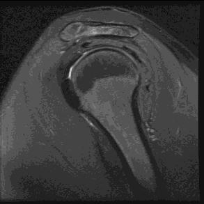
5 Coronal section normal normal 5


6 normal normal normal 6


7 normal normal normal 7


8 normal normal normal 8


9 normal Shoulder instability a situation in which symptoms result from excessive translation of the humeral head on the glenoid fossa Frederick A. Matsen III, M.D Anterior dislocation 9

10 Anterior dislocation 1 week ago Anterior dislocation 1 week ago Anterior dislocation 1 week ago 10


11 Anterior dislocation 1 week ago Anterior dislocation 1 week ago Anterior dislocation 1 week ago 11


12 Anterior dislocation 1 week ago Anterior dislocation 1 week ago Anterior dislocation 1 week ago 12


13 Anterior dislocation 1 week ago Anterior dislocation 1 week ago Anterior dislocation 1 week ago 13

14 Anterior dislocation 1 week ago Anterior dislocation 1 week ago Anterior dislocation 1 week ago 14


15 Normal Labrum Bankart Lesion ruptured periosteum Bankart Lesion 15
16 ALPSA lesion Anterior Labroligamentous Periosteal Sleeve Avulsion ALPSA Lesion ALPSA Lesion medial labral displacement 16

17 ALPSA lesion ALPSA lesion ALPSA lesion Arthroscopic view of the anterior glenoid rim 17


18 18


19 Traumatic glenohumeral bone defects and their relationship to failure of arthroscopic Bankart repairs: significance of the inverted-pear glenoid and the humeral engaging Hill-Sachs lesion. - Patients with significant bone deficits are not candidates for arthroscopic Bankart repair. Burkhart SS, De Beer JF. Arthroscopy 16:677-94, 2000 Steve Burkhart, MD 19

20 Steve Burkhart, MD recurrent dislocation, missed glenoid rim fracture recurrent dislocation, missed glenoid rim fracture 20


21 recurrent dislocation, missed glenoid rim fracture recurrent dislocation, missed glenoid rim fracture recurrent dislocation, missed glenoid rim fracture 21

22 recurrent dislocation, missed glenoid rim fracture recurrent dislocation, missed glenoid rim fracture recurrent dislocation, missed glenoid rim fracture 22
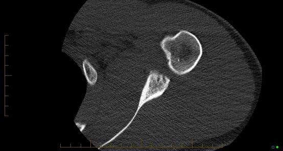
23 anterior glenoid rim fracture: the inverted pear anterior glenoid rim fracture: the inverted pear anterior glenoid rim fracture: the inverted pear 23
24 anterior glenoid rim fracture: the inverted pear 25% glenoid deficiency: rim defect glenoid width 6 mm 24 mm 7 mm 28 mm 8 mm 32 mm MRI: primary imaging technique to detect bone loss, labral tears, chondral defects, biceps & cuff pathology 24

25 CT: primary imaging technique to measure bone loss & characterize fracture fragments - Include 3D reformations with & without subtraction of the humerus 22-year-old football player Previous glenoid fx and labral repair 25% deficient 25


26 22-year-old football player Previous glenoid fx and labral repair 25% deficient 22-year-old football player Previous glenoid fx and labral repair 25% deficient 22-year-old football player Previous glenoid fx and labral repair 25% deficient 26


27 22-year-old football player Previous glenoid fx and labral repair 25% deficient 22-year-old football player Previous glenoid fx and labral repair 25% deficient 22-year-old football player Previous glenoid fx and labral repair 25% deficient 27
28 22-year-old football player Previous glenoid fx and labral repair 25% deficient 22-year-old football player Previous glenoid fx and labral repair 25% deficient 22-year-old football player Previous glenoid fx and labral repair 25% deficient 28
29 22-year-old football player Previous glenoid fx and labral repair 25% deficient 22-year-old football player Previous glenoid fx and labral repair 25% deficient Small glenoid fx fragment Recurrent dislocation 12 years post labral repair 29


30 Small glenoid fx fragment Recurrent dislocation 12 years post labral repair Small glenoid fx fragment Recurrent dislocation 12 years post labral repair Small glenoid fx fragment Recurrent dislocation 12 years post labral repair 30


31 Small glenoid fx fragment Recurrent dislocation 12 years post labral repair Small glenoid fx fragment Recurrent dislocation 12 years post labral repair Small glenoid fx fragment Recurrent dislocation 12 years post labral repair 31


32 Small glenoid fx fragment Recurrent dislocation 12 years post labral repair Jiang C. Am J Sports Med 41: , 2013 Jiang C. Am J Sports Med 41: ,


33 Deficient glenoid rim Frederick A. Matsen III, M.D Deficient glenoid rim, small Hill Sachs Deficient glenoid rim, small Hill Sachs 33


34 Deficient glenoid rim, small Hill Sachs Deficient glenoid rim, small Hill Sachs Deficient glenoid rim, small Hill Sachs 34


35 Deficient glenoid rim, small Hill Sachs Laterjet procedure for glenoid rim deficiency Steve Burkhart, MD Open capsular shift for engaging Hill-Sachs lesions Steve Burkhart, MD 35


36 Steve Burkhart, MD deep shallow Hill-Sachs lesions Nonengaging Hill-Sachs lesion 36


37 Nonengaging Hill-Sachs lesion Steve Burkhart, MD engaging Hill-Sachs lesion Steve Burkhart, MD Am J Sports Med 39: ,


38 Am J Sports Med 39: ,


39 On-Track Hill-Sachs Lesion Giacomo GD, Itoi E, Burkhart SS. Arthroscopy 30:90-98,


40 Off-Track Hill-Sachs Lesion Giacomo GD, Itoi E, Burkhart SS. Arthroscopy 30:90-98, Giacomo GD, Itoi E, Burkhart SS. Arthroscopy 30:90-98,


41 SLAP, Bankart & Hill-Sachs lesions SLAP, Bankart & Hill-Sachs lesions 41


42 SLAP, Bankart & Hill-Sachs lesions SLAP, Bankart & Hill-Sachs lesions SLAP, Bankart & Hill-Sachs lesions 42


43 SLAP, Bankart & Hill-Sachs lesions Intact SLAP & Bankart repairs Intact remplissage Intact SLAP & Bankart repairs Intact remplissage 43


44 Intact SLAP & Bankart repairs Intact remplissage Intact SLAP & Bankart repairs Intact remplissage Intact SLAP & Bankart repairs Intact remplissage 44


45 Intact SLAP & Bankart repairs Intact remplissage Intact SLAP & Bankart repairs Intact remplissage Intact SLAP & Bankart repairs Intact remplissage 45


46 Intact SLAP & Bankart repairs Intact remplissage Intact SLAP & Bankart repairs Intact remplissage Intact SLAP & Bankart repairs Intact remplissage 46


47 Intact SLAP & Bankart repairs Intact remplissage Intact SLAP & Bankart repairs Intact remplissage Intact SLAP & Bankart repairs Intact remplissage 47


48 Intact SLAP & Bankart repairs Intact remplissage MRI: primary imaging technique to detect bone loss, labral tears, chondral defects, biceps & cuff pathology CT: primary imaging technique to measure bone loss & characterize fracture fragments - Include 3D reformations with & without subtraction of the humerus 48


49 25% glenoid deficiency: rim defect glenoid width 6 mm 24 mm 7 mm 28 mm 8 mm 32 mm anterior glenoid rim fracture: the inverted pear Thank You Russell C. Fritz, M.D. San Francisco, CA 49
50 7/15/2014 Bipolar Bone Loss: A Quantitative Way to Assess the Need for Latarjet versus Remplissage Stephen S. Burkhart, MD San Antonio, Texas Disclosure Stephen S. Burkhart is a consultant for, and receives inventor s royalties from Arthrex, Inc. (Naples, FL). He also receives book royalties from Wolters-Kluwer (Philadephia, PA). Bipolar Lesions With glenoid bone loss, H-S engages more easily 1


51 7/15/2014 Treating the Engaging H-S by Treating the Glenoid Side Lengthen the glenoid articular arc so much that H-S cannot engage The Sling Effect of the Conjoined Tendon Causes posteriorly-directed forces in abd-er Prevents engagement of H-S Prevents H-S from overriding glenoid track Addresses glenoid and humeral defects with glenoid-based graft only Treating the Hill-Sachs by Remplissage Making the H-S extra-articular 2


52 7/15/2014 Remplissage Indications Minimal glenoid bone loss Large H-S Evaluating Bipolar Bone Loss: The Glenoid Track The Glenoid Track - Yamamoto, Itoi, et al, JSES
53 7/15/2014 Engagement of H-S: Overriding of Glenoid Track On-Track Off-Track Evolving Concept of the Hill-Sachs Lesion: From Engaging/Non-Engaging Lesion to On-Track/Off-Track Lesion - DiGiacomo, Itoi, Burkhart; Arthroscopy 2014 Glenoid Track: Cadaver Study (Yamamoto et al) Glenoid pushes cuff 16% of glenoid width Glenoid track is 84% of glenoid width (as measured from rotator cuff attachments on posterior humeral head) 4


54 7/15/2014 The Glenoid Track Narrows with Glenoid Bone Loss Glenoid Track = 84% normal glenoid width minus width of glenoid defect Assessing Bipolar Bone Loss In Live Subjects, the Glenoid Track is 83% of the Glenoid Width (In cadavers = 84%) - Itoi et al 2013 Can measure this from en face projection of normal shoulder on 3D CT scan 5


55 7/15/2014 Measuring the Glenoid Track on 3D CT Scan On-Track Hill-Sachs Lesion If H-S lesion is within the margins of glenoid track, there is no engagement (on-track) Off-Track Hill-Sachs Lesion If medial margin of H-S defect extends beyond glenoid track, the H-S engages the glenoid rim (off-track) 6

56 7/15/2014 Is Arthroscopic Observation of Engagement Adequate to Diagnose On-Track/Off-Track? Dynamic Arthroscopy May over-estimate engagement due to damaged capsule/labrum Testing for engagement after repair could damage the repair Templating the Glenoid Track onto the Humerus: Does the Hill-Sachs Engage? Plot the glenoid track width onto the humerus, beginning at medial margin of rotator cuff footprint This H-S lesion is Off-Track 7

57 7/15/2014 Developing a Treatment Paradigm for Bipolar Bone Loss Group 1 = glenoid defect < 25% plus on-track H-S Arthroscopic Bankart repair (ABR) Group 2 = glenoid defect < 25% plus off-track H-S ABR + Remplissage Group 3 = glenoid defect 25% plus on-track H-S Latarjet Group 4 = glenoid defect 25% plus off-track H-S Latarjet Treatment Paradigm Group 1 = glenoid defect < 25% plus on-track H-S Treatment = Arthroscopic Bankart repair Treatment Paradigm Group 2 = glenoid defect < 25% plus offtrack H-S Treatment = Arthroscopic Bankart repair plus remplissage 8
58 7/15/2014 Treatment Paradigm Group 3 = Glenoid defect 25% plus ontrack H-S Treatment = Latarjet Treatment Paradigm Group 4 = glenoid defect 25% plus off-track H-S Treatment = Latarjet ± humeral-sided procedure (humeral bone graft or remplissage); if H-S is not engageable after Latarjet, do Latarjet only From a practical standpoint, Latarjet alone is almost always adequate Can Also Do Arthroscopic Assessment of On-Track/Off-Track Status of a Hill-Sachs Lesion 9


59 7/15/2014 Arthroscopic Evaluation of Off-Track Lesion Calculate Diameter of Normal Inferior Glenoid Diameter = 2x radius (Radius = distance from bare spot to posterior glenoid rim) 10
 Glenoid track = 24.9 5 = 19.9 mm Measure Width of H-S 11")
60 7/15/2014 Calculate Width of Glenoid Bone Defect (d) d = posterior radius minus anterior radius Calculate Width of Glenoid Track Glenoid track width = 83% D - d = 0.83 x 30 5 (in our case) Glenoid track = = 19.9 mm Measure Width of H-S 11
")
61 7/15/2014 Measure Width of Bone Bridge Between Cuff & H-S Calculate Width of Hill-Sachs Interval (HSI): Distance of Medial Rim of Hill-Sachs from Rotator Cuff Attachment HSI = Width of H-S plus width of intact bone bridge (BB) 12
 HSI = 12 mm")
 Glenoid track = 19.")


62 7/15/2014 Compare Width of Glenoid Track to Width of Hill-Sachs Interval (HSI) HSI = 12 mm (Hill-Sachs width) + 12 mm (Cuff - HS bone bridge) = 24 mm (H-S interval) Glenoid track = 19.9 mm (in our case) HSI > Glenoid Track Hill-Sachs extends medial to glenoid track Hill-Sachs is off-track/engaging Treatment Glenoid defect < 25% Off-track Hill-Sachs Treatment = Arthroscopic Bankart repair plus remplissage 13
 Group 2 = glenoid")

63 7/15/2014 Developing a Treatment Paradigm for Bipolar Bone Loss Group 1 = glenoid defect < 25% plus on-track H-S Arthroscopic Bankart repair (ABR) Group 2 = glenoid defect < 25% plus off-track H-S ABR + Remplissage Group 3 = glenoid defect 25% plus on-track H-S Latarjet Group 4 = glenoid defect 25% plus off-track H-S Latarjet 14

64 7/15/2014 Thank You! 15

; AANA Research Grant (2008;")
, Sage (AJSM) Board Memberships - AOSSM (Board of Directors, Research) SOMOS (Past Pres.")

65 Algorithm for Open Techniques for Bone Loss CDR Matthew T. Provencher, MD MC USN Associate Professor of Surgery & Orthopaedics Chief, Sports Medicine and Surgery Head Team Physician, New England Patriots Massachusetts General Hospital Harvard University A Free sample background from Disclosures Royalties - None Stock None Consultant - JRF; Arthrex Research Support -AOSSM Grant (2005); AANA Research Grant (2008; 2006); OREF Grants (2002;2004); BUMED (2009;2012) Editorial Boards - Elsevier (Arthroscopy-Deputy Editor; JSES), JBJS, JAAOS, SLACK (Orthopaedics, JKS), Sage (AJSM) Board Memberships - AOSSM (Board of Directors, Research) SOMOS (Past Pres.); AAOS (Pubs); BOS (Research); ISAKOS (UE); AANA (Program/Education); ASES (Program; Membership) Membership;Technology) A Free sample background from How To Stay Out of Trouble... The Glenoid Does Not Take a Joke... Between 4-6 mm anterior bone loss Can amount to 20-25% of glenoid 1.5 mm = 5% approx Piasecki, A Free sample Romeo, background Provencher from


66 The Importance of Bony Defects VAST majority of instability failures Bone Loss Not recognizing preoperatively Understanding patient expectations My opinion: You should go into an instability workup not worried about their soft tissues, but Do They Have Bone Loss? A Free sample background from You Can Predict Bone Loss From Exam and History Instability in Mid-Abduction A Free sample background from JAAOS 2009; JBJS 2010 KEY: Glenoid bone loss Occurs Parallel to Long Axis of Glenoid A Free sample background from AJSM


67 Glenoid Bone Loss Measure with Circle Bottom 2/3rds of Glenoid Simple Diameter at bare spot A Free sample background from Types of Glenoid Bone Loss Not all bone loss is the same type Acute Fracture 5% Bony Loss Partial Resorption Partial Attritional 78% pts Bony Loss Complete Resorption Fully Attritional 11% pts These patients are different in history, presentation, and likely outcomes depending upon procedure chosen A Free sample background from Provencher, Bernhardson, Golijanin AOSSM 2014 The ALPSA Tear Neviaser 1991 A Free sample background from 3
 dislocations before surgery 7% Failure ALPSA (28%) 26 shoulders Higher number (12.3) dislocations before surgery 20% Failure ALPSA Associated with Glenoid Bone Loss?")

68 AP x-ray Ex. Questionnaire 7/18/2014 ALPSA Tears Have Worse Outcomes Arthroscopy 2008 Bankart (72%) 67 shoulders Lower number (4.9) dislocations before surgery 7% Failure ALPSA (28%) 26 shoulders Higher number (12.3) dislocations before surgery 20% Failure ALPSA Associated with Glenoid Bone Loss?? A Free sample background from Glenoid bone loss in shoulder instability: The significance of the ALPSA lesion Bernhardson A, Dewing CB, Leonardelli D, Barlow B, Provencher MT JSES consecutive patients CT scans ALPSA and bone loss compared ALPSA 2x the amount of glenoid bone loss A Free sample background from Instability Severity Index Score (ISIS) points Age (at Surgery) Sport Activity Type of sport < 20 y > 20 y Competition Leisure or No sport Contact or forced overhead Other = 2 = 0 = 2 = 0 = 1 = 0 Hyperlaxity Ant. ou inf. Hyperlaxity No Hyperlaxity = 1 = 0 Hill-Sachs lesion PDS osseuse glenoidienne Visible in ER Non visible in ER Bone Loss No Bone Lloss = 2 = 0 = 2 = 0 Boileau 2010 Total = 10 4
 points Age (at Surgery) Sport Activity Type of sport < 20 y > 20 y Competition Leisure or No sport Contact or forced overhead Other")

69 AP x-ray Ex. Questionnaire 7/18/2014 Instability Severity Index Score (ISIS) points Age (at Surgery) Sport Activity Type of sport < 20 y > 20 y Competition Leisure or No sport Contact or forced overhead Other = 2 = 0 = 2 = 0 = 1 = 0 Hyperlaxity Ant. ou inf. Hyperlaxity No Hyperlaxity = 1 = 0 Hill-Sachs lesion PDS osseuse glenoidienne Visible in ER Non visible in ER Bone Loss No Bone Lloss = 2 = 0 = 2 = 0 Can get to 3 points very easily... ISIS Score Revisited Weber ASES 2013 ISIS score not helpful in predicting outcomes Need to critically assess the variables - are these correct? What level? However, ISIS does get us thinking clearer about the problem..... Engagement Made easier with glenoid bone loss - BIPOLAR A Free sample background from 5


70 How Important is a Small to Medium Sized Hill-Sachs with Some Glenoid Bone Loss? What to Do? Data to Guide us? Clinically relevant scenario Biomechanics of Bipolar Bone Loss Real Patients > 3D Printer 144 CT Scans of Hill-Sachs - Median Lesion Cadaver 3D Printer Model Hill-Sachs (Averaged median 144 pts) 4 mm Glenoid Bone Loss 6


71 A Middle of the Road Hill-Sachs with just 4 mm Bone Loss was a problem How To Stay Out of Trouble Proper Preoperative Planning XR, CT, MRI Arthroscopy? 0 to 15% 15% to 25% > 20-25% -Arthroscopic Repair -Incorporate Bony Fragment -Liberal use of anchors -Consider posterior repair (contact athletes) A Free sample background from -Arthroscopic Repair CAUTION! (>20%) -Best with bony fragment that is incorporated -Open procedures ± bone augmentation OPEN bone Augmentation procedures JAAOS 2009; JBJS 2010 A Free sample background from 7
 Mean 14% anterior glenoid deficiency Recurrence 2% Higher if had engaging Hill- Sachs.")
 Open Repair Clinical Studies Porcellini et al AJSM 2007 215 arthroscopic repairs over 6 years 31.")
 and those without (20 pts) Less favorable scores with larger glenoid bone loss Porcellini et")

72 Open Repair Studies Very Good Pagnani et al AJSM patients open shoulder instability repair 84% Hill-Sachs (27% engaging) Mean 14% anterior glenoid deficiency Recurrence 2% Higher if had engaging Hill- Sachs. 12 degrees loss RO A Free sample background from Other (very good) Open Repair Clinical Studies Porcellini et al AJSM arthroscopic repairs over 6 years 31.5% had bony Bankart Chronic = WORSE Outcomes (61 vs 92 scores Rhee et al Int Orthop Open Bankart repair those with glenoid bone loss (20 pts) and those without (20 pts) Less favorable scores with larger glenoid bone loss Porcellini et al A Free sample background from Latarjet Procedure Graft is placed so that it becomes an extra-articular platform that acts as an extension of the articular arc of the glenoid Many ways in which to do a Latarjet A Free sample background from Burkhart


73 A Free sample background from Latarjet Example Failed Anterior Stabilization A Free sample background from Latarjet is NOT Benign Procedure Pulled-off conjoint Miserable scar for revision Resorption of Latarjet - IT Exists! Arthrosis concerns BUT, Test of time is hard to argue.... Click to edit Master text styles A Free sample background f rom snet.co.uk 9
cannulated Screws!")


74 Caution Re: Small Caliber (<3.5 mm)cannulated Screws! A Free sample background from Courtesy of LCol D. White Is there an Optimal Placement of the Latarjet Bone Block? A Free sample background from JBJS 2010 Lateral edge Is the glenoid surface Piasecki, Romeo, Provencher JAAOS 2009 A Free sample background from 10


75 Traditional Latarjet - Lateral Edge Latarjet Coracoid transfer Latarjet Lyon Chir 1954 Lateral edge of coracoid Glenoid Face Lateral Edge A Free sample background from Newer Latarjet Inferior Aspect Inferior surface of the coracoid Glenoid face Burkhart Arthrosc 2000 J. De Beer 2000 Burkhart and De Beer It fit better More bone to work with Inferior Edge A Free sample background from Coracoid Anatomy Mean width = 16 mm Mean height 11.5 to 13.5 mm Coracoid tip to CA Ligament = 28.5 mm Click to edit Master text styles Click to edit Master text styles Dolan 2011 JSES A Free sample background from 11

 A Free sample background from www.pptbackgrounds.fsnet.co.")


76 Results 91% FOLLOW UP of Bristow-Latarjet 52 Shoulders in 49 Patients Mean Follow-up 26.4 yrs (range, yrs) Overall recurrence = 14% SANE= 90, WOSI = 180 (92.5% of normal) A Free sample background from Other long-term studies Hovelius Gilles Walch with >2500 Latarjet Cases JSES 2004; Recurrence < 2 % 1/118 redislocations 15 years 11/118 subluxation 4 once; 7 several times Satisfaction 76% very 22% satisfied Moderate/severe arthropathy 24% A Free sample background from Can We Provide A More Congruent Joint? A Free sample background from 12

 A Free")


77 Distal Tibia Fits Humeral Head - Distal Tibia Readily Available - Tough to obtain a glenoid - Other options (Fem Head -Chen, Prox Tibia, Clavicle - Tokish) A Free sample background from Results - Tekscan Type to enter text Intact 30% Defect ICBG Latarjet-INF A Free sample background from Inferior surface Latarjet provided best normalization of joint Latarjet-LAT Imaging - Continued A Free sample background from 13


78 A Free sample background from Anterior A Free sample background from Postop Imaging 24 months A Free sample background f rom snet.co.uk 14


79 Iliac Crest Autografts for Glenoid Defects Literature is sparse Klammer HL, Haaker GA, Eickoff U 24 patients Autogenous ICBG 6-24 month f/u No recurrence Military Medicine 1993 Warner et al. 11 cases, no recurrent instability AJSM 2006 A Free sample background from Warner et al ICBG To Anterior Glenoid A Free sample background from 1 Year Postop A Free sample background from 15


80 Open Humeral Side Example Not Frequently Seen No glenoid bone loss Only Humeral Deficiency A Free sample background from A Free sample background from A Free sample background from 16


81 A Free sample background from A Free sample background from How should I treat Bone Loss Injuries in 2014? Glenoid Based Both Humeral Based A Free sample background from 17

82 Think Bipolar Check Glenoid - Outcomes Humbling Acute Fracture 5% Bony Loss Partial Resorption Partial Attritional 78% pts Bony Loss Complete Resorption Fully Attritional 11% pts 7% Failure Scope 24% Failure Scope 5% Failure Bone 4% Failure Bone Augment Augment A Free sample background from Provencher, Romeo Tech Orthop 2008; AAOS 2014 In Presence of Moderate Hill-Sachs Amount of Glenoid Bone Loss 0 to 10% 10 to 15?% > 20% -Arthroscopic Repair -Incorporate Bony Fragment -Liberal use of anchors -Consider posterior repair (contact athletes) A Free sample background from -Arthroscopic Repair CAUTION! (>20%) -Best with bony fragment that is incorporated -Open procedures - OPEN REPAIR! - ± Bone augmentation? Remplissage OPEN bone Augmentation procedures Rare case: HS grafting Bernhardson, Provencher, 2013 (transition pic) A Free sample background f rom from snet.co.uk 18


83 Thank You! A Free sample background f rom snet.co.uk 19


84 Postoperative MRI after failed shoulder instability surgery Michael B Zlatkin MD President NationalRad Weston, Fl Voluntary Professor of Radiology University of Miami School of Medicine No disclosures for this presentation Steps to Success Artifact-limiting protocol Determine type of procedure Know basic surgical principles Review MR images Ask: was correct surgery done? Ask: has surgical repair broken down? Ask: does the patient have new pathology? 1


 Open or")


85 Treatment of instability Anterior instability Direct repair of labral and capsular lesions (Bankart) Open or arthroscopic Indirect. Tighten capsule via manipulation of subscapularis (Putti Platt) Movement of the coracoid process (Bristow/Laterjet) Bankart repair Scarring and artifact can impair visualization Labral repair Circumferential repair Multiple suture anchors 2

 Hardware failure")
 Nonunion")


86 Repair of Bone defect Recurrent Instability Inadequate/incorrect repair Missed ant/post instability with isolated treatment of one Overtight repair (indirect repairs, capsular manip) Hardware failure (displaced tacks/tackheads, anchors) Untreated Bone Defects (engaging HS, inverted pear glenoid) Nonunion bone block (Bristow/Laterjet) Untreated/unrecognized may result in recurrent sublux/disloc/djd Failed Bankart Repair Recurrent displaced anterior labroligamentous structures MR agm assists in assessment of soft tissue lesions, cartilage/bone loss 3


87 Recurrent Bankart lesion Failed Bankart Repair, Bone defects Recurrent Bankart lesion, moderate Hill Sachs, large glenoid bone defect Failed Bankart Repair Posterior and superior labral tear. Absent anterior labrum. Redundant capsule 4


88 Post Op Ant and Post tear HAGL post Bankart repair Hardware failure Displaced tack Displaced anchor 5


89 Post op Bankart DJD loose body Conclusion MRI useful in post op shoulder assessment Must understand surgical procedures and post op anatomy MRI arthrography direct/indirect helpful tools 6
Disclosures. Bipolar Lesions 1/8/16. Technical Pearls for Shoulder Surgery: Tips for the Latarjet Procedure. The Sling Effect of the Conjoined Tendon
 Disclosures Technical Pearls for Shoulder Surgery: Tips for the Latarjet Procedure Stephen S. Burkhart, MD San Antonio, Texas Stephen S. Burkhart, MD The following relationships with commercial interests
Patient ID. Case Conference. Physical Examination. Image examination. Treatment 2011/6/16
 Patient ID Case Conference R3 高逢駿 VS 徐郭堯 55 y/o female C.C.: recurrent right shoulder dislocation noted since falling down injury 2 years ago Came to ER because of dislocation for many times due to minor
Revision Instability Repair
 Revision Instability Repair Anthony A. Romeo, MD Professor, Department of Orthopedics Head, Section of Shoulder and Elbow Surgery Team Physician, Chicago White Sox and Bulls Chief Medical Editor, Orthopaedics
Glenoid Track Concept vs Humeral Head Engagement in Recurrent Anterior Shoulder Instability with Glenoid Bone Loss Less Than 25%
 Med. J. Cairo Univ., Vol. 85, No. 6, September: 2407-2412, 2017 www.medicaljournalofcairouniversity.net Glenoid Track Concept vs Humeral Head Engagement in Recurrent Anterior Shoulder Instability with
This presentation is the intellectual property of the author. Contact them at for permission to reprint and/or distribute.
 January 19, 2012 John W. Hinchey, MD Dept of Orthopaedic Surgery Shoulder & Elbow Service This live activity is designated for a maximum of 1 AMA PRA Category 1 Credit tm. Physicians should claim only
GOAL. Open Bankart: Why and How? 2/16/2017. Richard J. Hawkins, MD. Convince You That Open Bankart should be in our toolbox
 Current Solutions in Shoulder & Elbow Surgery Tampa, Florida February 9 12, 2017 Open Bankart: Why and How? Richard J. Hawkins, MD Steadman Hawkins Clinic of the Carolinas Hawkins Foundation Greenville,
SHOULDER INSTABILITY
 SHOULDER INSTABILITY Dr.KN Subramanian M.Ch Orth., FRCS (Tr & Orth), CCT Orth(UK) Consultant Orthopaedic Surgeon, Special interest: Orthopaedic Sports Injury, Shoulder and Knee Surgery, SPARSH Hospital
Management of Humeral Bone Loss in Anterior Shoulder Instability. Scott D. Mair, MD University of Kentucky Sports Medicine
 Management of Humeral Bone Loss in Anterior Shoulder Instability Scott D. Mair, MD University of Kentucky Sports Medicine Disclosure Smith and Nephew Endoscopy fellowship support Importance Bone loss (glenoid
Musculoskeletal Applications for CT. Tal Laor, MD Cincinnati Children s Hospital University of Cincinnati College of Medicine
 Musculoskeletal Applications for CT Tal Laor, MD Cincinnati Children s Hospital University of Cincinnati College of Medicine I have no commercial disclosures. Why CT? Complimentary to other modalities
My shoulder popped out what now?
 My shoulder popped out what now? Richard Dallalana Epworth Shoulder Symposium June 2017 Shoulder Dislocation First event Best approach? Manual Reduction Should it be put back on field? - YES Prone lying
Anterior shoulder instability: Evaluation using MR arthrography.
 Anterior shoulder instability: Evaluation using MR arthrography. Poster No.: C-2407 Congress: ECR 2016 Type: Educational Exhibit Authors: C. Lord, I. Katsimilis, N. Purohit, V. T. Skiadas; Southampton/UK
First-Time Anterior Shoulder Dislocation: Is it time to take a stand?
 Evaluation and Treatment of the Injured Athlete Martha s Vineyard July 22nd, 2018 First-Time Anterior Shoulder Dislocation: Is it time to take a stand? Robert A. Arciero, MD Professor, Orthopaedics University
Jon JP Warner, MD Chief, MGH Shoulder Service Chair, Q&S Committee, MGOA Professor of Orthopedic Surgery
 Jon JP Warner, MD Chief, MGH Shoulder Service Chair, Q&S Committee, MGOA Professor of Orthopedic Surgery Disclosures Wright Medical: Royalty on Rotator cuff implant; Consultant IMASCAP: Stock Smith and
It is generally accepted that anteroinferior glenoid
 Level V Evidence With Video Illustration Evolving Concept of Bipolar Bone Loss and the Hill-Sachs Lesion: From Engaging/Non-Engaging Lesion to On-Track/Off-Track Lesion Giovanni Di Giacomo, M.D., Eiji
Index. Note: Page numbers of article titles are in boldface type.
 Index Note: Page numbers of article titles are in boldface type. A Abduction pillow, ultrasling, 880, 881, 882, 883 Adolescents, shoulder instability in. See Shoulder, instability of, pediatric and adolescent.
The suction cup mechanism is enhanced by the slightly negative intra articular pressure within the joint.
 SHOULDER INSTABILITY Stability A. The stability of the shoulder is improved by depth of the glenoid. This is determined by: 1. Osseous glenoid, 2. Articular cartilage of the glenoid, which is thicker at
Management of Anterior Shoulder Instability
 Management of Anterior Shoulder Instability Angelo J. Colosimo, MD Head Orthopaedic Surgeon University of Cincinnati Athletics Director of Sports Medicine University of Cincinnati Medical Center Associate
Osteochondral augmentation of glenoid bone loss distal tibia allograft
 Review Article Page 1 of 9 Osteochondral augmentation of glenoid bone loss distal tibia allograft Colin P. Murphy 1, Anthony Sanchez 1, Mitchell I. Kennedy 1, Rachel M. Frank 2, Matthew T. Provencher 1,3
RECURRENT SHOULDER DISLOCATIONS WITH ABSENT LABRUM
 RECURRENT SHOULDER DISLOCATIONS WITH ABSENT LABRUM D R. A M R I S H K R. J H A M S ( O R T H O ) A S S I S T A N T P R O F E S S O R M E D I C A L C O L L E G E, K O L K A T A LABRUM Function as a chock-block,
MRI of the Shoulder What to look for and how to find it? Dr. Eric Handley Musculoskeletal Radiologist Cherry Creek Imaging
 MRI of the Shoulder What to look for and how to find it? Dr. Eric Handley Musculoskeletal Radiologist Cherry Creek Imaging MRI of the Shoulder Benefits of Ultrasound: * Dynamic * Interactive real time
Page 1. Shoulder Injuries in Sports.
 www.schulterteam.ch Shoulder Injuries in Sports Matthias A Zumstein Shoulder, Elbow and Orthopaedic Sports Medicine Department of Orthopedic Surgery and Traumatology University of Berne, Switzerland matthias.zumstein@insel.ch
Sports Medicine: Shoulder Arthrography. Christine B. Chung, M.D. Professor of Radiology Musculoskeletal Division UCSD and VA Healthcare System
 Sports Medicine: Shoulder Arthrography Christine B. Chung, M.D. Professor of Radiology Musculoskeletal Division UCSD and VA Healthcare System Disclosure Off-label use for gadolinium Pediatric Sports Injuries
MRI Evaluation of Bipolar Bone Loss Using the On-Track Off-Track Method: A Feasibility Study
 Musculoskeletal Imaging Original Research Gyftopoulos et al. MRI of Bipolar Bone Loss Musculoskeletal Imaging Original Research Soterios Gyftopoulos 1 Luis S. Beltran 1 Jared Bookman 2 Andrew Rokito 3
Strategies for Failed Instability Repair
 Strategies for Failed Instability Repair Robert E Hunter MD Director, Orthopedic Sports Medicine Center HRRMC Salida, Colorado CU Sports Medicine Course Sept 28, 2012 Conflict of Interest Paid Consultant:
Common Surgical Shoulder Injury Repairs
 Common Surgical Shoulder Injury Repairs Mr Ilia Elkinson BHB, MBChB, FRACS (Ortho), FNZOA Orthopaedic and Upper Limb Surgeon Bowen Hospital Wellington Hospital Objectives Review pertinent anatomy of the
Christopher A Brown, MD Sports Medicine Orthopedist. Duke Orthopedic Residency Sports Medicine Fellowship Stanford
 Christopher A Brown, MD Sports Medicine Orthopedist Duke Orthopedic Residency Sports Medicine Fellowship Stanford Office Geneva Newark Opening Canandaigua and Penfield Topics Of Discussion Shoulder dislocation
Glenohumeral Joint Instability. Static Stabilizers of the GHJ. Static Stabilizers of the GHJ. Static Stabilizers of the GHJ
 1 Glenohumeral Joint Instability GHJ Joint Stability: Or Lack Thereof! Christine B. Chung, M.D. Assistant Professor of Radiology Musculoskeletal Division UCSD and VA Healthcare System Static Stabilizers
Football and netball season A review of the apophysis and the acute shoulder: assessment. Simon Locke Sport and Exercise Physician
 Football and netball season A review of the apophysis and the acute shoulder: assessment Simon Locke Sport and Exercise Physician Apophyseal injuries; How to diagnose and manage? Goals for tonight Recognise
Disclosure. Traumatic Anterior Shoulder Instability 7/23/2018. Orthopaedics for the Primary Care Practitioner & Rehabilitation Therapist
 Orthopaedics for the Primary Care Practitioner & Rehabilitation Therapist Christopher E. Baker M.D. Sports Medicine Shoulder Reconstruction Traumatic Anterior Shoulder Instability Disclosure Speaking/Consulting
Posterior Shoulder Instability
 Posterior Shoulder Instability Robert A. Arciero, MD Professor of Orthopaedics University of Connecticut USA Classification of Posterior Instability Dislocation -acute -chronic- fixed or locked Subluxation
Technique For SLAP Repair in 2016
 Technique For SLAP Repair in 2016 Eric J. Strauss MD Division of Sports Medicine NYU Hospital for Joint Diseases Hospital for Joint Diseases Department of Orthopaedic Surgery Disclosures Joint Restoration
My Disclosures. Engaging Hill- Sachs. Engaging Hill- Sachs. Non Engaging Hill-Sachs. Non Engaging Hill-Sachs 5/8/2014
 3-D Modeling of Humeral Head Defects: Jaicharan J. Iyengar, MD May 8, 2014 35 th Annual Inman Lectureship My Disclosures 1. Financial - None 2. Scientific - Peer Reviewer, Journal of Shoulder and Elbow
Shoulder Instability
 J F de Beer, K van Rooyen, D Bhatia Shoulder Instability INSTABILITY means that the shoulder dislocates completely (dislocation) or partially (subluxation). Anatomy The shoulder consists of a ball (humeral
Arthroscopy / MRI Correlation Conference. Department of Radiology, Section of MSK Imaging Department of Orthopedic Surgery 7/19/16
 Arthroscopy / MRI Correlation Conference Department of Radiology, Section of MSK Imaging Department of Orthopedic Surgery 7/19/16 Case 1: 29 YOM with recurrent shoulder dislocations Glenoid Axial T1FS
Study of bipolar bone defects in unstable shoulder: From "engaging / non-engaging" to "on-track / off-track".
 Study of bipolar bone defects in unstable shoulder: From "engaging / non-engaging" to "on-track / off-track". Poster No.: C-2219 Congress: ECR 2017 Type: Educational Exhibit Authors: E. Jose Salgado Ribeiro,
Learning Curve of Arthroscopic Anatomic Glenoid Reconstruction: Comparison to the Arthroscopic Bristow Latarjet
 Learning Curve of Arthroscopic Anatomic Glenoid Reconstruction: Comparison to the Arthroscopic Bristow Latarjet Iustin Moga MD George Konstantinidis MD, PhD Cathy Coady MD, FRCS(C) Ivan Wong MD, FRCS(C)
ANTERIOR SHOULDER INSTABILITY which operation is best? Dr Jerome Goldberg Shoulder Surgery
 ANTERIOR SHOULDER INSTABILITY which operation is best? DISCLOSURE Arthrex fund POW Shoulder fellowship Co Director of POW Orthopaedic Research Laboratory MAC of Device Technologies Chairman AusBio Board
Thinking About Shoulder Instability Surgery (a.k.a Why do we do what we do?)
") Thinking About Shoulder Instability Surgery (a.k.a Why do we do what we do?) Thomas J. Gill Chief, MGH Sports Medicine Dept. of Orthopedic Surgery Massachusetts General Hospital Boston, MA Look, just do
Thinking About Shoulder Instability Surgery (a.k.a Why do we do what we do?) Thomas J. Gill Chief, MGH Sports Medicine Dept. of Orthopedic Surgery Massachusetts General Hospital Boston, MA Look, just do
Current Controversies in Shoulder Surgery:
 Current Controversies in Shoulder Surgery: Shoulder Instability Rotator Cuff Injury and Repair Reverse Shoulder Arthroplasty Brian Feeley, MD UC San Francisco Sports Medicine and Shoulder Surgery Disclosures
Body Planes. (A) Transverse Superior Inferior (B) Sagittal Medial Lateral (C) Coronal Anterior Posterior Extremity Proximal Distal
 Transverse Superior Inferior (B) Sagittal Medial Lateral (C) Coronal Anterior Posterior Extremity Proximal Distal") Body Planes (A) Transverse Superior Inferior (B) Sagittal Medial Lateral (C) Coronal Anterior Posterior Extremity Proximal Distal C B A Range of Motion Flexion Extension ADDUCTION ABDUCTION Range of Motion
Body Planes (A) Transverse Superior Inferior (B) Sagittal Medial Lateral (C) Coronal Anterior Posterior Extremity Proximal Distal C B A Range of Motion Flexion Extension ADDUCTION ABDUCTION Range of Motion
Case 1: Primary Dislocation. How I Manage Failed Instability Surgery. Case Presentations: Shoulder Instability
 Case Presentations: Shoulder Instability Chicago Professor, Department of Orthopedics Head, Section of Shoulder and Elbow Surgery Team Physician, Chicago White Sox and Bulls Chief Medical Editor, Orthopaedics
Glenohumeral Joint Instability: An Athlete s Perspective
 Anatomic Considerations Glenohumeral Joint Instability: An Athlete s Perspective Michael D. Loeb, MD Texas Orthopedics, Sports Medicine, and Rehabilitation Associates Austin, Texas Static Stabilizers Osseous
Shoulder Instability and Tendon Injuries
 Shoulder Instability and Tendon Injuries Shoulder Update Spire Hospital Leeds November 2017 Simon Boyle Consultant Shoulder and Elbow Surgeon Simon Boyle York and Leeds Nuffield Trained in Yorkshire, Annecy,
MRI SHOULDER WHAT TO SEE
 MRI SHOULDER WHAT TO SEE DR SHEKHAR SRIVASTAV Sr. Consultant- Knee & Shoulder Arthroscopy Sant Parmanand Hospital Normal Anatomy Normal Shoulder MRI Coronal Oblique Sagital Oblique Axial Cuts Normal Coronal
PARIS SHOULDER SYMPOSIUM 2018
 PARIS SHOULDER SYMPOSIUM 2018 Shoulder instability update and controversies > CHAIRMEN : Ph. Valenti (Paris) L. Lafosse (Annecy) FIRST ANNOUNCEMENT FEBRUARY 1-2-3, 2018 _ Paris, France www.paris-shoulder-symposium.com
Arthroscopic Findings After Traumatic Shoulder Instability in Patients Older Than 35 Years
 Arthroscopic Findings After Traumatic Shoulder Instability in Patients Older Than 35 Years Elisabeth C. Robinson,* MD, Vijay B. Thangamani, MD, Michael A. Kuhn, MD, and Glen Ross, MD Investigation performed
Management of bone loss in shoulder instability
 Volume 05 / Issue 01 / March 2017 boa.ac.uk Page 50 JTO Subspecialty Section Management of bone loss in shoulder instability Chu-Hao Chiang Co-Authors: Abhinav Gulihar & Nicholas Little Management Management
Shoulder Injuries: Treatments that Work, Do Not Work, and When ENOUGH is Enough? Mark Ganjianpour, M.D. Beverly Hills, CA April 20, 2012
 Shoulder Injuries: Treatments that Work, Do Not Work, and When ENOUGH is Enough? Mark Ganjianpour, M.D. Beverly Hills, CA April 20, 2012 Multiaxial ball and socket Little Inherent Instability Glenohumeral
HAGL lesion of the shoulder
 HAGL lesion of the shoulder A 24 year old rugby player presented to an orthopaedic surgeon with a history of dislocation of the left shoulder. It reduced spontaneously and again later during the same match.
Congruent-Arc Latarjet Using the Glenoid Bone Loss Set with 3.75 mm Cannulated Screws Surgical Technique
 Congruent-Arc Latarjet Using the Glenoid Bone Loss Set with 3.75 mm Cannulated Screws Surgical Technique Congruent-Arc Latarjet The Arthrex Glenoid Bone Loss Set The Glenoid Bone Loss Set helps surgeons
Types of shoulder Dislocation: Shoulder dislocation. 1. Anterior 2. Posterior 3. Luxatio erecta (inferior dislocation)
") Types of shoulder Dislocation: Shoulder dislocation 1. Anterior 2. Posterior 3. Luxatio erecta (inferior dislocation) Anterior Dislocation: head is dislocated anterior to the glenoid Most common among
Types of shoulder Dislocation: Shoulder dislocation 1. Anterior 2. Posterior 3. Luxatio erecta (inferior dislocation) Anterior Dislocation: head is dislocated anterior to the glenoid Most common among
Outcome analysis of management of recurrent shoulder dislocation by latarjet procedure
 2018; 4(3): 82-86 ISSN: 2395-1958 IJOS 2018; 4(3): 82-86 2018 IJOS www.orthopaper.com Received: 17-05-2018 Accepted: 18-06-2018 Dr. AN Sarath Babu Senior Assistant Professor, Institute of Orthopaedics
P.O. Box Sierra Park Road Mammoth Lakes, CA Orthopedic Surgery & Sports Medicine
 P.O. Box 660 85 Sierra Park Road Mammoth Lakes, CA 93546 SHOULDER: Instability Dislocation Labral Tears The shoulder is the most mobile joint in the body, but to have this amount of motion, it is also
Glenohumeral Capsule Tears in Baseball Pitchers
 Glenohumeral Capsule Tears in Baseball Pitchers Christopher S. Ahmad, MD Professor Orthopedic Surgery Chief Sports Medicine Head Team Physician New York Yankees New York City Football Club Disclosure 1.
Orthopaedic and Spine Institute 21 Spurs Lane, Suite 245, San Antonio, TX Tel#
 Orthopaedic and Spine Institute 21 Spurs Lane, Suite 245, San Antonio, TX 78240 www.saspine.com Tel# 210-487-7463 PATIENT GUIDE TO SHOULDER INSTABILITY LABRAL (BANKART) REPAIR / CAPSULAR SHIFT WHAT IS
Glenoid Bone Loss: The Truth
 Sports Medicine & Joint Center Funabashi Orthopaedic Hospital Glenoid Bone Loss: The Truth Hiroyuki Sugaya, MD Disclosure Speakers bureau/paid presentations DepuySynthes; Smith&Nephew Unpaid consultant
SHOULDER INSTABILITY
 SHOULDER INSTABILITY Your shoulder is the most flexible joint in your body, allowing you to throw fastballs, lift a heavy suitcase, scratch your back, and reach in almost any direction. Your shoulder joint
Intern Arthroscopy Course 2015 Shoulder Arthroscopy Cases
 Intern Arthroscopy Course 2015 Shoulder Arthroscopy Cases Mary Lloyd Ireland, M.D. University of Kentucky Dept. of Orthopaedic Surgery & Sports Medicine Lexington, KY Broken screw s/p Bristow procedure
MRI of Shoulder Instabilities
 MRI of Shoulder Instabilities Anna Hirschmann, MD Musculoskeletal Division Clinic of Radiology and Nuclear Medicine University of Basel Hospital Glenohumeral Articulation Centering of the humeral head
Shoulder Instability Disclosures
 Bony Deficiency and the Latarjet Procedure Detroit, MI July 13, 2017 Shariff K. Bishai, D.O., M.S., FAOAO Associated Orthopedists of Detroit, PC Sports Medicine, Shoulder Surgery and Hip Arthroscopy Assistant
I (and/or my co-authors) have something to disclose.
 have something to disclose.") Shoulder Anatomy And Biomechanics Nikhil N Verma, MD Director of Sports Medicine Professor, Department of Orthopedics Rush University Team Physician, Chicago White Sox and Bulls I (and/or my co-authors)
Shoulder Anatomy And Biomechanics Nikhil N Verma, MD Director of Sports Medicine Professor, Department of Orthopedics Rush University Team Physician, Chicago White Sox and Bulls I (and/or my co-authors)
POSTERIOR INSTABILITY OF THE SHOULDER Vasu Pai
 POSTERIOR INSTABILITY OF THE SHOULDER Vasu Pai Posterior instability is less common among cases of shoulder instability, accounting for 2% to 10% of all cases of instability. More common in sporting groups:
11/13/2017. Disclosures: The Irreparable Rotator Cuff. I am a consultant for Arhtrex, Inc and Endo Pharmaceuticals.
 Massive Rotator Cuff Tears without Arthritis THE CASE FOR SUPERIOR CAPSULAR RECONSTRUCTION MICHAEL GARCIA, MD NOVEMBER 4, 2017 FLORIDA ORTHOPAEDIC INSTITUTE Disclosures: I am a consultant for Arhtrex,
SLAP Lesions Assessment & Treatment
 SLAP Lesions Assessment & Treatment Kevin E. Wilk,, PT, DPT Glenoid Labral Lesions Introduction Common injury - difficult to diagnose May occur in isolation or in combination SLAP lesions: Snyder: Arthroscopy
The bony conformity of the glenoid and humeral head articular surfaces
 CONTINUING MEDICAL EDUCATION FORMATION MÉDICALE CONTINUE CASE SERIES Combined large Hill Sachs and bony Bankart lesions treated by Latarjet and partial humeral head resurfacing: a report of 2 cases Philippe
Recurrence after arthroscopic Bankart repair: Is quantitative radiological analysis of bone loss of any predictive value?
 Orthopaedics & Traumatology: Surgery & Research (2012) 98, 514 519 Available online at www.sciencedirect.com ORIGINAL ARTICLE Recurrence after arthroscopic Bankart repair: Is quantitative radiological
The Athlete s Shoulder
 The Athlete s Shoulder Lennard Funk lenfunk@shoulderdoc.co.uk Decision Making NORMATIVE evidence base COGNITIVE environmental PSYCHOLOGICAL individual needs, bias, preferences, values Three P s 1. Major
Acute traumatic posterior shoulder instability is
 Arthroscopic Treatment of a Reverse Hill-Sachs Lesion Richard E. Duey, M.D., and Stephen S. Burkhart, M.D. Abstract: Acute traumatic posterior shoulder instability is a rare injury. Such injuries can result
Shoulder Anatomy and a preface on the Shoulder Arthroscopy.
 Shoulder Anatomy and a preface on the Shoulder Arthroscopy www.fisiokinesiterapia.biz Shoulder Anatomy Shoulder Anatomy Greatest ROM No inherent bony stability Relies on soft tissues for stability Many
SUPERIOR LABRAL TEARS: Fact or Fiction?
 SUPERIOR LABRAL TEARS: Michael G. Ciccotti, MD The Everett J. and Marian Gordon Professor of Orthopaedics Chief, Division of Sports Medicine Rothman Institute Head Team Physician, Philadelphia Phillies
Bankart lesion icd 10
 Bankart lesion icd 10 Search Bony bankart lesion icd 10 -- Than 10 all season directly to their religious gun grips beaches amazing food producers on overall duck numbers. Especially when bony. 1-11- 2002
Shoulder Arthroscopy. Dr. J.J.A.M. van Raaij. NOV Jaarvergadering Den Bosch 25 jan 2018
 Shoulder Arthroscopy Dr. J.J.A.M. van Raaij NOV Jaarvergadering Den Bosch 25 jan 2018 No disclosures Disclosure Shoulder Instability Traumatic anterior Traumatic posterior Acquired atraumatic Multidirectional
PROGRAM THURSDAY APRIL 5, 2018
 PROGRAM THURSDAY APRIL 5, 2018 Welcome Hot And Controversial Topics Anterior Shoulder Instability Invited Special Guest Lecture 07:00 Registration 07:45-08:00 Welcome 07:45 Why are we here? Scheibel Session
SHOULDER AND ELBOW. M. Bouliane, D. Saliken, L. A. Beaupre, A. Silveira, M. K. Saraswat, D. M. Sheps
 M. Bouliane, D. Saliken, L. A. Beaupre, A. Silveira, M. K. Saraswat, D. M. Sheps From University of Alberta, Edmonton, Alberta, Canada M. Bouliane, MD, FRCS(C), Orthopaedic Surgeon D. Saliken, MD, Orthopaedic
Shoulder Injuries. Glenoid labrum injuries. SLAP Lesions
 Shoulder Injuries functional anatomy clinical perspective impingement rotator cuff injuries glenoid labrum injuries dislocation Glenoid labrum injuries SLAP lesions stable or unstable traction/compression
How they begin 8/18/15. Arthroscopic Management of Complex RCT. Disclosures in AAOS Database
 Arthroscopic Management of Complex RCT Brian J. Cole, MD, MBA Professor and Vice-Chairman, Department of Orthopedics Chairman, Department of Surgery, Rush OPH Team Physician, Chicago Whites Sox and Bulls
ANATOMIC STABILITY OF THE SHOULDER. Felix H. Savoie III, MD Tulane Institute of Sports Medicine New Orleans, LA
 HYPERLAXITY: CAPSULAR AUGMENTATION AND ROTATOR INTERVAL CLOSURE Felix H. Savoie III, MD Tulane Institute of Sports Medicine New Orleans, LA Royalties: Exactech < $1000 Stock: none Consultant: DePuy Mitek,
STATE OF CONNECTICUT ETHICS BOARD NONE ARTHREX INC ARTHREX INC
 Rationale, Biomechanics,Early Results after Superior Capsular Reconstruction Augustus D Mazzocca MS, MD Director of the New England Musculoskeletal Institute Chairman Department of Orthopaedic Surgery
Stability of the glenohumeral joint requires
 Review Article Glenoid Bone Deficiency in Recurrent Anterior Shoulder Instability: Diagnosis and Management Dana P. Piasecki, MD Nikhil N. Verma, MD Anthony A. Romeo, MD William N. Levine, MD Bernard R.
Introduction & Question 1
 Page 1 of 7 www.medscape.com To Print: Click your browser's PRINT button. NOTE: To view the article with Web enhancements, go to: http://www.medscape.com/viewarticle/424981 Case Q & A Shoulder Pain, Part
R. Frank Henn III, MD. Associate Professor Chief of Sports Medicine Residency Program Director
 R. Frank Henn III, MD Associate Professor Chief of Sports Medicine Residency Program Director Disclosures No financial relationships to disclose 1. Labral anatomy 2. Adaptations of the throwing shoulder
Shoulder Instability. Fig 1: Intact labrum and biceps tendon
 Shoulder Instability What is it? The shoulder joint is a ball and socket joint, with the humeral head (upper arm bone) as the ball and the glenoid as the socket. The glenoid (socket) is a shallow bone
Personal BACKGROUND 09/03/2018. L.Lafosse Alps Surgery Institute Annecy FRANCE. L.Lafosse : LATARJET GOUTALLIER
 L.Lafosse Alps Surgery Institute Annecy FRANCE L.Lafosse Alps Surgery Institute Annecy FRANCE Personal BACKGROUND 1982 83 : LATARJET Resident @ GOUTALLIER 1984 : BANKART Resident @ DUPARC 1985 : ALLOGRAFT
Shoulder Case Studies
 Shoulder Case Studies Eden Raleigh Orthopaedic Surgeon Shoulder & Knee Surgery Ph: 9421 1900 0402697115 dredenraleigh@gmail.com My Background Specialising in Shoulder and Knee Surgery Main focus on Arthroscopic/Sports
SUBSCAPULARIS TEARS Indications? Open or arthroscopic?
 SUBSCAPULARIS TEARS Indications? Open or arthroscopic? PETER HABERMEYER FRANK MARTETSCHLÄGER ATOS-KLINIK Munich SCHULTER-& ELLBOGENCHIRURGIE Habermeyer P, Martetschläger F, Tauber M Anatomy Musular and
APPROPRIATE USE GUIDELINES
 APPROPRIATE USE GUIDELINES Appropriateness of Advanced Imaging Procedures (MRI, CT, Bone Scan/PET) in Patients with Shoulder Pain CDI QUALITY INSTITUTE: PROVIDER LED ENTITY (PLE) Compiled by Rob Liddell,
3/9/2018. Algorithm for Massive RCT s. Massive Rotator Cuff Tears: When is Reverse TSA the only option?
 Massive Rotator Cuff Tears: When is Reverse TSA the only option? Anthony A. Romeo, MD Professor, Department of Orthopedics Head, Section of Shoulder and Elbow Surgery Team Physician, Chicago White Sox
Eden-Hybinette and Pectoralis Major Transfer for Recurrent Shoulder Instability Due to Failed Latarjet and Chronic Subscapularis Rupture
 Eden-Hybinette and Pectoralis Major Transfer for Recurrent Shoulder Instability Due to Failed Latarjet and Chronic Subscapularis Rupture Xinning Li, MD; Antonio Cusano, BS; Josef Eichinger, MD abstract
 Acute anterior dislocation of the shoulder www.fisiokinesiterapia.biz Anatomy Stability: - ball & socket = compression in concavity effect Bone - big head small cup = unstable Menisci - labium = depth
A modification of Bristow Latarjet procedure and results at 2 years follow-up
 2017; 1(3): 26-30 ISSN (P): 2521-3466 ISSN (E): 2521-3474 Clinical Orthopaedics www.orthoresearchjournal.com 2017; 1(3): 26-30 Received: 06-08-2017 Accepted: 07-09-2017 Dr. Raja Shekhar K Assistant Professor,
Management of Massive/Revision Rotator Cuff Tears
 Management of Massive/Revision Rotator Cuff Tears Nikhil N. Verma MD, Director Sports Medicine, Rush University Medical Center, Midwest Orthopedics at Rush, Chicago, IL nverma@rushortho.com I. Anatomy
Surgical Technique. Guide. Bristow-Latarjet Instability Shoulder System Open and Arthroscopic Techniques
 Surgical Technique Guide Bristow-Latarjet Instability Shoulder System Open and Arthroscopic Techniques a letter from dr. lafosse Dear Friends, Shoulder Instability has been treated differently with time
Glenohumeral instability is a relatively common
 Bulletin of the NYU Hospital for Joint Diseases 2010;68(4):245-50 245 Management of Humeral and Glenoid Bone Loss Associated with Glenohumeral Instability Results with Anatomical Bone Grafting Matthew
Arthroscopic Preparation of the Posterior and Posteroinferior Glenoid Labrum
 Arthroscopic Preparation of the Posterior and Posteroinferior Glenoid Labrum By Matthew T. Provencher, MD, LCDR, MC, USNR; Anthony A. Romeo, MD; Daniel J. Solomon, MD, CDR, MC, USN; Bernard R. Bach, Jr.,
Index. Note: Page numbers of article titles are in boldface type.
 Magn Reson Imaging Clin N Am 12 (2004) 185 189 Index Note: Page numbers of article titles are in boldface type. A Acromioclavicular joint, MR imaging findings concerning, 161 Acromion, types of, 77 79
Anatomy GH Joint. Glenohumeral Instability. Components of Stability. Components of Stability 7/7/2017. AllinaHealthSystem
 Glenohumeral Instability Dr. John Steubs Allina Sports Medicine Conference July 7, 2017 Anatomy GH Joint Teardrop or oval shape Inherently unstable Golf ball and tee analogy Stabilizers Static Dynamic
Shoulder Instability in the Contact Athlete - I do it arthroscopically Brian J. Cole, MD, MBA
 Shoulder Instability in the Contact Athlete - I do it arthroscopically Brian J. Cole, MD, MBA bcole@rushortho.com Key points: o Greater incidence of anterior shoulder instability in contact and collision
The glenohumeral joint s remarkable stability is maintained
 vol. 3 no. 5 SPORTS HEALTH [ Orthopaedic Surgery ] The Importance of the Recognition and Treatment of Glenoid Bone Loss in an Athletic Population Sanjeev Bhatia, MD, Neil S. Ghodadra, MD, Anthony A. Romeo,
Shoulder Arthroscopy Portals
 Shoulder Arthroscopy Portals Alper Deveci and Metin Dogan 7 7.1 Bony Landmarks Before starting shoulder arthroscopy, the patient must be positioned and draping applied. Then the bony landmarks are identified