Altered Biomechanics and Spondylolysis
|
|
|
- Polly Lamb
- 5 years ago
- Views:
Transcription


1 Altered Biomechanics and Spondylolysis Terry R. Yochum DC, DACBR, Fellow ACCR Alicia M. Yochum RN, DC, DACBR, RMSK Does Altered Biomechanics Cause Bone Marrow Edema? Mark E. Schweitzer, MD and Lawrence M. White MD Department of Radiology Thomas Jefferson University Hospital Philadelphia, Pennsylvania Radiology 198:851 March
2 What is Bone Marrow Edema? Inflammation in the bone Injury to the trabeculae causing it to bleed Repetitive Impact Controversial Etiology Blood= Fluid Fluid= High signal Fluid = White Marrow (Fat)= Black Materials and Methods 12 Participants 6 Women 6 Men Age: (Mean 30) All asymptomatic and without abnormal pronation MRI baseline Bilateral foot, ankle, knee, and hip Scans utilizing a 1.5 Tesla magnet was done utilizing STIR imaging which suppresses fat signal and enhances water signal Insert 9/16 (1.4cm) longitudinal metatarsal arch pad inserted into the shoes of one foot of each volunteer Forces the participant into unilateral abnormal foot pronation 2
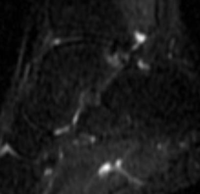
3 Results An additional MRI scan was done after 2 weeks of forced abnormal foot pronation utilizing the insert 11 participants developed bright signal consistent with fluid/water indicating bone marrow edema (BME) 1 participant showed involvement on the contralateral foot Location of BME Locations: Foot, Tibia, Femur Most were at metatarsal and phalangeal joints 8 phalanges 4 metatarsals The most common was the first ray Some were more pronounced than others with 2 appearing similar to a stress fracture 3


4 Initial 2 Weeks of Abnormal Pronation Before After 4
 to determine if the BME had resolved NO signal alteration was noted in")
5 Results Clinical Nearly all participants complained of pain or discomfort in the lower extremity during the study All volunteers were asymptomatic immediately after insert removal and at clinical follow up 1 day, 1 week, 1 month Imaging Follow Up 3 volunteers were images a 3 rd time (2 weeks after removal of the insert) to determine if the BME had resolved NO signal alteration was noted in the previous areas of BME in 2/3 One participant demonstrated minimal persistent edema that was more diffuse than when originally noted ALTERED BIOMECHANICS AND BONE MARROW EDEMA REVISITED Logan College of Chiropractic Research Study, St. Louis, MO PARTICIPATING INVESTIGATORS Dr. Alicia M. Yochum Principal Investigator Dr. Gary M. Guebert Dr. Jeff Thompson Dr. Terry R. Yochum Dr. Kim Christensen Dr. Reed B. Phillips Dr. Norman W. Kettner Dr. Mark Schweitzer (M.D.) 5

 All students are instructed to go about their normal activities of daily life to")
6 MATERIALS AND METHODS 22 total student participants 17 treatment participants 5 control participants Inclusion Criteria Normal BMI 20-30years old Exclusion Criteria Pre-existing abnormal pronation of the foot- Physical examination History of chronic low back or lower extremity pain in the last 6 months Use of opioid medications Runs more than 10 miles/week Preexisting conditions (metabolic, bone softening) Device that that would be incompatible with MRI (pacemaker) Methods 17 participants placed in unilateral FORCED pronation utilizing a 9/16 inch insert in their right shoe Control Group: 5 Randomly Selected Participants- No insert Undergo all other aspects of study (VAS, Biomechanical Pictures, MRI s) All students are instructed to go about their normal activities of daily life to include their normal exercise routine (running under 10mi/wk). 6


7 7
8 6 Week Protocol Time line Initial MRI scan to make sure participants do not have preexisting BME 2 Weeks- MRI Scan after insert was in place for 2 weeks 4 Weeks- MRI Scan after 2 additional weeks of abnormal pronation with the insert After this scan the insert was removed 6 Weeks- Follow up scan after 2 weeks without the pronation device to look for resolution of symptoms/edema At the time of each MRI scan, biomechanical pictures (overhead squat) were taken and a Numerical Rating Scale (NRS) was performed. IMAGING STUDIES All participants were scanned with a 1.5 Tesla MRI magnet. STIR images obtained Suppress all signal from fat so FLUID/EDEMA stands out White Bone marrow/trabecular bone is Black The areas scanned: BILATERAL Foot- Sagittal Ankle- Sagittal Knee- Coronal Hip- Coronal Sacroiliac Joint- Coronal Lower lumbar spine- Sagittal 8

9 Image Interpretation Two Radiologists certified by the American Chiropractic Board of Radiology (ACBR) Dr. Gary Guebert and Dr. Jeff Thompson Blinded as to which students have been pronated and which ones have not. Imaging RESULTS Talonavicular Joint- Initial Study NO BME 9


")


10 INITIAL MRI 2 MRI 3 MRI 4- Follow up 1 Posterior Lateral Talar Process (Stieda)


11 L5 Right MRI 2 Initial MRI 3 MRI 4- Follow up Lumbar Spine Pedicle L4 Left NO EDEMA Initial MRI 2 11
 All scores were 0 initially = Patients had NO low back pain or lower extremity")

12 MRI 3 MRI 4- Follow Up Pain Evaluation Done before the study began and every 2 weeks (MRI) All scores were 0 initially = Patients had NO low back pain or lower extremity pain Oswestry: Done before study began and at the time of the 3 rd MRI Right before the insert was taken out 13 participants developed pain in their foot and knee NO hip pain 12
 Not statistically significant although those that developed BME were all in the treatment group Not random incidence p value- 0.59 and 0.")
13 Oswestry Range: 6-58% Disability Average: 27% All began at 0% 17% of participants = SEVERE disability! 1 participant was 3% away from CRIPPLED Data Analysis Statistical Significance with p-value <0.05 Bone Marrow Edema Fisher s Exact (small sample size) Not statistically significant although those that developed BME were all in the treatment group Not random incidence p value and 0.77 (time point 2 and 3) Study is underpowered= not enough people Numerical Rating Scale Repeated ANOVA and T-Test Overall significance of pain over time: p-value <0.001 Significance between time point 1 and 2 as well as 3 and 4. Significance between the treatment and control: p-value <0.05 Significance in Knee pain in those who developed BME (p 0.01) Oswestry Pair-wise T-test Statistically significant difference in participants at the beginning of the study verses the end: p-value <0.001 Statistically significant difference in control verses treatment 13


14 What is pronation?... Normal part of the gait cycle Heel Strike: Supinated Midstance: Pronated Toe Off: Supinated Abnormal Pronation Toe out- Pronounced heel strike in supination Excessive pronation in midstance Increased load on 1 st toe at toe off Midstance Toe Off 3 ARCHES Heel Strike Biomechanical faults Possible Biomechanical effects of ABNORMAL Pronation Dropped Arch (Calcaneal eversion) Toe out Medial deviation of the knee Internal rotation and femur Genu valgus deformity Pelvic Unleveling Shoulder Unleveling 14
 3.")

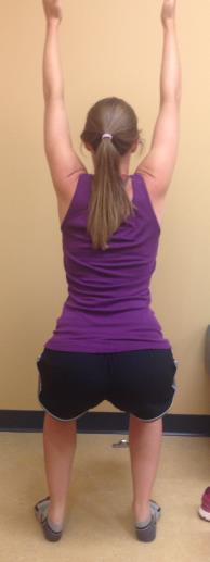
15 Comparison Without Custom Orthotics With Custom Orthotics- 1 Week Later What did we see in our study?... Overhead squat Analysis Most Common 1. Toe Out 2. Arch Drop (calcaneal eversion) 3. Knee Deviation (Medial/Lateral) 4. Forward Arms 5. Forward Lean Uncommon Forward Head Low Back Arch/Rounding Weight Shift NOT FOUND Heels Up PRE-EXISTING FUNCTIONAL BIOMECHANICAL FAULTS! 15


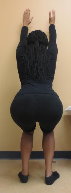


16 TOE OUT Dropped arch (calcaneal Eversion) 16


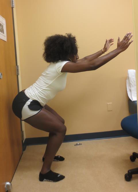


17 Knee Deviation More commonly encountered on the left Medial Lateral Forward Arms/Lean 17


18 What Causes Disc Degeneration?. Excessive mechanical loading! Causes a disc to degenerate by disrupting its structure and precipitating a cascade of nonreversible cell-mediated responses leading to further disruption. SPINE 2006 Sir Kirkaldy Willis MD 3 Stages of Degeneration Dysfunction Instability Stabilization PRE-EXISTING FUNCTIONAL BIOMECHANICAL FAULTS! 18


19 Calcaneal Inclination angle Normal: Hallux Valgus Bunion Normal: 5-10 Case Courtesy of Logan University 19


20 Knee Deviation VARUS VALGUS Hip OA Case Courtesy of Logan University 20


21 =Altered Mechanics 21


22 22
23 STRESS FRACTURES Fatigue Fracture Normal bone that fails under an abnormal load Insufficiency Fracture Abnormal bone that fails under a normal load Imaging Signs of Stress Fractures Solid Periosteal new bone formation (Plain Films) Transverse opaque bands when seen enface (Plain Films) Fracture line seldom seen on plain films but best seen on CT Positive bone scan (Planar) Bone marrow edema on MRI 23


24 INCIDENCE OF SPONDYLOLYSIS AND/OR SPONDYLOLISTHESIS CAUCASIAN: 5-7% AFRICAN AMERICANS: 2% ALASKAN ESKIMOS: 40% HIGHLY MOTIVATED ATHLETES PERFORMING HYPEREXTENSION: 15% GYMNASTS, DIVERS, POLE VAULTERS POWER WEIGHT LIFTERS: 40-50% 24

 AP Lumbar Spine No")
25 Tilt up view o cephalic tube tilt CR half way between umbilicus & pubic ramus (1.5 below ASIS) AP Lumbar Spine No tube tilt 25


26 Tilt up view L/S Jt. No tube tilt Tube tilt 26

27 Single leg hyperextension Stork test Physical examination revealed painful hyperextension with some relief during flexion. His pain was localized to the L4 segment The patient exhibited a positive Stork test Plain film radiographs, planar bone scan and two MRI scans (three months apart) were read as normal. 27


28 Planar Bone Scan Negative T2 Weighted Sagittal MRI Normal 28


29 Parasagittal T2- weighted MRI Scan - Bone Marrow Edema in the Left Pars Parasagittal T2- weighted MRI Scan - Bone Marrow Edema in the Right Pars 29


30 Axial T1-weighted MRI L4 SPECT Scan Single Photon Emission Computed Tomography Coronal 30

 Fat suppression technique or Fluid")
31 SPECT Scan Single Photon Emission Computed Tomography Parasagittal No more SPECT bone scans MRI with STIR (short tau inversion recovery) Fat suppression technique or Fluid sensitive imaging 31

32 Boston Brace International (508) EL RASSI, ET AL LUMBAR SPONDYLOLYSIS IN PEDIATRIC AND ADOLESCENT SOCCER PLAYERS AMERICAN JOURNAL OF SPORTS MEDICINE, , 2005 According to Rassi and colleagues, bracing itself, does not determine successful results, whereas physical activity restriction has a higher influence on clinical outcomes. The combination of both physical activity restriction and lumbar bracing would have a higher impact when the clinical outcome is compared to the use of either alone. 32
33 STEINER AND MICHELI TREATMENT OF SYMPTOMATIC SPONDYLOLYSIS AND SPONDYLOLISTHESIS WITH MODIFIED BOSTON BRACE SPINE:10: , 1985 According to Steiner and Micheli, clinical outcomes of patients undergoing conservative treatment, along with spinal bracing found that patients wearing the thoracolumbar orthosis obtained a higher functional outcome (100% excellent results), compared to those not wearing the braces (68% excellent results). Caution must be taken when attributing this increase function to the use of the thoracolumbar orthoses alone. Patients needing the brace were also patients needing a longer period of physical activity restriction. The conclusion is that the reduction in physical activity, rest and the thoracolumbar Boston Overlap Brace will yield the most positive clinical results. 33
DOES ALTERED BIOMECHANICS CAUSE BONE MARROW EDEMA?
 DOES ALTERED BIOMECHANICS CAUSE BONE MARROW EDEMA? Alicia M. Yochum RN, DC, DACBR, RMSK DOES ALTERED BIOMECHANICS CAUSE BONE MARROW EDEMA? Mark E. Schweitzer, MD and Lawrence M. White MD Department of
17/10/2017. Foot and Ankle
 17/10/2017 Alicia M. Yochum RN, DC, DACBR, RMSK Foot and Ankle Plantar Fasciitis Hallux Valgus Deformity Achilles Tendinosis Posterior Tibialis Tendon tendinopathy Stress Fracture Ligamentous tearing Turf
Case Studies: Low Back Pain in the Athlete. Jim Messerly DO
 Case Studies: Low Back Pain in the Athlete Jim Messerly DO Nothing to disclose Case #1 History 15 y/o male presents for evaluation of his low back pain. His pain has been present for several months. The
Sashil Kapur, MD Sports medicine fellow Lutheran General
 Sashil Kapur, MD Sports medicine fellow Lutheran General 1 Spondy-what? Presentation Diagnosis Treatment Return to play 2 Very common musculoskeletal complaint 10-15% of children and adolescents Adults
Dorsal surface-the upper area or top of the foot. Terminology
 It is important to learn the terminology as it relates to feet to properly communicate with referring physicians when necessary and to identify the relationship between the anatomical structure of the
Managing Tibialis Posterior Tendon Injuries
 Managing Tibialis Posterior Tendon Injuries by Thomas C. Michaud, DC Published April 1, 2015 by Dynamic Chiropractic Magazine Tibialis posterior is the deepest, strongest, and most central muscle of the
Hyperpronation of the foot causes many different
 IMMEDIATE CHANGES IN THE QUADRICEPS FEMORIS ANGLE AFTER INSERTION OF AN ORTHOTIC DEVICE D. Robert Kuhn, DC, a Terry R. Yochum, DC, b Anton R. Cherry, c and Sean S. Rodgers c ABSTRACT Objective: To measure
Balanced Body Movement Principles
 Balanced Body Movement Principles How the Body Works and How to Train it. Module 3: Lower Body Strength and Power Developing Strength, Endurance and Power The lower body is our primary source of strength,
Spondylolysis. Lysis (Greek λύσις, lýsis from lýein "to separate") refers to the breaking down.
 refers to the breaking down.") Spondylolysis Lysis (Greek λύσις, lýsis from lýein "to separate") refers to the breaking down. Thomas J Kishen Spine Surgeon Sparsh Hospital for Advanced Surgeries Bangalore Spondylolysis Defect in the
Spondylolysis Lysis (Greek λύσις, lýsis from lýein "to separate") refers to the breaking down. Thomas J Kishen Spine Surgeon Sparsh Hospital for Advanced Surgeries Bangalore Spondylolysis Defect in the
BIOMECHANICAL EXAMINATION OF THE PEDIATRIC LOWER EXTREMITY 2017
 BIOMECHANICAL EXAMINATION OF THE PEDIATRIC LOWER EXTREMITY 2017 B. RESSEQUE, D.P.M., D.A.B.P.O. Professor, N.Y. College of Podiatric Medicine ARCH HEIGHT OFF WEIGHTBEARING Evaluate arch height by placing
Sport Specific MRI. The symptoms of the majority, if not all sports injuries are experienced when upright, and weight-bearing
 Sport Specific MRI The symptoms of the majority, if not all sports injuries are experienced when upright, and weight-bearing A complete, accurate MRI assessment can only be made when in the position of
BIOMECHANICAL EXAMINATION OF THE PEDIATRIC LOWER EXTREMITY
 BIOMECHANICAL EXAMINATION OF THE PEDIATRIC LOWER EXTREMITY B.Resseque, D.P.M. ARCH HEIGHT OFF WEIGHTBEARING Evaluate arch height by placing a ruler from the heel to the first metatarsal head Compare arch
Spondylolysis To Brace or Not To Brace AMSSM 2014
 Spondylolysis To Brace or Not To Brace AMSSM 2014 Nothing to Disclose Disclosures To Brace? Goals of Treatment History/ Literature of Bracing Mechanics of Bracing Benefits to Brace. Earlier to return to
Evaluation of Gait Mechanics Using Computerized Plantar Surface Pressure Analysis and it s Relation to Common Musculoskeletal Problems
 Evaluation of Gait Mechanics Using Computerized Plantar Surface Pressure Analysis and it s Relation to Common Musculoskeletal Problems Laws of Physics effecting gait Ground Reaction Forces Friction Stored
The Athlete s Lumbar Spine: Current Concepts
 The Athlete s Lumbar Spine: Current Concepts Content / Objectives Anatomy Common injuries Treatment and prevention Pablo Vazquez Seoane, M.D. 44 th Annual Sports Medicine Symposium January 19 21, 2017
right Initial examination established that you have 'flat feet'. Additional information left Left foot is more supinated possibly due to LLD
 Motion analysis report for Feet In Focus at 25/01/2013 Personal data: Mathew Vaughan DEMO REPORT, 20 Churchill Way CF10 2DY Cardiff - United Kingdom Birthday: 03/01/1979 Telephone: 02920 644900 Email:
Quads (medicine ball)
") Saggital Front Reach Saggital Front Reach 1) Start position: Stand with feet hip width apart. Hold medicine ball or dumbbell at waist. 2) Step forward 2-3 feet with the heel striking first and lean torso
Saggital Front Reach Saggital Front Reach 1) Start position: Stand with feet hip width apart. Hold medicine ball or dumbbell at waist. 2) Step forward 2-3 feet with the heel striking first and lean torso
CHAPTER 8: THE BIOMECHANICS OF THE HUMAN LOWER EXTREMITY
 CHAPTER 8: THE BIOMECHANICS OF THE HUMAN LOWER EXTREMITY _ 1. The hip joint is the articulation between the and the. A. femur, acetabulum B. femur, spine C. femur, tibia _ 2. Which of the following is
IFAST Assessment. Name: Date: Sport: Review Health Risk Assessment on initial consult form. List Client Goals (what brings you here?
 IFAST Assessment Name: Date: Sport: Review Health Risk Assessment on initial consult form List Client Goals (what brings you here?) Cardiovascular Measurements Blood Pressure Resting Heart Rate Body Composition
2/24/2014. Outline. Anterior Orthotic Management for the Chronic Post Stroke Patient. Terminology. Terminology ROM. Physical Evaluation
 Outline Anterior Orthotic Management for the Chronic Post Stroke Patient Physical Evaluation Design Considerations Orthotic Design Jason M. Jennings CPO, LPO, FAAOP jajennings@hanger.com Primary patterning
CHRONIC FOOT PROBLEMS FOOT and ANKLE BASICS
 CHRONIC FOOT PROBLEMS FOOT and ANKLE BASICS ABC s of Comprehensive Musculoskeletal Care December 1 st, 2007 Stephen Pinney MD Chief, UCSF Foot and Ankle Service Chronic problems typically occur gradually
What Happens to the Paediatric Flat Foot? Peter J Briggs Freeman Hospital Newcastle upon Tyne
 What Happens to the Paediatric Flat Foot? Peter J Briggs Freeman Hospital Newcastle upon Tyne We don t know!! Population Studies 2300 children aged 4-13 years Shoe wearers Flat foot 8.6% Non-shoe wearers
One hundred and ten individuals participated in this study
 Purpose The purpose of this study was to compare gait characteristics in an asymptomatic population of younger and older adults to older OA patients of different severities Hypothesis(es) The following
Case Report: Metatarsalgia (by first ray insufficiency)
") Sergio Puigcerver (1) ; Juan Carlos González (1) ; Roser Part (1) ; Eduardo Brau (1) ; Felip Salinas (2) (1) Instituto de Biomecánica de Valencia, UPV. Valencia, España; ibv@ibv.upv.es ; www.ibv.org (2)
Sergio Puigcerver (1) ; Juan Carlos González (1) ; Roser Part (1) ; Eduardo Brau (1) ; Felip Salinas (2) (1) Instituto de Biomecánica de Valencia, UPV. Valencia, España; ibv@ibv.upv.es ; www.ibv.org (2)
Thoracolumbar Spine Conditions: Treatment and Return to Play
 Thoracolumbar Spine Conditions: Treatment and Return to Play C H R I S T O P H E R B U R K S, MD B I E N V I L L E O R T H O P A E D I C S P E C I A L I S T S O C E A N S P R I N G S, MS Thoracolumbar
Running Injuries in Children and Adolescents
 Running Injuries in Children and Adolescents Cook Children s SPORTS Symposium July 2, 2014 Running Injuries Overuse injuries Acute injuries Anatomic conditions 1 Overuse Injuries Pain that cannot be tied
Signal intensity changes of the posterior elements of the lumbar spine in symptomatic adults
 ORIGINAL ARTICLE SPINE SURGERY AND RELATED RESEARCH Signal intensity changes of the posterior elements of the lumbar spine in symptomatic adults Kosuke Sugiura, Toshinori Sakai, Fumitake Tezuka, Kazuta
Imaging of Ankle and Foot pain
 Imaging of Ankle and Foot pain Pramot Tanutit, M.D. Department of Radiology Faculty of Medicine, Prince of Songkla University 1 Outlines Plain film: anatomy Common causes of ankle and foot pain Exclude:
SpineFAQs. Lumbar Spondylolisthesis
 SpineFAQs Lumbar Spondylolisthesis Normally, the bones of the spine (the vertebrae) stand neatly stacked on top of one another. The ligaments and joints support the spine. Spondylolisthesis alters the
Movement Terminology. The language of movement is designed to allow us to describe how the body moves through space.
 Movement Terminology The language of movement is designed to allow us to describe how the body moves through space. In exercise it allows us to communicate with other movement professionals so we can describe
Clinical Evaluation and Imaging of the Patellofemoral Joint Common clinical syndromes
 Clinical Evaluation and Imaging of the Patellofemoral Joint Common clinical syndromes A. Panagopoulos Lecturer in Orthopaedics Medical School, Patras University Objectives Anatomy of patellofemoral joint
The BioMechanics Method
 The BioMechanics Method EXERCISE SOLUTIONS FOR CHRONIC PAIN The Fundamentals of Structural Assessment End of Section Self-Check There are many things that can make conducting a structural assessment more
Medical Terminology. Unit 2
 Medical Terminology Unit 2 Students will apply medical terminology. Objective 1: Identify and utilize anatomical positions, planes, and directional terms. Demonstrate what anatomical position is and how
Imaging of Trauma to the Spine. Orthopedic Diplomate Program University of Bridgeport College of Chiropractic
 Imaging of Trauma to the Spine Orthopedic Diplomate Program University of Bridgeport College of Chiropractic Jefferson Fracture Yee, LL: The Jefferson Fracture, Radiology Cases in Pediatric Emergency Medicine.
Medical Terminology. Anatomical Position, Directional Terms and Movements
 Medical Terminology Anatomical Position, Directional Terms and Movements What we will cover... Content Objectives Students will be able to gain a better understanding and application of medical terminology
Lumbar epidural hematoma associated with spondylolyses
 J Neurosurg Spine 8:174 180, 2008 Lumbar epidural hematoma associated with spondylolyses Report of 3 cases CHIMA O. OHAEGBULAM, M.D., 1 IAN F. DUNN, M.D., 2 PIERRE D HEMECOURT, M.D., 3 AND MARK R. PROCTOR,
THE SELECTIVE FUNCTIONAL MOVEMENT ASSESSMENT
 - 33 - THE SELECTIVE FUNCTIONAL MOVEMENT ASSESSMENT SFMA SCORING FP DP Active Cervical Flexion Active Cervical Extension Cervical Rotation Upper Extremity Pattern 1(MRE) Upper Extremity Pattern 2 (LRF)
BUCKS MSK: FOOT AND ANKLE PATHWAY GP MANAGEMENT. Hallux Valgus. Assessment: Early Management. (must be attempted prior to any referral to imsk):
:") Hallux Valgus Common condition: affecting around 28% of the adult population. Prevalence increases with age and in females. Observation: Lateral deviation of the great toe. May cause secondary irritation
Hallux Valgus Common condition: affecting around 28% of the adult population. Prevalence increases with age and in females. Observation: Lateral deviation of the great toe. May cause secondary irritation
6.4 The Ankle. Body Divided into Planes. Health Services: Unit 6 Arms and Legs. Body Movement Vocabulary
 6.4 The Ankle Body Movement Vocabulary When fitness professionals refer to movement of the body, the pattern of movement is described from the anatomical position This position can best be described as
Running Athlete: Part C. Case Analysis Materials
 Running Athlete: Part C Case Analysis Materials Case 1 Subjective Examination (performed offcamera) Runs very sporadically, but generally 2-3 x per week around 2-4 miles Play recreational soccer Denies
Cox Technic Case Report #169 published at (sent 5/9/17) 1
 1") Cox Technic Case Report #169 published at www.coxtechnic.com (sent 5/9/17) 1 Management of Lumbar Radiculopathy Associated with an Extruded L4 L5 disc and concurrent L5 S1 Spondylolytic Spondylolisthesis
Cox Technic Case Report #169 published at www.coxtechnic.com (sent 5/9/17) 1 Management of Lumbar Radiculopathy Associated with an Extruded L4 L5 disc and concurrent L5 S1 Spondylolytic Spondylolisthesis
A Patient s Guide to Flatfoot Deformity (Pes Planus) in Children
 in Children") A Patient s Guide to Flatfoot Deformity (Pes Planus) in Children 2350 Royal Boulevard Suite 200 Elgin, IL 60123 Phone: 847.931.5300 Fax: 847.931.9072 DISCLAIMER: The information in this booklet is compiled
A Patient s Guide to Flatfoot Deformity (Pes Planus) in Children 2350 Royal Boulevard Suite 200 Elgin, IL 60123 Phone: 847.931.5300 Fax: 847.931.9072 DISCLAIMER: The information in this booklet is compiled
Functional biomechanics of the lower limb
 Functional biomechanics of the lower limb Ben and Matt. 24th July 2011 Principles of function Gravity Ground reaction Eco-concentric eccentric loading (preload) of a muscle (or group) is essential for
Body Organizations Flashcards
 1. What are the two main regions of the body? 2. What three structures are in the Axial Region? 1. Axial Region (Goes down midline of the body) 2. Appendicular Region (limbs) 3. Axial Region (Goes down
Medical Terminology. Anatomical Position, Directional Terms and Movements
 Medical Terminology Anatomical Position, Directional Terms and Movements What we will cover... Content Objectives Students will be able to gain a better understanding and application of medical terminology
anchor point. Essentially, the more the body is parallel to the floor, the more difficult the exercise. The third set of pictures shows two common
 ST BOWLER SQUAT The bowler squat performed with a suspension trainer is a great little single leg squat/hinge combo! The suspension trainer assists with balance and I believe makes the exercise more effective.
Functional Movement Screen (Cook, 2001)
") Functional Movement Screen (Cook, 2001) TEST 1 DEEP SQUAT Purpose - The Deep Squat is used to assess bilateral, symmetrical, mobility of the hips, knees, and ankles. The dowel held overhead assesses bilateral,
Functional Movement Screen (Cook, 2001) TEST 1 DEEP SQUAT Purpose - The Deep Squat is used to assess bilateral, symmetrical, mobility of the hips, knees, and ankles. The dowel held overhead assesses bilateral,
Comprehension of the common spine disorder.
 Objectives Comprehension of the common spine disorder. Disc degeneration/hernia. Spinal stenosis. Common spinal deformity (Spondylolisthesis, Scoliosis). Osteoporotic fracture. Anatomy Anatomy Anatomy
Physical Examination of the Foot & Ankle
 Inspection Standing, feet straight forward facing toward examiner Swelling Deformity Flatfoot (pes planus and hindfoot valgus) High arch (pes cavus and hindfoot varus) Peek-a-boo heel Varus Too many toes
Case 1 7 yo male Right elbow injury 3 months ago Medial elbow pain and tenderness over medial epicondyle Long arm cast given but off himself 1 month a
 Case presentations Case 1 7 yo male Right elbow injury 3 months ago Medial elbow pain and tenderness over medial epicondyle Long arm cast given but off himself 1 month after Progressive limited elbow flexion
6/5/2018. Forefoot Disorders. Highgate Private Hospital (Royal Free London NHS Foundation Trust (Barnet & Chase Farm Hospitals) Hallux Rigidus
 Hallux Rigidus") Forefoot Disorders Mr Pinak Ray (MS, MCh(Orth), FRCS, FRCS(Tr&Orth)) Highgate Private Hospital (Royal Free London NHS Foundation Trust (Barnet & Chase Farm Hospitals) E: ray.secretary@uk-conslutants Our
Forefoot Disorders Mr Pinak Ray (MS, MCh(Orth), FRCS, FRCS(Tr&Orth)) Highgate Private Hospital (Royal Free London NHS Foundation Trust (Barnet & Chase Farm Hospitals) E: ray.secretary@uk-conslutants Our
Lumbar Spine Applied Anatomy. Jason Zafereo, PT, OCS, FAAOMPT Clinical Orthopedic Rehabilitation Education
 Lumbar Spine Applied Anatomy Jason Zafereo, PT, OCS, FAAOMPT Clinical Orthopedic Rehabilitation Education Objectives Discuss concepts relevant to pathophysiology and differential diagnosis for lumbar radiculopathy
10/22/15. Running Injury Mechanics. Excessive Pronation. Distribution of Running Injuries. 1. Patellofemoral Pain Syndrome
 Distribution of Running Injuries Metarsalgia/Stress Fx Running Injury Mechanics Irene S. Davis, PhD, PT, FACSM, FAPTA, FASB Dept. PM&R, Harvard Medical School Director, Spaulding National Running Center
USCF 6 th Annual Primary Care Sports Medicine Conference: ABCs of Musculoskeletal Care
 USCF 6 th Annual Primary Care Sports Medicine Conference: ABCs of Musculoskeletal Care Back Injuries in the Young Athlete Lyle J. Micheli, MD Mechanism of Injury 1. Acute macrotrauma 2. Repetitive microtrauma
the muscle that opposes the action of a joint about an axis
 Adams forward bend test Aetiology Agonist Ambulation Anisomelia Antagonist Antagonistic pelvic torsion the patient bends forward to emphasise any asymmetry in the rib cage or loin on the back for the clinical
LEG LENGTH INEQUALITY: Sports Medicine Perspective
 LEG LENGTH INEQUALITY: Sports Medicine Perspective Debra A. Zillmer, M.D. M&M Orthopaedics, Ltd 18 Year Old Experienced Cross Country Runner: Sept Sr Year Pain in left lower leg with running Pain now prevents
Redirect GRF to Affect Mobility, Stability or Load? Increase/Decrease Joint Moments to Reduce Stress Strain Relationships?
 5-1 SECTION 5 CRITICAL DECISION MAKING IN ORTHOTIC THERAPY QUESTIONS Answering the some critical (as in choosing between criteria) questions should help as a guide to selecting an appropriate orthosis,
BORGinsole Measurement devices
 BORGinsole Measurement devices BORGinsole Angle-Finder Dorsal Flexion of the first Metatarsophalangeal joint - P. is sitting up on the examination table, with legs straight. - T. is sitting at the end
Instructional Course Lecture 2011
 Instructional Course Lecture 2011 Yoon Hae Kwak Dept. of Orthopaedic Surgery Hallym University Sacred Heart Hospital Hallym University Medical Center Rotational and Angular variations of the lower extremities
Spondylolysis repair using a pedicle screw hook or claw-hook system. a comparison of bone fusion rates
 ORIGINAL ARTICLE SPINE SURGERY AND RELATED RESEARCH Spondylolysis repair using a pedicle screw hook or claw-hook system. a comparison of bone fusion rates Ko Ishida 1), Yoichi Aota 2), Naoto Mitsugi 1),
Nicky Schmidt PT, C/NDT 1
 Preparing the foot for third rocker and initial contact Nicky Schmidt PT, C/NDT copyright 2012 References Laboratory Strategies developed and taught by Nicky Schmidt, P.T. in the NDTA Approved Advanced
Posterior Tibialis Tendon Dysfunction & Repair
 1 Posterior Tibialis Tendon Dysfunction & Repair Surgical Indications and Considerations Anatomical Considerations: The posterior tibialis muscle arises from the interosseous membrane and the adjacent
OTM Lecture Gait and Somatic Dysfunction of the Lower Extremity
 OTM Lecture Gait and Somatic Dysfunction of the Lower Extremity Somatic Dysfunction Tenderness Asymmetry Range of Motion Tissue Texture Changes Any one of which must be present to diagnosis somatic dysfunction.
DB HAMMER CURL: 1-LEG SUPPORTED ALT- ARM + ISO-HOLD
 DB HAMMER CURL: 1-LEG SUPPORTED ALT- ARM + ISO-HOLD The single-leg supported alternating-arm DB hammer curl with iso-hold requires you to maintain a stable position on one leg while performing a biceps
ChiroCredit.com Presents Biomechanics: Focus on
 ChiroCredit.com Presents Biomechanics: Focus on the Knee Presented by: Ivo Waerlop, DC Shawn Allen, DC 1 Focus on The Knee 2 Pertinent Anatomy Femur Tibia Fibula Patella Prepatellar bursa Infrapatellar
Footwear, Orthotics, Taping and Bracing. Types of Feet. Types of Footwear. Types of Feet. Footwear, Orthotics, Bracing, and Taping Course Objectives
 Footwear, Orthotics, Bracing, and Taping Course Objectives Footwear, Orthotics, Taping and Bracing Laura Fralich, MD Primary Care Update Friday, May 4, 2017 Better understand types of footwear and the
TPW 's Shin Splints Menu
 TPW 's Shin Splints Menu # Sets Reps Duration E-cise 1 1 40 Supine Foot Circles & Point/Flexes 2 2 1 0:01:00 Supine Calf & Hamstring Stretch 3 1 1 0:02:00 Static Extension Position 4 1 1 0:02:00 Airbench
Intoeing: When to Worry? Sukhdeep K. Dulai SPORC 2018
 Intoeing: When to Worry? Sukhdeep K. Dulai SPORC 2018 What is it? Intoeing: When to worry? Why isn t it always cause for worry? What are the benign causes of intoeing? What are the pathologic causes of
Gait Analysis: Qualitative vs Quantitative What are the advantages and disadvantages of qualitative and quantitative gait analyses?
 Gait Analysis: Qualitative vs Quantitative What are the advantages and disadvantages of qualitative and quantitative gait analyses? Basics of Gait Analysis Gait cycle: heel strike to subsequent heel strike,
Spondylolysis DESCRIPTION EXPECTED OUTCOME POSSIBLE COMPLICATIONS COMMON SIGNS AND SYMPTOMS GENERAL TREATMENT CONSIDERATIONS CAUSES
 DESCRIPTION is a stress or fatigue fracture of the bones of the spine (vertebrae) that does not involve the main weight-bearing part of those bones, the body of the vertebra. Instead, it involves an area
Routine Guide EXAMINATION PROJECTION CASSETTE SIZE NOTES PRINT ORIENTATION. 14x17 CW* 14x17LW 14x17LW. 14x17LW 14x17LW 14x17LW
 EXAMINATION PROJECTION CASSETTE SIZE NOTES PRINT ORIENTATION A-C Joints without weights with weights 14x17 CW* One 14x17 divided; both shoulders on one exposure. *If part does not fit, do 10x12s CW. Both
Life. Uncompromised. The KineSpring Knee Implant System Surgeon Handout
 Life Uncompromised The KineSpring Knee Implant System Surgeon Handout 2 Patient Selection Criteria Patient Selection Criteria Medial compartment degeneration must be confirmed radiographically or arthroscopically
Active-Assisted Stretches
 1 Active-Assisted Stretches Adequate flexibility is fundamental to a functional musculoskeletal system which represents the foundation of movement efficiency. Therefore a commitment toward appropriate
DEEP SQUAT. Upper torso is parallel with tibia or toward vertical Femur below horizontal Knees are aligned over feet Dowel aligned over feet
 APPENDIX 9 SCORING CRITERIA DEEP SQUAT Upper torso is parallel with tibia or toward vertical Femur below horizontal Knees are aligned over feet Dowel aligned over feet Upper torso is parallel with tibia
Today s session. Common Problems in Rehab. Tim Keeley B.Phty, Cred.MDT, APA Principal Physiotherapist. physiofitness.com.au facebook.
 Tim Keeley B.Phty, Cred.MDT, APA Principal Physiotherapist physiofitness.com.au facebook.com/physiofitness Today s session Essential list for the lower body Rehab starting point Focussing on activation,
Body Planes & Positions
 Learning Objectives Objective 1: Identify and utilize anatomical positions, planes, and directional terms. Demonstrate what anatomical position is and how it is used to reference the body. Distinguish
Overuse Injuries & special skeletal injuries Dr M.Taghavi Director of sport medicine center of olympic academy
 Overuse Injuries & special skeletal injuries Dr M.Taghavi Director of sport medicine center of olympic academy Prevalence of Overuse Injuries 30 to 50% of all sport injuries are from overuse In some sports
Static Back. Instructions: Purpose: Hold this ecise for 05 min. prepared for Pain Free Posture MN
 1 Static Back Hold this ecise for 05 min. 1. Lie on your back with your legs up over a block or chair 2. Place your arms out to the sides at 45 degrees from your body with palms up 3. Relax your upper
ORTHOTIC REFERENCE GUIDE. srtprosthetics.com
 ORTHOTIC REFERENCE GUIDE srtprosthetics.com 1.866.633.3961 AFO, Ankle Foot Orthosis, Articulated (Custom-Molded) Provides adjustable control of the ankle and foot Designed with an articulated ankle and
The Relationship between Medial Longitudinal Arch and Peroneal Tubercle
 The Relationship between Medial Longitudinal Arch and Peroneal Tubercle -Anatomical Study- Department of Orthopaedic Surgery Kangwon National University Hospital Chuncheon, Republic of Korea Chang Hyun
SUBTALAR ARTHROEREISIS IN THE OLDER PATIENT
 C H A P T E R 1 7 SUBTALAR ARTHROEREISIS IN THE OLDER PATIENT William D. Fishco, DPM, MS INTRODUCTION Arthroereisis is a surgical procedure designed to limit the motion of a joint. Subtalar joint arthroereisis
Recognizing common injuries to the lower extremity
 Recognizing common injuries to the lower extremity Bones Femur Patella Tibia Tibial Tuberosity Medial Malleolus Fibula Lateral Malleolus Bones Tarsals Talus Calcaneus Metatarsals Phalanges Joints - Knee
Degenerative knee disorders. Management of knee pain An Orthotists perspective
 Degenerative knee disorders Management of knee pain An Orthotists perspective Orthotists role Reduce pain Help to preserve the joint Delay surgery Allow continued activity -Exercise /walking -Recreation
Evaluation and Management of Knee Pain. Michael Cassat, MD University of Arkansas for Medical Sciences
 Evaluation and Management of Knee Pain Michael Cassat, MD University of Arkansas for Medical Sciences Disclosure I have no actual or potential conflict of interest in relation to this program/presentation.
.org. Posterior Tibial Tendon Dysfunction. Anatomy. Cause. Symptoms
 Posterior Tibial Tendon Dysfunction Page ( 1 ) Posterior tibial tendon dysfunction is one of the most common problems of the foot and ankle. It occurs when the posterior tibial tendon becomes inflamed
Board Positions. Skill progression from beginner to advanced: 2 Half Balls 1 Half Ball 1 Half Ball and 1 Ball 2 Balls 1 Ball
 Feet facing forward on bolts General athletic stance for pushing and pulling Feet facing at an angle Movement transition for rotational and agility Wide in-line stance Simulate forward weight shift running
ORTHOSCAN MOBILE DI POSITIONING GUIDE
 ORTHOSCAN MOBILE DI POSITIONING GUIDE Table of Contents SHOULDER A/P of Shoulder... 4 Tangential (Y-View) of Shoulder... 5 Lateral of Proximal Humerus... 6 ELBOW A/P of Elbow... 7 Extended Elbow... 8 Lateral
University of Jordan. Professor Freih Abuhassan -
 Freih Odeh Abu Hassan F.R.C.S.(Eng.), F.R.C.S.(Tr.& Orth.). Professor of Orthopedics University of Jordan 1 A. Sacroiliitis History Trauma is very common Repetitive LS motion--lumbar rotation or axial
(July, 0) Conflicts of interest All the authors confirm that there are no conflicts of interest with people or organizations that could bias the natur
 Conflicts of interest All the authors confirm that there are no conflicts of interest with people or organizations that could bias the natur") (July, 0) Bony Healing of Discontinuous Laminar Stress Fractures due to Contralateral Pars Defect or Spina Bifida Occulta Toshinori Sakai, Tsuyoshi Goto, Kosuke Sugiura, Hiroaki Manabe, FumitakeTezuka,
(July, 0) Bony Healing of Discontinuous Laminar Stress Fractures due to Contralateral Pars Defect or Spina Bifida Occulta Toshinori Sakai, Tsuyoshi Goto, Kosuke Sugiura, Hiroaki Manabe, FumitakeTezuka,
Case Iselin's disease in a Thai boxer.
 Case 13609 Iselin's disease in a Thai boxer. Joris De Win 1, 3, Filip Vanhoenacker 2, 4, Els Goossens3 1: Department of Physical Medicine and Rehabilitation; University Ghent (UGent), Belgium; Email:de_win_joris@hotmail.com
Full Body (medicine ball) Saggital Front Reach
 Saggital Front Reach") Saggital Front Reach Saggital Front Reach 1) Start position: Stand with feet hip width apart. Hold medicine ball or dumbbell at waist. 2) Step forward 2-3 feet with the heel striking first and lean torso
Saggital Front Reach Saggital Front Reach 1) Start position: Stand with feet hip width apart. Hold medicine ball or dumbbell at waist. 2) Step forward 2-3 feet with the heel striking first and lean torso
Cruciform anterior spinal hyperextension (CASH) brace with round anterior chest pads.
 brace with round anterior chest pads.") General Description Thoracolumbar orthoses (TLOs) are used mainly to treat fractures between T10 and L2, because their mobility is not restricted by the ribs, unlike fractures between T2 and T9. Immobilization
General Description Thoracolumbar orthoses (TLOs) are used mainly to treat fractures between T10 and L2, because their mobility is not restricted by the ribs, unlike fractures between T2 and T9. Immobilization
MEDIAL TIBIAL STRESS, SHIN SPLINTS
 10 MEDIAL TIBIAL STRESS, SHIN SPLINTS What is Medial Tibial Stress Syndrome (MTSS)? Medial tibial stress syndrome (MTSS), commonly encompassed under the umbrella term shin splints, occurs along the bottom
emoryhealthcare.org/ortho
 COMMON SOCCER INJURIES Oluseun A. Olufade, MD Assistant Professor, Department of Orthopedics and PM&R 1/7/18 GOALS Discuss top soccer injuries and treatment strategies Simplify hip and groin injuries in
PREPARED FOR. Marsha Eichhorn DATE OF INJURY : N/A DATE OF ANALYSIS : 12/14/2016 DATE OF IMAGES : 12/8/2016. REFERRING DOCTOR : Dr.
 Accent on Health Chiropractic 405 Firemans Ave PREPARED FOR Marsha Eichhorn DATE OF INJURY : N/A DATE OF ANALYSIS : 12/14/2016 DATE OF IMAGES : 12/8/2016 REFERRING DOCTOR : Dr. David Bohn This report contains
SESSION #207 UNDERSTANDING FUNCTION FROM THE GROUND UP Greg Roskopf, MA Owner/developer of Muscle Activation Techniques
 SESSION #207 UNDERSTANDING FUNCTION FROM THE GROUND UP Greg Roskopf, MA Owner/developer of Muscle Activation Techniques PRESCRIBING EXERCISE AS A COMPONENT OF HEALTH: PEOPLE ARE COMING TO US TO GET HEALTHY!
ATHLETIC CONDITIONING ON THE ARC BARREL
 ATHLETIC CONDITIONING ON THE ARC BARREL page 1 INTRODUCTION The STOTT PILATES Athletic Conditioning stream serves as a bridge between STOTT PILATES standard repertoire and the CORE Athletic Conditioning
Appendix H: Description of Foot Deformities
 Appendix H: Description of Foot Deformities The following table provides the description for several foot deformities: hammer toe, claw toe, hallux deformity, pes planus, pes cavus and charcot arthropathy.
Evaluating the Athlete Questionnaire
 Evaluating the Athlete Questionnaire Prior to developing the strength and conditioning training plan the coach should first evaluate factors from the athlete s questionnaire that may impact the strength
Dingjun Hao, Baorong He, Liang Yan. Hong Hui Hospital, Xi an Jiaotong University College. of Medicine, Xi an, Shaanxi , China
 Xi an Hong Hui Hospital Xi an, Shaanxi, China The difference of occurring superior adjacent segment pathology after lumbar posterolateral fusion by using two different pedicle screw insertion techniques