DOES ALTERED BIOMECHANICS CAUSE BONE MARROW EDEMA?
|
|
|
- Bathsheba Sparks
- 5 years ago
- Views:
Transcription


1 DOES ALTERED BIOMECHANICS CAUSE BONE MARROW EDEMA? Alicia M. Yochum RN, DC, DACBR, RMSK DOES ALTERED BIOMECHANICS CAUSE BONE MARROW EDEMA? Mark E. Schweitzer, MD and Lawrence M. White MD Department of Radiology Thomas Jefferson University Hospital Philadelphia, Pennsylvania Radiology 198:851 March
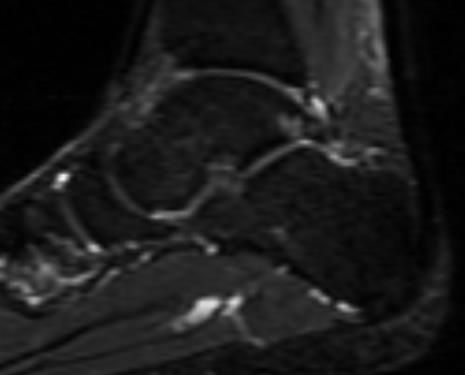
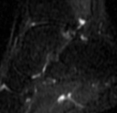
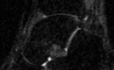
2 WHAT IS BONE MARROW EDEMA? Inflammation in the bone Injury to the trabeculae causing it to bleed Repetitive Impact Controversial Etiology Blood= Fluid Fluid= High signal Fluid = White Marrow (Fat)= Black 12 Participants 6 Women 6 Men Age: (Mean 30) All asymptomatic and without abnormal pronation MATERIALS AND METHODS MRI baseline Bilateral foot, ankle, knee, and hip Scans utilizing a 1.5 Tesla magnet was done utilizing STIR imaging which suppresses fat signal and enhances water signal Insert 9/16 (1.4cm) longitudinal metatarsal arch pad inserted into the shoes of one foot of each volunteer Forces the participant into unilateral abnormal foot pronation 2
3 RESULTS An additional MRI scan was done after 2 weeks of forced abnormal foot pronation utilizing the insert 11 participants developed bright signal consistent with fluid/water indicating bone marrow edema (BME) 1 participant showed involvement on the contralateral foot LOCATION OF BME Locations: Foot, Tibia, Femur Most were at metatarsal and phalangeal joints 8 phalanges 4 metatarsals The most common was the first ray Some were more pronounced than others with 2 appearing similar to a stress fracture 3


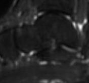
4 Initial STIR WEIGHTED IMAGE 2 Weeks of Abnormal Pronation Before After 4
 to determine if the BME had resolved NO signal alteration was noted in")
5 RESULTS Clinical Nearly all participants complained of pain or discomfort in the lower extremity during the study All volunteers were asymptomatic immediately after insert removal and at clinical follow up 1 day, 1 week, 1 month Imaging Follow Up 3 volunteers were images a 3 rd time (2 weeks after removal of the insert) to determine if the BME had resolved NO signal alteration was noted in the previous areas of BME in 2/3 One participant demonstrated minimal persistent edema that was more diffuse than when originally noted ALTERED BIOMECHANICS AND BONE MARROW EDEMA REVISITED Logan College of Chiropractic Research Study, St. Louis, MO PARTICIPATING INVESTIGATORS Dr. Alicia M. Yochum Principal Investigator Dr. Gary M. Guebert Dr. Jeff Thompson Dr. Terry R. Yochum Dr. Kim Christensen Dr. Reed B. Phillips Dr. Norman W. Kettner Dr. Mark Schweitzer (M.D.) 5

 All students are instructed to go about their normal activities of daily life to")
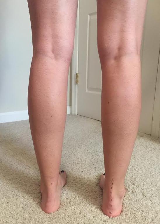
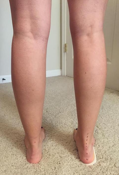
6 MATERIALS AND METHODS 22 total student participants 17 treatment participants 5 control participants Inclusion Criteria Normal BMI 20-30years old Exclusion Criteria Pre-existing abnormal pronation of the foot- Physical examination History of chronic low back or lower extremity pain in the last 6 months Use of opioid medications Runs more than 10 miles/week Preexisting conditions (metabolic, bone softening) Device that that would be incompatible with MRI (pacemaker) METHODS 17 participants placed in unilateral FORCED pronation utilizing a 9/16 inch insert in their right shoe Control Group: 5 Randomly Selected Participants- No insert Undergo all other aspects of study (VAS, Biomechanical Pictures, MRI s) All students are instructed to go about their normal activities of daily life to include their normal exercise routine (running under 10mi/wk). 6
7 7
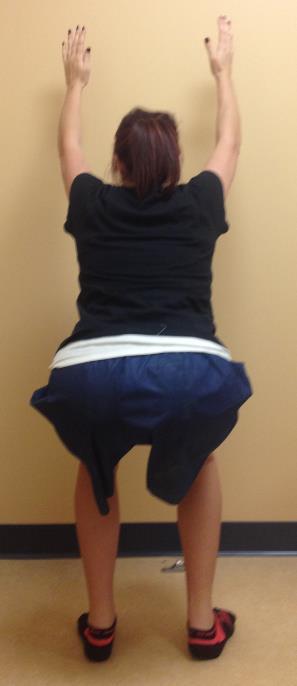
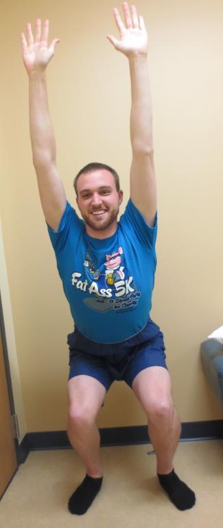
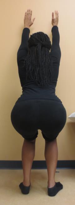
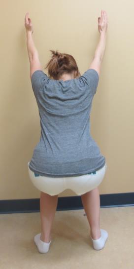
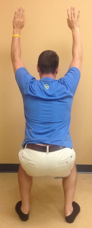
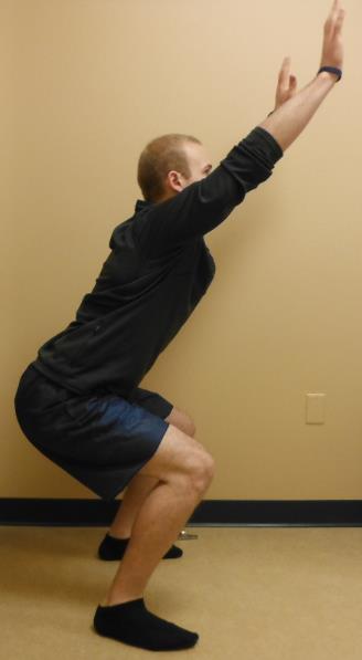
8 TIME LINE 6 Week Protocol Initial MRI scan to make sure participants do not have preexisting BME 2 Weeks- MRI Scan after insert was in place for 2 weeks 4 Weeks- MRI Scan after 2 additional weeks of abnormal pronation with the insert After this scan the insert was removed 6 Weeks- Follow up scan after 2 weeks without the pronation device to look for resolution of symptoms/edema At the time of each MRI scan, biomechanical pictures (overhead squat) were taken and a Numerical Rating Scale (NRS) was performed. IMAGING STUDIES All participants were scanned with a 1.5 Tesla MRI magnet. STIR images obtained Suppress all signal from fat so FLUID/EDEMA stands out White Bone marrow/trabecular bone is Black The areas scanned: BILATERAL Foot- Sagittal Ankle- Sagittal Knee- Coronal Hip- Coronal Sacroiliac Joint- Coronal Lower lumbar spine- Sagittal 8

9 Two Radiologists certified by the American Chiropractic Board of Radiology (ACBR) Dr. Gary Guebert and Dr. Jeff Thompson IMAGE INTERPRETATION Blinded as to which students have been pronated and which ones have not. Talonavicular Joint- Initial Study NO BME IMAGING RESULTS 9


")
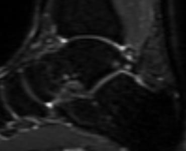
10 INITIAL MRI 2 MRI 3 MRI 4- Follow up 1 Posterior Lateral Talar Process (Stieda)


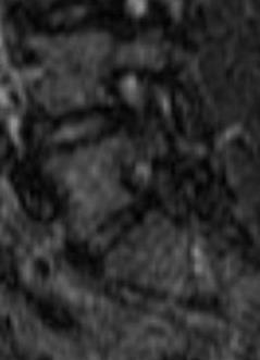
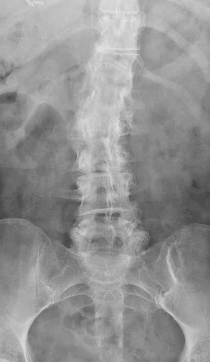
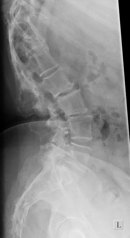
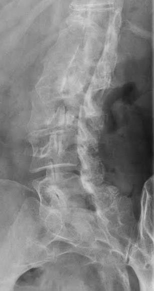
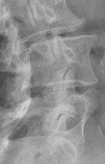
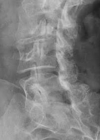
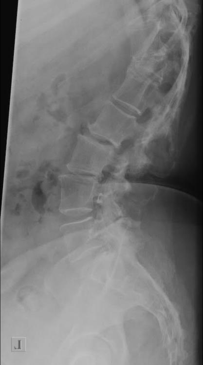
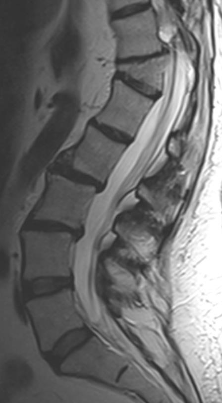
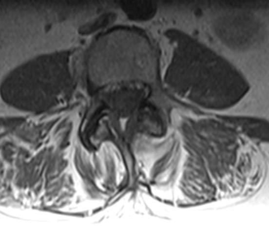
11 L5 RIGHT MRI 2 MRI 4- Follow up Initial MRI 3 LUMBAR SPINE PEDICLE L4 LEFT Initial MRI 2 11
 All scores were 0")


12 MRI 3 MRI 4- Follow Up PAIN EVALUATION Done before the study began and every 2 weeks (MRI) All scores were 0 initially = Patients had NO low back pain or lower extremity pain Oswestry: Done before study began and at the time of the 3 rd MRI Right before the insert was taken out 13 participants developed pain in their foot and knee NO hip pain 12
 Not statistically significant although those that developed BME were all in the treatment group Not random incidence p value- 0.59 and 0.")
13 Range: 6-58% Disability Oswestry Average: 27% All began at 0% 17% of participants = SEVERE disability! 1 participant was 3% away from CRIPPLED DATA ANALYSIS Statistical Significance with p-value <0.05 Bone Marrow Edema Fisher s Exact (small sample size) Not statistically significant although those that developed BME were all in the treatment group Not random incidence p value and 0.77 (time point 2 and 3) Study is underpowered= not enough people Numerical Rating Scale Repeated ANOVA and T-Test Overall significance of pain over time: p-value <0.001 Significance between time point 1 and 2 as well as 3 and 4. Significance between the treatment and control: p-value <0.05 Significance in Knee pain in those who developed BME (p 0.01) Oswestry Pair-wise T-test Statistically significant difference in participants at the beginning of the study verses the end: p-value <0.001 Statistically significant difference in control verses treatment 13


14 Normal part of the gait cycle Heel Strike: Supinated Midstance: Pronated Toe Off: Supinated WHAT IS PRONATION?... Abnormal Pronation Toe out- Pronounced heel strike in supination Excessive pronation in midstance Increased load on 1 st toe at toe off Toe Off Midstance 3 ARCHES Heel Strike 3 Arches Anterior Transverse Arch Lateral Longitudinal Medial Longitudinal 14
 Toe")


 4. Forward Arms 5.")

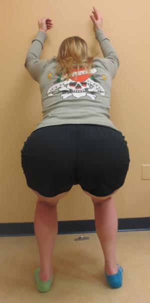
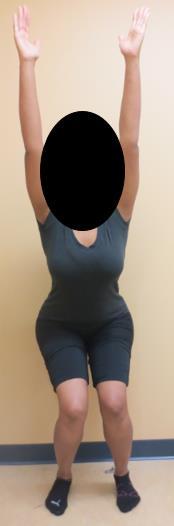
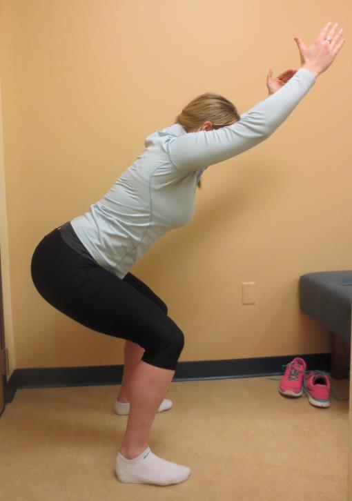
15 BIOMECHANICAL FAULTS Possible Biomechanical effects of ABNORMAL Pronation Dropped Arch (Calcaneal eversion) Toe out Medial deviation of the knee Internal rotation and femur Genu valgus deformity Pelvic Unleveling Shoulder Unleveling Most Common 1. Toe Out 2. Arch Drop (calcaneal eversion) 3. Knee Deviation (Medial/Lateral) 4. Forward Arms 5. Forward Lean WHAT DID WE SEE IN OUR STUDY?... OVERHEAD SQUAT ANALYSIS Uncommon Forward Head Low Back Arch/Rounding Weight Shift NOT FOUND Heels Up PRE-EXISTING FUNCTIONAL BIOMECHANICAL FAULTS! 15

16 TOE OUT Dropped arch (calcaneal Eversion) 16


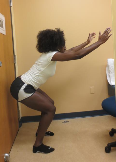
17 Knee Deviation More commonly encountered on the left Medial Lateral Forward Arms/Lean 17
18 Structural WOLFF S Imbalances LAW Leads to Dysfunction Wolff s Law: Any bone under stress, given time, may cause bone production in attempt to strengthen and/or stabilize the bony structure 18


19 SIR KIRKALDY WILLIS MD 3 Stages of Degeneration Dysfunction Instability Stabilization PRE-EXISTING FUNCTIONAL BIOMECHANICAL FAULTS! KNEE DEVIATION VARUS VALGUS 19


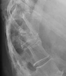
20 HIP OA Case Courtesy of Logan University Compression Fracture = Altered Mechanics 20

21 21
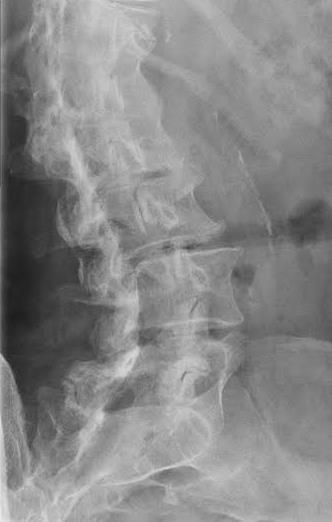
Altered Biomechanics and Spondylolysis
 Altered Biomechanics and Spondylolysis Terry R. Yochum DC, DACBR, Fellow ACCR Alicia M. Yochum RN, DC, DACBR, RMSK Does Altered Biomechanics Cause Bone Marrow Edema? Mark E. Schweitzer, MD and Lawrence
17/10/2017. Foot and Ankle
 17/10/2017 Alicia M. Yochum RN, DC, DACBR, RMSK Foot and Ankle Plantar Fasciitis Hallux Valgus Deformity Achilles Tendinosis Posterior Tibialis Tendon tendinopathy Stress Fracture Ligamentous tearing Turf
Dorsal surface-the upper area or top of the foot. Terminology
 It is important to learn the terminology as it relates to feet to properly communicate with referring physicians when necessary and to identify the relationship between the anatomical structure of the
Evaluation of Gait Mechanics Using Computerized Plantar Surface Pressure Analysis and it s Relation to Common Musculoskeletal Problems
 Evaluation of Gait Mechanics Using Computerized Plantar Surface Pressure Analysis and it s Relation to Common Musculoskeletal Problems Laws of Physics effecting gait Ground Reaction Forces Friction Stored
Hyperpronation of the foot causes many different
 IMMEDIATE CHANGES IN THE QUADRICEPS FEMORIS ANGLE AFTER INSERTION OF AN ORTHOTIC DEVICE D. Robert Kuhn, DC, a Terry R. Yochum, DC, b Anton R. Cherry, c and Sean S. Rodgers c ABSTRACT Objective: To measure
Sport Specific MRI. The symptoms of the majority, if not all sports injuries are experienced when upright, and weight-bearing
 Sport Specific MRI The symptoms of the majority, if not all sports injuries are experienced when upright, and weight-bearing A complete, accurate MRI assessment can only be made when in the position of
Managing Tibialis Posterior Tendon Injuries
 Managing Tibialis Posterior Tendon Injuries by Thomas C. Michaud, DC Published April 1, 2015 by Dynamic Chiropractic Magazine Tibialis posterior is the deepest, strongest, and most central muscle of the
Physical Examination of the Foot & Ankle
 Inspection Standing, feet straight forward facing toward examiner Swelling Deformity Flatfoot (pes planus and hindfoot valgus) High arch (pes cavus and hindfoot varus) Peek-a-boo heel Varus Too many toes
Functional biomechanics of the lower limb
 Functional biomechanics of the lower limb Ben and Matt. 24th July 2011 Principles of function Gravity Ground reaction Eco-concentric eccentric loading (preload) of a muscle (or group) is essential for
Balanced Body Movement Principles
 Balanced Body Movement Principles How the Body Works and How to Train it. Module 3: Lower Body Strength and Power Developing Strength, Endurance and Power The lower body is our primary source of strength,
The BioMechanics Method
 The BioMechanics Method EXERCISE SOLUTIONS FOR CHRONIC PAIN The Fundamentals of Structural Assessment End of Section Self-Check There are many things that can make conducting a structural assessment more
BIOMECHANICAL EXAMINATION OF THE PEDIATRIC LOWER EXTREMITY 2017
 BIOMECHANICAL EXAMINATION OF THE PEDIATRIC LOWER EXTREMITY 2017 B. RESSEQUE, D.P.M., D.A.B.P.O. Professor, N.Y. College of Podiatric Medicine ARCH HEIGHT OFF WEIGHTBEARING Evaluate arch height by placing
IFAST Assessment. Name: Date: Sport: Review Health Risk Assessment on initial consult form. List Client Goals (what brings you here?
 IFAST Assessment Name: Date: Sport: Review Health Risk Assessment on initial consult form List Client Goals (what brings you here?) Cardiovascular Measurements Blood Pressure Resting Heart Rate Body Composition
6.4 The Ankle. Body Divided into Planes. Health Services: Unit 6 Arms and Legs. Body Movement Vocabulary
 6.4 The Ankle Body Movement Vocabulary When fitness professionals refer to movement of the body, the pattern of movement is described from the anatomical position This position can best be described as
ChiroCredit.com Presents Biomechanics: Focus on
 ChiroCredit.com Presents Biomechanics: Focus on the Knee Presented by: Ivo Waerlop, DC Shawn Allen, DC 1 Focus on The Knee 2 Pertinent Anatomy Femur Tibia Fibula Patella Prepatellar bursa Infrapatellar
SESSION #207 UNDERSTANDING FUNCTION FROM THE GROUND UP Greg Roskopf, MA Owner/developer of Muscle Activation Techniques
 SESSION #207 UNDERSTANDING FUNCTION FROM THE GROUND UP Greg Roskopf, MA Owner/developer of Muscle Activation Techniques PRESCRIBING EXERCISE AS A COMPONENT OF HEALTH: PEOPLE ARE COMING TO US TO GET HEALTHY!
What Happens to the Paediatric Flat Foot? Peter J Briggs Freeman Hospital Newcastle upon Tyne
 What Happens to the Paediatric Flat Foot? Peter J Briggs Freeman Hospital Newcastle upon Tyne We don t know!! Population Studies 2300 children aged 4-13 years Shoe wearers Flat foot 8.6% Non-shoe wearers
BIOMECHANICAL EXAMINATION OF THE PEDIATRIC LOWER EXTREMITY
 BIOMECHANICAL EXAMINATION OF THE PEDIATRIC LOWER EXTREMITY B.Resseque, D.P.M. ARCH HEIGHT OFF WEIGHTBEARING Evaluate arch height by placing a ruler from the heel to the first metatarsal head Compare arch
Musculoskeletal Examination
 Musculoskeletal Examination Statement of Goals Know how to perform a complete musculoskeletal examination. Learning Objectives A. Describe the anatomy of the musculoskeletal system including the bony structures,
Instructional Course Lecture 2011
 Instructional Course Lecture 2011 Yoon Hae Kwak Dept. of Orthopaedic Surgery Hallym University Sacred Heart Hospital Hallym University Medical Center Rotational and Angular variations of the lower extremities
OTM Lecture Gait and Somatic Dysfunction of the Lower Extremity
 OTM Lecture Gait and Somatic Dysfunction of the Lower Extremity Somatic Dysfunction Tenderness Asymmetry Range of Motion Tissue Texture Changes Any one of which must be present to diagnosis somatic dysfunction.
10/22/15. Running Injury Mechanics. Excessive Pronation. Distribution of Running Injuries. 1. Patellofemoral Pain Syndrome
 Distribution of Running Injuries Metarsalgia/Stress Fx Running Injury Mechanics Irene S. Davis, PhD, PT, FACSM, FAPTA, FASB Dept. PM&R, Harvard Medical School Director, Spaulding National Running Center
right Initial examination established that you have 'flat feet'. Additional information left Left foot is more supinated possibly due to LLD
 Motion analysis report for Feet In Focus at 25/01/2013 Personal data: Mathew Vaughan DEMO REPORT, 20 Churchill Way CF10 2DY Cardiff - United Kingdom Birthday: 03/01/1979 Telephone: 02920 644900 Email:
Active-Assisted Stretches
 1 Active-Assisted Stretches Adequate flexibility is fundamental to a functional musculoskeletal system which represents the foundation of movement efficiency. Therefore a commitment toward appropriate
Board Positions. Skill progression from beginner to advanced: 2 Half Balls 1 Half Ball 1 Half Ball and 1 Ball 2 Balls 1 Ball
 Feet facing forward on bolts General athletic stance for pushing and pulling Feet facing at an angle Movement transition for rotational and agility Wide in-line stance Simulate forward weight shift running
THE SELECTIVE FUNCTIONAL MOVEMENT ASSESSMENT
 - 33 - THE SELECTIVE FUNCTIONAL MOVEMENT ASSESSMENT SFMA SCORING FP DP Active Cervical Flexion Active Cervical Extension Cervical Rotation Upper Extremity Pattern 1(MRE) Upper Extremity Pattern 2 (LRF)
5 minutes: Attendance and Breath of Arrival. 50 minutes: Problem Solving Ankles and Feet
 5 minutes: Attendance and Breath of Arrival 50 minutes: Problem Solving Ankles and Feet Punctuality- everybody's time is precious: o o Be ready to learn by the start of class, we'll have you out of here
DB HAMMER CURL: 1-LEG SUPPORTED ALT- ARM + ISO-HOLD
 DB HAMMER CURL: 1-LEG SUPPORTED ALT- ARM + ISO-HOLD The single-leg supported alternating-arm DB hammer curl with iso-hold requires you to maintain a stable position on one leg while performing a biceps
Evidence-Based Examination of the Foot Presented by Alexis Wright, PT, PhD, DPT, FAAOMPT Practice Sessions/Skill Check-offs
 Evidence-Based Examination of the Foot Presented by Alexis Wright, PT, PhD, DPT, FAAOMPT Practice Sessions/Skill Check-offs Module Five: Movement Assessment of the Foot/Ankle (1 hour CEU Time) Skilled
Evaluating the Athlete Questionnaire
 Evaluating the Athlete Questionnaire Prior to developing the strength and conditioning training plan the coach should first evaluate factors from the athlete s questionnaire that may impact the strength
Copyright 2004, Yoshiyuki Shiratori. All right reserved.
 Ankle and Leg Evaluation 1. History Chief Complaint: A. What happened? B. Is it a sharp or dull pain? C. How long have you had the pain? D. Can you pinpoint the pain? E. Do you have any numbness or tingling?
Quads (medicine ball)
") Saggital Front Reach Saggital Front Reach 1) Start position: Stand with feet hip width apart. Hold medicine ball or dumbbell at waist. 2) Step forward 2-3 feet with the heel striking first and lean torso
Saggital Front Reach Saggital Front Reach 1) Start position: Stand with feet hip width apart. Hold medicine ball or dumbbell at waist. 2) Step forward 2-3 feet with the heel striking first and lean torso
A Patient s Guide to Flatfoot Deformity (Pes Planus) in Children
 in Children") A Patient s Guide to Flatfoot Deformity (Pes Planus) in Children 2350 Royal Boulevard Suite 200 Elgin, IL 60123 Phone: 847.931.5300 Fax: 847.931.9072 DISCLAIMER: The information in this booklet is compiled
A Patient s Guide to Flatfoot Deformity (Pes Planus) in Children 2350 Royal Boulevard Suite 200 Elgin, IL 60123 Phone: 847.931.5300 Fax: 847.931.9072 DISCLAIMER: The information in this booklet is compiled
Movement Terminology. The language of movement is designed to allow us to describe how the body moves through space.
 Movement Terminology The language of movement is designed to allow us to describe how the body moves through space. In exercise it allows us to communicate with other movement professionals so we can describe
Aetiology: Pressure of Distal intermetatarsal ligament against common digital nerve. Lumbar radiculopathy Instability MTPJ joint or inflammatory MPJ
 MORTON S NEUROMA 80% III web space (next common is II). Never occurs in III or IV Common in females in fifties Aetiology: Pressure of Distal intermetatarsal ligament against common digital nerve Rule out
Redirect GRF to Affect Mobility, Stability or Load? Increase/Decrease Joint Moments to Reduce Stress Strain Relationships?
 5-1 SECTION 5 CRITICAL DECISION MAKING IN ORTHOTIC THERAPY QUESTIONS Answering the some critical (as in choosing between criteria) questions should help as a guide to selecting an appropriate orthosis,
TPW 's Shin Splints Menu
 TPW 's Shin Splints Menu # Sets Reps Duration E-cise 1 1 40 Supine Foot Circles & Point/Flexes 2 2 1 0:01:00 Supine Calf & Hamstring Stretch 3 1 1 0:02:00 Static Extension Position 4 1 1 0:02:00 Airbench
One hundred and ten individuals participated in this study
 Purpose The purpose of this study was to compare gait characteristics in an asymptomatic population of younger and older adults to older OA patients of different severities Hypothesis(es) The following
Running Athlete: Part C. Case Analysis Materials
 Running Athlete: Part C Case Analysis Materials Case 1 Subjective Examination (performed offcamera) Runs very sporadically, but generally 2-3 x per week around 2-4 miles Play recreational soccer Denies
Pilates for the Endurance Runner With Special Focus on the Hip Joint
 Pilates for the Endurance Runner With Special Focus on the Hip Joint Kellie McGeoy April 11 th, 2014 Aptos, CA 2013 1 Abstract: Endurance running is defined as any distance over 5 kilometers (3.1 miles)
BORGinsole Measurement devices
 BORGinsole Measurement devices BORGinsole Angle-Finder Dorsal Flexion of the first Metatarsophalangeal joint - P. is sitting up on the examination table, with legs straight. - T. is sitting at the end
Feet First. Michael K. Cooper, DO FACOFP Family Practice/OMM St John Clinic - Claremore OOA 2018 Annual Convention
 Feet First Michael K. Cooper, DO FACOFP Family Practice/OMM St John Clinic - Claremore OOA 2018 Annual Convention Disclaimer I have no conflict of interest. I am not on any pharmaceutical company payroll
ORTHOSCAN MOBILE DI POSITIONING GUIDE
 ORTHOSCAN MOBILE DI POSITIONING GUIDE Table of Contents SHOULDER A/P of Shoulder... 4 Tangential (Y-View) of Shoulder... 5 Lateral of Proximal Humerus... 6 ELBOW A/P of Elbow... 7 Extended Elbow... 8 Lateral
Plantar fasciopathy (PFs)
") Plantar fasciopathy (PFs) 2016. 04. 30. Jung-Soo Lee, M.D., Ph.D. Department of Rehabilitation Medicine, Uijeongbu St. Mary's Hospital, College of Medicine, The Catholic University of Korea Anatomy of
Plantar fasciopathy (PFs) 2016. 04. 30. Jung-Soo Lee, M.D., Ph.D. Department of Rehabilitation Medicine, Uijeongbu St. Mary's Hospital, College of Medicine, The Catholic University of Korea Anatomy of
Routine Guide EXAMINATION PROJECTION CASSETTE SIZE NOTES PRINT ORIENTATION. 14x17 CW* 14x17LW 14x17LW. 14x17LW 14x17LW 14x17LW
 EXAMINATION PROJECTION CASSETTE SIZE NOTES PRINT ORIENTATION A-C Joints without weights with weights 14x17 CW* One 14x17 divided; both shoulders on one exposure. *If part does not fit, do 10x12s CW. Both
2/24/2014. Outline. Anterior Orthotic Management for the Chronic Post Stroke Patient. Terminology. Terminology ROM. Physical Evaluation
 Outline Anterior Orthotic Management for the Chronic Post Stroke Patient Physical Evaluation Design Considerations Orthotic Design Jason M. Jennings CPO, LPO, FAAOP jajennings@hanger.com Primary patterning
Knee Pain Solutions. Assess Your Pain. Make a Plan. Take Action
 Knee Pain Solutions Assess Your Pain Make a Plan Take Action By Jared Evans Certified Strength and Conditioning Specialist Giammalva Fitness Director There are many different causes of knee pain and understanding
Intoeing: When to Worry? Sukhdeep K. Dulai SPORC 2018
 Intoeing: When to Worry? Sukhdeep K. Dulai SPORC 2018 What is it? Intoeing: When to worry? Why isn t it always cause for worry? What are the benign causes of intoeing? What are the pathologic causes of
Nicky Schmidt PT, C/NDT 1
 Preparing the foot for third rocker and initial contact Nicky Schmidt PT, C/NDT copyright 2012 References Laboratory Strategies developed and taught by Nicky Schmidt, P.T. in the NDTA Approved Advanced
Functional Movement Test. Deep Squat
 Functional Movement Test Put simply, the FMS is a ranking and grading system that documents movement patterns that are key to normal function. By screening these patterns, the FMS readily identifies functional
Medical Terminology. Anatomical Position, Directional Terms and Movements
 Medical Terminology Anatomical Position, Directional Terms and Movements What we will cover... Content Objectives Students will be able to gain a better understanding and application of medical terminology
Primary Movements. Which one? Rational - OHS. Assessment. Rational - OHS 1/1/2013. Two Primary Movement Assessment: Dynamic Assessment (other)
") Primary Movements Practical Application for Athletic Trainers Two Primary Movement Assessment: NASM-CES Overhead Squat Single-leg Squat Dynamic Assessment (other) Single-leg Step Off Functional Movement
Primary Movements Practical Application for Athletic Trainers Two Primary Movement Assessment: NASM-CES Overhead Squat Single-leg Squat Dynamic Assessment (other) Single-leg Step Off Functional Movement
anchor point. Essentially, the more the body is parallel to the floor, the more difficult the exercise. The third set of pictures shows two common
 ST BOWLER SQUAT The bowler squat performed with a suspension trainer is a great little single leg squat/hinge combo! The suspension trainer assists with balance and I believe makes the exercise more effective.
Appendix H: Description of Foot Deformities
 Appendix H: Description of Foot Deformities The following table provides the description for several foot deformities: hammer toe, claw toe, hallux deformity, pes planus, pes cavus and charcot arthropathy.
Obesity is associated with reduced joint range of motion (Park, 2010), which has been partially
, which has been partially") INTRODUCTION Obesity is associated with reduced joint range of motion (Park, 2010), which has been partially attributed to adipose tissues around joints limiting inter-segmental rotations (Gilleard, 2007).
INTRODUCTION Obesity is associated with reduced joint range of motion (Park, 2010), which has been partially attributed to adipose tissues around joints limiting inter-segmental rotations (Gilleard, 2007).
Patella Instability in Children and Adolescents
 Patella Instability in Children and Adolescents Description Patella Instability is an injury to the kneecap (patella) affecting the joint it forms with the thigh bone (femur) Patella Instability can occur
CHRONIC FOOT PROBLEMS FOOT and ANKLE BASICS
 CHRONIC FOOT PROBLEMS FOOT and ANKLE BASICS ABC s of Comprehensive Musculoskeletal Care December 1 st, 2007 Stephen Pinney MD Chief, UCSF Foot and Ankle Service Chronic problems typically occur gradually
GENERAL EXERCISES MID-BACK BMW MANUFACTURING CO. PZ-AM-G-US I July 2017
 GENERAL EXERCISES MID-BACK BMW MANUFACTURING CO. PZ-AM-G-US I July 2017 Disclosure: The exercises, stretches, and mobilizations provided in this presentation are for educational purposes only are not to
ANTERIOR KNEE PAIN. Explanation. Causes. Symptoms
 ANTERIOR KNEE PAIN Explanation Anterior knee pain is most commonly caused by irritation and inflammation of the patellofemoral joint of the knee (where the patella/kneecap connects to the femur/thigh bone).
CHAPTER 8: THE BIOMECHANICS OF THE HUMAN LOWER EXTREMITY
 CHAPTER 8: THE BIOMECHANICS OF THE HUMAN LOWER EXTREMITY _ 1. The hip joint is the articulation between the and the. A. femur, acetabulum B. femur, spine C. femur, tibia _ 2. Which of the following is
Toe walking gives rise to parental concern. Therefore, toe-walkers are often referred at the 3 years of age.
 IDIOPATHIC TOE WALKING Toe walking is a common feature in immature gait and is considered normal up to 3 years of age. As walking ability improves, initial contact is made with the heel. Toe walking gives
the muscle that opposes the action of a joint about an axis
 Adams forward bend test Aetiology Agonist Ambulation Anisomelia Antagonist Antagonistic pelvic torsion the patient bends forward to emphasise any asymmetry in the rib cage or loin on the back for the clinical
Case 1 7 yo male Right elbow injury 3 months ago Medial elbow pain and tenderness over medial epicondyle Long arm cast given but off himself 1 month a
 Case presentations Case 1 7 yo male Right elbow injury 3 months ago Medial elbow pain and tenderness over medial epicondyle Long arm cast given but off himself 1 month after Progressive limited elbow flexion
Foot Injuries. Dr R B Kalia
 Foot Injuries Dr R B Kalia Overview Dramatic impact on the overall health, activity, and emotional status More attention and aggressive management Difficult appendage to study and diagnose. Aim- a stable
BUCKS MSK: FOOT AND ANKLE PATHWAY GP MANAGEMENT. Hallux Valgus. Assessment: Early Management. (must be attempted prior to any referral to imsk):
:") Hallux Valgus Common condition: affecting around 28% of the adult population. Prevalence increases with age and in females. Observation: Lateral deviation of the great toe. May cause secondary irritation
Hallux Valgus Common condition: affecting around 28% of the adult population. Prevalence increases with age and in females. Observation: Lateral deviation of the great toe. May cause secondary irritation
Iliotibial Band Syndrome
 Iliotibial Band Syndrome Definition and Home Stretches Edited by Dr. Ryan Lambert-Bellacov Iliotibial Band: Definition The iliotibial band (ITB) is a dense fibrous band running from the lateral pelvis
Coaching the Injury Prone Athlete
 Coaching the Injury Prone Athlete Rob Thickpenny National Coach Mentor - Physical Preparation High Knee Drill Foot cocked, heel to butt, knee high, step over opposite knee, tall hips, extend hips in drive,
Runner s Injury Prevention Program
 Runner s Injury Prevention Program www.healthfitchiro.com Comprehensive Running Analysis Report Health-Fit Chiropractic & Sports Medicine Kevin M. Christie D.C. CSCS Report Summary (Phase 1) Dear Janet,
Trainers. Anne-Marie O Connor Musculoskeletal Podiatrist
 Trainers Anne-Marie O Connor Musculoskeletal Podiatrist Agenda Background Tarso-navicular stress fractures Case Study Interventions and research Further Research Anatomy Anatomically, wedged between the
Case Report: Metatarsalgia (by first ray insufficiency)
") Sergio Puigcerver (1) ; Juan Carlos González (1) ; Roser Part (1) ; Eduardo Brau (1) ; Felip Salinas (2) (1) Instituto de Biomecánica de Valencia, UPV. Valencia, España; ibv@ibv.upv.es ; www.ibv.org (2)
Sergio Puigcerver (1) ; Juan Carlos González (1) ; Roser Part (1) ; Eduardo Brau (1) ; Felip Salinas (2) (1) Instituto de Biomecánica de Valencia, UPV. Valencia, España; ibv@ibv.upv.es ; www.ibv.org (2)
Exam of the Knee and Ankle I HAVE NO FINANCIAL DISCLOSURES RELEVANT TO THIS PRESENTATION
 Exam of the Knee and Ankle I HAVE NO FINANCIAL DISCLOSURES RELEVANT TO THIS PRESENTATION Disclosures I have no relevant financial relationships with the manufacturers of any commercial products and or
Static Back. Instructions: Purpose: Hold this ecise for 05 min. prepared for Pain Free Posture MN
 1 Static Back Hold this ecise for 05 min. 1. Lie on your back with your legs up over a block or chair 2. Place your arms out to the sides at 45 degrees from your body with palms up 3. Relax your upper
Biomechanical Explanations for Selective Sport Injuries of the Lower Extremity
 Biomechanical Explanations for Selective Sport Injuries of the Lower Extremity American Osteopathic Academy of Sports Medicine Presentation April 23, 2015 Understanding Normalcy What is Normal? Rearfoot/heel
Scar Engorged veins. Size of the foot [In clubfoot, small foot]
![Scar Engorged veins. Size of the foot [In clubfoot, small foot]](/thumbs/78/77722241.jpg "Scar Engorged veins. Size of the foot [In clubfoot, small foot]") 6. FOOT HISTORY Pain: Walking, Running Foot wear problem Swelling; tingly feeling Deformity Stiffness Disability: At work; recreation; night; walk; ADL, Sports Previous Rx Comorbidities Smoke, Sugar, Steroid
6. FOOT HISTORY Pain: Walking, Running Foot wear problem Swelling; tingly feeling Deformity Stiffness Disability: At work; recreation; night; walk; ADL, Sports Previous Rx Comorbidities Smoke, Sugar, Steroid
FMS Corrective Exercises. This drill is designed to improve squatting mechanics by repatterning the squat from the bottom up.
 A. FMS Corrective Exercises. Toe Touch Squat This drill is designed to improve squatting mechanics by repatterning the squat from the bottom up. This drill is designed to improve squatting mechanics by
Recognizing common injuries to the lower extremity
 Recognizing common injuries to the lower extremity Bones Femur Patella Tibia Tibial Tuberosity Medial Malleolus Fibula Lateral Malleolus Bones Tarsals Talus Calcaneus Metatarsals Phalanges Joints - Knee
Cavus Foot: Subtle and Not-So-Subtle AOFAS Resident Review Course September 28, 2013
 Cavus Foot: Subtle and Not-So-Subtle Course September 28, 2013 Matthew M. Roberts, MD Associate Professor of Clinical Orthopaedic Surgery Co-Chief, Foot and Ankle Service Hospital for Special Surgery Disclosure
Terms of Movements by Prof. Dr. Muhammad Imran Qureshi
 Terms of Movements by Prof. Dr. Muhammad Imran Qureshi Three systems of the body work in coordination to perform various movements of the body. These are: A System of Bones (Osteology), A System of Muscles
Medical Terminology. Unit 2
 Medical Terminology Unit 2 Students will apply medical terminology. Objective 1: Identify and utilize anatomical positions, planes, and directional terms. Demonstrate what anatomical position is and how
What is Kinesiology? Basic Biomechanics. Mechanics
 What is Kinesiology? The study of movement, but this definition is too broad Brings together anatomy, physiology, physics, geometry and relates them to human movement Lippert pg 3 Basic Biomechanics the
Knee Pain. Pain in the pressure on. the kneecap. well as being supported (retinaculum) quadricep. Abnormal. to the knee. or dislocate.
 quadricep. Abnormal. to the knee. or dislocate.") Knee Pain in Children and Adolescents Description Pain in the knee can occur from various causess but is usually from increased pressure on the kneecap (patella) or abnormal motion. Softening of the cartilage
Knee Pain in Children and Adolescents Description Pain in the knee can occur from various causess but is usually from increased pressure on the kneecap (patella) or abnormal motion. Softening of the cartilage
Body Organizations Flashcards
 1. What are the two main regions of the body? 2. What three structures are in the Axial Region? 1. Axial Region (Goes down midline of the body) 2. Appendicular Region (limbs) 3. Axial Region (Goes down
Preventative Exercises for the Achilles
 Preventative Exercises for the Achilles Outline 1. Toe walk x 15 each foot 2. Feet out walk x 15 each foot 3. Feet in walk x 15 each foot 4. Ankle in walk x 10 each foot 5. Ankle out walk x 10 each foot
Evaluation and Management of Knee Pain. Michael Cassat, MD University of Arkansas for Medical Sciences
 Evaluation and Management of Knee Pain Michael Cassat, MD University of Arkansas for Medical Sciences Disclosure I have no actual or potential conflict of interest in relation to this program/presentation.
Anatomy and Physiology Unit 1 Review Sheet
 Anatomy and Physiology Unit 1 Review Sheet Chapter 1 Name Date Hour 1. investigates the body's structure, whereas investigates the processes or functions of living things. A. Physiology, cytology B. Physiology,
Functional Movement Screen (Cook, 2001)
") Functional Movement Screen (Cook, 2001) TEST 1 DEEP SQUAT Purpose - The Deep Squat is used to assess bilateral, symmetrical, mobility of the hips, knees, and ankles. The dowel held overhead assesses bilateral,
Functional Movement Screen (Cook, 2001) TEST 1 DEEP SQUAT Purpose - The Deep Squat is used to assess bilateral, symmetrical, mobility of the hips, knees, and ankles. The dowel held overhead assesses bilateral,
Clinical Evaluation and Imaging of the Patellofemoral Joint Common clinical syndromes
 Clinical Evaluation and Imaging of the Patellofemoral Joint Common clinical syndromes A. Panagopoulos Lecturer in Orthopaedics Medical School, Patras University Objectives Anatomy of patellofemoral joint
Michelle Felzmann Page 1
 Functional Fascia for Group Fitness Presented By: Michelle Felzmann Let's get out of our box in group fitness and try something new! Learn new exercise sequences that are designed to train the fascial
Plantar Fasciitis, Myofascial Connections and Yoga
 Friday, September 20, 2013 Plantar Fasciitis, Myofascial Connections and Yoga The therapeutic benefits of Hatha yoga arise from whole body energetic balancing combined with distinct biomechanical adjustments.
Lumbar Spine Applied Anatomy. Jason Zafereo, PT, OCS, FAAOMPT Clinical Orthopedic Rehabilitation Education
 Lumbar Spine Applied Anatomy Jason Zafereo, PT, OCS, FAAOMPT Clinical Orthopedic Rehabilitation Education Objectives Discuss concepts relevant to pathophysiology and differential diagnosis for lumbar radiculopathy
Foot and Ankle Physical Exam. The Big Picture: - Gait analysis - Exam standing - Exam sitting - Provocative maneuvers
 Foot and Ankle Physical Exam The Big Picture: - Gait analysis - Exam standing - Exam sitting - Provocative maneuvers 1. Gait analysis Physical Exam 2. Examination Standing Alignment Swelling 3. Examination
11/4/2018 SUBTLETIES OF LOWER EXTREMITY TRAUMA IMAGING SPEAKER DISCLOSURES
 SUBTLETIES OF LOWER EXTREMITY TRAUMA IMAGING Charles S. Resnik, M.D. Professor of Radiology University of Maryland School of Medicine Upon completion of this presentation, participants will be better able
The Pelvic Equilibrium Theory Part 2
 The Pelvic Equilibrium Theory Part 2 Understanding the abnormal motion patterns associated with The Pelvic Equilibrium Theory and Leg length Inequality. Aims of this section! To discuss the abnormal motion
Body Planes & Positions
 Learning Objectives Objective 1: Identify and utilize anatomical positions, planes, and directional terms. Demonstrate what anatomical position is and how it is used to reference the body. Distinguish
Medical Terminology. Anatomical Position, Directional Terms and Movements
 Medical Terminology Anatomical Position, Directional Terms and Movements What we will cover... Content Objectives Students will be able to gain a better understanding and application of medical terminology
CASE ONE CASE ONE. RADIAL HEAD FRACTURE Mason Classification. RADIAL HEAD FRACTURE Mechanism of Injury. RADIAL HEAD FRACTURE Imaging
 CASE ONE An eighteen year old female falls during a basketball game, striking her elbow on the court. She presents to your office that day with a painful, swollen elbow that she is unable to flex or extend
Main Menu. Ankle and Foot Joints click here. The Power is in Your Hands
 1 The Ankle and Foot Joints click here Main Menu Copyright HandsOn Therapy Schools 2009 K.8 http://www.handsonlineeducation.com/classes/k8/k8entry.htm[3/27/18, 1:40:03 PM] Ankle and Foot Joint 26 bones
A THREE-DIMENSIONAL JOINT-BY-JOINT APPROACH TO MOVEMENT
 C H A P T E R T W O A THREE-DIMENSIONAL JOINT-BY-JOINT APPROACH TO MOVEMENT We work because it is a chain reaction, each subject leads to the next. ~ C H A R L E S E A M E S A concept recently permeated
Gait Analysis: Qualitative vs Quantitative What are the advantages and disadvantages of qualitative and quantitative gait analyses?
 Gait Analysis: Qualitative vs Quantitative What are the advantages and disadvantages of qualitative and quantitative gait analyses? Basics of Gait Analysis Gait cycle: heel strike to subsequent heel strike,
MEDIROYAL FUNCTIONAL INSOLES
 MEDIROYAL FUNCTIONAL INSOLES mediroyal.se MOW MEDIAL ORTHOTIC WEDGE Your feet are the foundation that the body rests on. They should be able to stand up to four times the body weight and excessive loading