Imaging of Ankle and Foot pain
|
|
|
- Amos Campbell
- 5 years ago
- Views:
Transcription
1 Imaging of Ankle and Foot pain Pramot Tanutit, M.D. Department of Radiology Faculty of Medicine, Prince of Songkla University 1
2 Outlines Plain film: anatomy Common causes of ankle and foot pain Exclude: Acute trauma Joint diseases Osteomyelitis Bone tumors 2

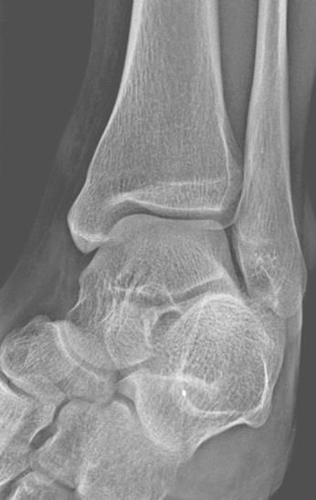
3 Radiographic Anatomy Fore foot Mid foot Hind foot Lisfranc joint Chopart s joint 3


4 Radiographic Anatomy 4 mm 4
5 Radiographic Anatomy 5


6 Special views Lateral views with weight-baring 6

7 Special views Calcaneal view (Skier s view) 7

8 Special views 8


9 Special views M L metatarsal ridge Sesamoid views Skyline view 9

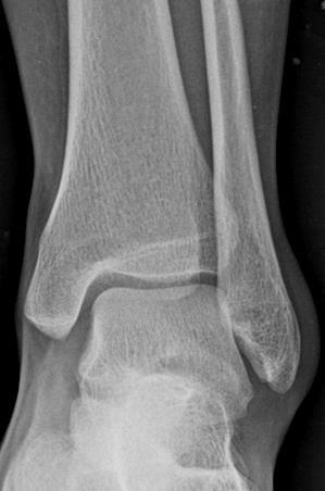
10 Bunion (Hallux valgus) A bony bump that forms on the joint at the base of the big toe due to the big toe pushes against the next toe forcing the joint get bigger and stick out. Red and sore skin Age > 40 years-old 10
11 Turf Toe Most common in football player: Hyperextension injury at 1 st MTP Injuries of the plantar capsuloligamentous complex of the first MTP joint or Plantar plate disruption MTP joint synovitis Flexor tendon sheath synovitis Persistent MTP hyperextension RadioGraphics 2001; 21:

12 Turf Toe: 12

13 Turf Toe Marrow edema Subchondral dark line Joint effusion Tenosynovitis of the FHL 13


14 Bipartite medial sesamoid Eur Radiol 2003; 13:L164-L177 14
15 Bipartite sesamoid vs fracture Fracture: Sharp Radiolucent Uncorticated line Two fragmnets often fit together MRI: marrow edema in recent fracture 99mTc-MDP bone scan: normal in bipartite Eur Radiol 2003; 13:L164-L177 15

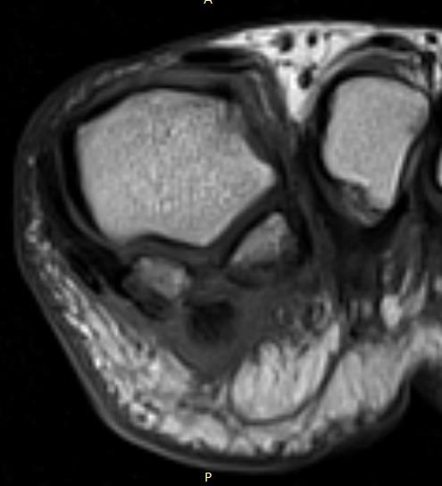
16 Axial PDW-FS: Sesamoids M L MCL: thicker LCL: thinner T: FHL tendon T 16
17 Sesamoiditis Repetitive injury Marrow edema ( DDx stress response) Abnormal on STIR (normal on T1W) Both sesamoids Reactive soft tissue abnormality; tendinitis, synovitis, bursitis etc. RadioGraphics 2001; 21:
18 Sesamoiditis 18
19 19

20 20
21 Morton s neuroma Not true neoplasm; perineural fibrosis and nerve degeneration Intermetatarsal space; 3 rd and 2 nd Intermetatarsal ligament-level Relate with bursitis Iso-slightly hyperintense on T1W (muscle), iso-hypointense on T2W (fat), intense enhancement Radiology 2010; 255: RadioGraphics 2001; 21:
22 22
23 Bursitis Intermetatarsal bursae Adventitial bursae beneath metatarsal heads (pressure point) Low SI on T1W, high SI on T2W and STIR with peripheral enhancement. <= 3-mm in transverse diameter in the 1 st, 2 nd and 3 rd may be physiologic changes. RadioGraphics 2001; 21:
24 Stress Fractures Common at metatarsal bones: middle or distal of 2 nd, 3 rd, 4 th MT Runners, ballet, danacers, gymnasts, military recruits Altered weight bearing Hallux valgus Recent surgery of hallux Flattened foot RadioGraphics 2001; 21:

25 Periosteal reaction 25

26 Abnormal? 26

27 Diagnosis? C-sign Loss of talocalcaneal facet Talocalcaneal coalition 27

28 Tarsal Coalition An abnormal bony, cartilage or fibrous union between two or more bones of hind-and midfoot Most common: calcaneonavicular, talocalcaneal Secondary signs of coalition on lateral radiographs C-sign Talar beak Anteater sign pes planus Non-visualized middle facet AJR 2004; 182:

29 Calcified and swollen Achilles tendon Plantar calcaneal spur Posterior calcaneal spur Soft tissue swelling Avulsive fragments Loose body Plantar fasciitis Anterior impingement Eur Radiol 2007; 17:
30 Plantar fasciitis Most common cause of plantar heel pain Repetitive trauma or enthesopathy (AS, Reiter, PsoA) Plantar calcaneal spur MRI: fascial thickening involving proximal portion extending to calcaneal insertion. intermediate SI on T1W or PDW, high SI on T2W or STIR *** Fluid signal and blood Signal: plantar fascia rupture Edema of the adjacent fat pad and underlying soft tissue Marrow edema RadioGraphics 2000; 20: RadioGraphics 2000; 20:S181-S197 30

31 Erosion Edema 31
32 Plantar fibromatosis Localized fibrous proliferation arising from the superficial and medial aspect of the plantar fascia One or more nodules Low-intermediate SI on T1W, low SI on T2W, variable enhancement RadioGraphics 2001; 21: RadioGraphics 2000; 20:S181-S197 32


33 Type2 accessory navicular bone Eur Radiol 2003; 13:L164-L177 33


34 Type3 accessory navicular bone Eur Radiol 2003; 13:L164-L177 34

35 Os peroneum Eur Radiol 2003; 13:L164-L177 35


36 Os trigonum syndrome Eur Radiol 2003; 13:L164-L177 Eur Radiol 2007; 17:

37 Stieda process of the talus Eur Radiol 2007; 17:


38 Anterior tibial spur Anteromedial talar spur Soft tissue swelling Anterior impingement Eur Radiol 2007; 17:


39 Osteochondrosis dissicans Lateral VS Medial Trauma VS Non-Trauma? 39

40 OCD, unstable 40

41 Abnormal? Effusion? 41

42 Bursitis 42
43 Stress response Amorphous area of decreased marrow SI on T1WI and high SI on T2W or STIR. Adjacent soft tissue edema and enhancement May progress to stress fracture: low signal band contiguous with cortex on T1WI and T2WI. RadioGraphics 2001; 21:
44 Achilles tendinopathy Normal: uniformly low SI, flattened or slightly concave anterior border, < 1-cm in AP thickness RadioGraphics 2000; 20:
45 Tarsal tunnel syndrome Tarsal tunnel: fibro-osseous canal bounded by flexor retinaculum, medial surfaces of Talus and calcaneus. Posterior tibial nerve, 3 medial tendons (PT, FDL, FHL), posterior tibial vessels. RadioGraphics 2000; 20:


46 Thank you 46
17/10/2017. Foot and Ankle
 17/10/2017 Alicia M. Yochum RN, DC, DACBR, RMSK Foot and Ankle Plantar Fasciitis Hallux Valgus Deformity Achilles Tendinosis Posterior Tibialis Tendon tendinopathy Stress Fracture Ligamentous tearing Turf
Evaluation of Pediatric Foot Pain
 May 2006 Evaluation of Pediatric Foot Pain John Flibotte, Harvard Medical School Year III Our Patient AP is a 10 year old boy with chronic R foot pain 2 Anatomy of the Foot Manusov EG, et al. (1996), Part
Ankle Tendons in Athletes. Laura W. Bancroft, M.D.
 Ankle Tendons in Athletes Laura W. Bancroft, M.D. Outline Protocols Normal Anatomy Tendinopathy, partial and complete tears Posterior tibial, Flexor Hallucis Longus, Achilles, Peroneal and Anterior Tibial
Ultrasound of Mid and Hindfoot Pathology
 Ultrasound of Mid and Hindfoot Pathology Levon N. Nazarian, M.D. Professor of Radiology Thomas Jefferson University Hospital Disclosures None relevant to this presentation Educational Objective Following
Sports Injuries of the Foot and Ankle. Mark McEleney, MD University of Iowa College of Medicine Refresher Course for the Family Physician 4/4/2018
 Sports Injuries of the Foot and Ankle Mark McEleney, MD University of Iowa College of Medicine Refresher Course for the Family Physician 4/4/2018 I. Objectives A. By the end of the lecture attendees will
THE JOURNAL OF NUCLEAR MEDICINE Vol. 56 No. 3 March 2015 Rauscher et al.
 Supplemental Figure 1 Correlation analysis of tracer between and subsequent as assessed by SUV max in focal lesions (A). x-axis displays quantitative values as obtained by, and y-axis displays corresponding
Why? Ultrasound of the Foot. Ultrasound of the Foot. General Rules. Plantar Fascia. Plantar Fasciitis 18/09/2018
 Ultrasound of the Foot Why? Ultrasound of the Foot Plantar fasciitis Plantar fascia fibromatosis Morton s neuroma Intermetatarsal bursitis Adventitial bursitis Plantar plate tears MTP joint synovitis Ganglia
Case 1 7 yo male Right elbow injury 3 months ago Medial elbow pain and tenderness over medial epicondyle Long arm cast given but off himself 1 month a
 Case presentations Case 1 7 yo male Right elbow injury 3 months ago Medial elbow pain and tenderness over medial epicondyle Long arm cast given but off himself 1 month after Progressive limited elbow flexion
MRI of the Ankle and Foot
 Acta Radiológica Portuguesa, Vol.XX, nº 79, pág. 55-63, Jul.-Set., 2008 MRI of the Ankle and Foot Mark Anderson University of Virginia Health Sciences Center, Charlottesville, Virginia discuss the basic
Anatomy of Foot and Ankle
 Anatomy of Foot and Ankle Surface anatomy of the ankle & foot Surface anatomy of the ankle & foot Medial orientation point medial malleous sustentaculum tali tuberosity of navicular TA muscle TP muscle
Aetiology: Pressure of Distal intermetatarsal ligament against common digital nerve. Lumbar radiculopathy Instability MTPJ joint or inflammatory MPJ
 MORTON S NEUROMA 80% III web space (next common is II). Never occurs in III or IV Common in females in fifties Aetiology: Pressure of Distal intermetatarsal ligament against common digital nerve Rule out
Index. Clin Sports Med 23 (2004) Note: Page numbers of article titles are in boldface type.
 Note: Page numbers of article titles are in boldface type.") Clin Sports Med 23 (2004) 169 173 Index Note: Page numbers of article titles are in boldface type. A Achilles enthesopathy, calcaneal spur with, 133 clinical presentation of, 135 136 definition of, 131
Clin Sports Med 23 (2004) 169 173 Index Note: Page numbers of article titles are in boldface type. A Achilles enthesopathy, calcaneal spur with, 133 clinical presentation of, 135 136 definition of, 131
Imaging of posterior ankle pain : Main etiologies and differential diagnosis
 Imaging of posterior ankle pain : Main etiologies and differential diagnosis Poster No.: C-2399 Congress: ECR 2017 Type: Educational Exhibit Authors: W. Frikha, M. MECHRI, S. boukriba, H. RIAHI, M. CHELLI
MRI of Pediatric Ankle and Foot. Mahesh Thapa, MD Associate Professor Seattle Children s University of Washington School of Medicine
 MRI of Pediatric Ankle and Foot Mahesh Thapa, MD Associate Professor Seattle Children s University of Washington School of Medicine Disclosures Under contract with Lippincott Williams and Wilkins (LWW)
Peggers Super Summaries: Foot Injuries
 Lisfranc Injury ANATOMY Roman arch with recessed 2 nd MT base AP medial side of intermediate cuneiform to 2 nd MT base Oblique medial side of lateral cuneiform with 3 rd MT base and 4 th with medial boarder
Tarsal Coalition On MR
 Tarsal Coalition On MR By William Renner, M.D. This and other topics will be discussed in Tarsal coalition is a congenital anomaly with fusion of the tarsal bones. The fusion may be bony, fibrous or cartilaginous.
radiologymasterclass.co.uk
 http://radiologymasterclass.co.uk Hip X-ray anatomy - Normal AP (anterior-posterior) Shenton's line is formed by the medial edge of the femoral neck and the inferior edge of the superior pubic ramus Loss
Case. 15 Y old boy presented with pain in the foot. No history of injury or any constitutional symptoms. Your diagnosis?
 Case 15 Y old boy presented with pain in the foot. No history of injury or any constitutional symptoms Your diagnosis? Diagnosis: Calcaneo-navicular tarsal coalition. C sign Talar beaking Ant eaters nose
Ultrasound Evaluation of Posteromedial Ankle Pathology. Andrew C Cordle, M.D., Ph.D. 9/21/2018
 Ultrasound Evaluation of Posteromedial Ankle Pathology Andrew C Cordle, M.D., Ph.D. 9/21/2018 Overview: Pathology of the Posteromedial Ankle Flexor Tendon Pathology Accessory Navicular Bone Pathology Tarsal
PAINFUL SESAMOID OF THE GREAT TOE Dr Vasu Pai ANATOMICAL CONSIDERATION. At the big toe MTP joint: Tibial sesamoid (medial) & fibular (lateral)
 & fibular (lateral)") PAINFUL SESAMOID OF THE GREAT TOE Dr Vasu Pai ANATOMICAL CONSIDERATION At the big toe MTP joint: Tibial sesamoid (medial) & fibular (lateral) They are contained within the tendons of Flexor Hallucis Brevis
PAINFUL SESAMOID OF THE GREAT TOE Dr Vasu Pai ANATOMICAL CONSIDERATION At the big toe MTP joint: Tibial sesamoid (medial) & fibular (lateral) They are contained within the tendons of Flexor Hallucis Brevis
Section 4: Tarsal Coalitions
 Case H (Figure 2): PedCat CBCT transverse plane reconstruction of right Lisfranc midfoot dislocation compared to normal left foot. Clinical Relevance of the PedCat Study: The weight bearing CBCT study
Shane A. Shapiro, M.D. Assistant Professor, Orthopedic Surgery Mayo Clinic 2012 MFMER slide MFMER slide-3
 Ultrasound Foot and Ankle Pathology Disclosures None relevant Shane A. Shapiro, M.D. Assistant Professor, Orthopedic Surgery Mayo Clinic Florida @ShaneShapiroMD 2012 MFMER slide-2 Foot and Ankle Fundamentals
MRI IN NONOSSEOUS ABNORMALITIES OF THE FOREFOOT: A PICTORIAL REVIEW
 MRI IN NONOSSEOUS ABNORMALITIES OF THE FOREFOOT: A PICTORIAL REVIEW I Delgado, P Melloni, M Veintemillas, R Valls, M Vilagran, A Valera UDIAT. Sabadell (Barcelona). Spain. PURPOSE To catalog the wide spectrum
Extraarticular Lateral Ankle Impingement
 Extraarticular Lateral Ankle Impingement Poster No.: C-1282 Congress: ECR 2016 Type: Educational Exhibit Authors: C. Cevikol; Keywords: Trauma, Diagnostic procedure, MR, CT, Musculoskeletal system, Musculoskeletal
Foot and Ankle Complaints.
 Foot and Ankle Complaints www.fisiokinesiterapia.biz INTRODUCTION Anatomy and Function Foot Ankle Common complaints Common diagnoses FOOT AND ANKLE ANATOMY 26 bones and 2 sesamoids Forefoot Metatarsals
Review relevant anatomy of the foot and ankle. Learn the approach to examining the foot and ankle
 Objectives Review relevant anatomy of the foot and ankle Learn the approach to examining the foot and ankle Learn the basics of diagnosis and treatment of ankle sprains Overview of other common causes
Section Three: The Leg, Ankle, and Foot Lecture: Review of Clinical Anatomy, Patterns of Dysfunction and Injury, and
 Section Three: The Leg, Ankle, and Foot Lecture: Review of Clinical Anatomy, Patterns of Dysfunction and Injury, and Treatment Implications for the Leg, Ankle, and Foot Levels I and II Demonstration and
Foot & Ankle Examination Workshop Morteza Khodaee, MD, MPH, FACSM, FAAFP Associate Professor Department of Family Medicine University of Colorado
 Foot & Ankle Examination Workshop Morteza Khodaee, MD, MPH, FACSM, FAAFP Associate Professor Department of Family Medicine University of Colorado School of Medicine July 4, 2013 Objectives Participants
2017 SAFSA CONGRESS PROGRAMME
 2017 SAFSA CONGRESS PROGRAMME THURSDAY, MAY 25 07h45 07h55: WELCOME & INTRODUCTIONS Forefoot I: Hallux Valgus and Lesser Toes (08h00-10h00 Lectures) 08h00 08h30: Surgical Management of Hallux Valgus Rippstein,
Impingement Syndromes of the Ankle. Noaman W Siddiqi MD 5/4/2006
 Impingement Syndromes of the Ankle Noaman W Siddiqi MD 5/4/2006 Ankle Impingement Overview Clinical DX Increasingly recognized cause of chronic ankle pain Etiology can be soft tissue or osseous Professional
Clinical Application of the EMS Swiss DolorClast
 Chapter 12.fm Page 119 Tuesday, November 21, 2006 6:38 PM 12 Clinical Application of the EMS Swiss DolorClast L. Gerdesmeyer, M. Henne, P. Diehl, H. Gollwitzer, M. Göbel In general, the following recommendations
Plantar fasciopathy (PFs)
") Plantar fasciopathy (PFs) 2016. 04. 30. Jung-Soo Lee, M.D., Ph.D. Department of Rehabilitation Medicine, Uijeongbu St. Mary's Hospital, College of Medicine, The Catholic University of Korea Anatomy of
Plantar fasciopathy (PFs) 2016. 04. 30. Jung-Soo Lee, M.D., Ph.D. Department of Rehabilitation Medicine, Uijeongbu St. Mary's Hospital, College of Medicine, The Catholic University of Korea Anatomy of
Ankle Injuries. Resident Guidebook. Achilles tendon sprain/tear. Peroneal tendinopathy Peroneal subluxation. Extensor Hallucis Longus Tenosynovitis
 Ankle Injuries Achilles tendon sprain/tear Peroneal tendinopathy Peroneal subluxation Extensor Hallucis Longus Tenosynovitis Weber Fracture Stress fracture Calcaneal bursitis Calcaneal fracture Base of
It's normal, but it hurts! Painful sesamoid and accessory bone syndromes of the foot.
 It's normal, but it hurts! Painful sesamoid and accessory bone syndromes of the foot. Poster No.: P-0120 Congress: ESSR 2016 Type: Educational Poster Authors: A. C. Vieira, A. Vieira, R. Cunha; Porto/PT
ABC of Emergency Radiology
 l ja ) $% _2) < j> ~~~~~~~~~~~~~~~~~foot ABC of Emergency Radiology THE FOOT D A Nicholson, D O'Keeffe, P A Driscoll Accurate clinical assessment of injuries to the foot will avoid unnecessary exposure
Anatomy 1% 29% 64% 6%
 Mortons Neuroma Perineural fibrosis of the plantar digital nerve Females 8-10 3 rd plantar webspace most commonly effected Burning pain Sensory changes 3&4 digits / interdigital space Etiology Excessive
Posterior Ankle Impingement: Don t Get Pinched
 Posterior Ankle Impingement: Don t Get Pinched 11 th Annual Sports Medicine Continuing Education Conference Gregory P Witkowski, MD Orthopaedic Trauma and Foot/Ankle Surgery Disclosures I have nothing
Ankle impingement syndromes - pictorial review.
 Ankle impingement syndromes - pictorial review. Poster No.: P-0148 Congress: ESSR 2015 Type: Educational Poster Authors: R. D. T. Mesquita, J. Pinto, J. L. Rosas, A. Vieira ; Porto/PT, 1 2 2 3 1 1 3 Matosinhos/PT,
Ankle impingement syndromes - pictorial review.
 Ankle impingement syndromes - pictorial review. Poster No.: P-0148 Congress: ESSR 2015 Type: Educational Poster Authors: R. D. T. Mesquita, J. Pinto, J. L. Rosas, A. Vieira ; Porto/PT, 1 2 2 3 1 1 3 Matosinhos/PT,
Foot. Dr. Heba Kalbouneh Associate Professor of Anatomy and Histology
 Foot Dr. Heba Kalbouneh Associate Professor of Anatomy and Histology Dorsum of the Foot Sole of the Foot Plantar aponeurosis It is a triangular thickening of deep fascia in the sole of the foot Attachments:
Foot and ankle update
 Foot and ankle update Mr Ian Garnham Consultant Foot and Ankle Surgeon Whipps Cross University Hospital Hallux Rigidus Symptoms first ray and 1st MTP pain and swelling worse with push off or forced dorsiflexion
*Rippstein, Trnka, Saragas, Hoffman
 THURS 25th MAY 07:00 07:10 Welcome and Introductions Paulo Ferrao Lecture 1: 07:10 09:45 Forefoot I: Hallux Valgus and Lesser Toes Mark Easley 40 mins 07:10 07:50 Surgical Management of Hallux Valgus 30
pedcat Clinical Case Studies
 pedcat Clinical Case Studies C u r v e B e a m 1 7 5 T i t u s A v e, S u i t e 3 0 0 W a r r i n g t o n, P A 1 8 9 7 6 267-4 8 3-8081 w w w. c u r v e b e a m. c o m PedCAT: Clinical Evidence of diagnostic
Osseous variants generating symptoms in ankle and foot
 Osseous variants generating symptoms in ankle and foot Poster No.: P-0023 Congress: ESSR 2013 Type: Scientific Exhibit Authors: S. Daineffe, B. G. H. G. Pilet, S. Van de Perre, E. De Smet, 1 2 1 3 5 1
OVERUSE AND SPORTS-RELATED INJURIES. Overuse and sports-related injuries of the ankle and hindfoot: MR imaging findings
 OVERUSE ND SPORTS-RELTED INJURIES CHPTER 10 Overuse and sports-related injuries of the ankle and hindfoot: MR imaging findings Elizabeth S. Sijbrandij 1, d P.G. van Gils 1, Eduard E. de Lange 2 From the
Avascular Necrosis of the Foot. Dr. Hema Choudur MD, FRCPC Associate Professor. Dept. of Radiology. McMaster University, Hamilton, Canada.
 Avascular Necrosis of the Foot Dr. Hema Choudur MD, FRCPC Associate Professor. Dept. of Radiology. McMaster University, Hamilton, Canada. Avascular Necrosis: Pathophysiology Ischemia to the bone from oxygen
Anatomy of the lower limb
 Anatomy of the lower limb Arches & sole of the foot Dr. Hayder ARCHES OF THE FOOT The foot as a mechanical unit performs two major functions: - It acts as a pliable platform to support the body weigh during
emoryhealthcare.org/ortho
 COMMON SOCCER INJURIES Oluseun A. Olufade, MD Assistant Professor, Department of Orthopedics and PM&R 1/7/18 GOALS Discuss top soccer injuries and treatment strategies Simplify hip and groin injuries in
Anatomy of Ankle & Foot. Chang-Hyung Lee, M.D., Ph.D. Physical Medicine & Rehabilitation Samsung Medical Center
 Anatomy of Ankle & Foot Chang-Hyung Lee, M.D., Ph.D. Physical Medicine & Rehabilitation Samsung Medical Center Ankle Introduction Most frequently injured major joint 3 main articulation: distal tibiofibular
Rippstein, Trnka, Saragas, Narramore
 THURS 25th MAY 07:45 07:55 Welcome and Introductions Paulo Ferrao Lecture 1: 08:00 10:20 Forefoot I: Hallux Valgus and Lesser Toes Mark Easley 30 mins 08:00 08:30 Surgical Management of Hallux Valgus Saragas,
Physical Examination of the Foot & Ankle
 Inspection Standing, feet straight forward facing toward examiner Swelling Deformity Flatfoot (pes planus and hindfoot valgus) High arch (pes cavus and hindfoot varus) Peek-a-boo heel Varus Too many toes
Copyright 2004, Yoshiyuki Shiratori. All right reserved.
 Ankle and Leg Evaluation 1. History Chief Complaint: A. What happened? B. Is it a sharp or dull pain? C. How long have you had the pain? D. Can you pinpoint the pain? E. Do you have any numbness or tingling?
Clinical Experience with Foot and Ankle SPECT/CT:- From Dark horse to Fore runner of Foot and Ankle Imaging
 Clinical Experience with Foot and Ankle SPECT/CT:- From Dark horse to Fore runner of Foot and Ankle Imaging Poster No.: C-0113 Congress: ECR 2015 Type: Educational Exhibit Authors: K. E. Low 1, Z. Saad
Burwood Road, Concord Dora Street, Hurstville 119 Lethbridge Street, Penrith 160 Belmore Road, Randwick
 www.orthosports.com.au 47 49 Burwood Road, Concord 29 31 Dora Street, Hurstville 119 Lethbridge Street, Penrith 160 Belmore Road, Randwick Turf Toe Injury By Todd Gothelf Foot, Ankle, Shoulder History
Outline. Ankle/Foot Anatomy Ankle Sprains Ottawa Ankle Rules DDx: The Sprain That Wasn t
 Ankle Injuries Outline Ankle/Foot Anatomy Ankle Sprains Ottawa Ankle Rules DDx: The Sprain That Wasn t Anatomy: Ankle Mortise Bony Anatomy Lateral Ligament Complex Medial Ligament Complex Ankle Sprains
BUCKS MSK: FOOT AND ANKLE PATHWAY GP MANAGEMENT. Hallux Valgus. Assessment: Early Management. (must be attempted prior to any referral to imsk):
:") Hallux Valgus Common condition: affecting around 28% of the adult population. Prevalence increases with age and in females. Observation: Lateral deviation of the great toe. May cause secondary irritation
Hallux Valgus Common condition: affecting around 28% of the adult population. Prevalence increases with age and in females. Observation: Lateral deviation of the great toe. May cause secondary irritation
The University Of Jordan Faculty Of Medicine FOOT. Dr.Ahmed Salman Assistant Prof. of Anatomy. The University Of Jordan
 The University Of Jordan Faculty Of Medicine FOOT Dr.Ahmed Salman Assistant Prof. of Anatomy. The University Of Jordan Tarsal Tunnel Syndrome Due to compression of Tibial nerve as it travels through the
EASILY MISSED FOOT AND ANKLE FRACTURES NORDIC TRAUMA COURSE 2016, AARHUS
 EASILY MISSED FOOT AND ANKLE FRACTURES NORDIC TRAUMA COURSE 2016, AARHUS Ken F. Linnau, MD, MS Emergency Radiology Harborview Medical Center University of Washington Seattle, WA Thanks to Claire K Sandstrom
Foot & Ankle Disorders
 Foot & Ankle Disorders Hillingdon PGMC 6-7-2013 Htwe Zaw FRCS (Tr&Orth) Consultant Foot & Ankle and Trauma Surgeon Hillingdon Hospitals NHS Foundation Trust Overview Anatomy: hindfoot-midfoot coupling
Columbia/NYOH FOOT and ANKLE ROTATION-SPECIFIC OBJECTIVES
 Updated 2/8/10 Columbia/NYOH FOOT and ANKLE ROTATION-SPECIFIC OBJECTIVES INTERPERSONAL AND COMMUNICATION SKILLS Resident will at all times demonstrate behavior that is beyond reproach. Residents must be
2013/10/18 GANGLION CYSTS ENDOSCOPIC GANGLIONECTOMY ARTHROSCOPIC GANGLIONECTOMY OPEN GANGLIONECTOMY COMPARED TO FOOT AND ANKLE
 GANGLION CYSTS gelatinous fluid filled, encapsulated soft tissue masses adjacent to a joint or tendon ENDOSCOPIC GANGLIONECTOMY OF THE FOOT AND ANKLE Dr TH Lui North District Hospital HKSAR Pain, mass
Dr Nabil khouri MD. MSc. Ph.D
 Dr Nabil khouri MD. MSc. Ph.D Foot Anatomy The foot consists of 26 bones: 14 phalangeal, 5 metatarsal, and 7 tarsal. Toes are used to balance the body. Metatarsal Bones gives elasticity to the foot in
Financial Disclosure. Turf Toe
 Seth O Brien, CP, LP Financial Disclosure Mr. Seth O'Brien has no relevant financial relationships with commercial interests to disclose. Turf Toe Common in athletes playing on firm, artificial turf Forceful
Case Report Painful Os Peroneum Syndrome: Underdiagnosed Condition in the Lateral Midfoot Pain
 Case Reports in Radiology Volume 2016, Article ID 8739362, 4 pages http://dx.doi.org/10.1155/2016/8739362 Case Report Painful Os Peroneum Syndrome: Underdiagnosed Condition in the Lateral Midfoot Pain
Peripheral Nerve Ultrasound
 Peripheral Nerve Ultrasound Jon A. Jacobson, M.D. Professor of Radiology Director, Division of Musculoskeletal Radiology University of Michigan Normal Peripheral Nerve Ultrasound appearance: Hypoechoic
MRI in Patients with Forefoot Pain Involving the Metatarsal Region
 MRI in Patients with Forefoot Pain Involving the Metatarsal Region Poster No.: C-0151 Congress: ECR 2015 Type: Authors: Keywords: DOI: Scientific Exhibit R. Vukojevi#, M. Mustapic, D. Marjan; Zagreb/HR
 P R E S E N T S Dr. Mufa T. Ghadiali is skilled in all aspects of General Surgery. His General Surgery Services include: General Surgery Advanced Laparoscopic Surgery Surgical Oncology Gastrointestinal
Accessory ossicles of the ankle and foot
 Accessory ossicles of the ankle and foot Poster No.: C-2598 Congress: ECR 2013 Type: Educational Exhibit Authors: Á. Gómez Trujillo; Madrid/ES Keywords: Education and training, Education, MR, Digital radiography,
Copyright 2012 by The McGraw-Hill Companies, Inc. All rights reserved. McGraw-Hill/Irwin
 CHAPTER 8: THE LOWER EXTREMITY: KNEE, ANKLE, AND FOOT KINESIOLOGY Scientific Basis of Human Motion, 12 th edition Hamilton, Weimar & Luttgens Presentation Created by TK Koesterer, Ph.D., ATC Humboldt State
The Spring Ligament, PTT Tear, and Adult Acquired Flatfoot Deformity On MRI
 The Spring Ligament, PTT Tear, and Adult Acquired Flatfoot Deformity On MRI (Part 2) By William Renner, M.D. This and other topics will be discussed in: The posterior tibial tendon is the primary stabilizer
Index. Note: Page numbers of article titles are in boldface type.
 Index Note: Page numbers of article titles are in boldface type. A Achilles tendon injury of, pathophysiology of, 10 peritendinitis of, 119 120 rupture of, 32 35, 117 135 anatomy of, 117 118 chronic, 126
Topics. Musculoskeletal Infection Extremities. Detection of Infection. Role of Imaging in Extremity Infection. Detection of Infection
 Topics Musculoskeletal Infection Extremities Nuttaya Pattamapaspong M.D. Department of Radiology, Faculty of Medicine, Chiang Mai University, Chiang Mai, Thailand Role of imaging in extremity infection
Results of Calcaneal Osteotomy & Flexor Digitorum Longus transfer in Stage II Acquired Flatfoot Deformity
 Results of Calcaneal Osteotomy & Flexor Digitorum Longus transfer in Stage II Acquired Flatfoot Deformity Mr Amit Chauhan Mr Prasad Karpe Ms Maire-claire Killen Mr Rajiv Limaye University Hospital of North
11/2/17. Lateral Collateral Complex Medial Collateral Complex Distal Tibiofibular Syndesmosis Spring Ligament
 Andrew J Grainger Leeds, UK Lateral Collateral Complex ial Collateral Complex Distal Tibiofibular Syndesmosis Spring Ligament Brief anatomy review Scan tips and tricks Pathological appearances andrewgrainger@nhs.net
ANKLE MRI. TENDONS: (dark on all sequences except distal PTT and Achilles)
") TENDONS: (dark on all sequences except distal PTT and Achilles) ANKLE MRI -ANTERIOR EXTENSOR ( Tom Hates Dick ) -Tibialis anterior (medial and largest; abnormal in grumpy old men with DM or Gout; may appear
TENDONS: (dark on all sequences except distal PTT and Achilles) ANKLE MRI -ANTERIOR EXTENSOR ( Tom Hates Dick ) -Tibialis anterior (medial and largest; abnormal in grumpy old men with DM or Gout; may appear
Clarification of Terms
 Clarification of Terms The plantar aspect of the foot refers to the role or its bottom The dorsal aspect refers to the top or its superior portion The ankle and foot perform three main functions: 1. shock
MR Imaging of normal ankle anatomy: What the radiologists need to know
 MR Imaging of normal ankle anatomy: What the radiologists need to know Poster No.: P-0110 Congress: ESSR 2013 Type: Scientific Exhibit Authors: M. M. Simonet Redondo, I. Santos Gomez, A. Marin Canete,
Pediatric sports injuries in foot and ankle
 Pediatric sports injuries in foot and ankle Poster No.: P-0013 Congress: ESSR 2014 Type: Scientific Poster Authors: Y. Kobashi 1, T. Mogami 1, S. Yamazoe 1, A. Baba 2, S. Ogiwara 1 ; 1 2 Chiba/JP, Tokyo/JP
분당제생병원재활의학과이태임 COMMON PAINFUL CONDITIONS OF PEDIATRIC FOOT
 분당제생병원재활의학과이태임 COMMON PAINFUL CONDITIONS OF PEDIATRIC FOOT CASE M/12 CC Insidous onset of heel pain 축구를좋아해서많이했다. 걷거나뛰면더아프다. PE pes planus heelcord tightness Tenderness over the posterior calcaneus Sqeeze
Accessory Ossicles of the Foot and Ankle
 Accessory Ossicles of the Foot and Ankle Poster No.: C-0978 Congress: ECR 2016 Type: Educational Exhibit Authors: U. Kesimal, M. OYNAK, C. Cevikol; Antalya/TR Keywords: Anatomy, Musculoskeletal bone, Musculoskeletal
Ultrasound of the Knee
 Ultrasound of the Knee Jon A. Jacobson, M.D. Professor of Radiology Director, Division of Musculoskeletal Radiology University of Michigan Disclosures: Consultant: Bioclinica Book Royalties: Elsevier Advisory
musculoskeletal system anatomy muscles of foot sheet done by: dina sawadha & mohammad abukabeer
 musculoskeletal system anatomy muscles of foot sheet done by: dina sawadha & mohammad abukabeer Extensor retinaculum : A- superior extensor retinaculum (SER) : originates from the distal ends of the tibia
 Ankle Injuries Ankle injuries fall into the same basic categories as do all athletic injuries: Contusions Sprains Strains Fractures www.fisiokinesiterapia.biz 85% of all ankle sprains involve some plantar
mechanical stresses on the tendon with repetitive loading
 Tendinopathy.. How does it happen? mechanical stresses on the tendon with repetitive loading Impingement of the tendon between adjacent structures (bones, ligaments) and impaired blood supply Presentation
Knee, Ankle, and Foot: Normal and Abnormal Features with MRI and Ultrasound Correlation. Disclosures. Outline. Joint Effusion. Suprapatellar recess
 Knee, Ankle, and Foot: Normal and Abnormal Features with MRI and Ultrasound Correlation Jon A. Jacobson, M.D. Professor of Radiology Director, Division of Musculoskeletal Radiology University of Michigan
CHRONIC FOOT PROBLEMS FOOT and ANKLE BASICS
 CHRONIC FOOT PROBLEMS FOOT and ANKLE BASICS ABC s of Comprehensive Musculoskeletal Care December 1 st, 2007 Stephen Pinney MD Chief, UCSF Foot and Ankle Service Chronic problems typically occur gradually
ANKLE PLANTAR FLEXION
 ANKLE PLANTAR FLEXION Evaluation and Measurements By Isabelle Devreux 1 Ankle Plantar Flexion: Gastrocnemius and Soleus ROM: 0 to 40-45 A. Soleus: Origin: Posterior of head of fibula and proximal1/3 of
Computed Tomographic Imaging of Foot and Ankle trauma
 Computed Tomographic Imaging of Foot and Ankle trauma Dr. Tudor H. Hughes M.D., FRCR Department of Radiology University of California School of Medicine San Diego, California CT of Foot and Ankle Trauma
A Patient s Guide to Foot Anatomy
 A Patient s Guide to Foot Anatomy Introduction Our feet are constantly under stress. It's no wonder that 80 percent of us will have some sort of problem with our feet at some time or another. Many things
Clin Podiatr Med Surg 19 (2002) Index
 Index") Clin Podiatr Med Surg 19 (2002) 335 344 Index Note: Page numbers of article titles are in bold face type. A Accessory soleus muscle, magnetic resonance imaging of, 300 Achilles tendon injury of, magnetic
Clin Podiatr Med Surg 19 (2002) 335 344 Index Note: Page numbers of article titles are in bold face type. A Accessory soleus muscle, magnetic resonance imaging of, 300 Achilles tendon injury of, magnetic
Arthroscopy Of the Ankle.
 Arthroscopy Of the Ankle www.fisiokinesiterapia.biz Ankle Arthroscopy Anatomy Patient setup Portal placement Procedures Complications Anatomy Portals Anterior Anteromedial Anterolateral Anterocentral Posterior
Leg. Dr. Heba Kalbouneh Associate Professor of Anatomy and Histology
 Leg Dr. Heba Kalbouneh Associate Professor of Anatomy and Histology Skin of the Leg Cutaneous Nerves Medially: The saphenous nerve, a branch of the femoral nerve supplies the skin on the medial surface
Rocker bottom foot in diabetic patients - a severe deformity of the diabetic Charcot s joint with serious sequelae
 Rocker bottom foot in diabetic patients - a severe deformity of the diabetic Charcot s joint with serious sequelae Poster No.: P-0079 Congress: ESSR 2015 Type: Educational Poster Authors: J. Brtkova, T.
PROBLEMS AND ORTHOTIC SOLUTIONS. Problem/Issue Underlying treatment goal Solution Pes Cavus foot
 PROBLEMS AND ORTHOTIC SOLUTIONS Problem/Issue Underlying treatment goal Solution Pes Cavus foot Usually also a supinated foot Rigid high arched foot with poor shock absorption and cushioning. Often roll
Orthopaedic (Ankles & Feet) Referral Guidelines
 Referral Guidelines") Orthopaedic (Ankles & Feet) Referral Guidelines Austin Health Orthopaedic Clinic holds weekly multidisciplinary meetings to discuss and plan the treatment of patients with Orthopaedic and Fracture conditions.
Orthopaedic (Ankles & Feet) Referral Guidelines Austin Health Orthopaedic Clinic holds weekly multidisciplinary meetings to discuss and plan the treatment of patients with Orthopaedic and Fracture conditions.
Running Injuries. Lower Extremity
 Running Injuries Lower Extremity Causes of Overuse Injuries Repetition Surface/Environment Postural Alignment Improper Biomechanics Poor Footwear/equipment Over-training/improper training Stress Injuries
Knee Contusions and Stress Injuries. Laura W. Bancroft, M.D.
 Knee Contusions and Stress Injuries Laura W. Bancroft, M.D. Objectives Review 5 types of contusion patterns Pivot shift Dashboard Hyperextension Clip Lateral patellar dislocation Demonstrate various stress
Traumatic Injuries to the Foot and Ankle
 Traumatic Injuries to the Foot and Ankle Dr. Joseph N. Daniel Clinical Associate Professor of Orthopaedic Surgery Foot and Ankle Service, The Rothman Institute Thomas Jefferson University Hospital Philadelphia,
A Pictorial Review of Congenital Tarsal Coalition
 A Pictorial Review of Congenital Tarsal Coalition Poster No.: C-2305 Congress: ECR 2011 Type: Educational Exhibit Authors: J. Jethwa, M. Tapp; Torquay/UK Keywords: Musculoskeletal joint, Musculoskeletal