Osteotomy System. Surgical Technique
|
|
|
- Mary Hawkins
- 5 years ago
- Views:
Transcription


1 Osteotomy System Surgical Technique


2 Acumed is a global leader of innovative orthopaedic and medical solutions. We are dedicated to developing products, service methods, and approaches that improve patient care. Acumed Osteotomy System The Acumed Osteotomy System features the Ulna Shortening Plate, designed to offer a low-profile plate with built-in osteotomy reference lines and an Osteotomy Guide. The reference lines on the plate help facilitate the creation of the osteotomy when a freehand cut is preferred. Designed in conjunction with William B. Geissler, MD, the low-profile Ulna Shortening Plate is designed to keep the screw heads as low as possible. The interfragmentary screw may be placed in one of two locations through the scalloped slot and is intended to compress the osteotomy securely when used as a lag screw. The plate offers the option to lock up to three screws distally and one proximally. Indications for an ulna shortening osteotomy include: Ulnar Impaction Syndrome due to ulnar-positive variance Distal radial ulnar joint (DRUJ) incongruity due to shortening of the radius Traumatic and degenerative tears of the triangular fibrocartilage complex (TFCC) associated with positive ulnar variance
3 Table of Contents System Features... 1 Instrumentation Overview...2 Surgical Technique Overview...3 Surgical Technique...5 Osteotomy Guide Assembly Instructions...5 Osteotomy With Guide Technique...6 Osteotomy Without Guide Technique Ordering Information...17


4 System Features Built-in Osteotomy Reference Guides Measurement reference lines on the side of the plate visually display the amount of shortening which can be obtained. Each 40 oblique laser line and spacing in between represents 2 mm of shortening. The perpendicular lines near the measurement slot are also spaced 2 mm apart and are designed to indicate the shortening obtained from the osteotomy. Measurement Slot Scalloped Lag Screw Slot Proximal Locking Hole.054" K-wire Hole Measurement Lines Compression Slot Osteotomy Measurement Reference Lines Three Distal Locking Holes Limited Contact Design Advanced Instrumentation The Ulnar Shortening Reduction Clamp ( ) utilizes a speed-lock wheel designed to maintain a hands-free compression of the osteotomy. The multipurpose temporary Ulnar Shortening Reduction Peg ( ) is partially threaded to help ensure that the far cortex is not tapped prior to it being replaced by a screw. The Ulnar Shortening Reduction Peg is designed to stabilize the ulna and help maintain rotational alignment while creating the osteotomy prior to being used with the reduction clamp. 1
 Ulnar Shortening Guide Locking Bolt (80-0421)")
 Osteotomy Saw Blade")
 Ulnar Shortening Guide Left (80-0418) 3.")
 Plate Tack (PL-PTACK) Ulnar Shortening")
 2.5 mm Quick Release Hex Driver (HPC-0025).")
 3.")
 Ulnar Shortening Reduction")
 Reduction Forceps with")
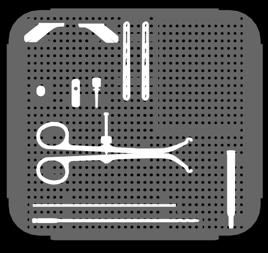
5 Instrumentation Overview 6-Hole Ulna Shortening Plate (PL-UL06) Ulnar Shortening Guide Locking Bolt ( ) Ulnar Shortening Guide Bottom Plate ( ) Ulnar Shortening Reduction Peg ( ) Osteotomy Saw Blade Hub Style DS ( S) Osteotomy Saw Blade Hub Style L ( S) Ulnar Shortening Guide Left ( ) 3.5 mm Nonlocking (COL-3XXX) 3.5 mm (CO-3XXX) Plate Tack (PL-PTACK) Ulnar Shortening Guide Right ( ) T15 Stick Fit Hexalobe Driver ( ) 2.5 mm Quick Release Hex Driver (HPC-0025).054" x 6" Guide Wire (WS-1406ST) Quick Release Drill ( ) 3.5 mm x 5" Quick Release Drill (MS-DC35) Osteotomy Saw Blade Hub Style S ( S) Ulnar Shortening Reduction Clamp ( ) Hexalobe Locking Drill Guide ( ) Locking Drill Guide ( ) Reduction Forceps with Serrated Jaw (PL-CL04) / 3.5 mm Thin Drill Guide (PL-2196) Large Cannulated Quick Release Driver Handle (MS-3200) 2


6 Surgical Technique Overview Align Insert Secure Osteotomy Guide Assembly Instructions Guide Placement K-wire Placement Placement Osteotomy with Guide Technique Osteotomy Without Guide Technique 3


7 Osteotomy Gap Reduction Screw Placement Final Confirmation 4

, slide the Ulnar Shortening Guide Bottom Plate (80-0420) into the appropriate")


8 Osteotomy Guide Assembly Instructions Figure 1 Figure 2 The Acumed Osteotomy Guide Assembly ( or , , ) offers the ability to make adjustments needed to perform the first and second cuts without the need for numerous guides. Additionally, by allowing continuous adjustment from 1 mm to 10 mm, the osteotomy guide assembly allows resection of the desired amount. 1 Assemble Guide and Bottom Plate Ensuring that the laser-marked arrows are aligned (Figure 1), slide the Ulnar Shortening Guide Bottom Plate ( ) into the appropriate Ulnar Shortening Guide ( or ). Ensure that the bottom plate is completely engaged into the ulnar shortening guide (Figure 2). Note: The subsequent technique is for a volar approach with the ulnar shortening guide. If a medial approach is taken, the opposite ulnar shortening guide can be used. For example, use the left ulnar shortening guide for a medial approach if the osteotomy is performed on the right ulna. Be sure the cutting slot lines up with the angled measurement reference lines on the plate. 2 Insert Locking Bolt Slide the guide bottom plate distal enough so that the Ulnar Shortening Guide Locking Bolt ( ) can be inserted through both pieces (Figure 3). Figure 3 Ulnar Shortening Guide Bottom Plate ( ) Ulnar Shortening Guide ( or ) Ulnar Shortening Guide Locking Bolt ( ) 5


9 Osteotomy With Guide Technique William B. Geissler, MD 1 Plate Placement Determine the amount of ulnar variance by reviewing preoperative X-rays. After exposing the volar side of the ulna, place the plate 3 5 cm proximal to the distal end of the ulna. Secure the 6-Hole Ulna Shortening Plate (PL-UL06) to the volar surface with one or more clamps, such as the Reduction Forceps with Serrated Jaw (PL-CL04). Make sure the proximal and distal orientation of the plate is correct, as noted by the laser marks on the plate. Figure 4 Proximal Distal Figure 5 2 Distal Screw and Reduction Peg Placement Depending on your choice of screw, drill the most distal locking hole using the appropriate Locking Drill Guide ( or see chart below) and Quick Release Drill ( ). Then insert the proper length Cortical or (COL-3XXX or 30-XXXX) with the proper Hex (HPC-0025) or Hexalobe ( ) Driver. In the proximal end of the measurement slot, drill bicortically and perpendicular to the plate and insert the temporary Ulnar Shortening Reduction Peg ( ) with a 2.5 mm Quick Release Hex Driver (HPC-0025). Option: Predrill the two remaining distal locking holes in the same manner with the appropriate locking drill guide but DO NOT INSERT SCREWS. This optional step can also occur after the osteotomy has been achieved based on surgeon preference. Drill Guide and Driver Selections Screw Drill Guide Driver Hex (Cortical) Screw Hexalobe Screw Locking Drill Guide ( ) Hexalobe Locking Drill Guide ( ) Figure mm Quick Release Hex Driver (HPC-0025) T15 Stick Fit Hexalobe Driver ( ) 6-Hole Ulna Shortening Plate (PL-UL06) Reduction Forceps with Serrated Jaw (PL-CL04) Locking Drill Guide ( ) Hexalobe Locking Drill Guide ( ) Quick Release Drill ( ) (COL-3XXX) 2.5 mm Quick Release Hex Driver (HPC-0025) T15 Stick Fit Hexalobe Driver ( ) Ulnar Shortening Reduction Peg ( ) 6
![Osteotomy With Guide Technique [continued] 3 Osteotomy Guide Placement Remove the clamp and insert the Osteotomy Guide Assembly (80-0418 or 80-0419, 80-0420, 80-0421) so that the Ulnar Shortening](/docs-images/87/96621890/images/10-0.jpg "Guide Locking Bolt (80-0421) is inserted into the third most distal locking hole closest to the lasered reference lines.")

10 Osteotomy With Guide Technique [continued] 3 Osteotomy Guide Placement Remove the clamp and insert the Osteotomy Guide Assembly ( or , , ) so that the Ulnar Shortening Guide Locking Bolt ( ) is inserted into the third most distal locking hole closest to the lasered reference lines. The cutting slot on the osteotomy guide assembly will be aligned with the angled laser lines on the plate. Figure 7 4 Locking Bolt Tightening Set the osteotomy guide assembly to the 1 mm mark in the measurement window and firmly tighten the locking bolt with a 2.5 mm Quick Release Hex Driver (HPC-0025) or T15 Stick Fit Hexalobe Driver ( ). Figure 8 Figure 9 Ulnar Shortening Guide ( or ) Ulnar Shortening Guide Bottom Plate ( ) Ulnar Shortening Guide Locking Bolt ( ) 2.5 mm Quick Release Hex Driver (HPC-0025) T15 Stick Fit Hexalobe Driver ( ) 7
![Osteotomy With Guide Technique [continued] 5 Provisional Wire Placement For additional rotational stability, a Plate Tack (PL-PTACK) may be inserted into the proximal locking hole and a.](/docs-images/87/96621890/images/11-0.jpg "054\" K-wire (WS-1406ST) can be inserted into the K-wire hole in the distal end of the plate. A second.")
 into the osteotomy guide assembly cutting slot and make the first cut.")
11 Osteotomy With Guide Technique [continued] 5 Provisional Wire Placement For additional rotational stability, a Plate Tack (PL-PTACK) may be inserted into the proximal locking hole and a.054" K-wire (WS-1406ST) can be inserted into the K-wire hole in the distal end of the plate. A second.054" K-wire may be inserted through the Osteotomy Guide Assembly ( or , , ) and into the bone for additional stability. Figure 10 6 Initial Osteotomy Creation Insert the Osteotomy Saw Blade ( S, S, or S) into the osteotomy guide assembly cutting slot and make the first cut. Generously irrigate the osteotomy. Note: The use of a generic saw blade with the Osteotomy System must meet the following specifications and is considered the responsibility of the user. The cutting slot is.68 mm (.027") wide. The saw blade used must be thinner than the cutting slot and should allow for a minimum cutting depth of 25 mm in order to pass through the guide and bone. Saw blades smaller than.5 mm may be too thin and can increase the chance of an unparallel cut. If the kerf of the blade does not clear the slot, it may be inserted by sliding the shaft of the blade through the open-end of the cutting slot. Optional Saw Blades Osteotomy Saw Blade Hub Style L ( S) Osteotomy Saw Blade Hub Style S ( S) Figure 11 Osteotomy Saw Blade Hub Style DS ( S) Each blade has a thickness of.5 mm (.020") along the shaft and.63 mm (.025") at the cutting edge (kerf). Plate Tack (PL-PTACK).054" K-wire (WS-1406ST) Ulnar Shortening Guide ( or ) Ulnar Shortening Guide Bottom Plate ( ) Ulnar Shortening Guide Locking Bolt ( ) Osteotomy Saw Blade Hub Style L ( S) Osteotomy Saw Blade Hub Style S ( S) Osteotomy Saw Blade Hub Style DS ( S) 8
![Osteotomy With Guide Technique [continued] Figure 12 7 Create Secondary Osteotomy Remove the.](/docs-images/87/96621890/images/12-0.jpg "054\" K-wire (WS-1406ST) inserted into the Osteotomy Guide Assembly (80-0418 or 80-0419, 80-0420, 80-0421) and loosen the Ulnar Shortening Guide Locking Bolt (80-0421) just enough to slide the Ulnar")
 or T15 Stick Fit Hexalobe Driver (80-0760).")
12 Osteotomy With Guide Technique [continued] Figure 12 7 Create Secondary Osteotomy Remove the.054" K-wire (WS-1406ST) inserted into the Osteotomy Guide Assembly ( or , , ) and loosen the Ulnar Shortening Guide Locking Bolt ( ) just enough to slide the Ulnar Shortening Guide ( or ) to the number corresponding to the amount of shortening preferred. Firmly retighten the locking bolt with the 2.5 mm Quick Release Hex Driver (HPC-0025) or T15 Stick Fit Hexalobe Driver ( ). Make sure that both sides of the ulna are re-aligned with each other and re-insert the K-wire through the osteotomy guide assembly into the bone. Make the second cut. Note: The numbers on the Ulnar Shortening Guide Bottom Plate ( ) are designed to correspond to the preferred amount of bone to be resected. For example, the 4 signifies 4 mm of resection. Figure 13 8 Bone Wafer Removal Remove both K-wires, the osteotomy guide, and Plate Tack (PL-PTACK). Slightly loosen (DO NOT REMOVE) the reduction peg in the measurement slot and excise the bone wafer. Figure " K-wire (WS-1406ST) Ulnar Shortening Guide ( or ) Ulnar Shortening Guide Bottom Plate ( ) Ulnar Shortening Guide Locking Bolt ( ) 2.5 mm Quick Release Hex Driver (HPC-0025) T15 Stick Fit Hexalobe Driver ( ) Plate Tack (PL-PTACK) 9
![Osteotomy With Guide Technique [continued] 9 Secondary Locking Drill Guide Placement Place a bone clamp over the distal portion of the ulna and plate to reduce the gap in between them.](/docs-images/87/96621890/images/13-0.jpg "In the third most distal locking hole closest to the osteotomy, drill using the Locking Drill Guide (80-0384 or 80-0668) and Quick Release Drill (80-0387) if predrilling was not performed in Step 2.")

13 Osteotomy With Guide Technique [continued] 9 Secondary Locking Drill Guide Placement Place a bone clamp over the distal portion of the ulna and plate to reduce the gap in between them. In the third most distal locking hole closest to the osteotomy, drill using the Locking Drill Guide ( or ) and Quick Release Drill ( ) if predrilling was not performed in Step 2. Insert the proper length Cortical or (COL-3XXX or 30-XXXX) or 3.5 mm Cortical or Nonlocking (CO-3XXX or 30-XXXX). Remove the bone clamp and place the locking drill guide into the second distal locking hole. Figure Osteotomy Gap Reduction Clamp ( ) around the Ulnar Shortening Reduction Peg ( ) and locking drill guide ( ). Reduce the osteotomy gap with the reduction clamp and tighten the speed-lock wheel on the clamp to maintain reduction hands-free. Place the Ulnar Shortening Reduction Note: If the gap does not close, examine and remove any excess bone in the osteotomy site near the plate. If excess bone is present in the osteotomy site, the proximal and distal ends of the bone may be rotated under the plate to remove any bone blocking reduction. Figure 16 Locking Drill Guide ( ) Hexalobe Locking Drill Guide ( ) Quick Release Drill ( ) (COL-3XXX) 3.5 mm (CO-3XXX) 3.5 mm Nonlocking Ulnar Shortening Reduction Clamp ( ) Ulnar Shortening Reduction Peg ( ) 10
![Osteotomy With Guide Technique [continued] Figure 17 11 Proximal Nonlocking Screw Placement While holding the compression, drill the proximal end of the compression slot with the Quick Release Drill](/docs-images/87/96621890/images/14-0.jpg "(80-0387), then measure and insert a 3.5 mm Cortical or Nonlocking (CO-3XXX or 30-XXXX). Ensure that the preferred amount of shortening has been achieved by X-ray. Figure 18: 3.")
, drill a glide hole in the near cortex at an angle across the osteotomy site (Figure 18).")
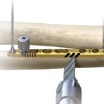
14 Osteotomy With Guide Technique [continued] Figure Proximal Nonlocking Screw Placement While holding the compression, drill the proximal end of the compression slot with the Quick Release Drill ( ), then measure and insert a 3.5 mm Cortical or Nonlocking (CO-3XXX or 30-XXXX). Ensure that the preferred amount of shortening has been achieved by X-ray. Figure 18: 3.5 mm x 5" Quick Release Drill 12 Drilling Glide Hole Quick Release Drill (MS-DC35) and the /3.5 mm Thin Drill Guide (PL-2196), drill a glide hole in the near cortex at an angle across the osteotomy site (Figure 18). Although the proximal or distal portion of the slot may be used depending on the osteotomy location and preferred interfragmentary screw placement, the proximal slot is preferred. Next, place the end of the drill guide into the 3.5 mm glide hole and use a Quick Release Drill ( ) to drill the far cortex (Figure 19). In the scalloped lag screw slot using a 3.5 mm Note: If the angle of the drill is too shallow, the drill may collide with the adjacent screw. Figure 19: Quick Release Drill Quick Release Drill ( ) 3.5 mm Nonlocking 3.5 mm (CO-3XXX) 3.5 mm x 5" Quick Release Drill (MS-DC35) Thin Drill Guide (PL-2196) 11
![Osteotomy With Guide Technique [continued] 13 Distal Locking Screw Placement Nonlocking (CO-3XXX or 30-XXXX) into the scalloped lag screw slot. Remove the Ulnar Shortening Reduction Clamp (80-0423).](/docs-images/87/96621890/images/15-0.jpg "Drill the second distal locking hole using the Quick Release Drill (80-0387) before removing the Locking Drill Guide (80-0384 or 80-0668).")
15 Osteotomy With Guide Technique [continued] 13 Distal Locking Screw Placement Nonlocking (CO-3XXX or 30-XXXX) into the scalloped lag screw slot. Remove the Ulnar Shortening Reduction Clamp ( ). Drill the second distal locking hole using the Quick Release Drill ( ) before removing the Locking Drill Guide ( or ). Measure and insert a Cortical and (COL-3XXX or 30-XXXX) into the remaining distal locking hole. Measure and insert a 3.5 mm Cortical or Figure Final Screw Placement ( ). Measure and replace with a 3.5 mm Cortical or Nonlocking (CO-3XXX or 30-XXXX). Drill, measure, and insert a Cortical and (COL-3XXX or 30-XXXX) in the remaining proximal locking hole. Remove the Ulnar Shortening Reduction Peg Figure mm Nonlocking 3.5 mm (CO-3XXX) Ulnar Shortening Reduction Clamp ( ) Quick Release Drill ( ) Hexalobe Locking Drill Guide ( ) Locking Drill Guide ( ) (COL-3XXX) Ulnar Shortening Reduction Peg ( ) 12


16 Osteotomy Without Guide Technique William B. Geissler, MD 1 Plate Placement Determine the amount of ulnar variance by reviewing preoperative X-rays. After exposing the volar side of the ulna, place the plate 3 5 cm proximal to the distal end of the ulna. Secure the 6-Hole Ulna Shortening Plate (PL-UL06) to the volar surface with one or more clamps, such as the Reduction Forceps with Serrated Jaw (PL-CL04). Make sure the proximal and distal orientation of the plate is correct, as noted by the laser marks on the plate. Figure 22 Proximal Distal Figure 23 Figure 24 Drill Guide and Driver Selections Screw Drill Guide Driver Hex (Cortical) Screw Hexalobe Screw Locking Drill Guide ( ) Hexalobe Locking Drill Guide ( ) 2.5 mm Quick Release Hex Driver (HPC-0025) T15 Stick Fit Hexalobe Driver ( ) 2 Distal Screw and Reduction Peg Placement Drill the most distal locking hole using the Locking Drill Guide ( or see chart below) and Quick Release Drill ( ) and insert the proper length Cortical or Hexalobe Screw (COL-3XXX or 30-XXXX) with proper Hex (HPC-0025) or Hexalobe ( ) Driver. In the proximal end of the measurement slot, drill bicortically perpendicular to the plate and insert the Ulnar Shortening Reduction Peg ( ) with a 2.5 mm Quick Release Hex Driver (HPC-0025). Option: Predrill the two remaining distal locking holes in the same manner with the locking drill guide but DO NOT INSERT SCREWS. This optional step can also occur after the osteotomy has been achieved based on surgeon preference. 6-Hole Ulna Shortening Plate (PL-UL06) Reduction Forceps with Serrated Jaw (PL-CL04) Locking Drill Guide ( ) Hexalobe Locking Drill Guide ( ) Quick Release Drill ( ) (COL-3XXX) 2.5 mm Quick Release Hex Driver (HPC-0025) T15 Stick Fit Hexalobe Driver ( ) Ulnar Shortening Reduction Peg ( ) 13
![Osteotomy Without Guide Technique [continued] 3 Osteotomy Creation Using the 40 reference marks as a visual guide, start the osteotomy at the most distal laser mark with the Osteotomy Saw Blade](/docs-images/87/96621890/images/17-0.jpg "(80-0739-S, 80-0740-S, or 80-2017-S). Generously irrigate the osteotomy. Create the osteotomy to the determined amount of shortening and excise the bone wafer. A.")

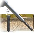
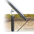
17 Osteotomy Without Guide Technique [continued] 3 Osteotomy Creation Using the 40 reference marks as a visual guide, start the osteotomy at the most distal laser mark with the Osteotomy Saw Blade ( S, S, or S). Generously irrigate the osteotomy. Create the osteotomy to the determined amount of shortening and excise the bone wafer. A.054" K-wire (WS-1406ST) in the distal end of the plate and a Plate Tack (PL-PTACK) in the proximal end may be used for additional stability. Figure 25 Note: Each 40 reference line and space is 2 mm wide. Optional Saw Blades Osteotomy Saw Blade Hub Style L ( S) Osteotomy Saw Blade Hub Style S ( S) Osteotomy Saw Blade Hub Style DS ( S) Each blade has a thickness of.5 mm (.020") along the shaft and.63 mm (.025") at the cutting edge (kerf). 4 Secondary Locking Drill Guide Placement Examine the osteotomy site near the plate. If excess bone is present in the osteotomy site, the proximal and distal ends of the bone may be rotated under the plate to remove any bone blocking the reduction. Place a bone clamp over the distal portion of the ulna and plate to reduce the gap in between them. In the third most distal locking hole closest to the osteotomy, drill using the Locking Drill Guide ( or ) and Cortical or (COL-3XXX or 30-XXXX) if predrilling was not performed in Step 2. Insert a 3.5 mm Cortical or Nonlocking (CO-3XXX or 30-XXXX). Figure 26 Osteotomy Saw Blade Hub Style L ( S) Osteotomy Saw Blade Hub Style S ( S) Osteotomy Saw Blade Hub Style DS ( S).054" K-wire (WS-1406ST) Plate Tack (PL-PTACK) Locking Drill Guide ( ) Hexalobe Locking Drill Guide ( ) (COL-3XXX) 3.5 mm (CO-3XXX). 3.5 mm Nonlocking 14
![Osteotomy Without Guide Technique [continued] Figure 27 5 Osteotomy Gap Reduction Remove the bone clamp and place the Locking Drill Guide (80-0384 or 80-0668) into the second distal locking hole.](/docs-images/87/96621890/images/18-0.jpg "Slightly loosen the reduction peg in the measurement slot. Place the Ulnar Shortening Reduction Clamp (80-0423) around the Ulnar Shortening Reduction Peg (80-0422) and locking drill guide.")
, measure and insert a 3.")
18 Osteotomy Without Guide Technique [continued] Figure 27 5 Osteotomy Gap Reduction Remove the bone clamp and place the Locking Drill Guide ( or ) into the second distal locking hole. Slightly loosen the reduction peg in the measurement slot. Place the Ulnar Shortening Reduction Clamp ( ) around the Ulnar Shortening Reduction Peg ( ) and locking drill guide. Reduce the osteotomy gap with the reduction clamp and tighten the speed-lock wheel on the clamp to maintain reduction hands-free. Figure 28 6 Proximal Nonlocking Screw Placement While holding the compression, drill the proximal end of the compression slot with a Quick Release Drill ( ), measure and insert a 3.5 mm Cortical or Nonlocking Hexalobe Screw (CO-3XXX or 30-XXXX) with a 2.5 mm Quick Release Hex Driver (HPC-0025) or T15 Stick Fit Hexalobe Driver ( ). Ensure that the preferred amount of shortening has been achieved by X-ray. Locking Drill Guide ( ) Hexalobe Locking Drill Guide ( ) Ulnar Shortening Reduction Clamp ( ) Ulnar Shortening Reduction Peg ( ) Quick Release Drill ( ) 3.5 mm (CO-3XXX) 3.5 mm Nonlocking 2.5 mm Quick Release Hex Driver (HPC-0025) T15 Stick Fit Hexalobe Driver ( ) 15
![Osteotomy Without Guide Technique [continued] 7 Drilling Glide Hole In the scalloped slot, using a 3.5 mm Quick Release Drill (MS-DC35) and the /3.](/docs-images/87/96621890/images/19-0.jpg "5 mm Thin Drill Guide (PL-2196), drill a glide hole in the near cortex at an angle across the osteotomy site (Figure 29). Next, place the end of the drill guide into the 3.")


19 Osteotomy Without Guide Technique [continued] 7 Drilling Glide Hole In the scalloped slot, using a 3.5 mm Quick Release Drill (MS-DC35) and the /3.5 mm Thin Drill Guide (PL-2196), drill a glide hole in the near cortex at an angle across the osteotomy site (Figure 29). Next, place the end of the drill guide into the 3.5 mm glide hole and use a Quick Release Drill ( ) to drill the far cortex (Figure 30). Measure and insert a 3.5 mm Cortical or Nonlocking (CO-3XXX or 30-XXXX). The proximal or distal portion of the slot may be used depending on the osteotomy location and preferred interfragmentary screw placement. The most proximal hole is preferred. Note: If the angle of the drill is too shallow, the drill may collide with the adjacent screw. Figure 29: 3.5 mm x 5" Quick Release Drill Figure 30: Quick Release Drill 8 Final Screw Placement Remove reduction clamp and drill the second distal locking hole before removing the Locking Drill Guide ( or ). Measure and insert a Cortical or (COL-3XXX or 30-XXXX) into the remaining distal locking hole. Remove the Ulnar Shortening Reduction Peg ( ). Measure and replace with a 3.5 mm Cortical or Nonlocking (CO-3XXX or 30-XXXX). Drill, measure and insert a 3.5 mm locking cortical or hexalobe screw in the remaining proximal locking hole. Figure mm x 5" Quick Release Drill (MS-DC35) /3.5 mm Thin Drill Guide (PL-2196) Quick Release Drill ( ) 3.5 mm (CO-3XXX) 3.5 mm Nonlocking Locking Drill Guide ( ) Hexalobe Locking Drill Guide ( ) (COL-3XXX) Ulnar Shortening Reduction Peg ( ) 16
20 Ordering Information Tray Components Ulna Shortening Plate 1 6-Hole Ulna Shortening Plate PL-UL06 Instrumentation 2 Ulnar Shortening Guide, Left Ulnar Shortening Guide, Right Ulnar Shortening Guide Locking Bolt Sterile Components Osteotomy Saw Blades Osteotomy Saw Blade Hub Style L* Osteotomy Saw Blade Hub Style S* Osteotomy Saw Blade Hub Style DS* Additional Components Instrumentation S S S 5 Ulnar Shortening Guide Bottom Plate Ulnar Shortening Reduction Peg mm Quick Release Hex Driver HPC-0025 T15 Stick-Fit Hexalobe Driver Ulnar Shortening Reduction Clamp Plate Tack PL-PTACK 8.054" x 6" Guide Wire WS-1406ST 9 Quick Release Drill Locking Drill Guide mm x 5" Quick Release Drill MS-DC35 Hexalobe Locking Drill Guide Tray Ulnar Shortening Tray Assembly *Optional Note: The Acumed Osteotomy System can be used with the following Acumed systems to access additional instrumentation not included in this tray: Clavicle Plating System, Elbow Plating System, and Acu-Loc 2 System. To learn more about the full line of Acumed innovative surgical solutions or order additional systems, please contact your local Acumed sales representative, call , or visit 17

21
22 Ordering Information Screws s 3.5 mm x 8 mm Locking COL mm x 10 mm Locking COL mm x 12 mm Locking COL mm x 14 mm Locking COL mm x 16 mm Locking COL mm x 18 mm Locking COL mm x 20 mm Locking COL mm s 3.5 mm x 8 mm CO mm x 10 mm CO mm x 12 mm CO mm x 14 mm CO mm x 16 mm CO mm x 18 mm CO mm x 20 mm CO-3200 s 3.5 mm x 8 mm Locking mm x 10 mm Locking mm x 12 mm Locking mm x 14 mm Locking mm x 16 mm Locking mm x 18 mm Locking mm x 20 mm Locking mm Nonlocking s 3.5 mm x 8 mm Nonlocking mm x 10 mm Nonlocking mm x 12 mm Nonlocking mm x 14 mm Nonlocking mm x 16 mm Nonlocking mm x 18 mm Nonlocking mm x 20 mm Nonlocking
23 Notes: 20
24 Acumed Headquarters 5885 NW Cornelius Pass Road Hillsboro, OR Office: Office: Fax: These materials contain information about products that may or may not be available in any particular country or may be available under different trademarks in different countries. The products may be approved or cleared by governmental regulatory organizations for sale or use with different indications or restrictions in different countries. Products may not be approved for use in all countries. Nothing contained on these materials should be construed as a promotion or solicitation for any product or for the use of any product in a particular way which is not authorized under the laws and regulations of the country where the reader is located. Specific questions physicians may have about the availability and use of the products described on these materials should be directed to their particular local sales representative. Specific questions patients may have about the use of the products described in these materials or the appropriateness for their own conditions should be directed to their own physician. HNW00-03-J Effective: 2016/ Acumed LLC US Patent No B2 Acumed is a registered trademark of Acumed, LLC
Osteotomy System. The reference lines on the plate help facilitate the creation of the osteotomy, when a free hand cut is preferred.
 Osteotomy System Osteotomy System Since 1988, Acumed has been designing solutions for the demanding situations facing orthopaedic surgeons, hospitals and their patients. Our strategy has been to know the
Acu-Loc Wrist Spanning Plate System. Surgical Technique
 Acu-Loc Wrist Spanning Plate System Surgical Technique Acumed is a global leader of innovative orthopaedic and medical solutions. We are dedicated to developing products, service methods, and approaches
Surgical Technique. Forearm Fracture Solutions
 Surgical Technique Forearm Fracture Solutions Acumed is a global leader of innovative orthopaedic and medical solutions. We are dedicated to developing products, service methods, and approaches that improve
Locking Ankle Plating System. Surgical Technique
 Locking Ankle Plating System Surgical Technique Acumed is a global leader of innovative orthopaedic and medical solutions. We are dedicated to developing products, service methods, and approaches that
Forearm Fracture Solutions. Surgical Technique
 Forearm Fracture Solutions Surgical Technique Acumed is a global leader of innovative orthopaedic and medical solutions. We are dedicated to developing products, service methods, and approaches that improve
Hand Fracture System with Small Bone External Fixation System. Product Overview
 Hand Fracture System with Small Bone External Fixation System Product Overview Acumed Hand Fracture System The Acumed Hand Fracture System is designed to provide both standard and fracture-specific fixation
Acu-Loc Wrist Plating System. Surgical Technique
 Acu-Loc Wrist Plating System Surgical Technique Acumed is a global leader of innovative orthopaedic and medical solutions. We are dedicated to developing products, service methods, and approaches that
Ratcheting Compression Plating System. Surgical Technique
 Ratcheting Compression Plating System Surgical Technique Acumed is a global leader of innovative orthopaedic and medical solutions. We are dedicated to developing products, service methods, and approaches
Elbow Plating System. Surgical Technique
 Elbow Plating System Surgical Technique Acumed is a global leader of innovative orthopaedic and medical solutions. We are dedicated to developing products, service methods, and approaches that improve
Forefoot/Midfoot Plating System. Surgical Technique
 Forefoot/Midfoot Plating System Surgical Technique Acumed is a global leader of innovative orthopaedic and medical solutions. We are dedicated to developing products, service methods, and approaches that
Elbow Plating System
 Elbow Plating System Elbow Plating System Acumed is a global leader of innovative orthopaedic and medical solutions. We are dedicated to developing products, service methods and approaches that improve
Tension Band Pin System 2. Surgical Technique
 Tension Band Pin System 2 Surgical Technique Acumed is a global leader of innovative orthopaedic and medical solutions. We are dedicated to developing products, service methods, and approaches that improve
Surgical Technique. Elbow Plating System
 Surgical Technique Elbow Plating System Acumed is a global leader of innovative orthopaedic and medical solutions. We are dedicated to developing products, service methods, and approaches that improve
Forearm Fracture Solutions. Product Overview
 Forearm Fracture Solutions Product Overview Acumed Forearm Fracture Solutions Acumed Forearm Fracture Solutions includes plating and rodding systems with a range of diaphyseal radius and ulna fracture
Locking Radial Head Plates
 Locking Radial Head Plates Locking Radial Head Plates Since 1988, Acumed has been designing solutions to the demanding situations facing orthopaedic surgeons, hospitals and their patients. Our strategy
Biotrak Resorbable Fixation System
 Surgical Technique Biotrak Resorbable Fixation System Acumed is a global leader of innovative orthopaedic and medical solutions. We are dedicated to developing products, service methods, and approaches
Forefoot/Midfoot Plating System
 Surgical Technique Forefoot/Midfoot Plating System Acumed is a global leader of innovative orthopaedic and medical solutions. We are dedicated to developing products, service methods, and approaches that
Volar Distal Radius Plating System Surgical Technique
 Volar Distal Radius Plating System Surgical Technique Acu-Loc 2 Volar Distal Radius Plating System Acumed is a global leader of innovative orthopaedic and medical solutions. We are dedicated to developing
Acutrak 2 Headless Compression Screw System Micro, Mini, and Standard Screws. Supplemental Use Guide Four Corner Fusion
 Acutrak 2 Headless Compression Screw System Micro, Mini, and Standard Screws Supplemental Use Guide Four Corner Fusion Acumed is a global leader of innovative orthopaedic and medical solutions. We are
Modular Hand System. Product Overview
 Modular Hand System Product Overview Modular Hand System Designed to address specific indications throughout the hand, from the carpals to the phalanges, the Acumed Modular Hand System offers a variety
AcUMEDr. FoREARM ROD SYSTEM
 AcUMEDr FoREARM ROD SYSTEM FoREARM ROD SYSTEM Since 1988 Acumed has been designing solutions to the demanding situations facing orthopedic surgeons, hospitals and their patients. Our strategy has been
Volar Distal Radius Plating System Surgical Technique
 Volar Distal Radius Plating System Surgical Technique Acu-Loc 2 Volar Distal Radius Plating System Acumed is a global leader of innovative orthopaedic and medical solutions. We are dedicated to developing
Surgical Technique. Tension Band Pin System
 Surgical Technique Tension Band Pin System Acumed is a global leader of innovative orthopaedic and medical solutions. We are dedicated to developing products, service methods, and approaches that improve
Surgical Technique. Ankle Plating System
 Surgical Technique Ankle Plating System Acumed is a global leader of innovative orthopaedic and medical solutions. We are dedicated to developing products, service methods, and approaches that improve
Cannulated Screw System. Product Overview
 Cannulated Screw System Product Overview Acumed Cannulated Screw System The Acumed Cannulated Screw System consists of screws, washers, and instruments and is intended for fixation of fractures, fusions,
AcUMEDr. Olecranon Threaded Compression Rod
 AcUMEDr Olecranon Threaded Compression Rod Olecranon Threaded Compression Rod Since 1988, Acumed has been designing solutions to the demanding situations facing orthopaedic surgeons, hospitals and their
AcUMEDr. Anatomic Midshaft Forearm Plates
 AcUMEDr Anatomic Midshaft Forearm Plates Anatomic Midshaft Forearm Plates Since 1988, Acumed has been designing solutions to the demanding situations facing orthopaedic surgeons, hospitals and their patients.
Acu-Loc 2 Wrist Plating System. Surgical Technique
 Acu-Loc 2 Wrist Plating System Surgical Technique Acumed is a global leader of innovative orthopaedic and medical solutions. We are dedicated to developing products, service methods, and approaches that
Locking Clavicle Plating System. Surgical Technique
 Locking Clavicle Plating System Surgical Technique Acumed is a global leader of innovative orthopaedic and medical solutions. We are dedicated to developing products, service methods, and approaches that
Surgical Technique. Fibula Rod System
 Surgical Technique Fibula Rod System Acumed is a global leader of innovative orthopaedic and medical solutions. We are dedicated to developing products, service methods, and approaches that improve patient
Hand Fracture System. Surgical Technique
 Hand Fracture System Surgical Technique Acumed is a global leader of innovative orthopaedic and medical solutions. We are dedicated to developing products, service methods, and approaches that improve
Supplemental Use Guide Standard Triple Arthrodesis
 Acutrak 2 Headless Compression Screw System 4.7 mm and 7.5 mm Screws Supplemental Use Guide Standard Triple Arthrodesis Acumed is a global leader of innovative orthopaedic and medical solutions. We are
Acu-Loc 2 Wrist Plating System
 Surgical Technique Acu-Loc 2 Wrist Plating System Acumed is a global leader of innovative orthopaedic and medical solutions. We are dedicated to developing products, service methods, and approaches that
Bone Graft Harvesting System. Surgical Technique
 Bone Graft Harvesting System Surgical Technique Acumed is a global leader of innovative orthopaedic and medical solutions. We are dedicated to developing products, service methods, and approaches that
Locking Forefoot/Midfoot Plating System
 Locking Forefoot/Midfoot Plating System Locking Forefoot/Midfoot Plating System Acumed is an industry leader in innovative solutions for extremities and trauma. We are dedicated to pioneering solutions
Surgical Technique. Wrist Plating System
 Surgical Technique Wrist Plating System Acumed is a global leader of innovative orthopaedic and medical solutions. We are dedicated to developing products, service methods, and approaches that improve
AcUMEDr. LoCKING CLAVICLE PLATE SYSTEM
 AcUMEDr LoCKING CLAVICLE PLATE SYSTEM LoCKING CLAVICLE PLATE SYSTEM Since 1988 Acumed has been designing solutions to the demanding situations facing orthopedic surgeons, hospitals and their patients.
LCP Ulna Osteotomy System 2.7. Low profile angular stable fixation for ulna shortening osteotomies.
 LCP Ulna Osteotomy System 2.7. Low profile angular stable fixation for ulna shortening osteotomies. Surgical Technique This publication is not intended for distribution in the USA. Instruments and implants
Tension Band Pin System
 Tension Band Pin System Tension Band Pin System Acumed is an industry leader in innovative solutions for extremities and trauma. We are dedicated to pioneering solutions that benefit the patient and drive
AcUMEDr. Arc Wrist Tower. For Wrist Arthroscopy And Fracture Reduction
 AcUMEDr Arc Wrist Tower For Wrist Arthroscopy And Fracture Reduction Arc Wrist Tower Since 1988, Acumed has been designing solutions to the demanding situations facing orthopaedic surgeons, hospitals and
Surgical Technique. Targeter Systems Overview
 Surgical Technique Targeter Systems Overview PERI-LOC Locked Plating System Targeter Systems Overview Table of contents Product overview... 2 Introduction... 2 Indications... 2 Design features and benefits...
AcUMEDr. Locking Proximal Humeral Plate. PoLARUSr PHPt
 AcUMEDr Locking Proximal Humeral Plate PoLARUSr PHPt PoLARUSr PHPt LOCKING PROXIMAL HUMERAL PLATE Since 1988 Acumed has been designing solutions to the demanding situations facing orthopedic surgeons,
Axi+LineTM Proximal Bunion CorrectionSystem. Surgical Technique
 Axi+LineTM Proximal Bunion CorrectionSystem Surgical Technique A i Line Proximal Bunion Correction System TM Desired angle correction is built into the plate, providing a self-reducing, 5-hole construct
Surgical Technique. CONQUEST FN Femoral Neck Fracture System
 Surgical Technique CONQUEST FN Femoral Neck Fracture System Table of Contents Introduction... 3 Indications... 3 Product Overview... 4 Surgical Technique... 5 Patient Positioning... 5 Reduce the Fracture...
Clavicle Plating System Acu-Sinch Repair System. Surgical Technique
 Clavicle Plating System Acu-Sinch Repair System Surgical Technique Acumed is a global leader of innovative orthopaedic and medical solutions. We are dedicated to developing products, service methods, and
Zimmer Small Fragment Universal Locking System. Surgical Technique
 Zimmer Small Fragment Universal Locking System Surgical Technique Zimmer Small Fragment Universal Locking System 1 Zimmer Small Fragment Universal Locking System Surgical Technique Table of Contents Introduction
Small Fragment Plating System. Securing optimal fixation through locked and compression plating technology
 Small Fragment Plating System Securing optimal fixation through locked and compression plating technology Contents Design Rationale Introduction Interfragmentary Fixation Insertion of a 3.5 mm Cortical
DISTAL ELBOW SET. proximal ulna plate SURGICAL TECHNIQUE GUIDE
 SURGICAL TECHNIQUE GUIDE DISTAL ELBOW SET proximal ulna plate As described by: Jorge L. Orbay, M.D. Miami Hand & Upper Extremity Institute Miami, Florida DISTAL ELBOW SET proximal ulna plate Indications
RAYHACK Kienbock Radial Shortening System
 RAYHACK Kienbock Radial Shortening System SURGICAL TECHNIQUE Surgical Technique as described by John M. Rayhack, MD RAYHACK Kienbock Radial Shortening System Surgical Technique Contents RAYHACK Kienbock
MSP TM Metatarsal Shortening System. Surgical Technique
 MSP TM Metatarsal Shortening System Surgical Technique Metaphyseal/Diaphyseal osteotomy does not result in plantar displacement of metatarsal head Osteotomy guide integrated into implant allows for controlled
Fibula Rod System. Lateral Malleolus Fracture Indications:
 Fibula Rod System Fibula Rod System Since 1988, Acumed has been designing solutions for the demanding situations facing orthopaedic surgeons, hospitals and their patients. Our strategy has been to know
3.5 mm LCP Hook Plate
 Part of the DePuy Synthes Locking Compression Plate (LCP ) System 3.5 mm LCP Hook Plate Surgical Technique Table of Contents Introduction 3.5 mm LCP Hook Plate 2 AO Principles 4 Indications 5 Clinical
For Distal Femur Fractures. 95º Condylar Plate. Quick Reference Chart
 For Distal Femur Fractures 95º Condylar Plate Quick Reference Chart 95 Condylar Plate. Quick reference chart for distal femur fractures. Insert guide wires Fix condylar fragments with 6.5 mm cancellous
MSP TM Metatarsal Shortening System. Surgical Technique
 MSP TM Metatarsal Shortening System Surgical Technique Metaphyseal/Diaphyseal osteotomy does not result in plantar displacement of metatarsal head Osteotomy guide integrated into implant allows for controlled
Small Fragment Plating System
 Small Fragment Plating System Securing optimal fixation through locked and compression plating technology SURGICAL TECHNIQUE RECOVERY FUNCTION SURVIVORSHIP DePuy believes in an approach to trauma surgery
SMV Scientific Bone Plate and Screw System Surgical Technique
 SMV Scientific Bone Plate and Screw System Surgical Technique Description: The SMV Scientific Bone Plate and Screw System consists of non-locking plates and bone screw fasteners in a variety of lengths,
AcUMEDr. ScAPHOID TARGETING GUIDE
 AcUMEDr ScAPHOID TARGETING GUIDE ScAPHOID TARGETING GUIDE Since 1988 Acumed has been designing solutions to the demanding situations facing orthopedic surgeons, hospitals and their patients. Our strategy
Surgical Technique. 3.5mm and 4.5mm Lateral Proximal Tibia Locking Plates
 Surgical Technique 3.5mm and 4.5mm Lateral Proximal Tibia Locking Plates PERI-LOC Periarticular Locked Plating System 3.5mm and 4.5mm Lateral Proximal Tibia Locking Plate Surgical Technique Contents Product
Technique Guide. LCP Proximal Femoral Hook Plate 4.5/5.0. Part of the LCP Periarticular Plating System.
 Technique Guide LCP Proximal Femoral Hook Plate 4.5/5.0. Part of the LCP Periarticular Plating System. Table of Contents Introduction Features and Benefits 2 AO ASIF Principles 4 Indications 5 Surgical
Wrist Fixation System
 Wrist Fixation System Anatomy / Fracture Implant EXTRA & SIMPLE ARTICULAR Volar Radius Volar Fixed Angle Plate Volar Bearing Plate Radial Peg Plate Volar Hook Plate Volar Buttress Pin Volar Shear Plate
Surgical Technique. Clavicle Locking Plate
 Surgical Technique Clavicle Locking Plate PERI-LOC Locked Plating System Clavicle Locking Plate Surgical Technique Table of Contents Introduction...2 Indications...3 Plate Features...3 Patient Positioning...4
Surgical Technique. Lower Extremity Plates and Straight Plates
 Surgical Technique Lower Extremity Plates and Straight Plates 2 Table of contents Overview...4 Indications... 4 Contraindications... 4 Screw Options... 5 Straight Plate Options... 6 Proximal Tibia Plate
Surgical Technique. Distal Humerus Locking Plate
 Surgical Technique Distal Humerus Locking Plate PERI-LOC Locked Plating System Distal Humerus Locking Plate Surgical Technique Table of Contents Introduction...2 Indications...3 Plate Features...3 Patient
Technique Guide. 3.5 mm LCP Low Bend Medial Distal Tibia Plate Aiming Instruments. Part of the 3.5 mm LCP Percutaneous Instrument System.
 Technique Guide 3.5 mm LCP Low Bend Medial Distal Tibia Plate Aiming Instruments. Part of the 3.5 mm LCP Percutaneous Instrument System. Table of Contents Introduction 3.5 mm LCP Low Bend Medial Distal
Surgical Technique. Proximal Humerus Locking Plate
 Surgical Technique Proximal Humerus Locking Plate PERI-LOC Upper Extremity Locked Plating System 3.5mm & 4.5mm Proximal Humerus Locking PlatesCatalog Infor Table of Contents Introduction.........................................................2
tep Ankle Fracture Plating System
 tep Response Ortho is a global orthopaedic trauma solutions manufacturer offering premium products created under its founding principles of innovation, excellence by design, and functional superiority.
Surgical Technique. Anterolateral and Medial Distal Tibia Locking Plates
 Surgical Technique Anterolateral and Medial Distal Tibia Locking Plates PERI-LOC Periarticular Locked Plating System Anterolateral and Medial Distal Tibia Locking Plates Surgical Technique Contents Product
MetaFix Ludloff Plate
 Merete MetaFix Ludloff Plate Low Profile Locking Bone Plate System Surgical Technique and Ordering Information - Content - Content 1. Description.................................................. 3 2.
Instrument and Implant for wrist fracture
 Instrument and Implant for wrist fracture Jansri Janpanya Product specialist The Bangkok Unitrade Co,.ltd. Objectives Type of LCP for distal radius Fx. The new LCP design for distal radius Fx. Have knowledge
LCP Medial Distal Tibia Plate, without Tab. The Low Profile Anatomic Fixation System with Angular Stability and Optimal Screw Orientation.
 LCP Medial Distal Tibia Plate, without Tab. The Low Profile Anatomic Fixation System with Angular Stability and Optimal Screw Orientation. Technique Guide LCP Small Fragment System Table of Contents Introduction
LCP Medial Proximal Tibial Plate 4.5/5.0. Part of the Synthes LCP periarticular plating system.
 LCP Medial Proximal Tibial Plate 4.5/5.0. Part of the Synthes LCP periarticular plating system. Technique Guide This publication is not intended for distribution in the USA. Instruments and implants approved
Surgical Technique Carpal Fusion
 Carpal Fusion Patent and Patent Pending CAUTION: Federal Law (USA) restricts this device to sale by or on the order of a physician. INDICATIONS FOR USE The Extremity Medical Lag Screw and X-Post System
LoCKING FOREFOOT/ MIDFOOT PLATES
 LoCKING FOREFOOT/ MIDFOOT PLATES LoCKING FOREFOOT/ MIDFOOT PLATES Since 1988 Acumed has been designing solutions to the demanding situations facing orthopedic surgeons, hospitals and their patients. Our
Distal Radius Plate Instrument and Implant Set. Discontinued December 2017 DSUS/TRM/0916/1063(1)
") Distal Radius Plate Instrument and Implant Set Surgical Technique Discontinued December 2017 DSUS/TRM/0916/1063(1) The Distal Radius Plates Indications For fixation of fractures and osteotomies, including
Distal Radius Plate Instrument and Implant Set Surgical Technique Discontinued December 2017 DSUS/TRM/0916/1063(1) The Distal Radius Plates Indications For fixation of fractures and osteotomies, including
Distal Cut First Femoral Preparation
 Surgical Technique Distal Cut First Femoral Preparation Primary Total Knee Arthroplasty LEGION Total Knee System Femoral preparation Contents Introduction...3 DCF femoral highlights...4 Preoperative planning...6
Surgical Technique. Olecranon Locking Plate
 Surgical Technique Olecranon Locking Plate PERI-LOC Locked Plating System Olecranon Locking Plate Surgical Techniquealog Infor Table of Contents Introduction...2 Indications...3 Plate Features...3 Patient
AcUMEDr. MoDULAR HAND SYSTEM
 AcUMEDr MoDULAR HAND SYSTEM MoDULAR HAND SYSTEM Since 1988, Acumed has been designing solutions to the demanding situations facing orthopedic surgeons, hospitals and their patients. Our strategy has been
PediLoc 3.5mm and 4.5mm Contour Femur Plate Surgical Technique
 PediLoc 3.5mm and 4.5mm Contour Femur Plate Surgical Technique Surgical Technique Contour Femur Plate The technique description herein is made available to the healthcare professional to illustrate the
SURGICAL TECHNIQUE GUIDE PROTEAN. radial head plate. As described by: Jorge L. Orbay, M.D. Miami Hand & Upper Extremity Institute Miami, Florida
 SURGICAL TECHNIQUE GUIDE PROTEAN radial head plate As described by: Jorge L. Orbay, M.D. Miami Hand & Upper Extremity Institute Miami, Florida PROTEAN radial head plate Indications for Use The PROTEAN
The Flower Medial Column Fusion Plate
 The Flower Medial Column Fusion Plate PROCEDURE GUIDE www.flowerortho.com The Flower Foot & Ankle Application NC FUSION PLATE 2-HOLE COMPRESSION PLATE TMT FUSION PLATE LAPIDUS FUSION PLATE COMPRESSION
A sequenced approach to flush graft placement. GLENOID BONE LOSS SYSTEM Procedural Solution
 A sequenced approach to flush graft placement GLENOID BONE LOSS SYSTEM Procedural Solution One comprehensive system, two ways of treating glenoid bone loss The GLENOID BONE LOSS SYSTEM provides you with
OSS Orthopedic Salvage System Compress Device. Surgical Technique
 OSS Orthopedic Salvage System Compress Device Surgical Technique Table of Contents Quick Reference Guide... 2 Device Description... 3 Resection... 4 No Face Adapter Provisional Construct... 4 Canal Preparation...
Surgical Technique International Version. Clavicle Locking Plate
 Surgical Technique International Version Clavicle Locking Plate PERI-LOC Upper Extremity Locked Plating System Clavicle Surgical Techniquefor Table of Contents Introduction........................................................2
Cannulated Pediatric Osteotomy System (CAPOS). A single system of osteotomy blade plates and cannulated instrumentation.
. A single system of osteotomy blade plates and cannulated instrumentation.") Cannulated Pediatric Osteotomy System (CAPOS). A single system of osteotomy blade plates and cannulated instrumentation. Technique Guide This publication is not intended for distribution in the USA. Instruments
Cannulated Pediatric Osteotomy System (CAPOS). A single system of osteotomy blade plates and cannulated instrumentation. Technique Guide This publication is not intended for distribution in the USA. Instruments
Correction System. Surgical Technique
 Nextra Hammertoe Correction System Surgical Technique Maximized Bone Purchase* Stable and Secure Phalanx Optimized Screw Design Adjustable Bone-to-Bone Apposition Progressive Ratchet Tightening Mechanism
System. Humeral Nail. Surgical Technique
 System Humeral Nail Surgical Technique Contents IMPLANT FEATURES 2 1. INDICATIONS 3 2. PRE-OPERATIVE PLANNING 3 3. PATIENT POSITIONING & FRACTURE REDUCTION 3 4. INCISION 4 5. ENTRY POINT 4-6 6. PROXIMAL
Cannulated Angled Blade Plate 3.5 and 4.5, 90.
 Cannulated Angled Blade Plate 3.5 and 4.5, 90. Technique Guide This publication is not intended for distribution in the USA. Instruments and implants approved by the AO Foundation. Table of Contents Introduction
LCP Distal Humerus Plates
 The anatomic fixation system for the distal humerus with angular stability Surgical technique LCP Locking Compression Plate Contents Indications and contraindications 2 Implants 3 Instruments 5 Preparation
Technique Guide. 3.5 mm LCP Olecranon Plates. Part of the Synthes locking compression plate (LCP) system.
 system.") Technique Guide 3.5 mm LCP Olecranon Plates. Part of the Synthes locking compression plate (LCP) system. Table of Contents Introduction 3.5 mm LCP Olecranon Plates 2 AO Principles 3 Indications 3 Clinical
Technique Guide 3.5 mm LCP Olecranon Plates. Part of the Synthes locking compression plate (LCP) system. Table of Contents Introduction 3.5 mm LCP Olecranon Plates 2 AO Principles 3 Indications 3 Clinical
DARCO. Bow 2 Plate SURGIC AL TECHNIQUE
 DARCO Bow 2 Plate SURGIC AL TECHNIQUE Contents 2 Preface 3 Chapter 1 4 Chapter 2 5 6 7 8 9 Appendix 10 10 11 Intended Use Indications/Contraindications Design Rationale Preoperative Planning Surgical Technique
3. Insert Tocar Sleeves Insert the NCB tissue protection sleeve assembly 1.6 to 10mm through a skin incision (Fig. 38).
.") NCB Proximal Humerus Plating System Surgical Technique 19 2. Temporary Plate Fixation The plate can be temporary fixed to the bone with 1.6mm K-wire through the proximal cannulated fixation screw of the
NCB Proximal Humerus Plating System Surgical Technique 19 2. Temporary Plate Fixation The plate can be temporary fixed to the bone with 1.6mm K-wire through the proximal cannulated fixation screw of the
LCP Medial Proximal Tibial Plate 3.5. Part of the Synthes small fragment Locking Compression Plate (LCP) system.
 system.") LCP Medial Proximal Tibial Plate 3.5. Part of the Synthes small fragment Locking Compression Plate (LCP) system. Technique Guide This publication is not intended for distribution in the USA. Instruments
LCP Medial Proximal Tibial Plate 3.5. Part of the Synthes small fragment Locking Compression Plate (LCP) system. Technique Guide This publication is not intended for distribution in the USA. Instruments
Zimmer Periarticular Proximal Humeral Locking Plate
 Zimmer Periarticular Proximal Humeral Locking Plate Surgical Technique The Science of the Landscape Zimmer Periarticular Proximal Humeral Locking Plate 1 Surgical Technique Table of Contents Introduction
tep Ankle Fracture Plating System
 tep Response Ortho is a global orthopaedic trauma solutions manufacturer offering premium products created under its founding principles of innovation, excellence by design and functional superiority.
Merete BLP. Surgical Technique. - Bunion Locking Plate - Low Profile Locking Bone Plate System
 Merete BLP - Bunion Locking Plate - Low Profile Locking Bone Plate System Surgical Technique Merete Medical, Inc. 49 Purchase Street Rye, N.Y. 10580 Phone: 914 967-1532 www.merete-medical.com - Surgical
Technique Guide. TomoFix Osteotomy System. A comprehensive plating system for stable fixation of osteotomies around the knee.
 Technique Guide TomoFix Osteotomy System. A comprehensive plating system for stable fixation of osteotomies around the knee. Table of Contents Introduction TomoFix Osteotomy System 2 AO Principles 4 Indications
MINI TIBIAL PLATEAU LEVELING OSTEOTOMY (TPLO) SYSTEM
 SYSTEM") MINI TIBIAL PLATEAU LEVELING OSTEOTOMY (TPLO) SYSTEM For stabilizing osteotomies of the canine and feline proximal tibia SURGICAL TECHNIQUE TABLE OF CONTENTS INTRODUCTION Mini Tibial Plateau Leveling
MINI TIBIAL PLATEAU LEVELING OSTEOTOMY (TPLO) SYSTEM For stabilizing osteotomies of the canine and feline proximal tibia SURGICAL TECHNIQUE TABLE OF CONTENTS INTRODUCTION Mini Tibial Plateau Leveling
3.5 mm LCP Low Bend Medial Distal Tibia Plate Aiming Instruments
 Part of the 3.5 mm LCP 3.5 mm LCP Low Bend Medial Distal Tibia Plate Aiming Instruments Surgical Technique TABLE OF CONTENTS INTRODUCTION 3.5 mm LCP Low Bend Medial Distal Tibia Plate 2 Aiming Instruments