3.5 mm LCP Hook Plate
|
|
|
- Quentin Fletcher
- 6 years ago
- Views:
Transcription


1 Part of the DePuy Synthes Locking Compression Plate (LCP ) System 3.5 mm LCP Hook Plate Surgical Technique
2 Table of Contents Introduction 3.5 mm LCP Hook Plate 2 AO Principles 4 Indications 5 Clinical Cases 6 Surgical Technique Preparation 7 Position Patient 8 Approach 9 Reduce Fracture 10 Perform Osteotomy and Fix Temporarily 11 Predrill for Hooks 13 Position 3.5 mm LCP Hook Plate 14 Insert Screws 15 Implant Removal (Optional) 20 Product Information Implants 21 Instruments 22 Product Information 26 MR Information The 3.5 mm LCP Hook Plate System has not been evaluated for safety and compatibility in the MR environment. It has not been tested for heating, migration or image artifact in the MR environment. The safety of the 3.5 mm LCP Hook Plate System in the MR environment is unknown. Scanning a patient who has this device may result in patient injury. Image intensifier control 3.5 mm LCP Hook Plate Surgical Technique DePuy Synthes 1
")


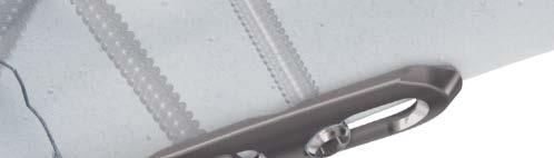
3 3.5 mm LCP Hook Plate. Part of the DePuy Synthes locking compression plate (LCP) System. Low profile Minimal hardware prominence Hooks provide additional points of fixation Locking screws provide a fixed-angle construct, which provides advantages in osteopenic bone where traditional screw purchase is compromised Precontoured Shaped to provide spring-effect, to aid in reduction Dual hook configuration facilitates placement Versatile A single 3-hole plate can be used in multiple locations, applied to either the right or left side of the anatomy, resulting in less inventory required Scalloped undercuts Preservation of blood supply to the bone Sharp hooks Aid in placement of the plate 1 DePuy Synthes 3.5 mm LCP Hook Plate Surgical Technique


4 Olecranon Distal fibula/ lateral malleolus Distal tibia/ medial malleolus Nonlocking hole between the hooks Allows fracture compression with a lag screw ( home-run screw ) Hole zero Elongated nonlocking hole Designed for plate flexibility, and aids in placement and fracture compression Hole one Elongated Combi holes For controlled compression and optimal plate placement Hole two Rounded edges Minimize soft tissue irritation Hole three 3.5 mm LCP Hook Plate Surgical Technique DePuy Synthes 3

5 AO Principles AO PRINCIPLES In 1958, the AO formulated four basic principles, which have become the guidelines for internal fixation. 1, 2 In 1958, the AO formulated four basic principles, which have become the guidelines for internal fixation 1, 2. 4_Priciples_03.pdf :08 Anatomic Anatomic reduction reduction Fracture Fracture reduction reduction and and fixation fixation to to restore restore anatomical anatomical relationships. relationships. 1 2 Stable Stable fixation fixation Fracture Fracture fixation fixation providing providing absolute absolute or relative or relative stability, stability, as required as by the required patient, by the the injury, patient, and the the injury, personality and the personality of the fracture. of the fracture. Early, Early, active mobilization Early Early and and safe safe mobilization and rehabilitation of of the injured part and and the the patient as as a whole. 4 3 Preservation Preservation of of blood blood supply supply Preservation Preservation of of the the blood blood supply supply to to soft soft tissues tissues and and bone bone by by gentle reduction gentle reduction techniques techniques and and careful careful handling. handling. 1. Müller ME, Allgöwer M, Schneider R, Willenegger H. Manual of Internal Fixation. 3rd ed. Berlin, Heidelberg, New York: Springer-Verlag; Rüedi TP, RE Buckley, CG Moran. AO Principles of Fracture Management. 1 Müller ME, M Allgöwer, R Schneider, H Willenegger. Manual of Internal 2nd ed. Stuttgart New York: Thieme; Fixation. 3rd ed. Berlin Heidelberg New York: Springer Rüedi TP, RE Buckley, CG Moran. AO Principles of Fracture Management. 2nd ed. Stuttgart, New York: Thieme DePuy Synthes 3.5 mm LCP Hook Plate Surgical Technique
6 Indications The 3.5 mm LCP Hook Plate is indicated for fractures, osteotomies and nonunions of small bones, including the ulna, radius, tibia and fibula, particularly in osteopenic bone. 3.5 mm LCP Hook Plate Surgical Technique DePuy Synthes 5


7 Clinical Cases Case 1* 78-year-old, gender unknown 21-B3 fracture: right arm Poor bone quality Preoperative lateral Postoperative AP Postoperative lateral Case 2* 61-year-old male Open distal humerus fracture: right arm Good bone quality Olecranon osteotomy required to repair distal humerus Preoperative lateral Postoperative AP Postoperative lateral * Results from case studies are not predictive of results in other cases. Results in other cases may vary. 6 DePuy Synthes 3.5 mm LCP Hook Plate Surgical Technique
 Note: The following technique is specific to the olecranon.")
8 Preparation Required set / Small Fragment LCP Instrument and Implant Set, with 3.5 mm Hex Drive Cortex Screws (stainless steel or titanium) Note: The following technique is specific to the olecranon. Steps 1, 2 and 3 are specific to this surgical site, and should be modified when applied to other anatomy and fracture locations. 3.5 mm LCP Hook Plate Surgical Technique DePuy Synthes 7

9 Position Patient 1 Position patient For an olecranon procedure, place the patient in lateral decubitus with the elbow flexed over a side rest. A small padded table can be placed under the forearm to support the elbow in extension if necessary. The supine position with forearm placed across the chest is also an acceptable position, especially with extended approaches to the lateral pillar or column. Note: For more information on fixation principles using conventional and locked plating techniques, please refer to the Small Fragment Locking Compression Plate (LCP) System Technique Guide. 8 DePuy Synthes 3.5 mm LCP Hook Plate Surgical Technique

10 Approach 2 Approach Make a posterior midline incision centered over the fracture or osteotomy site. The incision can also be slightly curved to the radial side to protect the ulnar nerve. 3.5 mm LCP Hook Plate Surgical Technique DePuy Synthes 9
11 Reduce Fracture 3a Reduce fracture Instruments / 2.0 mm Kirschner Wire, 150 mm, trocar point (stainless steel or titanium) or mm Kirschner Wire with Thread, 150 mm, trocar point, 5 mm thread length Reduce the fracture directly or indirectly depending on the type of fracture. Temporarily fix the fragment using Kirschner wires and/or forceps. Examine the reduction of the olecranon using image intensification. Ensure Kirschner wires or forceps will not interfere with plate placement. 11 DePuy Synthes 3.5 mm LCP Hook Plate Surgical Technique
12 Perform Osteotomy and Fix Temporarily 3b Perform osteotomy and fix temporarily Perform an incomplete osteotomy of the dorsal cortex of the olecranon using a thin oscillating saw blade. Complete the osteotomy with a chisel in order to obtain an interdigitating fracture line. The fracture line should ideally run through the bare area of the sigmoid notch. The olecranon fragment is then placed to the lateral side. Protect the ulnar nerve on the medial side and the muscular branch to the anconeus on the lateral side. Perform surgery on the distal humerus as required. 3.5 mm LCP Hook Plate Surgical Technique DePuy Synthes 11
13 Perform Osteotomy and Fix Temporarily 3b. Perform osteotomy and fix temporarily continued Instrument mm Kirschner Wire, 150 mm, trocar points on both ends Insert a 1.6 mm Kirschner wire with trocar points on both ends into the proximal olecranon fragment and reduce the osteotomy using an inside-out technique. Insert the wire into the fragment through the osteotomy from distal to proximal. The insertion point should be close to the articular surface and the wire should exit the fragment at the distal insertion line of the triceps tendon. Grasp wire from the tip exiting the proximal end of the olecranon. Retract wire proximally until its tip no longer protrudes from the osteotomy line. Reduce the olecranon. Check for an anatomic reduction of the articular surface. Advance the previously placed wire from proximal to distal until it passes through the cortex of the coronoid process. Insert additional Kirschner wires if greater preliminary stability is required. Note: Take care not to drill in a radial direction, as the tip of the Kirschner wires and potential screws may interfere with forearm rotation. 11 DePuy Synthes 3.5 mm LCP Hook Plate Surgical Technique

14 Predrill for Hooks 4 Predrill for hooks Instruments mm Drill Bit, quick coupling, 125 mm mm Universal Drill Guide Drill two holes for hook placement using the plate as a guide. The holes should lie approximately 4 mm proximal to the insertion line of the triceps and centered over the olecranon. Note: The holes are drilled through longitudinal splits in the tendon fibers. 3.5 mm LCP Hook Plate Surgical Technique DePuy Synthes 11
15 Position 3.5 mm LCP Hook Plate 5 Position 3.5 mm LCP hook plate Optional instruments Bending Pliers for 2.7 mm and 3.5 mm plate, 150 mm length and Bending Pliers for 2.7 mm and 3.5 mm plate, 150 mm length or Bending Pliers for 2.7 mm and 3.5 mm Reconstruction Plates, 180 mm length, (two required) Place the plate on the olecranon, sinking the hooks into the predrilled holes. Be careful not to damage the surgical gloves or the patient s surrounding soft tissues with the sharp hooks. Align the plate in the axis of the proximal ulna. Note: If required, the plate can be bent using bending pliers. Precautions: To prevent the locking section of the screw hole from being deformed, do not bend the plate over the locking holes. The plate hooks must not be bent. When placing the LCP Hook Plate 3.5, pay attention to not damage the surgical gloves or the patient s surrounding soft tissue with the sharp hooks. 11 DePuy Synthes 3.5 mm LCP Hook Plate Surgical Technique


16 Insert Screws 6 Insert cortex screws Instruments mm Drill Bit, quick coupling, 110 mm gold Small Hexagonal Screwdriver, with holding sleeve or Handle, with quick coupling, small and Small Hexagonal Screwdriver Shaft Depth Gauge mm Universal Drill Guide Self-centering Bone Forceps or Bone Holding Forceps, soft ratchet Drill a screw hole in the dynamic compression unit (DCU) portion of the middle elongated Combi hole, ensuring that the drill will not collide with the reduction Kirschner wires. Measure for screw length, using the depth gauge. Insert a 3.5 mm cortex screw. Do not fully tighten the screw. A 4.0 mm cancellous bone screw may also be used in this step. Next, drill a screw hole approximately 20 mm 30 mm from the end of the plate, through the near cortex only, and aligned with the centerline of the plate. Insert a 3.5 mm cortex screw, leaving the screwhead approximately 2 mm 4 mm above the bone. 3.5 mm LCP Hook Plate Surgical Technique DePuy Synthes 11

17 Insert Screws 6. Insert cortex screws continued Apply compression outside the plate using a bone holding forceps and the 3.5 mm cortex screw. Tighten the previously inserted 3.5 mm cortex screw in the middle elongated Combi hole of the plate. Remove the forceps and the independent 3.5 mm cortex screw that had been used to apply compression. Note: Although inserting a cortex screw eccentrically can gain limited compression, it is recommended that additional compression be obtained using external means. 11 DePuy Synthes 3.5 mm LCP Hook Plate Surgical Technique

18 7 Insert screw between hooks Instruments mm Drill Bit, quick coupling, 125 mm and mm Universal Drill Guide or mm Drill Bit, quick coupling, 110 mm gold and mm Universal Drill Guide Small Hexagonal Screwdriver, with holding sleeve or Handle, with quick coupling, small and Small Hexagonal Screwdriver Shaft Depth Gauge, for 2.7 mm and small screws Drill the screw hole between the hooks with a 2.5 mm drill bit. Be sure to angle the drill away from the long axis of the plate to avoid colliding with other screws in the plate. Measure for screw length, using the depth gauge. Ensure that the screw will not collide with the reduction Kirschner wires. Insert and tighten a 3.5 mm cortex screw. Alternatively, use a 2.7 mm cortex screw in this hole. Remove the forceps and the independent 3.5 mm cortex screw used to apply compression in Step mm LCP Hook Plate Surgical Technique DePuy Synthes 11
19 Insert Screws 8 Insert locking screws Instruments mm Drill Bit, quick coupling, 165 mm mm Threaded Drill Guide, for 3.5 mm Locking Screws StarDrive Screwdriver, T15 or Handle with quick coupling, small and StarDrive Screwdriver Shaft, T15, self-retaining, quick coupling Depth Gauge, for 2.7 mm and small screws Torque Limiting Attachment, 1.5 Nm, quick coupling or Torque Limiting Attachment, 1.5 Nm Insert the threaded drill guide into the locking portion of the other (open) elongated Combi hole in the plate shaft until fully seated. Use the 2.8 mm drill bit to the desired depth. Remove the drill guide and use the depth gauge to determine the screw length. Insert the self-tapping 3.5 mm locking screw using the StarDrive TM Screwdriver shaft with torque limiting attachment or using the StarDrive screwdriver. 11 DePuy Synthes 3.5 mm LCP Hook Plate Surgical Technique
20 Remove the 1.6 mm Kirschner wire. Precaution: Always use the torque limiter to restrict the maximum torque. Alternatively, follow the appropriate steps for inserting a 3.5 mm cortex screw into this hole. 3.5 mm LCP Hook Plate Surgical Technique DePuy Synthes 11

21 Implant Removal (Optional) Optional set Screw Removal Set Optional instruments Conical Extraction Screw Handle, with quick coupling To remove locking screws, unlock all screws from the plate, then remove the screws completely from the bone. This prevents simultaneous rotation of the plate when unlocking the last locking screw. If the screws cannot be removed with the screwdriver (e.g. if the hexagonal or StarDrive recess of a locking screw is damaged, or if the screws are stuck in the plate), insert the conical extraction screw with left-handed thread into the screwhead using the handle with quick coupling and loosen the locking screw by turning it counterclockwise. 22 DePuy Synthes 3.5 mm LCP Hook Plate Surgical Technique
22 Screws Used with the 3.5 mm LCP Hook Plate 2.7 mm Cortex Screws, self-tapping* May be used in the nonlocking hole between the hooks Compress the plate to the bone 3.5 mm Cortex Screws, self-tapping* May be used in the DCU portion of the Combi holes in the plate shaft May be used in the nonlocking hole between the hooks Compress the plate to the bone or create axial compression Hex Drive or T15 StarDrive recess 3.5 mm Locking Screws, self-tapping* Used in locking portion of the Combi holes Create a locked, fixed-angle screw/ plate construct 4.0 mm Cancellous Bone Screws* May be used in the DCU portion of the Combi holes in the plate shaft Compress the plate to the bone or create axial compression Fully or partially threaded shaft *Found in the Small Fragment LCP Screw Set 3.5 mm LCP Hook Plate Surgical Technique DePuy Synthes 11

23 Selected Instruments from the Small Fragment LCP Instrument and Implant Sets ( , ) mm Kirschner Wire, 150 mm, trocar point or * 1.6 mm Kirschner Wire, 150 mm, trocar point on both ends mm Kirschner Wire with Thread, 150 mm, trocar point, 5 mm thread length mm Drill Bit, quick coupling, 125 mm mm Drill Bit, quick coupling, 110 mm, gold mm Drill Bit, quick coupling, 165 mm mm Drill Bit, quick coupling, 110 mm * Also available 22 DePuy Synthes 3.5 mm LCP Hook Plate Surgical Technique


24 Handle, with quick coupling * Large Handle, with quick coupling mm Threaded Drill Guide Small Hexagonal Screwdriver, with holding sleeve Small Hexagonal Screwdriver Shaft StarDrive Screwdriver, T StarDrive Screwdriver Shaft, T15, self-retaining, quick coupling * Also available 3.5 mm LCP Hook Plate Surgical Technique DePuy Synthes 22
 319.01 Depth Gauge, for 2.7 mm and small screws 323.26 2.")


25 Selected Instruments from the Small Fragment LCP Instrument and Implant Set ( , ) Depth Gauge, for 2.7 mm and small screws mm Universal Drill Guide mm Universal Drill Guide Bending Iron, for 2.7 mm and 3.5 mm plates, 150 mm Used with Bending Iron, for 2.7 mm and 3.5 mm plates, 150 mm Used with Bending Iron, for 2.7 mm and 3.5 mm Reconstruction Plates 22 DePuy Synthes 3.5 mm LCP Hook Plate Surgical Technique


26 329.15* Bending Pliers, for 2.7 mm and 3.5 mm plates, 230 mm *** Self-Centering Bone Forceps, 10 mm serrated jaw, speed lock ** Bone Holding Forceps, soft ratchet for plates to 9 mm wide * Torque Limiting Attachment, 1.5 Nm Torque Limiting Attachment, 1.5 Nm, quick coupling *Also available **SS Set only ***Ti Set only 3.5 mm LCP Hook Plate Surgical Technique DePuy Synthes 22


27 Product Information 3.5 mm LCP Hook Plates mm LCP Hook Plate, 3 holes, 62 mm mm Titanium LCP Hook Plate, 3 holes, 62 mm The 3.5 mm LCP Hook Plate is a part of the following sets: mm LCP Elbow System Set mm Titanium LCP Elbow System Set mm LCP Olecranon Plate Set mm Titanium LCP Olecranon Plate Set The 3.5 mm LCP Hook Plate can be housed in the following graphic cases: mm LCP Olecranon Plate Graphic Case mm LCP Elbow System Graphic Case mm Titanium LCP Elbow System Graphic Case in combination with: Retrofit Kit for 3.5 mm Titanium LCP Elbow System Graphic Case The 3.5 mm LCP Hook Plate requires one of the following sets: Small Fragment LCP Instrument and Implant Set, with 3.5 mm StarDrive Cortex Screws Small Fragment LCP Instrument and Implant Set, with 3.5 mm Hex Drive Cortex Screws Small Fragment LCP Instrument and Implant Set, with 4 mm Cannulated Screws Small Fragment LCP Instrument and Titanium Implant Set, with 3.5 mm StarDrive Cortex Screws Small Fragment LCP Instrument and Titanium Implant Set, with 3.5 mm Hex Drive Cortex Screws Available nonsterile or sterile-packed. Add S to catalog number to order sterile product. Note: For additional information, please refer to package insert. For detailed cleaning and sterilization instructions, please refer to or sterilization instructions, if provided. 16 DePuy Synthes 3.5 mm LCP Hook Plate Surgical Technique
28 Also Available Screw Removal Set mm LCP Hook Plate Retrofit Kit for Olecranon Plate Tray mm LCP Olecranon Graphic Case Tray Retrofit Kit for 3.5 mm LCP Elbow System Graphic Case 3.5 mm LCP Hook Plate Surgical Technique DePuy Synthes 22
29 Limited Warranty and Disclaimer: DePuy Synthes products are sold with a limited warranty to the original purchaser against defects in workmanship and materials. Any other express or implied warranties, including warranties of merchantability or fitness, are hereby disclaimed. Please also refer to the package insert(s) or other labeling associated with the devices identified in this surgical technique for additional information. CAUTION: Federal Law restricts these devices to sale by or on the order of a physician. Some devices listed in this surgical technique may not have been licensed in accordance with Canadian law and may not be for sale in Canada. Please contact your sales consultant for items approved for sale in Canada. Not all products may currently be available in all markets. Manufactured or distributed by: Synthes USA Products, LLC 1302 Wrights Lane East West Chester, PA Synthes USA, LLC 1101 Synthes Avenue Monument, CO To order (USA): To order (Canada): Note: For recognized manufacturer, refer to the product label. DePuy Synthes All rights reserved. DSUS/TRM/0916/1097 4/17 DV
3.5 mm LCP Olecranon Plates
 Part of the DePuy Synthes Locking Compression Plate (LCP ) System 3.5 mm LCP Olecranon Plates Surgical Technique Table of Contents Introduction 3.5 mm LCP Olecranon Plates 2 AO Principles 3 Indications
3.5 mm LCP Extra-articular Distal Humerus Plate
 Part of the DePuy Synthes Locking Compression Plate (LCP ) System 3.5 mm LCP Extra-articular Distal Humerus Plate Surgical Technique Table of Contents Introduction 3.5 mm LCP Extra-articular Distal Humerus
Low Bend Distal Tibia Plates
 Part of the DePuy Synthes Locking Compression Plate (LCP ) System 3.5 mm LCP Low Bend Medial Distal Tibia Plates Surgical Technique Table of Contents Introduction 3.5 mm LCP Low Bend Medial Distal Tibia
Long Volar Plates for Diaphyseal-Metaphyseal Radius Fractures LCP. Dia-Meta Volar Distal Radius Plates. Surgical Technique
 Long Volar Plates for Diaphyseal-Metaphyseal Radius Fractures LCP Dia-Meta Volar Distal Radius Plates Surgical Technique Table of Contents Introduction LCP Dia-Meta Volar Distal Radius Plates 2 AO Principles
Technique Guide. 3.5 mm LCP Olecranon Plates. Part of the Synthes locking compression plate (LCP) system.
 system.") Technique Guide 3.5 mm LCP Olecranon Plates. Part of the Synthes locking compression plate (LCP) system. Table of Contents Introduction 3.5 mm LCP Olecranon Plates 2 AO Principles 3 Indications 3 Clinical
Technique Guide 3.5 mm LCP Olecranon Plates. Part of the Synthes locking compression plate (LCP) system. Table of Contents Introduction 3.5 mm LCP Olecranon Plates 2 AO Principles 3 Indications 3 Clinical
3.5 mm LCP Distal Humerus Plates
 Part of the DePuy Synthes Locking Compression Plate (LCP ) System 3.5 mm LCP Distal Humerus Plates Surgical Technique Table of Contents Introduction 3.5 mm LCP Distal Humerus Plates 2 AO Principles 4 Indications
2.7 mm/3.5 mm LCP Distal Fibula Plate
 Part of the DePuy Synthes Locking Compression Plate (LCP ) System 2.7 mm/3.5 mm LCP Distal Fibula Plate Surgical Technique Table of Contents Introduction 2.7 mm/3.5 mm LCP Distal Fibula Plates 2 AO Principles
3.5 mm LCP Clavicle Hook Plates
 Part of the Synthes Locking Compression Plate (LCP ) System 3.5 mm LCP Clavicle Hook Plates Surgical Technique Table of Contents Introduction 3.5 mm LCP Clavicle Hook Plates 2 AO Principles 4 Indications
3.5 mm LCP Low Bend Medial Distal Tibia Plate Aiming Instruments
 Part of the 3.5 mm LCP 3.5 mm LCP Low Bend Medial Distal Tibia Plate Aiming Instruments Surgical Technique TABLE OF CONTENTS INTRODUCTION 3.5 mm LCP Low Bend Medial Distal Tibia Plate 2 Aiming Instruments
3.5 mm Clavicle Hook Plates
 A Single Solution for Lateral Clavicle Fractures and Acromioclavicular Joint Dislocations 3.5 mm Clavicle Hook Plates Surgical Technique Discontinued December 2017 DSUS/TRM/1016/1126(1) Table of Contents
Part of the DePuy Synthes Locking Compression Plate (LCP ) System. 3.5 mm LCP Medial Proximal Tibia Plates
 System. 3.5 mm LCP Medial Proximal Tibia Plates") Part of the DePuy Synthes Locking Compression Plate (LCP ) System 3.5 mm LCP Medial Proximal Tibia Plates Surgical Technique Table of Contents Introduction 3.5 mm LCP Medial Proximal Tibia Plates 2 AO
Part of the DePuy Synthes Locking Compression Plate (LCP ) System 3.5 mm LCP Medial Proximal Tibia Plates Surgical Technique Table of Contents Introduction 3.5 mm LCP Medial Proximal Tibia Plates 2 AO
Technique Guide. 3.5 mm LCP Low Bend Medial Distal Tibia Plates. Part of the Synthes locking compression plate (LCP) system.
 system.") Technique Guide 3.5 mm LCP Low Bend Medial Distal Tibia Plates. Part of the Synthes locking compression plate (LCP) system. Table of Contents Introduction 3.5 mm LCP Low Bend Medial Distal Tibia Plates
Technique Guide 3.5 mm LCP Low Bend Medial Distal Tibia Plates. Part of the Synthes locking compression plate (LCP) system. Table of Contents Introduction 3.5 mm LCP Low Bend Medial Distal Tibia Plates
3.5 mm LCP Anterolateral Distal Tibia Plates
 Part of the DePuy Synthes Locking Compression Plate (LCP ) System 3.5 mm LCP Anterolateral Distal Tibia Plates Surgical Technique Table of Contents Introduction 3.5 mm LCP Anterolateral Distal Tibia Plates
3.5 mm Locking Attachment Plate
 For Treatment of Periprosthetic Fractures 3.5 mm Locking Attachment Plate Surgical Technique Table of Contents Introduction 3.5 mm Locking Attachment Plate 2 Indications 4 Surgical Technique Preparation
LCP Low Bend Medial Distal Tibia Plates 3.5 mm. Anatomic plates with low profile head for intra- and extraarticular fractures.
 LCP Low Bend Medial Distal Tibia Plates 3.5 mm. Anatomic plates with low profile head for intra- and extraarticular fractures. Surgical Technique This publication is not intended for distribution in the
3.5 mm LCP Distal Tibia T-Plates
 Part of the DePuy Synthes Locking Compression Plate (LCP ) System 3.5 mm LCP Distal Tibia T-Plates Surgical Technique Table of Contents Introduction 3.5 mm LCP Distal Tibia T-Plates 2 AO Principles 4 Indications
3.5 mm LCP Superior Anterior Clavicle Plates
 Part of the DePuy Synthes Locking Compression Plate (LCP ) System 3.5 mm LCP Superior Anterior Clavicle Plates Surgical Technique Table of Contents Introduction 3.5 mm LCP Superior Anterior Clavicle Plates
The Locking Calcaneal Plate Instrument and Implant Sets
 Part of the DePuy Synthes Locking Compression Plate (LCP ) System The Locking Calcaneal Plate Instrument and Implant Sets Surgical Technique Table of Contents Introduction Locking Calcaneal Plate 2 AO
2.4 mm Variable Angle LCP Volar Rim Distal Radius Plates
 For Fragment-Specific Fracture Fixation With Variable Angle Locking Technology 2.4 mm Variable Angle LCP Volar Rim Distal Radius Plates Surgical Technique Table of Contents Introduction 2.4 mm Variable
4.5 mm LCP Medial Proximal Tibia Plates
 Part of the DePuy Synthes LCP Periarticular Plating System 4.5 mm LCP Medial Proximal Tibia Plates Surgical Technique Table of Contents Introduction 4.5 mm LCP Medial Proximal Tibia Plates 2 AO Principles
Distal Radius Plate Instrument and Implant Set. Discontinued December 2017 DSUS/TRM/0916/1063(1)
") Distal Radius Plate Instrument and Implant Set Surgical Technique Discontinued December 2017 DSUS/TRM/0916/1063(1) The Distal Radius Plates Indications For fixation of fractures and osteotomies, including
Distal Radius Plate Instrument and Implant Set Surgical Technique Discontinued December 2017 DSUS/TRM/0916/1063(1) The Distal Radius Plates Indications For fixation of fractures and osteotomies, including
Technique Guide. 2.7 mm/3.5 mm LCP Distal Fibula Plates. Part of the Synthes locking compression plate (LCP) system.
 system.") Technique Guide 2.7 mm/3.5 mm LCP Distal Fibula Plates. Part of the Synthes locking compression plate (LCP) system. Table of Contents Introduction 2.7 mm/3.5 mm LCP Distal Fibula Plates 2 AO Principles
Technique Guide 2.7 mm/3.5 mm LCP Distal Fibula Plates. Part of the Synthes locking compression plate (LCP) system. Table of Contents Introduction 2.7 mm/3.5 mm LCP Distal Fibula Plates 2 AO Principles
For Distal Femur Fractures. 95º Condylar Plate. Quick Reference Chart
 For Distal Femur Fractures 95º Condylar Plate Quick Reference Chart 95 Condylar Plate. Quick reference chart for distal femur fractures. Insert guide wires Fix condylar fragments with 6.5 mm cancellous
Technique Guide. 3.5 mm LCP Low Bend Medial Distal Tibia Plate Aiming Instruments. Part of the 3.5 mm LCP Percutaneous Instrument System.
 Technique Guide 3.5 mm LCP Low Bend Medial Distal Tibia Plate Aiming Instruments. Part of the 3.5 mm LCP Percutaneous Instrument System. Table of Contents Introduction 3.5 mm LCP Low Bend Medial Distal
Small Fragment Locking Compression Plate (LCP ) System
 System") Stainless Steel and Titanium Small Fragment Locking Compression Plate (LCP ) System Surgical Technique Table of Contents Introduction Small Fragment Locking Compression Plate (LCP) System 2 AO Principles
Stainless Steel and Titanium Small Fragment Locking Compression Plate (LCP ) System Surgical Technique Table of Contents Introduction Small Fragment Locking Compression Plate (LCP) System 2 AO Principles
2.4 mm Variable Angle LCP Dorsal Distal Radius Plate
 For Fragment-Specific Fracture Fixation With Variable Angle (VA) Locking Technology 2.4 mm Variable Angle LCP Dorsal Distal Radius Plate Surgical Technique Table of Contents Introduction 2.4 mm VA LCP
LCP Medial Distal Tibia Plate, without Tab. The Low Profile Anatomic Fixation System with Angular Stability and Optimal Screw Orientation.
 LCP Medial Distal Tibia Plate, without Tab. The Low Profile Anatomic Fixation System with Angular Stability and Optimal Screw Orientation. Technique Guide LCP Small Fragment System Table of Contents Introduction
Technique Guide. LCP Distal Fibula Plates. Part of the Synthes locking compression plate (LCP) system.
 system.") Technique Guide LCP Distal Fibula Plates. Part of the Synthes locking compression plate (LCP) system. Table of Contents Introduction LCP Distal Fibula Plates 2 AO Principles 4 Indications 5 Surgical Technique
Technique Guide LCP Distal Fibula Plates. Part of the Synthes locking compression plate (LCP) system. Table of Contents Introduction LCP Distal Fibula Plates 2 AO Principles 4 Indications 5 Surgical Technique
Variable Angle LCP Locking Technology
 BUILDING ON SUCCESS Variable Angle LCP Locking Technology The Evolution of Plating Technology Round Screw Hole Dynamic Compression Plate Limited-Contact Dynamic Compression Less Invasive Stabilization
LCP DISTAL TIBIA PLATE
 LCP DISTAL TIBIA PLATE Instruments and implants approved by the AO Foundation. This publication is not intended for distribution in the USA. SURGICAL TECHNIQUE Image intensifier control This description
LCP Distal Fibula Plates. Part of the Synthes locking compression plate (LCP) system.
 system.") LCP Distal Fibula Plates. Part of the Synthes locking compression plate (LCP) system. Surgical Technique This publication is not intended for distribution in the USA. Instruments and implants approved
LCP Distal Fibula Plates. Part of the Synthes locking compression plate (LCP) system. Surgical Technique This publication is not intended for distribution in the USA. Instruments and implants approved
2.4 mm LCP Radial Head Plates. Part of the Synthes LCP Distal Radius Plate System.
 2.4 mm LCP Radial Head Plates. Part of the Synthes LCP Distal Radius Plate System. Technique Guide Instruments and Implants approved by the AO Foundation Table of Contents Introduction 2.4 mm LCP Radial
Cannulated Pediatric Osteotomy System (CAPOS)
") A Single System of Osteotomy Blade Plates and Cannulated Instrumentation Cannulated Pediatric Osteotomy System (CAPOS) Surgical Technique Table of Contents Introduction Cannulated Pediatric Osteotomy System
A Single System of Osteotomy Blade Plates and Cannulated Instrumentation Cannulated Pediatric Osteotomy System (CAPOS) Surgical Technique Table of Contents Introduction Cannulated Pediatric Osteotomy System
LCP Proximal Radius Plates 2.4. Plates for radial head rim and for radial head neck address individual fracture patterns of the proximal radius.
 LCP Proximal Radius Plates 2.4. Plates for radial head rim and for radial head neck address individual fracture patterns of the proximal radius. Surgical Technique This publication is not intended for
Technique Guide. 3.5 mm LCP Periarticular Proximal Humerus Plate. Part of the Synthes locking compression plate (LCP) system.
 system.") Technique Guide 3.5 mm LCP Periarticular Proximal Humerus Plate. Part of the Synthes locking compression plate (LCP) system. Table of Contents Introduction 3.5 mm LCP Proximal Humerus Plate 2 AO Principles
Technique Guide 3.5 mm LCP Periarticular Proximal Humerus Plate. Part of the Synthes locking compression plate (LCP) system. Table of Contents Introduction 3.5 mm LCP Proximal Humerus Plate 2 AO Principles
2.4 mm Variable Angle LCP Volar Extra-Articular Distal Radius System. For fragment-specific fracture fixation with variable angle locking technology.
 2.4 mm Variable Angle LCP Volar Extra-Articular Distal Radius System. For fragment-specific fracture fixation with variable angle locking technology. Surgical Technique This publication is not intended
Technique Guide. 2.4 mm Variable Angle LCP Distal Radius System. For fragment-specific fracture fixation with variable angle locking technology.
 Technique Guide 2.4 mm Variable Angle LCP Distal Radius System. For fragment-specific fracture fixation with variable angle locking technology. Table of Contents Introduction 2.4 mm Variable Angle LCP
LCP Proximal Radius Plates 2.4. Plates for radial head rim and for radial head neck address individual fracture patterns of the proximal radius.
 Technique Guide LCP Proximal Radius Plates 2.4. Plates for radial head rim and for radial head neck address individual fracture patterns of the proximal radius. Table of Contents Introduction LCP Proximal
Distal Radius Sterile Kit. Optimization And Efficiency You Can Rely On
 Distal Radius Sterile Kit Optimization And Efficiency You Can Rely On Introducing The Distal Radius Sterile Kit Improved OR Workflow The Distal Radius Sterile Kit provides high-quality single-use implants
Surgical Technique. This publication is not intended for distribution in the USA. Instruments and implants approved by the AO Foundation.
 LCP Extra-articular Distal Humerus Plate. The anatomically shaped and angular stable fixation system for extraarticular fractures of the distal humerus. Surgical Technique This publication is not intended
4.5 mm LCP Condylar Plate Aiming Instruments
 Part of the LCP Periarticular Aiming Instrument System (Large) 4.5 mm LCP Condylar Plate Aiming Instruments Surgical Technique Table of Contents Introduction 4.5 mm LCP Condylar Plate Aiming Instruments
LCP Distal Fibula Plates. Part of the Synthes locking compression plate (LCP) system.
 system.") LCP Distal Fibula Plates. Part of the Synthes locking compression plate (LCP) system. Surgical Technique This publication is not intended for distribution in the USA. Instruments and implants approved
LCP Distal Fibula Plates. Part of the Synthes locking compression plate (LCP) system. Surgical Technique This publication is not intended for distribution in the USA. Instruments and implants approved
OBSOLETED. LCP Medial Distal Tibia Plate, without Tab. The Low Profile Anatomic Fixation System with Angular Stability and Optimal Screw Orientation.
 LCP Medial Distal Tibia Plate, without Tab. The Low Profile Anatomic Fixation System with Angular Stability and Optimal Screw Orientation. Surgical Technique LCP Small Fragment System This publication
Technique Guide. Small Fragment Locking Compression Plate (LCP) System. Stainless steel and titanium.
 System. Stainless steel and titanium.") Technique Guide Small Fragment Locking Compression Plate (LCP) System. Stainless steel and titanium. Table of Contents Introduction Small Fragment Locking Compression Plate (LCP) System 2 AO Principles
Technique Guide Small Fragment Locking Compression Plate (LCP) System. Stainless steel and titanium. Table of Contents Introduction Small Fragment Locking Compression Plate (LCP) System 2 AO Principles
Technique Guide. TomoFix Osteotomy System. A comprehensive plating system for stable fixation of osteotomies around the knee.
 Technique Guide TomoFix Osteotomy System. A comprehensive plating system for stable fixation of osteotomies around the knee. Table of Contents Introduction TomoFix Osteotomy System 2 AO Principles 4 Indications
2.4 mm Variable Angle Locking Intercarpal Fusion System
 For Partial Wrist Arthrodesis With Variable Angle Locking Technology 2.4 mm Variable Angle Locking Intercarpal Fusion System Surgical Technique Table of Contents Introduction 2.4 mm Variable Angle Locking
TOMOFIX Medial Distal Femur Plate
 For Closed-Wedge Varus Femoral Osteotomies TOMOFIX Medial Distal Femur Plate Surgical Techniquee TABLE OF CONTENTS INTRODUCTION TOMOFIX Medial Distal Femur Plate 2 AO Principles 4 Indications 5 SURGICAL
Olecranon Osteotomy Nail. For simple fractures and osteotomies of the olecranon.
 Olecranon Osteotomy Nail. For simple fractures and osteotomies of the olecranon. Technique Guide Discontinued June 2016; AVAILABLE FOR IMPLANT REMOVAL PURPOSES ONLY DSEM/TRM/0517/0843 Table of Contents
VA-LCP Anterior Clavicle Plate. The anatomically precontoured fixation system with angular stability for clavicle shaft and lateral clavicle.
 Technique Guide VA-LCP Anterior Clavicle Plate. The anatomically precontoured fixation system with angular stability for clavicle shaft and lateral clavicle. Table of Contents Introduction VA-LCP Anterior
VA-LCP Olecranon Plates 2.7/3.5. The fracture-specific low-profile fixation system with variable angle locking technology.
 VA-LCP Olecranon Plates 2.7/3.5. The fracture-specific low-profile fixation system with variable angle locking technology. Surgical Technique This publication is not intended for distribution in the USA.
Large Fragment LCP Instrument and Implant Set
 Part of the DePuy Synthes Locking Compression Plate (LCP ) System Large Fragment LCP Instrument and Implant Set Surgical Technique Table of Contents Introduction Large Fragment LCP Instrument and Implant
LCP Distal Humerus Plates
 The anatomic fixation system for the distal humerus with angular stability Surgical technique LCP Locking Compression Plate Contents Indications and contraindications 2 Implants 3 Instruments 5 Preparation
Low Profile Neuro Plating System. Surgical Technique
 Low Profile Neuro Plating System Surgical Technique TABLE OF CONTENTS INTRODUCTION Low Profile Neuro Plating System 2 SURGICAL TECHNIQUE Technique 5 PRODUCT INFORMATION Low Profile Neuro Plates 10 Low
LCP Anterolateral Distal Tibia Plate 3.5. The low profile anatomic fixation system with optimal plate placement and angular stability.
 LCP Anterolateral Distal Tibia Plate 3.5. The low profile anatomic fixation system with optimal plate placement and angular stability. Technique Guide LCP Small Fragment System Table of Contents Introduction
modular ClaVICle PlaTe system
 modular ClaVICle PlaTe system 3.7 mm/3.3 mm VA LCP Anterior Clavicle Plates and 3.3 mm Superior Anterior Clavicle Plates surgical TeChnIque Table of Contents Introduction Modular Clavicle Plate System
LCP Condylar Plate 4.5/5.0. Part of the LCP Periarticular Plating System.
 LCP Condylar Plate 4.5/5.0. Part of the LCP Periarticular Plating System. Surgical Technique This publication is not intended for distribution in the USA. Instruments and implants approved by the AO Foundation.
LCP Extra-articular Distal Humerus Plate.
 Technique Guide LCP Extra-articular Distal Humerus Plate. The anatomically shaped and angular stable fixation system for extraarticular fractures of the distal humerus. Table of Contents Introduction
Variable Angle LCP Volar Rim Distal Radius Plate 2.4. For fragment-specific fracture fixation with variable angle locking technology.
 Technique Guide Variable Angle LCP Volar Rim Distal Radius Plate 2.4. For fragment-specific fracture fixation with variable angle locking technology. Image intensifier control Warning This description
2.4 mm Variable Angle LCP Volar Extra-Articular Distal Radius System. For fragment-specific fracture fixation with variable angle locking technology.
 Technique Guide 2.4 mm Variable Angle LCP Volar Extra-Articular Distal Radius System. For fragment-specific fracture fixation with variable angle locking technology. Table of Contents Introduction 2.4
LCP Anterolateral Distal Tibia Plate 3.5. The low profile anatomic fixation system with optimal plate placement and angular stability.
 LCP Anterolateral Distal Tibia Plate 3.5. The low profile anatomic fixation system with optimal plate placement and angular stability. Technique Guide LCP Small Fragment System Table of Contents Introduction
3.5 MM VA-LCP PROXIMAL TIBIA PLATE SYSTEM
 3.5 MM VA-LCP PROXIMAL TIBIA PLATE SYSTEM Part of the DePuy Synthes Variable Angle Periarticular Plating System SURGICAL TECHNIQUE TABLE OF CONTENTS INTRODUCTION 3.5 mm VA-LCP Proximal Tibial Plate 2 AO
Trochanter Stabilization Plate for DHS Implants
 Extends DHS Plate Construct to Help Stabilize Greater Trochanter Trochanter Stabilization Plate for DHS Implants Surgical Technique Table of Contents Introduction Trochanter Stabilization Plate for DHS
3.5 mm LCP Superior and Superior Anterior Clavicle Plates. Part of the Synthes modular clavicle plate system.
 3.5 mm LCP Superior and Superior Anterior Clavicle Plates. Part of the Synthes modular clavicle plate system. Technique Guide Instruments and implants approved by the AO Foundation Table of Contents Introduction
LCP Proximal Radius Plates 2.4. Plates for radial head rim and for radial head neck address individual fracture patterns of the proximal radius.
 LCP Proximal Radius Plates 2.4. Plates for radial head rim and for radial head neck address individual fracture patterns of the proximal radius. Surgical Technique This publication is not intended for
LCP Medial Proximal Tibial Plate 3.5. Part of the Synthes small fragment Locking Compression Plate (LCP) system.
 system.") LCP Medial Proximal Tibial Plate 3.5. Part of the Synthes small fragment Locking Compression Plate (LCP) system. Technique Guide This publication is not intended for distribution in the USA. Instruments
LCP Medial Proximal Tibial Plate 3.5. Part of the Synthes small fragment Locking Compression Plate (LCP) system. Technique Guide This publication is not intended for distribution in the USA. Instruments
2.7 mm/3.5 mm Variable Angle LCP Elbow System DJ9257-B 1
 2.7 mm/3.5 mm Variable Angle LCP Elbow System DJ9257-B 1 System overview Simply complete: A comprehensive system, consisting of five (5) distal humerus plates and three (3) types of olecranon plates Implant
LCP Anterior Ankle Arthrodesis Plates. Part of the Synthes Locking Compression Plate (LCP) System.
 System.") LCP Anterior Ankle Arthrodesis Plates. Part of the Synthes Locking Compression Plate (LCP) System. Technique Guide Instruments and implants approved by the AO Foundation Table of Contents Introduction
LCP Anterior Ankle Arthrodesis Plates. Part of the Synthes Locking Compression Plate (LCP) System. Technique Guide Instruments and implants approved by the AO Foundation Table of Contents Introduction
low ProfIle neuro PlaTIng system
 low ProfIle neuro PlaTIng system surgical TeChnIque Table of Contents Introduction Low Profile Neuro Cranial Plating System 2 Surgical Technique Technique 5 Product Information Low Profile Neuro Plates
2.7 mm/3.5 mm VA-LCP Anterior Clavicle Plates. Part of the Synthes modular clavicle plate system.
 2.7 mm/3.5 mm VA-LCP Anterior Clavicle Plates. Part of the Synthes modular clavicle plate system. Technique Guide Instruments and implants approved by the AO Foundation Table of Contents Introduction 2.7
Mini External Fixator
 Stabilize the Phalanges and Metacarpals Mini External Fixator Surgical Technique Table of Contents Introduction Mini External Fixator 2 Indications 4 Surgical Technique Technique Overview 5 Product Information
2.7 mm/3.5 mm Variable Angle LCP. Ankle Trauma System
 Part of the DePuy Synthes Variable Angle Locking Compression Plate (VA LCP ) System 2.7 mm/3.5 mm Variable Angle LCP Ankle Trauma System Surgical Technique Table of Contents Introduction 2.7 mm/3.5 mm
LCP Superior Clavicle Plate. The anatomically precontoured fixation system with angular stability for clavicle shaft and lateral clavicle.
 Technique Guide LCP Superior Clavicle Plate. The anatomically precontoured fixation system with angular stability for clavicle shaft and lateral clavicle. Table of Contents Introduction LCP Superior Clavicle
2.4 mm LCP Distal Radius System
 A Comprehensive Plating System to Address a Variety of Fracture Patterns 2.4 mm LCP Distal Radius System Surgical Technique Table of Contents Introduction 2.4 mm LCP Distal Radius System 2 AO Principles
VA-LCP Distal Humerus Plates 2.7/3.5. The low-profile fixation system with variable angle locking technology.
 VA-LCP Distal Humerus Plates 2.7/3.5. The low-profile fixation system with variable angle locking technology. Technique Guide This publication is not intended for distribution in the USA. Instruments and
Technique Guide. LCP Posterior Medial Proximal Tibial Plate 3.5. Part of the Synthes small fragment LCP system.
 Technique Guide LCP Posterior Medial Proximal Tibial Plate 3.5. Part of the Synthes small fragment LCP system. Table of Contents Introduction LCP Posterior Medial Proximal Tibial Plate 3.5 2 AO Principles
COMPACT ANKLE FRACTURE SYSTEM
 COMPACT ANKLE FRACTURE SYSTEM Consolidated ankle fracture solution for surgery centers SURGICAL TECHNIQUE TABLE OF CONTENTS INTRODUCTION Compact Ankle Fracture System 2 AO Principles 8 Indications 9 Clinical
Technique Guide. VA-LCP Distal Humerus Plates 2.7/3.5. The low-profile fixation system with variable angle locking technology.
 Technique Guide VA-LCP Distal Humerus Plates 2.7/3.5. The low-profile fixation system with variable angle locking technology. Image intensifier control Warning This description alone does not provide sufficient
LCP Medial Proximal Tibial Plate 4.5/5.0. Part of the Synthes LCP periarticular plating system.
 LCP Medial Proximal Tibial Plate 4.5/5.0. Part of the Synthes LCP periarticular plating system. Technique Guide This publication is not intended for distribution in the USA. Instruments and implants approved
LCP Superior Clavicle Plate. The anatomically precontoured fixation system with angular stability for clavicle shaft and lateral clavicle.
 LCP Superior Clavicle Plate. The anatomically precontoured fixation system with angular stability for clavicle shaft and lateral clavicle. Surgical Technique This publication is not intended for distribution
Technique Guide. LCP Proximal Femoral Hook Plate 4.5/5.0. Part of the LCP Periarticular Plating System.
 Technique Guide LCP Proximal Femoral Hook Plate 4.5/5.0. Part of the LCP Periarticular Plating System. Table of Contents Introduction Features and Benefits 2 AO ASIF Principles 4 Indications 5 Surgical
LCP Ulna Osteotomy System 2.7. Low profile angular stable fixation for ulna shortening osteotomies.
 LCP Ulna Osteotomy System 2.7. Low profile angular stable fixation for ulna shortening osteotomies. Surgical Technique This publication is not intended for distribution in the USA. Instruments and implants
LCP Distal Tibia Plate
 Surgical Technique LCP Locking Compression Plate Original Instruments and Implants of the Association for the Study of Internal Fixation AO/ASIF Table of contents Indications 3 Implants/Instruments 5 Surgical
Titanium Distal Femoral Nail System
 For Retrograde Insertion Titanium Distal Femoral Nail System Surgical Technique Table of Contents Introduction Titanium Distal Femoral Nail System 2 AO Principles 4 Indications 5 Clinical Cases 6 Surgical
Technique Guide. 4.5 mm LCP Proximal Tibia Plates. Part of the Synthes LCP Periarticular Plating System.
 Technique Guide 4.5 mm LCP Proximal Tibia Plates. Part of the Synthes LCP Periarticular Plating System. Table of Contents Introduction 4.5 mm LCP Proximal Tibia Plates 2 AO Principles 4 Indications 5 Surgical
DOUBLE/TRIPLE PELVIC OSTEOTOMY PLATES For Treating Coxofemoral Joint Instability and Subluxation in Immature Dogs
 DOUBLE/TRIPLE PELVIC OSTEOTOMY PLATES For Treating Coxofemoral Joint Instability and Subluxation in Immature Dogs Instruments and implants approved by the AO Foundation. This publication is not intended
2.7 mm/3.5 mm LCP Distal Fibula Plate System. Part of the Synthes locking compression plate (LCP) system.
 system.") 2.7 mm/3.5 mm LCP Distal Fibula Plate System. Part of the locking compression plate (LCP) system. Anatomically contoured Multiple screw options for fixedangle support Coaxial holes minimize screw head
2.7 mm/3.5 mm LCP Distal Fibula Plate System. Part of the locking compression plate (LCP) system. Anatomically contoured Multiple screw options for fixedangle support Coaxial holes minimize screw head
Compact Distal Radius System. Consolidated solution for Distal Radius Plating Systems
 Compact Distal Radius System. Consolidated solution for Distal Radius Plating Systems Designed to allow customized system configurations Variable Angle (VA) and Locking Compression Plate (LCP ) options
LCP Metaphyseal Plates. For extra-articular fractures.
 LCP Metaphyseal Plates. For extra-articular fractures. Surgical Technique This publication is not intended for distribution in the USA. Instruments and implants approved by the AO Foundation. Image intensifier
Olecranon Locking Plate II
 INDEX Indications Patient Position Fracture Reduction and Fixation Surgical Technique Step 1 Surgical Approach Step 2 Implantation Step 3 Proximal Locking Screw Insertion Step 4 Distal Screw Insertion
VA-LCP Distal Humerus Plates 2.7/3.5. The low-profile fixation system with variable angle locking technology.
 VA-LCP Distal Humerus Plates 2.7/3.5. The low-profile fixation system with variable angle locking technology. Surgical Technique This publication is not intended for distribution in the USA. Instruments
LCP Superior Anterior Clavicle Plate. The anatomically precontoured fixation system with angular stability for clavicle shaft and lateral clavicle.
 LCP Superior Anterior Clavicle Plate. The anatomically precontoured fixation system with angular stability for clavicle shaft and lateral clavicle. Surgical Technique This publication is not intended for
MINI TIBIAL PLATEAU LEVELING OSTEOTOMY (TPLO) SYSTEM
 SYSTEM") MINI TIBIAL PLATEAU LEVELING OSTEOTOMY (TPLO) SYSTEM For stabilizing osteotomies of the canine and feline proximal tibia SURGICAL TECHNIQUE TABLE OF CONTENTS INTRODUCTION Mini Tibial Plateau Leveling
MINI TIBIAL PLATEAU LEVELING OSTEOTOMY (TPLO) SYSTEM For stabilizing osteotomies of the canine and feline proximal tibia SURGICAL TECHNIQUE TABLE OF CONTENTS INTRODUCTION Mini Tibial Plateau Leveling
Technique Guide. PHILOS and PHILOS Long. The anatomic fixation system for the proximal humerus.
 Technique Guide PHILOS and PHILOS Long. The anatomic fixation system for the proximal humerus. Table of Contents Introduction PHILOS and PHILOS Long 2 AO Principles 4 Indications 5 Surgical Technique
2.0 mm Mandible Locking Plate System
 Advanced Plating System for Trauma, Microvascular Reconstruction, and Orthognathic Surgery 2.0 mm Mandible Locking Plate System Surgical Technique TABLE OF CONTENTS INTRODUCTION 2.0 mm Mandible Locking
VA-LCP Anterior Clavicle Plate. The anatomically precontoured fixation system with angular stability for clavicle shaft and lateral clavicle.
 VA-LCP Anterior Clavicle Plate. The anatomically precontoured fixation system with angular stability for clavicle shaft and lateral clavicle. Surgical Technique This publication is not intended for distribution
2.4 mm Variable Angle LCP Intercarpal Fusion System. Variable angle locking technology for mediocarpal partial arthrodesis.
 2.4 mm Variable Angle LCP Intercarpal Fusion System. Variable angle locking technology for mediocarpal partial arthrodesis. Technique Guide Instruments and implants approved by the AO Foundation Table
For Fast and Stable Fixation of the Sternum. Sternal ZIPFIX. System. Surgical Technique
 For Fast and Stable Fixation of the Sternum Sternal ZIPFIX System Surgical Technique TABLE OF CONTENTS INTRODUCTION Sternal ZIPFIX System 2 AO Principles 7 Indications and Contraindications 8 SURGICAL
6.5 mm midfoot fusion bolt
 6.5 mm midfoot fusion bolt For intramedullary fixation of the medial column of the foot SurgIcal technique Table of Contents Introduction 6.5 mm Midfoot Fusion Bolt 2 AO Principles 4 Indications 5 Surgical