3.5 mm LCP Low Bend Medial Distal Tibia Plate Aiming Instruments
|
|
|
- Myra Atkins
- 5 years ago
- Views:
Transcription


1 Part of the 3.5 mm LCP 3.5 mm LCP Low Bend Medial Distal Tibia Plate Aiming Instruments Surgical Technique
2 TABLE OF CONTENTS INTRODUCTION 3.5 mm LCP Low Bend Medial Distal Tibia Plate 2 Aiming Instruments AO Principles 4 Indications 4 SURGICAL TECHNIQUE Preparation 5 Approach 8 Reduce Articular Surface 9 Insert Plate 10 Insert Screws 14 PRODUCT INFORMATION Implants 24 Instruments 25 Set List 29 MR Information The 3.5 mm LCP Low Bend Medial Distal Tibia Plate System has not been evaluated for safety and compatibility in the MR environment. It has not been tested for heating, migration, or image artifact in the MR environment. The safety of the 3.5 mm LCP Low Bend Medial Distal Tibia Plate System in the MR environment is unknown. Scanning a patient who has this device may result in patient injury. Image intensifier control 3.5 mm LCP Low Bend Medial Distal Tibia Plate Aiming Instruments Surgical Technique DePuy Synthes 1


3 3.5 MM LCP LOW BEND MEDIAL DISTAL TIBIA PLATE AIMING INSTRUMENTS The 3.5 mm LCP Percutaneous Instrument System consists of a comprehensive series of aiming arms and instrumentation to facilitate the percutaneous, submuscular insertion of various plates. The 3.5 mm LCP Percutaneous Instrument System provides common instrumentation throughout the system including: Screwdrivers Threaded drill sleeves Drill bits Locking/ neutral guides Compression sleeves Trocars Pull reduction devices 3.5 mm LCP Low Bend Medial Distal Tibia Plates The 3.5 mm LCP Low Bend Medial Distal Tibia Plate is part of the DePuy Synthes Locking Compression Plate (LCP) System that merges locking screw technology with conventional plating techniques. The Combi holes in the LCP Plate shaft combine a dynamic compression unit (DCU) hole with a locking screw hole. Combi holes provide the flexibility of axial compression and locking capability throughout the length of the plate shaft. Fixation with the 3.5 mm LCP Low Bend Medial Distal Tibia Plate has many similarities to traditional plate fixation methods, with a few important improvements. Locking screws provide the ability to create a fixed-angle construct while using standard AO plating techniques. Locking capability is important for fixed-angle constructs in osteopenic bone or multifragment fractures where screw purchase is compromised. These screws do not rely on plate-to-bone compression to resist patient load, but function similarly to multiple, small, angled blade plates. 1 DePuy Synthes 3.5 mm LCP Low Bend Medial Distal Tibia Plate Aiming Instruments Surgical Technique

4 Aiming for all three positions of the Combi hole: Locking Compression Neutral Instruments snap into aiming arms for quick assembly and removal Color coding identifies instrument compatibility Insertion handles and aiming arms designated for specific anatomic plates 3.5 mm LCP Low Bend Medial Distal Tibia Plate Aiming Instruments Surgical Technique DePuy Synthes 3

5 AO PRINCIPLES AO PRINCIPLES In 1958, the AO formulated four basic principles, which have become the guidelines for internal fixation. 1,2 In 1958, the AO formulated four basic principles, which have become the guidelines for internal fixation 1, 2. 4_Priciples_03.pdf :08 Anatomic reduction Anatomic reduction Fracture reduction and fixation to Fracture reduction and fixation to restore anatomical relationships. restore anatomical relationships. 1 2 Stable fixation Stable fixation Fracture fixation providing absolute Fracture fixation providing absolute or relative stability, as or relative stability, as required by the patient, the injury, and the required by the patient, the injury, personality of the fracture. and the personality of the fracture. Early, active mobilization Early and safe mobilization and and rehabilitation of the injured part part and the patient as a whole. 4 3 Preservation of of blood supply Preservation of of the the blood blood supply supply to to soft tissues and and bone bone by by gentle reduction gentle reduction techniques techniques and and careful handling. INDICATIONS The Synthes LCP Distal Tibia Plates are intended for fixation of complex intra- and extra-articular fractures and osteotomies of the distal tibia, as a part of the Synthes Small Fragment LCP System. 1. Müller ME, Allgöwer M, Schneider R, Willenegger H. Manual of Internal Fixation. 3rd ed. Berlin, Heidelberg, New York: Springer-Verlag; Rüedi TP, RE Buckley, CG Moran. AO Principles of Fracture Management. 1 Müller ME, M Allgöwer, R Schneider, H Willenegger. Manual of Internal 2nd ed. Stuttgart New York: Thieme; Fixation. 3rd ed. Berlin Heidelberg New York: Springer Rüedi TP, RE Buckley, CG Moran. AO Principles of Fracture Management. 2nd ed. Stuttgart, New York: Thieme DePuy Synthes 3.5 mm LCP Low Bend Medial Distal Tibia Plate Aiming Instruments Surgical Technique
6 PREPARATION Required sets / Small Fragment LCP Instrument and Implant Set, with self-tapping screws (stainless steel or titanium) / 3.5 mm LCP Low Bend Medial Distal Tibia Plate Implant Set (stainless steel or titanium) mm LCP Percutaneous Instrument Set Optional sets Bone Forceps Set Small Battery Drive Set, with 14.4 V battery pack Large Distractor Set Minimally Invasive Reduction and Plate Insertion Instrument Set Complete a preoperative radiographic assessment and prepare the preoperative plan. Determine plate length and the distal screw locations to ensure proper plate position and screw placement in the distal tibial segment and above the articular surface. Note: For information on fixation principles using conventional and locked plating techniques. Please refer to the Small Fragment Locking Compression Plate (LCP) Technique Guide. 3.5 mm LCP Low Bend Medial Distal Tibia Plate Aiming Instruments Surgical Technique DePuy Synthes 5


7 Preparation 1 Position patient Position the patient supine on a radiolucent table. Viewing the distal tibia under fluoroscopy in both the lateral and AP views is necessary. 2 Attach insertion handle Instruments StarDrive Screwdriver, T15, self-retaining, 272 mm, for 3.5 mm LCP Percutaneous Instrument System / Insertion Handle, for 3.5 mm Low Bend Medial Distal Tibia Plate, right or left Attach the appropriate insertion handle to the plate by aligning the three locating tabs on the handle with the three dimples in the plate. Use the appropriate screwdriver to tighten the insertion handle connecting screw to secure the handle to the plate. Precaution: Excessive tightening of the connecting screw may damage the threads in the plate, resulting in loss of fixation. Note: The insertion handles are not compatible with the 3.5 mm LCP Medial Distal Tibia Plates, without tab ( or ). 2 DePuy Synthes 3.5 mm LCP Low Bend Medial Distal Tibia Plate Aiming Instruments Surgical Technique

8 Preparation 3 Secure aiming arm to plate distally Instrument / Aiming Arm, for 3.5 mm LCP Low Bend Medial Distal Tibia Plates, right or left Attach the appropriate aiming arm to the insertion handle. Align the body of the aiming arm with the shaft of the plate. Finger-tighten the connection bolt to secure the aiming arm to the insertion handle. Note: Flats on the connection bolt allow use of the 11 mm combination wrench for removing the aiming arm. Do not use the wrench to tighten the connecting bolt during assembly. Doing so could damage the connecting bolt. 3.5 mm LCP Low Bend Medial Distal Tibia Plate Aiming Instruments Surgical Technique DePuy Synthes 7

9 APPROACH Approach Make a medial incision through the skin and subcutis slightly above the level of ankle joint and over the medial malleolus and distal tibia. Identify, dissect, and protect the saphenous vein and accompanying saphenous nerve. Deep dissection allows exposure of the medial malleolus and the distal tibial metaphysis. An extraperiosteal approach is usually preferred. Precaution: Take care not to damage the saphenous nerve or saphenous vein. 8 DePuy Synthes 3.5 mm LCP Low Bend Medial Distal Tibia Plate Aiming Instruments Surgical Technique

10 REDUCE ARTICULAR SURFACE 1 Reduce articular surface Instruments Large Distractor Small Battery Drive Quick Coupling for K-Wires Reduce the fracture fragments and confirm reduction using image intensification. Secure fragments with appropriately placed bone screws. The plate may be secured temporarily with one of the following: Reduction forceps Independent K-wires Independent lag screws Large distractor External fixator K-wires through the plate Lag screws through the plate Locking screws through the plate Note: Locking screws do not provide interfragment compression; therefore, any compression must be achieved with standard lag screws. The articular fractures must be reduced and compressed before fixation of the plate with locking screws. 3.5 mm LCP Low Bend Medial Distal Tibia Plate Aiming Instruments Surgical Technique DePuy Synthes 9

11 INSERT PLATE 1 Insert plate Instruments Soft Tissue Retractor, small, extendible Soft Tissue Retractor, large, extendible Prior to insertion use a round smooth periosteal elevator or soft tissue retractor. Carefully insert the plate under the soft tissues and along the anteromedial face of the tibia. The aiming arm and insertion handle can be used to control the plate orientation during insertion. Be careful to avoid deviation of the plate from the tibia itself. Center the plate on the medial malleolus. 11 DePuy Synthes 3.5 mm LCP Low Bend Medial Distal Tibia Plate Aiming Instruments Surgical Technique

12 Insert Plate 2 Position plate and fix provisionally Confirm the plate orientation, length, and distal location with fluoroscopy in the anteroposterior and lateral planes. Confirm the fracture reduction. Make any adjustments before inserting screws. Note: This locking plate is precontoured to fit the medial distal tibia. If the plate contour is changed, it is important to check the position of the screws relative to the joint, using the screw placement verification technique (page 14). If the plate contour is changed, the aiming arm may not properly target the holes in the plate. Optional instrument Pull Reduction Device, for 3.5 mm LCP The plate may be temporarily held in place using: Pull reduction device 4.0 mm cancellous bone screw in a distal Combi hole Standard plate holding forceps K-wires through the plate Any of these options will allow moving the plate into a final position, and will also prevent plate rotation while inserting the first locking screw. The K-wire shown in the C-arm image is parallel to the joint and is inserted through the K-wire hole in the plate. Placing the K-wire in this location illustrates the proximity to the joint. Note: Ensure proper reduction before inserting the first locking screw. Once locking screws are inserted, further reduction is not possible without loosening the locking screws. 3.5 mm LCP Low Bend Medial Distal Tibia Plate Aiming Instruments Surgical Technique DePuy Synthes 11


 Remove the scalpel from the aiming arm.")


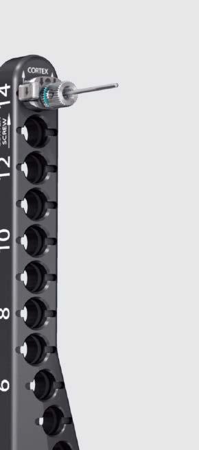
13 Insert Plate 3 Secure aiming arm to plate proximally Instruments Outer Sleeve, for 3.5 mm LCP Trocar with Handle, for 3.5 mm LCP Scalpel with Handle, for 3.5 mm LCP mm Percutaneous Threaded Wire Guide Determine the hole in the aiming arm corresponding to the most proximal hole in the plate. Attach a blade to the scalpel handle. The scalpel will pass through the aiming holes and assist in performing a minimally invasive and accurate incision. (The scalpel will pass through the aiming arm only as far as the top surface of the plate.) Remove the scalpel from the aiming arm. Note: Remove the scalpel blade before storing the handle in the graphic case. Assemble an outer sleeve onto the trocar with handle until it fully seats. While squeezing inward on the two latches of the outer sleeve, insert the trocar assembly into the appropriate aiming arm hole with the arrows on the outer sleeve oriented in the same direction as the LOCKING arrow on the aiming arm. Push the trocar assembly down to the plate through the incision until the trocar tip contacts the plate and the two latches of the outer sleeve securely snap into the aiming arm. Remove the trocar. 11 DePuy Synthes 3.5 mm LCP Low Bend Medial Distal Tibia Plate Aiming Instruments Surgical Technique

14 Insert Plate Instruments mm Drill Tip Guide Wire, 200 mm Handle for Percutaneous Threaded Drill Guides mm Percutaneous Threaded Wire Guide Thread the handle into the wire guide. Insert the handle and guide assembly through the outer sleeve, and securely thread it to the plate. Turn the handle counterclockwise to disengage and remove it from the guide. Note: Securely tighten the wire guide to the plate, to achieve a stable construct between the aiming arm and the plate. Following fluoroscopic confirmation of a satisfactory plate location on the tibia proximally, a 1.6 mm drill tip guide wire can be inserted into the bone through the percutaneous wire guide to secure the plate location. This 1.6 mm wire may be placed unicortically or bicortically. 3.5 mm LCP Low Bend Medial Distal Tibia Plate Aiming Instruments Surgical Technique DePuy Synthes 11


15 INSERT SCREWS 1 Screw placement verification (optional) Instruments mm Drill Tip Guide Wire, 200 mm mm Percutaneous Threaded Drill Guide mm Percutaneous Threaded Wire Guide mm Calibrated Drill Bit (with stop) Direct Measuring Device Small Battery Drive Quick Coupling for K-Wires * Torque Limiting Attachment, 1.5 Nm or Torque Limiting Attachment, 1.5 Nm, quick coupling The orientation and direction of the locking screws depend on the plate contour. The final screw position may be verified with a K-wire before insertion. This is especially important when the plate has been manually contoured with a bending press, drawn to the bone with a screw or reduction device, or applied near the joint. With the 1.6 mm percutaneous threaded wire guide in the locking hole, drive a 1.6 mm drill tip guide wire to the desired depth. Verify K-wire placement under image intensification to determine if final screw placement will be acceptable. Precaution: The K-wire position represents the final position of the locking screw. Confirm that the K-wire does not enter the joint. *Also available. 14 DePuy Synthes 3.5 mm LCP Low Bend Medial Distal Tibia Plate Aiming Instruments Surgical Technique


16 Insert Screws Measure for screw length by sliding the tapered end of the direct measuring device over the K-wire down to the wire sleeve. Remove the direct measuring device, K-wire and 1.6 mm percutaneous threaded wire guide. Insert a 2.8 mm percutaneous threaded drill guide into the locking hole and drill to the measured depth, using the 2.8 mm calibrated drill bit. Remove the threaded drill guide and insert the appropriate length locking screw. 3.5 mm LCP Low Bend Medial Distal Tibia Plate Aiming Instruments Surgical Technique DePuy Synthes 11
 03.113.028 Depth Gauge for 3.5 mm LCP 323.060 Direct Measuring Device 511.")


17 Insert Screws 2 Insert distal screws Instruments Screwdriver Shaft StarDrive, 165 mm mm Percutaneous Threaded Drill Guide Screwdriver StarDrive, T15, self-retaining, 272 mm, for 3.5 mm LCP Percutaneous Instrument System mm Calibrated Drill Bit (with stop) Depth Gauge for 3.5 mm LCP Direct Measuring Device * Torque Limiting Attachment, 1.5 Nm or Torque Limiting Attachment, 1.5 Nm, quick coupling Determine the combination of screws to be used for fixation. If a combination of locking and cortex screws will be used, cortex screws should be inserted first to pull the plate to the bone. If a locking screw will be used as the first screw, ensure the plate is held securely to the bone to prevent plate rotation as the screw is locked to the plate. In distal Combi holes For nonlocking screws, use standard AO screw insertion technique. The two Combi holes in the plate head accept 3.5 mm cortex or 4.0 mm cancellous bone screws. When using a cortex or cancellous bone screw in these Combi holes, the screwhead will be recessed in the hole. Note: When evaluating screw options, remember the 3.5 mm conical screws have a lower profile than the locking screws. 3.7 mm cannulated locking screws are also available. *Also available. 12 DePuy Synthes 3.5 mm LCP Low Bend Medial Distal Tibia Plate Aiming Instruments Surgical Technique

18 Insert Screws For distal locking screws Thread the 2.8 mm percutaneous threaded drill guide into a distal locking hole until fully seated. Use the 2.8 mm calibrated drill bit with stop to drill to the desired depth. Remove the drill and note the indicated drill depth on the gauge. Note: The plastic stop is designed to ride up against the 2.8 mm percutaneous drill guide. The side of the stop facing the drill guide indicates the correct drilling depth. Remove the drill guide. 3.5 mm LCP Low Bend Medial Distal Tibia Plate Aiming Instruments Surgical Technique DePuy Synthes 11
 Note: Use the tip of the handle for percutaneous threaded drill")


19 Insert Screws 2. Insert distal screws (continued) Note: Use the tip of the handle for percutaneous threaded drill guides as a pin wrench to loosen the drill guides from the plate. Inserting locking screws Insert the locking screw under power, using the torque limiting attachment and the StarDrive TM Screwdriver shaft, or insert manually using the StarDrive Screwdriver. Be sure the plate is held securely to the bone to prevent rotation as the screw is locked to the plate. When using the torque limiting attachment, the screw is securely locked into the plate when a click is heard. Note: Always use a torque limiting attachment when using power with the StarDrive Screwdriver shaft. 11 DePuy Synthes 3.5 mm LCP Low Bend Medial Distal Tibia Plate Aiming Instruments Surgical Technique
20 Insert Screws 3 Insert 3.5 mm cortex screws in shaft Instruments Outer Sleeve, for 3.5 mm LCP Trocar with Handle, for 3.5 mm LCP Scalpel with Handle, for 3.5 mm LCP Neutral Drill Guide, for 3.5 mm LCP Compression Drill Guide, for 3.5 mm LCP Stopper, for 3.5 mm LCP Percutaneous Instrument System mm Calibrated Drill Bit (with stop) * Depth Gauge, for 3.5 mm LCP Small Hexagonal Screwdriver with Holding Sleeve Small Hexagonal Screwdriver Shaft Direct Measuring Device Small Battery Drive Quick Coupling for Drill Bits Create an incision through the appropriate aiming arm hole. Assemble the trocar handle with an outer sleeve. *Also available. 3.5 mm LCP Low Bend Medial Distal Tibia Plate Aiming Instruments Surgical Technique DePuy Synthes 11
 While squeezing inward on the two latches of the outer")


21 Insert Screws 3. Insert 3.5 mm cortex screws in shaft (continued) While squeezing inward on the two latches of the outer sleeve, insert the trocar assembly into the appropriate aiming arm hole, with the arrows on the outer sleeve oriented in the same direction as the CORTEX arrow on the aiming arm. Push the trocar assembly down to the plate through the incision until the trocar tip contacts the plate and the two latches of the outer sleeve securely snap into the aiming arm. Remove the trocar. 22 DePuy Synthes 3.5 mm LCP Low Bend Medial Distal Tibia Plate Aiming Instruments Surgical Technique


22 Insert Screws Insert the appropriate drill guide, neutral or compression, into the outer sleeve until it securely snaps into place. Note: When using the compression guide, orient the tab on the compression guide with the slot on the outer sleeve. Use the 2.5 mm calibrated drill bit with stop to drill to the desired depth. Proper screw length can be determined from the calibration on the drill bit aligned with the top of the drill guide. Note: The side of the stop facing the drill guide indicates the correct drilling depth. Alternative technique Screw length can also be determined with the use of the depth gauge. Remove the drill guide and insert the depth gauge into the outer sleeve to the previously drilled depth. Proper screw length is indicated by the gauge marking aligned with the top of the outer sleeve. Remove the drill bit and drill guide. Insert proper length 3.5 mm cortex screw, using the appropriate screwdriver. Repeat this process to insert as many 3.5 mm cortex screws as necessary into the plate shaft. Mark each screw location in the aiming arm with a stopper. Precaution: All 3.5 mm cortex screws must be inserted before insertion of locking screws. Note: Inserting cortex screws into the plate may cause the plate to flex. If the plate contour is changed, the aiming arm may not properly target the holes in the plate. 3.5 mm LCP Low Bend Medial Distal Tibia Plate Aiming Instruments Surgical Technique DePuy Synthes 22

23 Insert Screws 4 Insert 3.5 mm locking screws in shaft Instruments Outer Sleeve, for 3.5 mm LCP Trocar with Handle, for 3.5 mm LCP Scalpel with Handle, for 3.5 mm LCP Handle for Percutaneous Threaded Drill Guides Screwdriver Shaft StarDrive, 165 mm mm Percutaneous Threaded Drill Guide Screwdriver StarDrive, T15, self-retaining, 272 mm, for 3.5 mm LCP Percutaneous Instrument System mm Calibrated Drill Bit (with stop) * Depth Gauge, for 3.5 mm LCP * Torque Limiting Attachment, 1.5 Nm or Torque Limiting Attachment, 1.5 Nm, quick coupling Create an incision through the appropriate aiming arm hole. Assemble an outer sleeve onto the trocar with handle until it is fully seated. While squeezing the two latches of the outer sleeve, insert the assembly into the aiming arm hole with the arrows on the outer sleeve aligned in same the direction as the LOCKING arrow on the aiming arm. Push down to the plate through the incision until the trocar tip contacts the plate. Push the outer sleeve down the trocar shaft the remainder of the way until the latches of the outer sleeve snap into the aiming arm. *Also available. 22 DePuy Synthes 3.5 mm LCP Low Bend Medial Distal Tibia Plate Aiming Instruments Surgical Technique

24 Insert Screws Remove the trocar. Thread the 2.8 mm percutaneous threaded drill guide into a locking hole until fully seated. Use the 2.8 mm calibrated drill bit with stop to drill to the desired depth. Remove the drill and note the drill depth indicated by the stop. Insert the proper length 3.5 mm locking screws in the same manner described for the distal locking screws. 5 Remove aiming arm and insertion handle Instruments Screwdriver StarDrive, T15, self-retaining, 272 mm, for 3.5 mm LCP Percutaneous Instrument System Combination Wrench, 11 mm width across flats Remove all outer sleeves. Turn the connecting bolt on the aiming arm counterclockwise to loosen and remove the aiming arm from the insertion handle. The 11 mm combination wrench may be used, if necessary. Remove the insertion handle from the plate by loosening the connecting screw with the T15 StarDrive Screwdriver. Remove any remaining guide wires. 3.5 mm LCP Low Bend Medial Distal Tibia Plate Aiming Instruments Surgical Technique DePuy Synthes 22


25 IMPLANTS SCREWS USED WITH THE 3.5 MM LCP LOW BEND MEDIAL DISTAL TIBIA PLATE STAINLESS STEEL AND TITANIUM 2.7 mm Cortex Screw May be used in the distal locking holes Compresses the plate to the bone 3.5 mm Cortex Screw May be used in the DCU portion of the Combi holes in the plate shaft Compresses the plate to the bone or creates axial compression Self-tapping tip 3.5 mm Locking Screw Creates a locked, fixed-angle screw/plate construct Self-tapping tip Used in the locking portion of the Combi holes or in round locking holes Threaded conical head 3.7 mm Cannulated Locking Screw Creates a locked, fixed-angle screw/plate construct Self-drilling, self-tapping tip Threaded conical head 3.7 mm Cannulated Conical Screw Compresses the plate to the lateral femoral condyle Smooth conical head Self-drilling, self-tapping tip 4.0 mm Cancellous Bone Screw May be used in the DCU portion of the Combi holes in the plate shaft Compresses the plate to the bone or creates axial compression Fully or partially threaded shaft 14 DePuy Synthes 3.5 mm LCP Low Bend Medial Distal Tibia Plate Aiming Instruments Surgical Technique

26 INSTRUMENTS Aiming Arm, for 3.5 mm LCP Low Bend Medial Distal Tibia Plate, right Aiming Arm, for 3.5 mm LCP Low Bend Medial Distal Tibia Plate, left Outer Sleeve, for 3.5 mm LCP Percutaneous Instrument System, self retaining Trocar with Handle, for 3.5 mm LCP Scalpel with Handle, for 3.5 mm LCP Neutral Drill Guide, for 3.5 mm LCP 3.5 mm LCP Low Bend Medial Distal Tibia Plate Aiming Instruments Surgical Technique DePuy Synthes 15

27 Instruments Compression Drill Guide, for 3.5 mm LCP Handle for Percutaneous Threaded Drill Guides Pull Reduction Device, for 3.5 mm LCP Nut for Pull Reduction Device with Stopper, for 3.5 mm LCP Percutaneous Instrument System Screwdriver Shaft StarDrive, 165 mm mm Percutaneous Threaded Drill Guide Screwdriver StarDrive, T15, self-retaining, 272 mm, for 3.5 mm LCP Percutaneous Instrument System 22 DePuy Synthes 3.5 mm LCP Low Bend Medial Distal Tibia Plate Aiming Instruments Surgical Technique


28 Instruments mm Percutaneous Threaded Wire Guide mm Calibrated Drill Bit mm Calibrated Drill Bit Insertion Handle for 3.5 mm LCP Low Bend Medial Distal Tibia Plate, right Insertion Handle, for 3.5 mm LCP Low Bend Medial Distal Tibia Plate, left Large Handle with quick coupling 3.5 mm LCP Low Bend Medial Distal Tibia Plate Aiming Instruments Surgical Technique DePuy Synthes 17

29 Instruments Small Hexagonal Screwdriver Shaft, long, for 3.5 mm cortex and 4.0 mm cancellous bone screws Small Hexagonal Screwdriver, long, for 3.5 mm cortex and 4.0 mm cancellous bone screws mm Cleaning Stylet mm Cleaning Stylet Combination Wrench, 11 mm width across flats Direct Measuring Device 18 DePuy Synthes 3.5 mm LCP Low Bend Medial Distal Tibia Plate Aiming Instruments Surgical Technique
30 LCP SMALL FRAGMENT PERCUTANEOUS INSTRUMENT SYSTEM, FOR 3.5 MM LCP LOW BEND MEDIAL DISTAL TIBIA PLATES ( ) Graphic Case for 3.5 mm LCP Percutaneous Instrument System, for 3.5 mm LCP Low Bend Medial Distal Tibia Plates Implant mm Drill Tip Guide Wire, 200 mm Instruments Aiming Arm, for 3.5 mm LCP Low Bend Medial Distal Tibia Plate, right Aiming Arm, for 3.5 mm LCP Low Bend Medial Distal Tibia Plate, left Outer Sleeve, for 3.5 mm LCP Percutaneous Instrument System, self retaining Trocar with Handle, for 3.5 mm LCP Scalpel with Handle, for 3.5 mm LCP Neutral Drill Guide, for 3.5 mm LCP Compression Drill Guide, for 3.5 mm LCP Handle for Percutaneous Threaded Drill Guides Pull Reduction Device, for 3.5 mm LCP Nut for Pull Reduction Device Stopper, for 3.5 mm LCP Percutaneous Instrument System Screwdriver Shaft StarDrive, 165 mm mm Percutaneous Threaded Drill Guide Screwdriver StarDrive, T15, self-retaining, 272 mm, for 3.5 mm LCP Percutaneous Instrument System mm Percutaneous Threaded Wire Guide mm Calibrated Drill Bit, quick coupling, 250 mm/95 mm mm Calibrated Drill Bit, quick coupling, 250 mm/95 mm Insertion Handle, for 3.5 mm LCP Low Bend Medial Distal Tibia Plate, right Insertion Handle, for 3.5 mm LCP Low Bend Medial Distal Tibia Plate, left Depth Gauge f/3.5 mm LCP Pertcutaneous Instrument System Large Handle with quick coupling Small Hexagonal Screwdriver Shaft, long, for 3.5 mm cortex and 4.0 mm cancellous bone screws Small Hexagonal Screwdriver, long, for 3.5 mm cortex and 4.0 mm cancellous bone screws mm Cleaning Stylet mm Cleaning Stylet Combination Wrench, 11 mm width across flats Direct Measuring Device Note: For additional information, please refer to package insert. For detailed cleaning and sterilization instructions, please refer to or sterilization instructions, if provided. 3.5 mm LCP Low Bend Medial Distal Tibia Plate Aiming Instruments Surgical Technique DePuy Synthes 22
31 Limited Warranty and Disclaimer: DePuy Synthes products are sold with a limited warranty to the original purchaser against defects in workmanship and materials. Any other express or implied warranties, including warranties of merchantability or fitness, are hereby disclaimed. Please also refer to the package insert(s) or other labeling associated with the devices identified in this surgical technique for additional information. CAUTION: Federal Law restricts these devices to sale by or on the order of a physician. Some devices listed in this surgical technique may not have been licensed in accordance with Canadian law and may not be for sale in Canada. Please contact your sales consultant for items approved for sale in Canada. Not all products may currently be available in all markets. Manufactured or distributed by: Synthes USA Products, LLC 1302 Wrights Lane East West Chester, PA Synthes USA, LLC 1101 Synthes Avenue Monument, CO To order (USA): To order (Canada): Note: For recognized manufacturer, refer to the product label. DePuy Synthes All rights reserved. DSUS/TRM/1016/1162 5/17 DV
Technique Guide. 3.5 mm LCP Low Bend Medial Distal Tibia Plate Aiming Instruments. Part of the 3.5 mm LCP Percutaneous Instrument System.
 Technique Guide 3.5 mm LCP Low Bend Medial Distal Tibia Plate Aiming Instruments. Part of the 3.5 mm LCP Percutaneous Instrument System. Table of Contents Introduction 3.5 mm LCP Low Bend Medial Distal
Low Bend Distal Tibia Plates
 Part of the DePuy Synthes Locking Compression Plate (LCP ) System 3.5 mm LCP Low Bend Medial Distal Tibia Plates Surgical Technique Table of Contents Introduction 3.5 mm LCP Low Bend Medial Distal Tibia
3.5 mm LCP Extra-articular Distal Humerus Plate
 Part of the DePuy Synthes Locking Compression Plate (LCP ) System 3.5 mm LCP Extra-articular Distal Humerus Plate Surgical Technique Table of Contents Introduction 3.5 mm LCP Extra-articular Distal Humerus
Technique Guide. 3.5 mm LCP Low Bend Medial Distal Tibia Plates. Part of the Synthes locking compression plate (LCP) system.
 system.") Technique Guide 3.5 mm LCP Low Bend Medial Distal Tibia Plates. Part of the Synthes locking compression plate (LCP) system. Table of Contents Introduction 3.5 mm LCP Low Bend Medial Distal Tibia Plates
Technique Guide 3.5 mm LCP Low Bend Medial Distal Tibia Plates. Part of the Synthes locking compression plate (LCP) system. Table of Contents Introduction 3.5 mm LCP Low Bend Medial Distal Tibia Plates
3.5 mm LCP Olecranon Plates
 Part of the DePuy Synthes Locking Compression Plate (LCP ) System 3.5 mm LCP Olecranon Plates Surgical Technique Table of Contents Introduction 3.5 mm LCP Olecranon Plates 2 AO Principles 3 Indications
LCP Low Bend Medial Distal Tibia Plates 3.5 mm. Anatomic plates with low profile head for intra- and extraarticular fractures.
 LCP Low Bend Medial Distal Tibia Plates 3.5 mm. Anatomic plates with low profile head for intra- and extraarticular fractures. Surgical Technique This publication is not intended for distribution in the
2.7 mm/3.5 mm LCP Distal Fibula Plate
 Part of the DePuy Synthes Locking Compression Plate (LCP ) System 2.7 mm/3.5 mm LCP Distal Fibula Plate Surgical Technique Table of Contents Introduction 2.7 mm/3.5 mm LCP Distal Fibula Plates 2 AO Principles
Part of the DePuy Synthes Locking Compression Plate (LCP ) System. 3.5 mm LCP Medial Proximal Tibia Plates
 System. 3.5 mm LCP Medial Proximal Tibia Plates") Part of the DePuy Synthes Locking Compression Plate (LCP ) System 3.5 mm LCP Medial Proximal Tibia Plates Surgical Technique Table of Contents Introduction 3.5 mm LCP Medial Proximal Tibia Plates 2 AO
Part of the DePuy Synthes Locking Compression Plate (LCP ) System 3.5 mm LCP Medial Proximal Tibia Plates Surgical Technique Table of Contents Introduction 3.5 mm LCP Medial Proximal Tibia Plates 2 AO
LCP Medial Distal Tibia Plate, without Tab. The Low Profile Anatomic Fixation System with Angular Stability and Optimal Screw Orientation.
 LCP Medial Distal Tibia Plate, without Tab. The Low Profile Anatomic Fixation System with Angular Stability and Optimal Screw Orientation. Technique Guide LCP Small Fragment System Table of Contents Introduction
3.5 mm LCP Anterolateral Distal Tibia Plates
 Part of the DePuy Synthes Locking Compression Plate (LCP ) System 3.5 mm LCP Anterolateral Distal Tibia Plates Surgical Technique Table of Contents Introduction 3.5 mm LCP Anterolateral Distal Tibia Plates
Long Volar Plates for Diaphyseal-Metaphyseal Radius Fractures LCP. Dia-Meta Volar Distal Radius Plates. Surgical Technique
 Long Volar Plates for Diaphyseal-Metaphyseal Radius Fractures LCP Dia-Meta Volar Distal Radius Plates Surgical Technique Table of Contents Introduction LCP Dia-Meta Volar Distal Radius Plates 2 AO Principles
3.5 mm Locking Attachment Plate
 For Treatment of Periprosthetic Fractures 3.5 mm Locking Attachment Plate Surgical Technique Table of Contents Introduction 3.5 mm Locking Attachment Plate 2 Indications 4 Surgical Technique Preparation
3.5 mm LCP Hook Plate
 Part of the DePuy Synthes Locking Compression Plate (LCP ) System 3.5 mm LCP Hook Plate Surgical Technique Table of Contents Introduction 3.5 mm LCP Hook Plate 2 AO Principles 4 Indications 5 Clinical
4.5 mm LCP Medial Proximal Tibia Plates
 Part of the DePuy Synthes LCP Periarticular Plating System 4.5 mm LCP Medial Proximal Tibia Plates Surgical Technique Table of Contents Introduction 4.5 mm LCP Medial Proximal Tibia Plates 2 AO Principles
3.5 mm LCP Clavicle Hook Plates
 Part of the Synthes Locking Compression Plate (LCP ) System 3.5 mm LCP Clavicle Hook Plates Surgical Technique Table of Contents Introduction 3.5 mm LCP Clavicle Hook Plates 2 AO Principles 4 Indications
3.5 mm Clavicle Hook Plates
 A Single Solution for Lateral Clavicle Fractures and Acromioclavicular Joint Dislocations 3.5 mm Clavicle Hook Plates Surgical Technique Discontinued December 2017 DSUS/TRM/1016/1126(1) Table of Contents
OBSOLETED. LCP Medial Distal Tibia Plate, without Tab. The Low Profile Anatomic Fixation System with Angular Stability and Optimal Screw Orientation.
 LCP Medial Distal Tibia Plate, without Tab. The Low Profile Anatomic Fixation System with Angular Stability and Optimal Screw Orientation. Surgical Technique LCP Small Fragment System This publication
3.5 mm LCP Distal Humerus Plates
 Part of the DePuy Synthes Locking Compression Plate (LCP ) System 3.5 mm LCP Distal Humerus Plates Surgical Technique Table of Contents Introduction 3.5 mm LCP Distal Humerus Plates 2 AO Principles 4 Indications
4.5 mm LCP Condylar Plate Aiming Instruments
 Part of the LCP Periarticular Aiming Instrument System (Large) 4.5 mm LCP Condylar Plate Aiming Instruments Surgical Technique Table of Contents Introduction 4.5 mm LCP Condylar Plate Aiming Instruments
3.5 mm LCP Distal Tibia T-Plates
 Part of the DePuy Synthes Locking Compression Plate (LCP ) System 3.5 mm LCP Distal Tibia T-Plates Surgical Technique Table of Contents Introduction 3.5 mm LCP Distal Tibia T-Plates 2 AO Principles 4 Indications
3.5 MM VA-LCP PROXIMAL TIBIA PLATE SYSTEM
 3.5 MM VA-LCP PROXIMAL TIBIA PLATE SYSTEM Part of the DePuy Synthes Variable Angle Periarticular Plating System SURGICAL TECHNIQUE TABLE OF CONTENTS INTRODUCTION 3.5 mm VA-LCP Proximal Tibial Plate 2 AO
Technique Guide. 3.5 mm LCP Olecranon Plates. Part of the Synthes locking compression plate (LCP) system.
 system.") Technique Guide 3.5 mm LCP Olecranon Plates. Part of the Synthes locking compression plate (LCP) system. Table of Contents Introduction 3.5 mm LCP Olecranon Plates 2 AO Principles 3 Indications 3 Clinical
Technique Guide 3.5 mm LCP Olecranon Plates. Part of the Synthes locking compression plate (LCP) system. Table of Contents Introduction 3.5 mm LCP Olecranon Plates 2 AO Principles 3 Indications 3 Clinical
LCP Anterolateral Distal Tibia Plate 3.5. The low profile anatomic fixation system with optimal plate placement and angular stability.
 LCP Anterolateral Distal Tibia Plate 3.5. The low profile anatomic fixation system with optimal plate placement and angular stability. Technique Guide LCP Small Fragment System Table of Contents Introduction
The Locking Calcaneal Plate Instrument and Implant Sets
 Part of the DePuy Synthes Locking Compression Plate (LCP ) System The Locking Calcaneal Plate Instrument and Implant Sets Surgical Technique Table of Contents Introduction Locking Calcaneal Plate 2 AO
LCP DISTAL TIBIA PLATE
 LCP DISTAL TIBIA PLATE Instruments and implants approved by the AO Foundation. This publication is not intended for distribution in the USA. SURGICAL TECHNIQUE Image intensifier control This description
LCP Anterolateral Distal Tibia Plate 3.5. The low profile anatomic fixation system with optimal plate placement and angular stability.
 LCP Anterolateral Distal Tibia Plate 3.5. The low profile anatomic fixation system with optimal plate placement and angular stability. Technique Guide LCP Small Fragment System Table of Contents Introduction
Technique Guide. 2.7 mm/3.5 mm LCP Distal Fibula Plates. Part of the Synthes locking compression plate (LCP) system.
 system.") Technique Guide 2.7 mm/3.5 mm LCP Distal Fibula Plates. Part of the Synthes locking compression plate (LCP) system. Table of Contents Introduction 2.7 mm/3.5 mm LCP Distal Fibula Plates 2 AO Principles
Technique Guide 2.7 mm/3.5 mm LCP Distal Fibula Plates. Part of the Synthes locking compression plate (LCP) system. Table of Contents Introduction 2.7 mm/3.5 mm LCP Distal Fibula Plates 2 AO Principles
Technique Guide. LCP Proximal Femoral Hook Plate 4.5/5.0. Part of the LCP Periarticular Plating System.
 Technique Guide LCP Proximal Femoral Hook Plate 4.5/5.0. Part of the LCP Periarticular Plating System. Table of Contents Introduction Features and Benefits 2 AO ASIF Principles 4 Indications 5 Surgical
3.5 mm LCP Superior Anterior Clavicle Plates
 Part of the DePuy Synthes Locking Compression Plate (LCP ) System 3.5 mm LCP Superior Anterior Clavicle Plates Surgical Technique Table of Contents Introduction 3.5 mm LCP Superior Anterior Clavicle Plates
Cannulated Pediatric Osteotomy System (CAPOS)
") A Single System of Osteotomy Blade Plates and Cannulated Instrumentation Cannulated Pediatric Osteotomy System (CAPOS) Surgical Technique Table of Contents Introduction Cannulated Pediatric Osteotomy System
A Single System of Osteotomy Blade Plates and Cannulated Instrumentation Cannulated Pediatric Osteotomy System (CAPOS) Surgical Technique Table of Contents Introduction Cannulated Pediatric Osteotomy System
LCP Medial Proximal Tibial Plate 3.5. Part of the Synthes small fragment Locking Compression Plate (LCP) system.
 system.") LCP Medial Proximal Tibial Plate 3.5. Part of the Synthes small fragment Locking Compression Plate (LCP) system. Technique Guide This publication is not intended for distribution in the USA. Instruments
LCP Medial Proximal Tibial Plate 3.5. Part of the Synthes small fragment Locking Compression Plate (LCP) system. Technique Guide This publication is not intended for distribution in the USA. Instruments
Technique Guide. LCP Distal Fibula Plates. Part of the Synthes locking compression plate (LCP) system.
 system.") Technique Guide LCP Distal Fibula Plates. Part of the Synthes locking compression plate (LCP) system. Table of Contents Introduction LCP Distal Fibula Plates 2 AO Principles 4 Indications 5 Surgical Technique
Technique Guide LCP Distal Fibula Plates. Part of the Synthes locking compression plate (LCP) system. Table of Contents Introduction LCP Distal Fibula Plates 2 AO Principles 4 Indications 5 Surgical Technique
Technique Guide. 3.5 mm LCP Periarticular Proximal Humerus Plate. Part of the Synthes locking compression plate (LCP) system.
 system.") Technique Guide 3.5 mm LCP Periarticular Proximal Humerus Plate. Part of the Synthes locking compression plate (LCP) system. Table of Contents Introduction 3.5 mm LCP Proximal Humerus Plate 2 AO Principles
Technique Guide 3.5 mm LCP Periarticular Proximal Humerus Plate. Part of the Synthes locking compression plate (LCP) system. Table of Contents Introduction 3.5 mm LCP Proximal Humerus Plate 2 AO Principles
For Distal Femur Fractures. 95º Condylar Plate. Quick Reference Chart
 For Distal Femur Fractures 95º Condylar Plate Quick Reference Chart 95 Condylar Plate. Quick reference chart for distal femur fractures. Insert guide wires Fix condylar fragments with 6.5 mm cancellous
LCP Distal Fibula Plates. Part of the Synthes locking compression plate (LCP) system.
 system.") LCP Distal Fibula Plates. Part of the Synthes locking compression plate (LCP) system. Surgical Technique This publication is not intended for distribution in the USA. Instruments and implants approved
LCP Distal Fibula Plates. Part of the Synthes locking compression plate (LCP) system. Surgical Technique This publication is not intended for distribution in the USA. Instruments and implants approved
Periarticular Aiming Arm Instruments for LCP Proximal Tibial Plate 4.5/5.0. Part of the LCP Periarticular Aiming Arm Instrument System (large).
.") Technique Guide Periarticular Aiming Arm Instruments for LCP Proximal Tibial Plate 4.5/5.0. Part of the LCP Periarticular Aiming Arm Instrument System (large). Image intensifier control Warning This description
Technique Guide Periarticular Aiming Arm Instruments for LCP Proximal Tibial Plate 4.5/5.0. Part of the LCP Periarticular Aiming Arm Instrument System (large). Image intensifier control Warning This description
Distal Radius Plate Instrument and Implant Set. Discontinued December 2017 DSUS/TRM/0916/1063(1)
") Distal Radius Plate Instrument and Implant Set Surgical Technique Discontinued December 2017 DSUS/TRM/0916/1063(1) The Distal Radius Plates Indications For fixation of fractures and osteotomies, including
Distal Radius Plate Instrument and Implant Set Surgical Technique Discontinued December 2017 DSUS/TRM/0916/1063(1) The Distal Radius Plates Indications For fixation of fractures and osteotomies, including
2.7 mm/3.5 mm Variable Angle LCP. Ankle Trauma System
 Part of the DePuy Synthes Variable Angle Locking Compression Plate (VA LCP ) System 2.7 mm/3.5 mm Variable Angle LCP Ankle Trauma System Surgical Technique Table of Contents Introduction 2.7 mm/3.5 mm
LCP Condylar Plate 4.5/5.0. Part of the LCP Periarticular Plating System.
 LCP Condylar Plate 4.5/5.0. Part of the LCP Periarticular Plating System. Surgical Technique This publication is not intended for distribution in the USA. Instruments and implants approved by the AO Foundation.
LCP Distal Fibula Plates. Part of the Synthes locking compression plate (LCP) system.
 system.") LCP Distal Fibula Plates. Part of the Synthes locking compression plate (LCP) system. Surgical Technique This publication is not intended for distribution in the USA. Instruments and implants approved
LCP Distal Fibula Plates. Part of the Synthes locking compression plate (LCP) system. Surgical Technique This publication is not intended for distribution in the USA. Instruments and implants approved
Technique Guide. 4.5 mm LCP Proximal Tibia Plates. Part of the Synthes LCP Periarticular Plating System.
 Technique Guide 4.5 mm LCP Proximal Tibia Plates. Part of the Synthes LCP Periarticular Plating System. Table of Contents Introduction 4.5 mm LCP Proximal Tibia Plates 2 AO Principles 4 Indications 5 Surgical
LCP Medial Proximal Tibial Plate 4.5/5.0. Part of the Synthes LCP periarticular plating system.
 LCP Medial Proximal Tibial Plate 4.5/5.0. Part of the Synthes LCP periarticular plating system. Technique Guide This publication is not intended for distribution in the USA. Instruments and implants approved
Technique Guide. TomoFix Osteotomy System. A comprehensive plating system for stable fixation of osteotomies around the knee.
 Technique Guide TomoFix Osteotomy System. A comprehensive plating system for stable fixation of osteotomies around the knee. Table of Contents Introduction TomoFix Osteotomy System 2 AO Principles 4 Indications
2.4 mm LCP Radial Head Plates. Part of the Synthes LCP Distal Radius Plate System.
 2.4 mm LCP Radial Head Plates. Part of the Synthes LCP Distal Radius Plate System. Technique Guide Instruments and Implants approved by the AO Foundation Table of Contents Introduction 2.4 mm LCP Radial
2.4 mm Variable Angle LCP Volar Rim Distal Radius Plates
 For Fragment-Specific Fracture Fixation With Variable Angle Locking Technology 2.4 mm Variable Angle LCP Volar Rim Distal Radius Plates Surgical Technique Table of Contents Introduction 2.4 mm Variable
LCP Proximal Radius Plates 2.4. Plates for radial head rim and for radial head neck address individual fracture patterns of the proximal radius.
 Technique Guide LCP Proximal Radius Plates 2.4. Plates for radial head rim and for radial head neck address individual fracture patterns of the proximal radius. Table of Contents Introduction LCP Proximal
Technique Guide. 3.5 mm LCP Proximal Tibia Plate. Part of the Synthes Small Fragment LCP System.
 Technique Guide 3.5 mm LCP Proximal Tibia Plate. Part of the Synthes Small Fragment LCP System. Table of Contents AO ASIF Principles of Internal Fixation 4 Indications/Contraindications 5 Surgical Technique
Small Fragment Locking Compression Plate (LCP ) System
 System") Stainless Steel and Titanium Small Fragment Locking Compression Plate (LCP ) System Surgical Technique Table of Contents Introduction Small Fragment Locking Compression Plate (LCP) System 2 AO Principles
Stainless Steel and Titanium Small Fragment Locking Compression Plate (LCP ) System Surgical Technique Table of Contents Introduction Small Fragment Locking Compression Plate (LCP) System 2 AO Principles
LCP Medial Proximal Tibial Plate 3.5. Part of the Synthes small fragment Locking Compression Plate (LCP) system.
 system.") LCP Medial Proximal Tibial Plate 3.5. Part of the Synthes small fragment Locking Compression Plate (LCP) system. Surgical Technique This publication is not intended for distribution in the USA. Instruments
LCP Medial Proximal Tibial Plate 3.5. Part of the Synthes small fragment Locking Compression Plate (LCP) system. Surgical Technique This publication is not intended for distribution in the USA. Instruments
Large Fragment LCP Instrument and Implant Set
 Part of the DePuy Synthes Locking Compression Plate (LCP ) System Large Fragment LCP Instrument and Implant Set Surgical Technique Table of Contents Introduction Large Fragment LCP Instrument and Implant
TOMOFIX Medial Distal Femur Plate
 For Closed-Wedge Varus Femoral Osteotomies TOMOFIX Medial Distal Femur Plate Surgical Techniquee TABLE OF CONTENTS INTRODUCTION TOMOFIX Medial Distal Femur Plate 2 AO Principles 4 Indications 5 SURGICAL
LCP Proximal Tibial Plate 4.5/5.0 with Periarticular Aiming Arm Instruments
 LCP Proximal Tibial Plate 4.5/5.0 with Periarticular Aiming Arm Instruments Surgical Technique This publication is not intended for distribution in the USA. Instruments and implants approved by the AO
Periarticular Aiming Arm Instruments for LCP Condylar Plate 4.5/5.0. Part of the LCP Periarticular Aiming Arm Instrument System (large).
.") Periarticular Aiming Arm Instruments for LCP Condylar Plate 4.5/5.0. Part of the LCP Periarticular Aiming Arm Instrument System (large). Surgical Technique This publication is not intended for distribution
Periarticular Aiming Arm Instruments for LCP Condylar Plate 4.5/5.0. Part of the LCP Periarticular Aiming Arm Instrument System (large). Surgical Technique This publication is not intended for distribution
Technique Guide. LCP Posterior Medial Proximal Tibial Plate 3.5. Part of the Synthes small fragment LCP system.
 Technique Guide LCP Posterior Medial Proximal Tibial Plate 3.5. Part of the Synthes small fragment LCP system. Table of Contents Introduction LCP Posterior Medial Proximal Tibial Plate 3.5 2 AO Principles
4.5 mm VA-LCP. Part of the Variable Angle Periarticular Plating System
 4.5 mm VA-LCP Curved Condylar Plate Part of the Variable Angle Periarticular Plating System Surgical Technique Table of Contents Introduction 4.5 mm VA-LCP Curved Condylar Plates 2 4.5 mm VA-LCP Curved
VA-LCP Anterior Clavicle Plate. The anatomically precontoured fixation system with angular stability for clavicle shaft and lateral clavicle.
 Technique Guide VA-LCP Anterior Clavicle Plate. The anatomically precontoured fixation system with angular stability for clavicle shaft and lateral clavicle. Table of Contents Introduction VA-LCP Anterior
LCP Distal Tibia Plate
 Surgical Technique LCP Locking Compression Plate Original Instruments and Implants of the Association for the Study of Internal Fixation AO/ASIF Table of contents Indications 3 Implants/Instruments 5 Surgical
VA-LCP Condylar Plate 4.5/5.0. Part of the Synthes Variable Angle Periarticular Plating System.
 VA-LCP Condylar Plate 4.5/5.0. Part of the Synthes Variable Angle Periarticular Plating System. Technique Guide This publication is not intended for distribution in the USA. Instruments and implants approved
LCP Proximal Radius Plates 2.4. Plates for radial head rim and for radial head neck address individual fracture patterns of the proximal radius.
 LCP Proximal Radius Plates 2.4. Plates for radial head rim and for radial head neck address individual fracture patterns of the proximal radius. Surgical Technique This publication is not intended for
Trochanter Stabilization Plate for DHS Implants
 Extends DHS Plate Construct to Help Stabilize Greater Trochanter Trochanter Stabilization Plate for DHS Implants Surgical Technique Table of Contents Introduction Trochanter Stabilization Plate for DHS
LCP Condylar Plate 4.5/5.0. Part of the LCP Periarticular Plating System.
 LCP Condylar Plate 4.5/5.0. Part of the LCP Periarticular Plating System. Surgical Technique This publication is not intended for distribution in the USA. Instruments and implants approved by the AO Foundation.
2.4 mm Variable Angle LCP Volar Extra-Articular Distal Radius System. For fragment-specific fracture fixation with variable angle locking technology.
 2.4 mm Variable Angle LCP Volar Extra-Articular Distal Radius System. For fragment-specific fracture fixation with variable angle locking technology. Surgical Technique This publication is not intended
2.4 mm Variable Angle LCP Dorsal Distal Radius Plate
 For Fragment-Specific Fracture Fixation With Variable Angle (VA) Locking Technology 2.4 mm Variable Angle LCP Dorsal Distal Radius Plate Surgical Technique Table of Contents Introduction 2.4 mm VA LCP
2.4 mm Variable Angle Locking Intercarpal Fusion System
 For Partial Wrist Arthrodesis With Variable Angle Locking Technology 2.4 mm Variable Angle Locking Intercarpal Fusion System Surgical Technique Table of Contents Introduction 2.4 mm Variable Angle Locking
Technique Guide. PHILOS and PHILOS Long. The anatomic fixation system for the proximal humerus.
 Technique Guide PHILOS and PHILOS Long. The anatomic fixation system for the proximal humerus. Table of Contents Introduction PHILOS and PHILOS Long 2 AO Principles 4 Indications 5 Surgical Technique
Technique Guide. 2.4 mm Variable Angle LCP Distal Radius System. For fragment-specific fracture fixation with variable angle locking technology.
 Technique Guide 2.4 mm Variable Angle LCP Distal Radius System. For fragment-specific fracture fixation with variable angle locking technology. Table of Contents Introduction 2.4 mm Variable Angle LCP
Titanium Distal Femoral Nail System
 For Retrograde Insertion Titanium Distal Femoral Nail System Surgical Technique Table of Contents Introduction Titanium Distal Femoral Nail System 2 AO Principles 4 Indications 5 Clinical Cases 6 Surgical
LCP Anterolateral Distal Tibia Plate 3.5. The low profile anatomic fixation system with optimal plate placement and angular stability.
 LCP Anterolateral Distal Tibia Plate 3.5. The low profile anatomic fixation system with optimal plate placement and angular stability. Surgical Technique LCP Small Fragment System This publication is not
VA-LCP Proximal Tibial Plate 3.5
 Part of the Synthes Variable Angle Periarticular Plating System VA-LCP Proximal Tibial Plate 3.5 Surgical Technique Image intensifier control This description alone does not provide sufficient background
LCP Superior Clavicle Plate. The anatomically precontoured fixation system with angular stability for clavicle shaft and lateral clavicle.
 Technique Guide LCP Superior Clavicle Plate. The anatomically precontoured fixation system with angular stability for clavicle shaft and lateral clavicle. Table of Contents Introduction LCP Superior Clavicle
Technique Guide. Locking Attachment Plate. For treatment of periprosthetic fractures.
 Technique Guide Locking Attachment Plate. For treatment of periprosthetic fractures. Table of Contents Introduction Locking Attachment Plate 2 Indications 4 Surgical Technique Patient Positioning 5 Preparation
Low Profile Neuro Plating System. Surgical Technique
 Low Profile Neuro Plating System Surgical Technique TABLE OF CONTENTS INTRODUCTION Low Profile Neuro Plating System 2 SURGICAL TECHNIQUE Technique 5 PRODUCT INFORMATION Low Profile Neuro Plates 10 Low
3.5 mm LCP Superior and Superior Anterior Clavicle Plates. Part of the Synthes modular clavicle plate system.
 3.5 mm LCP Superior and Superior Anterior Clavicle Plates. Part of the Synthes modular clavicle plate system. Technique Guide Instruments and implants approved by the AO Foundation Table of Contents Introduction
Variable Angle LCP Locking Technology
 BUILDING ON SUCCESS Variable Angle LCP Locking Technology The Evolution of Plating Technology Round Screw Hole Dynamic Compression Plate Limited-Contact Dynamic Compression Less Invasive Stabilization
Technique Guide. Small Fragment Locking Compression Plate (LCP) System. Stainless steel and titanium.
 System. Stainless steel and titanium.") Technique Guide Small Fragment Locking Compression Plate (LCP) System. Stainless steel and titanium. Table of Contents Introduction Small Fragment Locking Compression Plate (LCP) System 2 AO Principles
Technique Guide Small Fragment Locking Compression Plate (LCP) System. Stainless steel and titanium. Table of Contents Introduction Small Fragment Locking Compression Plate (LCP) System 2 AO Principles
Distal Radius Sterile Kit. Optimization And Efficiency You Can Rely On
 Distal Radius Sterile Kit Optimization And Efficiency You Can Rely On Introducing The Distal Radius Sterile Kit Improved OR Workflow The Distal Radius Sterile Kit provides high-quality single-use implants
DOUBLE/TRIPLE PELVIC OSTEOTOMY PLATES For Treating Coxofemoral Joint Instability and Subluxation in Immature Dogs
 DOUBLE/TRIPLE PELVIC OSTEOTOMY PLATES For Treating Coxofemoral Joint Instability and Subluxation in Immature Dogs Instruments and implants approved by the AO Foundation. This publication is not intended
LCP Proximal Radius Plates 2.4. Plates for radial head rim and for radial head neck address individual fracture patterns of the proximal radius.
 LCP Proximal Radius Plates 2.4. Plates for radial head rim and for radial head neck address individual fracture patterns of the proximal radius. Surgical Technique This publication is not intended for
Mini External Fixator
 Stabilize the Phalanges and Metacarpals Mini External Fixator Surgical Technique Table of Contents Introduction Mini External Fixator 2 Indications 4 Surgical Technique Technique Overview 5 Product Information
MINI TIBIAL PLATEAU LEVELING OSTEOTOMY (TPLO) SYSTEM
 SYSTEM") MINI TIBIAL PLATEAU LEVELING OSTEOTOMY (TPLO) SYSTEM For stabilizing osteotomies of the canine and feline proximal tibia SURGICAL TECHNIQUE TABLE OF CONTENTS INTRODUCTION Mini Tibial Plateau Leveling
MINI TIBIAL PLATEAU LEVELING OSTEOTOMY (TPLO) SYSTEM For stabilizing osteotomies of the canine and feline proximal tibia SURGICAL TECHNIQUE TABLE OF CONTENTS INTRODUCTION Mini Tibial Plateau Leveling
modular ClaVICle PlaTe system
 modular ClaVICle PlaTe system 3.7 mm/3.3 mm VA LCP Anterior Clavicle Plates and 3.3 mm Superior Anterior Clavicle Plates surgical TeChnIque Table of Contents Introduction Modular Clavicle Plate System
LCP Proximal Femoral Hook Plate 4.5/5.0. Part of the LCP Periarticular Plating System.
 LCP Proximal Femoral Hook Plate 4.5/5.0. Part of the LCP Periarticular Plating System. Surgical Technique This publication is not intended for distribution in the USA. Instruments and implants approved
2.4 mm Variable Angle LCP Volar Extra-Articular Distal Radius System. For fragment-specific fracture fixation with variable angle locking technology.
 Technique Guide 2.4 mm Variable Angle LCP Volar Extra-Articular Distal Radius System. For fragment-specific fracture fixation with variable angle locking technology. Table of Contents Introduction 2.4
LCP Superior Clavicle Plate. The anatomically precontoured fixation system with angular stability for clavicle shaft and lateral clavicle.
 LCP Superior Clavicle Plate. The anatomically precontoured fixation system with angular stability for clavicle shaft and lateral clavicle. Surgical Technique This publication is not intended for distribution
LCP Percutaneous Aiming System 3.5 for PHILOS. For less invasive surgery at the proximal humerus.
 LCP Percutaneous Aiming System 3.5 for PHILOS. For less invasive surgery at the proximal humerus. Surgical Technique This publication is not intended for distribution in the USA. Instruments and implants
2.7 mm/3.5 mm VA-LCP Anterior Clavicle Plates. Part of the Synthes modular clavicle plate system.
 2.7 mm/3.5 mm VA-LCP Anterior Clavicle Plates. Part of the Synthes modular clavicle plate system. Technique Guide Instruments and implants approved by the AO Foundation Table of Contents Introduction 2.7
Olecranon Osteotomy Nail. For simple fractures and osteotomies of the olecranon.
 Olecranon Osteotomy Nail. For simple fractures and osteotomies of the olecranon. Technique Guide Discontinued June 2016; AVAILABLE FOR IMPLANT REMOVAL PURPOSES ONLY DSEM/TRM/0517/0843 Table of Contents
Surgical Technique. This publication is not intended for distribution in the USA. Instruments and implants approved by the AO Foundation.
 LCP Extra-articular Distal Humerus Plate. The anatomically shaped and angular stable fixation system for extraarticular fractures of the distal humerus. Surgical Technique This publication is not intended
3.5 mm LCP Tibial Plateau Sets. Configuration options.
 3.5 mm LCP Tibial Plateau Sets. Configuration options. Multiple trays Multiple levels Multiple options 3.5 mm LCP Tibial Plateau Sets The tibial plateau trays allow the hospital to build a custom - ized
PHILOS and PHILOS Long. The anatomic fixation system for the proximal humerus.
 PHILOS and PHILOS Long. The anatomic fixation system for the proximal humerus. Surgical Technique This publication is not intended for distribution in the USA. Instruments and implants approved by the
LCP Anterior Ankle Arthrodesis Plates. Part of the Synthes Locking Compression Plate (LCP) System.
 System.") LCP Anterior Ankle Arthrodesis Plates. Part of the Synthes Locking Compression Plate (LCP) System. Technique Guide Instruments and implants approved by the AO Foundation Table of Contents Introduction
LCP Anterior Ankle Arthrodesis Plates. Part of the Synthes Locking Compression Plate (LCP) System. Technique Guide Instruments and implants approved by the AO Foundation Table of Contents Introduction
COMPACT ANKLE FRACTURE SYSTEM
 COMPACT ANKLE FRACTURE SYSTEM Consolidated ankle fracture solution for surgery centers SURGICAL TECHNIQUE TABLE OF CONTENTS INTRODUCTION Compact Ankle Fracture System 2 AO Principles 8 Indications 9 Clinical
VA-LCP Ankle Trauma System 2.7/3.5. Our most comprehensive ankle plating system.
 VA-LCP Ankle Trauma System 2.7/3.5. Our most comprehensive ankle plating system. Surgical Technique This publication is not intended for distribution in the USA. Instruments and implants approved by the
Large Distractor Femur
 Fracture Reduction and Provisional Stabilization Large Distractor Femur Surgical Technique Table of Contents Introduction Standard Femoral Distraction 2 Large Distractor System 4 Surgical Technique Prepare
low ProfIle neuro PlaTIng system
 low ProfIle neuro PlaTIng system surgical TeChnIque Table of Contents Introduction Low Profile Neuro Cranial Plating System 2 Surgical Technique Technique 5 Product Information Low Profile Neuro Plates
VA-LCP Ankle Trauma System 2.7/3.5. Our most comprehensive ankle plating system.
 VA-LCP Ankle Trauma System 2.7/3.5. Our most comprehensive ankle plating system. Surgical Technique This publication is not intended for distribution in the USA. Instruments and implants approved by the
Zimmer MIS Periarticular 3.5mm Proximal Tibial Locking Plate
 Zimmer MIS Periarticular 3.5mm Proximal Tibial Locking Plate Surgical Technique The Science of the Landscape Zimmer MIS Periarticular 3.5mm Proximal Tibial Locking Plate Surgical Technique 1 Zimmer MIS
LCP Metaphyseal Plates. For extra-articular fractures.
 LCP Metaphyseal Plates. For extra-articular fractures. Surgical Technique This publication is not intended for distribution in the USA. Instruments and implants approved by the AO Foundation. Image intensifier
LCP Periarticular Proximal Humerus Plate 3.5. The anatomic fixation system with anterolateral shaft placement.
 LCP Periarticular Proximal Humerus Plate 3.5. The anatomic fixation system with anterolateral shaft placement. Surgical Technique This publication is not intended for distribution in the USA. Instruments
LCP Superior Anterior Clavicle Plate. The anatomically precontoured fixation system with angular stability for clavicle shaft and lateral clavicle.
 LCP Superior Anterior Clavicle Plate. The anatomically precontoured fixation system with angular stability for clavicle shaft and lateral clavicle. Surgical Technique This publication is not intended for