Elbow Plating System. Surgical Technique
|
|
|
- Randall Mosley
- 5 years ago
- Views:
Transcription


1 Elbow Plating System Surgical Technique


2 Acumed is a global leader of innovative orthopaedic and medical solutions. We are dedicated to developing products, service methods, and approaches that improve patient care. Acumed Elbow Plating System Designed in conjunction with Shawn W. O Driscoll, MD, PhD, the Elbow Plating System is engineered to address fractures of the distal humerus, olecranon, and coronoid. The Elbow Plating System includes precontoured, indication-specific plates and a low-profile Olecranon Plate design with anatomic curvature and instrumentation to aid with plate and screw insertion. This system also includes the System with variable angle Tap-Loc Technology for the Medial and Lateral Distal Humerus Plates. Acumed offers Posterolateral Plates in addition to Medial and Lateral Distal Humerus Plates to provide multiple plating solutions for elbow fracture management. Indications for Use: Fractures of the distal humerus, olecranon, and coronoid Osteotomies of the olecranon Definition Warning Caution Note Indicates critical information about a potential serious outcome to the patient or the user. Indicates instructions that must be followed in order to ensure the proper use of the device. Indicates information requiring special attention.
3 Table of Contents System Features...2 Instrument Overview...6 Surgical Technique Overview...8 Surgical Techniques Olecranon Plate Olecranon Plate Osteotomy Cutting Jig Distal Humerus Plate Posterolateral Plate Coronoid Plate Ordering Information... 30


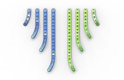


4 System Features Plates and Screws Olecranon Plates Coronoid Plates Posterolateral Distal HumerusPlates s Lateral Column Plates Medial Column Plates 2
![System Features [continued] Key Plate Features Plate lengths range from 3-hole to 15-hole (65 mm 100 mm in shaft](/docs-images/87/96808880/images/5-0.jpg "length) Extended Plates are now offered in two lengths (90 mm and 130 mm) 3-hole plate offers fixation of")


5 System Features [continued] Key Plate Features Plate lengths range from 3-hole to 15-hole (65 mm 100 mm in shaft length) Extended Plates are now offered in two lengths (90 mm and 130 mm) 3-hole plate offers fixation of olecranon osteotomies and fractures where a longer plate is not necessary Left and right specific plates are designed to provide anatomic fit, both proximally and distally along the ulnar shaft Olecranon Plate K-wire holes are included in the plates to facilitate provisional plate fixation Holes in the buttress portion of the plate are designed to allow screws to capture fragments Locking screws in the Standard Plates are designed to avoid collision with one another, regardless of screw length selection Coronoid Plate K-wire holes and prongs are provided for provisional fixation Offset screw hole is intended to help capture fractures of the sublime tubercle Posterolateral Distal Humerus Plate Limited-contact design Locking screws may be used in the distal holes Proximal taper is engineered to minimize stress concentrations Cluster of distal screws, angled distally and divergent from one another, is designed to allow the plate to sit more proximally to avoid potential impingement on the olecranon and to capture fracture fragments Long screws in the Lateral Column Plate are designed to interdigitate with screws from the Medial Column Plate, providing a parallel construct for the stabilization of distal humerus fractures Medial and Lateral Plates The Acumed Elbow Plating System was the first to offer precontoured, parallel distal humerus plates. 3
![System Features [continued] Key Screw, Instrumentation, and Tray Features Screws The 3.0 mm and 3.](/docs-images/87/96808880/images/6-0.jpg "5 mm screws are offered in lengths from 8 mm 65 mm, providing more options than previous generations 2.")


6 System Features [continued] Key Screw, Instrumentation, and Tray Features Screws The 3.0 mm and 3.5 mm screws are offered in lengths from 8 mm 65 mm, providing more options than previous generations 2.7 mm screws with a smaller diameter head allow clustering of screws in selected regions Hexalobe screws are Type II anodized Instrumentation Driver interface is intended to minimize screw stripping Cutting flute design is intended to facilitate screw insertion, particularly with longer screws A radiolucent targeting guide targets the four clustered 2.7 mm proximal screws in the plate Trays All-metal screw caddy design eliminates the use of plastic. The screw caddy lid is removable for ease of use Retractable handles in the screw caddy are designed to aid in removing the caddy from the tray Windows are on the sides and top of the system tray, so that contents can be viewed without opening and disassembling the tray 4
7 System Features [continued] Tap-Loc Technology The Acumed Tap-Loc technology is designed to be used only with the Medial and Lateral Distal Humerus Plates to insert locking screws with up to 20 degrees of angulation. Quick Release Instrumentation provides an efficient way to switch from 3.5 mm to 3.0 mm plate taps T-Handle Provides control when tapping plate holes 3.5 mm and 3.0 mm Plate Tap Sizes Color-coded taps accommodate the screw diameters provided in the system Laser Mark Indicates maximum tapping depth Tapping Threads Allow surgeons to tap the plate after drilling, creating threads in the plate and bone for locking screw insertion Tap Trajectory Guide Follows the drill path for accurate tap angle and screw placement 5
 Reduction Forceps With Serrated Jaw (PL-CL04) Medium Ratcheting Driver Handle (80-0663) 2.")
 Plate Holder Assembly (PL-2030) Plate Bender, large (PL-2045) Offset Drill")
 2.3 mm Locking Drill Guide 6 65 mm (80-0622) 2.")
.062 x 5.75 STT Guide Wire, Titanium (WT-1606STT) 2.")
8 Instrument Overview 8" Bone Reduction Forceps (MS-1280) Bone Reduction Forceps, 5.25 (MS-45300) Reduction Forceps With Serrated Jaw (PL-CL04) Medium Ratcheting Driver Handle ( ) 2.8 mm Drill Guide Cannula (PL-28CLAMP) Targeted Drill Guide (PL-CLAMP) 2.3 mm Drill Guide Cannula ( ) Plate Holder Assembly (PL-2030) Plate Bender, large (PL-2045) Offset Drill Guide (PL-2095) 2.8 mm/3.5 mm Thin Drill Guide (PL-2196) 2.0 mm/2.3 mm Narrow Drill Guide ( ) 2.8 mm Hexalobe Locking Drill Guide 6 65 mm ( ) 2.3 mm Locking Drill Guide 6 65 mm ( ) 2.0 mm Hexalobe Locking Drill Guide 4 32 mm ( ) Periosteal Elevator (MS-46212) Depth Gauge 6 65 mm ( ).035 x 5.75 STT Guide Wire, Titanium (WT-0906STT).062 x 5.75 STT Guide Wire, Titanium (WT-1606STT) 2.0 mm Quick Release Drill ( ) 15 mm Hohman Retractor (MS-46827) Sharp Hook (PL-CL06) CO/CA Countersink (PL-2080) 6
![Instrument Overview [continued] Plate Tap for 3.5 mm Screw (80-0661) Plate Tap for 3.](/docs-images/87/96808880/images/9-2.jpg "0 mm Screw (80-0659) T15 Stick Fit Hexalobe Driver (80-0760) T8 Stick Fit Hexalobe Driver (80-0759) 2.3 mm Quick Release Drill (80-0627) 2.")
 3.5 mm Cortical Screw Bone Tap (MS-LTT35) 2.0 mm Depth Probe (80-0643) 2.")
 Locking Bolt: M4 (80-0652) Targeting Guide Locking Bolt: 10-32 (80-2164) 70 mm Tension Band Pin (30-0098) 90 mm Tension Band Pin (30-0099)")
9 Instrument Overview [continued] Plate Tap for 3.5 mm Screw ( ) Plate Tap for 3.0 mm Screw ( ) T15 Stick Fit Hexalobe Driver ( ) T8 Stick Fit Hexalobe Driver ( ) 2.3 mm Quick Release Drill ( ) 2.8 mm Quick Release Drill ( ) Bone Tap for 2.7 mm s ( ) Bone Tap for 3.0 mm Non-locking Screws ( ) 3.5 mm x 5" Quick Release Drill (MS-DC35) 3.5 mm Cortical Screw Bone Tap (MS-LTT35) 2.0 mm Depth Probe ( ) 2.3 mm Depth Probe ( ) Quick Release T-Handle (MS-T1212) Olecranon Plate Osteotomy Cutting Jig ( ) Olecranon Plate Proximal Targeting Guide ( ) Locking Bolt: M4 ( ) Targeting Guide Locking Bolt: ( ) 70 mm Tension Band Pin ( ) 90 mm Tension Band Pin ( ) Tension Band Pin Snapper ( ) 2.0 mm x 9" ST Guide Wire (WS-2009ST).062" x 6" Guide Wire (WS-1607ST).045" x 6" ST Guide Wire (WS-1106ST) Plate Tack (PL-PTACK) 7


10 Surgical Technique Overview Fracture Reduction and Plate Placement Provisional Wire Placement Nonlocking Distal Screw Placement Proximal Locking Screw Placement Olecranon Plate Surgical Technique Provisional Fixation Pre-Drill Screw Holes Create Osteotomy Olecranon Osteotomy Cutting Jig Surgical Technique Articular Fragment Reduction Plate Placement and Provisional Fixation Screw Placement Compress Column Distal Humerus Plates Surgical Technique 8


11 Fracture Site Compression Final Screw Placement Postoperative Protocol Tap Distal Plate Holes Insert Distal Screws Insert Proximal Locking Screws Postoperative Protocol 9
![Surgical Technique Overview [continued]](/docs-images/87/96808880/images/12-0.jpg "Articular Fragment Reduction Plate")


12 Surgical Technique Overview [continued] Articular Fragment Reduction Plate Placement and Provisional Fixation Initial Proximal Screw Placement Distal Screw Fixation and Supracondylar Compression Posterolateral Plate Surgical Technique Fracture Fragment Fixation Plate Placement and Provisional Fixation Initial Central Nonlocking Screw Coronoid Fixation Coronoid Plates Surgical Technique 10


13 Insert Proximal Locking Screws Postoperative Protocol Insert Remaining Locking Screws Postoperative Protocol 11
14 Olecranon Plate Surgical Technique Shawn W. O Driscoll, MD, PhD Figure 1 Technical Objectives for Locking Olecranon Plates: Each screw should be as long as possible Locking screws should interlock to provide a stable fixed angle structure inside the bone fragment Plate should buttress against anterior pull of elbow flexors Plate should provide stable fixation of the ulnar shaft Plate should be applied with compression across the fracture Plate must be strong and stiff enough to resist bending before union occurs 1 Fracture Reduction and Plate Placement Attach the Olecranon Plate Proximal Targeting Guide ( ) to the Olecranon Plate (70-03XX) with the Locking Bolt: M4 ( ). Flex the elbow 90 degrees, reduce the fracture, and apply the plate. The prongs in the proximal end of the plate should penetrate the triceps tendon and provide provisional fixation. These prongs are not intended to compress the tendon and a gap between the plate and the bone should be visible on X-ray. Note: Plates designed for use on the left arm are blue. Plates designed for use on the right arm are green. Note: Using the Extended Olecranon Plate (70-03XX) requires splitting the triceps tendon. Figure 2 2 Provisional Wire Placement If a locking screw is to be utilized in the most proximal hole of the plate, thread the 2.3 mm Locking Drill Guide 6 65 mm ( ) into the plate hole. A 2.0 mm x 9" ST K-wire (WS-2009ST) is drilled through the locking drill guide and across the fracture site, penetrating the anterior metaphyseal cortex. Do not remove this wire until Step 6. Alternatively, two.062" x 6" K-wires (WS-1607ST) can be placed across the fracture, one on each side of the plate. Olecranon Plate Proximal Targeting Guide ( ) Olecranon Plate (70-03XX) Locking Bolt: M4 ( ) 2.3 mm Locking Drill Guide 6 65 mm ( ) 2.0 mm x 9" ST Guide Wire (WS-2009ST) Also used as a K-wire.062" x 6" Guide Wire (WS-1607ST) Also used as a K-wire 12
15 Olecranon Plate Surgical Technique [continued] 3 Nonlocking Distal Screw Placement With provisional reduction confirmed, drill with the 2.8 mm Quick Release Drill ( ) through a slotted hole, distal to the fracture site and into the ulnar shaft. Use the Depth Gauge 6 65 mm ( ) to measure for screw length. Connect the T15 Stick Fit Hexalobe Driver ( ) to the Medium Ratcheting Driver Handle ( ) and insert the appropriate 3.5 mm Nonlocking (30-02XX). Tighten the screw partially to allow for later compression. A 3.5 mm Cortical Screw Bone Tap (MS-LTT35) is available for patients with dense bone. Figure 3 Note: When implanting the Narrow 5-Hole Olecranon Plates ( or ), only the 2.7 mm locking and nonlocking hexalobe screws and associated instrumentation may be used throughout all plate holes. Note: 3.0 mm or 3.5 mm Nonlocking s (30-03XX or 30-02XX) can be used in the shaft of the plate. 4 Proximal Locking Screw Placement To insert two 2.7 mm locking hexalobe screws (30-03XX) into the proximal holes on either side of the 2.0 mm wire, begin by threading the 2.0 mm Locking Drill Guide 4 32 mm ( ) through the proximal targeting guide and into the most proximal locking holes. While drilling with the 2.0 mm Quick Release Drill ( ), be sure not to exit the bone. Drill depth may be read directly off the laser line on the drill through the locking drill guide or with the 2.0 mm Depth Probe ( ). To insert the appropriate 2.7 mm locking hexalobe screw, connect the T8 Stick Fit Hexalobe Driver ( ) to the Medium Ratcheting Driver Handle ( ). Note: When using the T8 driver, care should be taken not to overtighten the screw or apply more torque than necessary to seat the locking screw into the plate. Screws should be tightened by hand and not under power. Screw Diameter Drill Diameter 2.7 mm 2.0 mm 3.0 mm 2.3 mm 3.5 mm 2.8 mm Figure mm Quick Release Drill ( ) Depth Gauge 6 65 mm ( ) T15 Stick Fit Hexalobe Driver ( ) Medium Ratcheting Driver Handle ( ) 3.5 mm Nonlocking (30-02XX) 3.5 mm Cortical Screw Bone Tap (MS-LTT35) 2.7 mm Locking (30-03XX) 2.0 mm Locking Drill Guide 4 32 mm ( ) 2.0 mm Quick Release Drill ( ) 2.0 mm Depth Probe ( ) T8 Stick Fit Hexalobe Driver ( ) 13
![Olecranon Plate Surgical Technique [continued] Figure 5 5 Fracture Site Compression If the selected plate length has two or more compression slots, the fracture site is compressed in the following](/docs-images/87/96808880/images/16-0.jpg "manner. Insert a 3.5 mm Nonlocking Hexalobe Screw (30-02XX) in dynamic compression mode into a distal slot along the ulnar shaft using the Offset Drill Guide (PL-2095).")
16 Olecranon Plate Surgical Technique [continued] Figure 5 5 Fracture Site Compression If the selected plate length has two or more compression slots, the fracture site is compressed in the following manner. Insert a 3.5 mm Nonlocking Hexalobe Screw (30-02XX) in dynamic compression mode into a distal slot along the ulnar shaft using the Offset Drill Guide (PL-2095). The proximal shaft screw must be slightly loosened to allow for compression. If a longer plate is used and further compression is required, partially insert another nonlocking screw into a distal slot in dynamic compression mode and then loosen the first two screws to allow for plate movement. Figure 6 6 Final Screw Placement Remove the 2.0 mm K-wire from the most proximal plate hole. Thread the 2.8 mm Hexalobe Locking Drill Guide 6 65 mm ( ) into that hole and use the 2.8 mm Quick Release Drill ( ) in the path of the wire. Measure for screw length with the Depth Gauge 6 65 mm ( ) and insert the appropriately sized 3.5 mm Locking Hexalobe Screw (30-02XX). If a 3.0 mm Locking (30-02XX) is desired, the 2.3 mm Hexalobe Locking Drill Guide 6 65 mm ( ) and 2.3 mm Quick Release Drill ( ) are utilized. The remaining locking screws are then inserted at the surgeon s discretion. 3.5 mm Nonlocking (30-02XX) Offset Drill Guide (PL-2095) 2.8 mm Hexalobe Locking Drill Guide 6 65 mm ( ) 2.8 mm Quick Release Drill ( ) Depth Gauge 6 65 mm ( ) 3.5 mm Locking (30-02XX) 3.0 mm Locking (30-02XX) 2.3 mm Hexalobe Locking Drill Guide 6 65 mm ( ) 2.3 mm Quick Release Drill ( ) 14
![Olecranon Plate Surgical Technique [continued] 7 Postoperative Protocol Note: The following protocol may be replaced with an alternative protocol at the performing surgeon s discretion.](/docs-images/87/96808880/images/17-0.jpg "Immediately after closure, the elbow is placed in a bulky non-compressive Jones dressing with an anterior plaster slab to maintain the elbow in extension.")
17 Olecranon Plate Surgical Technique [continued] 7 Postoperative Protocol Note: The following protocol may be replaced with an alternative protocol at the performing surgeon s discretion. Immediately after closure, the elbow is placed in a bulky non-compressive Jones dressing with an anterior plaster slab to maintain the elbow in extension. The initial rehabilitation is planned according to the extent of soft-tissue damage. When the fracture is associated with severe soft-tissue damage, the extremity is kept immobilized with the elbow in extension for three to seven days postoperatively. If the fracture is closed and there is no severe swelling or fracture blisters, the Jones dressing is removed after two days and an elastic non-constrictive sleeve is applied over an absorbent dressing placed on the wound. A physical therapy program including active and passive motion is then initiated. Figure 7 8 Optional: Implant Removal Instructions To remove an Olecranon Plate, use a T15 Stick Fit Hexalobe Driver ( ) to remove all 3.5 and 3.0 mm screws and a T8 Stick Fit Hexalobe Driver ( ) for all 2.7 mm screws in conjunction with a Quick Release T-Handle (MS-T1212) before extracting the plate. Referencing the Screw Removal Brochure (SPF10-00) may aid in implant extraction if difficulty is experienced. Figure 8 T15 Stick Fit Hexalobe Driver ( ) T8 Stick Fit Hexalobe Driver ( ) Quick Release T-Handle (MS-T1212) 15
18 Olecranon Plate Osteotomy Cutting Jig Surgical Technique Shawn W. O Driscoll, MD, PhD Figure 9 1 Provisional Fixation Place the Olecranon Plate Osteotomy Cutting Jig ( ) onto the proximal portion of the olecranon with the elbow flexed at 90 degrees. The jig is designed to sit on top of the triceps tendon. Secure the jig provisionally by placing a Plate Tack (PL-PTACK) into the plate tack holes in the jig. A.062" x 6" K-wire (WS-1607ST) may also be placed in the small K-wire hole between the cutting slots. Figure 10 2 Pre-Drill Screw Holes The Olecranon Plate Osteotomy Cutting Jig ( ) allows pre-drilling of the screw holes that will be used with subsequent placement of the Olecranon Plate (70-03XX). Use a 2.8 mm Quick Release Drill ( ) to drill the slot for future placement of a 3.5 mm Nonlocking Hexalobe Screw (30-02XX). The 2.0 mm Quick Release Drill ( ) is utilized to drill the two smaller, proximal holes for future placement of the 2.7 mm Locking s (30-03XX). Figure 11 Olecranon Plate Osteotomy Cutting Jig ( ) Plate Tack (PL-PTACK).062" x 6" Guide Wire (WS-1607ST) Also used as a K-wire Olecranon Plate (70-03XX) 2.8 mm Quick Release Drill ( ) 3.5 mm Nonlocking (30-02XX) 2.0 mm Quick Release Drill ( ) 2.7 mm Locking (30-03XX) 16
![Olecranon Plate Osteotomy Cutting Jig Surgical Technique [continued] 3 Create Osteotomy Select the cutting slot that provides the most optimal position for the chevron osteotomy.](/docs-images/87/96808880/images/19-1.jpg "Using a thin-bladed oscillating saw (.025\" in thickness) (80-0739-S, 80-0740-S or 80-2017-S), create an osteotomy about one third of the way through the olecranon.")
19 Olecranon Plate Osteotomy Cutting Jig Surgical Technique [continued] 3 Create Osteotomy Select the cutting slot that provides the most optimal position for the chevron osteotomy. Using a thin-bladed oscillating saw (.025" in thickness) ( S, S or S), create an osteotomy about one third of the way through the olecranon. Remove the Olecranon Plate Osteotomy Cutting Jig ( ). Use the oscillating saw to join the two sides of the provisional cut. A thin-bladed osteotome is used to complete the osteotomy. Figure 12 Figure 13 Figure 14 Osteotomy Saw Blade Hub Style L ( S) Osteotomy Saw Blade Hub Style S ( S) Osteotomy Saw Blade Hub Style DS ( S) Olecranon Plate Osteotomy Cutting Jig ( ) 17
20 Distal Humerus Plates Surgical Technique Shawn W. O Driscoll, MD, PhD Technical Objectives Checklist: Every screw should pass through a plate Each screw engages a fragment on the opposite side that is also attached to a plate Each screw should be as long as possible Each screw should engage as many fragments as possible The screws in the distal fragments should lock together by interdigitation, creating a fixed angle structure Plates should be applied such that compression is achieved at the supracondylar level for both columns Plates must be strong and stiff enough to resist breaking or bending before union occurs Figure 15 1 Articular Fragment Reduction The articular fragments, which tend to be rotated toward each other in the axial plane, are reduced anatomically and provisionally held with two.045" x 6" ST K-wires (WS-1106ST). One or two strategically placed wires can then be used to provisionally hold the distal fragments in alignment with the humeral shaft. Note: Place the wires holding the articular fragments close to the subchondral level to avoid interference with later screw placement, and away from where the plates will be placed on the lateral and medial columns. 045" x 6" ST Guide Wire (WS-1106ST) Also used as a K-wire 18
![Distal Humerus Plates Surgical Technique [continued] 2 Plate Placement and Provisional Fixation The selected Medial and Lateral Distal Humerus Plates (PL-LEMXX and PL-LELXXX) are placed and held](/docs-images/87/96808880/images/21-0.jpg "apposed to the distal humerus, while one 2.")
21 Distal Humerus Plates Surgical Technique [continued] 2 Plate Placement and Provisional Fixation The selected Medial and Lateral Distal Humerus Plates (PL-LEMXX and PL-LELXXX) are placed and held apposed to the distal humerus, while one 2.0 mm x 9" ST Guide Wire (WS-2009ST) is inserted through hole #2 (numbered from distal to proximal) of each plate through the epicondyles and across the distal fragments to maintain provisional fixation. These 2.0 mm wires are left in place until Step 7 to aid in placing the locking screws in the distal fragments. Figure 16 Note: The Medial and Lateral Distal Humerus Plates are designed to accept 3.0 mm and 3.5 mm hexalobe screws. If using 3.0 mm screws, use the 2.3 mm Quick Release Drill ( ) and the 2.3 mm Locking Drill Guide, 6 65 mm ( ). If using 3.5 mm screws, use the 2.8 mm Quick Release Drill ( ) and the 2.8 mm Hexalobe Locking Drill Guide, 6 65 mm ( ). Note: The 2.7 mm hexalobe screws have a smaller head diameter and should NOT be used with the Medial and Lateral Distal Humerus Plates. Note: The Medial Plates are not left and right specific and are all fuchsia in color. The Lateral Plates are green for use on a right arm and blue for use on a left arm. 3 Initial Proximal Screw Placement With provisional reduction confirmed, drill through a proximal slotted hole along the shaft of the plate with the 2.8 mm Quick Release Drill ( ) and measure for depth with the Depth Gauge 6 65 mm ( ). Connect the T15 Stick Fit Hexalobe Driver ( ) to the Medium Ratcheting Driver Handle ( ) and insert the appropriate length of 3.5 mm Nonlocking (30-02XX). Tighten the screw partially, allowing some freedom for the plate to move proximally during compression in later steps. Figure 17 The undersurface of each plate is tubular in the metaphyseal and diaphyseal regions, so the screw in the slotted hole only needs to be tightened slightly to provide provisional fixation of the entire distal humerus. Note: The 3.5 mm Cortical Screw Bone Tap (MS-LTT35) is available for patients with dense bone. Medial Distal Humerus Plates (PL-LEMXX) Lateral Distal Humerus Plate (PL-LELXXX) 2.0 mm x 9" ST Guide Wire (WS-2009ST) 2.3 mm Quick Release Drill ( ) 2.3 mm Locking Drill Guide, 6 65 mm ( ) 2.8 mm Quick Release Drill ( ) 2.8 mm Hexalobe Locking Drill Guide, 6 65 mm ( ) Depth Gauge 6 65 mm ( ) T15 Stick Fit Hexalobe Driver ( ) Medium Ratcheting Driver Handle ( ) 3.5 mm Nonlocking (30-02XX) 3.5 mm Cortical Screw Bone Tap (MS-LTT35) 19
![Distal Humerus Plates Surgical Technique [continued] Screw Diameter Drill Diameter 3.0 mm 2.3 mm 3.5 mm 2.](/docs-images/87/96808880/images/22-1.jpg "8 mm Figure 18 4 Nonlocking Distal Screw Placement Drill and insert the appropriate lengths of 3.5 mm Nonlocking s (30-02XX) through hole #1 on both the medial and lateral side.")
22 Distal Humerus Plates Surgical Technique [continued] Screw Diameter Drill Diameter 3.0 mm 2.3 mm 3.5 mm 2.8 mm Figure 18 4 Nonlocking Distal Screw Placement Drill and insert the appropriate lengths of 3.5 mm Nonlocking s (30-02XX) through hole #1 on both the medial and lateral side. The Targeted Drill Guide (PL-CLAMP) cannot be used in hole #1 of the Locking Medial Plate (PL-LEMXX) if the angle of the nonlocking screw exceeds 20. After drilling, measure depth and insert the appropriate length 3.5 mm nonlocking hexalobe screw. The 3.0 mm Nonlocking s (30-03XX) may be used to enable more screws to be placed in the distal fragments to provide stability. Figure 19 5 Compress Lateral Column Using the 8" Bone Reduction Forceps (MS-1280) to provide interfragmentary compression across the fracture at the supracondylar level, the lateral column is first fixed. A 3.5 mm Nonlocking (30-02XX) is inserted in the Locking Lateral Plate (PL-LELXXX) in dynamic compression mode in a slotted hole proximal to the fracture site using the Offset Drill Guide (PL-2095). Tightening this screw further increases interfragmentary compression at the supracondylar level to the point of causing some distraction at the medial supracondylar ridge. The.045" wires used for provisional fixation may be removed at this point. Figure 20 6 Compress Medial Column The medial column is compressed in a similar manner using the 8" Bone Reduction Forceps (MS-1280). Insert a 3.5 mm Nonlocking (30-02XX) into a slotted hole proximal to the fracture site in the Locking Medial Plate (PL-LEMXX). The screw should be inserted in a dynamic compression mode. If the plates are slightly under-contoured, they can be compressed against the metaphysis with a large bone clamp, giving further supracondylar compression. Remove the 2.0 mm wires that were inserted in Step mm Nonlocking (30-02XX) Targeted Drill Guide (PL-CLAMP) Locking Medial Plate (PL-LEMXX) 3.0 mm Nonlocking (30-03XX) 8" Bone Reduction Forceps (MS-1280) Locking Lateral Plate (PL-LELXXX) Offset Drill Guide (PL-2095) 20
![Distal Humerus Plates Surgical Technique [continued] 7 Tap Distal Plate Holes Note: This is an optional step. Please follow Step 7 if locking screws are desired in the distal plate holes.](/docs-images/87/96808880/images/23-0.jpg "If nonlocking screws are preferred, please continue to Step 8. To tap the distal plate holes for a 3.5 mm Locking Hexalobe Screw (30-02XX), use the 2.8 mm Quick Release Drill (80-0387).")
23 Distal Humerus Plates Surgical Technique [continued] 7 Tap Distal Plate Holes Note: This is an optional step. Please follow Step 7 if locking screws are desired in the distal plate holes. If nonlocking screws are preferred, please continue to Step 8. To tap the distal plate holes for a 3.5 mm Locking Hexalobe Screw (30-02XX), use the 2.8 mm Quick Release Drill ( ). Measure drill depth with the Depth Gauge 6 65 mm ( ) to determine screw length. Connect the Plate Tap for 3.5 mm Screws ( ) to the Quick Release T-Handle (MS-T1212) and tap the plate. The front end of the tap will act as a guide to aid the locking screw in following the correct trajectory. Turning the tap one-half turn at a time, tap the plate, taking care not to insert the tap further than the start of the laser line on the tap threads (see Tapping Instructions below). The T-Handle should only be used with the plate taps and not for locking or nonlocking screw insertion. The proximal slotted holes are NOT to be tapped. Use of Plate Taps The taps are single-surgery use only and should be discarded after each surgery or if the tap becomes dull or unusable during surgery. Caution: Tapping a plate using a plate tap will generate titanium debris that should be removed. Failure to remove the plate debris can cause, among other complications, inflammation, cartilage damage, and patient discomfort Do not tap a slot Do not re-tap a hole (use a nonlocking screw) Tap by hand, not under power The angle of the tapped hole must not exceed 20 degrees If resistance increases while using a tap, discard the tap immediately. The tap may break due to excessive torque or levering and care should be taken to avoid such conditions. If the tap breaks, carefully remove all tap pieces Note: Irrigate hole prior to tapping Do not tap deeper than the start of the laser line Clean debris from tap after tapping each hole Figure mm Locking (30-02XX) 2.8 mm Quick Release Drill ( ) Depth Gauge 6 65 mm ( ) Plate Tap for 3.5 mm Screws ( ) Quick Release T-Handle (MS-T1212) 21
![Distal Humerus Plates Surgical Technique [continued] Figure 22 8 Insert Distal Screws Insert the appropriate length of 3.0 or 3.5 mm Locking s (30-02XX) for tapped plate holes or 3.0 or 3.5 mm Nonlocking s (30-03XX or 30-02XX) for untapped plate holes.](/docs-images/87/96808880/images/24-2.jpg "Note: Care should be taken not to overtighten the screw. The #3 holes on both the Medial and Lateral Plates are optional.")
24 Distal Humerus Plates Surgical Technique [continued] Figure 22 8 Insert Distal Screws Insert the appropriate length of 3.0 or 3.5 mm Locking s (30-02XX) for tapped plate holes or 3.0 or 3.5 mm Nonlocking s (30-03XX or 30-02XX) for untapped plate holes. Note: Care should be taken not to overtighten the screw. The #3 holes on both the Medial and Lateral Plates are optional. If these holes are used, be sure to use locking screws if locking screws have already been inserted in previous steps. Figure 23 9 Insert Proximal Locking Screws The remaining locking shaft screws may be inserted at the surgeon s discretion. Note that the plate holes in the humeral shaft are pre-threaded for fixed-angle screws. Thread the 2.8 mm Hexalobe Locking Drill Guide 6 65 mm ( ) into the locking plate holes and drill using the 2.8 mm Quick Release Drill ( ). Drill depth may be read directly off of the laser line on the drill or with the 2.3 mm Depth Probe ( ). Insert the appropriate length of 3.5 mm Locking s (30-02XX). Figure mm Locking (30-02XX) 3.5 mm Locking (30-02XX) 3.0 mm Nonlocking (30-03XX) 3.5 mm Nonlocking (30-02XX) 2.8 mm Hexalobe Locking Drill Guide 6 65 mm ( ) 2.8 mm Quick Release Drill ( ) 2.3 mm Depth Probe ( ) 22
25 Distal Humerus Plates Surgical Technique [continued] 10 Postoperative Protocol replaced with an alternative protocol at the performing surgeon s discretion. Note: The following protocol may be Immediately after closure, the elbow is placed in a bulky non-compressive Jones dressing with an anterior plaster slab to maintain the elbow in extension. The initial rehabilitation is planned according to the extent of soft-tissue damage. When the fracture is associated with severe soft-tissue damage, the extremity is kept immobilized with the elbow in extension for three to seven days postoperatively. If the fracture is closed and there is no severe swelling or fracture blisters, the Jones dressing is removed after two days and an elastic non-constrictive sleeve is applied over an absorbent dressing placed on the wound. A physical therapy program including active and passive motion is then initiated. Figure Optional: Implant Removal Instructions To remove a Medial or Lateral Plate, use a T15 Stick Fit Hexalobe Driver ( ) and a Quick Release T-Handle (MS-T1212) to remove all screws before extracting the plate. Referencing the Screw Removal Brochure (SPF10-00) may aid in implant extraction if difficulty is experienced. T15 Stick Fit Hexalobe Driver ( ) Quick Release T-Handle (MS-T1212) 23
26 Posterolateral Plate Surgical Technique Shawn W. O Driscoll, MD, PhD Figure 26 Figure 27 1 Articular Fragment Reduction Following exposure, the articular fragments are reduced anatomically and provisionally held using.045" x 6" ST Guide Wires (WS-1106ST), functioning as K-wires. The Bone Reduction Forceps, 5.25 (MS-45300) and the 8" Bone Reduction Forceps (MS-1280) are provided in the system to aid in fracture reduction. Note: Place the wires holding the articular fragments close to the subchondral level to avoid interference with later screw placement, and away from where the plates will be placed on the posterolateral column. Figure 28 Figure 29 2 Plate Placement and Provisional Fixation Apply the selected Posterolateral Plate (70-03XX) to the bone. K-wire holes are included on the plate for provisional fixation and accept.062" x 6" Guide Wires (WS-1607ST). Plate Tacks (PL-PTACK) may also be used through the plate holes to aid in provisional fixation. Note: Plates designed for use on the left arm are blue. Plates designed for use on the right arm are green. Screw Diameter Drill Diameter 2.7 mm 2.0 mm 3.0 mm 2.3 mm 3.5 mm 2.8 mm Figure 30 3 Initial Proximal Screw Placement With provisional reduction confirmed, drill with the 2.8 mm Quick Release Drill ( ), measure depth with the Depth Gauge 6 65 mm ( ), and insert a 3.5 mm Nonlocking (30-02XX) through the slotted hole that is located proximally on the plate. Connect the T15 Stick Fit Hexalobe Driver ( ) to the Medium Ratcheting Driver Handle ( ) and insert the screw. Note: Bone taps are provided and recommended for patients with dense bone. Note: If a 3.0 mm Nonlocking (30-03XX) is preferred, use the 2.3 mm Quick Release Drill ( )..045" x 6" ST Guide Wire (WS-1106ST) Also used as a K-wire Bone Reduction Forceps, 5.25 (MS-45300) 8" Bone Reduction Forceps (MS-1280) Posterolateral Plate (70-03XX).062" x 6" Guide Wire (WS-1607ST) Plate Tack (PL-PTACK) 2.8 mm Quick Release Drill ( ) Depth Gauge 6 65 mm ( ) 3.5 mm Nonlocking (30-02XX) T15 Stick Fit Hexalobe Driver ( ) Medium Ratcheting Driver Handle ( ) 3.0 mm Nonlocking (30-03XX) 2.3 mm Quick Release Drill ( ) 24
27 Posterolateral Plate Surgical Technique [continued] 4 Distal Screw Fixation and Supracondylar Compression The three most-distal locking screws are inserted first by threading the 2.0 mm Hexalobe Locking Drill Guide 4 32 mm ( ) into one of the three most-distal plate holes. Select the 2.0 mm Quick Release Drill ( ) and drill to the desired depth through the 2.0 mm locking drill guide. Drill depth may be read directly off the laser band on the drill or with a 2.0 mm Depth Probe ( ). The most-proximal of the four distal screws may be inserted for additional fixation of the distal fragments (shown in the illustration). Figure 31 Connect the T8 Stick Fit Hexalobe Driver ( ) to the Medium Ratcheting Driver Handle ( ) and insert a 2.7 mm Locking (30-03XX) until it is fully seated in the plate. Repeat this step for the remaining distal screws. Note: Care should be taken not to overtighten the locking screws. To achieve supracondylar compression, the screw in the slotted hole should be loosened and the fracture compressed at the supracondylar level. Figure 32 Note: To assist in threading the 2.0 mm Locking Drill Guide into the distal locking holes, an optional Posterolateral Distal Humerus Targeting Guide is an available optional part. Choose the Posterolateral Distal Humerus Targeting Guide Left ( ) for a left plate or the Posterolateral Distal Humerus Targeting Guide Right ( ) for a right plate. Place the appropriate guide over the distal locking holes and secure in place with the Targeting Guide Locking Bolt: ( ). The locking bolt is designed to be inserted through the locking bolt hole on the guide, which is the most proximal hole. 2.0 mm Hexalobe Locking Drill Guide 4 32 mm ( ) 2.0 mm Quick Release Drill ( ) 2.0 mm Depth Probe ( ) T8 Stick Fit Hexalobe Driver ( ) Medium Ratcheting Driver Handle ( ) 2.7 mm Locking (30-03XX) Posterolateral Distal Humerus Targeting Guide Left ( ) Posterolateral Distal Humerus Targeting Guide Right ( ) Targeting Guide Locking Bolt: ( ) 25
![Posterolateral Plate Surgical Technique [continued] Figure 33 5 Insert Proximal Locking Screws The remaining locking shaft screws may be inserted at the surgeon s discretion. To insert the 3.](/docs-images/87/96808880/images/28-1.jpg "5 mm Locking s (30-02XX) along the shaft, thread the 2.8 mm Hexalobe Locking Drill Guide 6 65 mm (80-0668) into the locking hole and drill using the 2.8 mm Quick Release Drill (80-0387).")
28 Posterolateral Plate Surgical Technique [continued] Figure 33 5 Insert Proximal Locking Screws The remaining locking shaft screws may be inserted at the surgeon s discretion. To insert the 3.5 mm Locking s (30-02XX) along the shaft, thread the 2.8 mm Hexalobe Locking Drill Guide 6 65 mm ( ) into the locking hole and drill using the 2.8 mm Quick Release Drill ( ). Drill depth may be read directly off the laser band on the drill or with the 2.3 mm Depth Probe ( ). Insert the appropriate length of 3.5 mm locking hexalobe screws. Figure 35 Figure 34 6 Postoperative Protocol Note: The following protocol may be replaced with an alternative protocol at the performing surgeon s discretion. Immediately after closure, the elbow is placed in a bulky noncompressive Jones dressing with an anterior plaster slab to maintain the elbow in extension, and the upper extremity is elevated. The arm should be brought down from the elevated position frequently enough (perhaps once per hour) to minimize the likelihood of compartment syndrome. The initial rehabilitation is planned according to the extent of soft-tissue damage. When the fracture is associated with severe soft-tissue damage, the extremity is kept immobilized and elevated with the elbow in extension for three to seven days postoperatively. If the fracture is closed and there is no severe swelling or fracture blisters, the Jones dressing is removed after three days and an elastic non-constrictive sleeve is applied over an absorbent dressing placed on the wound. A physical therapy program including active and passive motion is then initiated. 7 Optional: Implant Removal Instructions To remove a Medial or Lateral Plate, use a T15 Stick Fit Hexalobe Driver ( ) and a Quick Release T-Handle (MS-T1212) to remove all screws before extracting the plate. Referencing the Screw Removal Brochure (SPF10-00) may aid in implant extraction if difficulty is experienced. 3.5 mm Locking (30-02XX) 2.8 mm Hexalobe Locking Drill Guide 6 65 mm ( ) 2.8 mm Quick Release Drill ( ) 2.3 mm Depth Probe ( ) T15 Stick Fit Hexalobe Driver ( ) Quick Release T-Handle (MS-T1212) 26
 functioning as K-wires.")
29 Coronoid Plates Surgical Technique Shawn W. O Driscoll, MD, PhD 1 Fracture Fragment Fixation Expose the coronoid and ridge of the ulna through an anteromedial approach. Reduce and provisionally hold the fragments with smooth.045" x 6" ST Guide Wires (WS-1106ST) functioning as K-wires. Figure 36 2 Plate Placement and Provisional Fixation Apply the Coronoid Plate (70-041X) so that the two prongs on the proximal section grasp and buttress the anteromedial facet of the coronoid. If the sublime tubercle, on which the anterior bundle of the medial collateral ligament (MCL) inserts, is also fractured (Anteromedial Subtype III fracture), the offset screw hole should sit over that fragment for proper screw position. The distal portion of the plate should extend along the ridge on the anteromedial side of the ulna. Several.045" K-wires may be used for provisional plate fixation through the K-wire holes in the plate. Figure 37 Note: Use caution when handling the plate as it has sharp prongs. Repeated and excessive bending may damage the plate, causing it to not fit or function as intended. Note: Plates designed for use on the left arm are blue. Plates designed for use on the right arm are green. Screw Diameter Drill Diameter 2.7 mm 2.0 mm.045" x 6" ST Guide Wire (WS-1106ST) Also used as a K-wire Coronoid Plate (70-041X) 27
30 Coronoid Plates Surgical Technique [continued] Figure 38 3 Initial Central Nonlocking Screw The first screw inserted is a 2.7 mm Nonlocking (30-03XX) into hole #1, which is the central plate hole. Drill using the 2.0 mm Quick Release Drill ( ) and measure for the screw length using the Depth Gauge 6 65 mm ( ). Connect the T8 Stick Fit Hexalobe Driver ( ) to the Medium Ratcheting Driver Handle ( ) and insert the screw. When determining the screw lengths, make sure to compensate for any expected plate deformation if the bend does not fully seat the plate against the bone. As the screw is tightened, the plate will flex and contour to the bone. If the outer proximal hole begins to bend outward, tighten this first screw only partially, insert the most proximal screw, then go back to fully seat the central screw. Seating this screw may also cause the prongs on the proximal portion of the plate to buttress the coronoid and further compress the plate to the bone. Note: Tapping the bone prior to screw insertion with the Bone Tap for 2.7 mm s ( ) may be needed for patients with dense bone (see Tapping Instructions on page 21). Figure 39 4 Coronoid Fixation To fill the proximal 2.7 mm nonlocking hexalobe screw holes (holes #2 and #3), use the same technique as in Step 3. The offset screw hole, #4, is optional and can be filled with a nonlocking screw if the fracture extends to the sublime tubercle. As these nonlocking screws are inserted, the plate will continue to contour to the bone. Note: If K-wires were inserted for provisional fixation, they should be removed prior to drilling and inserting screws into the proximal portion of the plate. Note: Use of fluoroscopy is recommended to verify the trajectory of the nonlocking screws to ensure they avoid the articular surface. 2.7 mm Nonlocking (30-03XX) 2.0 mm Quick Release Drill ( ) Depth Gauge 6 65 mm ( ) T8 Stick Fit Hexalobe Driver ( ) Medium Ratcheting Driver Handle ( ) Bone Tap for 2.7 mm Hexalobe Screws ( ) 28
31 Coronoid Plates Surgical Technique [continued] 5 Insert Remaining Locking Screws To insert the 2.7 mm Nonlocking s (30-03XX), thread the 2.0 mm Locking Drill Guide ( ) into each distal plate hole (#5 and #6) and drill with the 2.0 mm Quick Release Drill ( ). Insert the locking screws with the T8 Stick Fit Hexalobe Driver ( ) and the Medium Ratcheting Driver Handle ( ). Nonlocking screws can be used at the surgeon s discretion. Figure 40 Note: Care should be taken not to overtighten the screws or apply excess torque on the driver. 6 Postoperative Protocol Note: The following protocol may be replaced with an alternative protocol at the performing surgeon s discretion. Immediately after closure, the elbow is placed in a bulky non-compressive Jones dressing with an anterior plaster slab to maintain the elbow in a relatively extended position and the upper extremity is kept elevated for three days, bringing it down from the elevated position each hour for 5 10 minutes to permit adequate perfusion. The initial rehabilitation is planned according to the stability of the elbow, the security of fracture fixation, and the extent of soft-tissue damage. Figure 41 7 Optional: Implant Removal Instructions To remove a Coronoid Plate, use a T8 Stick Fit Hexalobe Driver ( ) and a Quick Release T-Handle (MS-T1212) to remove all screws before extracting the plate. Referencing the Screw Removal Brochure (SPF10-00) may aid in implant extraction if difficulty is experienced. 2.7 mm Nonlocking (30-03XX) 2.0 mm Locking Drill Guide ( ) 2.0 mm Quick Release Drill ( ) T8 Stick Fit Hexalobe Driver ( ) Medium Ratcheting Driver Handle ( ) Quick Release T-Handle (MS-T1212) 29
32 Ordering Information Tray Components Distal Humerus Plates 1 Locking Lateral Plate, 20-hole, Right (206 mm) PL-LEL20R 11 Posterolateral Distal Humerus Plate, 5-hole, RT (78 mm) Locking Lateral Plate, 14-hole, Right (142 mm) PL-LEL14R 12 Locking Medial Plate, Short, 9-hole (95 mm) PL-LEM9S 3 Locking Lateral Plate, 10-hole, Right (100 mm) PL-LEL10R 13 Locking Medial Plate, Long, 9-hole (96 mm) PL-LEM9L 4 Locking Lateral Plate, 6-hole, Right (58 mm) PL-LEL6R 14 Locking Medial Plate, 8-hole (88 mm) PL-LEM8 5 Locking Lateral Plate, 6-hole, Left (58 mm) PL-LEL6L 15 Locking Medial Plate, 7-hole (84 mm) PL-LEM7 6 Locking Lateral Plate, 10-hole, Left (100 mm) PL-LEL10L 16 Locking Medial Plate, 12-hole (130 mm) PL-LEM12 7 Locking Lateral Plate, 14-hole, Left (142 mm) PL-LEL14L 17 Locking Medial Plate, 16-hole (175 mm) PL-LEM16 8 Locking Lateral Plate, 20-hole, Left (206 mm) PL-LEL20L 18 Posterolateral Distal Humerus Plate, 5-hole, Left (78 mm) Posterolateral Distal Humerus Plate, 11-hole, Right (152 mm) Posterolateral Distal Humerus Plate, 7-hole, Left (103 mm) Posterolateral Distal Humerus Plate, 7-hole, Right (103 mm) Posterolateral Distal Humerus Plate, 11-hole, Left (152 mm) Optional Components from Elbow Plating System Distal Humerus Plates Posterolateral Distal Humerus Plate, 15-hole, Left (203 mm) Posterolateral Distal Humerus Plate, 15-hole, Right (203 mm) Note: To learn more about the full line of Acumed innovative surgical solutions, please contact your authorized Acumed distributor, call , or visit 30

33
34 Ordering Information [continued] Tray Components Coronoid Plates Instrumentation 3 Coronoid Plate, Standard, Right mm Tension Band Pin Coronoid Plate, Standard, Left mm Tension Band Pin Instrumentation 26 Tension Band Pin Snapper Olecranon Plate Osteotomy Cutting Jig Olecranon Plate Prox Targeting Guide Locking Bolt: M Olecranon Plates Olecranon Plate, Extended, 9-hole, Left (130 mm) Olecranon Plate, Extended, 9-hole, Right (130 mm) Olecranon Plate, Standard, 5-hole, Left (90 mm) Olecranon Plate, Standard, 5-hole, Right (90 mm) Olecranon Plate, Standard, 3-hole, Left (65 mm) Olecranon Plate, Standard, 3-hole, Right (65 mm) Olecranon Plate, Standard, 7-hole, Left (110 mm) Olecranon Plate, Standard, 7-hole, Right (110 mm) Olecranon Plate, Standard, 11-hole, Left (150 mm) Olecranon Plate, Standard, 11-hole, Right (150 mm) Olecranon Plate, Extended, 5-hole, Left (90 mm) Olecranon Plate, Extended, 5-hole, Right (90 mm) Optional Components from Elbow Plating System Coronoid Plates Olecranon Plates 1 Coronoid Plate, Small, Right Olecranon Plate, Narrow, 5-hole, Left (85 mm) Coronoid Plate, Small, Left Olecranon Plate, Narrow, 5-hole, Right (85 mm) Olecranon Plate, Standard, 15-hole, Left (190 mm) Olecranon Plate, Standard, 15-hole, Right (190 mm)


35
36 Ordering Information [continued] Tray Components Instrumentation 1 Medium Ratcheting Driver Handle mm Cortical Screw Bone Tap MS-LTT35 2 Plate Tap for 3.5 mm Screw mm Depth Probe Plate Tap for 3.0 mm Screw mm Depth Probe T15 Stick Fit Hexalobe Driver mm Hexalobe Locking Drill Guide 6 65 mm T8 Stick Fit Hexalobe Driver mm Locking Drill Guide 6-65 mm CO/CA Countersink PL mm Hexalobe Locking Drill Guide 4 32 mm Quick Release T-Handle MS-T mm Hohman Retractor MS mm Quick Release Drill Sharp Hook PL-CL mm Quick Release Drill Offset Drill Guide PL mm Quick Release Drill mm/3.5 mm Thin Drill Guide PL Bone Tap for 2.7 mm Hexalobe Screws Bone Tap for 3.0 mm Non-Locking Screws mm/2.3 mm Narrow Drill Guide Depth Gauge 6 65 mm mm x 5" Quick Release Drill MS-DC35 34


37
38 Ordering Information [continued] Tray Components Instrumentation mm Drill Guide Cannula PL-28CLAMP 8.045" x 6" ST Guide Wire* WS-1106ST 2 Targeted Drill Guide PL-CLAMP mm Drill Guide Cannula x 5.75 STT Guide Wire, Titanium.035 x 5.75 STT Guide Wire, Titanium WT-1606STT WT-0906STT 4 Plate Holder Assembly PL Plate Tack PL-PTACK 5 Plate Bender, Large PL " Bone Reduction Forceps MS mm x 9" ST Guide Wire* WS-2009ST 13 Bone Reduction Forceps, 5.25 MS " x 6" Guide Wire* WS-1607ST 14 Reduction Forceps With Serrated Jaw PL-CL04 *Also used as a K-wire 15 Periosteal Elevator MS Additional Components 2.0 mm Quick Release Drill Targeting Guide Locking Bolt: Sterile Parts Osteotomy Saw Blade Hub Style L Osteotomy Saw Blade Hub Style S Osteotomy Saw Blade Hub Style DS S S S 36

39
40 Ordering Information [continued] Screws 3.5 mm Locking s 3.5 mm Nonlocking s 3.5 mm x 8 mm Locking mm x 8 mm Nonlocking mm x 10 mm Locking mm x 10 mm Nonlocking mm x 12 mm Locking mm x 12 mm Nonlocking mm x 14 mm Locking mm x 14 mm Nonlocking mm x 16 mm Locking mm x 16 mm Nonlocking mm x 18 mm Locking mm x 18 mm Nonlocking mm x 20 mm Locking mm x 20 mm Nonlocking mm x 22 mm Locking mm x 22 mm Nonlocking mm x 24 mm Locking mm x 24 mm Nonlocking mm x 26 mm Locking mm x 26 mm Nonlocking mm x 28 mm Locking mm x 28 mm Nonlocking mm x 30 mm Locking mm x 30 mm Nonlocking mm x 32 mm Locking mm x 32 mm Nonlocking mm x 34 mm Locking mm x 34 mm Nonlocking mm x 36 mm Locking mm x 36 mm Nonlocking mm x 38 mm Locking mm x 38 mm Nonlocking mm x 40 mm Locking mm x 40 mm Nonlocking mm x 45 mm Locking mm x 45 mm Nonlocking mm x 50 mm Locking mm x 50 mm Nonlocking mm x 55 mm Locking mm x 55 mm Nonlocking mm x 60 mm Locking mm x 60 mm Nonlocking mm x 65 mm Nonlocking
41 Ordering Information [continued] Screws 3.0 mm Locking s 3.0 mm Nonlocking s 3.0 mm x 8 mm Locking mm x 10 mm Locking mm x 12 mm Locking mm x 14 mm Locking mm x 16 mm Locking mm x 18 mm Locking mm x 20 mm Locking mm x 22 mm Locking mm x 24 mm Locking mm x 26 mm Locking mm x 28 mm Locking mm x 30 mm Locking mm x 32 mm Locking mm x 34 mm Locking mm x 36 mm Locking mm x 38 mm Locking mm x 40 mm Locking mm x 45 mm Locking mm x 50 mm Locking mm x 55 mm Locking mm x 60 mm Locking mm x 8 mm Nonlocking 3.0 mm x 10 mm Nonlocking 3.0 mm x 12 mm Nonlocking 3.0 mm x 14 mm Nonlocking 3.0 mm x 16 mm Nonlocking 3.0 mm x 18 mm Nonlocking 3.0 mm x 20 mm Nonlocking 3.0 mm x 22 mm Nonlocking 3.0 mm x 24 mm Nonlocking 3.0 mm x 26 mm Nonlocking 3.0 mm x 28 mm Nonlocking 3.0 mm x 30 mm Nonlocking 3.0 mm x 32 mm Nonlocking 3.0 mm x 34 mm Nonlocking 3.0 mm x 36 mm Nonlocking 3.0 mm x 38 mm Nonlocking 3.0 mm x 40 mm Nonlocking 3.0 mm x 45 mm Nonlocking 3.0 mm x 50 mm Nonlocking 3.0 mm x 55 mm Nonlocking 3.0 mm x 60 mm Nonlocking 3.0 mm x 65 mm Nonlocking
42 Ordering Information [continued] Screws 2.7 mm Locking s 2.7 mm Nonlocking s 2.7 mm x 8 mm Locking mm x 10 mm Locking mm x 12 mm Locking mm x 14 mm Locking mm x 16 mm Locking mm x 18 mm Locking mm x 20 mm Locking mm x 22 mm Locking mm x 24 mm Locking mm x 26 mm Locking mm x 28 mm Locking mm x 30 mm Locking mm x 32 mm Locking mm x 8 mm Nonlocking 2.7 mm x 10 mm Nonlocking 2.7 mm x 12 mm Nonlocking 2.7 mm x 14 mm Nonlocking 2.7 mm x 16 mm Nonlocking 2.7 mm x 18 mm Nonlocking 2.7 mm x 20 mm Nonlocking 2.7 mm x 22 mm Nonlocking 2.7 mm x 24 mm Nonlocking 2.7 mm x 26 mm Nonlocking 2.7 mm x 28 mm Nonlocking 2.7 mm x 30 mm Nonlocking 2.7 mm x 32 mm Nonlocking
43 Notes: 41
44 Acumed Headquarters 5885 NW Cornelius Pass Road Hillsboro, OR Office: Office: Fax: These materials contain information about products that may or may not be available in any particular country or may be available under different trademarks in different countries. The products may be approved or cleared by governmental regulatory organizations for sale or use with different indications or restrictions in different countries. Products may not be approved for use in all countries. Nothing contained on these materials should be construed as a promotion or solicitation for any product or for the use of any product in a particular way which is not authorized under the laws and regulations of the country where the reader is located. Specific questions physicians may have about the availability and use of the products described on these materials should be directed to their particular authorized Acumed distributor. Specific questions patients may have about the use of the products described in these materials or the appropriateness for their own conditions should be directed to their own physician. ELB00-05-M Effective: 2017/ Acumed LLC Acumed and Tap-Loc are registered trademarks of Acumed LLC
Elbow Plating System
 Elbow Plating System Elbow Plating System Acumed is a global leader of innovative orthopaedic and medical solutions. We are dedicated to developing products, service methods and approaches that improve
Surgical Technique. Elbow Plating System
 Surgical Technique Elbow Plating System Acumed is a global leader of innovative orthopaedic and medical solutions. We are dedicated to developing products, service methods, and approaches that improve
Osteotomy System. Surgical Technique
 Osteotomy System Surgical Technique Acumed is a global leader of innovative orthopaedic and medical solutions. We are dedicated to developing products, service methods, and approaches that improve patient
Locking Ankle Plating System. Surgical Technique
 Locking Ankle Plating System Surgical Technique Acumed is a global leader of innovative orthopaedic and medical solutions. We are dedicated to developing products, service methods, and approaches that
Mayo Clinic Congruent Elbow Plate System
 AcUMEDr Mayo Clinic Congruent Elbow Plate System Congruent Elbow Plate System Since 1988, Acumed has been designing solutions to the demanding situations facing orthopaedic surgeons, hospitals and their
Acu-Loc Wrist Spanning Plate System. Surgical Technique
 Acu-Loc Wrist Spanning Plate System Surgical Technique Acumed is a global leader of innovative orthopaedic and medical solutions. We are dedicated to developing products, service methods, and approaches
Locking Radial Head Plates
 Locking Radial Head Plates Locking Radial Head Plates Since 1988, Acumed has been designing solutions to the demanding situations facing orthopaedic surgeons, hospitals and their patients. Our strategy
Surgical Technique. Forearm Fracture Solutions
 Surgical Technique Forearm Fracture Solutions Acumed is a global leader of innovative orthopaedic and medical solutions. We are dedicated to developing products, service methods, and approaches that improve
Forearm Fracture Solutions. Surgical Technique
 Forearm Fracture Solutions Surgical Technique Acumed is a global leader of innovative orthopaedic and medical solutions. We are dedicated to developing products, service methods, and approaches that improve
Tension Band Pin System 2. Surgical Technique
 Tension Band Pin System 2 Surgical Technique Acumed is a global leader of innovative orthopaedic and medical solutions. We are dedicated to developing products, service methods, and approaches that improve
Osteotomy System. The reference lines on the plate help facilitate the creation of the osteotomy, when a free hand cut is preferred.
 Osteotomy System Osteotomy System Since 1988, Acumed has been designing solutions for the demanding situations facing orthopaedic surgeons, hospitals and their patients. Our strategy has been to know the
Acu-Loc Wrist Plating System. Surgical Technique
 Acu-Loc Wrist Plating System Surgical Technique Acumed is a global leader of innovative orthopaedic and medical solutions. We are dedicated to developing products, service methods, and approaches that
Surgical Technique. Distal Humerus Locking Plate
 Surgical Technique Distal Humerus Locking Plate PERI-LOC Locked Plating System Distal Humerus Locking Plate Surgical Technique Table of Contents Introduction...2 Indications...3 Plate Features...3 Patient
Surgical Technique. Tension Band Pin System
 Surgical Technique Tension Band Pin System Acumed is a global leader of innovative orthopaedic and medical solutions. We are dedicated to developing products, service methods, and approaches that improve
Surgical Technique. Ankle Plating System
 Surgical Technique Ankle Plating System Acumed is a global leader of innovative orthopaedic and medical solutions. We are dedicated to developing products, service methods, and approaches that improve
Forefoot/Midfoot Plating System. Surgical Technique
 Forefoot/Midfoot Plating System Surgical Technique Acumed is a global leader of innovative orthopaedic and medical solutions. We are dedicated to developing products, service methods, and approaches that
AcUMEDr. FoREARM ROD SYSTEM
 AcUMEDr FoREARM ROD SYSTEM FoREARM ROD SYSTEM Since 1988 Acumed has been designing solutions to the demanding situations facing orthopedic surgeons, hospitals and their patients. Our strategy has been
AcUMEDr. Olecranon Threaded Compression Rod
 AcUMEDr Olecranon Threaded Compression Rod Olecranon Threaded Compression Rod Since 1988, Acumed has been designing solutions to the demanding situations facing orthopaedic surgeons, hospitals and their
Forefoot/Midfoot Plating System
 Surgical Technique Forefoot/Midfoot Plating System Acumed is a global leader of innovative orthopaedic and medical solutions. We are dedicated to developing products, service methods, and approaches that
Biotrak Resorbable Fixation System
 Surgical Technique Biotrak Resorbable Fixation System Acumed is a global leader of innovative orthopaedic and medical solutions. We are dedicated to developing products, service methods, and approaches
Hand Fracture System. Surgical Technique
 Hand Fracture System Surgical Technique Acumed is a global leader of innovative orthopaedic and medical solutions. We are dedicated to developing products, service methods, and approaches that improve
AcUMEDr. Anatomic Midshaft Forearm Plates
 AcUMEDr Anatomic Midshaft Forearm Plates Anatomic Midshaft Forearm Plates Since 1988, Acumed has been designing solutions to the demanding situations facing orthopaedic surgeons, hospitals and their patients.
Surgical Technique. Fibula Rod System
 Surgical Technique Fibula Rod System Acumed is a global leader of innovative orthopaedic and medical solutions. We are dedicated to developing products, service methods, and approaches that improve patient
Hand Fracture System with Small Bone External Fixation System. Product Overview
 Hand Fracture System with Small Bone External Fixation System Product Overview Acumed Hand Fracture System The Acumed Hand Fracture System is designed to provide both standard and fracture-specific fixation
Forearm Fracture Solutions. Product Overview
 Forearm Fracture Solutions Product Overview Acumed Forearm Fracture Solutions Acumed Forearm Fracture Solutions includes plating and rodding systems with a range of diaphyseal radius and ulna fracture
SMV Scientific Bone Plate and Screw System Surgical Technique
 SMV Scientific Bone Plate and Screw System Surgical Technique Description: The SMV Scientific Bone Plate and Screw System consists of non-locking plates and bone screw fasteners in a variety of lengths,
Surgical Technique. Locking Small Fragment Overview
 Surgical Technique Locking Small Fragment Overview PERI-LOC Locked Plating System Locking Small Fragment Overview Surgical Technique Table of contents Product overview... 2 Introduction... 2 Indications...
Surgical Technique. Olecranon Locking Plate
 Surgical Technique Olecranon Locking Plate PERI-LOC Locked Plating System Olecranon Locking Plate Surgical Techniquealog Infor Table of Contents Introduction...2 Indications...3 Plate Features...3 Patient
Supplemental Use Guide Standard Triple Arthrodesis
 Acutrak 2 Headless Compression Screw System 4.7 mm and 7.5 mm Screws Supplemental Use Guide Standard Triple Arthrodesis Acumed is a global leader of innovative orthopaedic and medical solutions. We are
Tension Band Pin System
 Tension Band Pin System Tension Band Pin System Acumed is an industry leader in innovative solutions for extremities and trauma. We are dedicated to pioneering solutions that benefit the patient and drive
Surgical Technique. Proximal Humerus Locking Plate
 Surgical Technique Proximal Humerus Locking Plate PERI-LOC Upper Extremity Locked Plating System 3.5mm & 4.5mm Proximal Humerus Locking PlatesCatalog Infor Table of Contents Introduction.........................................................2
AcUMEDr. Locking Proximal Humeral Plate. PoLARUSr PHPt
 AcUMEDr Locking Proximal Humeral Plate PoLARUSr PHPt PoLARUSr PHPt LOCKING PROXIMAL HUMERAL PLATE Since 1988 Acumed has been designing solutions to the demanding situations facing orthopedic surgeons,
Surgical Technique. Targeter Systems Overview
 Surgical Technique Targeter Systems Overview PERI-LOC Locked Plating System Targeter Systems Overview Table of contents Product overview... 2 Introduction... 2 Indications... 2 Design features and benefits...
Surgical Technique. Clavicle Locking Plate
 Surgical Technique Clavicle Locking Plate PERI-LOC Locked Plating System Clavicle Locking Plate Surgical Technique Table of Contents Introduction...2 Indications...3 Plate Features...3 Patient Positioning...4
Locking Forefoot/Midfoot Plating System
 Locking Forefoot/Midfoot Plating System Locking Forefoot/Midfoot Plating System Acumed is an industry leader in innovative solutions for extremities and trauma. We are dedicated to pioneering solutions
Ratcheting Compression Plating System. Surgical Technique
 Ratcheting Compression Plating System Surgical Technique Acumed is a global leader of innovative orthopaedic and medical solutions. We are dedicated to developing products, service methods, and approaches
LCP Medial Distal Tibia Plate, without Tab. The Low Profile Anatomic Fixation System with Angular Stability and Optimal Screw Orientation.
 LCP Medial Distal Tibia Plate, without Tab. The Low Profile Anatomic Fixation System with Angular Stability and Optimal Screw Orientation. Technique Guide LCP Small Fragment System Table of Contents Introduction
Surgical Technique. Calcaneal Locking Plate
 Surgical Technique Calcaneal Locking Plate PERI-LOC Locked Plating System Calcaneal Locking Plate Surgical TechniqueCatalog Infor Table of Contents Introduction...2 Indications...3 Plate Features...3 Patient
A locking plate system that expands a surgeon s options in trauma surgery. Zimmer NCB Plating System
 A locking plate system that expands a surgeon s options in trauma surgery Zimmer NCB Plating System The Power of Choice The power of having true intraoperative options is at your fingertips. Using standard
Small Fragment Plating System. Securing optimal fixation through locked and compression plating technology
 Small Fragment Plating System Securing optimal fixation through locked and compression plating technology Contents Design Rationale Introduction Interfragmentary Fixation Insertion of a 3.5 mm Cortical
Fibula Rod System. Lateral Malleolus Fracture Indications:
 Fibula Rod System Fibula Rod System Since 1988, Acumed has been designing solutions for the demanding situations facing orthopaedic surgeons, hospitals and their patients. Our strategy has been to know
Acutrak 2 Headless Compression Screw System Micro, Mini, and Standard Screws. Supplemental Use Guide Four Corner Fusion
 Acutrak 2 Headless Compression Screw System Micro, Mini, and Standard Screws Supplemental Use Guide Four Corner Fusion Acumed is a global leader of innovative orthopaedic and medical solutions. We are
3. PATIENT POSITIONING & FRACTURE REDUCTION 3 8. DISTAL GUIDED LOCKING FOR PROXIMAL NAIL PROXIMAL LOCKING FOR LONG NAIL 13
 Contents IMPLANT FEATURES 2 1. INDICATIONS 3 2. PRE-OPERATIVE PLANNING 3 3. PATIENT POSITIONING & FRACTURE REDUCTION 3 4. INCISION 4 5. ENTRY POINT 4-6 6. PROXIMAL NAIL INSERTION 6-7 7. PROXIMAL LOCKING
Acu-Loc 2 Wrist Plating System. Surgical Technique
 Acu-Loc 2 Wrist Plating System Surgical Technique Acumed is a global leader of innovative orthopaedic and medical solutions. We are dedicated to developing products, service methods, and approaches that
Volar Distal Radius Plating System Surgical Technique
 Volar Distal Radius Plating System Surgical Technique Acu-Loc 2 Volar Distal Radius Plating System Acumed is a global leader of innovative orthopaedic and medical solutions. We are dedicated to developing
A locking plate system that expands a surgeon s options in trauma surgery. Zimmer NCB Plating System
 A locking plate system that expands a surgeon s options in trauma surgery Zimmer NCB Plating System The Power of Choice The power of having true intraoperative options is at your fingertips. Using standard
Conventus CAGE PH Surgical Techniques
 Conventus CAGE PH Surgical Techniques Conventus Orthopaedics The Conventus CAGE PH (PH Cage) is a permanent implant comprised of an expandable scaffold, made from nitinol and titanium, which is deployed
Zimmer Periarticular Proximal Humeral Locking Plate
 Zimmer Periarticular Proximal Humeral Locking Plate Surgical Technique The Science of the Landscape Zimmer Periarticular Proximal Humeral Locking Plate 1 Surgical Technique Table of Contents Introduction
Small Fragment Plating System
 Small Fragment Plating System Securing optimal fixation through locked and compression plating technology SURGICAL TECHNIQUE RECOVERY FUNCTION SURVIVORSHIP DePuy believes in an approach to trauma surgery
2.7 mm/3.5 mm Variable Angle LCP Elbow System DJ9257-B 1
 2.7 mm/3.5 mm Variable Angle LCP Elbow System DJ9257-B 1 System overview Simply complete: A comprehensive system, consisting of five (5) distal humerus plates and three (3) types of olecranon plates Implant
Technique Guide. 3.5 mm LCP Low Bend Medial Distal Tibia Plate Aiming Instruments. Part of the 3.5 mm LCP Percutaneous Instrument System.
 Technique Guide 3.5 mm LCP Low Bend Medial Distal Tibia Plate Aiming Instruments. Part of the 3.5 mm LCP Percutaneous Instrument System. Table of Contents Introduction 3.5 mm LCP Low Bend Medial Distal
Technique Guide. LCP Proximal Femoral Hook Plate 4.5/5.0. Part of the LCP Periarticular Plating System.
 Technique Guide LCP Proximal Femoral Hook Plate 4.5/5.0. Part of the LCP Periarticular Plating System. Table of Contents Introduction Features and Benefits 2 AO ASIF Principles 4 Indications 5 Surgical
LCP Medial Proximal Tibial Plate 3.5. Part of the Synthes small fragment Locking Compression Plate (LCP) system.
 system.") LCP Medial Proximal Tibial Plate 3.5. Part of the Synthes small fragment Locking Compression Plate (LCP) system. Technique Guide This publication is not intended for distribution in the USA. Instruments
LCP Medial Proximal Tibial Plate 3.5. Part of the Synthes small fragment Locking Compression Plate (LCP) system. Technique Guide This publication is not intended for distribution in the USA. Instruments
Surgical Technique. Anterolateral and Medial Distal Tibia Locking Plates
 Surgical Technique Anterolateral and Medial Distal Tibia Locking Plates PERI-LOC Periarticular Locked Plating System Anterolateral and Medial Distal Tibia Locking Plates Surgical Technique Contents Product
Surgical Technique. CONQUEST FN Femoral Neck Fracture System
 Surgical Technique CONQUEST FN Femoral Neck Fracture System Table of Contents Introduction... 3 Indications... 3 Product Overview... 4 Surgical Technique... 5 Patient Positioning... 5 Reduce the Fracture...
System. Humeral Nail. Surgical Technique
 System Humeral Nail Surgical Technique Contents IMPLANT FEATURES 2 1. INDICATIONS 3 2. PRE-OPERATIVE PLANNING 3 3. PATIENT POSITIONING & FRACTURE REDUCTION 3 4. INCISION 4 5. ENTRY POINT 4-6 6. PROXIMAL
Technique Guide. 3.5 mm LCP Olecranon Plates. Part of the Synthes locking compression plate (LCP) system.
 system.") Technique Guide 3.5 mm LCP Olecranon Plates. Part of the Synthes locking compression plate (LCP) system. Table of Contents Introduction 3.5 mm LCP Olecranon Plates 2 AO Principles 3 Indications 3 Clinical
Technique Guide 3.5 mm LCP Olecranon Plates. Part of the Synthes locking compression plate (LCP) system. Table of Contents Introduction 3.5 mm LCP Olecranon Plates 2 AO Principles 3 Indications 3 Clinical
AcUMEDr. LoCKING CLAVICLE PLATE SYSTEM
 AcUMEDr LoCKING CLAVICLE PLATE SYSTEM LoCKING CLAVICLE PLATE SYSTEM Since 1988 Acumed has been designing solutions to the demanding situations facing orthopedic surgeons, hospitals and their patients.
LCP Distal Humerus Plates
 The anatomic fixation system for the distal humerus with angular stability Surgical technique LCP Locking Compression Plate Contents Indications and contraindications 2 Implants 3 Instruments 5 Preparation
PediLoc 3.5mm and 4.5mm Contour Femur Plate Surgical Technique
 PediLoc 3.5mm and 4.5mm Contour Femur Plate Surgical Technique Surgical Technique Contour Femur Plate The technique description herein is made available to the healthcare professional to illustrate the
Volar Distal Radius Plating System Surgical Technique
 Volar Distal Radius Plating System Surgical Technique Acu-Loc 2 Volar Distal Radius Plating System Acumed is a global leader of innovative orthopaedic and medical solutions. We are dedicated to developing
Technique Guide. 3.5 mm LCP Low Bend Medial Distal Tibia Plates. Part of the Synthes locking compression plate (LCP) system.
 system.") Technique Guide 3.5 mm LCP Low Bend Medial Distal Tibia Plates. Part of the Synthes locking compression plate (LCP) system. Table of Contents Introduction 3.5 mm LCP Low Bend Medial Distal Tibia Plates
Technique Guide 3.5 mm LCP Low Bend Medial Distal Tibia Plates. Part of the Synthes locking compression plate (LCP) system. Table of Contents Introduction 3.5 mm LCP Low Bend Medial Distal Tibia Plates
Instrument and Implant for wrist fracture
 Instrument and Implant for wrist fracture Jansri Janpanya Product specialist The Bangkok Unitrade Co,.ltd. Objectives Type of LCP for distal radius Fx. The new LCP design for distal radius Fx. Have knowledge
Technique Guide. 3.5 mm LCP Periarticular Proximal Humerus Plate. Part of the Synthes locking compression plate (LCP) system.
 system.") Technique Guide 3.5 mm LCP Periarticular Proximal Humerus Plate. Part of the Synthes locking compression plate (LCP) system. Table of Contents Introduction 3.5 mm LCP Proximal Humerus Plate 2 AO Principles
Technique Guide 3.5 mm LCP Periarticular Proximal Humerus Plate. Part of the Synthes locking compression plate (LCP) system. Table of Contents Introduction 3.5 mm LCP Proximal Humerus Plate 2 AO Principles
LCP Medial Proximal Tibial Plate 4.5/5.0. Part of the Synthes LCP periarticular plating system.
 LCP Medial Proximal Tibial Plate 4.5/5.0. Part of the Synthes LCP periarticular plating system. Technique Guide This publication is not intended for distribution in the USA. Instruments and implants approved
Surgical Technique International Version. Clavicle Locking Plate
 Surgical Technique International Version Clavicle Locking Plate PERI-LOC Upper Extremity Locked Plating System Clavicle Surgical Techniquefor Table of Contents Introduction........................................................2
Surgical Technique International Version
 Surgical Technique International Version PERI-LOC VLP Variable-Angle Locked Plating System Surgical Technique Table of Contents Product Overview...2 Introduction...2 Indications and Contraindications...3
Acu-Loc 2 Wrist Plating System
 Surgical Technique Acu-Loc 2 Wrist Plating System Acumed is a global leader of innovative orthopaedic and medical solutions. We are dedicated to developing products, service methods, and approaches that
Zimmer Small Fragment Universal Locking System. Surgical Technique
 Zimmer Small Fragment Universal Locking System Surgical Technique Zimmer Small Fragment Universal Locking System 1 Zimmer Small Fragment Universal Locking System Surgical Technique Table of Contents Introduction
Surgical Technique. Wrist Plating System
 Surgical Technique Wrist Plating System Acumed is a global leader of innovative orthopaedic and medical solutions. We are dedicated to developing products, service methods, and approaches that improve
Cannulated Screw System. Product Overview
 Cannulated Screw System Product Overview Acumed Cannulated Screw System The Acumed Cannulated Screw System consists of screws, washers, and instruments and is intended for fixation of fractures, fusions,
Surgical Technique. 3.5mm and 4.5mm Lateral Proximal Tibia Locking Plates
 Surgical Technique 3.5mm and 4.5mm Lateral Proximal Tibia Locking Plates PERI-LOC Periarticular Locked Plating System 3.5mm and 4.5mm Lateral Proximal Tibia Locking Plate Surgical Technique Contents Product
3.5 mm LCP Extra-articular Distal Humerus Plate
 Part of the DePuy Synthes Locking Compression Plate (LCP ) System 3.5 mm LCP Extra-articular Distal Humerus Plate Surgical Technique Table of Contents Introduction 3.5 mm LCP Extra-articular Distal Humerus
The Flower Medial Column Fusion Plate
 The Flower Medial Column Fusion Plate PROCEDURE GUIDE www.flowerortho.com The Flower Foot & Ankle Application NC FUSION PLATE 2-HOLE COMPRESSION PLATE TMT FUSION PLATE LAPIDUS FUSION PLATE COMPRESSION
Olecranon Locking Plate II
 INDEX Indications Patient Position Fracture Reduction and Fixation Surgical Technique Step 1 Surgical Approach Step 2 Implantation Step 3 Proximal Locking Screw Insertion Step 4 Distal Screw Insertion
Modular Hand System. Product Overview
 Modular Hand System Product Overview Modular Hand System Designed to address specific indications throughout the hand, from the carpals to the phalanges, the Acumed Modular Hand System offers a variety
Simply Complete. VA-LCP Elbow Plating System.
 Simply Complete. VA-LCP Elbow Plating System. Building on success Founded upon the established principles of Synthes LCP technology and the first generation elbow plates, the Variable Angle LCP Elbow Plating
3.5 mm LCP Hook Plate
 Part of the DePuy Synthes Locking Compression Plate (LCP ) System 3.5 mm LCP Hook Plate Surgical Technique Table of Contents Introduction 3.5 mm LCP Hook Plate 2 AO Principles 4 Indications 5 Clinical
PediLoc 3.5mm and 4.5mm Bowed Femur Plate Surgical Technique
 PediLoc 3.5mm and 4.5mm Bowed Femur Plate Surgical Technique 2957 Bow Broch_REV_B.indd 1 2/10/11 12:47 PM Surgical Technique Bowed Femur Plate The technique description herein is made available to the
Olecranon Osteotomy Nail. For simple fractures and osteotomies of the olecranon.
 Olecranon Osteotomy Nail. For simple fractures and osteotomies of the olecranon. Technique Guide Discontinued June 2016; AVAILABLE FOR IMPLANT REMOVAL PURPOSES ONLY DSEM/TRM/0517/0843 Table of Contents
Zimmer MIS Periarticular 3.5mm Proximal Tibial Locking Plate
 Zimmer MIS Periarticular 3.5mm Proximal Tibial Locking Plate Surgical Technique The Science of the Landscape Zimmer MIS Periarticular 3.5mm Proximal Tibial Locking Plate Surgical Technique 1 Zimmer MIS
TABLE OF CONTENTS. 2 (8144 Rev 2)
") 1 (8144 Rev 2) TABLE OF CONTENTS Introduction Conventus CAGE TM - Proximal Humerus...3 Indications and Contraindications...4 Surgical Summary...5 Patient Positioning & Approach...6 Surgical Technique Plate
1 (8144 Rev 2) TABLE OF CONTENTS Introduction Conventus CAGE TM - Proximal Humerus...3 Indications and Contraindications...4 Surgical Summary...5 Patient Positioning & Approach...6 Surgical Technique Plate
DISTAL ELBOW SET. proximal ulna plate SURGICAL TECHNIQUE GUIDE
 SURGICAL TECHNIQUE GUIDE DISTAL ELBOW SET proximal ulna plate As described by: Jorge L. Orbay, M.D. Miami Hand & Upper Extremity Institute Miami, Florida DISTAL ELBOW SET proximal ulna plate Indications
tep Ankle Fracture Plating System
 tep Response Ortho is a global orthopaedic trauma solutions manufacturer offering premium products created under its founding principles of innovation, excellence by design, and functional superiority.
3.5 mm LCP Olecranon Plates
 Part of the DePuy Synthes Locking Compression Plate (LCP ) System 3.5 mm LCP Olecranon Plates Surgical Technique Table of Contents Introduction 3.5 mm LCP Olecranon Plates 2 AO Principles 3 Indications
Surgical Technique. Lower Extremity Plates and Straight Plates
 Surgical Technique Lower Extremity Plates and Straight Plates 2 Table of contents Overview...4 Indications... 4 Contraindications... 4 Screw Options... 5 Straight Plate Options... 6 Proximal Tibia Plate
Distal Radius Plate Instrument and Implant Set. Discontinued December 2017 DSUS/TRM/0916/1063(1)
") Distal Radius Plate Instrument and Implant Set Surgical Technique Discontinued December 2017 DSUS/TRM/0916/1063(1) The Distal Radius Plates Indications For fixation of fractures and osteotomies, including
Distal Radius Plate Instrument and Implant Set Surgical Technique Discontinued December 2017 DSUS/TRM/0916/1063(1) The Distal Radius Plates Indications For fixation of fractures and osteotomies, including
VariAx Elbow Locking Plate System
 VariAx Elbow Locking Plate System Operative Technique Distal Lateral Plate Distal Medial Plate Posterior Lateral Plate Posterior Medial Plate Olecranon Plate Locking and Non Locking Screws Dedicated Instrumentation
Locking Clavicle Plating System. Surgical Technique
 Locking Clavicle Plating System Surgical Technique Acumed is a global leader of innovative orthopaedic and medical solutions. We are dedicated to developing products, service methods, and approaches that
LCP Anterolateral Distal Tibia Plate 3.5. The low profile anatomic fixation system with optimal plate placement and angular stability.
 LCP Anterolateral Distal Tibia Plate 3.5. The low profile anatomic fixation system with optimal plate placement and angular stability. Technique Guide LCP Small Fragment System Table of Contents Introduction
A sequenced approach to flush graft placement. GLENOID BONE LOSS SYSTEM Procedural Solution
 A sequenced approach to flush graft placement GLENOID BONE LOSS SYSTEM Procedural Solution One comprehensive system, two ways of treating glenoid bone loss The GLENOID BONE LOSS SYSTEM provides you with
Distal Ulnar Locking Plate
 INDEX Indications Patient Position Surgical Technique - Step 1 Approach - Step 2 Plate Contouring - Step 3 Fracture Reduction - Step 4 Distal Plate Fixation - Step 5 Confirm Proper Reconstruction - Step
3.5 mm LCP Low Bend Medial Distal Tibia Plate Aiming Instruments
 Part of the 3.5 mm LCP 3.5 mm LCP Low Bend Medial Distal Tibia Plate Aiming Instruments Surgical Technique TABLE OF CONTENTS INTRODUCTION 3.5 mm LCP Low Bend Medial Distal Tibia Plate 2 Aiming Instruments
3.5 mm LCP Distal Humerus Plates
 Part of the DePuy Synthes Locking Compression Plate (LCP ) System 3.5 mm LCP Distal Humerus Plates Surgical Technique Table of Contents Introduction 3.5 mm LCP Distal Humerus Plates 2 AO Principles 4 Indications
OBSOLETED. LCP Medial Distal Tibia Plate, without Tab. The Low Profile Anatomic Fixation System with Angular Stability and Optimal Screw Orientation.
 LCP Medial Distal Tibia Plate, without Tab. The Low Profile Anatomic Fixation System with Angular Stability and Optimal Screw Orientation. Surgical Technique LCP Small Fragment System This publication
Low Bend Distal Tibia Plates
 Part of the DePuy Synthes Locking Compression Plate (LCP ) System 3.5 mm LCP Low Bend Medial Distal Tibia Plates Surgical Technique Table of Contents Introduction 3.5 mm LCP Low Bend Medial Distal Tibia
LCP Anterolateral Distal Tibia Plate 3.5. The low profile anatomic fixation system with optimal plate placement and angular stability.
 LCP Anterolateral Distal Tibia Plate 3.5. The low profile anatomic fixation system with optimal plate placement and angular stability. Technique Guide LCP Small Fragment System Table of Contents Introduction
VariAx 2 Elbow Locking Plate System. Operative technique
 VariAx 2 Elbow Locking Plate System Operative technique VariAx 2 Clavicle Locking Plate System Operative technique VariAx 2 Elbow Locking Plate System Contents 1. Introduction... 3 2. Indications, precautions