Deformational Plagiocephaly
|
|
|
- Raymond Francis
- 5 years ago
- Views:
Transcription


1 Deformational Plagiocephaly A GUIDE TO DIAGNOSIS AND TREATMENT
2 Introduction In 1992, the American Academy of Pediatrics began to recommend supine sleeping for infants to reduce the incidence of Sudden Infant Death Syndrome (SIDS), and in 1994, the Back to Sleep campaign began. This campaign is credited with reducing the incidence of SIDS by over 50 percent. 1,2 However, increased incidence of cranial deformities has been an unanticipated consequence of this change in sleep position. Recent studies estimate the incidence of cranial deformation, also referred to as flat head syndrome, is as high as 46 percent in otherwise healthy infants. 3 Cranial deformation results from pressure on the head during its most rapid period of growth. Mechanics of Cranial Deformation As the brain grows at a rapid pace in early development, the cranial bones are pushed outward, similar to filling a balloon with water. When external force (such as a laying on a flat surface) limits expansion in a particular area, growth will continue in the areas of least resistance. However, unlike the soft surface of a water balloon that reverts to a round shape when external force is removed, the firm bones of the cranium retain deformation. The process of deformation occurs over time and is caused by restriction of growth. Sample of Plagiocephaly (Misshapen Head) 2
3 OVERVIEW OF PLAGIOCEPHALY Variations of Cranial Deformity Head shape is commonly described using terms of latin origin. The four shapes described below are the most common abnormal head shapes a pediatrician will see. These four abnormal head shapes can be caused by either mechanical cranial deformation or by craniosynostosis, a condition caused by premature closure of a cranial suture (growth plate). Craniosynostosis is a pathologic process and usually requires surgery; therefore, it is important to differentiate the two phenomena. Plagiocephaly The term plagiocephaly describes an asymmetric head. This can be predominantly anterior (forehead flattening) or posterior (occipital flattening). Posterior deformational plagiocephaly is the most common abnormal head shape a pediatrician will see. Flattening is accompanied by anterior displacement of the ear, forehead, and in severe cases, the orbit and cheek. Brachycephaly Brachycephaly describes a short, wide head. The occiput flattens and there is bilateral widening in the temporo-parietal regions. In deformational brachycephaly, there is often bulging above the ears. Asymmetric brachycephaly Asymmetric brachycephaly is the combination of plagiocephaly and brachycephaly. It is characterized by asymmetric occipital flattening accompanied by bilateral parietal widening. Scaphocephaly Scaphocephaly (also known as dolichocephaly ) is characterized by flattening of the sides of the head and elongation from anterior to posterior. Deformational scaphocephaly is most often seen in premature infants. 3
4 CRANIOSYNOSTOSIS VS. CRANIAL DEFORMATION Differentiating Craniosynostosis from Cranial Deformation Premature closure of the cranial sutures, or growth plates, is called craniosynostosis. Early closure limits bone growth perpendicular to the suture, causing growth restriction and abnormal head shape. Craniosynostosis is rare, with an incidence of 1/2,100 1/3,225 infants. 4,5 Each form of craniosynostosis is associated with a characteristic head shape that can usually be differentiated from deformation by physical examination. The differences between deformational head Metopic suture Sagittal suture Posterior fontanelle Coronal sutures shape changes and craniosynostosis are detailed below. Further details may be found in the article by Dr. Gary F. Rogers titled, Deformational Plagiocephaly, Brachycephaly, and Scaphocephaly Part 1: Terminology, Diagnosis, and Etiopathogenesis. 6 If cranioynostosis is suspected, prompt referral to a craniofacial plastic surgeon or neurosurgeon is indicated. Sagittal synostosis (synostotic scaphocephaly) Anterior fontanelle Lambdoid sutures As the most common form of craniosynostosis, 7 sagittal synostosis can be differentiated from deformational scaphocephaly common in premature infants by its characteristic forehead and low occipital bossing and the more severe degree of cranial narrowing. Unilateral coronal synostosis (anterior plagiocephaly) Unilateral coronal synostosis is the second most common form of craniosynostosis. The forehead is flat on the affected side, the orbit is shallow and drawn upward, and the nasal root deviates away from the affected side. With infants routinely sleeping on their backs, deformational anterior plagiocephaly is rare. 4

5 Bilateral coronal synostosis (synostotic brachycephaly) Bilateral coronal synostosis is usually associated with syndromes, such as Apert Syndrome or Crouzon syndrome. The brachycephaly associated with bilateral coronal synostosis can be differentiated from common deformational brachycephaly by the severe degree of widening, compensatory vertical growth ( turricephaly ), and sometimes, the presence of syndromic features. Metopic synostosis (trigonocephaly) The metopic suture is the only suture to close in infancy or childhood. Metopic synostosis causes a protruberant forehead with recessed lateral orbital rims, hypotelorism, and bilateral parietal fullness. There is no deformational equivalent for trigonocephaly. Lambdoid synostosis (posterior plagiocephaly) This rare form of craniosynostosis can be mistaken for its very common deformational equivalent. However, the two causes of posterior plagiocephaly have different clinic features. From the vertex view, a patient with lambdoid synostosis has a trapezoid shaped head. The occiput is flat on the affected side, and there is contralateral occipital and forehead bossing. The ear shifts posteriorly, toward the affected suture, and there is prominence of the mastoid. Conversely, a patient with deformational plagiocephaly has a parallelogram shaped head. The occiput is flat on the affected side, the ear moves anteriorly, away from the affected area, and there is ipsilateral forehead bossing. 5

6 RISK FACTORS AND PREVENTION Risk Factors for Deformational Plagiocephaly Understanding risk factors for deformational plagiocephaly is essential for prevention, diagnosis, and treatment. While supine sleeping is often blamed for causing cranial deformation, multiple risk factors are usually present in infants with plagiocephaly. Risk factors include: 8,9 multiple gestation premature birth developmental delay first-born status male gender torticollis restricted movement while supine (such as in a carseat, swing, or Rock n Play) Because torticollis (sternocleidomastoid muscle imbalance) is a strong risk factor for deformational plagiocephaly, primary providers should ask whether or not the infant has a head positional preference during the first well-baby visit. A preferred head position, or difference in cervical rotation may be a sign of torticollis. Even in the absence of limited cervical rotation, positional preference is often associated with deformational plagiocephaly. 6

7 Prevention and Treatment of Deformational Plagiocephaly Given that cranial growth is necessary for shape deformation, growth is also needed to correct head shape. The growth rate of an infant s head is most rapid at birth, continuing through the first few months of life. By three months of age, the cranial growth rate declines and does so progressively over the first years of life. Standard CDC head circumference growth charts demonstrate the rate of change. Because correction relies on cranial growth, shape is most expeditiously and effectively corrected during periods of rapid growth early in life, regardless of the treatment modality employed. Prevention Measures to prevent cranial deformation should be employed immediately after birth and have been shown to reduce the rate of plagiocephaly by over half 9. When possible, pressure on the back of the head should be avoided, such as by carrying baby facing toward the parent or by doing tummy time. For infants old enough, supported sitting, such as in a Bumbo Seat, avoids pressure on the head. When supine position is necessary, such as during sleeping, unhindered movement should be encouraged. This includes avoiding prolonged time in carseat, baby swing, and other carriers. In addition, alter position of items of interest frequently, such as by rotating toys from side to side or by alternating direction in the crib. It is important to establish a habit of parental involvement early. Use of a contoured sleep surface may help prevent plagiocephaly. Numerous products are on the market, ranging from donut pillows to wedges to carseat inlays. Data on the effectiveness of these products is lacking, and items like donut pillows may pose a suffocation risk when used in the crib. For infants with multiple risk factors for plagiocephaly, the PlagioCradle may be considered for prevention. The PlagioCradle is a modified foam sleep surface contoured for the infant s body and head that relieves pressure on the occiput while in the supine position. 7

8 PLAGIOCEPHALY TREATMENT Guided repositioning While deformational plagiocephaly is increasingly more common, most cases are mild and can be treated with repositioning. Repositioning focuses on many of the same strategies described above for prevention of plagiocephaly. In addition, strategies to help eliminate neck rotational preference and direct pressure to the prominent side of the occiput are used to help direct growth. These maneuvers include tucking a small towel under baby s back and shoulder while restrained in a carseat and altering feeding position. For infants with torticollis, physical therapy should be used in conjunction with repositioning. A guide for repositioning can be downloaded from the NOPCO website. PlagioCradle In partnership with Boston Children s Hospital, Boston Brace developed the PlagioCradle, an alternative sleep surface, custom fit to each patient using layers of foam. The concept of cradle treatment is based on the notion that if an infant s maturing head deforms against a flat surface, then a concave surface would enable the head to grow in a normal, rounded fashion. This foam is dense enough to maintain its form under the weight of the infant, yet is soft enough to remain comfortable during long hours of use. The infant s head rests in a concave space allowing unrestricted occipital growth. The neck is supported in an anatomically correct position by a semi-circular shape in the foam layers. The PlagioCradle works best when started in the first two months of life. It is used for a minimum of 12 hours each day, or whenever the infant is supine. Once an infant is able to roll over (usually around three to four months of age), cradle therapy is discontinued. At this age, if needed, the patient is then able to begin helmeting as a means of correcting their cranial asymmetry. The PlagioCradle has been shown to be effective for treatment of deformational plagiocephaly. 10,11 The PlagioCradle is effective for treatment of plagiocephaly and there is growing data to support its use for scaphocephaly. Because the cradle can promote widening of the head, it is not recommended for brachycephaly. 8


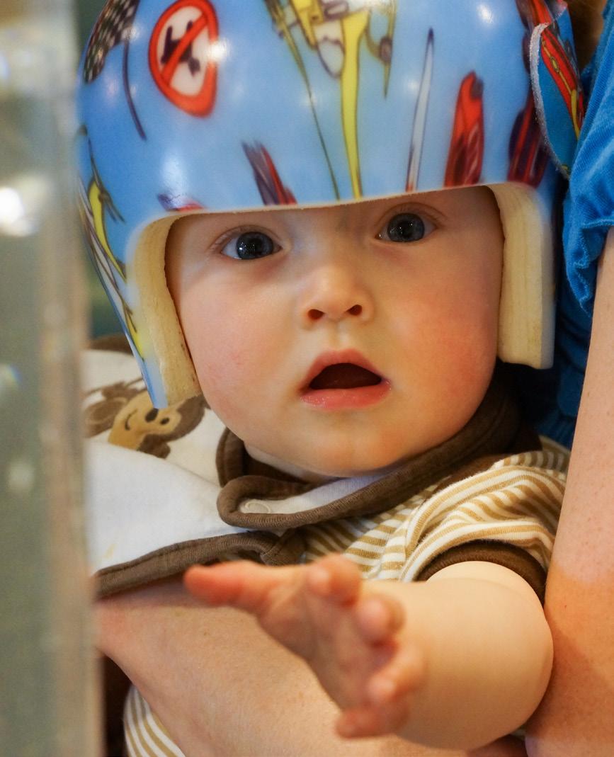
9 Orthotic helmet therapy (Boston Band) Numerous studies demonstrate effectiveness of orthotic helmeting for treatment of deformational plagiocephaly. 12,13,14,15 An orthotic helmet works by restricting further asymmetric growth, guiding the flattened areas to grow into the empty space of the helmet. The orthotic device is made of lightweight foam that is cut away in layers as the head grows. Helmets are custom made using cranial laser-scans taken during initial clinical visits. Helmets fit loosely and work by passively directing growth, rather than actively pushing on the cranium. Helmets are worn at all times, including sleep (a minimum of 23 hours/day), and are well-tolerated by most patients. The duration of helmet therapy at our center is typically 3 5 months. Because the head grows fastest early in life, the sooner helmet therapy begins, the faster the rate of correction. Orthotic helmeting is usually started at 4 months of age or older, but may be considered as early as 3 months of age in select cases. Maximum improvement is achieved when started by 6 7 months of age, 12,13 but good results are achieved within the first year of life. While improvement in cranial shape using orthotic helmeting has been documented in patients as old as 18 months, 14 the rate of change will be slower and the degree of correction achieved smaller. In addition, our experience has been that older patients do not tolerate wearing the helmet as well as younger patients, making lengthy periods of helmeting challenging. For that reason, we recommend starting helmet therapy early. 9
10 CLINICAL OUTCOME NOPCO Clinical Outcome Data Approximately 500 patients with abnormal head shape are treated at Boston-area NOPCO clinics annually. Clinical outcomes are regularly reviewed to ensure consistent quality care. Below are treatment outcomes for NOPCO patients with deformational plagiocephaly and brachycephaly, who completed treatment between January and May, Correction of plagiocephaly is measured using the Cranial Vault Asymmetry Index (CVAI), the percent difference between the diagonal dimensions of the head. 16 Brachycephaly is measured using the Cranial Ratio (CR), the width to length ratio of the head. 17 A/P Diagonal A Diagonal B M/L (A-B) X 100 CVAI = A or B (whichever is greater) CR = M/L A/P X100 PlagioCradle Thirty-five patients completed treatment with the PlagioCradle for deformational plagiocephaly. The mean treatment duration was 7.8 weeks, ending when the patient is able to roll out of the cradle. On average, patients started with a moderate degree of asymmetry and measured at the low end of mild asymmetry at the end of treatment. Note the slight widening of the head (CI) with cradle use, making the cradle inappropriate for treatment of brachycephaly. Mean Characteristics and Treatment Outcomes for Patients with Plagiocephaly Treated with the PlagioCradle (January May, 2013) Patients (n) Treatment Duration (weeks) Initial CI (%) Discharge CI (%) Initial CVAI (%) Discharge CVAI (%) (normal) 90.1 (normal) 7.8 (moderate) 4.0 (mild) 10
11 Orthotic Helmet Therapy (Boston Band) Two hundred and eighteen patients completed helmet therapy for plagiocephaly, brachycephaly, and asymmetric brachycephaly. On average, therapy was initiated at 6.5 months of age, with average duration in the helmet ranging from weeks. On average, patients started with a moderate degree of deformity and measured at the upper limit of normal at the end of treatment. Mean Characteristics and Treatment Outcomes for Patients Treated with the Boston Band (January May, 2013) Patients (n) Age at Fitting (Months) Treatment Duration (weeks) Initial CI (%) Discharge CI (%) Initial CVAI (%) Discharge CVAI (%) (moderate) 3.5 (normal) (moderate) 92.7 (normal) (moderate) 92.1 (normal) 7.3 (moderate) 2.5 (normal) Plagiocephaly Brachycephaly Asymmetric brachycephaly 11
12 REFERENCES 1. National Center for Health Statistics, Centers for Disease Control and Prevention, DHHS. (2006). Deaths: Final data for 2003 [Electronic version]. 2. Eunice Kennedy Shriver National Institute of Child Health and Human Development. (2006). Continuing Education Program on SIDS Risk Reduction. NIH Pub No Accessed Dec. 6, SIDS_rev.pdf 3. Mawji A, Vollman AR, Hatfield J, McNeil DA, Sauvé R. The incidence of positional plagiocephaly: a cohort study. Pediatrics Aug;132(2): Lajeunie E, Le Merrer M, Bonaïti-Pellie C, Marchac D, Renier D. Genetic study of nonsyndromic coronal craniosynostosis. Am J Med Genet Feb 13;55(4): French LR, Jackson IT, Melton LJ 3rd. A population-based study of craniosynostosis. J Clin Epidemiol. 1990;43(1): Rogers GF. Deformational plagiocephaly, brachycephaly, and scaphocephaly. Part I: terminology, diagnosis, and etiopathogenesis. J Craniofac Surg Jan;22(1): Shillito J Jr, Matson DD. Craniosynostosis: a review of 519 surgical patients. Pediatrics Apr;41(4): Oh AK, Hoy EA, Rogers GF. Predictors of severity in deformational plagiocephaly. J Craniofac Surg Mar;20 Suppl 1: Cavalier A, Picot MC, Artiaga C, Mazurier E, Amilhau MO, Froye E, Captier G, Picaud JC. Prevention of deformational plagiocephaly in neonates. Early Hum Dev Aug;87(8): Rogers GF, Miller J, Mulliken JB. Comparison of a modifiable cranial cup versus repositioning and cervical stretching for the early correction of deformational posterior plagiocephaly. Plast Reconstr Surg Mar;121(3): Seruya M, Oh AK, Sauerhammer TM, Taylor JH, Rogers GF. Correction of deformational plagiocephaly in early infancy using the plagio cradle orthotic. J Craniofac Surg Mar;24(2): Kluba S, Kraut W, Reinert S, Krimmel M. What is the optimal time to start helmet therapy in positional plagiocephaly? Plast Reconstr Surg Aug;128(2):
13 13. Yoo HS, Rah DK, Kim YO. Outcome analysis of cranial molding therapy in nonsynostotic plagiocephaly. Arch Plast Surg Jul;39(4): Seruya M, Oh AK, Taylor JH, Sauerhammer TM, Rogers GF. Helmet treatment of deformational plagiocephaly: the relationship between age at initiation and rate of correction. Plast Reconstr Surg Jan;131(1):55e-61e. 15. Couture DE, Crantford JC, Somasundaram A, Sanger C, Argenta AE, David LR. Efficacy of passive helmet therapy for deformational plagiocephaly: report of 1050 cases. Neurosurg Focus Oct;35(4):E Plank L, Giavedoni B, Lombardo J, et al. The use of a non-invasive laser data acquisition system to quantify and compare infant head shape with non-synostotic positional plagiocephaly. Paper presented at the Association of Children s Prosthetic Orthotic Clinics Annual Meeting; Banff, Alberta, Canada; March 24 27, Hutchison BL, Hutchison LA, Thompson JM, Mitchell EA. Quantification of plagiocephaly and brachycephaly in infants using a digital photographic technique. Cleft Palate Craniofac J Sep;42(5):

14 LOCATIONS NOPCO locations Massachusetts NOPCO of Boston Children s Hospital 300 Longwood Avenue Boston, MA New Hampshire NOPCO of Exeter 1 Hampton Road, Suite 106A Exeter, NH NOPCO of Boston Children s at Waltham 9 Hope Avenue, Suite 200 Waltham, MA NOPCO of Somersworth 224 Route 108, Unit A Somersworth, NH NOPCO of Boston Longwood Medical 431 Brookline Avenue Boston, MA NOPCO of Burlington 50 Mall Road, Suite G-10 Burlington, MA NOPCO of Lawrence 25 Marston Street, Suite 201 Lawrence, MA NOPCO of Weymouth, Stetson Building 541 Main Street, Suite 214 Weymouth, MA
15 New Jersey NOPCO of Neptune 3700 Route 33, Suite LL02 Neptune, NJ Pennsylvania NOPCO of Pennsylvania 3550 Market Street Philadelphia, PA NOPCO of New Jersey 585 Cranbury Road, Suite B East Brunswick, NJ NOPCO of Pennsylvania Children s Seashore House 3405 Civic Center Blvd. Philadelphia, PA NOPCO of New Jersey Children s Specialized Hospital 150 New Providence Road Mountainside, NJ ext NOPCO of Voorhees 200 Bowman Drive, Suite D279 Voorhees, NJ Boston Children s locations Boston Children s Hospital 300 Longwood Avenue Boston, MA Boston Children s at Waltham 9 Hope Avenue Waltham, MA Boston Children s Physicians at Weymouth The Stetson Building 541 Main Street Weymouth, MA

16 #3084 Deformational Plagiocephaly A GUIDE TO DIAGNOSIS AND TREATMENT
MEDICAL POLICY MEDICAL POLICY DETAILS POLICY STATEMENT POLICY GUIDELINES. Page: 1 of 5. Medical Policy Title CRANIAL ORTHOTICS Policy Number 1.01.
 Page: 1 of 5 MEDICAL POLICY MEDICAL POLICY DETAILS Medical Policy Title CRANIAL ORTHOTICS Policy Number 1.01.32 Category Equipment/Supplies Effective Date 10/18/01 Revised Date 06/27/02, 07/24/03, 06/24/04,
Department of Neurosurgery. Differentiating Craniosynostosis from Positional Plagiocephaly
 Department of Neurosurgery Differentiating Craniosynostosis from Positional Plagiocephaly The number of infants with head shape deformities has risen over the past several years, likely due to increased
Prevention Diagnosis
 Prevention and Management of Positional Skull Deformities in Infants John Persing, MD, Hector James, MD, Jack Swanson, MD, John Kattwinkel, MD, Committee on Practice and Ambulatory Medicine, Section on
Interesting Case Series. The Danger of Posterior Plagiocephaly
 Interesting Case Series The Danger of Posterior Plagiocephaly Susan Orra, BA, a,b Kashyap Komarraju Tadisina, BS, a Bahar Bassiri Gharb, MD, PhD, a Antonio Rampazzo, MD, PhD, a Gaby Doumit, MD, a and Francis
Craniosynostosis. Diagnosis and Treatment
 Craniosynostosis Diagnosis and Treatment 2015 For more information about the Weill Cornell Craniofacial Program ABOUT The Weill Cornell Craniofacial Program takes a multidisciplinary approach to treating
Policy #: 008 Latest Review Date: May 2007 Category: Durable Medical Equipment
 Name of Policy: Dynamic Orthotic Cranioplasty (DOC) Policy #: 008 Latest Review Date: May 2007 Category: Durable Medical Equipment Policy Grade: D Background: As a general rule, benefits are payable under
Nonsurgical, nonorthotic treatment of occipital plagiocephaly: what is the natural history of the misshapen neonatal head?
 Nonsurgical, nonorthotic treatment of occipital plagiocephaly: what is the natural history of the misshapen neonatal head? S. David Moss, M.D. Phoenix Children's Hospital, Phoenix, Arizona Plagiocephaly
Craniosynostosis and Plagiocephaly
 Craniosynostosis and Plagiocephaly Andrew Jea MD MHA FAAP Professor and Chief Section of Pediatric Neurosurgery Riley Hospital for Children Department of Neurosurgery Indiana University School of Medicine
Effective Treatment of Craniosynostosis and Deformational Plagiocephaly Improves with Early Diagnosis:
 Effective Treatment of Craniosynostosis and Deformational Plagiocephaly Improves with Early Diagnosis: Watchful Waiting May Not Be the Best Option for Evaluating Abnormal Head Shape in Infants April, 2016
Adjustable Cranial Orthoses for Positional Plagiocephaly and Craniosynostoses Corporate Medical Policy
 Adjustable Cranial Orthoses for Positional Plagiocephaly and Craniosynostoses Corporate Medical Policy File name: Adjustable Cranial Orthoses for Positional Plagiocephaly and Craniosynostoses File code:
Table 1: Helmet vs Conservative Treatment
 Table 1: Helmet vs Conservative Treatment Author (Year) Title Study Description Data Class, Quality, and Van Wijk et al (2014) Helmet Therapy in Infants with Positional Skull Deformation: Randomized Controlled
Occipital flattening in the infant skull
 Occipital flattening in the infant skull Kant Y. Lin, M.D., Richard S. Polin, M.D., Thomas Gampper, M.D., and John A. Jane, M.D., Ph.D. Departments of Plastic Surgery and Neurological Surgery, University
The prevalence of deformational plagiocephaly PEDIATRIC/CRANIOFACIAL
 PEDIATRIC/CRANIOFACIAL Effectiveness of Conservative Therapy and Helmet Therapy for Positional Cranial Deformation Jordan P. Steinberg, M.D., Ph.D. Roshni Rawlani, B.A. Laura S. Humphries, M.D. Vinay Rawlani,
Adjustable Cranial Orthoses for Positional Plagiocephaly and Craniosynostoses Corporate Medical Policy
 Adjustable Cranial Orthoses for Positional Plagiocephaly and Craniosynostoses Corporate Medical Policy File name: Adjustable Cranial Orthoses for Positional Plagiocephaly and Craniosynostoses File code:
Cranial Remolding Restoring Bodies, Rebuilding Lives Toll Free:
 Cranial Remolding Restoring Bodies, Rebuilding Lives www.wcbl.com Toll Free: 1-888-552-2555 Causes of Positional Plagiocephaly During the first weeks of life, the infant s skull is very malleable. This
International Journal of Current Research and Academic Review ISSN: Volume 3 Number 1 (January-2015) pp
 pp") International Journal of Current Research and Academic Review ISSN: 47 Volume Number (January) pp. 66 www.ijcrar.com Clinical Profile of Patients with Craniosynostosis: A Descriptive Study Nagaraj V. Gadwal*
International Journal of Current Research and Academic Review ISSN: 47 Volume Number (January) pp. 66 www.ijcrar.com Clinical Profile of Patients with Craniosynostosis: A Descriptive Study Nagaraj V. Gadwal*
MP Adjustable Cranial Orthoses for Positional Plagiocephaly and Craniosynostoses. Related Policies None
 Medical Policy BCBSA Ref. Policy: 1.01.11 Last Review: 03/29/2018 Effective Date: 03/29/2018 Section: Durable Medical Equipment Related Policies None DISCLAIMER Our medical policies are designed for informational
Policy Specific Section: June 9, 1999 October 7, 2011
 Medical Policy Cranial Remodeling Orthosis Type: Medical Necessity/Not Medical Necessity Policy Specific Section: Durable Medical Equipment Original Policy Date: Effective Date: June 9, 1999 October 7,
The incidence of cranial deformities has increased since
 ORIGINAL RESEARCH ARTICLE Orthotic Treatment of Cranial Asymmetries: Comparison between Early and Late Interventions Carolina Gomes Matarazzo, MSPT, Fernando Campos Gomes Pinto, MD, PhD, Maria Stella Peccin,
Coding For Craniosynostosis. Peggy Feeley RHIA, CCS, CCS-P, COC AHIMA Approved ICD-10-CM/PCS Trainer
 Coding For Craniosynostosis Peggy Feeley RHIA, CCS, CCS-P, COC AHIMA Approved ICD-10-CM/PCS Trainer Cranial sagittal Synostosis Cranium job is to protect the brain The top portion of the skull, which protects
T HERE is an unusual and interesting variety of craniosynostosis in
 SURGICAL TREATMENT OF CONGENITAL ANOMALIES OF THE CORONAL AND METOPIC SUTURES TECHNICAL NOTE DONALD D. MATSON, M.D. Neurosurgical Service, The Children's Medical Center, and Deparlment of Surgery, Itarvard
Research Article Human Anatomy Case Report Bathrocephaly: a case report of a head shape associated with a persistent mendosal suture
 IJAE Vol. 119, n. 3: 263-267, 2014 ITALIAN JOURNAL OF ANATOMY AND EMBRYOLOGY Research Article Human Anatomy Case Report Bathrocephaly: a case report of a head shape associated with a persistent mendosal
Clinical Guide. Pediatric Cranial Deformities using STAR Cranial Remolding Orthoses
 Clinical Guide Orthotic Management of Pediatric Cranial Deformities using STAR Cranial Remolding Orthoses STARband Clinical Guide By Dulcey Lima, CO, OTR/L Introduction The number of skull deformities
a guide to understanding craniosynostosis a publication of children s craniofacial association
 a guide to understanding craniosynostosis a publication of children s craniofacial association 1 a guide to understanding craniosynostosis this parent s guide to craniosynostosis is designed to answer
4.1 Classification of Craniosynostosis: Therapeutical implications.
 ISPN course 23 rd Nov, 2015 Cranial & Craniofacial disorders 4.1 Classification of Craniosynostosis: Therapeutical implications. Kazuaki Shimoji, Masakazu Miyajima and Hajime Arai Department of Neurosurgery,
Craniosynostosis. chapter. Jeffrey Weinzweig, MD / Stephen B. Baker, MD, DDS / Mitchel Seruya, MD AQ1
 chapter 16 AQ1 Craniosynostosis Jeffrey Weinzweig, MD / Stephen B. Baker, MD, DDS / Mitchel Seruya, MD AQ2 PATIENT EVALUATION AND SELECTION Craniosynostosis is defined as the premature closure of a cranial
HANGER CLINIC CAP PROGRAM
 HANGER CLINIC CAP PROGRAM CRANIAL ASYMMETRY The Hanger Clinic Cranial Asymmetry Protocol (CAP) Program is built around your child s needs and a set of clearly defined clinical practice guidelines. You
Multidisciplinary care of craniosynostosis
 Journal of Multidisciplinary Healthcare open access to scientific and medical research Open Access Full Text Article Multidisciplinary care of craniosynostosis Review edward P Buchanan 1 Yunfeng Xue 1
What is Craniosynostosis?
 What is Craniosynostosis? Craniosynostosis is defined as the premature closure of the cranial sutures (what some people refer to as soft spots). This results in restricted and abnormal growth of the head.
Original Article. Annals of Rehabilitation Medicine INTRODUCTION
 Original Article Ann Rehabil Med 2013;37(6):785-795 pissn: 2234-0645 eissn: 2234-0653 http://dx.doi.org/10.5535/arm.2013.37.6.785 Annals of Rehabilitation Medicine Comparison of Helmet Therapy and Counter
Craniosynostosis & Craniofacial Surgery A Parent s Guide
 Craniosynostosis & Craniofacial Surgery A Parent s Guide Steven R. Buchman, MD Karin M. Muraszko, MD Carolyn Walborn, RN, MS, CPNP Laura Zang, MS, RN University of Michigan Medical Center C.S. Mott Children
Authors: Shitel Patel, Rami R Hallac, Pang-yun Chou, Min-Jeong Cho, Neil Stewart, Ana Nava, James Seaward, Alex Kane, Christopher Derderian
 Authors: Shitel Patel, Rami R Hallac, Pang-yun Chou, Min-Jeong Cho, Neil Stewart, Ana Nava, James Seaward, Alex Kane, Christopher Derderian Title: Location and Time of Maximal Head Shape Change in Strip
De f o r m at i o n a l or positional plagiocephaly is the
 J Neurosurg Pediatrics 5:000 000, 5:368 374, 2010 Comparison of perceptions and treatment practices between neurosurgeons and plastic surgeons for infants with deformational plagiocephaly Clinical article
Emerging Surgical Technologies: Open vs. Endoscopic Craniosynostosis Repair
 Emerging Surgical Technologies: Open vs. Endoscopic Craniosynostosis Repair Petra M. Meier, MD, DEAA Senior Associate of the Department of Anesthesiology, Perioperative and Pain Medicine, Boston Children
High-frequency ultrasound confirmation of positional plagiocephaly
 J Neurosurg (5 Suppl Pediatrics) 105:413 417, 2006 High-frequency ultrasound confirmation of positional plagiocephaly JAN REGELSBERGER, M.D., GÜNTER DELLING, M.D., MICHAEL TSOKOS, M.D., KNUTH HELMKE, M.D.,
Occipital plagiocephaly
 ritish Journal of Plastic Surgery (2000), 53, 367 377 9 2000 The ritish ssociation of Plastic Surgeons doi:10.1054/bjps.2000.3329 RITISH JOURNL OF [ ~ ] PLSTIC SURGERY Occipital plagiocephaly D. J. David
Cranial Base Changes Following. Surgical Treatment of Craniosynostosis. JEFFREY L. MARSH, M.D. MicHaAeL W. VaAnniEer, M.D.
 Cranial Base Changes Following Surgical Treatment of Craniosynostosis JEFFREY L. MARSH, M.D. MicHaAeL W. VaAnniEer, M.D. Three-dimensional osseous surface images from CT scans have been used to study the
Neurodevelopmental Implications of Deformational Plagiocephaly
 0196-206X/05/2605-0379 Developmental and Behavioral Pediatrics Vol. 26, No. 5, October 2005 Copyright # 2005 by Lippincott Williams & Wilkins, Inc. Printed in U.S.A. Review Article Neurodevelopmental Implications
Craniosynostosis - making the head fit the hat
 Craniosynostosis - making the head fit the hat Poster No.: R-0173 Congress: 2014 CSM Type: Scientific Exhibit Authors: S. Constantine, B. Clark; NORTH ADELAIDE/AU Keywords: Head and neck, Bones, Pediatric,
Numerous techniques have been developed to treat
 clinical article J Neurosurg Pediatr 18:674 678, 2016 Endoscope-assisted management of sagittal synostosis: wide vertex suturectomy and barrel stave osteotomies versus narrow vertex suturectomy Brian J.
Advaneement-onlay: an improved technique of fronto-orbital remodeling in craniosynostosis *
 Advances & operative techniques mcns 9 Springer-Verlag 1991 Child's Nerv Syst (1991) 7:264-271 Advaneement-onlay: an improved technique of fronto-orbital remodeling in craniosynostosis * Steven R. Cohen
Unilateral fusion of the coronal suture is the most. Isolated frontosphenoidal synostosis: a rare cause of synostotic frontal plagiocephaly
 J Neurosurg Pediatrics 13:553 558, 2014 AANS, 2014 Isolated frontosphenoidal synostosis: a rare cause of synostotic frontal plagiocephaly Clinical aicle Tina M. Sauerhammer, M.D., Albe K. Oh, M.D., Michael
Evidence-Based Care of the Child With Deformational Plagiocephaly, Part I: Assessment and Diagnosis
 ARTICLE Continuing Education Evidence-Based Care of the Child With Deformational Plagiocephaly, Part I: Assessment and Diagnosis Wendy S. Looman, PhD, RN, CNP, & Amanda B. Kack Flannery, MS, RN, CNP OBJECTIVES
Clinical Policy Title: Cranial orthotic devices
 Clinical Policy Title: Cranial orthotic devices Clinical Policy Number: 11.02.01 Effective Date: September 1, 2013 Initial Review Date: February 18, 2013 Most Recent Review Date: September 21, 2017 Next
Cranial molding helmet therapy and establishment of practical criteria for management in Asian infant positional head deformity
 DOI 10.1007/s00381-014-2471-y ORIGINAL PAPER Cranial molding helmet therapy and establishment of practical criteria for management in Asian infant positional head deformity Yasuo Aihara & Kana Komatsu
Characteristics, head shape measurements and developmental delay in 287 consecutive infants attending a plagiocephaly clinic
 Acta Pædiatrica ISSN 0803 5253 REGULAR ARTICLE Characteristics, head shape measurements and developmental delay in 287 consecutive infants attending a plagiocephaly clinic BL Hutchison (bl.hutchison@auckland.ac.nz),
HEAD TO FOOT EXAMINATION DR JP,ASST. PROF.ICH,GOVT MEDICAL COLLEGE KOTTAYAM
 HEAD TO FOOT EXAMINATION DR JP,ASST. PROF.ICH,GOVT MEDICAL COLLEGE KOTTAYAM 1.CRANIUM Is the size of head normal.?(measure ofc).is the skull shape abnormal( LOOK FROM ABOVE)? Are there any swellings on
Chapter 7: Head & Neck
 Chapter 7: Head & Neck Osteology I. Overview A. Skull The cranium is composed of irregularly shaped bones that are fused together at unique joints called sutures The skull provides durable protection from
CASE REPORT Pan-Suture Synostosis After Posterior Vault Distraction
 CASE REPORT Pan-Suture Synostosis After Posterior Vault Distraction Katrina F. Chu, BA, a Stephen R. Sullivan, MD, MPH, a,b and Helena O. Taylor, MD, PhD a,b a Warren Alpert Medical School of Brown University;
August 31, Appeals Coordinator United Healthcare P.O. Box Atlanta, GA RE: Patient: Employee: ID#: Group#: Group:
 August 31, 2001 Appeals Coordinator United Healthcare P.O. Box 740800 Atlanta, GA 30374-0800 RE: Patient: Employee: ID#: Group#: Group: To Whom It May Concern: We received your denial of coverage from
Surgery for craniosynostosis has evolved rapidly
 Surgical Treatment of Craniosynostosis: Outcome Analysis of 250 Consecutive Patients Gerald M. Sloan, MD*; Karin C. Wells, MD ; Corey Raffel, MD, PhD ; and J. Gordon McComb, MD ABSTRACT. Objective. Surgery
OMT FOR CONCUSSIONS KIMBERLY WOLF, D.O. FEBRUARY 17, 2017
 OMT FOR CONCUSSIONS KIMBERLY WOLF, D.O. FEBRUARY 17, 2017 POTENTIAL SEQUENCE Address lymphatics including all transition zones/diaphragms Address somatic dysfunction in spine Focus on upper cervical spine
Length of synostosis and segmented intracranial volume correlate with age in patients with non-syndromic sagittal synostosis
 https://helda.helsinki.fi Length of synostosis and segmented intracranial volume correlate with age in patients with non-syndromic sagittal synostosis Heliövaara, Arja 2018-03 Heliövaara, A, Leikola, J,
Skeletal System. Prof. Dr. Malak A. Al-yawer Department of Anatomy/Embryology Section
 Skeletal System Prof. Dr. Malak A. Al-yawer Department of Anatomy/Embryology Section Learning objectives At the end of this lecture, the medical student will be able to: State the embryonic origin of skeletal
2016 Congress of Neurological Surgeons 1
 CONGRESS OF NEUROLOGICAL SURGEONS SYSTEMATIC REVIEW AND EVIDENCE-BASED GUIDELINE FOR THE DIAGNOSIS OF PATIENTS WITH POSITIONAL PLAGIOCEPHALY: THE ROLE OF IMAGING Sponsored by Congress of Neurological Surgeons
Neurodevelopment in Children with Single-Suture Craniosynostosis and Plagiocephaly without Synostosis
 Neurodevelopment in Children with Single-Suture Craniosynostosis and Plagiocephaly without Synostosis Jayesh Panchal, M.B.B.S., F.R.C.S., M.B.A., Hamid Amirsheybani, M.D., Robin Gurwitch, Ph.D., Vicki
2016 Congress of Neurological Surgeons 1
 CONGRESS OF NEUROLOGICAL SURGEONS SYSTEMATIC REVIEW AND EVIDENCE-BASED GUIDELINE ON THE MANAGEMENT OF PATIENTS WITH POSITIONAL PLAGIOCEPHALY: THE ROLE OF REPOSITIONING Sponsored by Congress of Neurological
4.3 Surgical Management of anterior skull synostosis
 ISPN course 23 rd Nov, 2015 Cranial & Craniofacial disorders 4.3 Surgical Management of anterior skull synostosis Kazuaki Shimoji, Masakazu Miyajima and Hajime Arai Department of Neurosurgery, Juntendo
Prenatal Diagnosis of Cleft Lip
 Commentary Prenatal Diagnosis of Cleft Lip What the Sonologist Needs to Tell the Surgeon John. Mulliken, MD, eryl R. enacerraf, MD Division of Plastic Surgery, Children s Hospital (J..M.) Department of
Children with Metopic Ridge
 DOI: 10.5137/1019-5149.JTN.16886-15.2 Received: 28.12.2015 / Accepted: 04.03.2016 Published Online: 25.04.2016 Original Investigation Children with Metopic Ridge Tufan HICDONMEZ Dr. Lutfi Kirdar Education
Craniofacial Fellowship. Boston Children s Hospital and Harvard Medical School, Boston Massachusetts, July 2010-June 2011
 Kamlesh B. Patel Washington University- Saint Louis Assistant Professor, Division of Plastic and Reconstructive Surgery Associate Program Director 660 S. Euclid Ave. Campus Box 8238, St. Louis, MO 63130
Sagittal craniosynostosis is the most common of the. Combined metopic and sagittal craniosynostosis: is it worse than sagittal synostosis alone?
 Neurosurg Focus 31 (2):E2, 2011 Combined metopic and sagittal craniosynostosis: is it worse than sagittal synostosis alone? Jordan S. Terner, B.A., 1 Roberto Travieso, B.A., 1 Su-shin Lee, M.D., 2 Antonio
Deformational plagiocephaly and chiropractic care: A narrative review and case report
 Deformational plagiocephaly and chiropractic care: A narrative review and case report By Jennifer L. Hash, DC Jennifer L. Hash, DC, private practice, Lisle, Illinois, USA Contact: jhashdc@gmail.com ABSTRACT
pressure 6-7 and cranial growth restriction.
 Josephine Jung graduated from Medical School in Berlin in 2014. After completing her M.D. Thesis in Berlin she moved to London and is working as a Clinical Research Fellow in Neurosurgery at King s College
DOI: /01.PRS
 CME Management of Craniosynostosis Jayesh Panchal, M.D., M.B.A., and Venus Uttchin, B.A. Oklahoma City, Okla. Learning Objectives: After studying this article, the participant should be able to: 1. Review
Craniosynostosis is the early fusion of one or more. Cranial vault remodeling for sagittal craniosynostosis in older children
 Neurosurg Focus 31 (2):E3, 2011 Cranial vault remodeling for sagittal craniosynostosis in older children S. Alex Rottgers, M.D., 1,2 Peter D. Kim, M.D., Ph.D., 1,3 Anand Raj Kumar, M.D., 1,2 James J. Cray,
Craniosynostosis : Updates in Radiologic Diagnosis
 www.jkns.or.kr http://dx.doi.org/10.3340/jkns.2016.59.3.219 J Korean Neurosurg Soc 59 (3) : 219-226, 2016 Print ISSN 2005-3711 On-line ISSN 1598-7876 Copyright 2016 The Korean Neurosurgical Society Pediatric
Sample page. Neurosurgery/Neurology. A comprehensive illustrated guide to coding and reimbursement CODING COMPANION
 CODING COMPNION 2018 Neurosurgery/Neurology comprehensive illustrated guide to coding and reimbursement POWER UP YOUR CODING with Optum360, your trusted coding partner for 32 years. Visit optum360coding.com.
A New Ultrasound Method for Assessment of Head Shape Change in Infants With Plagiocephaly Jin Kyung Kim, MD, Dong Rak Kwon, MD, Gi-Young Park, MD
 Original Article Ann Rehabil Med 2014;38(4):541-547 pissn: 2234-0645 eissn: 2234-0653 http://dx.doi.org/10.5535/arm.2014.38.4.541 Annals of Rehabilitation Medicine A New Ultrasound Method for Assessment
1/27/2017. Nursing Care of the Pediatric Neurosurgical Patient. Assessment is the key...
 Nursing Care of the Pediatric Neurosurgical Patient Julie Deibel, APN, NP-C, CNE Pediatric Neurosurgery Illinois Neurological Institute Assessment is the key..... Overview Consideration of development
Chaired by Irene M.J. Mathijssen, MD, PhD
 ORIGINAL ARTICLE Guideline for Care of Patients With the Diagnoses of Craniosynostosis: Working Group on Craniosynostosis Irene M.J. Mathijssen, MD, PhD (J Craniofac Surg 2015;26: 1735 1807) Chaired by
Pitfalls in counselling: the craniosynostoses
 J7 Med Genet 1991; 28: 117-121 Pitfalls in counselling: the craniosynostoses Romana Marini, Karen Temple, Lyn Chitty, Sally Genet, Michael Baraitser Abstract We describe three families to highlight the
Evaluation of Staggered Osteotomy in Surgical Treatment of Trigonocephaly
 Evaluation of Staggered Osteotomy... Hassanpour et al. 28 Original 28 Evaluation of Staggered Osteotomy in Surgical Treatment of Trigonocephaly Seyed Esmail Hassanpour¹ Mohammad Reza Hadi Sichani 2 Mohammadreza
Sponsored by. Congress of Neurological Surgeons (CNS) and the Section on Pediatric Neurosurgery. Endorsed by
 and the Section on Pediatric Neurosurgery. Endorsed by") CONGRESS OF NEUROLOGICAL SURGEONS SYSTEMATIC REVIEW AND EVIDENCE-BASED GUIDELINE ON THE MANAGEMENT OF PATIENTS WITH POSITIONAL PLAGIOCEPHALY: THE ROLE OF PHYSICAL THERAPY Sponsored by Congress of Neurological
CONGRESS OF NEUROLOGICAL SURGEONS SYSTEMATIC REVIEW AND EVIDENCE-BASED GUIDELINE ON THE MANAGEMENT OF PATIENTS WITH POSITIONAL PLAGIOCEPHALY: THE ROLE OF PHYSICAL THERAPY Sponsored by Congress of Neurological
PEDIATRIC NEUROSURGICAL DISEASE
 PEDIATRIC SURGERY FOR THE PRIMARY CARE PEDIATRICIAN, PART 11 0031-3955/98 $8.00 +.OO PEDIATRIC NEUROSURGICAL DISEASE Mark S. Dias, MD, and Veetai Li, MD Although clinicians often worry about missing occult
Treatment planning of nonskeletal problems. in preadolescent children
 In the name of GOD Treatment planning of nonskeletal problems in preadolescent children Presented by: Dr Somayeh Heidari Orthodontist Reference: Contemporary Orthodontics Chapter 7 William R. Proffit,
Pediatric Radiology Update
 Pediatric Radiology Update Douglas Rivard, DO Vice Chairman, Radiology Dept Children s Mercy Hospital Asst Professor of Radiology University of Missouri-Kansas City Objectives Review radiation biology
The Normal Standards of Anterior Fontanel Size in Iraqi Neonates. Nahla Al-Gabban
 Abstract Background: The diagnosis of abnormal anterior fontanel requires an understanding of the wide variation of normal. Objectives: The study is an attempt to establish the normal value for anterior
RE: CHILD S NAME DOB: XXXXXXX Denial of DOC Band coverage XXXXXXXXX Care First Blue Cross Blue Shield Contract #: XXXXXX Group #: XXXXXXXXXXXXXX
 YOUR ADDRESS Oct. 9, 2002 Care First Blue Cross Blue Shield Attention: Appeals/Supervisor Control Plan Claims Unit P.O. Box 1739 Cumberland, Maryland 21501 RE: CHILD S NAME DOB: XXXXXXX Denial of DOC Band
Pathological condition that results from premature fusion of one or more sutures in the cranial vault;
 Pathological condition that results from premature fusion of one or more sutures in the cranial vault; Associated with a deformity of the vault and cranial base. Development Bones of the cranium The skull
Interactions of the endocrine system, bone and oral health
 Interactions of the endocrine system, bone and oral health All bones are not equal! Dense high proportion of cortical bone High proportion of trabecular bone Mandible Functions: mastication, respiration,
Component parts of Chrome Cobalt Removable Partial Denture
 Lec. 5 د.بسام الطريحي Component parts of Chrome Cobalt Removable Partial Denture Major connectors: Are either bars or plates, the difference between them is in the amount of tissue covers. Plates are broad
OMT Without An OMT Table Workshop. Dennis Dowling, DO FAAO Ann Habenicht, DO FAAO FACOFP
 OMT Without An OMT Table Workshop Dennis Dowling, DO FAAO Ann Habenicht, DO FAAO FACOFP Cervical Somatic Dysfunction (C5 SR RR) - Seated 1. Patient position: seated. 2. Physician position: standing facing
CRANIOFACIAL SURGERY OVER 30 YEARS IN GÖTEBORG
 Scandinavian Journal of Surgery 92: 274 280, 2003 CRANIOFACIAL SURGERY OVER 30 YEARS IN GÖTEBORG C. Lauritzen, P. Tarnow Department of Plastic Surgery, Sahlgrenska University Hospital, Göteborg University,
Anatomy and Physiology. Bones, Sutures, Teeth, Processes and Foramina of the Human Skull
 Anatomy and Physiology Chapter 6 DRO Bones, Sutures, Teeth, Processes and Foramina of the Human Skull Name: Period: Bones of the Human Skull Bones of the Cranium: Frontal bone: forms the forehead and the
Dr. N. Retnakumari. MDS, M.Phil, Dr. Manuja Vargheese, Dr. Madhu.S, Dr. Divya. S
 IOSR Journal of Dental and Medical Sciences (IOSR-JDMS) e-issn: 2279-0853, p-issn: 2279-0861. Volume 12, Issue 5 (Nov.- Dec. 2013), PP 11-15 A new approach in Presurgical Infant Orthopedics using an Active
Craniosynostosis - Recognition, clinical characteristics, and treatment
 BOSNIAN JOURNAL OF BASIC MEDICAL SCIENCES WWW.BJBMS.ORG Craniosynostosis - Recognition, clinical characteristics, and treatment Nina Kajdic 1,2, Peter Spazzapan 2, Tomaz Velnar 2 * 1 Chair of Surgery,
Headlines Craniofacial Support
 Other leaflets available from Headlines Craniofacial Support Please contact Group Administrator Justine Tweedie - info@headlines.org.uk for details of how to obtain copies 1 What causes Craniosynostosis?
The Swimmer s Shoulder: An Osteopathic Approach
 The Swimmer s Shoulder: An Osteopathic Approach Mary Solomon, D.O. Rainbow Babies and Children s Hospital Cleveland, OH 440-914-7865 1 I have no relevant relationships/affiliations with any proprietary
Imaging in the Evaluation of Children with Suspected Craniosynostosis
 4 Imaging in the Evaluation of Children with Suspected Craniosynostosis Daniel N. Vinocur and L. Santiago Medina I. What is the role of imaging in the diagnosis of craniosynostosis? II. What is the cost
Human, Probable Female, Scaphocephaly
 Human, Probable Female, Scaphocephaly Product Number: Specimen Evaluated: Skeletal Inventory: BC-193 Bone Clones replica 1 intact cranium 1 intact mandible General observations: In general, the molding
It has been proposed that partially edentulous maxillectomy
 CLASSICAL ARTICLE Basic principles of obturator design for partially edentulous patients. Part II: Design principles Mohamed A. Aramany, DMD, MS* Eye and Ear Hospital of Pittsburgh and University of Pittsburgh,
Skeletal System: Skull.
 Skeletal System: Skull www.fisiokinesiterapia.biz Bones of the Skull SPLANCHNOCRANIUM Nasal (2) Maxilla (2) Lacrimal (2) Zygomatic (2) Palatine (2) Inferior concha (2) Vomer Mandible NEUROCRANIUM Frontal
Bones of the skull & face
 Bones of the skull & face Cranium= brain case or helmet Copyright The McGraw-Hill Companies, Inc. Permission required for reproduction or display. The cranium is composed of eight bones : frontal Occipital
The key to facial beauty and optimal patient health - Part 1
 The key to facial beauty and optimal patient health - Part 1 By John Flutter, BDS the reason the upper jaw size remained stable and the occlusion improved, was due to the fact that after expanding the
Cleft-Craniofacial Center
 Cleft-Craniofacial Center A Pioneering T eam 2 Welcome to the Cleft-Craniofacial Center at Children s Hospital of Pittsburgh The Cleft-Craniofacial Center at Children s Hospital of Pittsburgh has been
Plastic Surgeon, Middlesbrough General Hospital, Stockton Children's Hospital, Newcastle Regional Hospital Board
 THE NASAL TIP IN BILATERAL HARE LIP By J. POTTER, F.R.C.S.Ed. Plastic Surgeon, Middlesbrough General Hospital, Stockton Children's Hospital, Newcastle Regional Hospital Board IN the problem of the bilateral
UCL Repair: Emphasis on Muscle Dissection and Reconstruction
 UCL Repair: Emphasis on Muscle Dissection and Reconstruction Unilateral cleft lip repair is performed using rotation-advancement technique. Markings are made on columella base, redlines, Cupid s bow on
... . ' I I I I I. '. I HISTORY. March 13, Date of Visit: February 24, Dear Dr.
 1.2.6 March 13, 2016 Date of Visit: February 24, 2016 Dear Dr. We recently had the opportunity to see - when he returned for follow-up consultation accompanied by his mother,_ and his sister. Concerns