movement occurs by a process of diffusion across the endothelium. We shall discuss if
|
|
|
- Felix Casey
- 5 years ago
- Views:
Transcription
1 Corneal channels Daljit Singh For the cornea to maintain its hydration, transparency and metabolism, it should have a fluid movement throughout the stroma. We have learnt from the text books that the fluid movement occurs by a process of diffusion across the endothelium. We shall discuss if there is any movement in the form of channels? The cornea is transparent for all practical purposes. That makes it difficult to visualize the transparent channels inside the cornea if any. We have been interested in the conjunctival lymphatics for many years. Conjunctival lymphatics could be demonstrated by injecting trypan blue dye in the peripheral cornea, rather than subconconjunctival injection brought out the lymphatics better We also tried to chart lymphatics in the cornea in painful blind eyes undergoing glaucoma surgery.the dye was injected in the mid-peripheral corneal stroma. It created a small ball of the dye, at the edges of which there were small projections towards the center and the periphery. Slit lamp examination of the injected cornea showed a diffuse presence of the dye only.over the next few days, the dye appeared to become faint. It showed no definite movement towards the center, but disappeared completely in a few days, obviously towards the periphery, in to the limbal lymphatics.

2 Once, studying the pictures of dye injected conjunctival lymphatics, I found that it had also created a thin circular line in the periphery of the cornea. Obviously it was not in Canal of Schlemm. Fig.1: The dye that was injected in the peripheral cornea, not only demonstrated the conjunctival lymphatics, but also a blue colored arc in the corneal periphery Later, in an eye undergoing enucleation, the dye was injected with great force in the cornea some distance from the limbus. It happened to produce a ring like appearance along the limbus, indicating the presence of a space that could hold the dye.

3 Fig.2: Trypan blue injected in the cornea has assumed a circular form inside the limbus.
4 Any kind of invisible corneal channels if present in the cornea, are expected to be transparent and fashioned on the lines of sinusoids, rather than definitive vessels likes arteries and veins. But suppose there is a corneal pathology that renders the corneal tissue translucent or semi-opaque, it could make the channels visible. This is just like the transparent limbal lymphatics that get visualized by the presence of slight pigment around them. This possibility occured to me when I first saw a 10 years patient of keratoglobus who had an opacity in the center of the cornea. There were prominent network like darkish lines that divided the large nebular corneal opacity in to many geometrical compartments. The lines became fainter and then disappeared towards transparent peripheral cornea. They did not seem to be some kind of pathology, but were visible due to the tissue changes around them. They had a nearly uniform size. On optical section they were seen to be in the stroma. The endothelial side was normal. We have been watching this patient for over 9 years. His corneal condition has remained the same.

5 Fig 3: ECCE and iris claw lens implantation was done in this case of megalocornea strange corneal opacities, 9 years before. The patient was then 10 years old.


6 ! Fig 4:The first picture was kept as a curiosity of congenital corneal condition co-existing with megalocornea. Nine years later, it became an important document. The appearance of the channels remains essentially the same.

7 Fig.5: Optical section of the cornea of the same megalocornea case as above at the age of 19 years. Clearly there are channels that have a three dimensional existence.
8 A case of postoperative striate keratitis throws light on the existence of the corneal channels. There is seen edema of the cornea and some sort of channel network. Optical section shows that the channels are in the corneal stroma. The appearance is not due to changes in the Descemet membrane. How does so much postoperative keratitis/edema disappear in a matter of hours or a couple of days? It probability happens because the channels in the cornea act like flood drains, as they do elsewhere in the body.


9 ! Fig.6: A case of severe striate keratitis showing island formation due to the showing up of a network of corneal channels. The optical section shows that they are located everywhere in the corneal stroma.
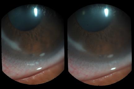
10 Soon we learnt that the best place to study corneal channels is the area in and around the arcus senilis. The semi-opacification of all grades is there to highlight the channels. The pattern of the channels is practically the same as in the case of keratoglobus shown above. The channels go right up to the lucid interval of arcus senilis. Many of the network channels end in to the lucid interval. The lucid interval in the arcus senilis is lucid because it seems to have more sinusoidal channels than elsewhere. An optical section of a lucid interval shall show a roughly triangular appearance, the base being towards the periphery with semi-opaque plates of the corneal tissue on both sides.

11

12 Figs 7a and b: A 82 years old patient showing a profusion of large and small network channels. Optical section shows that the wider channels are situated in the deeper layers and they have connections anteriorly. It is inconceivable that there should exist channels that drain no where. Thus the corneal channels can sometimes be seen to very clearly connected to the peripheral corneal channel, at regular intervals. Around 3 dozen such connections can be easily seen.

13 Fig.8: Network of corneal channels. Notice a periodic merging of the channels in to the channel of the lucid interval. The lucid interval channel also has to drain somewhere. For this we have a profusion of limbal lymphatics.

14 Fig.9. Optical section through the corneal periphery.the arcus senilis shows a prominent lucid interval.fine channels in the arcus and lymphatics at the edge of the lucid interval are also visible. Are there finer channels still, which escaped notice thus far? Probably. If we examine cases of non-descript keratitis under high magnification. We see very fine feathery appearances. If these are the finest ramifications of the corneal sinusoidal channels, they could establish most intimate contact with every element of the corneal tissue.

15 Fig.10 The finest chinks in the corneal structure get visible in a variety of stromal conditions.
16 Where and how does the fluid go out of the cornea? There is a myriad of lymphatic channels sitting on the limbus. Their corneal ends disappear in to the corneal periphery and their proximal ends merge in to the conjunctival lymphatics. It is not far fetched to say that there is a continuity between the corneal sinusoidal system and the limbal and conjunctival lymphatics. One can liken whole of the eyeball as a sponge, the porosity of the channels varying from one tissue to the other. We have been trained to study the tissues on histopathology. When we do not find a structure, it is supposed not to exist. The corneal channels can be studied at present only in vivo under a slit lamp microscope.

17 Fig.10: Showing numerous lymphatics over the arcus senilis and at the edge of the lucid interval.

18 Fig.11:A picture of the limbus showing intimate relationship between the limbal lymphatics,and channels in lucid interval and arcus senilis Relationship to the intraocular aqueous:
19 When such a dense network exists close to the limbus right on top of canal of Schlemm, it is inconceivable that that connections do not exist between them. Uptil now we do not have a photographic proof of interconnecting channels. But we have a clinical situation that points to the existence of that connection as follows. The cornea becomes edematous in cases of acute glaucoma. The moment the pressure is released by paracentesis, the cornea clears up within seconds. This miraculous change in water logging could not occur without the assumed channels and their connections to the source of raised intraocular pressure. Why is it that many advanced cases of open angle glaucoma, with intraocular pressure as high as 50 or more do not show corneal edema. It is possible that the channels system has been adapting and increasing the flow along the channels, so that the cornea remains transparent, while the optic disc is getting damaged all the while. Another point : What is the function of limbal capillaries? Don t they function the way they function in the rest of the body? We should consider a regular formation of lymph all round the cornea that nourishes the cornea through a network of channels, that we are considering. This line of thinking makes the fluid movement a dynamic activity, making the cornea a living and throbbing entity, rather than a piece of transparent tissue. A clinical proof of a peripheral channel:
20 Over 6 years ago, a 25 years old patient showed an unusual infection, in that the hypopeon level was not horizontal, but oblique. The infection appeared to extend to the level, where a star has been marked. I felt very uneasy about this patient. He was put on maximum local and oral medical both for fungal and bacterial infections. The patient was recalled after 24 hours. It was found that the infection had extended further along a predetermined path, i.e. along the peripheral corneal channel. This was my first clinical encounter with the pathology in the area of the lucid interval. I had to go abroad for a meeting. I referred the patient to PGI Chandigarh as an emergency, giving my impressions of the nature of the infection. The patient never came back to me, so I am not sure where he got further treatment and what was the final result.

21 Fig.12: A strange obliquely placed collection of exudates in the corneal periphery. The central side has a sharp edge. The opacity shows less opacification towards the center. Small satellite opacities are seen towards 3 O clock. The end of the process is marked with a star.

22 Fig.13: Within 24 hours, the picture has worsened dramatically. The hypopeon has a duplicated form towards 3 O clock.

23 Fig.14:If we look carefully, the infection has reached to almost 12 O clock in 24 hours time. Considering the speed at which the infection was spreading, I doubt if this eye got saved. No antibiotic could inside a space, in which the fluid was not showing any movement. Its natural course would have been a ring abscess leading to ring ulcer and its extension towards the center and sloughing of the cornea. Recent experience with similar cases: Forewarned with this kind of infection, it has been possible to save a couple of eyes, by treating the patients in ways different than before. One example is given below.
24 A 40 years old female came with the complaints of pain and redness in the eye of 4 days duration. She had not taken any treatment before.there was no corneal ulcer, but a definite streak of infection could be seen in the peripheral corneal channel. The exudates were broken in between, but a connection between them was there for sure. Going by the previous experience of a failed treatment, a more form of treatment was planned.conjunctival smear no kind of infection. It is possible that the source of infection was systemic, or the corneal route had closed. The lucid interval channel was injected at 12 O clock towards 3 O clock with 1 % voriconazole once.the lower infected crescent was opened with a hockey stick knife and the material removed was tested.it showed fungus. An intensive treatment with local voriconzole and supportive antibiotic drops was started, along with oral itraconazole 150 mg twice a day. The condition was considered as healed after a treatment of 30 days. Her visual acuity was saved at 6/6.

25 !

26 Fig.15: The infected peripheral corneal circular space, on the day of presentation. The infection has spread widely. Notice that the eye does not show much congestion.

27 Fig 16:The lower part of the peripheral corneal space has been opened and subconjunctival voriconazole and moxifloxacin given. The eye apparently appears in worst shape that before.


28 !

29 Fig.17: The condition of the eye 15 days from the start of the treatment.

30 Fig.18: The eye at the end of one month treatment. It is possible that had we not opened the infected peripheral corneal channel, this eye would have been lost from ring abscess and ulcer leading to rapid sloughing of the cornea.in such cases the central corneal sloughing occurs, since it loses all fluid moving and circulating connections with the limbus. To sum up:
31 The concept of corneal channels, dynamically connected to the anterior chamber through corneal endothelium on one side and connected to general circulation through peripheral circular corneal channel ( call it Singh channel) and limbal and conjunctival channels is based on solid photographically recorded observations. One should not lose sight of them when faced with any kind of corneal pathology. References: Singh, D., et al. The Conjunctival Lymphatic System. Annals of Ophthalmology. 2003;35, 2;
32
Relationship between limbal incisions. angle. and the structures of the anterior chamber
 Brit. _7. Ophthal. (I 973) 57, 722 Relationship between limbal incisions and the structures of the anterior chamber angle MOHAMED I. AYOUB AND AHMED H. SAID Department of Ophthalmology, Faculty of Medicine,
Diabetic retinopathy damage to the blood vessels in the retina. Cataract clouding of the eye s lens. Cataracts develop at an earlier age in people
 Diabetic Retinopathy What is diabetic eye disease? Diabetic eye disease refers to a group of eye problems that people with diabetes may face as a complication of diabetes. All can cause severe vision loss
Eye Care for Animals Micki Armour VMD DACVO THE CORNEA
 Eye Care for Animals Micki Armour VMD DACVO THE CORNEA ANATOMY 0.5-0.6mm thick 4 primary layers Epithelium (5-7 cell layers) Stroma (90% total thickness) Descemet s membrane Endothelium (1 layer) ANATOMY-
_ Assessment of the anterior chamber. Review of anatomy of the angle
 Assessment of the anterior chamber Dr Simon Barnard PhD BSc FCOptom FAAO DCLP Department of Optometry & Visual Science City University London, UK Review of anatomy of the angle Figure 1. Anatomical section
VISIONCARE S IMPLANTABLE MINIATURE TELESCOPE (by Dr. Isaac Lipshitz)
") PATIENT INFORMATION BOOKLET PAGE 1 OF 32 VISIONCARE S IMPLANTABLE MINIATURE TELESCOPE (by Dr. Isaac Lipshitz) AN INTRAOCULAR TELESCOPE FOR TREATING SEVERE TO PROFOUND VISION IMPAIRMENT DUE TO BILATERAL
PATIENT INFORMATION BOOKLET PAGE 1 OF 32 VISIONCARE S IMPLANTABLE MINIATURE TELESCOPE (by Dr. Isaac Lipshitz) AN INTRAOCULAR TELESCOPE FOR TREATING SEVERE TO PROFOUND VISION IMPAIRMENT DUE TO BILATERAL
Senile: flattening of vertical meridian, thinning of periphery, lack of luster
 Pterygia Etiology: triangular, fibrovascular, connective tissue overgrowths of bulbar conjunctiva onto cornea; distribution of ultraviolet energy- heat, wind, dust, dry atmosphere,higher prevalence nearer
THE CHRONIC GLAUCOMAS
 THE CHRONIC GLAUCOMAS WHAT IS GLAUCOMA? People with glaucoma have lost some of their field of all round vision. It is often the edge or periphery that is lost. That is why the condition can be missed until
THE CHRONIC GLAUCOMAS
 THE CHRONIC GLAUCOMAS WHAT IS GLAUCOMA People with glaucoma have lost some of their field of all round vision. It is often the edge or periphery that is lost. That is why the condition can be missed until
 measure of your overall performance. An isolated glucose test is helpful to let you know what your sugar level is at one moment, but it doesn t tell you whether or not your diabetes is under adequate control
PRE-DESCEMET S ENDOTHELIAL KERATOPLASTY (PDEK) DR ASHVIN AGARWAL
 DR ASHVIN AGARWAL") PRE-DESCEMET S ENDOTHELIAL KERATOPLASTY (PDEK) DR ASHVIN AGARWAL Endothelial keratoplasty (EK) has evolved at a brisk pace and the volume of data accumulated over the past 10 years has demonstrated that
PRE-DESCEMET S ENDOTHELIAL KERATOPLASTY (PDEK) DR ASHVIN AGARWAL Endothelial keratoplasty (EK) has evolved at a brisk pace and the volume of data accumulated over the past 10 years has demonstrated that
Meet Libby. Corneal Dysgenesis, Degeneration, and Dystrophies Definitions. Dr. Victor Malinovsky
 Meet Libby Corneal Dysgenesis, Degeneration, and Dystrophies 2006 Dr. Victor Malinovsky Definitions Dysgenesis: (congenital anomalies) A development disorder that results in a congenital malformation of
Glaucoma. Cornea. Iris
 Glaucoma Introduction Glaucoma is a group of eye diseases that can lead to blindness if not treated. Openangle glaucoma, the most common form of glaucoma, affects about 3 million Americans. Half of those
Glaucoma. What is glaucoma? Eye Words to Know. What causes glaucoma?
 2014 2015 Glaucoma What is glaucoma? Glaucoma is a disease that damages your eye s optic nerve. It usually happens when fluid builds up in the front part of your eye. That extra fluid increases the pressure
Written by Administrator Wednesday, 13 January :27 - Last Updated Thursday, 21 January :34
 angle closure glaucoma A type of glaucoma caused by a sudden and severe rise in eye pressure. Occurs when the pupil enlarges too much or too quickly, and the outer edge of the iris blocks the eye s drainage
Journal of Ophthalmic Medical Technology. Fuchs Dystrophy Amy Hischier
 Journal of Ophthalmic Medical Technology Volume 8, Number 1 October 2013 www.jomtonline.com Fuchs Dystrophy Amy Hischier Patient History: A 55 year old female complained that both of her eyes were red,
LEUKAEMIA*t INFILTRATION OF THE IRIS IN CHRONIC LYMPHATIC. pattemn * Received for pubiication November io, i967.
 Brit. J. Ophthal. (1968) 52, 781 INFILTRATION OF THE IRIS IN CHRONIC LYMPHATIC LEUKAEMIA*t BY BRIAN MARTIN The General Infirmary, Leeds OCULAR involvement is common in the leukaemias though the anterior
2/26/2017. Sameh Galal. M.D, FRCS Glasgow. Lecturer of Ophthalmology Research Institute of Ophthalmology
 Sameh Galal M.D, FRCS Glasgow Lecturer of Ophthalmology Research Institute of Ophthalmology No financial interest in the subject presented 1 Managing cataracts in children remains a challenge. Treatment
Clinical Commissioning Policy Proposition: Keratoprosthesis for corneal blindness
 Clinical Commissioning Policy Proposition: Keratoprosthesis for corneal blindness Reference: NHS England 1618 First published: Month Year Prepared by NHS England Specialised Services Clinical Reference
Some of the ophthalmic surgeries
 Some of the ophthalmic surgeries Some of the ophthalmic surgeries performed at the DMV Center. This document presents some types of the surgeries performed by the ophthalmology service at the DMV veterinary
PATIENT INFORMATION ON CORNEAL GRAFT
 PATIENT INFORMATION ON CORNEAL GRAFT (TRANSPLANT) SURGERY M ANANDAN What is the cornea? The clear window of the eye approximately 0.5mm thick and 12mm across. It lies in front of the fluid filled anterior
Corneal blood staining after hyphaema
 Brit. J_. Ophthal. (I 972) 56, 589 after hyphaema J. D. BRODRICK Sheffield has been described as a rare complication of contusion injury in which a hyphaema of relatively long duration and a raised intraocular
SAFE, PERMANENT EYE-COLOR CHANGE
 SAFE, PERMANENT EYE-COLOR CHANGE Prepared by Gregg Homer JSD (PhD) February 1, 2012 THE PIGMENTARY GLAUCOMA ISSUE Glaucoma Defined Glaucoma is currently defined as a disturbance of the structural or functional
AQUEOUS VEINS IN RABBITS*
 Brit. J. Ophthal., 35, 119. AQUEOUS VEINS IN RABBITS* BY D. P. GREAVES AND E. S. PERKINS Institute of Ophthalmology, London Director of Research, Sir Stewart Duke-Elder IN the course of investigations
n Corneal epithelium is derived from surface ectoderm n Composed of stratified squamous epith. n 5% of total corneal thickness (50-90micro m thick)
") Cornea overview Dr. Sarita Tuladhar MD, Ophthalmology Gandaki Medical College Embryology CORNEA: n Corneal epithelium is derived from surface ectoderm n Corneal stroma, descement memb, bowman s layer,
Cornea overview Dr. Sarita Tuladhar MD, Ophthalmology Gandaki Medical College Embryology CORNEA: n Corneal epithelium is derived from surface ectoderm n Corneal stroma, descement memb, bowman s layer,
CORNEAL CONDITIONS CORNEAL TRANSPLANTATION
 GENERAL INFORMATION CORNEAL CONDITIONS CORNEAL TRANSPLANTATION WHAT ARE CORNEAL CONDITIONS? The cornea is the clear outer layer of the eye. Shaped like a dome, it helps to protect the eye from foreign
CASE PRESENTATION BY Dr. Prashanti OPHTHALMOLOGY Ist YR
 CASE PRESENTATION BY Dr. Prashanti OPHTHALMOLOGY Ist YR PERSONAL DETAILS NAME : xxx AGE :57 SEX : Male IP/OP NO- 20170828623 OCCUPTION : Farmer CHIEF COMPLAINTS Redness Pain Watering Blurring of vision
OPACIFICATION IN PERFORATING CORNEAL GRAFTS*t
 Brit. J. Ophthal. (1954) 38, 10. OPACIFICATION IN PERFORATING CORNEAL GRAFTS*t BY A. G. LEIGH From the Institute of Ophthalmology, London As the success ofa grafting operation ultimately depends upon the
PLATE 34. (Received for publication, June 6, 1921.)
") Published Online: 1 November, 1921 Supp Info: http://doi.org/10.1084/jem.34.5.435 Downloaded from jem.rupress.org on October 18, 2018 REMOTE RESULTS OF COMPLETE HOMOTRANSPLAN- TATION OF THE CORNEA, BY
Published Online: 1 November, 1921 Supp Info: http://doi.org/10.1084/jem.34.5.435 Downloaded from jem.rupress.org on October 18, 2018 REMOTE RESULTS OF COMPLETE HOMOTRANSPLAN- TATION OF THE CORNEA, BY
XUE HUI Department of Histology& Embryology, Basic Medicine College of Jilin University
 SENSE ORGAN XUE HUI Department of Histology& Embryology, Basic Medicine College of Jilin University EYE fibrous globe lens photosensitive cells a system of cells and nerves concentric layers the sclera
The Orbit. The Orbit OCULAR ANATOMY AND DISSECTION 9/25/2014. The eye is a 23 mm organ...how difficult can this be? Openings in the orbit
 The eye is a 23 mm organ...how difficult can this be? OCULAR ANATOMY AND DISSECTION JEFFREY M. GAMBLE, OD COLUMBIA EYE CONSULTANTS OPTOMETRY & UNIVERSITY OF MISSOURI DEPARTMENT OF OPHTHALMOLOGY CLINICAL
Photodynamic therapy for IMMK in horses
 Photodynamic therapy for IMMK in horses Overview Immune mediated keratitis in horses Traditional treatment options Sustained release implant Photodynamic therapy Use in veterinary medicine Treatment for
is the clear, transparent part at the front of the eye. It allows light to enter the eye and it also refracts (focuses) the light onto the retina.
 the light onto the retina.") Senses- Vision Light is a small part (1/70th) of the total electromagnetic (EM) spectrum. The EM band extends from radio waves at one extreme to x-rays at the other. The eye detects light and converts
Senses- Vision Light is a small part (1/70th) of the total electromagnetic (EM) spectrum. The EM band extends from radio waves at one extreme to x-rays at the other. The eye detects light and converts
02/03/2014. Average Length: 23mm (Infant ~16mm) Approximately the size of a quarter Volume: ~5mL
 Approximately the size of a quarter Volume: ~5mL") Identify the anatomy of the eye. Explain the basic physiology of the parts of the eye. Briefly discuss various surgeries related to different parts of the anatomy. Average Length: 23mm (Infant ~16mm) Approximately
Identify the anatomy of the eye. Explain the basic physiology of the parts of the eye. Briefly discuss various surgeries related to different parts of the anatomy. Average Length: 23mm (Infant ~16mm) Approximately
Eye Fluids. Dr. Mohamed Saad Daoud
 Eye Fluids 1 Reference Books: Text Book of Medical physiology (Guyton and Hall) Eleventh edition 2 Fluid System of the Eye (Intraocular Fluid) The eye is filled with intraocular fluid, which maintains
generic name brand name duration
 SLIT LAMP MICROSCOPY SKILL SESSION The evaluation of all eye complaints begins with the history. All patients with eye complaints should have their visual acuity checked. Tetanus status R/O alkali/chemical
Retinal Tear and Detachment
 Retinal Tear and Detachment Introduction The retina is the layer of tissue in the back of the eye that is responsible for vision. It is attached to the choroid tissue, which supplies the retina with blood.
arthritis "Contact lens" cornea in rheumatoid (opposite). Brit. J. Ophthal. (I970) 54, 410 Peterborough District Hospital
. Brit. J. Ophthal. (I970) 54, 410 Peterborough District Hospital") Brit. J. Ophthal. (I970) 54, 410 "Contact lens" cornea in rheumatoid arthritis A. J. LYNE Peterborough District Hospital It has been noted that patients suffering from long-standing rheumatoid arthritis
Brit. J. Ophthal. (I970) 54, 410 "Contact lens" cornea in rheumatoid arthritis A. J. LYNE Peterborough District Hospital It has been noted that patients suffering from long-standing rheumatoid arthritis
Diabetes & Your Eyes
 Diabetes & Your Eyes Diabetes is a disease that occurs when the pancreas does not secrete enough insulin or the body is unable to process it properly. Insulin is the hormone that regulates the level of
Retinopathy Of Prematurity (or) Retrolental Fibroplasia )
 Retrolental Fibroplasia )") Medical Information Document On Retinopathy Of Prematurity (or) Retrolental Fibroplasia ) What we see is made in the brain from signals given to it by the eyes. What we see is in fact made in the brain.
Medical Information Document On Retinopathy Of Prematurity (or) Retrolental Fibroplasia ) What we see is made in the brain from signals given to it by the eyes. What we see is in fact made in the brain.
Histology of the Eye
 Histology of the Eye Objectives By the end of this lecture, the student should be able to describe: The general structure of the eye. The microscopic structure of:»cornea.»retina. EYE BULB Three coats
The Sense Organs 10/13/2016. The Human Eye. 1. Sclera 2. Choroid 3. Retina. The eye is made up of three layers:
 The human body gathers information from the outside world by using the five senses of: The Sense Organs 12.3 Sight Hearing Taste Smell Touch This information is essential in helping the body maintain homeostasis.
Unit VIII Problem 8 Anatomy: Orbit and Eyeball
 Unit VIII Problem 8 Anatomy: Orbit and Eyeball - The bony orbit: it is protecting our eyeball and resembling a pyramid: With a base directed: anterolaterally. And an apex directed: posteromedially. Notes:
Malawi. Ocular leprosy in. Clinical and therapeutic survey of 8,325 leprosy. patients
 Brit. 5. Ophthal. (I 970) 54, I 07 Ocular leprosy in Malawi Clinical and therapeutic survey of 8,325 leprosy patients U. TICHO AND I. BEN SIRA Eye Department, Hadassah Medical Centre, Jerusalem, Israel
Understanding Glaucoma
 Understanding Glaucoma What is glaucoma? Glaucoma is the name for a group of eye conditions in which the optic nerve is damaged at the point at which it leaves the eye. As the diagram below shows, this
Ophthalmology. Glaucoma
 Ophthalmology Glaucoma The Ophthalmology service offers the latest and most comprehensive eye care for patients. With a dedicated team of eye surgeons and consultants, we treat vision problems ranging
IMAGE OF THE MOMENT PRACTICAL NEUROLOGY
 178 PRACTICAL NEUROLOGY IMAGE OF THE MOMENT Gawn G. McIlwaine*, James H. Vallance* and Christian J. Lueck *Princess Alexandra Eye Pavilion, Chalmers Street, Edinburgh UK; The Canberra Hospital, P.O. Box
A Case of Carotid-Cavernous Fistula
 A Case of Carotid-Cavernous Fistula By : Mohamed Elkhawaga 2 nd Year Resident of Ophthalmology Alexandria University A 19 year old male patient came to our outpatient clinic, complaining of : -Severe conjunctival
THE RED EYE Cynthia McNamara, MD Week 25
 THE RED EYE Cynthia McNamara, MD Week 25 Educational Objectives: 1. Know the differential diagnosis and presentation of specific etiologies of the red eye 2. Be able to evaluate patients presenting with
Frequently Asked Questions about General Ophthalmology:
 1. Normal Eye Structure The eye is a slightly asymmetrical globe, about an inch in diameter. The parts of the eye include: Cornea (a clear dome over the iris), Iris (the pigmented part); Pupil (the black
Optometric Postoperative Cataract Surgery Management
 Financial Disclosures Optometric Postoperative Cataract Surgery Management David Dinh, OD Oak Cliff Eye Clinic Dallas Eye Consultants March 10, 2015 Comanagement Joint cooperation between two or more specialists
Lamellar Keratoplasty for the Treatment of Fungal Keratitis
 Cornea 21(1): 33 37, 2002. 2002 Lippincott Williams & Wilkins, Inc., Philadelphia Lamellar Keratoplasty for the Treatment of Fungal Keratitis Lixin Xie, M.D., Weiyun Shi, M.D., Zhaosheng Liu, M.D., and
PRECISION PROGRAM. Injection Technique Quick-Reference Guide. Companion booklet for the Video Guide to Injection Technique
 Injection Technique Quick-Reference Guide PRECISION PROGRAM Companion booklet for the Video Guide to Injection Technique Available at www.ozurdexprecisionprogram.com Provides step-by-step directions with
CHOLESTEROL CRYSTALS IN THE ANTERIOR CHAMBER*
 Brit. J. Ophthal. (1963) 47, 295. CHOLESTEROL CRYSTALS IN THE ANTERIOR CHAMBER* BY Muslim University Institute of Ophthalmology. Aligarh, India THE occurrence of crystals in the anterior chamber of the
Specialist Referral Service Willows Information Sheets. Cataract surgery
 Specialist Referral Service Willows Information Sheets Cataract surgery An operating microscope in use A total cataract - the normally black pupil is bluish white Cataract surgery These notes do not cover
An Injector s Guide to OZURDEX (dexamethasone intravitreal implant) 0.7 mg
 0.7 mg") An Injector s Guide to OZURDEX (dexamethasone intravitreal implant) 0.7 mg This guide is intended to provide injectors with information on the recommended injection technique and the important risks related
An Injector s Guide to OZURDEX (dexamethasone intravitreal implant) 0.7 mg This guide is intended to provide injectors with information on the recommended injection technique and the important risks related
INTRA-CORNEAL LAMELLAR KERATOPLASTY*
 Brit. J. Ophthal. (1960) 44, 629. INTRA-CORNEAL LAMELLAR KERATOPLASTY* BY TADEUSZ KRWAWICZ Ophthalmological Clinic, Medical Academy, Lublin, Poland THE operative technique of lamellar keratoplasty is still
Vision I. Steven McLoon Department of Neuroscience University of Minnesota
 Vision I Steven McLoon Department of Neuroscience University of Minnesota 1 Eye Cornea Sclera Conjunctiva 2 Eye The conjunctiva lines the inner surface of the eyelids and outer surface of the sclera. 3
Factsheet. Glaucoma. Are there different types of glaucoma? Yes. There are four main types.
 What is glaucoma? Glaucoma is the name for a group of eye conditions in which the optic nerve is damaged at the point where it leaves the eye. This nerve carries information from the light sensitive layer
A LITTLE ANATOMY. three layers of eye: 1. outer: corneosclera. 2. middle - uvea. anterior - iris,ciliary body. posterior - choroid
 GLAUCOMA A LITTLE ANATOMY three layers of eye: 1. outer: corneosclera 2. middle - uvea anterior - iris,ciliary body posterior - choroid connection at the pars plana between post and ant uvea 3. retina
Glaucoma What You Should Know
 Glaucoma What You Should Know U.S. DEPARTMENT OF HEALTH AND HUMAN SERVICES National Institutes of Health National Eye Institute The National Eye Institute (NEI) conducts and supports research that leads
Diabetic Retinopathy WHAT IS DIABETIC RETINOPATHY? WHAT CAUSES DIABETIC RETINOPATHY? WHAT ARE THE STAGES OF DIABETIC RETINOPATHY?
 Diabetic Retinopathy WHAT IS DIABETIC RETINOPATHY? Diabetic retinopathy affects 8 million Americans with diabetes. A leading cause of blindness in American adults, it is caused by damage to the small blood
Glaucoma Glaucoma is a complication which has only recently been confirmed as a feature of
 1.2.4 OPHTHALMOLOGICAL ABNORMALITIES Ocular abnormalities are well documented in patients with NPS 6 62 81 95. 1.2.4.1 Glaucoma Glaucoma is a complication which has only recently been confirmed as a feature
CORNEAL TRANSPLANT CONSENT FORM
 CORNEAL TRANSPLANT CONSENT FORM Peninsula Laser Eye Medical Group 1174 Castro Street, Ste. 100 Mountain View, CA 94040 (650) 961-2585 www.lasik2020.com Introduction The cornea is the clear dome-shaped
4/22/16. Eye. External Anatomy of Eye. Accessory Structures. Bio 40B Dr. Kandula
 Eye Bio 40B Dr. Kandula External Anatomy of Eye Accessory Structures l Eyebrows l Levator Palpebrae Superioris - opens eye l Eyelashes l Ciliary glands modified sweat glands l Small sebaceous glands l
The Special Senses: Part A
 PowerPoint Lecture Slides prepared by Janice Meeking, Mount Royal College CHAPTER 15 The Special Senses: Part A Warm Up What is the function of the eyeball? List any structures of the eyeball that you
Diffuse infiltrating retinoblastoma
 Brit. 1. Ophthal. (I 971) 55, 6oo Diffuse infiltrating retinoblastoma GWYN MORGAN Department of Pathology, Institute of Ophthalmology, University of London The term "diffuse infiltrating retinoblastoma"
Structure of the eye and retina
 1 of 10 3/6/2012 1:06 PM Syllabus pdf file Course Schedule Structure of the eye and retina 2 of 10 3/6/2012 1:06 PM In-class demo: do Virtual Lab activity 3-6 (Visual Path in the Eyeball) Focusing, changes
THE EYE: RETINA AND GLOBE
 Neuroanatomy Suzanne Stensaas February 24, 2011, 10:00-12:00 p.m. Reading: Waxman Ch. 15. Your histology and gross anatomy books should be useful. Reading: Histology of the Eye from any histology book
HYPERPLASIA OF THE ANTERIOR LAYER OF THE IRIS STROMA*t
 Brit. J. Ophthal. (1965) 49, 516 HYPERPLASIA OF THE ANTERIOR LAYER OF THE IRIS STROMA*t BY MALCOLM N. LUXENBERG From the Bascom Palmer Eye Institute, Department of Ophthalmology, University of Miami School
Around The Globe in 60 Minutes
 Around The Globe in 60 Minutes Around the GLOBE in Sixty Minutes Basic Ocular Anatomy, Examination, and Diagnostic Techniques Introduction Focusing on canine and feline ocular anatomy and basic examination
Your Ophthalmologist has prescribed you. Poly (carboxymethylglucose sulfate) Medical device. Patient Information
 Medical device. Patient Information") Your Ophthalmologist has prescribed you Poly (carboxymethylglucose sulfate) Medical device Patient Information Why is it so important to treat diseases of the cornea? The cornea plays a major role in sight.
Your Ophthalmologist has prescribed you Poly (carboxymethylglucose sulfate) Medical device Patient Information Why is it so important to treat diseases of the cornea? The cornea plays a major role in sight.
INTRODUCTION: ****************************************************************************************************
 BIOLOGY 211: HUMAN ANATOMY & PHYSIOLOGY **************************************************************************************************** EYES AND VISION ****************************************************************************************************
 Traumatic Cataract Orbital Wall Fracture Vitreous Hemorrhage Optic Disc Hemorrhage a) Amblyopia b) Strabismus c) Trauma Playing with other children Sports Fire works BB gun Injecting needles .
Specialist Referral Service Willows Information Sheets. Lens luxation
 Specialist Referral Service Willows Information Sheets Lens luxation A dislocated (luxated) lens in the front chamber of the eye. The arrows mark the edge of the lens Lens luxation What is the lens? The
Dr Jo-Anne Pon. Dr Sean Every. 8:30-9:25 WS #70: Eye Essentials for GPs 9:35-10:30 WS #80: Eye Essentials for GPs (Repeated)
") Dr Sean Every Ophthalmologist Southern Eye Specialists Christchurch Dr Jo-Anne Pon Ophthalmologist Southern Eye Specialists, Christchurch Hospital, Christchurch 8:30-9:25 WS #70: Eye Essentials for GPs
Dr Sean Every Ophthalmologist Southern Eye Specialists Christchurch Dr Jo-Anne Pon Ophthalmologist Southern Eye Specialists, Christchurch Hospital, Christchurch 8:30-9:25 WS #70: Eye Essentials for GPs
Acute Eyes for ED. Enis Kocak. The Alfred Ophthalmology
 Acute Eyes for ED Enis Kocak The Alfred Ophthalmology The problem with eyes Things to cover Ocular anatomy Basic assessment Common presentations Eye first aid and procedures Ophthalmic emergencies What
NEPTUNE RED BANK BRICK
 NEPTUNE RED BANK BRICK Diabetes & The Eye Diabetics are more likely to develop Cataracts at a younger age. Diabetics are twice as likely to develop Glaucoma when compared to non-diabetics. The primary
Vascular changes in the iris in chronic
 Vascular changes in the iris in chronic anterior uveitis LEILA LAATIKAINEN From the Department of Ophthalmology, University of Helsinki, Finland British Journal of Ophthalmology, 1979, 63, 145-149 SUMMARY
GENERAL INFORMATION GLAUCOMA GLAUCOMA
 GENERAL INFORMATION GLAUCOMA GLAUCOMA WHAT IS GLAUCOMA? Glaucoma is commonly known as the sneak thief of sight because it can cause irreversible vision loss without any obvious symptoms. The term glaucoma
RNIB UNDERSTANDING GLAUCOMA
 RNIB UNDERSTANDING GLAUCOMA Eye Info Understanding glaucoma Summary: Designed to help you understand more about your eye condition, this guide has been written by our experienced eye health team. What
STAB INCISION GLAUCOMA SURGERY (SIGS)
") STAB INCISION GLAUCOMA SURGERY (SIGS) Dr. Soosan Jacob, MS, FRCS, DNB Senior Consultant Ophthalmologist, Dr. Agarwal's Eye Hospital, Chennai, India dr_soosanj@hotmail.com Videos available in Youtube channel:
STAB INCISION GLAUCOMA SURGERY (SIGS) Dr. Soosan Jacob, MS, FRCS, DNB Senior Consultant Ophthalmologist, Dr. Agarwal's Eye Hospital, Chennai, India dr_soosanj@hotmail.com Videos available in Youtube channel:
Distinction layer by layer. HRT II Rostock Cornea Module
 Distinction layer by layer HRT II Rostock Cornea Module Homogenously illuminated, undistorted images Movie capture Manual Pachymetry Epithelial and intra-corneal pachymetry Full corneal thickness Post-LASIK
By Darlene Jones, Nurse. May 2017
 By Darlene Jones, Nurse May 2017 Disclosure of potential conflict of interest Darlene Jones, Nurse I have no conflict of interest Course objectives Become familiar with the different pathologies in ophthalmology
Descemet s membrane endothelial keratoplasty (DMEK) surgery
 surgery") Patient information Descemet s membrane endothelial keratoplasty (DMEK) surgery This information leaflet tells you what to expect if you have DMEK surgery an operation on the cornea of the eye along with
Patient information Descemet s membrane endothelial keratoplasty (DMEK) surgery This information leaflet tells you what to expect if you have DMEK surgery an operation on the cornea of the eye along with
ADVANCED DIAGNOSTIC TECHNIQUES
 DIVISION OF VISION SCIENCES SESSION: 2008/2009 DIET: 1ST ADVANCED DIAGNOSTIC TECHNIQUES VISP216 LEVEL:2 MODULE LEADER: DR GUNTER LOFFLER B.Sc/B.Sc. (HONS) OPTOMETRY MAY 2009 DURATION: 2 HOURS CANDIDATES
Cataract. A cataract is a clouding of the lens in your eye. It
 Cataract A cataract is a clouding of the lens in your eye. It affects your vision. Cataracts are very common in older people. By age 80, more than half of all Americans either have a cataract or have had
Infra-red transillumination stereophotography of the iris in Fuchs's heterochromic cyclitis
 British Journal of Ophthalmology, 1978, 62, 110-115 Infra-red transillumination stereophotography of the iris in Fuchs's heterochromic cyclitis M. SAARI, I. VUORRE, AND H. NIEMINEN From the University
ASSESSING THE EYES. Structures. Eyelids Extraocularmuscles Eyelashes Lacrimal glands: Lacrimal ducts Cornea Conjunctiva Sclera Pupils Iris.
 ASSESSING THE EYES Structures External Eyelids Extraocularmuscles Eyelashes Lacrimal glands: Lacrimal ducts Cornea Conjunctiva Sclera Pupils Iris 1 2 Structures Internal Optic disc Physiological cup Retinal
PAINFUL PAINLESS Contact lens user BOV
 Common Causes Allergies Infections Ocular Cornea, uveitis, endophthalmitis Orbital Orbital cellulitis Inflammation Uveitis Scleritis / episcleritis Glaucomas Trauma Foreign bodies Chemical injuries History
Diabetic Retinopathy Information
 http://www.midwestretina.com Phone: (614)-339-8500 Toll Free: (866)-373-8462 Sugat S. Patel, M.D. Louis J. Chorich III, M.D. Dino D. Klisovic, M.D. Lisa M. Borkowski, M.D. Dominic M. Buzzacco, M.D. Johnstone
Visian ICL (Implantable Collamer Lens) For Nearsightedness. Facts You Need To Know About STAAR Surgical s Visian ICL SURGERY
 For Nearsightedness. Facts You Need To Know About STAAR Surgical s Visian ICL SURGERY") Visian ICL (Implantable Collamer Lens) For Nearsightedness Facts You Need To Know About STAAR Surgical s Visian ICL SURGERY PATIENT INFORMATION BOOKLET For Nearsightedness (Myopia) between 3 to 20 Diopters
Visian ICL (Implantable Collamer Lens) For Nearsightedness Facts You Need To Know About STAAR Surgical s Visian ICL SURGERY PATIENT INFORMATION BOOKLET For Nearsightedness (Myopia) between 3 to 20 Diopters
Medical School Histology Basics. VIBS 289 lab. Eye
 Medical School Histology Basics VIBS 289 lab Eye Larry Johnson Texas A&M University Aqueous humor OUTLINE OVERVIEW CELLULAR STRUCTURES THROUGH WHICH LIGHT PASSES A. CORNEA B. LENS C. RETINA STRUCTURES
QUALIT ATIVE CHARACTERIZATION OF SOME PA THOLOGIC CORNEAL DISEASES USING CONTACT SPECULAR MICROSCOPY
 QUALIT ATIVE CHARACTERIZATION OF SOME PA THOLOGIC CORNEAL DISEASES USING CONTACT SPECULAR MICROSCOPY JOSE DAVID F. MARIN, JR. EVANGELINE MARION A. ABENDANIO ROSSINA LYDIA ALEJO-RAMIREZ SAL V AOOR R. SALCEDA
Subject Index. Atopic keratoconjunctivitis (AKC) management 16 overview 15
 management 16 overview 15") Subject Index Acanthamoeba keratitis, see Infective keratitis Acute allergic conjunctivitis AKC, see Atopic keratoconjunctivitis Allergy acute allergic conjunctivitis 15 atopic keratoconjunctivitis 15
Subject Index Acanthamoeba keratitis, see Infective keratitis Acute allergic conjunctivitis AKC, see Atopic keratoconjunctivitis Allergy acute allergic conjunctivitis 15 atopic keratoconjunctivitis 15
Brampton Hurontario Street Brampton, ON L6Y 0P6
 Diabetic Retinopathy What is Diabetic Retinopathy Diabetic retinopathy is one of the leading causes of blindness world-wide. Diabetes damages blood vessels in many organs of the body including the eyes.
TEST BANK FOR EBERSOLE AND HESS TOWARD HEALTHY AGING 9TH EDITION BY TOUHY
 Link full download: http://testbankair.com/download/test-bank-for-newebersole-and-hess-toward-healthy-aging-9th-edition-by-touhy/ TEST BANK FOR EBERSOLE AND HESS TOWARD HEALTHY AGING 9TH EDITION BY TOUHY
Scrub In. What is the function of vitreous humor? What does the pupil do when exposed to bright light? a. Maintain eye shape and provide color vision
 Scrub In What is the function of vitreous humor? a. Maintain eye shape and provide color vision b. Maintain eye shape and refract light rays c. Provide night vision and color vision d. Provide night vision
Ophthalmology. Cataract
 Ophthalmology Cataract The Ophthalmology service offers the latest and most comprehensive eye care for patients. With a dedicated team of eye surgeons and consultants, we treat vision problems ranging
OCCLUSIVE VASCULAR DISORDERS OF THE RETINA
 OCCLUSIVE VASCULAR DISORDERS OF THE RETINA Learning outcomes By the end of this lecture the students would be able to Classify occlusive vascular disorders (OVD) of the retina. Correlate the clinical features