Maratus personatus, a masked peacock spider from Cape Riche, Western Australia (Araneae: Salticidae: Euophryinae)
|
|
|
- Juniper Clark
- 6 years ago
- Views:
Transcription
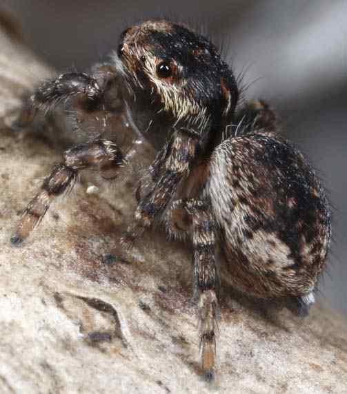
1 PECKHAMIA., 8 July 0, 0 urn:lsid:zoobank.org:pub:f8a9a-d f8090 (registered JUL 0) ISSN 8 (print) ISSN 9 80 (online), a masked peacock spider from Cape Riche, Western Australia (Araneae: Salticidae: Euophryinae) Jürgen C. Otto and David E. Hill 9 Grevillea Avenue, St. Ives, New South Wales 0, Australia, jurgenotto@optusnet.com.au Wild Horse Creek Drive, Simpsonville, SC 980-, USA, platycryptus@yahoo.com Key words: courtship, euophryine, jumping spider, Maratus, peacock spider, salticid Abstract: A new peacock spider,, is described from Western Australia. Nine new peacock spiders (Maratus Karsch 88) have been described recently from areas near the southern coast of Western Australia: M. caeruleus, M. karrie, M. melindae and M. sarahae Waldock 0; M. madelineae Waldock 0; M. avibus Otto & Hill 0a; M. pardus Otto & Hill 0b; M. maritimus and M. montanus Otto & Hill 0c. To this collection we add a distinctive new species that has been found at a single locality at Cape Riche, east of Albany., new species Type specimens. One holotype male ( #0), eight paratype males ( #-, -), and eight paratype females ( #-, 8-) will be deposited in the Western Australian Museum. Three additional males ( #-9) and four additional females ( #-, ) were examined but not preserved. #-9 and #- were collected at Cape Riche (S ', E 8 ', 8 OCT 0, coll. J. Otto); #0- and #- were the offspring of the aforementioned field-collected females, reared to adulthood. Etymology. The species group name (personatus, Latin, m., adj., English translation masked) refers to the presence of a prominent mask of deep blue scales covering the face of adult males of this species. Diagnosis. With their mask of bright blue scales, male M. personatus cannot be confused with any other Maratus. Unlike most (but not all) Maratus, M. personatus males do not rear or display their dorsal opisthosoma during courtship. However, the structure of the male pedipalp and the female epigynum, as well as the presence of a dorsal opisthosomal plate, support the generic placement of this species. Description of male (Figures -). Males (n=8) ranged from.8 to.9 mm in length.


2 #0 #0 #0 #0 #0 #0 8 9 #0 #0 #0 0 #0 #0 #0 Figure. Views of the living adult male holotype for., From the front, deep blue scales surround the blue anterior eyes, offset by white setae of the eye region, above, and the pedipalps, below., Like other Maratus, M. personatus has a dorsal opisthosomal plate with a distinct edge (arrow), as well as a triangular tuft of white colular setae.


3 # # 8 # # # # 9 # # # 0 # # # Figure. Four different living adult male M. personatus., The scales of this male have rubbed off, exposing the black cuticle of the dorsal opisthosomal plate. 9, The small chelicerae are black and glabrous, separated on either side from a marginal band of white setae.


4 # # # # # 8 # posterior leg R Figure. Four different living adult male M. personatus. 9 #8 # # anterior leg L


5 # # # # # # detail of right pedipalp # 8 Figure. Three different living adult male M. personatus. # 9 #


6 Figure. Two views of the underside of a living adult male. Coxae and trochanters of legs I and II are dark brown; those of legs III and IV are light brown or translucent. #0 #0 #0 #0 #0 Figure. Views of the adult male holotype M. personatus in ethanol., The eyes and facial scales of this specimen retained their blue colouration, suggesting that their colours are structural.


. General")


7 # # # # # # # # # # #0 #0 #0 #0 # 8 # 9 0 # # # Figure. Medial to lateral view of the left pedipalp of six different male. Separation of the inner and outer apex of the embolus can be seen in lateral views (distinctly in, less distinctly in,, 9, and 8). General features, including the shape and relative size of the embolus, the presence of medial tegular sclerotization proximal to the embolus, the pedipalp of the male M. personatus is much like those of other Maratus. #
8 8 The carapace is black in life, fading to dark brown in ethanol. The clypeus is black and glabrous, lacking the longer setae found in most Maratus. The chelicerae are also black and glabrous. The width across both chelicerae is less than / the width of the carapace. All four anterior eyes are deep blue in reflected light, each bordered with deep blue scales that match the scale cover of the entire face in colouration. The ALE are approximately / the diameter of the AME, separated from the AME on either side by about / of the diameter of an AME. The dorsal carapace including the eye region is covered with scales or setae, but appears dark and reflective, with a band of bright white scales anteriorly, and mixed white to orangebrown scales on the sides forming a band that extends to the rear along the top of the carapace, below the lateral eyes. A narrow, indistinct medial thoracic tract of white to orange-brown scales extends to the rear behind the eye region. There is a prominent white marginal band on either side, separated by a glabrous black area from the chelicerae on either side. The PME are distinctly closer to the PLE than to the ALE. A dorsal plate of the opisthosoma is present and can be identified by the presence of a distinct margin (Figure :), but this has no lateral flaps and it is neither elevated nor expanded during courtship display. Typically this plate is covered with a distinct but variable pattern of white to tan or brown scales, surrounding a dark figure (Figures :8, :, :, :), but in older individuals white scales may have rubbed off rendering the opisthosomal plate mostly black (Figure :). A white triangle comprised of bright white scales is usually associated with the colulus just above the brown to grey spinnerets. The ventral opisthosoma is mostly glabrous and brown, bordered by tracts of white setae to the front and rear, and on the sides (Figure ). The coxae and trochanters of legs III and IV are light-brown or translucent, as are the proximal femora of all legs. Coxae and trochanters of legs I and II, the sternum, the labium, and the endites are all dark brown and glabrous. Legs I and II are about the same length, much shorter than legs III and IV. Legs III are the longest. Legs I and II are strongly banded, particularly from the front, with dark bands alternating with bands of long white setae. The most prominent white bands are associated with the femuro-patellar joint, the patellartibial joint, and mid-way along the length of each tibia, where the band is somewhat diagonal (not transverse) with respect to the axis of the leg. From their proximal to distal end legs III are decorated with long white setae on the front of each otherwise brown or translucent proximal femur, followed by a longer black middle section of the femur, a light brown or translucent distal end of the femur and patella bearing long white setae, a black tibia fringed with long black setae, a brown or translucent metatarsus fringed with long white setae, barely separated distally from the tarsus by a black ring, and a brown or translucent tarsus bearing long white setae. As viewed from below (Figure ), the structure of the pedipalp is much like that of other Maratus species, with an inner an outer apex of the embolus, and sclerotization of the medial tegulum proximal to the embolus. The dorsal pedipalp (Figures :, :9) is, however, distinctive with long white fringes proximal to the cymbium, shorter black setae on the cymbium, and grey setae distally. When the pedipalps are held together in front of the chelicerae (Figure :), these fringes form a single white line that contrasts with the deep blue colour of the face. Description of female (Figures 8-). Females (n=) ranged from.8 to.8 mm in length. They closely resemble females of other Maratus with respect to shape and colouration. Chelicerae are brown, translucent and glabrous. The clypeus is brown and translucent, with long white setae directed anteromedially above the chelicerae. Ivory to light-brown scales surround the eyes in front and on the sides of the eye region, in irregular tracts extending to the rear on either side behind the PLE. The medio-dorsal carapace is mostly black to dark brown and glabrous, but a median thoracic tract of ivory to light brown scales may be present. The sides of the carapace are mostly brown and translucent, and there is no marginal band. The PME are slightly closer to the PLE than to the ALE.


9 9 # # # # # # # # 8 Figure 8. Views of three different living female. From above, these are mostly dark brown to black, with tracts of off-white or ivory scales on the sides of the carapace and opisthosoma. Two or more pairs of spots (scale patches) may be seen on the dorsal opisthosoma.


10 0 # # # # # 8 # # # # 9 #8 0 #8 Figure 9. Views of five different living female. #8


11 #0 #0 #0 # # # # Figure 0. Views of three different living female., In some individuals four distinct chevrons at the rear of the dorsal opisthosoma can be seen. In other individuals these are indistinct.


12 # # Figure. Underside of two different living female. #8 # # #8 #8 #8 Figure. Two different female in ethanol. Colours vary according to the state of preservation.


13 #0 # # # 8 #0 # # # Figure. Three different female in ethanol. # #9 #0 # #8 # Figure. Ventral view of epigynum of seven different female in ethanol. #
14 The dorsal opisthosoma varies from dark brown and glabrous in older individuals that have lost scales (Figure 0:) to dark brown covered with a pattern of off-white to brown scales (Figure 0:). For individuals with a distinct pattern, there are two pairs of distinct ivory spots (scale patches) anteriorly, and four chevrons of brown scales toward the rear. Margins of the opisthosoma are light brown, covered with off-white or ivory setae. The venter (Figure ) is light brown or tan, with a covering of white to ivory setae and scattered brown spots, and a central figure consisting of two brown lines comprised of coalesced spots, converging toward the rear. The coxae, sternum, labium, and endites are mostly glabrous, translucent, and almost colourless. Scattered, longer white setae radiate out along the margins of the sternum. All of these translucent structures appear as solid white or light yellow in ethanol. Legs I and II are shorter than legs III and IV and nearly equal in length, and leg III is the longest. The legs are distinctly banded, with dark pigmentation at the distal end of each segment, but otherwise they are light brown and translucent. The pedipalp is of similar colour. The epigynum (Figure ) is similar in general form to that of other Maratus, with prominent fossae and a pair of large posterior spermathecae. Although females collected with the distinctive males of this species varied little with respect to their general appearance and the placement of scale tracts (e.g., the paired spots and chevrons on the opisthosoma), they varied greatly with respect to the detailed structure of the epigynum. We have not observed this extent of variation in a Maratus species before, and it challenges the widely-held assumption (however see Crews 009 for counterexamples) that fine distinctions in the structure of the epigynum can be used for taxonomic purposes. Variations included separate versus contiguous spermathecae, fossae larger or much smaller than the spermathecae, narrow versus wide septum between the fossae, degree of sclerotization of the fossae, and both complexity and placement of the sclerotized ducts visible beneath the fossae. Yet both the association with and willingness to mate with males of this species in the field, the rearing of male offspring from several of these females, and the agreement of the form and appearance of developing males and females with adult females (Figures -9) confirm that they are indeed the same species. Immatures. A number of different conventions have been used to label the developmental stages of spiders, some of which identify one or more post-eclosion stages as prelarvae or larvae (Pfannenstiel 008, Mittmann & Wolff 0). Here we follow the convention of labeling the post-eclosion, pre-first moult stage as the first instar, the post-first moult stage as second instar, and so on. At the time that a salticid emerges from the egg it is soft and undeveloped with a gut filled with yolk (Figure :-). These first instars develop quickly within the egg sac (nest) with growth of the appendages and internal structures of the eyes, including the pigmented retinae is evident (Figure :-9). First instars have a pair of claws at the end of each leg and limited mobility, but appear to be blind and do not feed. After their first moult, the second instars have a working set of eyes and a pattern of setation that suggests that of the adult female (Figure ). As they continue to develop to the penultimate or th instar, both males (Figures -8) and females (Figure 9) look more and more like adult females. The enlarged pedipalps of the th instar males (Figures :, 8:, ) distinguish these from females. In both sexes the th instar is the adult stage.

, they are little")


15 8 9 Figure. Development of first instar. When these first emerge from the egg (-), they are little more than naked embryos, with short legs and unpigmented eyes that continue to grow and develop prior to the first moult. Some authors call these prelarvae. As these grow, pigment of the retinae, dorsal carapace, and opisthosoma appears, and the legs develop to the point that the spiderlings can move about in the egg sac. Although development is continuous during most of the first instar, the more developed pre-first moult spiderlings are sometimes called larvae.


 with")


16 8 9 0 Figure. Second instar. After their first moult, these spiderlings emerge from the egg-sac (nest) with well-developed eyes and footpads, and a covering of setae that suggests the appearance of an adult female., The adult female (left) is about times the length of a second instar (~. mm).


17 #0: nd #0: nd #0: nd #0: rd #0: #0: th 0 #0: th #0: rd rd #0: th 8 #0: th #0: th 9 #0: th Figure. Developmental stages of the holotype male., The penultimate male (th instar) is readily identified by its expanded pedipalps, but still has white clypeal setae like those of the female. The carapace of the rd instar is about wide.


18 #: th 8 #: th #: #: th #: th #: th th #: rd #: th #: th 8 9 #: th 0 #: th #: th Figure 8. Developmental stages of two different male.


19 #8: rd 9 #8: th #8: th #8: rd #8: #8: th #9: nd th #8: th 8 #9: th #9: th #9: rd #9: th 9 0 #0: th #0: th #0: rd #0: th #0: th #: #: th th #: rd Figure 9. Developmental stages of four different female. Note the light () and dark () colour variation of penultimates (th instar).

 when stepping from side to side in front of a female (Figures -).")

.")

20 0 Courtship display by males (Figures 0-; see Otto 0 for an online video of this display). As they display to females, males raise and wave their legs III, either unilaterally or bilaterally, but they do not raise or display their opisthosoma. Usually a male lifts and waves only the leading leg III (rate ~0-/s) when stepping from side to side in front of a female (Figures -). Side-stepping is also accompanied by movement of the pedipalps up and down. When a male holds his position during display, both legs III are extended and waved in a lateral plane, with the axis of each leg oriented in a variable direction, ranging from near horizontal (Figure :0) to vertical (Figure 0:). As it is waved, each leg may be 'flopped' at the femuro-patellar joint (Figure :-). The pedipalps may be held in a stationary position to expose the black chelicerae during this bilateral display (Figure ). # # # # # # Figure 0. Bilateral display positions by a male. As the extended and elevated legs III are waved in a transverse plane, the distal segments (beyond the femur) are often 'flopped' relative to the axis of the leg, as shown here (, ). Legs III may also be waved in a near vertical position (, ), and brought together above the spider (, ). Bilaterally symmetric displays like these are usually seen when the male is in a stationary position facing the female, and not stepping from side to side.


 and")
")


21 # # # # # # # 8 # #0 # 0 9 # #0 # # Figure. Bilateral (-, -) and unilateral (, -) display positions by six different male. When stepping from side to side (-), males raise and wave the leading leg III. Bilateral display may be brief, when the male is not stepping to one side or the other.
 video frames (FPS, exposure 0 msec/frame) showing a male stepping to the left and waving the leading leg LIII while facing a female.")
22 . 0.00s 9. 0.s. 0.s. 0.0s 0. 0.s s. 0.08s. 0.0s 9. 0.s. 0.s. 0.s 0. 0.s. 0.s. 0.8s. 0.80s. 0.0s. 0.s. 0.8s. 0.s. 0.s. 0.88s s. 0.0s. 0.9s Figure. Sequential (-) video frames (FPS, exposure 0 msec/frame) showing a male stepping to the left and waving the leading leg LIII while facing a female. To enhance comparison of position, sequential frames are arranged in columns (as in Figures -). -, During a brief (~0. s) pause in lateral movement, one wave cycle was completed, accompanied by up and down movement of the pedipalps.
 video frames (FPS, exposure 0 msec/frame) showing a male stepping to either side and waving the leading leg LIII while facing a female.")
23 . 0.00s 9. 0.s. 0.s. 0.9s. 0.0s 0. 0.s s..00s. 0.08s. 0.0s 9. 0.s..0s. 0.s. 0.s 0. 0.s 8..08s. 0.s. 0.8s. 0.80s 9..s. 0.0s. 0.s. 0.8s 0..s. 0.s. 0.s. 0.88s..0s s. 0.0s. 0.9s..s Figure. Sequential (-) video frames (FPS, exposure 0 msec/frame) showing a male stepping to either side and waving the leading leg LIII while facing a female. -, Bilateral display from a fixed position in front of the female. -, Unilateral display of the leading right leg III while stepping to the right. -0, Bilateral display in position., Unilateral display of the leading left leg III while stepping to the left.
 but non-consecutive video frames showing a male M.")
24 . 0.00s. 0.0s. 0.s s. 0.s s. 0.s 0..0s. 0.s..8s. 0.s..s Figure. Sequential (-) but non-consecutive video frames showing a male M. personatus displaying to a female (foreground) while stepping to the right.
 but non-consecutive video frames showing a male M.")
25 . 0.00s. 0.s. 0.08s s. 0.s 9. 0.s. 0.0s 0. 0.s. 0.8s. 0.9s. 0.s..00s Figure. Sequential (-) but non-consecutive video frames showing a male M. personatus displaying to a female (foreground) while stepping to the left.
26 . 0.00s. 0.0s. 0.08s. 0.s. 0.s. 0.0s. 0.s s 9. 0.s 0. 0.s. 0.0s. 0.s. 0.8s. 0.s. 0.s. 0.0s. 0.s s 9. 0.s 0. 0.s Figure. Sequential (-8, continued on next two pages) video frames (FPS, exposure 0 msec/frame) showing a male displaying to a female from one position. Here consecutive frames are arranged in rows., Movement of legs III from a horizontal to a vertical orientation was recorded here in a single frame (estimated duration 0-0 msec). Note the constant movement of legs III from frame to frame, including 'floppy' flexion at the femuro-patellar joint (, 8). During pauses between rapid leg movement, legs III were generally in one of three positions: near-horizontal, near-vertical, and intermediate (V-shaped position). There was little visible movement of the pedipalps during this sequence, as the pedipalps were held in a lower position to expose the black, glabrous clypeus and chelicerae.
27 . 0.80s. 0.8s. 0.88s. 0.9s. 0.9s..00s..0s 8..08s 9..s 0..s..0s..s..8s..s..s..0s..s 8..8s 9..s 0..s..0s..s..8s..s Figure (continued). Brief intervals of no movement (e.g., -) alternated with very rapid movement or vibration of the extended legs (e.g.,, 9, and ).
.")
28 8..s..80s..8s 8..88s 9..9s 0..9s..00s..0s..08s..s..s..0s..s 8..8s 9..s 0..s..0s..s..8s..s..s..0s..s 8..8s Figure (continued). Rapid movement of legs III was sometimes asynchronous during this bilateral display (, 8-0,, 9-, ).
,")


 raising")
.")


. As a pair mates, the opisthosoma of the female is rotated by")
29 9 Display by the courted female. As we have described for other Maratus species (Otto & Hill 0b, 0), a female M. personatus may turn away from a courting male, raise her opisthosoma, and wave it from side to side (Figure ). This appears to communicate rejection of the male. # # # # Figure. Display by a female in response to male courtship display. -, Sequential frames from a video showing a female (at left) raising and turning her opisthosoma from side to side while facing away from a nearby male (arrows). -, Male displaying to a female that has turned away and raised her opisthosoma. Mating. As we have observed in other Maratus, the male lowers his laterally extended legs III and makes contact with the carapace of the female by reaching forward with legs I during his final approach (Figure 8:-). As a pair mates, the opisthosoma of the female is rotated by 80 (Figure 8:). Figure 8. Final approach of a male (-) and mating beneath a stem (). Habitat. This species was found among small herbaceous plants and in leaf litter at Cape Riche, east of Albany, Western Australia (Figure 9).

30 0 Figure 9. Leaf litter and ground cover at Cape Riche where M. personatus was found. Acknowledgments We thank David Knowles for sharing his discovery of this new species with us, and the Department of Parks and Wildlife of Western Australia for permission to collect and export the spiders used in this study. All photographs presented here are copyright J. C. Otto. References Crews, S. C Assessment of rampant genitalic variation in the spider genus Homalonychus (Araneae, Homalonychidae). Invertebrate Biology 8 (): 0-. Mittmann, B. and C. Wolff. 0. Embryonic development and staging of the cobweb spider Parasteatoda tepidariorum C. L. Koch, 8 (syn.: Achaearanea tepidariorum; Araneomorphae; Theridiidae). Development Genes and Evolution (): 89-. Otto, J. C. 0. Peacock Spider. Online at: Otto, J. C. and D. E. Hill. 0a. Spiders of the mungaich group from Western Australia (Araneae: Salticidae: Euophryinae: Maratus), with one new species from Cape Arid. Peckhamia.: -. Otto, J. C. and D. E. Hill. 0b. Description of a new peacock spider from Cape Le Grand, Western Australia, with observations on display by males and females and comparative notes on the related Maratus volans (Araneae: Salticidae: Euophryinae: Maratus). Peckhamia.: -8. Otto, J. C. and D. E. Hill. 0c. Peacock spiders of the pavonis group from southern Australia (Araneae: Salticidae: Euophryinae: Maratus). Peckhamia.: -. Otto, J. C. and D. E. Hill. 0. Maratus elephans, a new member of the volans group from New South Wales (Araneae: Salticidae: Euophryinae). Peckhamia.: -9. Pfannenstiel, R. S Development of the cursorial spider, Cheiracanthium inclusum (Araneae: Miturgidae), on eggs of Helicoverpa zea (Lepidoptera: Noctuidae). Journal of the Entomological Society (): 8-. Karsch, F. 88. Diagnoses Attoidarum aliquot novarum Novae Hollandiae collectionis Musei zoologici Berolinensis [Descriptions of several new salticids from Australia in the collection of the Berlin Museum]. Mittheilungen des Münchener Entomologischen Vereins (): -. Waldock, J. M. 0. A review of the peacock spiders of the Maratus mungaich species-group (Araneae: Salticidae), with descriptions of four new species. Records of the Western Australian Museum 8: -8. Waldock, J. M. 0. Two new species of peacock spider of the Maratus mungaich species-group (Araneae: Salticidae) from south-western Australia. Records of the Western Australian Museum 9: 9-8.
New record of the jumping spider Epeus exdomus from Nepal (Araneae: Salticidae: Plexippina)
") PECKHAMIA 5., 5 September 07, 5 urn:lsid:zoobank.org:pub:8bcf7-60-5f-adc-d9db9ddf68b (registered SEP 07) ISSN 6 856 (print) ISSN 9 80 (online) New record of the jumping spider (Araneae: Salticidae: Plexippina)
PECKHAMIA 5., 5 September 07, 5 urn:lsid:zoobank.org:pub:8bcf7-60-5f-adc-d9db9ddf68b (registered SEP 07) ISSN 6 856 (print) ISSN 9 80 (online) New record of the jumping spider (Araneae: Salticidae: Plexippina)
Spiders of the mungaich group from Western Australia (Araneae: Salticidae: Euophryinae: Maratus), with one new species from Cape Arid
, with one new species from Cape Arid") Peckhamia. PECKHAMIA., January 0, urn:lsid:zoobank.org:pub:bd909-07c-e9e-b8f-c80e78af (registered 0 DEC 0) ISSN 8 (print) ISSN 9 80 (online) Spiders of the mungaich group from Western Australia (Araneae:
Peckhamia. PECKHAMIA., January 0, urn:lsid:zoobank.org:pub:bd909-07c-e9e-b8f-c80e78af (registered 0 DEC 0) ISSN 8 (print) ISSN 9 80 (online) Spiders of the mungaich group from Western Australia (Araneae:
RE-DESCRIPTION OF THE CRAB SPIDER, Thomisus citrinellus Simon, 1875 (ARANEAE, THOMISIDAE) FROM EGYPT
 FROM EGYPT") Indian Society of Arachnology ISSN 2278-1587 RE-DESCRIPTION OF THE CRAB SPIDER, Thomisus citrinellus Simon, 1875 (ARANEAE, THOMISIDAE) FROM EGYPT Plant Protection Research Institute, Agricultural Research
Indian Society of Arachnology ISSN 2278-1587 RE-DESCRIPTION OF THE CRAB SPIDER, Thomisus citrinellus Simon, 1875 (ARANEAE, THOMISIDAE) FROM EGYPT Plant Protection Research Institute, Agricultural Research
A NEW SPECIES OF LINOTHELE FROM COLOMBIA (ARANEAE, MYGALOMORPHAE, DIPLURIDAE)
") Paz S., N. and R. J. Raven. A new species of Linothele from Colombia (Araneae, Mygalomorphae, Dipluridae). J. Arachnol., 18 :79-86. A NEW SPECIES OF LINOTHELE FROM COLOMBIA (ARANEAE, MYGALOMORPHAE, DIPLURIDAE)
Paz S., N. and R. J. Raven. A new species of Linothele from Colombia (Araneae, Mygalomorphae, Dipluridae). J. Arachnol., 18 :79-86. A NEW SPECIES OF LINOTHELE FROM COLOMBIA (ARANEAE, MYGALOMORPHAE, DIPLURIDAE)
Genus Lycosa was erected by Latreille
 Pakistan J. Zool., vol. 38(3), pp. 185-189, 2006. Some New Species of Family Lycosidae from Agricultural Fields of Punjab, Pakistan ABDA BUTT, RAMZA ANWAR AND MUHMMAD TAHR Department of Zoology, University
New species and new records of jumping spiders (Araneae: Salticidae: Heliophaninae) from the Lake Victoria area
 from the Lake Victoria area") 2011. The Journal of Arachnology 39:482 489 New species and new records of jumping spiders (Araneae: Salticidae: Heliophaninae) from the Lake Victoria area Wanda Wesołowska: Institute of Zoology, Wrocław
2011. The Journal of Arachnology 39:482 489 New species and new records of jumping spiders (Araneae: Salticidae: Heliophaninae) from the Lake Victoria area Wanda Wesołowska: Institute of Zoology, Wrocław
FIRST RECORD OF THREE JUMPING SPIDERS (ARANEAE: SALTICIDAE) IN MERGASOR (ERBIL-IRAQ)
 IN MERGASOR (ERBIL-IRAQ)") FIRST RECORD OF THREE JUMPING SPIDERS (ARANEAE: SALTICIDAE) IN MERGASOR (ERBIL-IRAQ) Samir Mirkhan Ahmed*, Sherwan Taeeb Ahmed** *Mergasor hospital,,ministry of Health, Kurdistan region, Erbil, Iraq **Department
FIRST RECORD OF THREE JUMPING SPIDERS (ARANEAE: SALTICIDAE) IN MERGASOR (ERBIL-IRAQ) Samir Mirkhan Ahmed*, Sherwan Taeeb Ahmed** *Mergasor hospital,,ministry of Health, Kurdistan region, Erbil, Iraq **Department
Notes on the jumping spider Siler semiglaucus (Simon, 1901) in Thailand (Araneae: Salticidae: Heliophaninae)
 in Thailand (Araneae: Salticidae: Heliophaninae)") PECKHAMIA 26., May 205, 5 urn:lsid:zoobank.org:pub:a392c27-9890-4c5f-826d-f68e892d576f (registered 8 MAY 205) ISSN 26 8526 (print) ISSN 944 820 (online) Notes on the jumping spider Siler semiglaucus (Simon,
PECKHAMIA 26., May 205, 5 urn:lsid:zoobank.org:pub:a392c27-9890-4c5f-826d-f68e892d576f (registered 8 MAY 205) ISSN 26 8526 (print) ISSN 944 820 (online) Notes on the jumping spider Siler semiglaucus (Simon,
Ansienulina, a new genus of jumping spiders from tropical Africa (Araneae: Salticidae: Thiratoscirtinae)
") African Invertebrates Vol. 56 (2): 477 482 Pietermaritzburg 12 August 2015 Ansienulina, a new genus of jumping spiders from tropical Africa (Araneae: Salticidae: Thiratoscirtinae) Wanda Wesołowska Department
African Invertebrates Vol. 56 (2): 477 482 Pietermaritzburg 12 August 2015 Ansienulina, a new genus of jumping spiders from tropical Africa (Araneae: Salticidae: Thiratoscirtinae) Wanda Wesołowska Department
ANNOTATIONES ZOOLOGICAE JAPONENSES. Volume 55, No. 2-June Published by the Zoological Society of Japan
 ANNOTATIONES ZOOLOGICAE JAPONENSES Volume 55, No. 2-June 1982 Published by the Zoological Society of Japan AOKI and Mr. H. HARADA, Yokohama National University, in Eastern Kalimantan, Borneo. Included
Zoology Exercise #13: Chelicerata Lab Guide
 Zoology Exercise #13: Chelicerata Lab Guide Arthropods are diverse phylum that includes chelicerates (spiders, scorpions, mites, ticks), crustaceans, myriapods (millipedes & centipedes), and hexapods (insects).
POSTERIOR 1. situated behind: situated at or toward the hind part of the body :
 ANATOMICAL LOCATION Anatomy is a difficult subject with a large component of memorization. There is just no way around that, but we have made every effort to make this course diverse and fun. The first
A new species of the genus Castoponera (Araneae, Corinnidae) from Sarawak, Borneo, with comparison to a related species
 from Sarawak, Borneo, with comparison to a related species") ZooKeys 596: 13 25 (2016) A new species of the genus Castoponera (Araneae, Corinnidae) from Sarawak, Borneo... 13 doi: 10.3897/zookeys.596.8525 http://zookeys.pensoft.net RESEARCH ARTICLE A peer-reviewed
ZooKeys 596: 13 25 (2016) A new species of the genus Castoponera (Araneae, Corinnidae) from Sarawak, Borneo... 13 doi: 10.3897/zookeys.596.8525 http://zookeys.pensoft.net RESEARCH ARTICLE A peer-reviewed
Introduction in human anatomy
 Introduction in human anatomy Overview of Anatomy Anatomy is the study of the body structure and the relationships of the various parts of the body Gross or macroscopic (visible structures) Microscopic
The male of Marengo nitida with the description of M. rattotensis new species from Sri Lanka (Araneae: Salticidae)
") Zootaxa : 25 36 (2006) www.mapress.com/zootaxa/ Copyright 2006 Magnolia Press ISSN 1175-5326 (print edition) ZOOTAXA ISSN 1175-5334 (online edition) The male of Marengo nitida with the description of M.
Zootaxa : 25 36 (2006) www.mapress.com/zootaxa/ Copyright 2006 Magnolia Press ISSN 1175-5326 (print edition) ZOOTAXA ISSN 1175-5334 (online edition) The male of Marengo nitida with the description of M.
TWO NEW PURSE-WEB SPIDERS OF THE GENUS ATYPUS (ARANEAE, ATYPIDAE) FROM KOREA
 FROM KOREA") 2006. The Journal of Arachnology 34:170 175 TWO NEW PURSE-WEB SPIDERS OF THE GENUS ATYPUS (ARANEAE, ATYPIDAE) FROM KOREA Seung-Tae Kim, Hun-Sung Kim, Myung-Pyo Jung, Joon-Ho Lee: Entomology Program, School
2006. The Journal of Arachnology 34:170 175 TWO NEW PURSE-WEB SPIDERS OF THE GENUS ATYPUS (ARANEAE, ATYPIDAE) FROM KOREA Seung-Tae Kim, Hun-Sung Kim, Myung-Pyo Jung, Joon-Ho Lee: Entomology Program, School
Raveniola niedermeyeri from Iran: redescription and new data on distribution (Araneae, Nemesiidae)
") ZooKeys 57: 51 57 (2010) doi: 10.3897/zookeys.57.497 www.pensoftonline.net/zookeys Raveniola niedermeyeri: redescription and distribution 51 RESEARCH ARTICLE A peer-reviewed open-access journal Launched
ZooKeys 57: 51 57 (2010) doi: 10.3897/zookeys.57.497 www.pensoftonline.net/zookeys Raveniola niedermeyeri: redescription and distribution 51 RESEARCH ARTICLE A peer-reviewed open-access journal Launched
Definition of Anatomy. Anatomy is the science of the structure of the body and the relation of its parts.
 Definition of Anatomy Anatomy is the science of the structure of the body and the relation of its parts. Basic Anatomical Terms Anatomical terms for describing positions: Anatomical position: Supine position:
New species of Anelosimus (Araneae: Theridiidae) from Africa and Southeast Asia, with notes on sociality and color polymorphism
 from Africa and Southeast Asia, with notes on sociality and color polymorphism") Zootaxa : 1 34 (2006) www.mapress.com/zootaxa/ Copyright 2006 Magnolia Press ISSN 1175-5326 (print edition) ZOOTAXA ISSN 1175-5334 (online edition) New species of Anelosimus (Araneae: Theridiidae) from
Zootaxa : 1 34 (2006) www.mapress.com/zootaxa/ Copyright 2006 Magnolia Press ISSN 1175-5326 (print edition) ZOOTAXA ISSN 1175-5334 (online edition) New species of Anelosimus (Araneae: Theridiidae) from
AMERICAN MUSEUM NOVlTATES
 AMERICAN MUSEUM NOVlTATES Number 1037 Published by THE AMERICAN MUSEUM OF NATURAL HISTORY New York City REPORT ON A NEW RICINULEID FROM TEXAS BY W. J. GERTSCH AND S. MULAIK August 11, 1939 The curious,
Redescription of Phalangium riedeli Staręga, 1973 (Opiliones: Phalangiidae) from Turkey with the First Description of the Female
 from Turkey with the First Description of the Female") Redescription of Phalangium riedeli Staręga, 1973 (Opiliones: Phalangiidae) from Turkey with the First Description of the Female Author(s): Kemal Kurt Source: Entomological News, 124(3):186-192. Published
Redescription of Phalangium riedeli Staręga, 1973 (Opiliones: Phalangiidae) from Turkey with the First Description of the Female Author(s): Kemal Kurt Source: Entomological News, 124(3):186-192. Published
Unionicola (Chambardicola) banguiensis Vidrine, Borsari and Bastian- Stanford 2005
 banguiensis Vidrine, Borsari and Bastian- Stanford 2005") Unionicola (Chambardicola) banguiensis Vidrine, Borsari, and Bastian- Stanford 2005 SYNONOMY: Unionicola (Chambardicola) banguiensis Vidrine, Borsari and Bastian- Stanford 2005b in Vidrine et al.2006,
Unionicola (Chambardicola) banguiensis Vidrine, Borsari, and Bastian- Stanford 2005 SYNONOMY: Unionicola (Chambardicola) banguiensis Vidrine, Borsari and Bastian- Stanford 2005b in Vidrine et al.2006,
The Human Body: An Orientation
 The Human Body: An Orientation Body standing upright Anatomical Position feet slightly apart palms facing forward thumbs point away from body Directional Terms Superior and inferior toward and away from
Arachnophobe to Arachnophile. Presented by: Wes Robertson Henrico County Standing Water Initiative
 Arachnophobe to Arachnophile Presented by: Wes Robertson Henrico County Standing Water Initiative Introducing the Spectacular Spider Found worldwide Found in a variety of climates 42,000 plus species 110
Figure 7: Bones of the lower limb
 BONES OF THE APPENDICULAR SKELETON The appendicular skeleton is composed of the 126 bones of the appendages and the pectoral and pelvic girdles, which attach the limbs to the axial skeleton. Although the
Article.
 Zootaxa 3608 (6): 511 520 www.mapress.com/zootaxa/ Copyright 2013 Magnolia Press Article http://dx.doi.org/10.11646/zootaxa.3608.6.4 http://zoobank.org/urn:lsid:zoobank.org:pub:96f9d43c-feec-4b1b-b043-5284e5a188f9
ANACRONEURIA PAKARAIMA AND A. WOKOMUNG, TWO NEW STONEFLY SPECIES FROM GUYANA (PLECOPTERA: PERLIDAE)
") ANACRONEURIA PAKARAIMA AND A. WOKOMUNG, TWO NEW STONEFLY SPECIES FROM GUYANA (PLECOPTERA: PERLIDAE) Bill P. Stark Box 4045, Department of Biology, Mississippi College, Clinton, Mississippi, U.S.A. 39058
ANACRONEURIA PAKARAIMA AND A. WOKOMUNG, TWO NEW STONEFLY SPECIES FROM GUYANA (PLECOPTERA: PERLIDAE) Bill P. Stark Box 4045, Department of Biology, Mississippi College, Clinton, Mississippi, U.S.A. 39058
American Arachnological Society
 American Arachnological Society Opiliones of the Family Phalangodidae Found in Costa Rica Author(s): Clarence J. Goodnight and Marie L. Goodnight Source: Journal of Arachnology, Vol. 11, No. 2 (Summer,
258 DANIEL A. TEXTORIS Vol. 63
 258 DANIEL A. TEXTORIS Vol. 63 STUDIES OF THE GYPONINAE 1. THE GENUS MARGANANA DELONG* (HOMOPTERA: CICADELLIDAE) DWIGHT M. DELONG AND PAUL H. PREYTAG Department of Zoology and Entomology, The Ohio State
AMERICAN MUSEUM NOVITATES
 AMERICAN MUSEUM NOVITATES Number 3824, 59 pp. February 13, 2015 Three new genera of soft-bodied goblin spiders (Araneae, Oonopidae) from Mexico, Belize, and Guatemala ANGELO BOLZERN, 1 NORMAN I. PLATNICK,
A Frame of Reference for Anatomical Study. Anatomy and Physiology Mr. Knowles Chapter 1 Liberty Senior High School
 A Frame of Reference for Anatomical Study Anatomy and Physiology Mr. Knowles Chapter 1 Liberty Senior High School Anatomical Terms of Direction and Position Created for communicating the direction and
The Language of Anatomy. (Anatomical Terminology)
") The Language of Anatomy (Anatomical Terminology) Terms of Position The anatomical position is a fixed position of the body (cadaver) taken as if the body is standing (erect) looking forward with the upper
The Language of Anatomy (Anatomical Terminology) Terms of Position The anatomical position is a fixed position of the body (cadaver) taken as if the body is standing (erect) looking forward with the upper
Body Planes & Positions
 Learning Objectives Objective 1: Identify and utilize anatomical positions, planes, and directional terms. Demonstrate what anatomical position is and how it is used to reference the body. Distinguish
KONINKL. NEDERL. AKADEMIE VAN WETENSCHAPPEN - AMSTERDAM Reprinted from Proceedings, Series C, 66, No. 1, 1963
 KONINKL. NEDERL. AKADEMIE VAN WETENSCHAPPEN - AMSTERDAM Reprinted from Proceedings, Series C, 66, No. 1, 1963 ZOOLOGY INVERTEBRATE ZOOLOGY Crustacea TWO NEW SPECIES OF FRESH-WATER SHRIMP (CRUSTACEA DECAPODA)
Subphylum Cheliceriformes. Biology 300 Invertebrates in Film. Spiders, ticks, mites, scorpions, horseshoe crabs. Arachnid Biology
 Biology 300 Invertebrates in Film Subphylum Cheliceriformes Spiders, ticks, mites, scorpions, horseshoe crabs Arachnid Biology General Characteristics Body composed of two tagmata; the prosoma and opisthoma.
Morphometry and Principle Component Analysis (PCA) Of Red House Spider NesticodesRufipes of South Bangalore, Karnataka
 Of Red House Spider NesticodesRufipes of South Bangalore, Karnataka") IOSR Journal of Pharmacy and Biological Sciences (IOSR-JPBS) e-issn: 2278-3008, p-issn:2319-7676. Volume 9, Issue 6 Ver. I (Nov -Dec. 2014), PP 35-40 Morphometry and Principle Component Analysis (PCA)
IOSR Journal of Pharmacy and Biological Sciences (IOSR-JPBS) e-issn: 2278-3008, p-issn:2319-7676. Volume 9, Issue 6 Ver. I (Nov -Dec. 2014), PP 35-40 Morphometry and Principle Component Analysis (PCA)
C.A.W. Jeekel. Museum, Amsterdam) middle, more laterally straight to even very. width of collum about in. weak anteriorly, more.
 middle, more laterally straight to even very. width of collum about in. weak anteriorly, more.") Beaufortia SERIES OF MISCELLANEOUS PUBLICATIONS ZOOLOGICAL MUSEUM - AMSTERDAM No. 29 Volume 2 March 31, 1953 Two new Strongylosomidae from Indochina (Diplopoda, Polydesmida) by C.A.W. Jeekel (Zoological
Beaufortia SERIES OF MISCELLANEOUS PUBLICATIONS ZOOLOGICAL MUSEUM - AMSTERDAM No. 29 Volume 2 March 31, 1953 Two new Strongylosomidae from Indochina (Diplopoda, Polydesmida) by C.A.W. Jeekel (Zoological
Article. Revision of the genus Sinopoda Jäger, 1999 in Laos with discovery of the first eyeless huntsman spider species (Sparassidae: Heteropodinae)
") Zootaxa 3415: 37 57 (2012) www.mapress.com/zootaxa/ Copyright 2012 Magnolia Press Article ISSN 1175-5326 (print edition) ZOOTAXA ISSN 1175-5334 (online edition) Revision of the genus Sinopoda Jäger, 1999
Zootaxa 3415: 37 57 (2012) www.mapress.com/zootaxa/ Copyright 2012 Magnolia Press Article ISSN 1175-5326 (print edition) ZOOTAXA ISSN 1175-5334 (online edition) Revision of the genus Sinopoda Jäger, 1999
Introduction to Anatomical Terms. Packet #3
 Introduction to Anatomical Terms Packet #3 Directional Terms Directional terms describe the positions of structures relative to other structures or locations in the body. Introduction Superior vs. Inferior
Crayfish Observation and Dissection
 Name Period Date Crayfish Observation and Dissection Purpose: In this lab, you will observe the external structures of a crayfish and dissect it to study its internal structures and systems. Materials:
Note: Exercise 1 should be completed before your assigned lab time.
 Keying and Animal Taxonomy Lab Learning Objectives: 1 - Become familiar with the construction of an identification key 2 - Accurately use a key to identify unknowns 3 - Accurately apply common name, phylum
(Araneae, Haplogynae) PLATNICK' ABSTRACT. new, four-eyed species N. calderoni; Taintnops, for. the new, two-eyed species T. goloboffi; and Tisentnops,
 PLATNICK' ABSTRACT. new, four-eyed species N. calderoni; Taintnops, for. the new, two-eyed species T. goloboffi; and Tisentnops,") AMERICANt MUSEUM Novltates PUBLISHED BY THE AMERICAN CENTRAL PARK WEST AT 79TH Number 3113, 10 pp., 26 figures MUSEUM OF NATURAL HISTORY STREET, NEW YORK, N.Y. 10024 December 27, 1994 A Review of the Chilean
AMERICANt MUSEUM Novltates PUBLISHED BY THE AMERICAN CENTRAL PARK WEST AT 79TH Number 3113, 10 pp., 26 figures MUSEUM OF NATURAL HISTORY STREET, NEW YORK, N.Y. 10024 December 27, 1994 A Review of the Chilean
Dorsal simple eye, compound eyes (paired), prosoma, opisthosoma (cephalothorax and abdomen), movable spines, telson (tail)
, prosoma, opisthosoma (cephalothorax and abdomen), movable spines, telson (tail)") Phylum Arthropoda Subphylum Cheliceriformes Class Celicerata Subclass Merostomata = smallest living group of chelicerates, only 4 species are known, most ancient arthropods (Ordovician) Limulus polyphemus
Phylum Arthropoda Subphylum Cheliceriformes Class Celicerata Subclass Merostomata = smallest living group of chelicerates, only 4 species are known, most ancient arthropods (Ordovician) Limulus polyphemus
Development of male pedipalps prior to the final moulting in Pholcus phalangioides (Fuesslin) (Araneae, Pholcidae)
 (Araneae, Pholcidae)") Proc. 16th Europ. ColI. Arachnol. 27-35 Sied1ce, 10.03.1997 Development of male pedipalps prior to the final moulting in Pholcus phalangioides (Fuesslin) (Araneae, Pholcidae) Maciej BARTOS University of
Proc. 16th Europ. ColI. Arachnol. 27-35 Sied1ce, 10.03.1997 Development of male pedipalps prior to the final moulting in Pholcus phalangioides (Fuesslin) (Araneae, Pholcidae) Maciej BARTOS University of
Field identification of katipo
 Field identification of katipo Marion E. Sutton, Brendon R. Christensen, and John A. Hutcheson DOC RESEARCH & DEVELOPMENT SERIES 237 Published by Science & Technical Publishing Department of Conservation
Directions: Read and annotate the passage below and be prepared to watch a short video. Glue this paper in a your science notebook!
 Directions: Read and annotate the passage below and be prepared to watch a short video. Glue this paper in a your science notebook! Anatomy uses a precise language to communicate specific areas and structures
The jumping behavior of jumping spiders: a review (Araneae: Salticidae) 1
 1") Peckhamia 167.1 jumping behavior of jumping spiders 1 PECKHAMIA 167.1, 23 May 2018, 1 8 ISSN 2161 8526 (print) ISSN 1944 8120 (online) The jumping behavior of jumping spiders: a review (Araneae: Salticidae)
Peckhamia 167.1 jumping behavior of jumping spiders 1 PECKHAMIA 167.1, 23 May 2018, 1 8 ISSN 2161 8526 (print) ISSN 1944 8120 (online) The jumping behavior of jumping spiders: a review (Araneae: Salticidae)
Citation 熱帯医学 Tropical medicine 15(3). p169-
. p169-") NAOSITE: Nagasaki University's Ac Title Two New Intertidal Flies from Malay Author(s) Miyagi, Ichiro Citation 熱帯医学 Tropical medicine 15(3). p169- Issue Date 1973-10-20 URL http://hdl.handle.net/10069/4145
NAOSITE: Nagasaki University's Ac Title Two New Intertidal Flies from Malay Author(s) Miyagi, Ichiro Citation 熱帯医学 Tropical medicine 15(3). p169- Issue Date 1973-10-20 URL http://hdl.handle.net/10069/4145
SHOULDER JOINT ANATOMY AND KINESIOLOGY
 SHOULDER JOINT ANATOMY AND KINESIOLOGY SHOULDER JOINT ANATOMY AND KINESIOLOGY The shoulder joint, also called the glenohumeral joint, consists of the scapula and humerus. The motions of the shoulder joint
KEY TO THE SPECIES OF THE UMBROSUS GROUP ADULT FEMALES
 SOUTHEAST ASIAN J TROP MED PUBLIC HEALTH KEY TO THE SPECIES OF THE UMBROSUS GROUP ADULT FEMALES From PLATE 6: Hindtarsomere 5 dark-scaled PLATE 9 Palpus with pale bands, apical segment usually entirely
Unionicola (Unionicolides) calnani Vidrine 1986d. Plates in Vidrine (1996a)
 calnani Vidrine 1986d. Plates in Vidrine (1996a)") Unionicola (Unionicolides) calnani Vidrine 1986d Plates 53-55 in Vidrine (1996a) Synonomy-- Unionicola sp. nov. type 6 in Vidrine (1980a). Unionicola (Unionicolides) calnani Vidrine 1986d, Vidrine 1986e,
Unionicola (Unionicolides) calnani Vidrine 1986d Plates 53-55 in Vidrine (1996a) Synonomy-- Unionicola sp. nov. type 6 in Vidrine (1980a). Unionicola (Unionicolides) calnani Vidrine 1986d, Vidrine 1986e,
Component parts of Chrome Cobalt Removable Partial Denture
 Lec. 5 د.بسام الطريحي Component parts of Chrome Cobalt Removable Partial Denture Major connectors: Are either bars or plates, the difference between them is in the amount of tissue covers. Plates are broad
In this lab, you will observe the external structures of a crayfish and dissect it to study its internal structures and systems.
 Crayfish Dissection Objectives: Describe the appearance of various organs found in a crayfish. Name the organs that make up systems of the crayfish. Materials: safety goggles, gloves, magnifying glass,
Make sure you examine the colours of the head and the borders of the segment behind the head (the pronotum)
") Written by Adrian Chalkley County Recorder Freshwater Invertebrates Find me ontwitter at https://twitter.com/box_valley www.sns.org.uk Suffolk records to aquatics@sns.org.uk It is generally known that
Written by Adrian Chalkley County Recorder Freshwater Invertebrates Find me ontwitter at https://twitter.com/box_valley www.sns.org.uk Suffolk records to aquatics@sns.org.uk It is generally known that
SPECIES, A MEXICAN TROGLOBITE
 MEXITERPE8 SABINUS, NEW GENUS AND NEW SPECIES, A MEXICAN TROGLOBITE (DIPLOPODA TRICHOPETALIDAE)* BY NELL B. CAUSEY University of Arkansas, Fayetteville, Arkansas This troglobitic milliped is of unusual
Dorsal surface-the upper area or top of the foot. Terminology
 It is important to learn the terminology as it relates to feet to properly communicate with referring physicians when necessary and to identify the relationship between the anatomical structure of the
Blue Crab Dissection
 Name: Blue Crab Dissection External Anatomy Examine your crab and note that, unlike more primitive decapods such as shrimps and crayfish, the body is very wide and is dorsoventrally flattened. Most of
SD School Anatomy Program 1: Bones QuikNotes. Student Notes
 QuikNotes The transverse plane runs from right to left and divides the body into superior (upper) and inferior (lower) sections. Student Notes The frontal plane lies vertically along the body from head
A NEW DINOSAUR FROM THE LANCE
 SMITHSONIAN MISCELLANEOUS COLLECTIONS VOLUME 61. NUMBER 5 A NEW DINOSAUR FROM THE LANCE FORMATION OF WYOMING BY CHARLES W. GILMORE (Publication 2184) CITY OF WASHINGTON PUBLISHED BY THE SMITHSONIAN INSTITUTION
Anatomy & Physiology. An Introduction
 Anatomy & Physiology An Introduction An Overview of Anatomy Anatomy - The study of the structure of the human body Physiology - The study of body function Branches of Anatomy Surface anatomy Gross anatomy
Crayfish Dissection. Objectives: Describe the appearance of various organs found in a crayfish. Name the organs that make up systems of the crayfish.
 Crayfish Dissection Objectives: Describe the appearance of various organs found in a crayfish. Name the organs that make up systems of the crayfish. Background: Like all crustaceans, a crayfish has a fairly
Functional Movement Test. Deep Squat
 Functional Movement Test Put simply, the FMS is a ranking and grading system that documents movement patterns that are key to normal function. By screening these patterns, the FMS readily identifies functional
Courtship and male-male interaction behaviour of Orsima ichneumon (Simon, 1901), an ant-mimicking jumper spider (Arachnida: Salticidae)
, an ant-mimicking jumper spider (Arachnida: Salticidae)") Wee et al.: Orsima ichneumon spider behaviour RAFFLES BULLETIN OF ZOOLOGY 65: 426 439 Date of publication: 4 September 2017 http://zoobank.org/urn:lsid:zoobank.org:pub:b4c95199-cff1-406f-887a-60c29a9e2f47
Wee et al.: Orsima ichneumon spider behaviour RAFFLES BULLETIN OF ZOOLOGY 65: 426 439 Date of publication: 4 September 2017 http://zoobank.org/urn:lsid:zoobank.org:pub:b4c95199-cff1-406f-887a-60c29a9e2f47
ACANTHOMULUS. NOTE XIII. On two new species of the genus Acanthodrilus, Perr. from Liberia. Dr. R. Horst. Perr. from New-Caledonia,
 ACANTHOMULUS. 103 NOTE XIII. On two new species of the genus Acanthodrilus, Perr. from Liberia BY Dr. R. Horst Among the Invertebrates collected by Büttikofer and the late Sala during their journey in
A REVISION OF THE SPIDER GENUS BARRISCA (ARANEAE, RHOICININAE ) Norman I. Platnick
 Norman I. Platnick") Platnick, N. I. 1979. A revision of the spider genus Barrisca (Araneae, Rhoicininae). J. Arachnol., 6 :213-217. A REVISION OF THE SPIDER GENUS BARRISCA (ARANEAE, RHOICININAE ) Norman I. Platnick Department
Platnick, N. I. 1979. A revision of the spider genus Barrisca (Araneae, Rhoicininae). J. Arachnol., 6 :213-217. A REVISION OF THE SPIDER GENUS BARRISCA (ARANEAE, RHOICININAE ) Norman I. Platnick Department
CARBINEA, A NEW SPIDER GENUS FROM NORTH QUEENSLAND, AUSTRALIA (ARANEAE, AMAUROBIOIDEA, KABABININAE)
") 1999. The Journal of Arachnology 27:25 36 CARBINEA, A NEW SPIDER GENUS FROM NORTH QUEENSLAND, AUSTRALIA (ARANEAE, AMAUROBIOIDEA, KABABININAE) Valerie Todd Davies: Australia Queensland Museum, P.O. Box
1999. The Journal of Arachnology 27:25 36 CARBINEA, A NEW SPIDER GENUS FROM NORTH QUEENSLAND, AUSTRALIA (ARANEAE, AMAUROBIOIDEA, KABABININAE) Valerie Todd Davies: Australia Queensland Museum, P.O. Box
TONAPI, G. T Studies on the aquatic insect fauna of Poona (Aquatic Heteroptera). Proc. natn. Inst. Sci., India, 25B (6):
. Proc. natn. Inst. Sci., India, 25B (6):") 126 RecoriJs 0/ tke zootogtcat Survey oj 1 nilta TONAPI, G. T. 1959. Studies on the aquatic insect fauna of Poona (Aquatic Heteroptera). Proc. natn. Inst. Sci., India, 25B (6): 3Z1-32. 1 0NAPI, G. T. AND
126 RecoriJs 0/ tke zootogtcat Survey oj 1 nilta TONAPI, G. T. 1959. Studies on the aquatic insect fauna of Poona (Aquatic Heteroptera). Proc. natn. Inst. Sci., India, 25B (6): 3Z1-32. 1 0NAPI, G. T. AND
The behavior of Eris militaris (Araneae: Salticidae)
") 1 PECKHAMIA 24.2, 15 August 2008, 1-8 ISSN 1944-8120 The behavior of Eris militaris (Araneae: Salticidae) David Edwin Hill1 1 213 Wild Horse Creek Drive, Simpsonville, SC, 29680-6513 USA, email platycryptus@yahoo.com
1 PECKHAMIA 24.2, 15 August 2008, 1-8 ISSN 1944-8120 The behavior of Eris militaris (Araneae: Salticidae) David Edwin Hill1 1 213 Wild Horse Creek Drive, Simpsonville, SC, 29680-6513 USA, email platycryptus@yahoo.com
Peripatus ceramensis, n. sp.
 PEBIPATCTS OEK-AMBNSIS. 737 Peripatus ceramensis, n. sp. By F. Miiii- si ml.. C. Kcrsliaw. With Plate 19. Female. Antennas dark grey, the articulations thinly ringed with ochreous. Eyes shiny black. Oral
Novlttates. Genus Neocteniza (Araneae, Actinopodidae) PUBLISHED BY NATURAL HISTORY THE AMERICAN MUSEUM
 PUBLISHED BY NATURAL HISTORY THE AMERICAN MUSEUM") AMERICAN MUSEUM Novlttates PUBLISHED BY THE AMERICAN MUSEUM OF NATURAL HISTORY CENTRAL PARK WEST AT 79TH STREET NEW YORK, N.Y. 10024 U.S.A. NUMBER 2603 AUGUST 11, 1976 NORMAN I. PLATNICK AND MOHAMMAD U.
AMERICAN MUSEUM Novlttates PUBLISHED BY THE AMERICAN MUSEUM OF NATURAL HISTORY CENTRAL PARK WEST AT 79TH STREET NEW YORK, N.Y. 10024 U.S.A. NUMBER 2603 AUGUST 11, 1976 NORMAN I. PLATNICK AND MOHAMMAD U.
SEXING TARANTULAS REVISITED
 SEXING TARANTULAS REVISITED Dr. Robert Gale Breene III College of the Southwest, Carlsbad, New Mexico USA Every couple of years, enough new ATS members haven t heard the latest on sexing immature tarantulas.
7a A&P: Introduction to the Human Body - Body Compass
 7a A&P: Introduction to the Human Body - Body Compass 7a A&P: Introduction to the Human Body - Body Compass! Class Outline" 5 minutes" "Attendance, Breath of Arrival, and Reminders " 10 minutes "Grade
Principles of Anatomy and Physiology
 Principles of Anatomy and Physiology 14 th Edition CHAPTER 8 The Skeletal System: The Appendicular Skeleton The Appendicular Skeleton The 126 bones of the appendicular skeleton are primarily concerned
On four species of the genus Mistaria Lehtinen, 1967 (Araneae, Agelenidae) from Kenya
 from Kenya") African Invertebrates 59(2): 111 126 (2018) On four species of the genus Mistaria Lehtinen, 1967 (Araneae, Agelenidae) from Kenya 111 doi: 10.3897/AfrInvertebr.59.26617 http://africaninvertebrates.pensoft.net
African Invertebrates 59(2): 111 126 (2018) On four species of the genus Mistaria Lehtinen, 1967 (Araneae, Agelenidae) from Kenya 111 doi: 10.3897/AfrInvertebr.59.26617 http://africaninvertebrates.pensoft.net
and K n e e J o i n t Is the most complicated joint in the body!!!!
 K n e e J o i n t K n e e J o i n t Is the most complicated joint in the body!!!! 1-Consists of two condylar joints between: A-The medial and lateral condyles of the femur and The condyles of the tibia
LABORATORY SPAWNING AND EARLY DEVELOPMENT OF PARAPENAEOPSIS ACCLIVIROSTRIS (ALCOCK) (DECAPODA: PENAEIDAE)
 (DECAPODA: PENAEIDAE)") J. mar. biol. Ass. India, 1974, 16 (3): 731-740 LABORATORY SPAWNING AND EARLY DEVELOPMENT OF PARAPENAEOPSIS ACCLIVIROSTRIS (ALCOCK) (DECAPODA: PENAEIDAE) M. M. THOMAS, M. KATHIRVEL AND N. N. PILLAI Central
J. mar. biol. Ass. India, 1974, 16 (3): 731-740 LABORATORY SPAWNING AND EARLY DEVELOPMENT OF PARAPENAEOPSIS ACCLIVIROSTRIS (ALCOCK) (DECAPODA: PENAEIDAE) M. M. THOMAS, M. KATHIRVEL AND N. N. PILLAI Central
CKSS 2012 Exercise Science Section 1: The Anatomical Position An Introduction to Health and Physical Education
 CKSS 2012 Exercise Science Section 1: The Anatomical Position An Introduction to Health and Physical Education Ted Temertzoglou Paul Challen ISBN 1-55077-132-9 Text Books, Work Book and Reading List Introductions
Medical Terminology. Unit 2
 Medical Terminology Unit 2 Students will apply medical terminology. Objective 1: Identify and utilize anatomical positions, planes, and directional terms. Demonstrate what anatomical position is and how
Forensic Archaeology & Forensic Anthropology. ADJ14 Advanced Criminal Investigations
 Forensic Archaeology & Forensic Anthropology ADJ14 Advanced Criminal Investigations Anthropology & Archaeology Anthropology is the study of the biological and cultural aspects of all humans in all places
The Skeletal System THE APPENDICULAR SKELETON
 The Skeletal System THE APPENDICULAR SKELETON The appendicular skeleton consists of the girdles and the skeleton of the limbs. The upper (anterior) limbs are attached to the pectoral (shoulder) girdle
Functional Movement Screen (Cook, 2001)
") Functional Movement Screen (Cook, 2001) TEST 1 DEEP SQUAT Purpose - The Deep Squat is used to assess bilateral, symmetrical, mobility of the hips, knees, and ankles. The dowel held overhead assesses bilateral,
Functional Movement Screen (Cook, 2001) TEST 1 DEEP SQUAT Purpose - The Deep Squat is used to assess bilateral, symmetrical, mobility of the hips, knees, and ankles. The dowel held overhead assesses bilateral,
MORPHOLOGICAL STUDIES ON THE FEMUR, TIBIOTARSUS AND FIBULA OF PEAHEN (Pavo cristatus)
") MORPHOLOGICAL STUDIES ON THE FEMUR, TIBIOTARSUS AND FIBULA OF PEAHEN (Pavo cristatus) A.R. Sreeranjini 1, N. Ashok, V. R. Indu, K. M. Lucy, S. Maya and K.V. Syam Department of Veterinary Anatomy and Histology
MORPHOLOGICAL STUDIES ON THE FEMUR, TIBIOTARSUS AND FIBULA OF PEAHEN (Pavo cristatus) A.R. Sreeranjini 1, N. Ashok, V. R. Indu, K. M. Lucy, S. Maya and K.V. Syam Department of Veterinary Anatomy and Histology
BIO Lab 18: Dissection of the Earthworm
 The Earthworm Harken to me, you that know what is just, my people who have My law in their heart: Fear not the reproach of men and be not afraid of their blasphemies. For the worm shall eat them up as
The first Record of the Family Menthidae Chamberlin (Arachnida: Pseudoscorpiones) from Iran
 from Iran") International Journal of Research Studies in Zoology (IJRSZ) Volume 1, Issue 3, 2015, PP 27-31 ISSN 2454-941X (Online) www.arcjournals.org The first Record of the Family Menthidae Chamberlin (Arachnida:
International Journal of Research Studies in Zoology (IJRSZ) Volume 1, Issue 3, 2015, PP 27-31 ISSN 2454-941X (Online) www.arcjournals.org The first Record of the Family Menthidae Chamberlin (Arachnida:
Body Organizations Flashcards
 1. What are the two main regions of the body? 2. What three structures are in the Axial Region? 1. Axial Region (Goes down midline of the body) 2. Appendicular Region (limbs) 3. Axial Region (Goes down
Copyright 2010 Pearson Education, Inc.
 E. VERTEBRAL COLUMN 1. The vertebral column extends from the skull to the pelvis and forms the vertical axis of the skeleton. 2. The vertebral column is composed of vertebrae that are separated by intervertebral
Biology 164 Laboratory
 Biology 164 Laboratory Transmission Genetics: Inheritance of Mutant Traits in Drosophila Fruit Flies Introduction To reinforce your understanding of basic eukaryotic genetic principles, you will study
Normal TTE/TEE Examinations
 Normal TTE/TEE Examinations Geoffrey A. Rose, MD FACC FASE Sanger Heart & Vascular Institute Before you begin imaging... Obtain the patient s Height Weight BP PLAX View PLAX View Is apex @ 9-10 o clock?
Copyright 2003 Pearson Education, Inc. publishing as Benjamin Cummings. Dr. Nabil Khouri MD, MSc, Ph.D
 Dr. Nabil Khouri MD, MSc, Ph.D Pelvic Girdle (Hip) Organization of the Lower Limb It is divided into: The Gluteal region The thigh The knee The leg The ankle The foot The thigh and the leg have compartments
An Identification Guide to the Spider Families of Trinidad and Tobago, West Indies
 An Identification Guide to the Spider Families of Trinidad and Tobago, West Indies Jo-Anne Nina Sewlal Sewlal, J.N. 2006. An Identification Guide to the Spider Families of Trinidad and Tobago, West Indies.
Unionicola (Breaudatax) megachela Vidrine 1985c Plates in Vidrine (1996a)
 megachela Vidrine 1985c Plates in Vidrine (1996a)") Synonomy-- Unionicola (Breaudatax) megachela Vidrine 1985c Plates 169-173 in Vidrine (1996a) Unionicola sp. nov. type R in Vidrine 1974a and 1980a Unionicola (Polyatax) megachela Vidrine 1985c and 1986e
Synonomy-- Unionicola (Breaudatax) megachela Vidrine 1985c Plates 169-173 in Vidrine (1996a) Unionicola sp. nov. type R in Vidrine 1974a and 1980a Unionicola (Polyatax) megachela Vidrine 1985c and 1986e
Dr.Israa H. Mohsen. Lecture 5. The vertebral column
 Anatomy Lecture 5 Dr.Israa H. Mohsen The vertebral column The vertebral column a flexible structure consisting of 33 vertebrae holds the head and torso upright, serves as an attachment point for the legs,
C. Bones of the Pelvic Girdle
 C. Bones of the Pelvic Girdle 1. 2 coxal bones (a.k.a hip bones): -bony pelvis is made up of hip bones, sacrum, & coccyx -pelvic bones are large & heavy & attach to the axial skeleton via sacrum/coccyx
Calculating volume scotomas for patients with central scotomas
 APPENDIX Calculating volume scotomas for patients with central scotomas This appendix provides formulas to derive the shape and extent of volume scotomas for some simple examples of bilateral central field
Name Date Per. HANDOUT Frog Dissection Lab
 Name Date Per UNIT 6 HANDOUT Frog Dissection Lab Purpose: To observe the anatomy of an amphibian To discover characteristics of complex vertebrates To compare anatomy of the frog to that of other organisms
11.1 The Aortic Arch General Anatomy of the Ascending Aorta and the Aortic Arch Surgical Anatomy of the Aorta
 456 11 Surgical Anatomy of the Aorta 11.1 The Aortic Arch 11.1.1 General Anatomy of the Ascending Aorta and the Aortic Arch Surgery of the is one of the most challenging areas of cardiac and vascular surgery,
Module: #15 Lumbar Spine Fusion. Author(s): Jenni Buckley, PhD. Date Created: March 27 th, Last Updated:
: Jenni Buckley, PhD. Date Created: March 27 th, Last Updated:") Module: #15 Lumbar Spine Fusion Author(s): Jenni Buckley, PhD Date Created: March 27 th, 2011 Last Updated: Summary: Students will perform a single level lumbar spine fusion to treat lumbar spinal stenosis.
Module: #15 Lumbar Spine Fusion Author(s): Jenni Buckley, PhD Date Created: March 27 th, 2011 Last Updated: Summary: Students will perform a single level lumbar spine fusion to treat lumbar spinal stenosis.
Chapter 7 Skeletal System. Skeletal System: Bone Functions: Describe the role the skeletal system plays in each of the following functions.
 Chapter 7 Skeletal System Skeletal System: Bone Functions: Describe the role the skeletal system plays in each of the following functions. support protection muscle attachment - movement blood production
Muscle Testing of Knee Extensors. Yasser Moh. Aneis, PhD, MSc., PT. Lecturer of Physical Therapy Basic Sciences Department
 Muscle Testing of Knee Extensors Yasser Moh. Aneis, PhD, MSc., PT. Lecturer of Physical Therapy Basic Sciences Department Muscle Testing of Knee Extensors othe Primary muscle Quadriceps Femoris -Rectus
Remember from the first year embryology Trilaminar disc has 3 layers: ectoderm, mesoderm, and endoderm
 Development of face Remember from the first year embryology Trilaminar disc has 3 layers: ectoderm, mesoderm, and endoderm The ectoderm forms the neural groove, then tube The neural tube lies in the mesoderm