Mechanisms Underlying the Adverse Consequences of Stress: A Role for the Dynorphin/Kappa Opioid Receptor System, p38α MAPK,
|
|
|
- Dennis Holland
- 5 years ago
- Views:
Transcription
1 Mechanisms Underlying the Adverse Consequences of Stress: A Role for the Dynorphin/Kappa Opioid Receptor System, p38α MAPK, and the Serotonin Transporter Abigail G. Schindler A dissertation submitted in partial fulfillment of the requirements for the degree of Doctor of Philosophy University of Washington 2012 Reading Committee: Charles Chavkin, Chair Paul Phillips Stanley McKnight Program Authorized to Offer Degree: Pharmacology
2 University of Washington Abstract Mechanisms Underlying the Adverse Consequences of Stress: A Role for the Dynorphin/Kappa Opioid Receptor System, p38α MAPK, and the Serotonin Transporter Abigail G. Schindler Chair of the Supervisory Committee: Professor Charles Chavkin Department of Pharmacology Unlike the proadaptive effects of acute stress exposure, repeated stress exposure results in maladaptive responses by mechanisms that are not completely understood. These maladaptive responses can lead to debilitating diseases such as depression, anxiety, and drug addiction. During a stressful event, the dynorphins, a family of endogenous opioids, are released and subsequently bind to and active the kappa opioid receptor (KOR), but how the dynorphin/kor system mediates these adverse behaviors remains unknown. My dissertation sought to further our understanding of the role of the dynorphin/kor system in stress-induced aversion and stress-induced potentiation of cocaine conditioned place preference (cocaine-cpp), and to
3 elucidate downstream molecular targets. First, I attempted to understand the behavioral mechanisms underlying stress-induced potentiation of cocaine-cpp by modulating the temporal relation between cocaine treatment and stress exposure. I determined that stress-induced potentiation of cocaine-cpp was through modulation of cocaine or cocaine-associated cues, and not through modulation of associative learning or memory recall mechanisms. From this, I hypothesized that stress-induced aversion mediates potentiation of drug reward through modulation of drug or drug-associated cues. I next set out to determine whether the signal transduction mechanisms underlying stress-induced aversion and stress-induced potentiation of cocaine-cpp are the same. I determined that, like stress-induced aversion, stress-induced potentiation of drug reward is mediated by GRK3 and p38α MAPK in serotonergic neurons. These results support the hypothesis that stress-induced activation of the dynorphin/kor system mediates the aversive component of stress to produce potentiation of drug reward. Finally, I investigated potential downstream targets of p38α MAPK in order to further understand the molecular processes underlying the adverse consequences of repeated stress exposure. I demonstrated that stress-induced KOR activation increases the surface expression of the serotonin transporter (SERT) specifically in the ventral striatum in a GRK3 and p38α MAPK dependent manner, and hypothesize that this regulation leads to a hyposertonergic tone. Future work elucidating the downstream effects of hyposerotonergic tone in the ventral striatum is required, but the current thesis advances the knowledge base
4 regarding the role of the dynorphin/kor system and serotonin in the adverse consequences of repeated stress exposure.
5 TABLE OF CONTENTS Page List of Figures... ii Chapter 1: Introduction to Stress, Depression, Drug Addiction, the Dynorphin/KOR System, p38 MAPK, and Serotonin Chapter 2: The Rewarding Valence of Cocaine-Associated Cues is Increased by Stress-Induced Activation of the Dynorphin/Kappa Opioid Receptor System Without Affecting Associative Learning or Memory Retrieval Mechanisms Chapter 3: KOR Activation Mimicking That Used to Produce Conditioned Place Aversion Increases the Function of the Serotonin Transporter Without Modulating the Function of Low-Affinity, High-Capacity Transporter Chapter 4: Stress Produces Aversion and Potentiates Cocaine Reward by Releasing Endogenous Dynorphins in the Ventral Striatum to Locally Stimulate Serotonin Reuptake Chapter 5: Perspectives and Conclusions References Vita..144 i
6 LIST OF FIGURES Figure Page 2.1 A cartoon diagramming the components underlying the CPP training paradigm Exposure to FSS prior to CPP training affects the cocaine-cpp doseresponse relationship Pretreatment with KOR agonist prior to the final preference test is sufficient to produce a time and dose-dependent potentiation of cocaine-cpp that is norbni sensitive Prior exposure to repeated forced swim stress, but not acute forced swim stress, prior to the final preference test is sufficient to produce a potentiation of cocaine-cpp KOR agonist-induced activation has a time dependent effect on memory retrieval as assessed by the NOR assay Investigation of 5HT uptake by SERT GRK3 and p38α MAPK in serotonergic neurons mediate stress-induced potentiation of cocaine-cpp R-FSS, direct KOR activation, withdrawal from repeated nicotine, but not A- FSS, increases the rate of 5-HT uptake by SERT in a norbni dependent manner R-FSS increases SERT Vmax without affecting SERT Km and does not modulate DAT or low-affinity, high-capacity transporters R-FSS increases SERT Vmax by increasing SERT surface expression R-FSS induced increases in 5-HT uptake by SERT and SERT surface expression require GRK3 and p38α MAPK in serotonergic neurons SERT surface expression is selectively increased in the ventral striatum following R-FSS Local KORs mediate increased SERT surface expression in the ventral striatum following R-FSS SERT is required for kappa-mediated conditioned place aversion 106 ii
7 ACKNOWLEDGEMENTS Thanks to Dr. Charles Chavkin for convincing me to stay in graduate school before I even really started, for being a fantastic mentor, for allowing me enough space to explore my own ideas, and for believing that I will be successful in whatever career I may pursue. Thanks to all of my labmates, you have been amazing to work with and learn from. Thanks for bringing a non-sweet option for lab meetings. Thanks for being there for me through the ups and downs of graduate school. Thanks to John Neumaier, Catherine Hagan, and the rest of the Neumaier lab for making me feel at home during my time in your lab and for letting me use your RDEV equipment. Thanks to my committee, Paul Phillips, Stanley McKnight, Debra Schwinn, Ilene Bernstein, and Jeansok Kim for guidance and instruction. Thanks to my Seattle friends for giving me a break from lab work and science, for understanding when I couldn t make/afford an event, for cheering me up when lab work wasn t going well, and for always believing, even if I didn t, that one day I would graduate. Thanks to my parents. Thank you for everything, I really could not have done this without you. iii
8 DEDICATION To Laura. Since the first day I met you, you have inspired and motivated me to be a better scientist and a better woman. Thank you. iv
9 Chapter 1. Introduction to Stress, Depression, Drug Addiction, the Dynorphin/KOR System, p38 MAPK, and Serotonin Stress Homeostasis is a body s ability to regulate and maintain stability and function, even in the face of major changes to internal or external parameters. A body s homeostasis is constantly challenged by environmental factors such as disease, toxins, and other stressful events. Additionally, because an organism must expend energy and resources in order to bring its body back to equilibrium and homeostasis, maintaining homeostasis can also be considered a form of metabolic stress. Hans Selye was the first to describe stress as both the initial insult to homeostasis and the subsequent response by the body to restore equilibrium (Selye, 1956). Exposure to a brief acute stressor is generally thought to be proadaptive. An example of a proadaptive stress response is the flight or fight response, which prepares an organism to respond to a challenge. During the flight or fight response activation of the sympathetic nervous system occurs, increasing heart rate, blood pressure, and alertness, and inactivation of nonessential body functions such as the immune and digestive systems occur. A second example is the enhanced learning and memory sometimes seen after an acute stress exposure (Schwarzer, 2009). Conversely, exposure to a prolonged or repeated stressor is generally thought to produce a maladaptive response. Chronic stress can lead to immune system dysfunction, chronic fatigue, 1
10 hypertension and ulcers (Anderson, 1998; Metcalfe et al., 2003). Additionally, repeated stress can result in the increased risk for mood disorders such as depression and anxiety, and the increased risk for drug abuse and relapse of drug seeking behaviors in humans and experimental animals. (Kreek and Koob, 1998; Gold and Chrousos, 2002; Krishnan and Nestler, 2008). While the mechanisms underlying the physiological response to acute stress are well understood, much remains unknown regarding the mechanisms underlying responses to chronic stress. My thesis aimed to understand the behavioral and biochemical mechanisms underlying the adverse consequences of repeated stress exposure. Dynorphin/Kappa Opioid Receptor System During a stressful event, a variety of neurotransmitters and neuromodulators are released, including the dynorphin opioid peptides (Bruchas et al., 2010). The endogenous dynorphin peptides were discovered in the early 1980s (Goldstein et al., 1981; Seizinger et al., 1981; Weber et al., 1981; Cone et al., 1983). Dynorphin A (amino acids 1-17) was the first to be discovered and was considered a principal endogenous opioid ligand like enkephalin and b- endorphin, but it was soon followed by other dynorphins in this family including big-dynorphin (amino acids 1-32; dynorphin A and dynorphin B linked by Lys- Arg), dynorphin A1-8 (amino acids 1-8), dynorphin B1-13 (tridecapeptide; rimorphin), and α and β-neoendorphin (Kangawa and Matsuo, 1979; Minamino et al., 1980; Goldstein et al., 1981; Minamino et al., 1981; Fischli et al., 1982b, a). 2
11 All the dynorphins were found to be derived from a common biologically inactive precursor protein: prodynorphin, and preprodynorphin processing is carried out by various endopeptidases (Schwarzer, 2009). Importantly, the prodynorphin gene contains four CRE sites that may be responsible for the regulation of prodynorphin seen following neuronal excitation (Schwarzer, 2009). Each dynorphin peptide shares structural features conferring selectivity for the kappa opioid receptor (KOR) (Chavkin and Goldstein, 1981; Chavkin et al., 1982; Chavkin, 2000). KOR is widely expressed throughout the brain, spinal cord, and peripheral tissue. The physiological effects of these peptides were first examined in the peripheral nervous system including the guinea pig ileum and mouse vas deferens where KOR stimulation resulted in an inhibition of smooth muscle contraction (Chavkin and Goldstein, 1981; Cox and Chavkin, 1983) and antinociceptive responses (Herman and Goldstein, 1985; Spampinato and Candeletti, 1985). Additionally, dynorphin was shown to have a role in the central nervous system where release results in a blockade of long-term potentiation (LTP) induction in the hippocampus (Wagner et al., 1993). KOR activation has also been shown to play a role in temporal lobe epilepsy (Schwarzer, 2009). KOR is a seven transmembrane spanning, G-protein coupled receptor (GPCR) coupled to Gi/o protein and thus KOR-mediated effects are pertussis toxin sensitive (Akil et al., 1984). Based on pharmacology, multiple subtypes have been suggested but likely result from alternative splicing and posttranslational modifications (Dhawan et al., 1996). Although KOR normally 3
12 couples to inhibitory G-proteins, it is important to note that sub-nanomolar ligand concentrations can result in coupling to the stimulatory Gα s protein (Bruchas and Chavkin, 2010). Stimulation of the inhibitory Gα i/o protein following KOR activation leads to inhibition of adenylyl cyclase and thus camp synthesis through the G alpha subunit (Sharma et al., 1977) and decreased calcium and increased potassium channel conductances through the G beta-gamma subunit (Surprenant et al., 1990; Dhawan et al., 1996). The net effect of activation of this GPCR is decreased cell firing and neurotransmitter release. Specifically, KOR activation can regulate glutamatergic and GABAergic synaptic transmission and the release of dopamine (DA) and serotonin (5HT) (Hjelmstad and Fields, 2001; Tao and Auerbach, 2002; Hjelmstad and Fields, 2003; Chefer et al., 2005). Following prolonged ligand binding and subsequent KOR activation, G- protein receptor kinase 3 (GRK3) is recruited through interaction with the G betagamma subunit (Daaka et al., 1997). The c-terminal tail of KOR is phosphorylated by GRK3 at serine 369 which increases the affinity of betaarrestin for the receptor complex (Appleyard et al., 1999). GRK3 and betaarrestin recruitment are required for agonist dependent desensitization and internalization of KOR (McLaughlin et al., 2003b). Arrestin binding to the c- terminal tail of the receptor prevents any further activation of the G-protein leading to desensitization (Pitcher et al., 1998). Additionally, agonist-stimulated internalization results when the adaptor protein 2 (AP2) complex associates with KOR following beta-arrestin binding and leads to clathrin-mediated endocytosis (Ferguson, 2001). 4
13 Sustained KOR activation has also been demonstrated to lead to activation of the mitogen-activated protein kinase (MAPK) pathways including the extracellular signal-regulated kinase (ERK), c-jun N-terminal kinase (JNK), and p38 MAPK (Belcheva et al., 2005; Bruchas et al., 2006; Bruchas et al., 2007b; Bruchas et al., 2008). KOR activation leads to prolonged ERK phosphorylation via phosphoinositide 3-kinase, Protein Kinase C zeta (PKCζ), and Ca 2+ mobilization (Belcheva et al., 2005). KOR-mediated p38 MAPK activation requires receptor phosphorylation by GRK3 and beta-arrestin recruitment (Bruchas et al., 2006). KOR and Stress and Mood Major depressive disorder is a debilitating condition that can affect all aspects of an individual s life. Major depressive disorder is characterized by a generally depressed mood, decreased interest in activities that were once enjoyed, a decrease in energy levels, and difficulty concentrating and paying attention. Suicidal thoughts and intentions are also common in people with major depressive disorder. Current therapies focus on increasing one or more of the monoamines (dopamine, serotonin and norepinephrine). Although there is a plethora of drugs available, many patients are not responsive to current antidepressant therapies, and the molecular basis of mood disorders is not fully understood. In addition to the monoamines, numerous other signaling molecules such as corticotropin releasing factor (CRF), brain derived neurotrophic factor, 5
14 and dynorphin have been implicated in mood disorders such as major depressive disorder. Prolonged stress produces a multitude of maladaptive responses. Stressinduced dynorphin relsease and subsequent KOR activation produces dysphoria in humans (Pfeiffer et al., 1986) and depressive like behaviors in rodents (Mague et al., 2003). Additionally, using in situ hybridization histochemistry, changes in prodynorphin mrna have been detected in human beings with major depression, biopolar disorder, or suicidal tendencies as compared to normal control subjects (Hurd et al., 1997; Hurd, 2002), implicating the dynorphin/kor system in mood disorders. Common experimental stressors used in animal research are forced swim stress (FSS), social defeat stress (SDS), foot shock stress, administration of CRF (which leads to the subsequent release of dynorphin) or direct KOR activation by the agonists U50,488 or Salvinorin A. Stress-induced KOR activation produces analgesia in rodent models, seen as an increase in tail-withdrawal latency in the tail flick assay, that is sensitive to the KOR antagonist norbni and is absent in mice lacking dynorphin (McLaughlin et al., 2003a). Additionally, stress-induced KOR activation mediates swim stress-induced immobility in the FSS assay as immobility is not seen in mice pretreated with norbni or in mice lacking the KOR or prodynorphin genes (McLaughlin et al., 2003a). During acute swim stress exposure, an animal engages in active coping behaviors such as swimming and climbing. On subsequent swims, the animal transitions from active coping to more passive coping behaviors such as immobility. Thus, this swim stress- 6
15 induced immobility has been characterized as a depressive-like behavior and is sensitive to antidepressant treatment (Porsolt et al., 1977). Repeated social defeat stress produce defeated postural responses, again seen as passive coping and depressive-like, that can be blocked by pretreatment with norbni and are absent in prodynorphin knockout mice (McLaughlin et al., 2006a). KOR antagonists were demonstrated to have anxiolytic properties in rodents, and KOR antagonists can block CRF1-R induced anxiety like behavior (Knoll et al., 2007; Bruchas et al., 2009c). Finally, U50,488 produces an increase in intracranial selfstimulation (ICSS) threshold, an effect thought to represent a decrease in brain reward function (Carlezon et al., 2006; Tomasiewicz et al., 2008). ICSS thresholds are thought of as a measure of reward function, as decreases in threshold occur in response to rewarding stimuli such as drugs of abuse and increases in threshold occur in response to aversive stimuli such as KOR activation or drug withdrawal. KOR also plays a role in the motivational properties of stress; mice exposed to an odorant during FSS will later avoid that odorant when presented in the T-maze, opposite a neutral odorant not previously paired with FSS (Land et al., 2008). This avoidance behavior was blocked by pretreatment with norbni and absent in prodynorphin knockout mice (Land et al., 2008). Direct KOR activation by U50,488 also produced conditioned place aversion (CPA); when U50,488 is paired with a specific environmental context an animal will later avoid that context and this behavior is norbni sensitive (Shippenberg and Herz, 1986; Bals-Kubik et al., 1993). 7
16 Stress-induced KOR activation occurs in multiple brain regions and neurotransmitter systems implicated in emotional control (Bruchas et al., 2009a; Schwarzer, 2009). KOR activation leads to the phosphorylation of serine-369 on its c-terminal tail, so in order to visualize activated KOR using immunohistochemistry, immunocytochemistry, or immunoblotting techniques, an antibody that recognizes phosphorylated serine-369 was developed (McLaughlin et al., 2003b). Land et al., (2008) used this antibody to study the brain regions and cell types showing KOR activation following stress exposure. Stress-induced KOR activation was seen in the amygdala, bed nucleus of the stria terminalis, throughout the cortex, dorsal raphe nucleus, hippocampus, nucleus accumbens, ventral pallidum, striatum, and ventral tegmental area (Slowe et al., 1999; Clarke et al., 2003; Bruchas et al., 2007a; Land et al., 2008). Additionally, stressinduced KOR activation was seen to colocalize with serotonergic, dopaminergic, GABAergic, and astrocytic cells. Microinjection of U50,488 is sufficient to produce CPA when injected directly into the medial prefrontal cortex (PFC), nucleus accumbens, lateral hypothalamus, and ventral tegmental area but not the dorsal striatum or substantia nigra (Bals-Kubik et al., 1993). Together these data demonstrate that KORs are situated to modulate the excitability and signal transduction pathways of numerous brain regions and cell types implicated in stress and neuropsychiatric disorders such as depression and anxiety. One main goal of this dissertation is to understand the mechanisms underlying stressinduced depression. 8
17 KOR and Drugs of Abuse Drug addiction is a chronic relapsing disease where a person persists in their drug use even in the face of adverse consequences. Drug tolerance and withdrawal also result from prolonged drug use and drug use is seen as compulsive and repetitive. Initial drug intake is driven by the euphorigenic and rewarding properties of the drug of abuse. Prolonged drug use results in drug intake driven by a balance between the rewarding effects of the drug and avoidance of the negative withdrawal symptoms of drug use (Koob and Le Moal, 2005). Prolonged drug use also increases the incentive salience of drugassociated stimuli (Robinson and Berridge, 2003) and results in a transition to habit driven use (Everrit and Robbins, 2005) and dysfunction of decision making (Damasio, 1996). Stress has long been thought to play a negative role in drug abuse and addiction. Like conditioned drug associated stimuli, stress can lead to increased drug reward and reinstatement of drug seeking and taking (Bruchas et al., 2010; Shalev et al., 2010). The dynorphin/kor system has also long been thought to play a role in drug reward and addiction, and has been implicated in cocaine, amphetamine, nicotine, alcohol, and opioid abuse (Bruchas et al., 2010; Tejeda et al., 2012). Post-mortem human and animal tissues demonstrate changes in prodynorphin expression in reward-related regions following both acute and chronic psychostimulant intake (Hurd and Herkenham, 1993; Spangler et al., 1997; Turchan et al., 1998; Frankel et al., 2008; Isola et al., 2009). At least in the 9
18 striatum, increased prodynorphin and dynorphin expression requires D1 and NMDA receptor activation and is postulated to be a compensatory mechanism to the increased medium spiny neuron (MSN) activity seen following psychostimulant administration (Daunais and McGinty, 1996; Carlezon and Thomas, 2009). Whether KOR activation inhibits or enhances drug reward has been a topic of controversy (Bruchas et al., 2010; Tejeda et al., 2012). Early reports suggested an inhibitory role for dynorphin on the rewarding effects of cocaine. The selective kappa agonist U50,488 decreases cocaine self-administration (Glick et al., 1995) and behavioral sensitization in rats (Heidbreder et al., 1993). Additionally, kappa agonists inhibit cocaine-cpp when administered immediately prior to cocaine conditioning (Suzuki et al., 1992; Shippenberg et al., 1996; Zhang et al., 2004a). These affects have been attributed to KOR agonist s ability to decrease psychostimulant-induced increases in striatal DA levels and thus act as a brake (Zhang et al., 2004b, a). Contrary to the above results, our lab and others have demonstrated proaddictive qualities of stress-induced KOR activation (Bruchas et al., 2010). Potentiation of cocaine-cpp following stress or direct KOR activation has been demonstrated (McLaughlin et al., 2003a; McLaughlin et al., 2006b; McLaughlin et al., 2006a). KOR activation 60 min prior to the cocaine-conditioning phase resulted in a near doubling (potentiation) of time spent in the drug-paired compartment during the subsequent preference test as compared to control animals. Similar results were seen with forced swim stress and social defeat 10
19 stress (McLaughlin et al., 2003a; McLaughlin et al., 2006a). The potentiation of preference was blocked by pretreatment with the KOR antagonist norbni prior to stress exposure and cocaine conditioning and was absent in animals with gene deletion for either prodynorphin or KOR. Stress-induced KOR activation also leads to reinstatement of cocaine seeking behavior. Forced swim stress, footshock stress, or direct KOR activation by agonist administration produced cocaine-cpp reinstatement that was sensitive to norbni (Carey et al., 2007; Redila and Chavkin, 2008). Furthermore, stress-induced reinstatement of cocaine self-administration is attenuated by KOR antagonists and in KOR knockout mice (Beardsley et al., 2005), and direct KOR activation produces reinstatement (Valdez et al., 2007). One way to reconcile these differences is if repeated stress or agonist treatment resulted in phosphorylation, desensitization and thus depletion of the tonic inhibitory KOR tone (releasing the brake ). This possibility seems unlikely considering that norbni pretreatment alone did not cause a potentiation of cocaine-cpp as would be expected if KORs were playing an inhibitory role in cocaine-cpp. More likely the discrepancy seen between studies from our lab and other reports has to deal with the timing of agonist treatment; studies demonstrating an inhibitory role for KOR administered the agonist 5-10 minutes prior to cocaine whereas studies demonstrating a potentiating role for KOR administered U50, minutes prior to cocaine. Importantly, the time dependence of these KOR-mediated behaviors raises the possibility that different signal transduction networks underlie the opposing effects. An alternative 11
20 explanation is that stress-induced KOR activation affects the associative learning or memory recall mechanisms required for CPP and self-administration. In contrast to the brake or the associative learning hypothesizes, our lab proposes an alternative explanation where stress-induced KOR activation produces a dysphoric state that enhances the rewarding valence of the psychostimulant or the cues associated with the psychostimulant (Bruchas et al., 2010). Further work is required to differentiate between these alternatives and it will be important to determine if the signal transduction pathways required for stressinduced, KOR-mediated dysphoria are the same as those required for potentiation of drug reward and reinstatement. Thus, a major goal of this dissertation is to understand the behavioral mechanisms underlying stressinduced potentiation of drug reward. Stress/KOR and p38 MAPK As stated above, stress-induced or direct KOR activation has been shown to activate p38 MAPK (Bruchas et al., 2006; Bruchas et al., 2007a). This activation requires GRK3 activation and β-arrestin recruitment (Bruchas et al., 2006). Importantly, p38 MAPK activation is only seen following repeated, but not acute stress (Bruchas et al., 2007a). There are four isoforms of p38 MAPK: α, β, δ, γ, with p38α and β being expressed in neurons and glia (Zhang et al., 2007). Using immunoprecipitation with isoform-selective antibodies and subsequent western blot analysis with a phospho-p38 selective antibody, we recently 12
21 determined that the p38 MAPK isoform specifically activated following KOR activation is p38α MAPK (Bruchas et al., 2011). Previous studies have demonstrated a role for GRK3 and p38 MAPK in KOR mediated behaviors (Bruchas et al., 2007a; Land et al., 2009a). Mice lacking GRK3 show decreased immobility in the FSS assay and do not show CPA to an environment previously paired with the KOR agonist U50,488 (Bruchas et al., 2007a). Likewise, mice pretreated with the p38 MAPK inhibitor SB show decreased FSS immobility and do not show CPA to U50,488 (Bruchas et al., 2007a). Finally, increased FSS immobility, CPA to U50,488, and social defeat stress-induced cocaine-cpp reinstatement are not seen in mice lacking p38α MAPK in serotonergic neurons (Bruchas et al., 2011), implicating p38 MAPK and the serotonergic systems in these stress-induced behaviors. These effects seem specific to p38α MAPK in serotonergic neurons as animals with GFAP positive astrocytes lacking p38α MAPK show normal stress-induced behaviors (Bruchas et al., 2011). Stress/KOR, p38 MAPK, and Serotonin The serotonergic system has long been implicated in stress and mood disorders (Coppen and Doogan, 1988; Torres et al., 2003; Haenisch and Bonisch, 2011; Paul et al., 2011). The dorsal raphe is the main serotonergic nuclei in the brain and sends dense projections to multiple forebrain areas implicated in stress-induced behaviors (Hensler, 2006; Land et al., 2009a). Once serotonin (5-HT) is released into the synaptic cleft, the main high-affinity reuptake 13
22 mechanism for 5-HT is the Na + /Cl dependent 12-transmembrane serotonin transporter (SERT; SLC6A4) (Torres et al., 2003). Like 5-HT levels, altered SERT function has also been linked to stress, depression, and addiction (Lesch et al., 1996; Heinz et al., 1998; Malison et al., 1998; Sora et al., 2001; Lira et al., 2003; Wellman et al., 2007), and SERT is a major target of traditional antidepressant treatments. SERT knockout mice display increased anxiety, stress responses, and cocaine reward responses (Holmes et al., 2003). Additionally, polymorphisms in the human serotonin transporter have been linked with increased anxiety, depression, and drug abuse (Holmes et al., 2003). KOR activation can decrease 5-HT release in both the dorsal raphe and the nucleus accumbens, although the mechanism underlying these effects remain unknown (Tao and Auerbach, 2002). Direct p38 MAPK activation can regulate plasma membrane SERT (Zhu et al., 2005), the net effect being a decrease in extracellular 5-HT. These data imply the potential for KOR modulation of SERT function as an underlying mechanism for the adverse consequences of stress exposure. The final chapter of this dissertation investigates if stress-induced KOR activation can regulate SERT function and whether this could be a potential explanation for the maladaptive responses to repeated stress. 14
23 Chapter 2. The Rewarding Valence of Cocaine-Associated Cues is Increased by Stress- Induced Activation of the Dynorphin/Kappa Opioid Receptor System Without Affecting Associative Learning or Memory Retrieval Mechanisms* *This chapter was formatted for this thesis from the following article previously published: Behavioral stress may increase the rewarding valence of cocaine-associated cues through a dynorphin/kappa opioid receptor mediated mechanism without affecting associative learning or memory retrieval mechanisms. Schindler AG, Li S, Chavkin C (2010) Neuropsychopharmacology 35: A.G.S. designed and performed all of the experiments in this chapter except those in Figure 2.2 (S.L.) and wrote the paper. 15
24 Introduction In humans, stress exposure increases cocaine craving and induces relapse of cocaine use (Sinha et al., 2006), and in animal models, stressors facilitate cocaine self-administration, enhance conditioned place preference (CPP), and reinstate cocaine seeking (Goeders and Guerin, 1994; Haney et al., 1995; Shaham et al., 2000; McLaughlin et al., 2003a; Covington and Miczek, 2005; Carey et al., 2007; Redila and Chavkin, 2008; Land et al., 2009b). Although the adaptive responses to stress are generally protective, repeated and uncontrollable stress exposure can increase the risk of mood disorders (Gold and Chrousos, 2002). The mechanisms underlying these effects are still being resolved, but it is known that stress results in the release of key stress mediators including the dynorphin opioids (McLaughlin et al., 2003a; Bruchas et al., 2007a; Land et al., 2008). Dynorphins are a family of endogenous neuropeptides derived from a common precursor and sharing structural features conferring selectivity for the kappa opioid receptor (KOR) (Chavkin and Goldstein, 1981; Chavkin et al., 1982; Chavkin, 2000). In addition, the dynorphins seem to encode the dysphoric and aversive responses to stress (Koob and Le Moal, 2005; Land et al., 2008; Zhou et al., 2008; Knoll and Carlezon, 2009; Land et al., 2009b). Together these data suggest that KOR activation by endogenous dynorphins may mediate the negative affective changes caused by stress that increase drug addiction risk. 16
25 Previous reports demonstrated that prior activation of KOR potentiated the rewarding properties of cocaine subsequently administered in the CPP paradigm (McLaughlin et al., 2003a; McLaughlin et al., 2006b; McLaughlin et al., 2006a). In these studies KOR activation was caused either by exposure to repeated forced swim stress (FSS), repeated social defeat stress (SDS), or pharmacological activation of KOR by the selective agonist U50,488. We interpret these data to suggest that KOR activation by endogenous dynorphins induces a dysphoric state that enhances the rewarding properties of the subsequently experienced euphorigenic drug; however, the underlying mechanisms by which dynorphin may alter cocaine-cpp responses are not yet known. To define those mechanisms consistent with Pavlovian learning theory (Tzschentke 2007), the sequence of events during the CPP assay can be conceptualized as follows: 1) the animal is administered cocaine [an unconditioned stimulus (US)], 2) experiences a subjective rewarding euphorigenic effect [an unconditioned response (UR)], 3) learns to associate this UR with a specific environmental context [a conditioned stimulus (CS)], and when subsequently presented with the CS, 4) the animal exhibits approach behavior [a conditioned response (CR)] to the previously cocaine-paired CS. The rewarding valence of the US is not static (Koob and Le Moal, 2008), and the rewarding properties of cocaine (Figure 2.1; coefficient a) and the valence of the cocainepaired cues (Figure 2.1; coefficient c) may be controlled by external or internal parameters including the intrinsic hedonic state of the animal (Koob and Le Moal, 2005; Koob and Kreek, 2007; Koob and Le Moal, 2008). In the context of these 17
26 experiments, rewarding valence is defined as the magnitude as well as the positive or negative sign of the hedonic response to cocaine. Based on this conceptual scheme, the increase in the CR amplitude caused by prior KOR activation could be a consequence of an increased rewarding valence of the cocaine ( a ) or of the cocaine-associated cues ( c ). Alternatively, because stress exposure is also known to affect associative learning mechanisms (Schwarzer, 2009) and memory retrieval (de Quervain et al., 1998), stress-induced dynorphin release may potentiate the CPP by enhancing the associative learning mechanisms ( b ) or memory retrieval processes ( d ) involved. To distinguish among these alternatives, we first measured the dose-response relationship of cocaine-cpp in non-stressed and stressed animals. We distinguished between the valence and the associative learning hypotheses by adjusting the temporal relationship between KOR activation and cocaine administration. We tested whether KOR activation prior to the final preference test, but after the associative learning phases were already complete, also increased the CR. We hypothesize that if KOR activation enhances associative learning events, then the potentiation would be evident only when KOR activation and cocaine were temporally associated during the conditioning phase, but if KOR activation affects the rewarding valence of the stimulus or associated cues, then KOR activation should also potentiate CPP if presented just prior to the CS as diagrammed in Figure 2.3A. Finally, we utilized the novel object recognition (NOR) assay to investigate the effects of KOR activation on memory retrieval and expression mechanisms. 18
 is a technique used to evaluate preference for environmental stimuli that have been associated with a rewarding or aversive stimuli.")
27 Figure 2.1. A cartoon diagramming the components underlying the CPP training paradigm. Conditioned place preference (CPP) is a technique used to evaluate preference for environmental stimuli that have been associated with a rewarding or aversive stimuli. In this conceptual analysis, the Unconditioned Stimulus (US) is cocaine which produces an Unconditioned Response (UR) as a consequence of its euphorigenic (rewarding) effects. Stress experienced prior to cocaine may adjust the magnitude of the UR caused by cocaine (i.e. its valence) by the action of dynorphin on KOR (coefficient a ). Conditioned stimuli (CS) are the environmental cues in the CPP conditioning chamber that become associated with the UR during the training phase. Factors controlling the strength of the associative learning events are denoted by coefficient b. The Conditioned Response (CR) is the approach behavior to the drug-associated CS cues assessed during the final preference test, and we are testing the hypothesis that KOR activation may also affect the response to the CS by affecting the rewarding valence or memory retrieval processes controlling the amplitude of coefficient c. 19
28 Materials and Methods Animals and housing. Male C57Bl/6 mice (Charles River Laboratories, Wilmington, MA) weighing g were used. Mice were group housed, 2-4 per cage, in self-standing plastic cages (28 cm L 16 cm W 13 cm H) lined with Bed-o Cobs in an isolated, decentralized housing room. Housing room was maintained on a 12-h light/dark cycle (lights on at 07:00) with food pellets and water available ad libitum. Animal procedures were approved by the University of Washington Institutional Animal Care and Use Committee. Drugs and chemicals. Cocaine-HCl, Norbinaltorphimine (norbni)-hcl and (±)U50,488 were provided by the National Institute of Drug Abuse Drug Supply Program (Bethesda, MD). Drugs were dissolved in 0.9% saline just prior to injection. Forced swim stress. Wild type (WT) C57Bl/6 mice were exposed to a modified Porsolt forced swim stress (FSS) as described previously (Porsolt et al., 1977; McLaughlin et al., 2003a). For studies involving KOR activation prior to drug training (Figure 2.2A), the experimental protocol was as previously described (McLaughlin et al., 2003a). Studies involving KOR activation prior to the final preference test examined the effects of a single episode of stress exposure (Figure 2.4A) or repeated stress exposure (Figure 2.4B) in different sets of mice. To examine the effects of acute stress exposure, mice were exposed to one 15 20
29 min swim in 30 ± 1 C water on day 4, 10 or 45 min prior to the final preference test. To examine the effects of repeated stress exposure, mice were exposed to one 15 min swim in 30 ± 1 C water 2 4 hr after completion of cocaine training on day 3, and then four 6 min swims in 30 ± 1 C water on day 4, 10 min prior to the final preference test. After each trial, mice were removed, towel dried, and returned to their home cage for at least 6 min before further testing. Time spent immobile was recorded using video capture (Canon ZR90) from above and analyzed using Noldus Ethovision software (version 3.0; Noldus, Wageningen, The Netherlands). To confirm dynorphin release and subsequent KOR activation following FSS, a modified warm water (52.5 C) tail withdrawal assay was used (McLaughlin et al., 2003a). Conditioned place preference. WT C57Bl/6 mice were used in a threecompartment place-conditioning apparatus as described previously (McLaughlin et al., 2003a). The CPP apparatus consisted of two large Plexiglas outer compartments separated by a smaller inner compartment. The two outer compartments were made visually distinct with 2.5 cm wide alternating black and while strips oriented either vertically or horizontally, while the smaller inner chamber was completely white. Movement through each compartment was recorded using video capture (Canon ZR90) from above and analyzed using Noldus Ethovision software (version 3.0; Noldus, Wageningen, The Netherlands). For studies involving KOR activation prior to CPP training (Figure 2.2A) the experimental protocol was as previously described (McLaughlin et al., 21
30 2003a). Briefly, on day 1 of testing initial place preference bias was assessed by placing each animal in the small central compartment and recording time spent in each compartment while having free access to the entire apparatus for 30 min. Following initial preference testing, some mice were exposed to FSS. On days 2 and 3, mice received cocaine (15 mg/kg, s.c.) in the morning and confined to their drug-paired compartment for 30 min, and then 4 h later received saline (10 ml/kg of body weight, s.c.) in the afternoon and confined to their assigned salinepaired compartment for 30 min. On day 2, cocaine training began within 10 min of the last FSS trial. On day 4, the final preference test was assessed by placing each animal in the small central compartment and allowing free access to the entire apparatus for 30 min and recording time spent in each compartment. Based on this behavioral protocol, it is conceivable that the chamber associated with exposure to FSS may become positively associated with relief from stress. We previously addressed this potential confound by training animals with saline in both chambers but having one of the chambers consistently associated with removal from the second day of FSS (the relief chamber). Vehicle conditioned animals did not show a preference or aversion to the relief chamber when either unstressed or exposed to FSS (McLaughlin et al., 2003a). For studies involving KOR activation prior to the final preference test, the outer compartment floors were both covered with a depth of approximately 1-2 cm of shredded wood chip bedding (Beta chip, NEPCO, Warrensburg, NY), while the center chamber remained uncovered. To ensure a low basal stress level, experimental animals were briefly handled each day for 4 days and each mouse 22
31 received one mock s.c. and one mock i.p. injection prior to behavioral testing ( mock means needle inserted but no fluid injected). On day 1 of testing, initial place preference bias was assessed by placing each animal in the small central compartment and recording time spent in each compartment while having free access to the entire apparatus for 30 min; mice spending more than 720 sec in the inner chamber or spending double the amount of time in one of the outer chambers over the other outer chamber were excluded from the study (n = 34). An unbiased design was used: approximately half the animals received cocaine in their non-preferred chamber (n = 134) and half in their preferred chamber (n = 130), and pretest time spent in the subsequently drug paired box was equivalent to pretest time spent in the subsequently saline paired box (drug paired box mean = 669 ± 9.3 sec (n = 264); saline paired box mean = 680 ± 10.3 sec (n = 264); unpaired samples t test; p>0.05). On days 2 and 3, mice received saline (10 ml/kg of body weight, s.c.) in the morning and confined to their assigned saline-paired compartment for 30 min, and then 4 h later received cocaine (15 mg/kg, s.c.) in the afternoon and confined to their drug-paired compartment for 30 min. On day 4, the final preference test was performed by placing each animal in the small central compartment and allowing free access to the entire apparatus for 30 min and recording time spent in each compartment. Cocaine-CPP scores were calculated as time spent in the drug-paired compartment pre-training subtracted from time spent in the drug-paired compartment post-training and plotted using Graph Pad Prism 4.0 (San Diego, CA). For studies involving U50,488 administration prior to the final preference test: animals were 23
32 administered saline (10 ml/kg of body weight, i.p.) 60 min prior or U50,488 (5 mg/kg, i.p.) 5, 30, 60, 90, or 120 min prior to beginning the final preference test. Another set of animals were administered U50,488 (2.5, 5, or 10 mg/kg, i.p.) 60 min prior to beginning the final preference test. Some animals received norbni (10 mg/kg, i.p.) 2-18 hours prior to administration of either saline or U50,488 (5 mg/kg, i.p.) 60 min prior to beginning the final preference test. NorBNI s antagonistic effects are maximal at 1hr after administration and persist more than 21 days (Bruchas et al., 2007b). For studies involving acute FSS exposure prior to the final preference test: animals were exposed to one 15 min FSS 10 or 45 min prior to beginning the final preference test. Some animals received norbni (10 mg/kg, i.p.) 2-18 hours prior to exposure to one 15 min FSS 10 min prior to beginning the final preference test. For studies involving repeated FSS exposure prior to the final preference test: animals were exposed to one 15 min FSS 4 hours following completion of cocaine training on day 3. On day 4, animals were exposed to four 6 min swims and began the final preference test 10 min after FSS completion. Some animals received norbni 2 hours prior to exposure to FSS on day 3. Novel Object Recognition (NOR) assay. The effects of KOR agonist administration on memory retrieval was assessed using the NOR assay. The NOR assay depends on an animal s natural tendency to explore a novel object more than a familiar object (Ennaceur and Delacour, 1988). The NOR apparatus was an open field (40 x 20 x 20 cm). To ensure a low basal stress level, 24
33 experimental animals were briefly handled each day for 5 days, and each mouse received one mock i.p. injection prior to behavioral testing. On days 1-3, animals were habituated in the empty open field for 30 min. Multiple habituations were used to further lower basal stress levels. On day 4, animals received three 6 min training sessions each separated by 10 min in which they were presented with two identical objects (A1 and A2). The objects were coverslip boxes and were placed opposite each other 3.5 cm from each wall (see Figure 2.5Bi.) On day 5, animals were administered saline 60 min prior or U50,488 (5 mg/kg, i.p.) 15 or 60 min prior to beginning the 6 min NOR test. Some animals were pretreated with norbni (10 mg/kg, i.p.) 2 h prior to saline or U50,488 administration. In the NOR test, animals were exposed to a familiar object (A1) and a novel object (B) (see Figure 2.5Bi). The novel object was a top from a 1L bottle and A1 and B were placed in the same locations as A1 and A2 were placed in the training sessions. Object exploration was measured by stopwatch and exploration was defined as sniffing or touching the object with the nose. Behavior was not scored as exploration when the animal was using the object to rear up or when the animal was sitting on the object. Data are expressed as a recognition index (RI). For each training session: RI = time exploring A1 / (total time spent exploring A1 + A2), and for the NOR test: RI = time exploring B / (total time spent exploring B + A2). In order to ensure that a pre-training bias did not exist for one object over the other, a separate group of mice were habituated to the open field for 30 min and then 10 min later received a NOR test. No basal differences in exploration of object A and B were detected (RI = 0.49 ± 0.02, n = 8). To assess locomotion, 25
34 each session was recorded using video capture (Canon ZR90) from above and analyzed using Noldus Ethovision software. Data analysis. Data are expressed as means ± SEM. Differences between groups were determined using one-way ANOVA followed by Dunnet s post hoc (for comparisons between relevant groups to the control group) or Bonferroni post hoc test if the main effect was significant at p<0.05. Difference in maximal response was determined using a paired or unpaired samples t test. For experiments having a 2 2 factorial design, two-way ANOVAs followed by Fisher s LSC Multiple-Comparison post hoc test or Bonferroni post hoc test if an interaction effect was significant at p<0.05. For the NOR assay, within-group comparisons were made using a paired samples t test in order to determine if there was an increase in RI from the training phase to the testing phase. Statistical analyses were conducted using Graph Pad Prism 4.0 (San Diego, CA). Results Forced swim stress prior to CPP training changes the cocaine-cpp doseresponse relationship. Previous reports have shown that activation of the kappa opioid receptor system prior to cocaine-cpp training, either via administration of the kappa agonist U50,488 or exposure to FSS, produce a time-dependent potentiation of cocaine-cpp (McLaughlin et al., 2003a; McLaughlin et al., 2006b). Mice in those 26
35 studies were trained using 15 mg/kg (s.c.) cocaine only; therefore, we first determined the dose-response relationship for cocaine-cpp following FSS and for non-stressed controls. In non-stressed controls, doses of 10, 15, and 30 mg/kg cocaine gave equivalent CPP responses (10 mg/kg mean = 324 ± 43.2 sec (n = 12), 15 mg/kg mean = 334 ± 40.2 sec (n = 22), 30 mg/kg mean = 297 ± 47.5 sec (n = 16), one-way ANOVA; F 2,49 = 0.19, p>0.5) (Figure 2.2). Mice subjected to repeated FSS prior to 3, 10, or 15 mg/kg cocaine conditioning showed enhanced CPP responses (3 mg/kg mean = 346 ± 46.1 sec (n = 17), 10 mg/kg mean = 462 ± 49.6 sec (n = 15), 15 mg/kg mean = 488 ± 59.0 sec (n = 11), one-way ANOVA; F 2,40 = 2.29, p>0.5). The CPP responses were significantly potentiated at each of these doses (two-way ANOVA; main effect of swim, F 1,129 =7.54, p<0.05; main effect of cocaine dose, F 4,129 =11.54, p<0.0001; interaction of swim times cocaine dose, F 4,129 =2.45, p<0.05; Fisher s LSD Multiple Comparison post hoc test, p<0.05) (Figure 2.2). In contrast, FSS did not significantly potentiate the CPP response to 30 mg/kg (Figure 2.2). The effect of FSS under these training conditions was to shift the cocaine-cpp dose-response curve upward (as shown by significantly enhanced response at 3, 10, and 15 mg/kg cocaine) and to the left (as shown by a significant preference at 3 mg/kg cocaine for FSS exposed animals but not in mice not subjected to FSS. Analysis of the dose-response relationship indicates that the potentiation was not solely observed at one dose and suggests that the rewarding properties of cocaine were generally increased by prior stress-exposure. The lack of potentiation at 30 27
36 mg/kg cocaine is consistent with previous reports of an inverted U-shaped cocaine dose response (Bardo et al 1995; Caine et al 2000; Tzschentke 2007). 28
37 Figure 2.2. Exposure to FSS prior to CPP training affects the cocaine-cpp doseresponse relationship. A. diagram summarizing the training protocol. Mice were exposed to repeated FSS or allowed to remain in their home cage without swimming, prior to two days of cocaine and saline conditioning (see Materials and Methods). B. Mice were trained using cocaine doses of either 1, 3, 5, 10, 15, 30 mg/kg (s.c.) and subsequent place preference times were measured to generate dose response relationships for stressed and unstressed animals. Data show a potentiation of cocaine-cpp in animals exposed to FSS prior to training as compared to untreated controls. Data are mean ± SEM. Untreated control mice trained with 10, 15 or 30 mg/kg cocaine showed significant place preference. * p<0.05, one-sample t test, n = 9-22 mice. FSS caused a significant potentiation compared with unstressed controls at 3, 10 or 15 mg/kg cocaine. & p<0.05, two-way ANOVA followed by Fisher s LSC Multiple-Comparison post hoc test, n = 9-22 mice. 29
38 Potentiation of cocaine-cpp following direct KOR agonist-induced activation prior to expression of cocaine-cpp. Mice were trained in the standard cocaine-cpp protocol, using 15 mg/kg cocaine (s.c.) without experiencing either U50,488 or FSS prior to the conditioning sessions. On day 4, cocaine-conditioned mice were administered U50,488 (5 mg/kg, i.p.) 5, 30, 60, 90, or 120 min prior to final preference testing (as illustrated in Figure 2.3A). Kappa opioid receptor activation by U50, min prior to expression of cocaine-cpp significantly potentiated cocaine-cpp compared to saline-pretreated mice (n = 10-21; one-way ANOVA; F 5,83 = 4.96, p<0.0005; Dunnet s post hoc U50, min (n = 21) vs. saline (n = 21), p<0.05). In contrast, kappa opioid receptor activation 5, 30, 90, or 120 min prior to expression of cocaine-cpp did not significantly alter place preference expression (p>0.05) (Figure 2.3A). We also asked if potentiation of cocaine-cpp following KOR activation by U50,488 prior to the final preference test was dosedependent by administering either 2.5, 5, or 10 mg/kg U50,488 (i.p.) at 60 min prior to expression of cocaine-cpp. Significant cocaine-cpp potentiation was obtained at the U50,488 dose of 5 mg/kg (n = 12 21; one-way ANOVA; F 3,68 = 3.09, p<.0328; Dunnet s post hoc U50,488-5 mg/kg (n = 21) vs. saline (n = 21), p<0.05) but was absent at 2.5 and 10 mg/kg U50,488 (p>0.05), although there was a trend towards significance at 10 mg/kg U50,488 (Figure 2.3C). The 30
39 narrow dose-effect window may be a consequence of the hypo-locomotor effects at 10 mg/kg U50,488 as compared to 2.5 or 5 mg/kg U50,488 (Kuzmin et al., 2000). Potentiation of cocaine-cpp by direct KOR agonist-induced activation prior to expression of cocaine-cpp is blocked by norbni pretreatment. To confirm KOR mediation, mice were pretreated with the selective kappa receptor antagonist norbni (10 mg/kg, i.p.). Pretreated mice did not show potentiation of cocaine-cpp following U50,488 (5 mg/kg, i.p.) administration 60 min prior to the final preference test (n = 10-19; two-way ANOVA; interaction of norbni x U50,488, F 1,66 =9.00, p<0.004; followed by Bonferroni s post hoc, U50,488 (n = 21) vs norbni + U50,488 (n = 19) p<0.01) (Figure 2.3D). Additionally, norbni alone did not affect expression of cocaine-cpp as saline controls and mice administered norbni prior to administration of saline show comparable cocaine-cpp. These results suggest that potentiation following U50,488 administration prior to expression of cocaine-cpp is mediated by activation of the kappa opioid receptor system. 31

40 Figure 2.3. Pretreatment with KOR agonist prior to the final preference test is sufficient to produce a time and dose-dependent potentiation of cocaine-cpp that is norbni sensitive. A. Timeline of the protocol in which U50,488 was given prior to the final preference test. B. Preference test data demonstrating a timedependent U50,488 induced potentiation of cocaine-cpp. *p<0.05, significant difference in cocaine-cpp for mice treated with U50, min prior to preference test compared to saline treated mice, one-way ANOVA followed by Dunnet s post hoc test, n = mice per bar. C. Preference test data demonstrating a dose-dependent U50,488 induced potentiation of cocaine-cpp. Data show significant potentiation of cocaine-cpp with administration of U50,488 at 5 mg/kg but not 2.5 or 10 mg/kg. *p<0.05, significant difference in cocaine- CPP of U50,488 (5 mg/kg, i.p.) treated mice as compared to saline treated mice, one-way ANOVA followed by Dunnet s post hoc test, n = D. Preference test data demonstrating a norbni sensitive, U50,488 induced potentiation of cocaine-cpp. Data show that potentiation of cocaine-cpp by U50,488 was blocked by pretreatment with norbni. NorBNI alone had no effect on cocaine- CPP as compared to saline treated animals. *p<0.05, significant difference in cocaine-cpp of U50,488 treated mice as compared to saline controls, **p<0.01, significant difference in cocaine-cpp of U50,488 treated animals as compared to norbni pretreated, U50,488 treated animals, two-way ANOVA followed by Bonferroni s post hoc test, n = Data are mean ± SEM. 32
41 Potentiation of cocaine-cpp following repeated FSS, but not acute FSS, prior to expression of cocaine-cpp. To assess whether acute exposure to FSS would also potentiate cocaine- CPP when given after conditioning, mice were exposed to one 15 min FSS episode and then began preference testing within 10 min of being taken out of the water and towel dried (Figure 2.4A). As evident in Figure 2.4C, mice exposed to a single FSS episode did not show potentiation of cocaine-cpp (n = 11-20; one-way ANOVA; F 3,64 =0.953, p>0.05 compared to non-stressed saline controls). Pretreatment of acutely stressed animals with 10 mg/kg norbni (i.p.) also did not affect cocaine-cpp (Figure 2.4C). To confirm that the single FSS exposure released dynorphin, a modified warm water tail withdrawal assay was used (McLaughlin et al., 2003a). As previously shown, the single 15 min episode of FSS produced significant norbni-sensitive increase in tail withdrawal latency (mean pre-swim = 1.41 ± 0.08 sec; post-swim = 3.4 ± 0.19 sec, norbni-treated pre-swim = 1.15 ± 0.07 sec, norbni-treated post-swim = 1.98 ± 0.14 sec; n = 18-20; two-way ANOVA; interaction of norbni times swim, F 1,73 =5.82, p<0.018). Potentiation was seen following U50,488 administration at 60 but not 30 min prior to the final preference test. The acute swim began 25 min prior to the final preference test and thus may not have resulted in potentiation because this time point fell before the required hour time point seen with U50,488. Therefore, a different set of animals was exposed to one 15 min FSS and then began the final preference test 45 min after being taken out and towel dried (Figure 2.4A). As with the acute swim occurring 25 min prior to the final preference test, these 33
42 animals did not display cocaine-cpp potentiation as compared to control animals (n = 11-20; one-way ANOVA; F 3,64 =0.9525, p>0.05) (Figure 2.4C). As with the 25 min group, these animals also demonstrated an increase in tail withdrawal latency (mean pre-swim = 1.8 ± 0.07 sec; post-swim = 3.1 ± 0.24 sec) suggesting that the dynorphin/kor system was activated. To assess whether repeated exposure to FSS would potentiate cocaine- CPP when given after conditioning, mice were exposed to one 15 min FSS on day three, 2-4 hours following completion of cocaine associative learning and four 6 min swims on day 4, ending 10 min prior to expression of place preference (Figure 2.4B). Mice exposed to repeated FSS showed significant potentiation of cocaine-cpp as compared to saline controls (n = 10-21; one-way ANOVA; F 2,39 =4.14, p<0.023; followed by Bonferroni s post hoc, saline (n = 21) vs saline + swim (n = 10) p<0.05) (Figure 2.4D). The magnitude of potentiation was equivalent to that produced by direct KOR activation via administration of U50,488 (5 mg/kg, i.p.) 60 min prior to final preference test (mean U50, min = 334 ± 32.1 sec; n = 21; mean FSS = 319 ± 39.8 sec; n = 10.) The potentiation produced by repeated FSS was blocked by pretreatment with KOR antagonist norbni (10 mg/kg, i.p.); administration of norbni 2 hr prior to the first FSS on day 3 produced a subsequent cocaine-cpp response that did not significantly differ from untreated saline controls, but did differ from repeated FSS animals pretreated with saline (Bonferroni s post hoc, saline (n = 21) vs swim + norbni (n = 13) p>0.05 and swim (n = 10) vs norbni + swim (n = 13) p<0.05). After the second day of FSS, tail-withdrawal latencies were also significantly 34
43 increased in a norbni-sensitive manner (mean pre-swim = 1.84 ± 0.16 sec, postswim = 2.98 ± 0.15 sec, norbni-pretreated pre-swim = 1.74 ± 0.11 sec, norbnipretreated post-swim = 2.2 ± 0.18 sec; n = 10-20; two-way ANOVA; interaction of norbni times swim, F 1,37 =12.45, p<0.032) confirming that stress-induced dynorphin release was initiated following repeated FSS. Together, these results suggest that KOR activation by either agonist or repeated FSS, but not acute FSS, induced release of endogenous dynorphins to enhance the rewarding valence of the cocaine-associated cues. 35
44 Figure 2.4. Prior exposure to repeated forced swim stress, but not acute forced swim stress, prior to the final preference test is sufficient to produce a potentiation of cocaine-cpp. A. Timeline for acute FSS prior to the final preference test of cocaine-cpp. B. Timeline for repeated FSS prior to the final preference test of cocaine-cpp. C. Preference test data demonstrating a lack of cocaine-cpp potentiation following acute exposure to FSS. Data show no potentiation of cocaine-cpp in animals exposed to acute FSS occurring either 10 or 45 min prior to final preference testing. n.s. p>0.42, one-way ANOVA followed by Bonferroni s post hoc test, n = D. Preference test data demonstrating repeated FSS-induced potentiation of cocaine-cpp. *p<0.05, significant difference in cocaine-cpp of repeated FSS exposed animals as compared to saline treated animals and as compared to norbni pretreated repeated FSS exposed animals, one-way ANOVA followed by Bonferroni s post hoc test, n = Data are mean ± SEM. 36
45 U50,488 administration 60 min prior to NOR testing does not alter memory retrieval. The NOR assay was used to investigate the effects of KOR agonistinduced activation on memory retrieval (Ennaceur and Delacour, 1988). Mice were given saline (10 ml/kg of body weight, i.p.) or U50,488 (5 mg/kg, i.p.) 60 min prior to NOR testing on day 5 (Figure 2.5A). Saline treated mice displayed an increase in recognition index (RI) during NOR testing as compared to the NOR training phase (n = 16; paired samples t test; p<0.005) (Figure 2.5C). Animals administered U50, min prior to NOR testing also showed an increase in RI during NOR testing as compared to the NOR training phase (n = 12; paired samples t test; p<0.0001). Separate groups of animals were pretreated with norbni (10 mg/kg, i.p.) 2 hours prior to saline or U50,488 administration. Animals pretreated with norbni and then administered saline prior to NOR testing displayed an increase in RI (n = 10; paired samples t test; p<0.003) and animals pretreated with norbni and then administered U50, min prior to NOR testing also displayed an increase in RI (n = 8; paired samples t test; p<0.04). All four groups showed equivalent RIs during training and equivalent RI increase during NOR testing (training: n = 8-16; one-way ANOVA; F 4,47 =2.2, p>0.05; testing: n = 8-16; one-way ANOVA; F 4,47 =1.8, p>0.05) (Figure 2.5C). Because KOR activation can produce anxiety like responses (Bruchas et al., 2009c), we recorded distance traveled during the NOR test and found no difference across the groups (n = 8-16; one-way ANOVA; F 4,47 =0.7, p>0.05) (Figure 2.5D). Both saline and U50,488 treated animals crossed the center of the 37
46 open field and representative traces of locomotion are shown for saline (Figure 2.5Bii) and U50,488 (Figure 2.5Biii) treated animals. Previous reports demonstrated an effect of acute KOR activation on memory retrieval (Castellano et al., 1988; Daumas et al., 2007), therefore we also investigated the effects of acute U50,588 administration on NOR retrieval. Animals receiving U50,488 (5 mg/kg, i.p.) 15 min prior to NOR testing did not display a significant increase in RI (n = 8; paired sample t test; p>0.07) (Figure 2.5C) confirming that acute U50,488 administration inhibits memory retrieval. 38
 and the object on the right is the familiar object (A2).")
47 Figure 2.5. KOR agonist-induced activation has a time dependent effect on memory retrieval as assessed by the NOR assay. A. Timeline of NOR assay. B.i. Representative picture of the NOR testing chamber. The object of the left is the novel object (B) and the object on the right is the familiar object (A2). During training, novel object B was replaced with another object identical to A2 and is called A1. B.ii. Representative trace of a 6 min locomotion pattern for an animal administered saline 60 min prior to NOR testing. B.iii. Representative trace of a 6 min locomotion pattern for an animal administered U50, min prior to NOR testing. C. U50,488 treatment 15 but not 60 min prior to NOR testing inhibits memory retrieval. The recognition index (RI) increase from training session to NOR testing does not differ for animals administered U50, min prior to NOR testing as compared to saline controls. Pretreatment with norbni also does not change the RI increase as compared to saline controls. Animals receiving U50, min prior to NOR testing show an inhibition of memory retrieval.*p<0.05 significant increase in RI from training to testing sessions, paired samples t test, n= D. Locomotion in NOR testing does not change following either U50,488 and/or norbni administration. There were no significant differences between treatment groups. n.s. p >0.63, one-way ANOVA, n= 8-16). Data are means ± SEM. 39
48 Discussion The principal finding of this study is that stress-induced dynorphin release or U50,488 administration and subsequent KOR activation prior to the presentation of cocaine-associated cues enhances approach behaviors to those cues. Based on our evidence, the alternative hypotheses that KOR mediated potentiation of cocaine-cpp is dependent on enhanced associative learning mechanisms or memory retrieval were not supported. These results suggest that the stress-induced potentiation of cocaine-cpp previously described (McLaughlin et al., 2003a; McLaughlin et al., 2006b; McLaughlin et al., 2006a) maybe caused by a dynorphin/kor-dependent increase in the rewarding valence of cocaine and cocaine-associated cues. In order to investigate the possibility that stress may shift the pharmacological dose-response relationship between cocaine and subsequent place preference, we characterized the amount of place preference conditioned by administering different doses of cocaine to mice that had either been exposed or not to FSS. These data suggest that FSS-treated animals show a leftwards shift in their cocaine dose-response relationship as FSS-treated animals demonstrated a significant preference for 3 mg/kg cocaine whereas control, unstressed animals did not. Additionally, the potentiation of cocaine-cpp does not appear to be equivalent to an increase in dose; maximal cocaine-cpp response was significantly greater in those animals exposed to FSS than those not exposed. This result is consistent with Haile et al, (2001) who demonstrated a 40
49 leftward shift in the dose-response relationship for cocaine-cpp following chronic unpredictable stress in rats. Additionally, rhesus monkeys trained to choose between cocaine and food that were administered U50,488 also showed a leftward shift in the cocaine-choice, dose-response curve and a decrease in ED 50 for U50,488 treated animals at 0.32 mg/kg/h cocaine (Negus, 2004). Interestingly, mice trained on 30 mg/kg cocaine did not show a significant potentiation of cocaine-cpp following FSS exposure. Previous studies have shown that cocaine has an inverted U-shaped dose-response where the highest doses produce a diminished response (Bardo et al 1995; Caine et al 2000; Tzschentke 2007). This inverted U-shaped dose-response curve in a CPP assay is rationalized as resulting from aversive effects of high cocaine doses. The lower response of FSS-treated mice at 30 than 15 mg/kg cocaine is consistent with a leftward shift in the inverted U-shaped dose-response. Although a full inverted U- shaped dose-response curve was not generated for control mice in the present study, it seems plausible that doses higher than 30 mg/kg would be aversive and decrease place preference based on published work (Bardo et al 1995; Caine et al 2000; Tzschentke 2007). Haile et al, (2001) also found similar results; rats exposed to chronic unpredictable stress demonstrated a potentiated place preference to 5 mg/kg cocaine as compared to controls but a lower response to 7.5 mg/kg cocaine, suggesting an inverted U-shaped dose-response for animals exposed to unpredictable stress. Interestingly, control animals in that prior study also did not demonstrate a full inverted U-shaped dose-response, which is similar to the dose-response function generated using our control mice. Thus, our data 41
50 fit with the model that stress causes shifts in the dose-response relationship, and we conclude that FSS exposure increases the apparent efficacy for cocaine. The increase in apparent cocaine efficacy seen following FSS may contribute to a dynorphin/kor-dependent increase in the rewarding valence of cocaine and cocaine-associated cues and thus underlie stress-induced potentiation of cocaine-cpp. The alternative explanation that stress-induced dynorphin release may potentiate cocaine-cpp amplitude by enhancing associative learning processes was also plausible (Schwarzer, 2009). However, dynorphin is generally thought to suppress learning mechanisms, and activation of KOR blocks LTP induction in the hippocampus (Wagner et al., 1993), an important neuronal process and brain region for learning and memory. In addition, animals having elevated hippocampal dynorphin levels show memory deficits, not enhancements (Sandin et al., 1998). CA3 hippocampal microinjections of U50,488 decreased contextinduced freezing in a fear conditioning paradigm (Daumas et al., 2007), and prior exposure to FSS produced deficits in a novel object recognition task (Carey et al., 2009) suggesting a negative effect of KOR activation on learning. However, following administration of various KOR agonists, some reports have shown positive effects on learning (Hiramatsu and Hoshino, 2004; Hiramatsu and Watanabe, 2006; Kuzmin et al., 2006), although in these prior studies norbni did not completely reverse the effects and thus the agonist may be acting through non-kappa opioid sites. In the present study, potentiation of cocaine CPP could be evoked by stress-induced dynorphin release or U50,488-induced KOR 42
51 activation after the conditioning had been completed, thus, we conclude that an enhancement of associative learning mechanisms was not responsible. The other phases of learning and memory subsequent to the initial associative learning phase are consolidation, retrieval and expression processes. Consolidation occurs during the hours following a learning event (McGaugh et al., 1996; McGaugh, 1999), and could potentially be affected by KOR activation. However, U50,488 administration 30 min after cocaine conditioning failed to affect place preference (McLaughlin et al 2006 a). In the present paradigm, the potentiation of cocaine CPP by U50,488 occurred more than 24hrs after the associative learning sessions were completed. Thus, we conclude that an effect on consolidation mechanisms is unlikely. Acutely, KOR activation has been shown to inhibit memory retrieval (Castellano et al 1988), and this was confirmed in the present study using the NOR test. When U50,488 was administered prior to passive avoidance testing, mice displayed decreased memory retention (Castellano et al., 1988). Additionally, rats showed a decreased performance in a water-maze spatial memory task following footshock given prior to retention testing (de Quervain et al., 1998). One advantage of the NOR test of memory retrieval for this analysis of stress mechanisms is that both the passive avoidance and the water-maze assay are inherently stressful to the animal, whereas NOR is not (Ennaceur and Delacour, 1988). Although a separate effect on memory expression could not be distinguished from an effect on retrieval in the NOR assay, no evidence of enhancement of either retrieval or expression by agonist induced KOR activation 43
52 was evident in the present study. Lack of enhancement theoretically may have been the result of a ceiling effect in the NOR assay, although based on previous literature demonstrating an inhibitory, not enhancing, effect of stress or KOR activation on memory retrieval, this alternative explanation seems unlikely. In sum, because KOR activation acutely suppresses learning and memory mechanisms and no enhancement of these parameters was evident during the critical time interval necessary for KOR-dependent enhancement of cocaine CPP, the most parsimonious explanation for the potentiation of CPP is that KOR activation enhanced the rewarding valence of cocaine without affecting associative learning mechanisms responsible for acquisition, consolidation, retrieval or expression. The potentiation of cocaine CPP by repeated FSS or U50,488 was blocked by norbni pretreatment in this study, and in previous studies potentiation was not evident in knockout mice lacking functional dynorphin of KOR genes (McLaughlin et al 2003; McLaughlin et al 2006a). These results indicate that stress-induced release of dynorphin and subsequent activation of the KOR system mediates the potentiation of cocaine reward. While dynorphin dependent effects are evident following an acute swim session (Redila and Chavkin, 2008; Bruchas et al., 2009c), multiple swims are required to see stress-dependent effects such as immobility in the FSS (McLaughlin et al., 2003a) and activation of the mitogen activated protein kinases (MAPK) p38 and ERK (Bruchas et al., 2007a; Bruchas et al., 2008). Additionally, activation of p38 MAPK is required for KOR-mediated place aversion (Bruchas et al., 2007a; Land et al., 2009b), 44
53 therefore lack of cocaine-cpp potentiation seen following a single FSS may be a result of lack of p38 MAPK activation and the animal s subsequent aversive experience. In contrast, p38 MAPK activation is evident following a single injection of U50,488, which suggests that pharmacological activation may be a more sustained or robust stimulus. Consistent with this distinction, a single injection of U50,488 was found to potentiate CPP in this study, whereas multiple FSS trials were required. Stress induced potentiation of the rewarding properties of drugs of abuse has been previously established (Piazza et al., 1990; Will et al., 1998; McLaughlin et al., 2003a; Covington and Miczek, 2005; Koob and Kreek, 2007). Stress or direct KOR activation has also been shown to reinstate cocaine-cpp (Sanchez and Sorg, 2001; Carey et al., 2007; Redila and Chavkin, 2008; Land et al., 2009b). Stress-induced dynorphin release and pharmacological activation of KOR also results in conditioned place aversion in rodents (Shippenberg and Herz 1986; Land et al 2008), and kappa agonists induce reports of dysphoria in humans (Pfeiffer et al 1986). Whether the dynorphin-dependent mechanisms underlying potentiation, reinstatement and aversion are identical is not yet clear. They may have roots in similar dysphoria/anxiety mechanisms or may result from actions on distinct neuronal circuits. Further analysis of the brain circuits, cellular sites, and signaling mechanisms responsible will be important in identifying the fundamental mechanisms. The concept that modulation of hedonic state can affect cocaine valence and subsequent motivation has been previously suggested (Ahmed et al., 2002; 45
54 Negus, 2004; Solinas et al., 2008) and draws from the allostatic model of addiction (Koob and Le Moal, 2008; Bruchas et al., 2009b). Ahmed et al, (2002) demonstrated that increases in ICSS threshold lead to increases in cocaine intake and attributed these effects to a decrease in hedonic state resulting from prolonged cocaine withdrawal. Using a concurrent self-administration procedure Negus (2004) investigated the effects of U50,488 on cocaine self-administration in rhesus monkeys. Importantly, the author attributes the U50,488 induced leftward shift in the cocaine-choice dose-effect curve to increased reinforcing value of cocaine in comparison to food. Alternatively, Solinas et al (2008) demonstrated that environmental enrichment, a manipulation shown to be antidepressive, attenuated cocaine-cpp. KOR activation produces dysphoria in humans (Pfeiffer et al., 1986) and depression-like behaviors in rodents (Mague et al., 2003). Specifically KOR activation results in swim stress induced immobility and social defeat behaviors during social defeat stress (SDS) (McLaughlin et al., 2003a; McLaughlin et al., 2006a) and an increase in ICSS (Carlezon et al., 2006; Tomasiewicz et al., 2008). More recent work has examined the effects of KOR activation on the motivational effects of stress (Land et al., 2008). In this study, mice that learned to associate a neutral odorant with FSS later showed conditioned place aversion (CPA) to that odorant. Dynorphin gene deletion or administration of the selective KOR antagonist norbni blocked this avoidance behavior. Additionally, direct KOR activation, via treatment with the kappa agonist U69593, results in CPA (Shippenberg and Herz, 1986). Although the present study did not directly test the conclusion that KOR activation can 46
55 modulate reward valence, from the current results and previous data, it is plausible to hypothesize that KOR activation produces hedonic deficits that may subsequently increase the rewarding and motivational properties of cocaine and/or associated cues in order to return the animal to a state of hedonic equilibrium In conclusion, this study supports the hypothesis that KOR activation potentiates place preference by modulating the valence of cocaine and/or cocaine-associated cues and suggests a possible mechanism for stress-induced potentiation of cocaine-cpp. This study highlights the potential for the dynorphin/kor system as drug targets for treatment of disorders such as addiction and depression. 47
56 Chapter 3. KOR Activation Mimicking That Used to Produce Conditioned Place Aversion Increases the Function of the Serotonin Transporter Without Modulating the Function of Low-Affinity, High-Capacity Transporters* *This chapter was formatted for this thesis from the following article previously published. Selective p38alpha MAPK deletion in serotonergic neurons produces stress resilience in models of depression and addiction. Bruchas MR, Schindler AG, Shankar H, Messinger DI, Miyatake M, Land BB, Lemos JC, Hagan CE, Neumaier JF, Quintana A, Palmiter RD, Chavkin C (2011) Neuron 71: A.G.S. designed and performed all of the experiments in this chapter except those in Figure 3.1A (M.R.B.). 48
57 Introduction Stress-induced or direct KOR activation by agonist treatment leads to the downstream activation of p38 MAPK (Bruchas et al., 2006; Bruchas et al., 2007a). This activation requires phosphorylation of KOR by GRK3 and the subsequent recruitment of β-arrestin (Bruchas et al., 2006). p38 MAPK activation is seen following repeated, but not acute, stress exposure (Bruchas et al., 2007a). GRK3 and p38 MAPK activation are required for swim stress-induced immobility and kappa-mediated conditioned place aversion (Bruchas et al., 2007a; Land et al., 2009a), but the molecular targets of p38 MAPK following stress exposure are unknown. In transfected cells and midbrain synaptosomes, direct p38 MAPK activation has been shown to increase the function of the serotonin transporter (SERT) (Samuvel et al., 2005; Zhu et al., 2005). Additionally, administration of the kappa antagonist norbni into either the dorsal raphe or the nucleus accumbens, the main serotonergic nuclei and a projection area of the dorsal raphe respectively, blocks kappa-mediated CPA (Land et al., 2009a), implicating the serotonin system in stress-induced behaviors. In order to investigate if stress-induced p38 MAPK activation regulates the serotonin transporter I used rotating disk electrode voltammetry (RDEV), a biochemical technique used to measure neurotransmitter uptake kinetics. In addition to measuring 5-HT uptake by SERT, I also investigated the effects of stress on 5-HT uptake by low-affinity, high capacity transporters. Serotonin has 49
58 long been implicated in stress, mood disorders and drugs of abuse (Coppen and Doogan, 1988; Torres et al., 2003; Haenisch and Bonisch, 2011; Paul et al., 2011). Therefore, stress-induced regulation of SERT may be a mechanism underlying the adverse consequences of stress. Methods Animals: Male C57BL/6 mice (20-30 gm) were group-housed, four to a cage, in ventilated mouse cages (Thoren Caging Systems, Hazelton, PA) within the Animal Core Facility at the University of Washington, given access to food pellets and water ad libitum, and maintained in specific pathogen-free housing. Mice were transferred at least 1 wk before testing into a colony room adjacent to the behavioral testing room to acclimate to the study environment. Housing rooms were illuminated on a 12-hr light/dark cycle with lights on at 7 A.M. All procedures with mice were approved by the University of Washington Institutional Animal Care and Use Committee in accordance with the National Institutes of Health Guide for the Care and Use of Laboratory Animals. Generation of serotonin-specific conditional knockout mice: Mice (Nishida et al., 2004) with loxp sites flanking the third exon of p38α MAPK (Mapk14 lox/+ ) were obtained from the RIKEN Bioresearch Center. Mapk14 lox/+ mice were crossed to Mox2-Cre mice (Tallquist and Soriano, 2000) to generate Mapk D/+ mice so that the null allele would not be susceptible to Cre recombination and thereby guard 50
59 against ectopic germline Cre-mediated excision of the Mapk14 lox allele by Cre recombinase. Backcrossing with C57BL/6 wild-type mice allowed the Mapk D allele to be segregated away from the Mox2-Cre allele. Slc6a4-Cre has the Cre gene knocked into the promoter region of the endogenous serotonin transporter gene locus. epet Cre is a transgene driven by an enhancer element for the Pet1 transcription factor that is uniquely expressed in serotonergic neurons (Scott et al., 2005). These mice were then mated with Mapk14 lox/lox mice, which in the case of Mapk D/+ :Slc6a4-Cre parent would yield Mapk D/lox conditional knockout progeny as well as Mapk D/+ progeny with or without lacking Slc6a4-Cre, regarded as littermate controls carrying one or two functional p38α alleles, respectively. These were the three primary classes of mice used in behavioral and biochemical studies. All types of p38αcko mice were produced in expected Mendelian frequency and showed no discernible differences in growth, lifespan or overt health from either their p38α Δ/lox (heterozygote) or functionally wild-type (p38α +/lox ) littermates. As reported previously (Nishida et al, 2004), p38α lox/lox mice are behaviorally indistinguishable from wild type p38α +/+. Genotyping of mouse lines: DNA was isolated from tail tissue obtained from weanling mice (21-28 days of age), and PCR screening was performed using the following primers: A3 (5 -ATGAGATGCAGTACCCTTGGAGACCAGAAG-3 ) and A4 (5 -AGCCAGGGCTATACAGAGAAAAACCCTGTG-3 ) for the floxed and wild 51
60 type (+) p38α alleles, giving bands of 230 and 180, respectively. Primers A1 (5 - CCACAGAAGAGATGGAGCTATATGGATCTC-3 ) and A4 were used to detect the null p38α Δ allele as a 420-bp PCR product. The SERT Cre allele (450-bp band) was differentiated from wild type (350-bp) using 5 -CATCCGACCACT GACTGACCA-3, 5 -GGCACTAACCTCCACCATTCTG-3 and 5 -GAACGAAC CTGGTCGAAATCAG-3, while the Mox2 and epet1 Cre transgenes were detected using 5 -AGCGTTCGAACGCACTGATTTCG-3 and 5 - CGCCGTAAATCAATCGATGAGTTG-3, yielding a 330-bp band. Conditioned Place Aversion: Mice were trained in an unbiased, balanced threecompartment conditioning apparatus as described (Land et al., 2009, Bruchas et al., 2007). Briefly, mice were pre tested by placing individual animals in the small central compartment and allowing them to explore the entire apparatus for 30 min. Time spent in each compartment was recorded with a video camera (ZR90; Canon) and analyzed using Ethovision software (Noldus). Mice were randomly assigned to saline and drug compartments and received saline in the morning (10 ml/kg, i.p.) and drug in the afternoon at least 4 h after the morning training on 2 consecutive days (3 for CPP reinstatement). CPA was assessed on day 4 by allowing the mice to roam freely in all three compartments and recording the time spent in each. Scores were calculated by subtracting the time spent in the drug paired compartment post-test minus the pre-test. 52
61 Purification and biotinylation of synaptosomes: Synatosomes were prepared from whole brain according to published protocols (Hagan et al., 2010; Ramamoorthy, 2007). Briefly, brain was homogenized in 4 ml homogenizing buffer (300 mm Sucrose, 10 mm Hepes) using a Dounce homogenizer. The homogenates were transferred to polycarbonate tube (Beckman, Palo Alto, CA) and centrifuged at 1000xg (~3000 rpm) for 10 min at 4 C using a JA-21 rotor. The supernatants were transferred to fresh tubes and centrifuged at 15,900 x g. The crude synaptosomal pellets were then washed with ml of Krebs-Ringer-Hepes buffer (KRH, 124 mm NaCl, 1.8 mm KCl, 1.3 mm MgSO 4, 1.24 mm KH 2 PO 4, 2.5 mm CaCl 2, 26 mm NaHCO 3, 10 mm glucose) and spun at 15,900 x g two times. The synaptosomal preparations were re-suspended in 5 ml pre-oxygenated Krebs-Ringer-Hepes buffer and maintained blanketed with 95% O 2, 5% CO 2 gas in a 50 ml conical tube on ice. Rotating Disk Electrode Voltammetry: Rotating disk electrode voltammetry (RDEV) was used to measure initial velocities of serotonin (5-HT) transport into mouse synaptosomal preparations as previously described (Hagan et al. 2010). Synaptosomes (480 µl) were placed in the glass electrochemical well, a constant +550 mv potential (the previously defined optimum working potential for serotonin; Hagan et al, 2010) was applied to the carbon electrode, and the electrode was rotated at 3000 rpm. Each synaptosomal aliquot was allowed to stabilize for 10 min, and once a stable baseline was reached, 10 µl of 5 µm 5HT was added (100 nm final concentration) and uptake was recorded for 3 min. All 53
62 experiments were performed in the presence of 1 µm GBR12935 (Sigma-Aldrich) and 100 nm nisoxetine (Sigma-Aldrich). SERT specific uptake was defined as the difference in the initial rates in the presence and absence of 1 um paroxetine (Sigma-Aldrich). Results p38α MAPK and KOR modulate SERT activity To define the mechanism for the effects of p38α MAPK, we looked to studies in heterologous gene expression systems that previously suggested the plasma membrane serotonin transporter could be a p38 MAPK substrate (Zhu et al., 2005; Samuvel et al., 2005). Building on in vitro data showing that p38 MAPK increases SERT activity, we first asked whether the serotonergic p38αdependent CPA response was sensitive to the selective SERT reuptake inhibitor citalopram (Ravna et al., 2003). Mice were conditioned as previously described with a KOR agonist and then assayed for preference to the stressor-paired context. Control mice showed normal place aversion to the U50,488-paired compartment, whereas citalopram pretreated mice (15 mg/kg, i.p 30 min prior to KOR agonist) showed significantly less U50,488 place aversion (Figure 3.1A) (ANOVA, F (2,15) = 4.082, Bonferroni, p < 0.05 vs. saline). These behavioral data strongly implicate the regulation of extracellular serotonin as a plausible mechanism for p38α-dependent effects. 54
63 To determine if p38α MAPK activation actually modulates SERT function in vivo, we used rotating disk electroe voltammetry (RDEV), a validated measure of monoamine transport kinetics (McElvain 1992; Burnette et al. 1996; Earles and Schenk, 1998; Hagan et al., 2010), to measure 5HT uptake rates in synaptosomes isolated from stressed or unstressed mice. To isolate G-protein coupled receptor mediated p38α MAPK activation and to mimic the conditioned aversion paradigm described above, mice received either saline or U50,488 (2.5 mg/kg, i.p.) 24 hr prior to and again 30 min prior to preparation of whole-brain synaptosomes. Synaptosomes isolated from mice injected with KOR agonist (Figure 3.1C) showed a marked increase rate of SERT specific 5HT clearance compared with synaptosomes from control, saline injected mice (Figure 3.1B,D). This increase in uptake rate was blocked by in vivo pretreatment with norbni (2x2 ANOVA, significant effect of pretreatment, p < 0.05) (Figure 3.1D). We then determined whether deletion of p38α in serotonergic cells blocked the KOR induced increase in SERT uptake. Both wild type (p38α +/+ ) (t-test vs. saline control, p < 0.05) and control Mapk14 Δ/lox mice (t-test vs. saline control, p < 0.001) showed a significant U50,488-mediated increases in SERT uptake as compared to saline treated animals of the same genotype (Figure 3.1E). In contrast, KOR stimulation did not significantly increase 5HT uptake in p38αcko SERT (Mapk14 Δ/lox : Slc6a4-Cre) mice (t-test vs. control, p < 0.01) (Figure 3.5E), suggesting that p38α MAPK deletion prevented modulation of SERT activity. Because 5HT can also be taken up by a low-affinity, high-capacity transporter (Daws, 2009), we also examined the rate of 5HT uptake in the 55
64 combined presence of selective NET, SERT, DAT inhibitors. The low affinity transport was not significantly changed by treatment with KOR agonist in vivo (Figure 3.1F). Taken together these results strongly suggest that SERT activity in nerve terminals of serotonergic neurons is positively modulated in a p38αmapkdependent manner. 56
65 Figure 3.1. Investigation of 5HT uptake by SERT. (A) Place preference scores (± SEM) following conditioning of wild type mice treated either with U50,488 (2.5 mg/kg) (U50/Saline), with the selective SERT reuptake inhibitor citalopram (CPM) (15 mg/kg, i.p., 30 min prior to U50,488) (U50/CPM), or with citalopram alone (Saline/CPM). Citalopram prior to KOR agonist significantly blocked U50,488 CPA (ANOVA, p < 0.05, n =8-10) (B,C). Representative RDEV traces of 5-HT uptake from paroxetine (red traces) and non-paroxetine (black traces) treated synaptosomes isolated from control (B) or U50,488 (2.5 mg/kg, i.p. x2) treated animals (C). Note the larger difference in slope for U50,488 treated than control animals. (D) Administration of U50,488 (2.5 mg/kg, i.p. x2 24hr apart) to mice, 30 min prior to synaptosomal isolation, increased 5-HT uptake by SERT compared to saline treated controls (n = 10-16, * P < 0.01). This effect of U50,488 was blocked by pretreatment of the mice with norbni (10 mg/kg). (E) Administration of U50,488 (2.5 mg/kg, i.p., x2), increased serotonin uptake by SERT in synaptosomes generated from p38α +/+, p38α Δ/lox, and p38α +/+,SERTcre mice, but not from p38αcko SERT mice (n =10-16, * p < 0.05). (F) Administration of U50,488 (2.5 mg/kg, i.p. x2) 30 min prior to preparation of synaptosomes, did not significantly increase serotonin uptake by the low-affinity transporters (n=10-16). 57
66 Discussion The main finding of the current study was that agonist-induced KOR activation modulates serotonin transporter function in a p38α dependent manner. Kappa agonist administration in a dose and timing that mimics those used in conditioned place aversion increased the rate of serotonin uptake by SERT. This study demonstrated the previously unknown link between KOR activation and p38α MAPK regulation of SERT function. Increased SERT function would lead to a hyposerotonergic state, and decreased serotonin has been associated with stress exposure, depression, and drug addiction and withdrawal. These results, in combination with the data demonstrating that SSRI administration blocks U50,488 CPA, suggest that kappa-mediated regulation of SERT may be a potential mechanism by which kappa agonists lead to conditioned place aversions and dysphoria. Additional experiments are required to elucidate whether stress-induced kappa activation also regulates SERT, whether kappamediated SERT regulation is a function of increased Vmax or decreased Km (or both), and which brain regions expressing both KOR and SERT are modulated in this fashion. 58
67 Chapter 4. Increased Serotonin Transporter Function in the Ventral Striatum Mediates the Prodepressive and Proaddictive Effects of Stress Exposure* *This chapter was formatted for this thesis from the following article previously prepared. The article is currently under review by the Editors of the Journal of Neuroscience. Stress produces aversion and potentiates cocaine reward by releasing endogenous dynorphins in the ventral striatum to locally stimulate serotonin reuptake. Schindler AG, Smith, JS, Haripriya H, Messinger DI, Gustin RM, Schattauer S, Chavkin NW, Hagan CE, Neumaier JF, Chavkin C (2012) Submitted. A.G.S. designed and performed all of the experiments in this chapter. 59
68 Introduction Although acute stress exposure normally produces proadaptive responses, prolonged stress exposure can produce maladaptive responses including increased risk for mood disorders and drug addiction (Koob, 2008; Krishnan and Nestler, 2008). The mechanisms underlying these stress-induced responses are not yet resolved. Repeated stress results in release of the endogenous dynorphin opioids (McLaughlin et al., 2003a; Land et al., 2008), which in turn bind to and activate the kappa opioid receptor (KOR) (Chavkin and Goldstein, 1981; Chavkin et al., 1982). Stress-induced KOR activation produces dysphoric or aversive responses, potentiates cocaine conditioned place preference (cocaine-cpp), and causes reinstatement of cocaine preference (Bruchas et al., 2007a; Land et al., 2008; Redila and Chavkin, 2008; Schindler et al., 2010). Additionally, many of these stress-induced behaviors require KOR activation in the serotonergic dorsal raphe nucleus (DRN) and the dopaminergic and serotonergic target region the nucleus accumbens (NAc) (Land et al., 2009a), raising the possibility that stress, through KOR activation, modulates the serotonin and/or dopamine systems to produce prodepressive and proaddictive behaviors. Following release into the synaptic cleft, the main high-affinity transport mechanism for 5-HT clearance is the serotonin transporter (SERT, SLC6A4), and for DA clearance is the dopamine transporter (DAT, SLC6A3) (Torres et al., 2003). Altered SERT and DAT function have also been linked to stress, 60
69 prodepressive and proaddictive behaviors (Kuhar, 1992; Lesch et al., 1996; Heinz et al., 1998; Malison et al., 1998; Laasonen-Balk et al., 1999; Sora et al., 2001; Lira et al., 2003; Wellman et al., 2007). Interestingly, previous reports have demonstrated a role for p38 mitogen-activated protein kinase (MAPK) in the modulation of SERT and DAT function in vitro (Zhu et al., 2004; Samuvel et al., 2005; Zhu et al., 2005), implicating this MAPK in monoamine regulation. Sustained KOR activation leads to the phosphorylation and activation of MAPK pathways including p38 (Bruchas and Chavkin, 2010). Previous studies have demonstrated a role for p38 MAPK in stress/kor mediated aversion (Bruchas et al., 2007a; Land et al., 2009a; Bruchas et al., 2011), although a role for p38 MAPK in stress/kor mediated potentiation of cocaine-cpp has not been studied. Recently, KOR-mediated regulation of SERT by p38α MAPK has been suggested (Bruchas et al., 2011), although the underlying kinetic mechanisms, brain region(s) involved, and if stress-induced KOR activation regulates DAT function remains unknown. Monoamines can also be transported by low-affinity, high-capacity transporters such as the organic cation transporters (Oct) and the plasmalemmal monoamine transporters (PMAT) (Daws, 2009; Hagan et al., 2011). Interestingly, stress exposure has been shown to decrease the function of Oct3 at micromolar concentrations of 5-HT (Baganz et al., 2010), although a role for KOR was not investigated. In the present study, we used rotating disk electrode voltammetry (RDEV) to measure neurotransmitter uptake kinetics in synaptosomal preparations 61
70 (Earles and Schenk, 1998; Schenk et al., 2005; Hagan et al., 2010). To ascertain if stress exposure can regulate these various transporters, we measured uptake of 5-HT by SERT, DA by DAT, and 5-HT and DA by low-affinity, high-capacity transporters following acute or repeated stress exposure, direct KOR activation, or during nicotine withdrawal. We also used various knockout mice to investigate the signal transduction pathway(s) involved in KOR-mediated regulation of these monoamine transporter, KOR-mediated aversion, and stress-induced potentiation of cocaine-cpp. Methods Animals and housing: Male C57Bl/6 mice (Charles River Laboratories, Wilmington, MA), or transgenic mice on a C57BL/6 genetic background weighing g were used in these experiments. Homozygous GRK3 knockout (-/-), SERT knockout (-/-), and respective wild-type (+/+) littermate control mice were prepared by heterozygous crosses and genotyped as described previously (Xu et al., 2004; Hagan et al., 2010). Mice were group housed, 2-4 per cage, and the housing rooms were maintained on a 12-hr light/dark cycle (lights on at 07:00) with food pellets and water available ad libitum. Animal procedures were approved by the University of Washington Institutional Animal Care and Use Committee. Generation of p38α Conditional Knockout (p38α CKO epet ) transgenic mice: 62
71 Breeding: A floxed p38α MAPK line (Nishida et al., 2004) with loxp sites flanking the third exon of p38α was obtained from the RIKEN Bioresearch Center (Tsukuba, Japan). p38α lox/+ heterozygotes were crossed to mice broadly expressing Cre recombinase under the Mox2 promoter (Tallquist and Soriano, 2000) in order to generate p38α Δ/+ ; Mox2 Cre/+ heterozygotes bearing a null p38α allele no longer susceptible to Cre recombination, thereby guarding against ectopic germ-line excision of the floxed p38α allele during later generations of breeding. The null p38α Δ allele was segregated away from the Mox2-Cre allele by backcrossing with C57BL/6 wild-type mice. The p38α Δ/+ mice globally heterozygous for p38α were then crossed to the epet1-cre line (Scott et al., 2005) to yield p38α Δ/+ mice also heterozygous for epet1-cre. epet1-cre is a transgene driven by an enhancer element for a transcription factor (Pet1) uniquely expressed in serotonergic neurons (Scott et al., 2005). These mice were then mated with p38α lox/lox mice to give p38α Δ/lox ; epet1 Cre conditional knockout progeny (p38αcko epet ) as well as p38α Δ/lox (p38α Δ/lox ) and p38α lox/+ (p38α +/+ ) mice, that can be regarded as littermate controls carrying one or two functional p38α alleles, respectively. Conditional knockout mice showed no apparent differences in growth, lifespan or overt health from either their p38α Δ/lox (heterozygote) or functionally wild-type littermates, and were produced in expected Mendelian frequency (Bruchas et al., 2011). Genotyping: Mice weaned at 28 days of age were briefly anesthetized with isoflurane (Hospira, Lake Forest, IL) and a 0.5 cm tail biopsy was obtained. Tail tissue was digested by proteinase K overnight and genomic DNA purified using 63
72 Qiagen DNEasy columns (Qiagen Inc., Valencia, CA) according to manufacturer s instructions. Tail DNA was then used as a template for polymerase chain reaction (PCR) using Promega GoTaq Flexi polymerase (Promega, Madison, WI) with one of two buffers (5X Green GoTaq Flexi, Promega Cat. #M8911 or Taq DNA polymerase 10X PCR reaction buffer, Promega Cat. #M1902) used depending on the reaction. PCR products were then resolved on a 1.5% agarose electrophoresis gel and photographed under UV illumination for analysis. PCR screening was performed using the following primers: A3 (5 - ATGAGATGCAGTACCCTTGGAGACCAGAAG-3 ) and A4 (5 - AGCCAGGGCTATACAGAGAAAAACCCTGTG-3 ) for the floxed and wild type p38α alleles, giving bands of 230 and 180, respectively. Primers A1 (5 - CCACAGAAGAGATGGAGCTATATGGATCTC-3 ) and A4 were used to detect the null p38α Δ allele as a 420-bp PCR product. The Mox2-Cre and epet1-cre transgenes were detected using 5 -AGCGTTCGAACGCACTGATTTCG-3 and 5 -CGCCGTAAATCAATCGATGAGTTG-3, yielding a 330-bp band. Drugs and chemicals: Norbinaltorphimine (norbni)-hcl and (±)U50,488 were provided by the National Institute of Drug Abuse Drug Supply Program (Bethesda, MD) and were dissolved in 0.9% saline. ( )-Nicotine hydrogen tartrate salt [( )-1-methyl-2-(3-pyr-idyl)pyrrolidine (+)-bitartrate salt] (nicotine) was purchased from Sigma (St. Louis, MO) and was dissolved in 0.9% saline. KCl, MgSO 4, paraformaldahyde, and Tween-20 were from Fisher Scientific (Pittsburg, 64
73 PA). CaCl 2, KH 2 PO 4, NaCl, and NaHCO 3 were from JT Baker Chemical Co. (Philipsburg, NJ). Bovine serum albumin (BSA), CHAPS, EDTA, glucose, HEPES, Na-deoxycholate, Ponceau, sodium dodecyl sulfate, sucrose, Tris, and Triton-X 100 were from Sigma (St. Louis, MO). GBR (DAT inhibitor), nisoxetine hydrochloride (NET inhibitor), and paroxetine hydrochloride (SERT inhibitor) were from Sigma (St. Louis, MO) and were dissolved in physiological buffer as previously described (Hagan et al., 2010). Dopamine hydrochloride and serotonin hydrochloride were from Sigma (St. Louis, MO) and were dissolved in ph 7.2 PBS as previously described (Hagan et al., 2010). Glycine and milk were from Biorad (Hercules, CA). EZ link Sulfo NHS-SS-Biotin was from Pierce (Rockford, IL). Forced swim stress: Mice were exposed to a modified Porsolt forced swim stress as previously described (Porsolt et al., 1977; McLaughlin et al., 2003a). Acute stress exposure (A-FSS): mice were exposed to one 15 min swim in 30 ± 1 C water 10 min prior to decapitation and synaptosome generation. Repeated stress exposure (R-FSS): mice were exposed to one 15 min swim in 30 ± 1 C water and 24 hr later were exposed to four 6 min swims in 30 ± 1 C water 10 min prior to decapitation and synaptosome generation or final cocaine-cpp posttest (see below). Repeated stress exposure recovery (R-Recovery): mice were exposed to one 15 min swim in 30 ± 1 C water, 24 hr later were exposed to four 6 min swims, each separated by a 6-min break in the home cage, in 30 ± 1 C water, and then 24 hr later were decapitated and synaptosomes were 65
74 prepared. Acute stress exposure recovery (A-Recovery): mice were exposed to one 15 min swim in 30 ± 1 C water, and then 24 hr later were decapitated and synaptosomes were prepared. After each trial, mice were removed, towel dried, and returned to their home cage for at least 6 min before further testing. Cocaine conditioned place preference: Mice were used in a balanced threecompartment place-conditioning apparatus as previously described (Schindler et al., 2010). Noldus Ethovision software (version 3.0; Norldus, Wageningen, The Netherlands) was used to analyze movement through each compartment previously recorded using video capture (Canon ZR90) from above. Briefly, on day 1 mice were tested for initial place preference bias. Mice spending more than 720 sec in the middle chamber or spending double the amount of time in one of the outer chambers over the other outer chamber were excluded from the study. An unbiased design was used. On day 2 and 3, mice were administered saline (10 ml/kg of body weight, s.c.) and confined to their assigned saline-paired compartment for 30 min in the morning, and then 4 h later were administered cocaine (15 mg/kg, s.c.) and confined to their assigned drug-paired compartment for 30 min in the afternoon. On day 4 of testing mice were assessed for final place preference. Some mice were exposed to one 15 min FSS 2 hours following completion of cocaine training on day 3 and then on day 4 were exposed to four 6 min swims prior to final preference testing. Cocaine-CPP scores were calculated as time spent in the drug-paired compartment pre-training subtracted from time spent in the drug-paired compartment post-training. 66
75 Conditioned place aversion (CPA): Methods are the same as those used for cocaine-cpp except on day 2 and 3, mice were administered saline (10 ml/kg of body weight, i.p.) and confined to their assigned saline-paired compartment for 30 min in the morning, and then 4 h later were administered U50,488 (2.5 mg/kg, i.p.) and confined to their assigned drug-paired compartment for 30 min in the afternoon. U50,488 CPA scores were calculated as time spent in the drug-paired compartment pre-training subtracted from time spent in the drug-paired compartment post-training. Chronic nicotine exposure: Mice were administered saline (4 injections of 10 ml/kg of body weight, s.c., 2 hours apart) or nicotine (20 mg/kg/day s.c.; 4 injections of 5 mg/kg, 2 hours apart) for six days. RDEV was carried out on day 8. Preparation of brain synaptosomes: Mice were decapitated, whole or specific brain regions were dissected, and synaptosomes prepared as previously described (Hagan et al., 2010). The synaptosomal preparations were resuspended in 5 ml pre-oxygenated Krebs-Ringer-Hepes buffer (KRH, 124 mm NaCl, 1.8 mm KCl, 1.3 mm MgSO 4, 1.24 mm KH 2 PO 4, 2.5 mm CaCl 2, 26 mm NaHCO 3, 10 mm glucose) and maintained blanketed with 95% O 2, 5% CO 2 gas in a 50 ml conical tube on ice. 67
76 Rotating disk electrode voltammetry (RDEV): RDEV is an electrochemical technique used to measure neurotransmitter uptake kinetics (Earles and Schenk, 1998; Schenk et al., 2005; Hagan et al., 2010). A potential sufficient to oxidize an electroactive neurotransmitter is applied to the synaptosome solution. When 5-HT or DA is added, a small proportion of substrate transfers electrons to the electrode surface, producing a highly temporally resolved detection current proportional to the concentration of extra-synaptosomal 5-HT or DA. RDEV can be used to measure uptake kinetics for DAT, NET, SERT, and low-affinity, highcapacity transporter systems (Burnette et al., 1996; Earles and Schenk, 1998; Schenk et al., 2005; Hagan et al., 2010; Hagan et al., 2011). As previously described (Hagan et al., 2010), a Pine Instruments Inc. (Grove City, PA) AFMD03 glassy carbon electrode was used. The electrode was lowered into an electrochemical well and rotated at 3,000 rpm for 5-HT uptake studies and 2000 rpm for DA uptake studies. A constant potential of +550 mv was applied for 5-HT uptake studies and +450 mv for DA uptake studies relative to a Ag/AgCl reference electrode. After background subtraction, the initial velocities of 5-HT or DA uptake by synaptosomal preparations were calculated from the linear slope of the initial zero order portion of the plot of [5-HT] vs. time or [DA] vs. time. Data was normalized to protein concentration using a bicinchoninic acid (BCA) colorimetric based assay (Pierce; Rockford, IL) using BSA as the standard. Uptake rate was expressed as fmol 5-HT or DA / sec*mg protein. 68
77 5-HT studies: SERT specific uptake: For control experiments, synaptosomal preparations were allowed to stabilize for 10 min in the presence of 100 nm nisoxetine and 1 µm GBR (Figure 4.2A control trace). To measure nonspecific 5-HT uptake, synaptosomal preparations were allowed to stabilize for 10 min in the presence of 100 nm nisoxetine, 1 µm GBR 12935, and 1 µm paroxetine (Figure 4.2A paroxetine trace). Once a stable baseline was reached, 5-HT was added (10, 30, 100, 300, or 1000 nm final concentration) and uptake was recorded for 3 min. The slope of uptake by the paroxetine (paroxetine) treated synaptosomes was subtracted from the slope of uptake by the non-paroxetine (control) treated synaptosomes to obtain SERT specific uptake velocities (see Figure 4.2A). For all studies except the SERT kinetic analysis experiments, 100 nm 5-HT (final concentration) was used, as 100 nm was previously determined to be the SERT Km using RDEV and a synaptosomal preparation (Hagan et al, 2010). Low-Affinity uptake: Synaptosomal preparations were allowed to stabilize for 10 min in the presence of 100 nm nisoxetine, 1 µm GBR 12935, and 1 µm paroxetine. Once a stable baseline was reached, 5-HT was added (10, 30, 100, 300, or 1000 nm final concentration) and uptake was recorded for 3 min. The slope of uptake by the paroxetine (paroxetine) treated synaptosomes was used to obtain 5-HT uptake by low-affinity, high-capacity transporters (see Figure 4.2A). DA studies: DAT specific uptake: For control experiments, synaptosomal preparations were allowed to stabilize for 10 min in the presence of 100 nm nisoxetine and 1 µm paroxetine. To measure nonspecific DA uptake, 69
78 synaptosomal preparations were allowed to stabilize for 10 min in the presence of 100 nm nisoxetine, 1 µm paroxetine, and 1 µm GBR Once a stable baseline was reached, DA was added (30, 100, 300, 1000, or 3000 nm final concentration) and uptake was recorded for 3 min. The slope of uptake by the GBR (GBR 12935) treated synaptosomes was subtracted from the slope of uptake by the non-gbr (control) treated synaptosomes to obtain DAT specific uptake velocities. Low-Affinity uptake: Synaptosomal preparations were allowed to stabilize for 10 min in the presence of 100 nm nisoxetine, 1 µm paroxetine, and 1 µm GBR Once a stable baseline was reached, DA was added (30, 100, 300, 1000, or 3000 nm final concentration) and uptake was recorded for 3 min. The slope of uptake by the GBR (GBR 12935) treated synaptosomes was used to obtain DA uptake by low-affinity transporters. Purification, biotinylation and western blotting of synaptosomes: Whole and brain region specific synaptosomes were prepared as previously described (Hagan et al.) except protease and phosphatase inhibitors (Calbiochem, La Jolla, CA) were added to each buffer. To purify, synaptosomes were layered over a sucrose gradient consisting of 2.6 ml each of 0.85 M, 1.0 M, 1.2 M sucrose (top to bottom) and were centrifuged at 85,000 X g for 2 hr at 4 C. Purified synaptosomes were collected and appeared as a creamy colored band at the interface of the 1.0 M and 1.2M sucrose fractions. The purified synaptosomes were washed once with 0.32 M sucrose and then once with KRH buffer. After protein concentration determination, the purified synaptosomes were biotinylated 70
79 using the EZ-link-Sulfo-NHS-SS-Biotin to label cell surface proteins, according to manufacturer s instructions. Excess biotin was quenched with 100 mm glycine and synaptosomes were lysed in RIPA lysis buffer (10 mm Tris-HCL, ph 7.4, 150 mm NaCl, 1 mm EDTA, 1% Triton-X-100, 0.1% sodium dodecyl sulfate, 1% sodium deoxycholate, 1% CHAPS, and protein and phosphatase inhibitors). The lysates were rocked at 4 C for 45 min and then centrifuged at 15,000 rpm for 45 min. Supernatants were incubated with neutravidin beads (Pierce Biotechnology, Rockford, IL) overnight at 4 C to capture the cell surface biotinylated proteins. Beads were then pelleted, an aliquot of the supernatant (nonbiotinylated proteins) was saved, and the remaining supernatant was aspirated. Beads were then washed with RIPA buffer and bound proteins were eluted with Laemmli s buffer. To obtain total SERT protein levels, the biotinylated lysates (prior to binding to beads) were eluted with Laemmli s buffer. Surface (biotinylated), intracellular (nonbiotinylated), and total samples were then electrophoresed in a 10% tris-glycine gel and processed for western blotting. The blots were first stained with Ponceau stain. Blots were then washed repeatedly with water and 1X Tris buffered saline, Tween-20 (TBS-T), and then blocked for 1 hr in block buffer (2.5% milk, 2.5% BSA, 1X TBS-T). Blots were then probed overnight at room temperature in 2.5% BSA, 2.5% Milk, 1X TBS-Tween 20 with the anti- SERT antibody (1:500, Santa Cruz Biotechnology, Santa Cruz, CA) which recognizes predominantly the 75 kda SERT species and the anti-calnexin antibody (1:4000, Enzo Life Sciences, Farmingdale, NY) to asses the extent of biotinylation of intracellular proteins. Blots were washed 3 times with 1X TBS- 71
80 Tween 20 and then incubated for 1 hr at room temperature with 680 donkey antigoat or 800 donkey anti-rabbit (1:10000, Li-Cor, Lincoln, NE) in Li-Cor Blocking buffer and 2.5% BSA, 2.5% Milk, 1X TBS-Tween 20 (1:1 dilution). Blots were then washed 3 times with 1X TBS-Tween 20 and imaged as previously described (Bruchas et al., 2007). The band densities for surface and total SERT samples were quantified and normalized by the densities of calnexin in the corresponding total SERT sample as previously described (Samuvel et al., 2005). Viral vector design and production: Lenti-hSERT was developed based on the lentiviral construct expressing the beta2 subunit of the nicotinic acetylcholine receptor under the mouse phosphoglycerol kinase (PGK) promoter, published by the Changeux group (Maskos et al., 2005). The beta2 subunit was replaced with the human SERT sequence using XhoI and XbaI restriction sites. The lentihsert vector is a bicistronic construct expressing human SERT and GFP; egfp is preceded by an internal ribosomal entry sequence (IRES2) allowing for separate translation of egfp from the same transcript. Gene expression is under the control of the PGK promoter. The integrated virus is rendered replication incompetent by deletion of the U3 region of the 3 long terminal repeat (Zufferey et al., 1998; Sirven et al., 2001). Sequences have been incorporated to enhance RNA stability, transgene expression, and infection of nondividing cells (Maskos et al., 2005). The viral expression plasmid was inserted into the puc18 plasmid. As previously described (Land et al., 2009a), a commercial service at the Fred Hutchinson Cancer Research Center produced the viral particles. In 72
81 brief, viral particles were produced by cotransfection of the vector plasmid with a packaging plasmid and the VSV-G envelope plasmid; media was collected and viral particles isolated by filtration and ultracentrifugation h following transfection. Viral titer obtained was 6.2x10 7 TU/ml. Virus was tested for replication competent lentivirus by ELISA against the p24 capsid protein over a course of 4 weeks. Stereotaxic microinjections (norbni and Lentiviral constructs): Isofluraneanesthetized mice were mounted on a stereotaxic alignment system (David Kopf Instruments). Mice were injected bilaterally in the ventral striatum (1.00 mm lateral, 0.98 mm anterior, 5 mm depth from bregma) or in unilaterally in the dorsal raphe (0.00 mm lateral, 4.65 mm posterior, 3.85 mm depth) with 2.5 ug/side of norbni or the lentiviral construct (dorsal raphe only), as described previously (Bruchas et al., 2009, Smith et al., 2012). Animals were allowed to recover for at least 5 days after norbni injection before sacrifice, or 3 weeks after lentiviral injection before CPA testing. Data analysis: Data are expressed as means ± SEM. Student s unpaired, twosample t-test was used to determine statistical differences between pair-wise comparisons. Differences between groups were determined one-way ANOVA followed by Bonferroni post hoc test if the main effect was significant at p<0.05. For experiments having a 2 2 factorial design, a two-way ANOVA was used followed by Bonferroni post hoc test. Concentration-response curves were fit 73
82 using non-linear regression analysis (Michaelis-Menten equation) to obtain bestfit values for K m and V max, and to determine if best-fit values for each parameter were significantly different after R-FSS exposure. Statistical analyses were conducted using Graph Pad Prism 4.0 (San Diego, CA). Results Stress-induced potentiation of cocaine-cpp is mediated by GRK3 and p38α MAPK in serotonergic neurons. GRK3 and p38α MAPK in serotonergic neurons are required for KORmediated CPA but the signal transduction pathway required for stress-induced potentiation of cocaine-cpp remains unknown. To assess the role of GRK3 in KOR-mediated potentiation of cocaine-cpp, GRK3 (-/-) and GRK3 (+/+) littermates were exposed to one 15 min FSS on day 3, 2 h after completion of cocaine training, and four 6 min FSS on day 4, finishing 10 min before the final CPP posttest. GRK3 (+/+) mice exposed to repeated FSS showed a significant potentiation of cocaine-cpp (n = 13-20; two-way ANOVA; interaction of genotype + R-FSS, F 1,57 = 9.638, p<0.003; followed by Bonferroni s post hoc, GRK3 (+/+) control vs. GRK3 (+/+) R-FSS p<0.05) (Figure 4.1A). Alternatively, GRK3 (-/-) mice exposed to repeated FSS did not show a change in cocaine-cpp (Bonferroni s post hoc, GRK3 (-/-) control vs. GRK3 (-/-) R-FSS p>0.05) (Figure 4.1A). These data suggest that GRK3 is required for stress-induced potentiation 74
83 of cocaine-cpp and raises the possibility that this effect may be p38 MPAK mediated. To directly assess the role for p38 MAPK in R-FSS induced potentiation of cocaine-cpp, we generated conditional knockout (CKO) mice that lack p38α MAPK in serotonergic cells (see methods for detailed description of CKO generation). p38α Δ/lox and p38αcko epet mice were exposed to one 15 min FSS on day 3, 2 h after completion of cocaine training, and four 6 min FSS on day 4, finishing 10 min before the final CPP posttest. p38α Δ/lox control mice exposed to repeated FSS showed a significant potentiation of cocaine-cpp (n = 15-19; twoway ANOVA; interaction of genotype + R-FSS, F 1,58 = 5.729, p<0.02; followed by Bonferroni s post hoc, p38α Δ/lox control vs. p38α Δ/lox R-FSS p<0.05) (Figure 4.1B). Alternatively, p38αcko epet mice exposed to repeated FSS did not show a change in cocaine-cpp (Bonferroni s post hoc, p38αcko epet control vs. p38αcko epet R-FSS p>0.05), demonstrating that p38α MAPK in serotonergic neurons is required for stress-induced potentiation of cocaine-cpp. 75
 Preference test data demonstrating that GRK3 is required for stress-induced potentiation of cocaine-cpp. * denotes p<0.")
84 Figure 4.1. GRK3 and p38α MAPK in serotonergic neurons mediate stressinduced potentiation of cocaine-cpp. (a) Preference test data demonstrating that GRK3 is required for stress-induced potentiation of cocaine-cpp. * denotes p<0.05, significant difference for cocaine-cpp of R-FSS exposed GRK3 (+/+) mice as compared to control GRK3 (+/+) mice, *** denotes p<0.001, significant difference for cocaine-cpp of R-FSS exposed GRK3 (+/+) mice as compared to R-FSS (GRK3 -/-) mice, two-way ANOVA followed by Bonferroni s post hoc test, n = (b) Preference test data demonstrating that p38α MAPK in serotonergic neurons is required for stress-induced potentiation of cocaine-cpp. * denotes p<0.05, significant difference for cocaine-cpp of R-FSS exposed p38α Δ/lox mice as compared to control p38α Δ/lox mice or as compared to R-FSS p38αcko epet mice, two-way ANOVA followed by Bonferroni s post hoc test, n = Data are mean ± SEM. 76
85 Repeated swim stress, nicotine withdrawal-induced stress or pharmacological stress increases 5-HT uptake by SERT in a norbni dependent manner. To determine if stress exposure modulates 5-HT uptake by SERT, rotating disk electrode voltammetry (RDEV) was used to measure SERT function following the addition of 5-HT to the electrochemical well containing a suspension of mouse brain synaptosom (Hagan et al., 2010). Representative traces showing the rate of 100 nm 5-HT uptake by synaptosomes prepared from C57Bl/6 mice exposed to repeated forced swim stress (R-FSS) (Figure 4.2B) showed a greater difference in the 5-HT clearance rates of control treated vs. paroxetine treated synaptosomes as compared to traces by synaptosomes prepared from control C57Bl/6 mice (Figure 4.2A). SERT (+/+) littermate mice showed 5-HT uptake via SERT, whereas SERT (-/-) mice showed no significant 5-HT uptake via SERT (Figure 4.2A inset), demonstrating that SERT specific uptake had been isolated. Synaptosomes prepared from mice exposed to R-FSS showed a significantly increased rate of 100 nm 5-HT uptake by SERT as compared to synaptosomes prepared from control animals (n = 6-9; two-way ANOVA; effect of pretreatment, F 1,28 = 9.077, p<0.005, effect of treatment, F 1,28 = 7.19, p<0.012; followed by Bonferroni s post hoc, Control vs. R-FSS p<0.05) (Figure 4.2C). The increase in SERT uptake rate by R-FSS was blocked by pretreatment with the KOR antagonist norbni (10 mg/kg, i.p.) 24 hr prior to the initial swim (Bonferroni s post hoc, R-FSS vs. norbni + R-FSS p<0.05) and was not evident when 77
86 synaptosomes were prepared 24 hr following R-FSS (n = 6-8; unpaired, twotailed, t-test; t 12 = 0.48; p>0.05) (Figure 4.2C). KOR activation was evident following acute FSS exposure (A-FSS), but was not sufficient to cause KOR-induced potentiation of cocaine-cpp (Schindler et al., 2010). In order to investigate if A-FSS exposure could increase 100 nm 5- HT uptake by SERT, C57Bl/6 mice were exposed to one 15 min swim prior to synaptosomal preparation. A-FSS exposure did not increase 100 nm 5-HT uptake rate by SERT as compared to control mice (n = 6-8; one-way ANOVA; F 2,19 = 0.063, p>0.05) (Figure 4.2D). R-FSS exposure occurred over two days. Thus, it is possible that A-FSS exposure could cause an increase in 5-HT uptake by SERT if a 24 hr period passed between A-FSS exposure and synaptosomal generation. To investigate this possibility, mice were exposed to A-FSS and then synaptosomes were prepared 24 hr later (A-Recovery) (Figure 4.2D). Synaptosomes form mice exposed to A-Recovery did not show a significantly different 100 nm 5-HT uptake rate from control mice (n = 6-8; one-way ANOVA; F 2,19 = 0.063, p>0.05). Together, these results suggest that repeated KOR activation by R-FSS caused an increase in 100 nm 5-HT uptake by SERT that recovered 24 hr after the final swim. To assess the generality of the stress-induced changes in SERT function, we next examined changes in SERT function during withdrawal from repeated nicotine exposure, which can be considered stressful (Koob and Le Moal, 2005) and aversive (Kenny and Markou, 2001). Synaptosomes prepared from mice in withdrawal 24 hr following repeated exposure to nicotine (20 mg/kg/day s.c) for 78
87 six days, showed a significantly increased rate of 100 nm 5-HT uptake by SERT as compared to synaptosomes prepared from mice administered saline (10 ml/kg of body weight, i.p.) on the same schedule (n = 7-8; two-way ANOVA; interaction of norbni + Nicotine, F 1,27 = 8.712, p<0.007; followed by Bonferroni s post hoc, Saline vs. Nicotine p<0.05) (Figure 4.2E). The increase in SERT uptake rate evident during nicotine withdrawal was blocked by pretreatment with the KOR antagonist norbni (10 mg/kg, i.p.) 1 hr prior to the start of the nicotine treatment paradigm (Bonferroni s post hoc, Nicotine vs. norbni + Nicotine p<0.01), and norbni pretreatment alone had no effect on 100 nm 5-HT uptake by SERT (Bonferroni s post hoc, Saline vs. norbni + Saline p>0.05). NorBNI is a selective KOR antagonist whose effects last >21 days following a single dose (Horan et al., 1992; Bruchas et al., 2007). The KOR agonist U50,488 can be used as a form of pharmacological stressor and may provide a more selective KOR activation than FSS or withdrawal from repeated nicotine administration. Synapsotomes prepared from mice administered U50,488 (5 mg/kg, i.p.) 60 min prior to synaptosome generation showed a significantly increased rate of 100 nm 5-HT uptake by SERT as compared to synaptosomes prepared from mice administered saline (10 ml/kg of body weight, i.p.) 60 min prior to synaptosome generation (n = 9-11; two-way ANOVA; effect of pretreatment, F 1,35 = 4.234, p<0.047, effect of treatment, F 1,35 = 4.825, p<0.035; followed by Bonferroni s post hoc, Saline vs. U50,488 p<0.05) (Figure 4.2F). This dose and timing was used based on behavioral studies in which U50,488 administration significantly potentiated 79
88 cocaine-cpp when given 60 min prior to cocaine (Schindler et al.). The increase in SERT uptake rate by U50,488 was blocked by pretreatment with the KOR antagonist norbni (10 mg/kg, i.p.) 24 hr prior to U50,488 administration (Bonferroni s post hoc, U50,488 vs. norbni + U50,488 p<0.05), and norbni pretreatment alone had no effect on 100 nm 5-HT uptake by SERT (Bonferroni s post hoc, Saline vs. norbni + saline p>0.05). 80
89 81
90 Figure 4.2. R-FSS, direct KOR activation, withdrawal from repeated nicotine, but not A-FSS, increases the rate of 5-HT uptake by SERT in a norbni dependent manner. (a) Representative traces from control and paroxetine treated synaptosomes prepared from control mice. Inset: 5-HT uptake by SERT is present in SERT (+/+) littermates, but absent in SERT (-/-) mice, demonstrating isolation of SERT specific uptake. (b) Representative traces from control and paroxetine treated synaptosomes prepared from mice exposed to R-FSS. (c) RDEV data demonstrating that R-FSS increases the rate of 5-HT uptake by SERT, that is blocked by pretreatment with norbni and is recovered 24 hr post final stress. * denotes p<0.05, significant difference in 100 nm 5-HT uptake by SERT for synaptosomes prepared from mice exposed to R-FSS as compared with synaptosomes prepared from control mice or prepared from norbni pretreated, R-FSS exposed mice; two-way ANOVA followed by Bonferroni s post hoc test, n = 6-9. (d) RDEV data demonstrating that A-FSS does not increase the rate of 100 nm 5-HT uptake by SERT, and that 24 hr incubation period is not required for a FSS effect. (e) RDEV data demonstrating that withdrawal from repeated nicotine increases the rate of 5-HT uptake by SERT, and is blocked by pretreatment with norbni. * denotes p<0.05, significant difference in 100 nm 5- HT uptake by SERT for synaptosomes prepared from mice exposed to withdrawal from repeated nicotine as compared with synaptosomes prepared from mice administered saline; ** denotes p<0.01, significant difference in 100 nm 5-HT uptake by SERT for synaptosomes prepared from mice exposed to withdrawal from repeated nicotine as compared with synaptosomes prepared from norbni pretreated mice exposed to withdrawal from repeated nicotine; twoway ANOVA followed by Bonferroni s post hoc test, n = 7-8. (f) RDEV data demonstrating that direct KOR activation by U50,488 increases 100 nm 5-HT uptake by SERT, and is blocked by pretreatment with norbni. * denotes p<0.05, significant difference in 5-HT uptake by SERT for synaptosomes prepared from mice administered U50,488 as compared with synaptosomes prepared from mice administered saline or prepared from norbni pretreated, U50,488 administered mice; two-way ANOVA followed by Bonferroni s post hoc test, n = Data are mean ± SEM. 82
91 Repeated swim stress increases SERT Vmax without affecting SERT Km. An increase in 5-HT uptake by SERT following R-FSS exposure could result from a stress-induced increase in SERT Vmax (increased SERT synthesis or increased SERT at the membrane) or a decrease in SERT Km (increased catalytic activity of SERT). In order to obtain values for Vmax and Km, saturation kinetic analyses of 5-HT uptake by SERT were conducted with synaptosomes prepared from control or R-FSS exposed C57Bl/6 mice. R-FSS significantly increased the rate of 5-HT uptake by SERT at 100 nm (n = 6-10; two-way ANOVA; interaction of concentration and R-FSS, F 5,74 = 2.348, p<0.049; followed by Bonferroni s post hoc, Control vs. R-FSS p<0.05), 300 nm (Bonferroni s post hoc, Control vs. R-FSS p<0.05), and 1 µm (Bonferroni s post hoc, Control vs. R- FSS p<0.001) 5-HT, but not at 10 or 30 nm (Bonferroni s post hoc, Control vs. R- FSS p>0.05) 5-HT, as compared to the rate of 5-HT uptake by SERT from control synaptosomes at the corresponding 5-HT concentration (Figure 4.3A). R-FSS significantly increased SERT Vmax for 5-HT (n = 6-10; nonlinear regression followed by comparison of fits; F 1,82 = 4.913, p<0.029) without affecting SERT Km for 5-HT (n=6-10; nonlinear regression followed by comparison of fits; F 1,82 = 0.356, p>0.05) (see Figure 4.3A inset). 83
92 Figure 4.3. R-FSS increases SERT Vmax without affecting SERT Km and does not modulate DAT or low-affinity, high-capacity transporters. (a) Kinetic RDEV data demonstrating that R-FSS exposure increases SERT Vmax but does not change SERT Km. * denotes p<0.05 and *** denotes p<0.001, significant difference in 5-HT uptake by SERT for synaptosomes prepared from mice exposed to R-FSS as compared with synaptosomes prepared from control mice at the same 5-HT concentration; two-way ANOVA followed by Bonferroni s post hoc, n = (b) Kinetic RDEV data demonstrating that R-FSS exposure does not change the rate of DA uptake by DAT at any concentration tested; two-way ANOVA, n = (c) Kinetic RDEV data demonstrating that R-FSS does not 84
93 modulate 5-HT uptake by low-affinity, high-capacity transporters at any of the concentrations tested; two-way ANOVA, n = (d) Kinetic RDEV data demonstrating that R-FSS does not modulate DA uptake by low-affinity, highcapacity transporters at any of the concentrations tested; two-way ANOVA, n = Data are mean ± SEM. 85
94 Repeated swim stress does not increase DA uptake by DAT or 5-HT or DA uptake by low-affinity, high-capacity transporters. In order to obtain values for DAT Vmax and Km (not previously established for this animal species, cellular preparation, or technique), saturation kinetic analyses of DA uptake by DAT were conducted with synaptosomes prepared from control or R-FSS exposed C57Bl/6 mice. Synaptosomes prepared from mice exposed to R-FSS did not show a significantly different rate of DA uptake by DAT at any concentration of DA tested (n = 6-11; two-way ANOVA; interaction of concentration and R-FSS, F 5,75 = 1.240, p<0.299; followed by Bonferroni s post hoc, Control vs. R-FSS p>0.05), as compared to synaptosomes prepared from control mice at the corresponding 5-HT concentration (Figure 4.3B). Similarly, R-FSS did not significantly change DAT Vmax for DA (n = 6-11; nonlinear regression followed by comparison of fits; F 1,83 = 0.359, p>0.05) or DAT Km for DA (n=6-11; nonlinear regression followed by comparison of fits; F 1,83 = 0.009, p>0.05) (see Figure 4.3B inset) suggesting that R-FSS does not modulate DA uptake by DAT under these assay conditions. In addition to 5-HT uptake by SERT and DA uptake by DAT, both monoamines can be cleared by low-affinity, high-capacity transporters (Daws, 2009; Hagan et al., 2011). The contribution to uptake of these low-affinity, highcapacity transporters increases as 5-HT concentration increases (Baganz et al., 2010; Hagan et al., 2011). In order to investigate if stress-induced KOR activation regulates these alternative uptake mechanisms, we examined the uptake rate of 5-HT or DA in the presence of DAT, NET, and SERT inhibitors 86
95 following repeated stress (Figure 4.3C and 4.D). A concentration response curve was constructed from synaptosomes prepared from control and R-FSS exposed mice. Synaptosomes prepared from mice exposed to R-FSS did not show a significantly different rate of 5-HT uptake by low-affinity, high-capacity transporters at any concentration of 5-HT tested (n = 6-9; two-way ANOVA; interaction of concentration and R-FSS, F 5,74 = 0.717, p<0.612; followed by Bonferroni s post hoc, Control vs. R-FSS p>0.05), as compared to synaptosomes prepared from control mice at the corresponding 5-HT concentration (Figure 4.3C). One µm 5-HT is still below a saturating concentration for these low-affinity, high-capacity transporters, and it is therefore possible that R-FSS has effects at concentrations higher than those used in the current study. Similar to R-FSS exposure on 5-HT uptake by low-affinity, high-capacity transporters, no effect of R-FSS was seen at any concentration of DA tested as compared to synaptosomes prepared from control mice at the corresponding DA concentration (n = 6-11; two-way ANOVA; interaction of concentration and R- FSS, F 5,73 = 0.259, p<0.934; followed by Bonferroni s post hoc, Control vs. R- FSS p>0.05) (Figure 4.3D). 3 µm DA is still below a saturating concentration for these low-affinity, high-capacity transporters, and it is therefore possible that R- FSS has effects at concentrations higher than those used in the current study. Together, these data suggest that at the monoamine concentrations tested, KOR activation specifically increases 5-HT uptake by SERT but does not affect DA uptake by DAT or 5-HT or DA uptake by low-affinity, high-capacity transporters. 87
96 Repeated swim stress increases SERT surface expression An increase in SERT Vmax could arise from an increase in SERT surface expression or an increase in SERT synthesis. To determine if R-FSS increased SERT surface expression, purified synaptosomes from control, R-FSS, norbni, norbni + R-FSS, and R-Recovery exposed C57Bl/6 mice were biotinylated to label cell surface proteins. In order to determine if intracellular proteins were also labeled with the cell impermeant biotin, surface, intracellular, and total samples were eluted with Laemmli s buffer and processed for western blotting with the calnexin antibody. Calnexin is an integral protein of the endoplasmic reticulum and thus should not be available for labeling by biotin (Samuvel et al., 2005). As shown in figure 4.4A, calnexin-ir is seen in the flowthrough and total protein samples but is absent in the surface samples, suggesting that the majority of synaptosomes in each preparation were intact and biotin labeling of intracellular proteins did not occur. To determine if R-FSS increased SERT surface expression, surface proteins were eluted with Laemmli s buffer and processed for western blotting with the SERT antibody. We found that R-FSS increased surface SERT-ir compared to controls (n = 6-8; two-way ANOVA; effect of pretreatment, F 1,24 = 7.284, p<0.013, effect of treatment, F 1,24 = 10.02, p<0.004; followed by Bonferroni s post hoc, Control vs. R-FSS p<0.01) (Figure 4.4B). The increase in SERT surface expression by R-FSS was blocked by pretreatment with the KOR antagonist norbni (10 mg/kg, i.p.) 24 hr prior to the initial swim (Bonferroni s post hoc, R-FSS vs. norbni + R-FSS p<0.05) and was not evident when 88
97 synaptosomes were prepared 24 hr following R-FSS (n = 6-8; unpaired, twotailed, t-test; t 12 = 0.339; p>0.05) (Figure 4.4B). To determine if R-FSS increased total SERT expression, total proteins were eluted with Laemmli s buffer and processed for western blotting with the SERT antibody. We found no change in total SERT-ir following various forms of RSS (n = 6; two-way ANOVA; interaction of norbni and R-FSS, F 1,20 = 0.528, p>0.05) (Figure 4.4C), suggesting that the increase in SERT Vmax following R- FSS was not a result of increased synthesis of SERT. Together these data suggest that R-FSS increases SERT Vmax for 5-HT by increasing SERT plasma membrane surface expression. 89
 Representative western blot images and corresponding analysis showing that R-FSS increases SERT surface expression in a norbni dependent manner that is recovered 24 hours post-swim.")
98 Figure 4.4. R-FSS increases SERT Vmax by increasing SERT surface expression. (a) Representative western blot images demonstrating biotinylation of only surface proteins. (b) Representative western blot images and corresponding analysis showing that R-FSS increases SERT surface expression in a norbni dependent manner that is recovered 24 hours post-swim. ** denotes p<0.01, significant difference in SERT surface expression for synaptosomes prepared from mice exposed to R-FSS as compared with synaptosomes prepared from control mice, * denotes p<0.05, significant difference in SERT surface expression for synaptosomes prepared from mice exposed to R-FSS as compared with synaptosomes prepared from norbni + R-FSS exposed mice; two-way ANOVA followed by Bonferroni s post hoc, n = 6-8. (c) Representative western blot images and corresponding analysis showing that the R-FSS does not change total SERT expression. Data are mean ± SEM. 90
99 GRK3 and p38α MAPK are required for R-FSS induced increases in SERT function and surface expression. Activation of p38 MAPK has previously been shown to regulate SERT function and surface membrane expression (Zhu et al., 2004; Samuvel et al., 2005; Zhu et al., 2005), and kappa receptor activation of p38 MAPK has been previously shown to occur by a GRK3-arrestin dependent mechanism in vivo and in vitro (Bruchas et al., 2006). To assess the role of GRK3 in KOR-mediated SERT regulation, GRK3 (-/-) and GRK3 (+/+) littermates were exposed to R-FSS or remained in the home cage prior to synaptosomal generation. R-FSS significantly increased the rate of 100 nm 5-HT uptake by SERT in GRK3 (+/+) littermate controls (n = 6-8; two-way ANOVA; interaction of genotype x R-FSS, F 1,24 = 16.82, p<0.0004; followed by Bonferroni s post hoc, GRK3 (+/+) control vs. GRK3 (+/+) R-FSS p<0.001) (Figure 4.5A). GRK3 (-/-) control mice did not differ from GRK3 (+/+) control mice (Bonferroni s post hoc, GRK3 (+/+) control vs. GRK3 (-/-) control p>0.05), suggesting that GRK3 does not basally regulate SERT function. Alternatively, GRK3 (-/-) mice exposed to R-FSS did not show an increase in SERT uptake rates as compared to GRK3 (-/-) control mice (Bonferroni s post hoc, GRK3 (-/-) control vs. GRK3 (-/-) R-FSS p>0.05) (Figure 4.5A). These data suggest that GRK3 is required for KOR-induced increases in 5-HT uptake by SERT and raises the possibility that this effect may be p38 MAPK mediated. To determine if p38α MAPK in serotonergic neurons is required for R-FSS induced increase in 5-HT uptake by SERT, p38α Δ/lox and p38αcko epet mice were 91
100 exposed to R-FSS or remained in their home cage prior to synaptosomal generation. R-FSS increased the rate of 100 nm 5-HT uptake by SERT in p38α Δ/lox mice (n = 6-8; two-way ANOVA; interaction of genotype x R-FSS, F 1,24 = 6.073, p<0.021; followed by Bonferroni s post hoc, p38α Δ/lox control vs. p38α Δ/lox R-FSS p<0.01) as compared to control mice of the same genotype (Figure 4.5B). R-FSS had no effect on the rate of 100 nm 5-HT uptake by SERT in p38αcko epet mice (Bonferroni s post hoc, p38αcko epet control vs. p38αcko epet R-FSS p>0.05) (Figure 4.5B), demonstrating that p38α was required for R-FSS effects on SERT function. To determine if GRK3 was required for R-FSS induced increase in SERT surface expression, we isolated purified synaptosomes from control and R-FSS exposed GRK3 (+/+) and GRK3 (-/-) mice, eluted surface proteins with Laemmli s buffer, and performed western blotting with the SERT antibody. We found that R-FSS increased surface SERT-ir as compared to controls in the GRK3 (+/+) genotype (n = 4; two-way ANOVA; interaction of genotype and R-FSS, F 1,12 = 7.267, p<0.02; followed by Bonferroni s post hoc, GRK3 (+/+) control vs. GRK3 (+/+) R-FSS p<0.01) (Figure 4.5C). The increase in SERT surface expression by R-FSS was not seen in the GRK3 (-/-) genotype (Bonferroni s post hoc, GRK3 (- /-) control vs. GRK3 (-/-) R-FSS p>0.05) (Figure 4.5C), suggesting that GRK3 was required for stress-induced increase in surface SERT expression. To determine if p38 MAPK in serotonergic neurons was required for R- FSS induced increase in SERT surface expression, we isolated purified synaptosomes from control and R-FSS exposed p38α Δ/lox and p38αcko epet mice, 92
101 eluted surface proteins with Laemmli s buffer, and performed western blotting with the SERT antibody. We found that R-FSS increased surface SERT-ir as compared to controls in the p38α Δ/lox genotype (n = 5-8; two-way ANOVA; interaction of genotype and R-FSS, F 1,22 = 4.604, p<0.043; followed by Bonferroni s post hoc, p38α Δ/lox control vs. p38α Δ/lox R-FSS p<0.01) (Figure 4.5D). The increase in SERT surface expression by R-FSS was not seen in the p38αcko epet genotype (Bonferroni s post hoc, p38αcko epet control vs. p38αcko epet R-FSS p>0.05) (Figure 4.5D), demonstrating that p38α MAPK in serotonergic neurons was required for R-FSS effects on SERT surface expression. To assess if GRK3 or p38α MAPK expression changes total SERT levels, total proteins were eluted with Laemmli s buffer and processed for western blotting with the SERT antibody. As with WT mice, we found no change in total SERT-ir across GRK3 genotypes or following R-FSS (n = 4; two-way ANOVA; interaction of genotype and R-FSS, F 1,12 = 2.676, p>0.05) (Figure 4.5E). Similarly, we found no change in total SERT-ir across p38α genotypes or following R-FSS (n = 6-8; two-way ANOVA; interaction of genotype and R-FSS, F 1,18 = 0.853, p>0.05) (Figure 4.5F). Together these data suggest that the increase in SERT function and surface expression following R-FSS is mediated by GRK3 and p38α MAPK in serotonergic neurons. 93
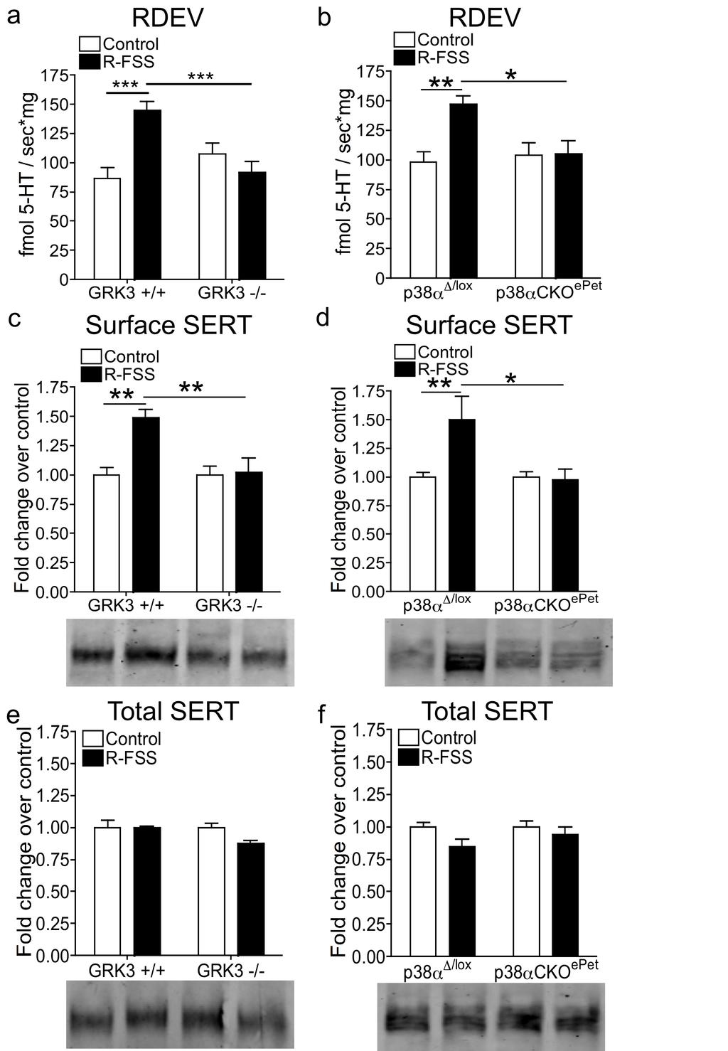
102 94
103 Figure 4.5. R-FSS induced increases in 5-HT uptake by SERT and SERT surface expression require GRK3 and p38α MAPK in serotonergic neurons. (a) RDEV data demonstrating that the R-FSS induced increase in the rate of 5-HT uptake by SERT requires GRK3. *** denotes p<0.001, significant difference in 5- HT uptake by SERT for synaptosomes prepared from GRK3 (+/+) mice exposed to R-FSS as compared with synaptosomes prepared from GRK3 (+/+) control mice or prepared from GRK3 -/-, R-FSS exposed mice; two-way ANOVA followed by Bonferroni s post hoc test, n = 6-8. (b) RDEV data demonstrating that R-FSS increases the rate of 5-HT uptake by SERT in p38α Δ/lox but not in p38αcko epet mice. ** denotes p<0.01, significant difference in 100 nm 5-HT uptake by SERT for synaptosomes prepared from p38α Δ/lox mice exposed to R- FSS as compared with synaptosomes prepared from p38α Δ/lox control mice, * denotes p<0.05, significant difference in 100 nm 5-HT uptake by SERT for synaptosomes prepared from p38α Δ/lox mice exposed to R-FSS as compared with synaptosomes prepared from p38αcko epet mice exposed to R-FSS; two-way ANOVA followed by Bonferroni s post hoc test, n = 6-8. (c) Representative western blot images and corresponding analysis showing that R-FSS increases SERT surface expression in a GRK3 dependent manner. ** denotes p<0.01, significant difference in SERT surface expression for synaptosomes prepared from GRK3 (+/+) mice exposed to R-FSS as compared with synaptosomes prepared from GRK3 (+/+) control mice or GRK3 (-/-) mice exposed to R-FSS; two-way ANOVA followed by Bonferroni s post hoc test, n = 4. (d) Representative western blot images and corresponding analysis showing that R-FSS increases SERT surface expression in a p38α MAPK dependent manner. ** denotes p<0.05, significant difference in SERT surface expression for synaptosomes prepared from p38α Δ/lox mice exposed to R-FSS as compared with synaptosomes prepared from p38α Δ/lox control mice, * denotes p<0.05, significant difference in SERT surface expression for synaptosomes prepared from p38α Δ/lox mice exposed to R-FSS as compared with synaptosomes prepared from p38αcko epet mice exposed to R-FSS; two-way ANOVA followed by Bonferroni s post hoc test, n = 5-8. (e) Representative western blot images and corresponding analysis showing that the GRK3 genotype or R-FSS does not change total SERT expression; two-way ANOVA, n = 4. (f) Representative western blot images and corresponding analysis showing that the p38α MAPK genotype or R-FSS does not change total SERT expression; two-way ANOVA, n = 6-8. Data are mean ± SEM. 95
104 Repeated swim stress selectively increases SERT surface expression in the ventral striatum. SERT expression is seen in multiple brain regions (Torres et al., 2003). In order to determine which brain region(s) show increased surface SERT expression following R-FSS, brain region specific purified synaptosomes from control and R-FSS exposed mice were biotinylated to label cell surface proteins. Surface proteins were eluted with Laemmli s buffer and processed for western blotting with the SERT antibody. Surprisingly, we found that R-FSS increased surface SERT-ir compared to controls only in the ventral striatum (VS) (n = 6; unpaired, two-tailed, t-test; t 10 = 2.862, p<0.017) (Figure 4.6A, red box). R-FSS did not change surface SERT-ir compared to controls in the medial prefrontal cortex (mpfc) (n = 4; unpaired, two-tailed, t-test; t 6 = 0.171, p>0.05), hippocampus (Hippo) (n = 6; unpaired, two-tailed, t-test; t 10 = 0.312, p>0.05), dorsal striatum (DS) (n = 6; unpaired, two-tailed, t-test; t 10 = 0.773, p>0.05), amygdala (Amyg) (n = 4; unpaired, two-tailed, t-test; t 6 = 0.879, p>0.05), or the dorsal raphe (DR) (n = 4; unpaired, two-tailed, t-test; t 6 = 0.776, p>0.05) (Figure 4.6A). To assess if R-FSS changes total SERT levels in each brain region, total proteins were eluted with Laemmli s buffer and processed for western blotting with the SERT antibody. As with WT mice, we found no change in total SERT-ir across the brain regions examined: mpfc (n = 6; unpaired, two-tailed, t-test; t 10 = 0.294, p>0.05), Hippo (n = 6; unpaired, two-tailed, t-test; t 10 = 1.487, p>0.05), DS (n = 6; unpaired, two-tailed, t-test; t 10 = 0.073, p>0.05), VS (n = 6; unpaired, 96
105 two-tailed, t-test; t 10 = 1.573, p>0.05), Amyg (n = 3-4; unpaired, two-tailed, t-test; t 5 = 0.886, p>0.05), and DR (n = 4; unpaired, two-tailed, t-test; t 6 = 1.053, p>0.05) (Figure 4.6B). Together these data demonstrate that R-FSS increases surface SERT expression specifically in the ventral striatum without changing total SERT levels or affecting other brain regions. 97
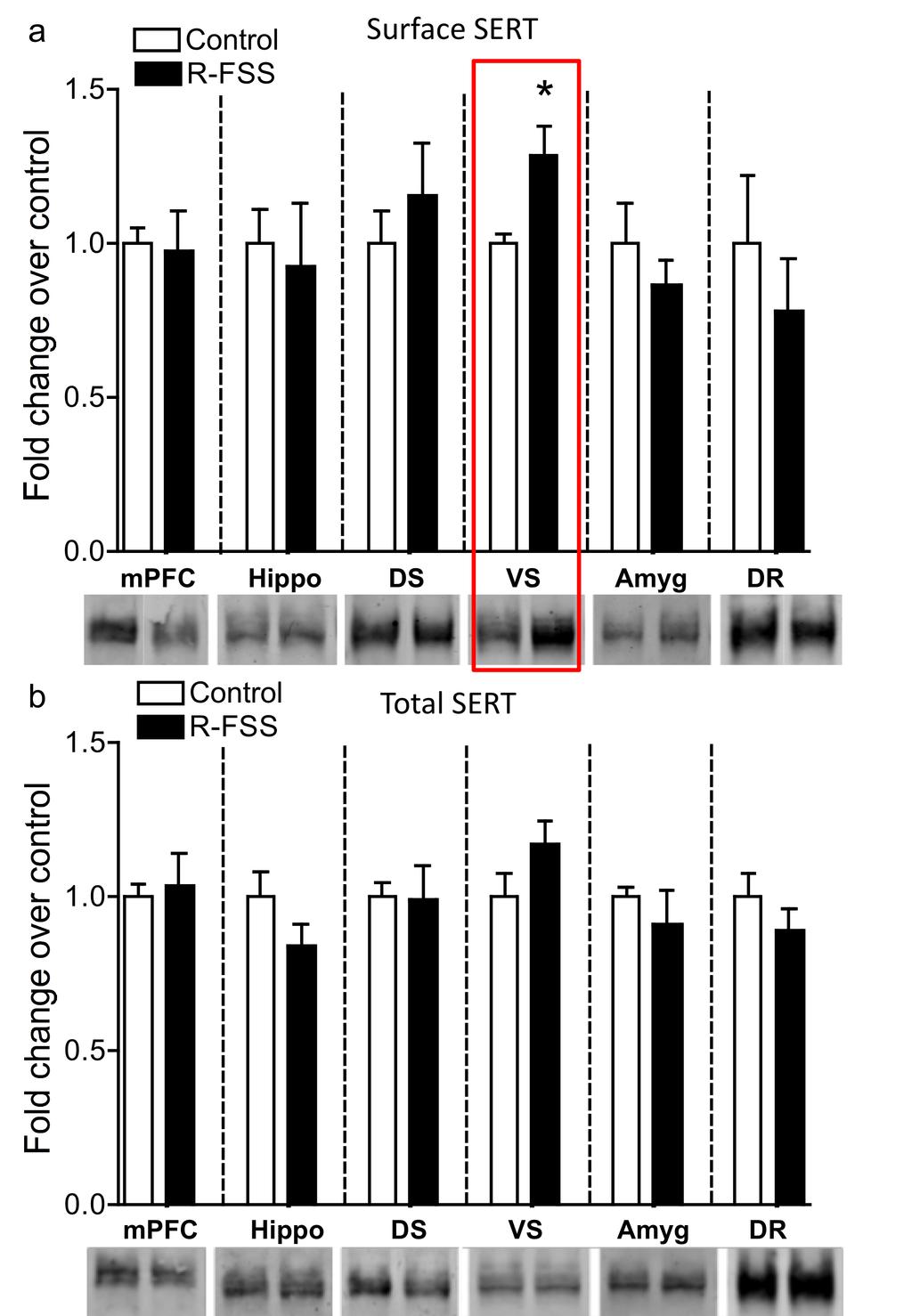
106 98
107 Figure 4.6. SERT surface expression is selectively increased in the ventral striatum following R-FSS. (a) Representative western blot images and corresponding analysis showing that R-FSS increases SERT surface expression only in the ventral striatum. * denotes p<0.05, significant difference in SERT surface expression for synaptosomes prepared from mice exposed to R-FSS as compared to control mice; unpaired, two-tailed, t-test, n = 4-6. (b) Representative western blot images and corresponding analysis showing that R-FSS does not change total SERT expression in any of the brain regions investigated; two-way ANOVA, n = 4-6. Data are mean ± SEM. mpfc = medial prefrontal cortex, Hippo = hippocampus, DS = dorsal striatum, VS = ventral striatum, Amyg = amygdala, DR = dorsal raphe. 99
108 Repeated swim stress increases surface SERT expression in the ventral striatum through activation of local kappa receptors. KOR receptors are expressed on the cell bodies of serotonergic DR neurons and on the afferent nerve terminals projecting to the VS (Tao and Auerbach, 2002; Land et al., 2009a), thus it is possible that KOR activation in the DR or VS could mediate the increase in surface SERT seen following R-FSS. In order to determine which pool of KORs mediate this effect we stereotaxically injected the long-lasting KOR antagonist norbni into the DR or VS. Following recovery from surgery, VS purified synaptosomes from control and R-FSS exposed mice were biotinylated to label cell surface proteins. Surface proteins were eluted with Laemmli s buffer and processed for western blotting with the SERT antibody. We found that R-FSS increased surface SERT-ir in the VS as compared to controls when norbni was injected into the DR (n = 3-5; two-way ANOVA; interaction of brain region and R-FSS, F 1,12 = 13.50, p<0.003; followed by Bonferroni s post hoc, norbni DR control vs. norbni R-FSS p<0.01) (Figure 4.7A). Conversely, the increase in SERT surface expression in the VS by R-FSS was not seen when norbni was injected into the VS (Bonferroni s post hoc, norbni VS control vs. norbni VS R-FSS p>0.05) (Figure 4.7A), suggesting that local KOR in the VS mediate the stress-induced increase in surface SERT expression. To assess if norbni into the DR or VS changes total SERT levels in the VS, total proteins were eluted with Laemmli s buffer and processed for western blotting with the SERT antibody. As with WT mice, we found no change in total 100
109 SERT-ir regardless of the region norbni was injected or R-FSS exposure (n = 3-5; two-way ANOVA; interaction of brain region and R-FSS, F 1,12 = 0.477, p>0.05) (Figure 4.7B). Figure 4.7C shows the proposed mechanism by which stressinduced KOR activation leads to increased SERT surface expression in the VS. 101
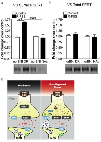
110 102
Kappa Opioid Receptor Activation Potentiates the Cocaine-Induced Increase in Evoked Dopamine Release Recorded In Vivo in the Mouse Nucleus Accumbens
 (214) 39, 336 348 & 214 American College of. All rights reserved 893-133X/14 www.neuropsychopharmacology.org Kappa Opioid Receptor Activation Potentiates the aine-induced Increase in Evoked Dopamine Release
Repeated stress exposure causes strain-dependent shifts in the behavioral economics of cocaine in rats
 bs_bs_banneraddiction Biology PRECLINICAL STUDY doi:10.1111/adb.12123 Repeated stress exposure causes strain-dependent shifts in the behavioral economics of cocaine in rats Peter A. Groblewski 1, Chad
MOLECULAR BIOLOGY OF DRUG ADDICTION. Sylvane Desrivières, SGDP Centre
 1 MOLECULAR BIOLOGY OF DRUG ADDICTION Sylvane Desrivières, SGDP Centre Reward 2 Humans, as well as other organisms engage in behaviours that are rewarding The pleasurable feelings provide positive reinforcement
Food restriction: enhancing effects on drug reward and striatal cell signaling
 Food restriction: enhancing effects on drug reward and striatal cell signaling K.D. Carr Departments of Psychiatry & Pharmacology NYU School of Medicine Common Neural Substrates for Incentive-Motivating
Opioid Receptor Antagonism and Prodynorphin Gene Disruption Block Stress-Induced Behavioral Responses
 5674 The Journal of Neuroscience, July 2, 2003 23(13):5674 5683 Behavioral/Systems/Cognitive Opioid Receptor Antagonism and Prodynorphin Gene Disruption Block Stress-Induced Behavioral Responses Jay P.
Classes of Neurotransmitters. Neurotransmitters
 1 Drugs Outline 2 Neurotransmitters Agonists and Antagonists Cocaine & other dopamine agonists Alcohol & its effects / Marijuana & its effects Synthetic & Designer Drugs: Ecstasy 1 Classes of Neurotransmitters
Neurotransmitter Systems III Neurochemistry. Reading: BCP Chapter 6
 Neurotransmitter Systems III Neurochemistry Reading: BCP Chapter 6 Neurotransmitter Systems Normal function of the human brain requires an orderly set of chemical reactions. Some of the most important
Adolescent Prozac Exposure Enhances Sensitivity to Cocaine in Adulthood INTRODUCTION
 INTRODUCTION Epidemiologic reports indicate that mood disorders in children and adolescents are quite common, with up to 70% of depressed children and adolescents experiencing a recurrence within 5 years
Neurobiology of Addiction JeanAnne Johnson Talbert, DHA, APRN BC, FNP, CARN AP
 Neurobiology of Addiction JeanAnne Johnson Talbert, DHA, APRN BC, FNP, CARN AP Disclosures This speaker has no conflicts of interest to disclose Objectives Define drug abuse and addiction Identify the
Selective p38a MAPK Deletion in Serotonergic Neurons Produces Stress Resilience in Models of Depression and Addiction
 Neuron Article Selective p38a MAPK Deletion in Serotonergic Neurons Produces Stress Resilience in Models of Depression and Addiction Michael R. Bruchas, 1,6, * Abigail G. Schindler, 1 Haripriya Shankar,
Stress-induced reinstatement of cocaine seeking is mediated by the kappa opioid system
 DOI 10.1007/s00213-008-1122-y ORIGINAL INVESTIGATION Stress-induced reinstatement of cocaine seeking is mediated by the kappa opioid system Van A. Redila & Charles Chavkin Received: 16 October 2007 / Accepted:
Neurotransmitter Systems I Identification and Distribution. Reading: BCP Chapter 6
 Neurotransmitter Systems I Identification and Distribution Reading: BCP Chapter 6 Neurotransmitter Systems Normal function of the human brain requires an orderly set of chemical reactions. Some of the
BRAIN MECHANISMS OF REWARD AND ADDICTION
 BRAIN MECHANISMS OF REWARD AND ADDICTION TREVOR.W. ROBBINS Department of Experimental Psychology, University of Cambridge Many drugs of abuse, including stimulants such as amphetamine and cocaine, opiates
- Neurotransmitters Of The Brain -
 - Neurotransmitters Of The Brain - INTRODUCTION Synapsis: a specialized connection between two neurons that permits the transmission of signals in a one-way fashion (presynaptic postsynaptic). Types of
If you give any person a prescription of something like Valium and have them take it on
 As always I am happy to do this presentation, which is my favorite topic in addiction medicine. I am an internist, and I have done healthcare for the homeless in Springfield as well as been the medical
The Nervous System. Chapter 4. Neuron 3/9/ Components of the Nervous System
 Chapter 4 The Nervous System 1. Components of the Nervous System a. Nerve cells (neurons) Analyze and transmit information Over 100 billion neurons in system Four defined regions Cell body Dendrites Axon
Cogs 107b Systems Neuroscience lec9_ neuromodulators and drugs of abuse principle of the week: functional anatomy
 Cogs 107b Systems Neuroscience www.dnitz.com lec9_02042010 neuromodulators and drugs of abuse principle of the week: functional anatomy Professor Nitz circa 1986 neurotransmitters: mediating information
Ghrelin mediates stressinduced. behavior in mice. Chuang et al 2011 L3: Love, Lust, Labor
 Ghrelin mediates stressinduced food-reward behavior in mice Chuang et al 2011 L3: Love, Lust, Labor Agenda Introduction What is Ghrelin? Previous Models New model Methods Results Discussion Conclusion
MeCP2 and psychostimulantinduced behavioral adaptations. Anne E. West, M.D., Ph.D. Department of Neurobiology Duke University Medical Center
 MeCP2 and psychostimulantinduced behavioral adaptations Anne E. West, M.D., Ph.D. Department of Neurobiology Duke University Medical Center Psychostimulants and antidepressants slowly change behavior Psychostimulants
NIH Public Access Author Manuscript Future Neurol. Author manuscript; available in PMC 2011 March 1.
 NIH Public Access Author Manuscript Published in final edited form as: Future Neurol. 2010 May 1; 5(3): 393 401. doi:10.2217/fnl.10.14. Neurobiology of dysregulated motivational systems in drug addiction
Neurophysiology and Neurochemistry in PsychoGeriatrics
 Tel Aviv University Sackler Faculty of Medicine CME in Psychiatry Neurophysiology and Neurochemistry in PsychoGeriatrics Nicola Maggio, MD, PhD Sackler Faculty of Medicine Tel Aviv University Department
Chemical Control of Behavior and Brain 1 of 9
 Chemical Control of Behavior and Brain 1 of 9 I) INTRO A) Nervous system discussed so far 1) Specific 2) Fast B) Other systems extended in space and time 1) Nonspecific 2) Slow C) Three components that
processes in the central nervous system (CNS), affecting many of the during the course of ethanol treatment. Ethanol stimulates the release of
, affecting many of the during the course of ethanol treatment. Ethanol stimulates the release of") INTRODUCTION INTRODUCTION Neuroscience research is essential for understanding the biological basis of ethanol-related brain alterations and for identifying the molecular targets for therapeutic compounds
INTRODUCTION INTRODUCTION Neuroscience research is essential for understanding the biological basis of ethanol-related brain alterations and for identifying the molecular targets for therapeutic compounds
Drugs, addiction, and the brain
 Drugs, addiction, and the brain Topics to cover: What is addiction? How is addiction studied in the lab? The neuroscience of addiction. Caffeine Cocaine Marijuana (THC) What are the properties of addiction?
Receptors and Neurotransmitters: It Sounds Greek to Me. Agenda. What We Know About Pain 9/7/2012
 Receptors and Neurotransmitters: It Sounds Greek to Me Cathy Carlson, PhD, RN Northern Illinois University Agenda We will be going through this lecture on basic pain physiology using analogies, mnemonics,
Kappa Opioid Regulation of Stress-Related Behavior
 McNair Scholars Journal Volume 14 Issue 1 Article 6 2010 Kappa Opioid Regulation of Stress-Related Behavior Erin Harshberger Grand Valley State University Follow this and additional works at: http://scholarworks.gvsu.edu/mcnair
Basal Ganglia General Info
 Basal Ganglia General Info Neural clusters in peripheral nervous system are ganglia. In the central nervous system, they are called nuclei. Should be called Basal Nuclei but usually called Basal Ganglia.
Neurotransmitter Functioning In Major Depressive Disorder
 Neurotransmitter Functioning In Major Depressive Disorder Otsuka Pharmaceutical Development & Commercialization, Inc. 2017 Otsuka Pharmaceutical Development & Commercialization, Inc., Rockville, MD January
The Nervous System Mark Stanford, Ph.D.
 The Nervous System Functional Neuroanatomy and How Neurons Communicate Mark Stanford, Ph.D. Santa Clara Valley Health & Hospital System Addiction Medicine and Therapy Services The Nervous System In response
NEUROBIOLOGY ALCOHOLISM
 NEUROBIOLOGY ALCOHOLISM THERE HAS BEEN A MAJOR THEORETICAL SHIFT IN MEDICATION DEVELOPMENT IN ALCOHOLISM Driven by animal models of intermittent ethanol administration followed by termination, then access
Neurobiology of Addiction
 Neurobiology of Addiction Domenic A. Ciraulo, MD Director of Alcohol Pharmacotherapy Research Center for Addiction Medicine Department of Psychiatry Massachusetts General Hospital Disclosure Neither I
Neurobiology of Addiction
 Neurobiology of Addiction Tiffany Love, Ph.D. Department of Psychiatry The University of Utah What is Addiction? Addiction is a chronic, relapsing, and treatable brain disorder. Compulsive drug seeking
by popular demand: Addiction II
 by popular demand: Addiction II PSY/NEU338: Animal learning and decision making: Psychological, computational and neural perspectives drug addiction huge and diverse field of research (many different drugs)
Stress-Induced p38 Mitogen-Activated Protein Kinase Activation Mediates -Opioid-Dependent Dysphoria
 11614 The Journal of Neuroscience, October 24, 2007 27(43):11614 11623 Behavioral/Systems/Cognitive Stress-Induced p38 Mitogen-Activated Protein Kinase Activation Mediates -Opioid-Dependent Dysphoria Michael
General introduction. Chapter 1
 General introduction Chapter 1 General introduction Historical aspects of drug use Religious, medicinal and recreational use of mind-altering substances by humans has a history of thousands of years 1.
CHAPTER 2. THE NEUROBIOLOGY OF SUBSTANCE USE, MISUSE, AND ADDICTION
 CHAPTER 2. THE NEUROBIOLOGY OF SUBSTANCE USE, MISUSE, AND ADDICTION Chapter 2 Preview A substantial body of research has accumulated over several decades and transformed our understanding of substance
Brain Reward Pathway and Addiction
 STO-131 Brain Reward Pathway and Addiction Part 1: Is Addictive? Medical Report Ray is feeling horrible. He is very depressed and nervous. He has a headache and nausea, and he is having trouble sleeping.
SUPPLEMENTARY INFORMATION
 Supplementary Figure 1. Behavioural effects of ketamine in non-stressed and stressed mice. Naive C57BL/6 adult male mice (n=10/group) were given a single dose of saline vehicle or ketamine (3.0 mg/kg,
Council on Chemical Abuse Annual Conference November 2, The Science of Addiction: Rewiring the Brain
 Council on Chemical Abuse Annual Conference November 2, 2017 The Science of Addiction: Rewiring the Brain David Reyher, MSW, CAADC Behavioral Health Program Director Alvernia University Defining Addiction
TO BE MOTIVATED IS TO HAVE AN INCREASE IN DOPAMINE. The statement to be motivated is to have an increase in dopamine implies that an increase in
 1 NAME COURSE TITLE 2 TO BE MOTIVATED IS TO HAVE AN INCREASE IN DOPAMINE The statement to be motivated is to have an increase in dopamine implies that an increase in dopamine neurotransmitter, up-regulation
Na + /Cl - -dependent Neurotransmitter Transporters. Outline. Synaptic Transmission. How did the neurotransmitter cross the membrane?
 Na /Cl - -dependent Neurotransmitter Transporters Stine Meinild Lundby The Neurobiology Group Molecular, Cellular, and Integrative Physiology Section November 26 th, 2007 slide 1 Outline Synaptic neurotransmission
The Neuroscience of Addiction: A mini-review
 The Neuroscience of Addiction: A mini-review Jim Morrill, MD, PhD MGH Charlestown HealthCare Center Massachusetts General Hospital Disclosures Neither I nor my spouse/partner has a relevant financial relationship
Mk-801 Administration in Adolescent Male Rats and Cocaine Conditioned Place
 Mk-801 Administration in Adolescent Male Rats and Cocaine Conditioned Place Preference Stephanie Willis, Jonnique Adjmul, Shabaaz Sandhu, Antoniette M. Maldonado-Devincci, Cheryl Kirsten ABSTRACT The present
Notes: Synapse. Overview. PSYC Summer Professor Claffey PDF. Conversion from an signal to a signal - electrical signal is the
 PSYC 170 - Summer 2013 - Professor Claffey Notes: Synapse PDF Overview Conversion from an signal to a signal - electrical signal is the - chemical signal is the Presynaptic - refers to that sends/receives
Behavioral Neuroscience: Fear thou not. Rony Paz
 Behavioral Neuroscience: Fear thou not Rony Paz Rony.paz@weizmann.ac.il Thoughts What is a reward? Learning is best motivated by threats to survival Threats are much better reinforcers Fear is a prime
Kappa Opioid Regulation of Dopamine in Dysphoria and Addiction. Jonathan M. Ehrich. A dissertation. submitted in partial fulfillment of the
 Kappa Opioid Regulation of Dopamine in Dysphoria and Addiction Jonathan M. Ehrich A dissertation submitted in partial fulfillment of the requirements for the degree of Doctor of Philosophy University of
SUPPLEMENTARY INFORMATION
 doi:10.1038/nature11436 Supplementary Figure 1. CRF peptide is localized cholinergic interneurons throughout the nucleus accumbens. Immunofluorescent images demonstrating localization of CRF peptide, tyrosine
Understanding Addiction and Its Impact on the Brain. SDSMA Webinar Matthew Stanley, DO
 Understanding Addiction and Its Impact on the Brain SDSMA Webinar Matthew Stanley, DO Estimated Economic Cost to Society Due to Substance Abuse and Addiction: Illegal drugs: Alcohol: Tobacco: $181 billion/year
Portions from Chapter 6 CHAPTER 7. The Nervous System: Neurons and Synapses. Chapter 7 Outline. and Supporting Cells
 CHAPTER 7 The Nervous System: Neurons and Synapses Chapter 7 Outline Neurons and Supporting Cells Activity in Axons The Synapse Acetylcholine as a Neurotransmitter Monoamines as Neurotransmitters Other
Supporting Online Material for
 www.sciencemag.org/cgi/content/full/328/5983/1288/dc1 Supporting Online Material for Induction of Fear Extinction with Hippocampal-Infralimbic BDNF Jamie Peters, Laura M. Dieppa-Perea, Loyda M. Melendez,
Key Concepts. Machinery of the Mind. The Growing Cycle. Six Brain Mind Functions. Machinery of the Mind 6/1/2012
 Machinery of the Mind How High-risk Choices Redesign the Brain Key Concepts Brain and Mind are reciprocal Brain/Mind Functions intertwine We are hardwired and we have software Your brain changes all the
INTERACTION DRUG BODY
 INTERACTION DRUG BODY What the drug does to the body What the body does to the drug Receptors - intracellular receptors - membrane receptors - Channel receptors - G protein-coupled receptors - Tyrosine-kinase
Understanding Addiction: Why Can t Those Affected Just Say No?
 Understanding Addiction: Why Can t Those Affected Just Say No? 1 The Stigma of Addiction There continues to be a stigma surrounding addiction even among health care workers. Consider the negative opinions
Basics of Pharmacology
 Basics of Pharmacology Pekka Rauhala Transmed 2013 What is pharmacology? Pharmacology may be defined as the study of the effects of drugs on the function of living systems Pharmacodynamics The mechanism(s)
BIPN 140 Problem Set 6
 BIPN 140 Problem Set 6 1) The hippocampus is a cortical structure in the medial portion of the temporal lobe (medial temporal lobe in primates. a) What is the main function of the hippocampus? The hippocampus
Behavioral Neuroscience: Fear thou not. Rony Paz
 Behavioral Neuroscience: Fear thou not Rony Paz Rony.paz@weizmann.ac.il Thoughts What is a reward? Learning is best motivated by threats to survival? Threats are much better reinforcers? Fear is a prime
Nervous System, Neuroanatomy, Neurotransmitters
 Nervous System, Neuroanatomy, Neurotransmitters Neurons Structure of neurons Soma Dendrites Spines Axon Myelin Nodes of Ranvier Neurons Structure of neurons Axon collaterals 1 Neurons Structure of neurons
SUPPLEMENTARY INFORMATION
 SUPPLEMENTARY INFORMATION doi:10.1038/nature12024 entary Figure 1. Distribution of the number of earned cocaine Supplementary Figure 1. Distribution of the number of earned cocaine infusions in Shock-sensitive
BIPN 140 Problem Set 6
 BIPN 140 Problem Set 6 1) Hippocampus is a cortical structure in the medial portion of the temporal lobe (medial temporal lobe in primates. a) What is the main function of the hippocampus? The hippocampus
Neurotransmitter Systems II Receptors. Reading: BCP Chapter 6
 Neurotransmitter Systems II Receptors Reading: BCP Chapter 6 Neurotransmitter Systems Normal function of the human brain requires an orderly set of chemical reactions. Some of the most important chemical
Drug Receptor Interactions and Pharmacodynamics
 Drug Receptor Interactions and Pharmacodynamics Dr. Raz Mohammed MSc Pharmacology School of Pharmacy 22.10.2017 Lec 6 Pharmacodynamics definition Pharmacodynamics describes the actions of a drug on the
The Biological Perspective. Jørg Mørland Senior researcher, Norwegian Institute of Public Health Professor em of Medicine University of Oslo
 The Biological Perspective Jørg Mørland Senior researcher, Norwegian Institute of Public Health Professor em of Medicine University of Oslo The Biological Perspective What is it? More than «the» one biological
Chapter 2: Cellular Mechanisms and Cognition
 Chapter 2: Cellular Mechanisms and Cognition MULTIPLE CHOICE 1. Two principles about neurons were defined by Ramón y Cajal. The principle of connectional specificity states that, whereas the principle
Selective p38a MAPK Deletion in Serotonergic Neurons Produces Stress Resilience in Models of Depression and Addiction
 Article Selective p38a MAPK Deletion in Serotonergic Neurons Produces Stress Resilience in Models of Depression and Addiction Michael R. Bruchas, 1,6, * Abigail G. Schindler, 1 Haripriya Shankar, 1 Daniel
The Neurobiology of Addiction
 The Neurobiology of Addiction Jodi Gilman, Ph.D. Center for Addiction Medicine Massachusetts General Hospital Associate Professor, Department of Psychiatry Harvard Medical School What is Addiction? commonly
 PSY 302 Lecture 6: The Neurotransmitters (continued) September 12, 2017 Notes by: Desiree Acetylcholine (ACh) CoA + Acetate Acetyl-CoA (mitochondria) (food, vinegar) + Choline ChAT CoA + ACh (lipids, foods)
9.01 Introduction to Neuroscience Fall 2007
 MIT OpenCourseWare http://ocw.mit.edu 9.01 Introduction to Neuroscience Fall 2007 For information about citing these materials or our Terms of Use, visit: http://ocw.mit.edu/terms. Working memory short-term
REVIEwS. Opioid receptors: drivers to addiction?
 REVIEwS Opioid receptors: drivers to addiction? Emmanuel Darcq 1 and Brigitte Lina Kieffer 1,2* Abstract Drug addiction is a worldwide societal problem and public health burden, and results from recreational
nucleus accumbens septi hier-259 Nucleus+Accumbens birnlex_727
 Nucleus accumbens From Wikipedia, the free encyclopedia Brain: Nucleus accumbens Nucleus accumbens visible in red. Latin NeuroNames MeSH NeuroLex ID nucleus accumbens septi hier-259 Nucleus+Accumbens birnlex_727
CASE 49. What type of memory is available for conscious retrieval? Which part of the brain stores semantic (factual) memories?
 memories?") CASE 49 A 43-year-old woman is brought to her primary care physician by her family because of concerns about her forgetfulness. The patient has a history of Down syndrome but no other medical problems.
CASE 49 A 43-year-old woman is brought to her primary care physician by her family because of concerns about her forgetfulness. The patient has a history of Down syndrome but no other medical problems.
America is a drugged society
 Overview of Drug Abuse Basic Considerations. M. Imad Damaj, Ph.D. Associate Professor Dept. of Pharmacology/Toxicology, Virginia Commonwealth University America is a drugged society 90% of all drugs manufactured
Receptors Families. Assistant Prof. Dr. Najlaa Saadi PhD Pharmacology Faculty of Pharmacy University of Philadelphia
 Receptors Families Assistant Prof. Dr. Najlaa Saadi PhD Pharmacology Faculty of Pharmacy University of Philadelphia Receptor Families 1. Ligand-gated ion channels 2. G protein coupled receptors 3. Enzyme-linked
Neurotransmitters acting on G-protein coupled receptors
 Neurotransmitters acting on G-protein coupled receptors Part 1: Dopamine and Norepinephrine BIOGENIC AMINES Monoamines Diamine Overview of Neurotransmitters and Their Receptors Criteria for defining a
Synaptic Transmission: Ionic and Metabotropic
 Synaptic Transmission: Ionic and Metabotropic D. Purves et al. Neuroscience (Sinauer Assoc.) Chapters 5, 6, 7. C. Koch. Biophysics of Computation (Oxford) Chapter 4. J.G. Nicholls et al. From Neuron to
Lack of GPR88 enhances medium spiny neuron activity and alters. motor- and cue- dependent behaviors
 Lack of GPR88 enhances medium spiny neuron activity and alters motor- and cue- dependent behaviors Albert Quintana, Elisenda Sanz, Wengang Wang, Granville P. Storey, Ali D. Güler Matthew J. Wanat, Bryan
Synaptic transmission
 Outline Synaptic transmission Sompol Tapechum M.D., Ph.D. Department of Physiology Faculty of Medicine Siriraj Hospital, Bangkok, Thailand. sisth@mahidol.ac.th 2 Structure of synapse Modes of synaptic
Kappa Opioid Receptors Regulate Stress-Induced Cocaine Seeking and Synaptic Plasticity
 Article Kappa Opioid Receptors Regulate Stress-Induced Cocaine Seeking and Synaptic Plasticity Nicholas M. Graziane, 1,3 Abigail M. Polter, 1 Lisa A. Briand, 2 R. Christopher Pierce, 2 and Julie A. Kauer
Action Potentials and Synaptic Transmission. BIO 219 Napa Valley College Dr. Adam Ross
 Action Potentials and Synaptic Transmission BIO 219 Napa Valley College Dr. Adam Ross Review of action potentials Nodes of Ranvier Nucleus Dendrites Cell body In saltatory conduction, the nerve impulses
The Biology of Addiction
 The Biology of Addiction Risk factors for addiction: Biological/Genetic Family history of addiction Being male Having mental illness Exposure to substances in utero * The genes that people are born with
Neural Communication. Central Nervous System Peripheral Nervous System. Communication in the Nervous System. 4 Common Components of a Neuron
 Neural Communication Overview of CNS / PNS Electrical Signaling Chemical Signaling Central Nervous System Peripheral Nervous System Somatic = sensory & motor Autonomic = arousal state Parasympathetic =
Psych 181: Dr. Anagnostaras
 Psych 181: Dr. Anagnostaras Lecture 5 Synaptic Transmission Introduction to synaptic transmission Synapses (Gk., to clasp or join) Site of action of most psychoactive drugs 6.5 1 Synapses Know basic terminology:
ADHD Medications & How They Work. Gail C. Rodin, Ph.D. January 21, 2008
 ADHD Medications & How They Work Gail C. Rodin, Ph.D. January 21, 2008 Agenda How the (ADHD) Brain Works (or doesn t) Neurons and neurotransmitters NE & DA: the major players in ADHD Channel vs. state
Synapse. Structure & Function. Neurotransmitter Sequence. Integration. History: 10/4/12 original version
 Synapse History: 10/4/12 original version Structure & Function (This content is covered in Sinjin's presentation, see link in calendar) Neurotransmitters Synaptic cleft Post-synaptic potential Excitation
Exam 2 PSYC Fall (2 points) Match a brain structure that is located closest to the following portions of the ventricular system
 Match a brain structure that is located closest to the following portions of the ventricular system") Exam 2 PSYC 2022 Fall 1998 (2 points) What 2 nuclei are collectively called the striatum? (2 points) Match a brain structure that is located closest to the following portions of the ventricular system
Exam 2 PSYC 2022 Fall 1998 (2 points) What 2 nuclei are collectively called the striatum? (2 points) Match a brain structure that is located closest to the following portions of the ventricular system
MANAGING PAIN IN PATIENTS WITH SUBSTANCE USE DISORDER
 MANAGING PAIN IN PATIENTS WITH SUBSTANCE USE DISORDER Melissa B. Weimer, DO, MCR Chief of Behavioral Health & Addiction Medicine St. Peter s Health Partners Grand Rounds October 11, 2017 Disclosures One
CU Scholar. University of Colorado, Boulder. Drew Schreiner University of Colorado Boulder. Spring 2013
 University of Colorado, Boulder CU Scholar Undergraduate Honors Theses Honors Program Spring 2013 Assessing cocaine addiction vulnerability and neurobiological correlates within the nigrostriatal pathway
Synaptic Communication. Steven McLoon Department of Neuroscience University of Minnesota
 Synaptic Communication Steven McLoon Department of Neuroscience University of Minnesota 1 Course News The first exam is next week on Friday! Be sure to checkout the sample exam on the course website. 2
Section: Chapter 5: Multiple Choice. 1. The structure of synapses is best viewed with a(n):
:") Section: Chapter 5: Multiple Choice 1. The structure of synapses is best viewed with a(n): p.155 electron microscope. light microscope. confocal microscope. nissle-stained microscopic procedure. 2. Electron
Section: Chapter 5: Multiple Choice 1. The structure of synapses is best viewed with a(n): p.155 electron microscope. light microscope. confocal microscope. nissle-stained microscopic procedure. 2. Electron
Transcriptional and Epigenetic Mechanisms of Addiction
 Transcriptional and Epigenetic Mechanisms of Addiction Eric J. Nestler Mount Sinai School of Medicine New York, NY Dr. Ray Fuller There is every reason to be optimistic that in the future we will find
At a Glance. Background Information. Lesson 3 Drugs Change the Way Neurons Communicate
 Lesson 3 Drugs Change the Way Neurons Communicate Overview Students build upon their understanding of neurotransmission by learning how different drugs of abuse disrupt communication between neurons. Students
Prof. Anagnostaras, Lecture 7: Fear
 Historical views that thought and emotion were processed separately in the brain Prof. Anagnostaras, Lecture 7: So far, fear is the best understood What is fear? Dictionary: A feeling of agitation and
Aberrant approach-avoidance conflict resolution following repeated cocaine pre-exposure
 DOI 10.1007/s00213-015-4006-y ORIGINAL INVESTIGATION Aberrant approach-avoidance conflict resolution following repeated cocaine pre-exposure David Nguyen 1 & Anett Schumacher 1 & Suzanne Erb 1 & Rutsuko
The Cerebral Cortex and Higher Intellectual Functions
 The Cerebral Cortex and Higher Intellectual Functions Lobes in a lateral view of left hemisphere Atlas Fig.2-11 The Insula The Hidden Lobe Atlas Fig. 2-11 Atlas Fig. 2-39 Lobes in a lateral view of left
Effects of Caffeine on Memory in Rats
 Western University Scholarship@Western 2015 Undergraduate Awards The Undergraduate Awards 2015 Effects of Caffeine on Memory in Rats Cisse Nakeyar Western University, cnakeyar@uwo.ca Follow this and additional
Psychotropic Drugs Critical Thinking - KEY
 Open Your Class with This Tomorrow Chasing the Scream: The First and Last Days of the War on s Psychotropic s Critical Thinking - KEY Background: The blood-brain barrier is a network of tightly packed
It s Not Just Serotonin: Neurosignaling in Mental Illness
 It s Not Just Serotonin: Neurosignaling in Mental Illness Barbara J. Limandri, DNSc, APRN, BC Professor of Nursing Linfield College Learning Outcomes Distinguish between metabotropic and ionotropic neuroreceptors
Session ID: 1001 June 14, 2012
 It s Not Just Serotonin: Neurosignaling in Mental Illness Barbara J. Limandri, DNSc, APRN, BC Professor of Nursing Linfield College Learning Outcomes Distinguish between metabotropic and ionotropic neuroreceptors
Relationship Between Stress and Substance Use Disorders: Neurobiologic Interface
 Relationship Between Stress and Substance Use Disorders: Neurobiologic Interface Kathleen Brady, M.D., Ph.D. Professor of Psychiatry Associate Dean of Clinical and Translational Research Medical University
Eighth Edition. Part I: Current Science of Addiction, Relapse & Recovery: Dispelling The Stigmas
 Eighth Edition Part I: Current Science of Addiction, Relapse & Recovery: Dispelling The Stigmas 1 < < < Evolution of our Human Brain: Spinal Cord to Diencephalon to Mammalian-Meso Cortex to Neo Cortex