Vasculature and innervation of the heart. A. Bendelic Human Anatomy Department
|
|
|
- Jocelyn McDaniel
- 5 years ago
- Views:
Transcription
1 Vasculature and innervation of the heart A. Bendelic Human Anatomy Department
2 Plan: 1. Arterial blood supply of the heart. Coronary arteries 2. Venous drainage of the heart. Cardiac veins 3. Innervation of the heart. Cardiac plexus 4. Afferent and efferent (sympathetic and parasympathetic) innervation of the heart
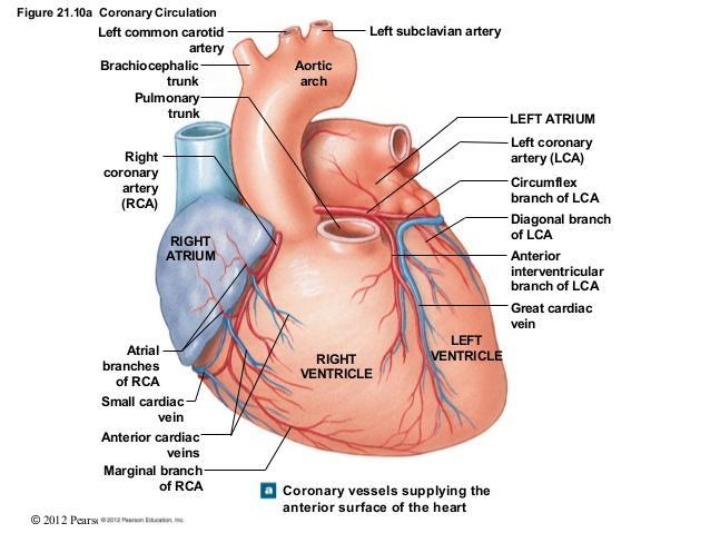
3 Arterial blood supply of the heart The coronary arteries (right and left), the first branches of the aorta, supply the myocardium and epicardium. The left and right coronary arteries arise from the corresponding aortic (coronary) sinuses. The coronary arteries supply both the atria and the ventricles, however the atrial branches are usually small and not readily apparent in the cadaveric heart.


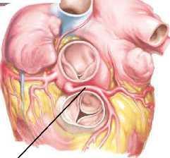
4 Aortic valve, aortic sinuses and coronary ostia


5 Aortic valve, aortic sinuses and coronary ostia


6 Anomalous origin of coronary arteries
7 Coronary arteries Coronary arteries and their major branches are usually located subepicardially. Some individuals, however have regions in which a bunch of cardiac muscle fibers passes over the coronary artery like a bridge. The following terms have been used to describe this anatomical finding: myocardial bridge or intramural coronary artery. The anterior interventricular branch of the left coronary artery is the branch most frequently involved with myocardial bridges.

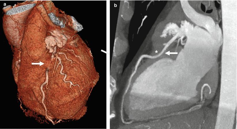
8 Myocardial bridging
9 Right coronary artery Origin right aortic sinus (right coronary sinus); Course follows coronary sulcus between atria and ventricles; Distribution right atrium, SA and AV nodes, most of right ventricle, part of left ventricle (diaphragmatic surface); posterior part of interventricular septum (one third).
10 Branches of the right coronary artery Sinu-atrial nodal branch (in approximately 60% of people); Atrial branches (anterior, lateral and posterior); Anterior ventricular branches (usually 2-3); Right marginal branch is greater in caliber than the other anterior ventricular branches and long enough to reach the apex; Posterior ventricular branches (usually 2-3); Posterior interventricular branch (major branch which descends into the posterior interventricular sulcus), its interventricular septal branches supply the posterior one third of the interventricular septum; Atrioventricular nodal branch (in approximately 80% of people) is the largest interventricular septal artery); In the most common pattern (67%), the right coronary artery is dominant, giving rise to the posterior interventricular branch.

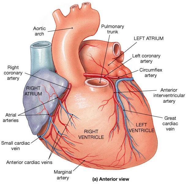
11 Blood supply of the heart
12 Left coronary artery Origin left aortic sinus (left coronary sinus); Course runs in coronary sulcus and gives off 2 branches: a) anterior interventricular branch and b)circumflex branch; Distribution left atrium, most of left ventricle, part of right ventricle, most of interventricular septum (anterior two thirds), SA node.
13 Branches of the left coronary artery Anterior interventricular branch (passes along the anterior interventricular sulcus); A. Anterior right ventricular branches are small and rarely number more than one or two; B. Anterior left ventricular branches (from two to nine); C. Left diagonal artery (reported in 33-50% individuals) is one of the anterior left ventricular branches; D. Interventricular septal branches (supply anterior 2/3 of the interventricular septum). Circumflex branch (follows the coronary sulcus around the left border of the heart to the posterior surface of the heart); A. Atrial branches; B. Left marginal branch; C. Posterior left ventricular branches; D. (Posterior interventricular branch (in 33% of cases)); E. (Sinu-atrial nodal branch (in approximately 40% of people)); F. (Atrioventricular nodal branch (in approximately 20% of people)).

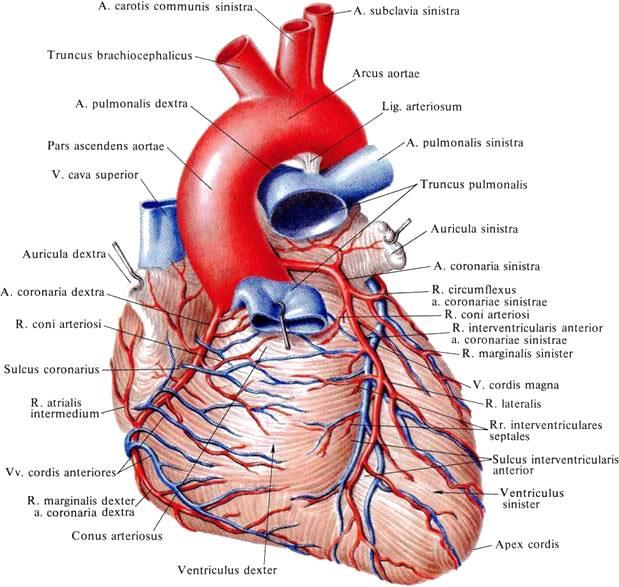
14 Coronary arteries
15 Coronary dominance Variations of the branching patterns and distribution of the coronary arteries are common. The term `dominant` is used to refer to the coronary artery giving off the posterior interventricular branch. In the most common right dominant pattern (right-dominance), present in approximately 67% of people, RCA gives rise to the posterior interventricular branch; the RCA and LCA share about equally in the blood supply of the heart. In approximately 15% of people, the LCA is dominant (leftdominance) in that the posterior interventricular branch is a branch of the circumflex artery (from LCA). There is codominance in approximately 18% of people, in which branches of both the RCA and LCA give rise to the posterior interventricular branches.

16 Variations of coronary arteries
17 Variations of the coronary arteries A few people have only a single coronary artery. In other people, the circumflex branch arises from the right aortic sinus. Approximately 4% of people have an accessory coronary artery. Anomalous coronary origins (from pulmonary trunk)
18 Coronary collateral circulation The branches of the coronary arteries are considered to be end arteries, arteries that supply regions of myocardium lacking sufficient anastomoses from other large branches to maintain viability of tissue should occlusion occur. However, anastomoses do exist between branches of the coronary arteries (subepicardial or myocardial) and between these arteries and extracardiac vessels (such as thoracic vessels). Anastomoses exist between the terminations of the RCA and LCA in the coronary sulcus and between the interventricular branches around the apex in approximately10% of people.
19 Coronary artery disease or coronary heart disease Coronary arteries disease is one of the leading causes of death. An area of myocardium that has undergone necrosis (pathological tissue death) constitutes a myocardial infarction. The most common cause of ischemic heart disease is coronary artery insufficiency resulting from atherosclerosis. Coronary bypass graft shunts blood from the aorta to a stenotic coronary artery to increase the flow distal to the obstruction. A segment of an artery (the radial or internal thoracic arteries) or a vein (the great saphenous vein) is connected to the ascending aorta and to the coronary artery distal to the stenosis.

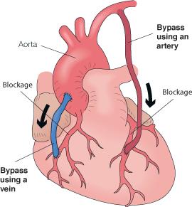
20 Coronary bypass graft
21 Coronary artery disease or coronary heart disease Coronary angioplasty (percutaneous transluminal coronary angioplasty) in which a catheter with a small inflatable balloon attached to its tip is introduced into the obstructed coronary artery. When the catheter reaches the obstruction the balloon is inflated, flattening the atherosclerotic plague against the vessel`s wall and the vessel increases the size of the lumen.
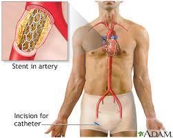
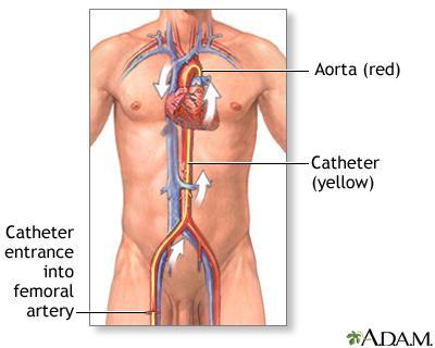
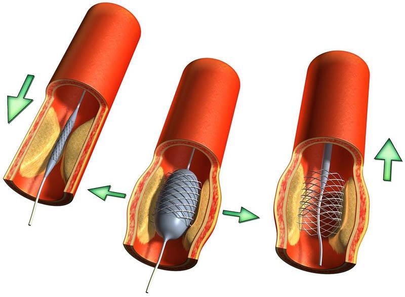
22 Coronary angioplasty
23 Venous drainage of the heart (cardiac veins) The heart is drained mainly by veins that empty into the coronary sinus and partly by small veins (anterior cardiac veins and smallest veins of the heart) that empty into the right atrium. The coronary sinus, the main vein of the heart, is a wide venous channel that runs from the left to the right in the posterior part of the coronary sulcus.
24 A. Coronary sinus The coronary sinus receives: a) the great cardiac vein (v. cordis magna), the main tributary of the coronary sinus, its first part (anterior interventricular vein) starts near the apex of the heart and ascends with the anterior interventricular branch of the LCA; b) the middle cardiac vein (v. cordis media), also named posterior interventricular vein, accompanies the posterior interventricular branch of the RCA; c) the small cardiac vein (v. cordis parva) accompanies the right marginal branch of the RCA; d) the posterior vein of left ventricle runs on the diaphragmatic surface of the left ventricle; e) the oblique vein of left atrium.
25 A. Coronary sinus The oblique vein of the left atrium is a small vessel, relatively unimportant postnatally, that descends over the posterior wall of left atrium. The remnant of the embryonic left superior vena cava, which usually atrophies during the fetal period, but occasionally persists in adults (replacing or augmenting the right superior vena cava.). It is a small vein (Marshall oblique vein) that merges with great cardiac vein to form the coronary sinus. Occasionally persists as a left superior vena cava.

26 Cardiac veins
27 B. Small cardiac veins Some cardiac veins do not drain via the coronary sinus. Several small anterior cardiac veins (vv. cordis anteriores) begin over the anterior surface of the right ventricle, cross the coronary sulcus, and usually end directly in the right atrium. The smallest cardiac veins (vv. cordis minimae) are minute vessels that begin in the capillary beds of the myiocardium and open directly into the chambers of the heart, chiefly the atria. Although called veins, they are valveless communications with the capillary beds of the myocardium and may carry blood from the heart chambers to the myocardium.
28 Lymphatic drainage of the heart Lymphatic vessels in the myocardium and subendocardial connective tissue pass to the subendocardial lymphatic plexus. Vessels from this plexus pass to the coronary groove and follow the coronary arteries. The lymphatic vessels that follow the right coronary artery empty into the anterior mediastinal lymph nodes or brachiocephalic lymph nodes. The lymphatic vessels that follow the left coronary artery empty into the inferior tracheobronchial lymph nodes.

29 Lymphatic drainage of the heart
30 Innervation of the heart The heart is supply by the vegetative nerve fibers from the cardiac plexus, which is often (quite artificially) divided into the superficial and deep portions. This nerve network is most commonly described as lying on the anterior surface of the bifurcation of the trachea. The cardiac plexus is formed of both sympathetic and parasympathetic fibers. Fibers are distributed along and to the coronary arteries, and to the components of conducting system of the heart, particularly the SA and AV nodes.

31 Cardiac plexus
32 Sources of formation of cardiac plexus A. Cardiac nerves of sympathetic trunk: Superior cervical cardiac nerve (from superior cervical ganglion); Middle cervical cardiac nerve (from middle cervical ganglion); Inferior cervical cardiac nerve (from inferior cervical ganglion or cervicothoracic ganglion); Thoracic cardiac nerves (from superior thoracic ganglia of the sympathetic trunk).
33 Sources of formation of cardiac plexus B. Cardiac branches of vagus nerve: Superior cervical cardiac branches (from cervical part of vagus nerve); Inferior cervical cardiac branches (from recurrent laryngeal nerve); Thoracic cardiac branches (from thoracic part of vagus nerve).
34 Two parts of (extraorganic) cardiac plexus Superficial (or ventral) part is located below the aortic arch, between it and pulmonary trunk. It is formed by the left superior cervical cardiac nerve (from left superior cervical ganglion) and the left superior cervical cardiac branches (from the left vagus nerve). Deep (or dorsal) part is situated behind of the aortic arch and in front of the bifurcation of the trachea. It is formed by the cervical and thoracic cardiac nerves (from cervical and thoracic paravertebral ganglia of sympathetic trunk) and the cervical and thoracic cardiac branches (from vagus nerve), except the nerves which form the superficial part.

35 Two parts of cardiac plexus
36 Intraorganic cardiac plexus Right coronary plexus, formed by superficial and deep parts of cardiac plexus, accompanies the right coronary artery and supply the right atrium and the right ventricle. Left coronary plexus, formed by deep part of cardiac plexus, accompanies the left coronary artery and supply the left atrium and left ventricle. Intracardiac branches of cardiac plexus, right and left coronary plexuses supply the conducting system of the heart, myocardium and coronary arteries.
37 The branches of vagus nerve supply: sinuatrial (SA) node, atrioventricular (AV) node, myocardium of atria. The right vagus nerve especially supply the SA node, however the left vagus nerve especially supply the AV node.
38 The branches of sympathetic trunk supply: sinuatrial (SA) node, atrioventricular (AV) node, myocardium of atria, myocardium of ventricles, bundle of His and fibers of Purkinje. The right cardiac nerves supply the conducting system of the heart, however the left cardiac nerves supply the myocardium.

39 Innervation of the heart
40 Afferent innervation of the heart The afferent (sensory) innervation of the heart is provided by the pseudounipolar neurons, located in the spinal ganglia and in the ganglia of vagus nerve. These neurons are responsible for sending information to the CNS. Sensory neurons innervating the heart also have been identified in intrinsic cardiac ganglia. These sensory neurons are in a position to initiate the local reflexes and, therefore, have a significant impact on the heart rate and contractility via the modulation of the activity of efferent parasympathetic neurons located in the intrinsic cardiac ganglia.
41 Afferent innervation of the heart The peripheral stimuli are transmitted through the sympathetic afferents to the spinal ganglia (of dorsal roots) and then, principally via the spinothalamic tract to the posterior thalamus. Through the vagal afferents the peripheral stimuli reach the superior and inferior ganglia of the vagus nerve and solitary tract nucleus, and from there to the posterior thalamus. The nucleus of tractus solitarius is a vital integrating center for the reflex control of cardiac circulation. The efferent fibers from this nucleus pass to the dorsal nucleus of vagus nerve, to the nuclei of reticular formation of the brain steam, to the hypothalamus and other centers of the brain.
")
42 Afferent (sensory) innervation
43 Efferent innervation of the heart Both the sympathetic and parasympathetic portions of the autonomic nervous system have a two neurons pathway from the central nervous system to the peripheral organ: 1. Preganglionic neurons located in the foci (centers) of autonomic nervous system; 2. Postganglionic neurons located in the autonomic (vegetative) nerve ganglia. (Mediator of the preganglionic (sympathetic and parasympathetic) neurons is acetylcholine (cholinergic neurons); mediator of sympathetic postganglionic neurons adrenalin and noradrenalin (adrenergic neurons); mediator of parasympathetic postganglionic neurons acetylcholine (cholinergic neurons)).
44 Efferent sympathetic innervation of the heart The sympathetic preganglionic neurons are located in the intermediolateral nucleus of the superior five or six thoracic segments of the spinal cord. The sympathetic postganglionic neurons are located in the cervical and thoracic (paravertebral) ganglia of the sympathetic trunks. The postganglionic fibers travers the cardiac nerves and the cardiac plexus to end in the SA and AV nodes, in the myocardium and on the coronary arteries. Sympathetic stimulation causes increased heart rate, impulse conduction, force of contraction, and, at the same time, increased blood flow through the coronary vessels to support the increased activity.

45 Efferent sympathetic innervation of the heart
46 Efferent parasympathetic innervation of the heart The parasympathetic preganglionic neurons are located in the dorsal nucleus of vagus nerve. The preganglionic fibers (axons of the preganglionic neurons) pass through the cardiac branches of vagus nerve to reach the cardiac plexus. The parasympathetic postganglionic neurons lie in the terminal (intrinsic) ganglia situated in the atrial wall and interatrial septum near the SA and AV nodes and along the coronary arteries. Parasympathetic stimulation slows the heart rate, reduces the force of the contraction, and constricts the coronary arteries, saving energy between periods of increased demand.

47 Efferent parasympathetic innervation of the heart

48 Cardiac plexus

49 Autonomic cardiovascular control

50 Thank you!
THE VESSELS OF THE HEART
 1 THE VESSELS OF THE HEART The vessels of the heart include the coronary arteries, which supply the heart and the veins and lymph vessels, which drain the heart. THE CORONARY ARTERIES These are the blood
The Heart & Pericardium Dr. Rakesh Kumar Verma Assistant Professor Department of Anatomy KGMU UP Lucknow
 The Heart & Pericardium Dr. Rakesh Kumar Verma Assistant Professor Department of Anatomy KGMU UP Lucknow Fibrous skeleton Dense fibrous connective tissue forms a structural foundation around AV & arterial
Blood supply of the Heart & Conduction System. Dr. Nabil Khouri
 Blood supply of the Heart & Conduction System Dr. Nabil Khouri Arterial supply of Heart Right coronary artery Left coronary artery 3 Introduction: Coronary arteries - VASAVASORUM arising from aortic sinuses
human anatomy 2016 lecture thirteen Dr meethak ali ahmed neurosurgeon
 Heart The heart is a hollow muscular organ that is somewhat pyramid shaped and lies within the pericardium in the mediastinum. It is connected at its base to the great blood vessels but otherwise lies
2. right heart = pulmonary pump takes blood to lungs to pick up oxygen and get rid of carbon dioxide
 A. location in thorax, in inferior mediastinum posterior to sternum medial to lungs superior to diaphragm anterior to vertebrae orientation - oblique apex points down and to the left 2/3 of mass on left
11/10/2014. Muscular pump Two atria Two ventricles. In mediastinum of thoracic cavity 2/3 of heart's mass lies left of midline of sternum
 It beats over 100,000 times a day to pump over 1,800 gallons of blood per day through over 60,000 miles of blood vessels. During the average lifetime, the heart pumps nearly 3 billion times, delivering
Middle mediastinum---- heart & pericardium. Dep. of Human Anatomy Zhou Hongying
 Middle mediastinum---- heart & pericardium Dep. of Human Anatomy Zhou Hongying eaglezhyxzy@163.com Subdivisions of the mediastinum Contents of Middle mediastinum Heart Pericardium: a serous sac enclosing
The Heart. The Heart A muscular double pump. The Pulmonary and Systemic Circuits
 C H A P T E R 19 The Heart The Heart A muscular double pump circuit takes blood to and from the lungs Systemic circuit vessels transport blood to and from body tissues Atria receive blood from the pulmonary
Human Anatomy, First Edition
 Human Anatomy, First Edition McKinley & O'Loughlin Chapter 22 : Heart 1 Functions of the Heart Center of the cardiovascular system, the heart. Connects to blood vessels that transport blood between the
Dr. Weyrich G07: Superior and Posterior Mediastina. Reading: 1. Gray s Anatomy for Students, chapter 3
 Dr. Weyrich G07: Superior and Posterior Mediastina Reading: 1. Gray s Anatomy for Students, chapter 3 Objectives: 1. Subdivisions of mediastinum 2. Structures in Superior mediastinum 3. Structures in Posterior
CV Anatomy Quiz. Dr Ella Kim Dr Pip Green
 CV Anatomy Quiz Dr Ella Kim Dr Pip Green Q1 The location of the heart is correctly described as A) lateral to the lungs. B) medial to the sternum. C) superior to the diaphragm. D) posterior to the spinal
Chapter 14. The Cardiovascular System
 Chapter 14 The Cardiovascular System Introduction Cardiovascular system - heart, blood and blood vessels Cardiac muscle makes up bulk of heart provides force to pump blood Function - transports blood 2
Ch.15 Cardiovascular System Pgs {15-12} {15-13}
 Ch.15 Cardiovascular System Pgs {15-12} {15-13} E. Skeleton of the Heart 1. The skeleton of the heart is composed of rings of dense connective tissue and other masses of connective tissue in the interventricular
Lab Activity 23. Cardiac Anatomy. Portland Community College BI 232
 Lab Activity 23 Cardiac Anatomy Portland Community College BI 232 Cardiac Muscle Histology Branching cells Intercalated disc: contains many gap junctions connecting the adjacent cell cytoplasm, creates
10/23/2017. Muscular pump Two atria Two ventricles. In mediastinum of thoracic cavity 2/3 of heart's mass lies left of midline of sternum
 It beats over 100,000 times a day to pump over 1,800 gallons of blood per day through over 60,000 miles of blood vessels. During the average lifetime, the heart pumps nearly 3 billion times, delivering
The Heart. Happy Friday! #takeoutyournotes #testnotgradedyet
 The Heart Happy Friday! #takeoutyournotes #testnotgradedyet Introduction Cardiovascular system distributes blood Pump (heart) Distribution areas (capillaries) Heart has 4 compartments 2 receive blood (atria)
Introduction to Anatomy. Dr. Maher Hadidi. Bayan Yanes. April/9 th /2013
 Introduction to Anatomy Dr. Maher Hadidi Bayan Yanes 27 April/9 th /2013 KEY POINTS: 1) Right side of the heart 2) Papillary muscles 3) Left side of the heart 4) Comparison between right and left sides
Chapter 4: The thoracic cavity and heart. The Heart
 Chapter 4: The thoracic cavity and heart The thoracic cavity is divided into right and left pleural cavities by a central partition, the mediastinum. The mediastinum is bounded behind by the vertebral
Identify the lines used in anatomical surface descriptions of the thorax. median line mid-axillary line mid-clavicular line
 L 14 A B O R A T O R Y Thorax THORACIC WALL Identify the lines used in anatomical surface descriptions of the thorax. median line mid-axillary line mid-clavicular line Identify the surface landmarks of
OBJECTIVE: To obtain a fundamental knowledge of the root of the neck with respect to structure and function
 The root of the neck Jeff Dupree, Ph.D. e mail: jldupree@vcu.edu OBJECTIVE: To obtain a fundamental knowledge of the root of the neck with respect to structure and function READING ASSIGNMENT: Moore and
Human Anatomy Biology 351
 nnnnn 1 Human Anatomy Biology 351 Exam #2 Please place your name on the back of the last page of this exam. You must answer all questions on this exam. Because statistics demonstrate that, on average,
10. Thick deposits of lipids on the walls of blood vessels, called, can lead to serious circulatory issues. A. aneurysm B. atherosclerosis C.
 Heart Student: 1. carry blood away from the heart. A. Arteries B. Veins C. Capillaries 2. What is the leading cause of heart attack and stroke in North America? A. alcohol B. smoking C. arteriosclerosis
Using Figure 14.1, match the following: 1) Myelin sheath. 1) 2) Cell body of ANS preganglionic neuron. 2)
 Myelin sheath. 1) 2) Cell body of ANS preganglionic neuron. 2)") Practice Exam 1 AP 2 chapters 14 and 18 Name MATCHING: Match labeled areas with the appropriate terminology from the list below. Figure 14.1 Using Figure 14.1, match the following: 1) Myelin sheath. 1)
Practice Exam 1 AP 2 chapters 14 and 18 Name MATCHING: Match labeled areas with the appropriate terminology from the list below. Figure 14.1 Using Figure 14.1, match the following: 1) Myelin sheath. 1)
THE CARDIOVASCULAR SYSTEM. Part 1
 THE CARDIOVASCULAR SYSTEM Part 1 CARDIOVASCULAR SYSTEM Blood Heart Blood vessels What is the function of this system? What other systems does it affect? CARDIOVASCULAR SYSTEM Functions Transport gases,
HUMAN HEART. Learn the following structures on the heart models.
 HUMAN HEART Learn the following structures on the heart models. The human heart has four chambers that consist of the right atrium, left atrium, right ventricle, and left ventricle. The atria are smaller
Chapter 5: Other mediastinal structures. The Large Arteries. The Aorta. Ascending aorta
 Chapter 5: Other mediastinal structures The Large Arteries The Aorta The aorta is the main arterial trunk of the systemic circulation and in the healthy state its wall contain a large amount of yellow
The Cardiovascular System: The Heart
 PowerPoint Lecture Slides prepared by Meg Flemming Austin Community College C H A P T E R 12 The Cardiovascular System: The Heart Chapter 12 Learning Outcomes 12-1 12-2 Describe the anatomy of the heart,
Figure ) The specific chamber of the heart that is indicated by letter A is called the. Diff: 1 Page Ref: 364
 The specific chamber of the heart that is indicated by letter A is called the. Diff: 1 Page Ref: 364") Essentials of Anatomy and Physiology, 9e (Marieb) Chapter 11 The Cardiovascular System Short Answer Figure 11.1 Using Figure 11.1, identify the following: 1) The Purkinje fibers are indicated by label.
Essentials of Anatomy and Physiology, 9e (Marieb) Chapter 11 The Cardiovascular System Short Answer Figure 11.1 Using Figure 11.1, identify the following: 1) The Purkinje fibers are indicated by label.
THE GOOFY ANATOMIST QUIZZES
 THE GOOFY ANATOMIST QUIZZES 5. NERVES Q1. Which of the following classifications of the nervous systems is correct? A. The autonomic nervous system is composed of the brain, cranial nerves and spinal nerves.
Heart & Pericardium. December, 2015
 Heart & Pericardium December, 2015 2 Pericardium Definition Fibro-serous sac that encloses the heart and the roots of great vessels Function Restrict excessive movements of the heart as a whole Serve as
The Cardiovascular System
 The Cardiovascular System The Manila Times College of Subic Prepared by: Stevens B. Badar, RN, MANc THE HEART Anatomy of the Heart Location and Size approx. the size of a person s fist, hollow and cone-shaped,
MODULE 2: CARDIOVASCULAR SYSTEM ANTOMY An Introduction to the Anatomy of the Heart and Blood vessels
 MODULE 2: CARDIOVASCULAR SYSTEM ANTOMY An Introduction to the Anatomy of the Heart and Blood vessels The cardiovascular system includes a pump (the heart) and the vessels that carry blood from the heart
slide 23 The lobes in the right and left lungs are divided into segments,which called bronchopulmonary segments
 Done By : Rahmeh Alsukkar Date : 26 /10/2017 slide 23 The lobes in the right and left lungs are divided into segments,which called bronchopulmonary segments Each segmental bronchus passes to a structurally
the Cardiovascular System I
 the Cardiovascular System I By: Dr. Nabil A Khouri MD, MsC, Ph.D MEDIASTINUM 1. Superior Mediastinum 2. inferior Mediastinum Anterior mediastinum. Middle mediastinum. Posterior mediastinum Anatomy of
Ch 19: Cardiovascular System - The Heart -
 Ch 19: Cardiovascular System - The Heart - Give a detailed description of the superficial and internal anatomy of the heart, including the pericardium, the myocardium, and the cardiac muscle. Trace the
Chapter 20 (2) The Heart
 The Heart") Chapter 20 (2) The Heart ----------------------------------------------------------------------------------------------------------------------------------------- Describe the component and function of
Chapter 20 (2) The Heart ----------------------------------------------------------------------------------------------------------------------------------------- Describe the component and function of
CARDIOVASCULAR SYSTEM
 CARDIOVASCULAR SYSTEM Overview Heart and Vessels 2 Major Divisions Pulmonary Circuit Systemic Circuit Closed and Continuous Loop Location Aorta Superior vena cava Right lung Pulmonary trunk Base of heart
Cardiovascular System
 Cardiovascular System The Heart Cardiovascular System The Heart Overview What does the heart do? By timed muscular contractions creates pressure gradients blood moves then from high pressure to low pressure
The Heart. Size, Form, and Location of the Heart. 1. Blunt, rounded point; most inferior part of the heart.
 12 The Heart FOCUS: The heart is composed of cardiac muscle cells, which are elongated, branching cells that appear striated. Cardiac muscle cells behave as a single electrical unit, and the highly coordinated
4. The two inferior chambers of the heart are known as the atria. the superior and inferior vena cava, which empty into the left atrium.
 Answer each statement true or false. If the statement is false, change the underlined word to make it true. 1. The heart is located approximately between the second and fifth ribs and posterior to the
Chapter 20 (1) The Heart
 The Heart") Chapter 20 (1) The Heart Learning Objectives Describe the location and structure of the heart Describe the path of a drop of blood from the superior vena cava or inferior vena cava through the heart out
Chapter 20 (1) The Heart Learning Objectives Describe the location and structure of the heart Describe the path of a drop of blood from the superior vena cava or inferior vena cava through the heart out
Human Anatomy. Autonomic Nervous System
 Human Anatomy Autonomic Nervous System 1 Autonomic Nervous System ANS complex system of nerves controls involuntary actions. Works with the somatic nervous system (SNS) regulates body organs maintains
Mediastinum and pericardium
 Mediastinum and pericardium Prof. Abdulameer Al-Nuaimi E-mail: a.al-nuaimi@sheffield.ac.uk E. mail: abdulameerh@yahoo.com The mediastinum: is the central compartment of the thoracic cavity surrounded by
AP2 Lab 3 Coronary Vessels, Valves, Sounds, and Dissection
 AP2 Lab 3 Coronary Vessels, Valves, Sounds, and Dissection Project 1 - BLOOD Supply to the Myocardium (Figs. 18.5 &18.10) The myocardium is not nourished by the blood while it is being pumped through the
Mediastinum It is a thick movable partition between the two pleural sacs & lungs. It contains all the structures which lie
 Dr Jamila EL medany OBJECTIVES At the end of the lecture, students should be able to: Define the Mediastinum. Differentiate between the divisions of the mediastinum. List the boundaries and contents of
Anatomy of the Heart. Figure 20 2c
 Anatomy of the Heart Figure 20 2c Pericardium & Myocardium Remember, the heart sits in it s own cavity, known as the mediastinum. The heart is surrounded by the Pericardium, a double lining of the pericardial
Anatomy of the Heart
 Biology 212: Anatomy and Physiology II Anatomy of the Heart References: Saladin, KS: Anatomy and Physiology, The Unity of Form and Function 8 th (2018). Required reading before beginning this lab: Chapter
The Cardiovascular System. anatom.ua 1
 The Cardiovascular System anatom.ua 1 The Closed Circulatory System Humans have a closed circulatory system, typical of all vertebrates, in which blood is confined to vessels and is distinct from the interstitial
Cardiovascular System
 Cardiovascular System I. Structure of the Heart A. Average adult heart is 14 cm long and 9 cm wide. B. Lies in the mediastinum. C. Enclosed in the pericardium. 1. Fibrous pericardium- Outer, tough connective
If I Only Had a Brain
 If I Only Had a Brain A Heart. (The Nerve!) Regions of the Brain Cerebral hemisphere Diencephalon Cerebellum (b) Adult brain Brain stem Regions of the Brain: Cerebrum Precentral gyrus Frontal lobe Central
Large Arteries of Heart
 Cardiovascular System (Part A-2) Module 5 -Chapter 8 Overview Arteries Capillaries Veins Heart Anatomy Conduction System Blood pressure Fetal circulation Susie Turner, M.D. 1/5/13 Large Arteries of Heart
This lab activity is aligned with Visible Body s A&P app. Learn more at visiblebody.com/professors
 1 This lab activity is aligned with Visible Body s A&P app. Learn more at visiblebody.com/professors 2 PRE-LAB EXERCISES: A. Watch the video 29.1 Heart Overview and make the following observations: 1.
Approximately the size of your fist Location. Pericardial physiology
 Heart Anatomy Approximately the size of your fist Location Superior surface of diaphragm Left of the midline Anterior to the vertebral column, posterior to the sternum Wednesday, March 28, 2012 Muscle
Principles of Anatomy and Physiology
 Principles of Anatomy and Physiology 14 th Edition CHAPTER 15 The Autonomic Nervous System Comparison of Somatic and Autonomic Nervous Systems The somatic nervous system includes both sensory and motor
The Nervous System: Autonomic Nervous System
 17 The Nervous System: Autonomic Nervous System PowerPoint Lecture Presentations prepared by Steven Bassett Southeast Community College Lincoln, Nebraska Introduction The autonomic nervous system functions
Anatomy and Physiology, Spring 2015 Exam II: Form A April 9, Name Student Number
 Anatomy and Physiology, Spring 2015 Exam II: Form A April 9, 2015 Name Student Number For Questions 1 2 refer to the following table. 1 Ventricular pressure is greater than aortic 6 AV valve is open 2
The Heart. C h a p t e r. PowerPoint Lecture Slides prepared by Jason LaPres Lone Star College - North Harris
 C h a p t e r 20 The Heart PowerPoint Lecture Slides prepared by Jason LaPres Lone Star College - North Harris Copyright 2009 Pearson Education, Inc., publishing as Pearson Benjamin Cummings Introduction
The Cardiovascular System
 The Cardiovascular System The Cardiovascular System A closed system of the heart and blood vessels The heart pumps blood Blood vessels allow blood to circulate to all parts of the body The function of
The Nervous System: Autonomic Nervous System Pearson Education, Inc.
 17 The Nervous System: Autonomic Nervous System Introduction The autonomic nervous system: Functions outside of our conscious awareness Makes routine adjustments in our body s systems The autonomic nervous
Chapter 20: Cardiovascular System: The Heart
 Chapter 20: Cardiovascular System: The Heart I. Functions of the Heart A. List and describe the four functions of the heart: 1. 2. 3. 4. II. Size, Shape, and Location of the Heart A. Size and Shape 1.
Read Chapters 21 & 22, McKinley et al
 ACTIVITY 9: BLOOD AND HEART OBJECTIVES: 1) How to get ready: Read Chapters 21 & 22, McKinley et al., Human Anatomy, 5e. All text references are for this textbook. Read dissection instructions BEFORE YOU
Conduction System of the Heart 4. Faisal I. Mohammed, MD, PhD
 Conduction System of the Heart 4 Faisal I. Mohammed, MD, PhD 1 Objectives List the parts that comprise the conduction system Explain the mechanism of slow response action potential (pacemaker potential)
Right lung. -fissures:
 -Right lung is shorter and wider because it is compressed by the right copula of the diaphragm by the live.. 2 fissure, 3 lobes.. hilum : 2 bronchi ( ep-arterial, hyp-arterial ), one artery mediastinal
ANATOMY. lecture#: Date : Lecturer : Maher Hadidi
 ANATOMY 28 lecture#: Date : Lecturer : Maher Hadidi Superior vena -=- Blood inflow part is rough and outflow part is smooth. - _Arch of aorta Pulmonary trunk Tricuspid valve Right auricle Right atrium
Junctional Tissues of Human Heart
 1 Junctional Tissues of Human Heart Mr. Rupajit Das, Associate Professor, M.B.B. College, Agartala Cardiac muscle consists essentially of certain specialised structures which are responsible for initiation
Organisation of the nervous system
 Chapter1 Organisation of the nervous system 1. Subdivisions of the nervous system The nervous system is divided: i) Structurally The central nervous system (CNS) composed of the brain and spinal cord.
Anatomy Review: The Heart Graphics are used with permission of A.D.A.M. Software, Inc. and Benjamin/Cummings Publishing Co.
 Anatomy Review: The Heart Graphics are used with permission of A.D.A.M. Software, Inc. and Benjamin/Cummings Publishing Co. Anatomy Views Label the diagrams of the heart below: Interactive Physiology Study
Biology 218 Human Anatomy
 Chapter 20 Adapted form Tortora 10 th ed. LECTURE OUTLINE A. Introduction (p. 632) 1. The autonomic nervous system (ANS) regulates the activity of smooth muscle, cardiac muscle, and certain glands. 2.
Cardiovascular Anatomy Dr. Gary Mumaugh
 Cardiovascular Anatomy Dr. Gary Mumaugh Location of Heart Approximately the size of your fist Location o Superior surface of diaphragm o Left of the midline in mediastinum o Anterior to the vertebral column,
Sympathetic Nervous System
 Sympathetic Nervous System Lecture Objectives Review the subdivisions of the nervous system. Review the general arrangement and compare the sympathetic and parasympathetic parts. Describe the following
The HEART. What is it???? Pericardium. Heart Facts. This muscle never stops working It works when you are asleep
 This muscle never stops working It works when you are asleep The HEART It works when you eat It really works when you exercise. What is it???? Located between the lungs in the mid thoracic region Apex
ACTIVITY 9: BLOOD AND HEART BLOOD
 ACTIVITY 9: BLOOD AND HEART OBJECTIVES: 1) How to get ready: Read Chapters 21 & 22, McKinley et al., Human Anatomy, 4e. All text references are for this textbook. Read dissection instructions BEFORE YOU
ANOMALOUS BRANCHING PATTERN OF CORONARY VESSELS
 ANOMALOUS BRANCHING PATTERN OF CORONARY VESSELS *Y. Mamatha and Chaitanya Sridhar Department of Anatomy, Hassan Medical College, Karnataka *Author for Correspondence ABSTRACT Anatomical variations in relation
Large veins of the thorax Brachiocephalic veins
 Large veins of the thorax Brachiocephalic veins Right brachiocephalic vein: formed at the root of the neck by the union of the right subclavian & the right internal jugular veins. Left brachiocephalic
Collin County Community College. ! BIOL Anatomy & Physiology! WEEK 5. The Heart
 Collin County Community College! BIOL. 2402 Anatomy & Physiology! WEEK 5 The Heart 1 (1578-1657) A groundbreaking work in the history of medicine, English physician William Harvey s Anatomical Essay on
Cardiovascular Physiology. Heart Physiology. Introduction. The heart. Electrophysiology of the heart
 Cardiovascular Physiology Heart Physiology Introduction The cardiovascular system consists of the heart and two vascular systems, the systemic and pulmonary circulations. The heart pumps blood through
Dana Alrafaiah. - Moayyad Al-Shafei. -Mohammad H. Al-Mohtaseb. 1 P a g e
 - 6 - Dana Alrafaiah - Moayyad Al-Shafei -Mohammad H. Al-Mohtaseb 1 P a g e Quick recap: Both lungs have an apex, base, mediastinal and costal surfaces, anterior and posterior borders. The right lung,
Lecturer: Ms DS Pillay ROOM 2P24 25 February 2013
 Lecturer: Ms DS Pillay ROOM 2P24 25 February 2013 Thoracic Wall Consists of thoracic cage Muscle Fascia Thoracic Cavity 3 Compartments of the Thorax (Great Vessels) (Heart) Superior thoracic aperture
Read Me. covering the Heart Anatomy. Labs. textbook. use. car: you
 Heart Anatomy Lab Pre-Lab Exercises Read Me These exercises should be done before coming to lab, after watching the videos covering the Heart Anatomy Labs. Answer the questions in this guide using the
Dr David Begley Papworth Hospital, Cambridge HRUK Certificate of Accreditation Course: Core Heart Rhythm Congress 2011
 Dr David Begley Papworth Hospital, Cambridge HRUK Certificate of Accreditation Course: Core Heart Rhythm Congress 2011 The AV node is the soul of the heart, and whoever understands its anatomy and electrophysiology
Cardiac Radiology In-Training Test Questions for Diagnostic Radiology Residents
 Cardiac Radiology In-Training Test Questions for Diagnostic Radiology Residents March, 2013 Sponsored by: Commission on Education Committee on Residency Training in Diagnostic Radiology 2013 by American
Human Anatomy and Physiology - Problem Drill 15: The Autonomic Nervous System
 Human Anatomy and Physiology - Problem Drill 15: The Autonomic Nervous System Question No. 1 of 10 Which of the following statements is correct about the component of the autonomic nervous system identified
LECTURE 5. Anatomy of the heart
 LECTURE 5. Anatomy of the heart Main components of the CVS: Heart Blood circulatory system arterial compartment haemomicrocirculatory (=microvascular) compartment venous compartment Lymphatic circulatory
Day 5 Respiratory & Cardiovascular: Respiratory System
 Day 5 Respiratory & Cardiovascular: Respiratory System Be very careful not to damage the heart and lungs while separating the ribs! Analysis Questions-Respiratory & Cardiovascular Log into QUIA using your
The Cardiovascular System. Chapter 15. Cardiovascular System FYI. Cardiology Closed systemof the heart & blood vessels. Functions
 Chapter 15 Cardiovascular System FYI The heart pumps 7,000 liters (4000 gallons) of blood through the body each day The heart contracts 2.5 billion times in an avg. lifetime The heart & all blood vessels
Cardiovascular System
 Cardiovascular System Purpose Transport oxygen and nutrients Take waste products away from tissues & organs Things we learned Blood pressure: the force of blood pushing against the walls of blood vessels
THE CARDIOVASCULAR SYSTEM. Heart 2
 THE CARDIOVASCULAR SYSTEM Heart 2 PROPERTIES OF CARDIAC MUSCLE Cardiac muscle Striated Short Wide Branched Interconnected Skeletal muscle Striated Long Narrow Cylindrical PROPERTIES OF CARDIAC MUSCLE Intercalated
Pearson's Comprehensive Medical Assisting Administrative and Clinical Competencies
 Pearson's Comprehensive Medical Assisting Administrative and Clinical Competencies THIRD EDITION CHAPTER 27 The Cardiovascular System Lesson 1: Overview of the Cardiovascular System Lesson Objectives Upon
DEVELOPMENT OF THE CIRCULATORY SYSTEM L E C T U R E 5
 DEVELOPMENT OF THE CIRCULATORY SYSTEM L E C T U R E 5 REVIEW OF CARDIAC ANATOMY Heart 4 chambers Base and apex Valves Pericardial sac 3 layers: epi, myo, endo cardium Major blood vessels Aorta and its
Figure 10.1A Transparency Master 79
 Brain Carotid arteries Jugular vein Right front leg Lungs (inflated) Cranial Right atrium To left front leg Left subclavian Bronchus capillaries Brachiocephalic vein Left atrium Dorsal aorta Right ventricle
Human Anatomy and Physiology Chapter 19 Worksheet 1- The Heart
 Human Anatomy and Physiology Chapter 19 Worksheet 1- The Heart Name Date Period 1. The "double pump" function of the heart includes the right side, which serves as the circuit pump, while the left side
The Cardiovascular System
 C H A P T E R 1 4 The Cardiovascular System OBJECTIVES After studying this chapter, you should be able to: 1. Describe how the heart is positioned in the thoracic cavity. 2. List and describe the layers
Chapter 14 The Autonomic Nervous System Chapter Outline
 Chapter 14 The Autonomic Nervous System Chapter Outline Module 14.1 Overview of the Autonomic Nervous System (Figures 14.1 14.3) A. The autonomic nervous system (ANS) is the involuntary arm of the peripheral
INTRODUCTORY REMARKS:
 INTRODUCTORY REMARKS: The circulatory system provides a way for the blood to be transported throughout the body. This provides nutrients to the cells and allows wastes to be removed. Open vs. Closed Circulatory
The Cardiovascular System: The Heart: Part A
 PowerPoint Lecture Slides prepared by Janice Meeking, Mount Royal College CHAPTER 18 The Cardiovascular System: The Heart: Part A Heart Anatomy Approximately the size of a fist Location In the mediastinum
efferent fibers from t.. Heart Surface anatomy and heart sounds -Dry lecture -Gray s 169,
 A patient is diagnosed with ischemia (i.e., lack of blood flow) in a left lobar pulmonary vein. The attending physician determines that the ischemia is due to a vasospastic episode. Constriction of this
Part 1. Copyright 2011 Pearson Education, Inc. Figure Copyright 2011 Pearson Education, Inc.
 PowerPoint Lecture Slides prepared by Leslie Hendon University of Alabama, Birmingham C H A P T E R The Heart 19 Part 1 The Heart A muscular double pump circuit vessels transport blood to and from the
Chapter 18 - Heart. I. Heart Anatomy: size of your fist; located in mediastinum (medial cavity)
") Chapter 18 - Heart I. Heart Anatomy: size of your fist; located in mediastinum (medial cavity) A. Coverings: heart enclosed in double walled sac called the pericardium 1. Fibrous pericardium: dense connective
Chapter 18 - Heart I. Heart Anatomy: size of your fist; located in mediastinum (medial cavity) A. Coverings: heart enclosed in double walled sac called the pericardium 1. Fibrous pericardium: dense connective
LAB 12-1 HEART DISSECTION GROSS ANATOMY OF THE HEART
 LAB 12-1 HEART DISSECTION GROSS ANATOMY OF THE HEART Because mammals are warm-blooded and generally very active animals, they require high metabolic rates. One major requirement of a high metabolism is
CARDIAC AND CORONARY ARTERY ANATOMY NO DISCLOSURES. Axial Anatomy of Heart. Axial Anatomy of Heart. Axial Anatomy of Heart
 CARDIAC AND CORONARY ARTERY ANATOMY NO DISCLOSURES NASCI MEETING, ORLANDO FLORIDA 2009 KOSTAKI G. BIS, MD, FACR DEPARTMENT OF RADIOLOGY WILLIAM BEAUMONT HOSPITAL Royal Oak, Michigan OBJECTIVES CARDIAC