LECTURE 5. Anatomy of the heart
|
|
|
- Denis Watson
- 5 years ago
- Views:
Transcription


1 LECTURE 5. Anatomy of the heart
2 Main components of the CVS: Heart Blood circulatory system arterial compartment haemomicrocirculatory (=microvascular) compartment venous compartment Lymphatic circulatory system lymphomicrocirculatory compartment lymphatic vessels, trunks and ducts


3 Four Functions of the Cardiovascular system. Pump Material through body Transport blood Deliver O2 and nutrients and remove wastes Return lymph to circulation FOUR FUNCTIONS


4 Cardiovascular System Arteries Arterioles Capillaries


5 Cardiovascular System Veins differ from arteries because they carry blood toward heart, have valves, and have thinner walls; they are more numerous and have larger capacity
6 Blood Vessel Function Vessel Function Examples Artery Vein Capillary Transports blood AWAY from the heart Transports blood TOWARDS the heart Material exchange between blood and body Aorta Pulmonary artery Vena cava Pulmonary vein Capillary beds found throughout body

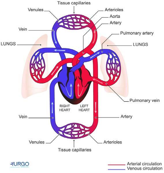
7 Systemic and Pulmonary Circulation Pulmonary circulation The flow of blood from the heart through the lungs back to the heart Picks up oxygen and releases carbon dioxide in the lungs Systemic circulation The flow of blood from the heart through the body back to the heart Delivers oxygen and picks up carbon dioxide in the body s tissues Cardiac circulation: blood flow within the heart
8 The heart beat is strong enough to squirt blood 30 feet The human heart beats ~35 million times per year Your heart beats 100,000 times and pumps ~2000 gallons of blood every day The heart pumps ~1,000,000 barrels of blood in a lifetime Most heart attacks occur between 8-9 a.m.
9 The blue whale has the largest heart it weights ~ one ton The hummingbird has a heart that beats 1000 times per minute Your entire volume of blood goes through your entire body once every minute Humans have ~60,000 miles of blood vessels in their bodies (more than twice the circumference of the earth!) Pig and baboon hearts have been transplanted into humans
10 Functions of the Heart 1. Generating blood pressure Required for blood flow through the blood vessels 2. Routing blood Two pumps, moving blood through the pulmonary and systemic circulations 3. Regulating blood supply Adjusts blood flow by changing the rate and force of heart contractions as needed

 at the level of the")

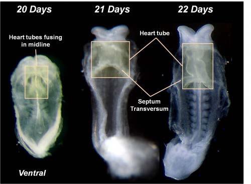
11 ONTOGENESIS Heart develops from the mesoderm on 3rd week embryogenesis a pair of tubes (sacks) at the level of the pharynx to either side of the primary gut between endoderm and splanchnopleura. 2 weeks
12

13 ONTOGENESIS 2.5 weeks 3 weeks The tubular heart with a two-layer wall. In the future, the inner layer of the tube formed endocardium, and from the outside - the myocardium and epicardium. Primordium the heart is located in the neck.
14

15 Development of the heart
16
17

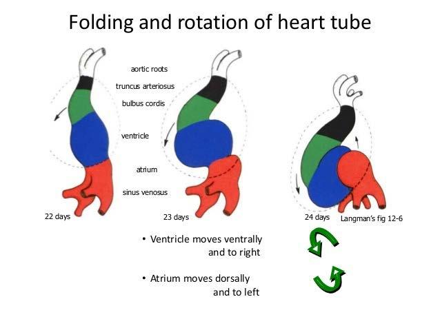
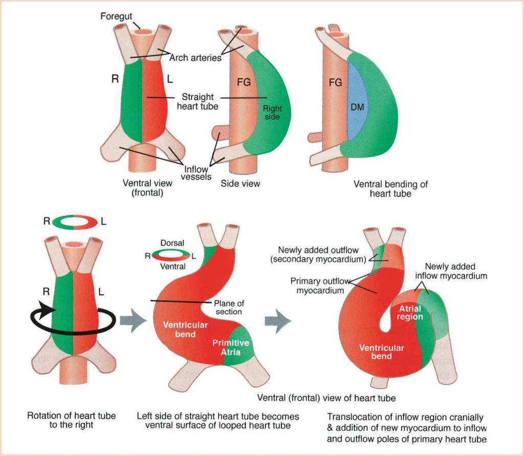
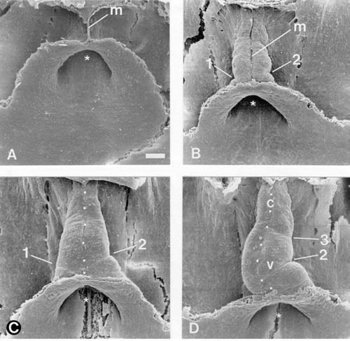
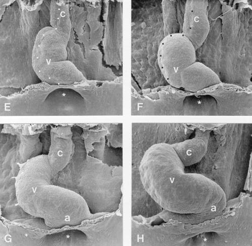
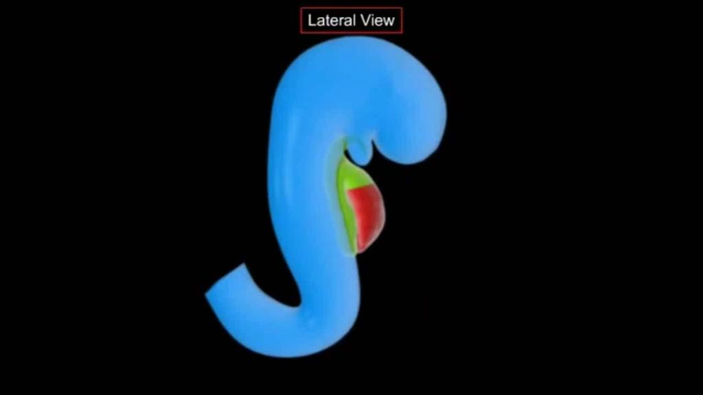
18 Folding and rotation of the heart tube Straight heart tube C- shaped tube S - shaped tube
19 Development of the heart

20 Natural embryonic heart after the torsion
21
22

23 Development of the heart On the 4th week of embryogenesis from upper rear of the inner surface of the total atrial growing primary interatrial septum toward the atrioventricular opening and separetes the common atrium into right and left.

24 This separation is not complete, because stored in the partition wide primary atrial opening. Right from the primary partition of the upper posterior wall of the atrium is growing secondary interatrial septum, which merges with the primary, completely separating the two atria. Cranial part of the secondary partitions breaks, forming a secondary atrial hole (foramen ovale).

25 At the beginning of the 8th week of embryogenesis on low back wall of the ventricles the fold is formed - the future interventricular septum, which grows upward and forward
26

27 Skeletopy of the Heart Oblique Position Apex: in the 5 th intercostal space 1cm from the left mammilary line toward the midline; Superior border passes on a level with the superior margin of the 3 rd costal cartilages; Right border passes 2-3cm to the right of the right sternal border between the 3 rd and 5 th ribs; Inferior border stretches transversely from the cartilage of the 5 th right rib to the heart apex; Left border passes from the cartilage of the 3 rd rib to the heart apex.
 surface comprises the: right atrium, atrioventricular groove and both ventricles separated by the interventricular groove.")
28 The heart surfaces: The anterior (sternocostal) surface comprises the: right atrium, atrioventricular groove, right ventricle, a small strip of left ventricle and the auricle of the left atrium. The inferior (diaphragmatic) surface comprises the: right atrium, atrioventricular groove and both ventricles separated by the interventricular groove. The posterior surface (base) comprises the left atrium receiving the four pulmonary veins. The Heart

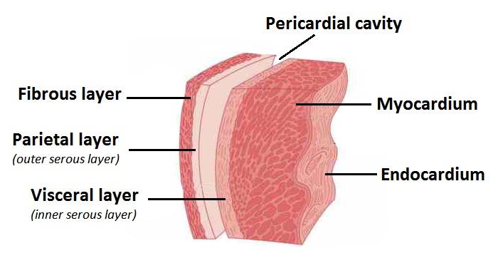
29 Heart Wall The heart wall has three layers Epicardium Visceral layer of the serous pericardium (visceral pericardium) Provides protection against the friction of rubbing organs Myocardium Cardiac muscle layer forming the bulk of the heart Responsible for contraction Endocardium Endothelial layer over crisscrossing, interlacing layer of connective tissue Inner endocardium reduces the friction resulting from the passage of blood through the heart Ventricles have ridges called trabeculae carneae The inner surfaces of the atria are mainly smooth Auricles have raised areas called musculi pectinati

30 Myocardium

31 Papillary Muscles Flaps of muscles connected to various valves on one end by chordae tendinae and to ventricular walls on the other Stabilize valves

32 Fibrous Skeleton of the Heart Consists of a plate of fibrous connective tissue Forms fibrous rings around the AV and SL valves for support Provides a point of attachment for heart muscle Electrically insulates the atria from the ventricles

33 Fibrous skeleton (rings, trigons) and myocardium Connective tissue heart frame: 1 - fibrous base mouth LA; 2 - fibrous base of the aorta; 3 - right atrial arch of fibrous ring; 4 - back arch of right fibrous ring; 5 - posterior arch of the left fibrous ring; 6 - a forward arch of left fibrous ring; 7 - left fibrotic triangle; 8 - right fibrous triangle; 9 - the front part of the right fibrous triangle; 10 posterior part of right fibrous triangle


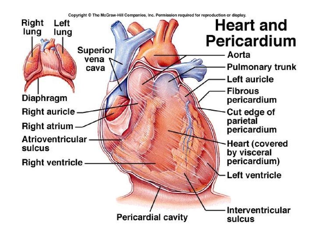

34 The Heart Pericardium includes the parietal and visceral layers with potential cavity
35 The Heart Chambers Atria Features small, thin-walled chambers Functions receiving chambers for blood returning to the heart from the circulation push the blood into the adjacent ventricles.
 Left side Pulmonary Veins")
36 The Heart Chambers Atria Receive blood from Right side Superior and Inferior Vena Cava Coronary Sinus (draining the myocardium) Left side Pulmonary Veins
37 Receives deoxygenated blood from the inferior vena cava below and from the superior vena cava above. Receives the coronary sinus in its lower part. The upper end of the atrium projects to the left of the superior vena cava as the right auricle. The Right Atrium
38 Receives oxygenated blood from four pulmonary veins which drain posteriorly. The cavity is smooth walled except for the atrial appendage. On the septal surface a depression marks the fossa ovalis. The Left Atrium
39 Ventricles Features The Heart Chambers make up most of the mass of the heart the walls of the left ventricle are thicker than those of the right
, Aorta (left ventricle)")
40 Ventricles Functions The Heart Chambers discharging chambers of the heart propel blood to Pulmonary Trunk (right ventricle), Aorta (left ventricle)
41 The Right Ventricle Receives blood from the right atrium through the tricuspid valve. The edges of the valve cusps are attached to chordae tendineae which are, in turn, attached below to papillary muscles.
42 The Right Ventricle The wall of the right ventricle is thicker than that of the atria but not as thick as that of the left ventricle. The wall contains a mass of muscular bundles known as trabeculae carneae. The infundibulum is the smooth walled outflow tract of the right ventricle.

43 The Right Ventricle The pulmonary valve is situated at the top of the infundibulum. It is composed of three semilunar cusps. Blood flows through the valve and into the pulmonary arteries via the pulmonary trunk to be oxygenated in the lungs.

44 The vestibule is a smooth walled part of the left ventricle which is located below the aortic valve. The Left Ventricle

45 Heart valves ensure unidirectional blood flow through the heart Composed of an endocardium with a connective tissue core. Two major types Atrioventricular valves Semilunar valves The Heart Valves
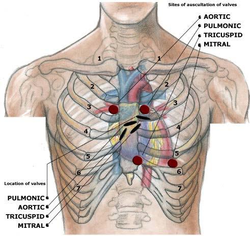
46 The Heart Valves Atrioventricular valves Right AV (Tricuspid) separates the right atrium from the right ventricle. Prevents backflow into atrium. Left AV (Bicuspid) separates the left atrium from the left ventricle. Prevents backflow into atrium. Semilunar valves Pulmonary valve separates the right ventricle from the pulmonary arteries. Prevents backflow after ventricular contraction. Aortic valve separates the left ventricle from the aorta. Prevents backflow after ventricular contraction.
47 Atrioventricular (AV) Valves Atrioventricular (AV) valves lie between the atria and the ventricles R-AV valve = tricuspid valve L-AV valve = bicuspid or mitral valve AV valves prevent backflow of blood into the atria when ventricles contract Chordae tendineae anchor AV valves to papillary muscles of ventricle wall Prevent prolapse of valve back into atrium
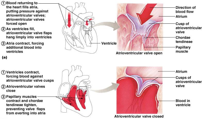
48 Atrioventricular Valve
 due to valves closing Lub - closing of atrioventricular valves Dub - closing of semilunar")
49 Semilunar Heart Valves Semilunar valves prevent backflow of blood into the ventricles Have no chordae tendinae attachments Aortic semilunar valve lies between the left ventricle and the aorta Pulmonary semilunar valve lies between the right ventricle and pulmonary trunk Heart sounds ( lub-dup ) due to valves closing Lub - closing of atrioventricular valves Dub - closing of semilunar valves

50 VALVULAR APPARATUS OF THE HEART Right AV (tricuspid) valve Chordai tendineae Papillary muscle Pulmonary semilunar valve Aortic semilunar valve Left AV (bicuspid) valve
51
52 Cardiac Cycle Overview of Systole and Diastole During systole AV valves close Pressure increases in the ventricles Semilunar valves are forced to open Blood flows into the aorta and pulmonary trunk At the beginning of diastole Pressure in the ventricles decreases Semilunar valves close to prevent backflow of blood from the aorta and pulmonary trunk into the ventricles When the pressure in the ventricles is lower than in the atria, the AV valves open and blood flows from the atria into the ventricles During atrial systole, the atria contract and complete the filling of the ventricles
53
54

55 FINISH!
THE CARDIOVASCULAR SYSTEM. Part 1
 THE CARDIOVASCULAR SYSTEM Part 1 CARDIOVASCULAR SYSTEM Blood Heart Blood vessels What is the function of this system? What other systems does it affect? CARDIOVASCULAR SYSTEM Functions Transport gases,
Anatomy of the Heart. Figure 20 2c
 Anatomy of the Heart Figure 20 2c Pericardium & Myocardium Remember, the heart sits in it s own cavity, known as the mediastinum. The heart is surrounded by the Pericardium, a double lining of the pericardial
2. right heart = pulmonary pump takes blood to lungs to pick up oxygen and get rid of carbon dioxide
 A. location in thorax, in inferior mediastinum posterior to sternum medial to lungs superior to diaphragm anterior to vertebrae orientation - oblique apex points down and to the left 2/3 of mass on left
Chapter 20 (1) The Heart
 The Heart") Chapter 20 (1) The Heart Learning Objectives Describe the location and structure of the heart Describe the path of a drop of blood from the superior vena cava or inferior vena cava through the heart out
Chapter 20 (1) The Heart Learning Objectives Describe the location and structure of the heart Describe the path of a drop of blood from the superior vena cava or inferior vena cava through the heart out
Heart Anatomy. 7/5/02 Stephen G Davenport 1
 Heart Anatomy Copyright 1999, Stephen G. Davenport, No part of this publication may be reproduced, stored in a retrieval system, or transmitted, in any form without prior written permission. 7/5/02 Stephen
the Cardiovascular System I
 the Cardiovascular System I By: Dr. Nabil A Khouri MD, MsC, Ph.D MEDIASTINUM 1. Superior Mediastinum 2. inferior Mediastinum Anterior mediastinum. Middle mediastinum. Posterior mediastinum Anatomy of
THE HEART. A. The Pericardium - a double sac of serous membrane surrounding the heart
 THE HEART I. Size and Location: A. Fist-size weighing less than a pound (250 to 350 grams). B. Located in the mediastinum between the 2 nd rib and the 5 th intercostal space. 1. Tipped to the left, resting
human anatomy 2016 lecture thirteen Dr meethak ali ahmed neurosurgeon
 Heart The heart is a hollow muscular organ that is somewhat pyramid shaped and lies within the pericardium in the mediastinum. It is connected at its base to the great blood vessels but otherwise lies
THE HEART OBJECTIVES: LOCATION OF THE HEART IN THE THORACIC CAVITY CARDIOVASCULAR SYSTEM
 BIOLOGY II CARDIOVASCULAR SYSTEM ACTIVITY #3 NAME DATE HOUR THE HEART OBJECTIVES: Describe the anatomy of the heart and identify and give the functions of all parts. (pp. 356 363) Trace the flow of blood
Chapter 14. The Cardiovascular System
 Chapter 14 The Cardiovascular System Introduction Cardiovascular system - heart, blood and blood vessels Cardiac muscle makes up bulk of heart provides force to pump blood Function - transports blood 2
CV Anatomy Quiz. Dr Ella Kim Dr Pip Green
 CV Anatomy Quiz Dr Ella Kim Dr Pip Green Q1 The location of the heart is correctly described as A) lateral to the lungs. B) medial to the sternum. C) superior to the diaphragm. D) posterior to the spinal
Human Anatomy, First Edition
 Human Anatomy, First Edition McKinley & O'Loughlin Chapter 22 : Heart 1 Functions of the Heart Center of the cardiovascular system, the heart. Connects to blood vessels that transport blood between the
The Heart. Happy Friday! #takeoutyournotes #testnotgradedyet
 The Heart Happy Friday! #takeoutyournotes #testnotgradedyet Introduction Cardiovascular system distributes blood Pump (heart) Distribution areas (capillaries) Heart has 4 compartments 2 receive blood (atria)
Ch 19: Cardiovascular System - The Heart -
 Ch 19: Cardiovascular System - The Heart - Give a detailed description of the superficial and internal anatomy of the heart, including the pericardium, the myocardium, and the cardiac muscle. Trace the
Heart Dissection. 5. Locate the tip of the heart or the apex. Only the left ventricle extends all the way to the apex.
 Heart Dissection Page 1 of 6 Background: The heart is a four-chambered, hollow organ composed primarily of cardiac muscle tissue. It is located in the center of the chest in between the lungs. It is the
The HEART. What is it???? Pericardium. Heart Facts. This muscle never stops working It works when you are asleep
 This muscle never stops working It works when you are asleep The HEART It works when you eat It really works when you exercise. What is it???? Located between the lungs in the mid thoracic region Apex
Ch.15 Cardiovascular System Pgs {15-12} {15-13}
 Ch.15 Cardiovascular System Pgs {15-12} {15-13} E. Skeleton of the Heart 1. The skeleton of the heart is composed of rings of dense connective tissue and other masses of connective tissue in the interventricular
The Cardiovascular System Part I: Heart Outline of class lecture After studying part I of this chapter you should be able to:
 The Cardiovascular System Part I: Heart Outline of class lecture After studying part I of this chapter you should be able to: 1. Describe the functions of the heart 2. Describe the location of the heart,
Cardiovascular Anatomy Dr. Gary Mumaugh
 Cardiovascular Anatomy Dr. Gary Mumaugh Location of Heart Approximately the size of your fist Location o Superior surface of diaphragm o Left of the midline in mediastinum o Anterior to the vertebral column,
Cardiovascular System. Heart Anatomy
 Cardiovascular System Heart Anatomy 1 The Heart Location & general description: Atria vs. ventricles Pulmonary vs. systemic circulation Coverings Walls The heart is found in the mediastinum, the medial
The Heart. The Heart A muscular double pump. The Pulmonary and Systemic Circuits
 C H A P T E R 19 The Heart The Heart A muscular double pump circuit takes blood to and from the lungs Systemic circuit vessels transport blood to and from body tissues Atria receive blood from the pulmonary
Circulation. Circulation = is a process used for the transport of oxygen, carbon! dioxide, nutrients and wastes through-out the body
 Circulation Circulation = is a process used for the transport of oxygen, carbon! dioxide, nutrients and wastes through-out the body Heart = muscular organ about the size of your fist which pumps blood.
The Cardiovascular System (Heart)
") The Cardiovascular System The Cardiovascular System (Heart) A closed system of the heart and blood vessels The heart pumps blood Blood vessels allow blood to circulate to all parts of the body The function
The Cardiovascular System The Cardiovascular System (Heart) A closed system of the heart and blood vessels The heart pumps blood Blood vessels allow blood to circulate to all parts of the body The function
LAB 12-1 HEART DISSECTION GROSS ANATOMY OF THE HEART
 LAB 12-1 HEART DISSECTION GROSS ANATOMY OF THE HEART Because mammals are warm-blooded and generally very active animals, they require high metabolic rates. One major requirement of a high metabolism is
The Cardiovascular System
 11 PART A The Cardiovascular System PowerPoint Lecture Slide Presentation by Jerry L. Cook, Sam Houston University ESSENTIALS OF HUMAN ANATOMY & PHYSIOLOGY EIGHTH EDITION ELAINE N. MARIEB The Cardiovascular
The Cardiovascular System. Chapter 15. Cardiovascular System FYI. Cardiology Closed systemof the heart & blood vessels. Functions
 Chapter 15 Cardiovascular System FYI The heart pumps 7,000 liters (4000 gallons) of blood through the body each day The heart contracts 2.5 billion times in an avg. lifetime The heart & all blood vessels
Read Chapters 21 & 22, McKinley et al
 ACTIVITY 9: BLOOD AND HEART OBJECTIVES: 1) How to get ready: Read Chapters 21 & 22, McKinley et al., Human Anatomy, 5e. All text references are for this textbook. Read dissection instructions BEFORE YOU
The Heart. Size, Form, and Location of the Heart. 1. Blunt, rounded point; most inferior part of the heart.
 12 The Heart FOCUS: The heart is composed of cardiac muscle cells, which are elongated, branching cells that appear striated. Cardiac muscle cells behave as a single electrical unit, and the highly coordinated
Cardiovascular System. I. Structures of the heart A. : Pericardium sack that surrounds the heart
 Cardiovascular System I. Structures of the heart A. : Pericardium sack that surrounds the heart 1. : Pericardial Cavity serous fluid filled space between the heart and the pericardium B. Heart Wall 1.
Figure ) The specific chamber of the heart that is indicated by letter A is called the. Diff: 1 Page Ref: 364
 The specific chamber of the heart that is indicated by letter A is called the. Diff: 1 Page Ref: 364") Essentials of Anatomy and Physiology, 9e (Marieb) Chapter 11 The Cardiovascular System Short Answer Figure 11.1 Using Figure 11.1, identify the following: 1) The Purkinje fibers are indicated by label.
Essentials of Anatomy and Physiology, 9e (Marieb) Chapter 11 The Cardiovascular System Short Answer Figure 11.1 Using Figure 11.1, identify the following: 1) The Purkinje fibers are indicated by label.
Read Me. covering the Heart Anatomy. Labs. textbook. use. car: you
 Heart Anatomy Lab Pre-Lab Exercises Read Me These exercises should be done before coming to lab, after watching the videos covering the Heart Anatomy Labs. Answer the questions in this guide using the
Anatomy of the Heart
 Biology 212: Anatomy and Physiology II Anatomy of the Heart References: Saladin, KS: Anatomy and Physiology, The Unity of Form and Function 8 th (2018). Required reading before beginning this lab: Chapter
Introduction to Anatomy. Dr. Maher Hadidi. Bayan Yanes. April/9 th /2013
 Introduction to Anatomy Dr. Maher Hadidi Bayan Yanes 27 April/9 th /2013 KEY POINTS: 1) Right side of the heart 2) Papillary muscles 3) Left side of the heart 4) Comparison between right and left sides
Lab Activity 23. Cardiac Anatomy. Portland Community College BI 232
 Lab Activity 23 Cardiac Anatomy Portland Community College BI 232 Cardiac Muscle Histology Branching cells Intercalated disc: contains many gap junctions connecting the adjacent cell cytoplasm, creates
Pearson's Comprehensive Medical Assisting Administrative and Clinical Competencies
 Pearson's Comprehensive Medical Assisting Administrative and Clinical Competencies THIRD EDITION CHAPTER 27 The Cardiovascular System Lesson 1: Overview of the Cardiovascular System Lesson Objectives Upon
This lab activity is aligned with Visible Body s A&P app. Learn more at visiblebody.com/professors
 1 This lab activity is aligned with Visible Body s A&P app. Learn more at visiblebody.com/professors 2 PRE-LAB EXERCISES: A. Watch the video 29.1 Heart Overview and make the following observations: 1.
The Cardiovascular System
 Essentials of Human Anatomy & Physiology Elaine N. Marieb Slides 11.1 11.19 Seventh Edition Chapter 11 The Cardiovascular System Functions of the Cardiovascular system Function of the heart: to pump blood
Chapter 20: Cardiovascular System: The Heart
 Chapter 20: Cardiovascular System: The Heart I. Functions of the Heart A. List and describe the four functions of the heart: 1. 2. 3. 4. II. Size, Shape, and Location of the Heart A. Size and Shape 1.
ACTIVITY 9: BLOOD AND HEART BLOOD
 ACTIVITY 9: BLOOD AND HEART OBJECTIVES: 1) How to get ready: Read Chapters 21 & 22, McKinley et al., Human Anatomy, 4e. All text references are for this textbook. Read dissection instructions BEFORE YOU
STRUCTURES OF THE CARDIOVASCULAR SYSTEM
 STRUCTURES OF THE CARDIOVASCULAR SYSTEM CARDIOVASCULAR SYSTEM Also called the circulatory system Consists of the heart, arteries, veins, and capillaries Main function is to pump/circulate oxygenated blood
The Cardiovascular System
 Essentials of Human Anatomy & Physiology Elaine N. Marieb Seventh Edition Chapter 11 The Cardiovascular System Slides 11.1 11.19 Lecture Slides in PowerPoint by Jerry L. Cook The Cardiovascular System
Chapter 18 - Heart. I. Heart Anatomy: size of your fist; located in mediastinum (medial cavity)
") Chapter 18 - Heart I. Heart Anatomy: size of your fist; located in mediastinum (medial cavity) A. Coverings: heart enclosed in double walled sac called the pericardium 1. Fibrous pericardium: dense connective
Chapter 18 - Heart I. Heart Anatomy: size of your fist; located in mediastinum (medial cavity) A. Coverings: heart enclosed in double walled sac called the pericardium 1. Fibrous pericardium: dense connective
Chapter 14. Circulatory System Images. VT-122 Anatomy & Physiology II
 Chapter 14 Circulatory System Images VT-122 Anatomy & Physiology II The mediastinum Dog heart Dog heart Cat heart Dog heart ultrasound Can see pericardium as distinct bright line Pericardial effusion Fluid
CIRCULATORY SYSTEM BLOOD VESSELS
 Name: Block: CIRCULATORY SYSTEM Multicellular organisms (above the level of roundworms) rely on a circulatory system to bring nutrients to, and take wastes away from, cells. In higher organisms such as
Approximately the size of your fist Location Superior surface of diaphragm Left of the midline in mediastinum Anterior to the vertebral column,
 Dr. Gary Mumaugh Approximately the size of your fist Location Superior surface of diaphragm Left of the midline in mediastinum Anterior to the vertebral column, posterior to the sternum Posteriorly the
THE HEART. Unit 3: Transportation and Respiration
 THE HEART Unit 3: Transportation and Respiration The Circulatory System Also called the Cardiovascular System Circulates blood in the body Transports nutrients, oxygen, carbon dioxide, hormones, and blood
Anatomy lab -1- Imp note: papillary muscle Trabeculae Carneae chordae tendineae
 Anatomy lab -1- Imp note: the arrangement of this sheet is different than the lab recording, it has been arranged in a certain way to make it easier to study. When you open the left ventricle you can see
Middle mediastinum---- heart & pericardium. Dep. of Human Anatomy Zhou Hongying
 Middle mediastinum---- heart & pericardium Dep. of Human Anatomy Zhou Hongying eaglezhyxzy@163.com Subdivisions of the mediastinum Contents of Middle mediastinum Heart Pericardium: a serous sac enclosing
Human Anatomy and Physiology Chapter 19 Worksheet 1- The Heart
 Human Anatomy and Physiology Chapter 19 Worksheet 1- The Heart Name Date Period 1. The "double pump" function of the heart includes the right side, which serves as the circuit pump, while the left side
Cardiovascular System Module 3: Heart Anatomy *
 OpenStax-CNX module: m49683 1 Cardiovascular System Module 3: Heart Anatomy * Donna Browne Based on Heart Anatomy by OpenStax This work is produced by OpenStax-CNX and licensed under the Creative Commons
Cardiovascular System
 Cardiovascular System Purpose Transport oxygen and nutrients Take waste products away from tissues & organs Things we learned Blood pressure: the force of blood pushing against the walls of blood vessels
Circulatory Systems. All cells need to take in nutrients and expel metabolic wastes.
 Circulatory Systems All cells need to take in nutrients and expel metabolic wastes. Single celled organisms: nutrients from the environment can diffuse (or be actively transported) directly in to the cell
The Cardiovascular System
 PowerPoint Lecture Slide Presentation by Patty Bostwick-Taylor, Florence-Darlington Technical College The Cardiovascular System 11 PART A The Cardiovascular System A closed system of the heart and blood
The Cardiovascular System: The Heart: Part A
 PowerPoint Lecture Slides prepared by Janice Meeking, Mount Royal College CHAPTER 18 The Cardiovascular System: The Heart: Part A Heart Anatomy Approximately the size of a fist Location In the mediastinum
The Heart. Made up of 3 different tissue: cardiac muscle tissue, nerve tissue, and connective tissue.
 The Heart The Heart Made up of 3 different tissue: cardiac muscle tissue, nerve tissue, and connective tissue. Your heart pumps with a regular beat (Heart Rate) Your heart rate can change depending on
Danil Hammoudi.MD 1/12/2009
 Danil Hammoudi.MD Aorta the biggest and longest artery (a blood vessel carrying blood away from the heart) in the body. It carries oxygen rich blood from the left ventricle of the heart to the body.inferior
10. Thick deposits of lipids on the walls of blood vessels, called, can lead to serious circulatory issues. A. aneurysm B. atherosclerosis C.
 Heart Student: 1. carry blood away from the heart. A. Arteries B. Veins C. Capillaries 2. What is the leading cause of heart attack and stroke in North America? A. alcohol B. smoking C. arteriosclerosis
37 1 The Circulatory System
 H T H E E A R T 37 1 The Circulatory System The circulatory system and respiratory system work together to supply cells with the nutrients and oxygen they need to stay alive. a) The respiratory system:
Heart & Pericardium. December, 2015
 Heart & Pericardium December, 2015 2 Pericardium Definition Fibro-serous sac that encloses the heart and the roots of great vessels Function Restrict excessive movements of the heart as a whole Serve as
Blood and Heart. Student Learning Objectives:
 Blood and Heart Student Learning Objectives: Identify the major components of the blood. Identify the primary structures associated with the heart Follow the blood through the path of the circulation.
An Illustrated 1. Dissection Guide. To The... Mammalian. rr= Heart. Right ventricle+---, by David H. Hall
 An Illustrated 1. Dissection Guide. To The... Mammalian rr= Heart ventricle+---, by David H. Hall The Mam.malian Heart Because mammals are warm blooded (endothermic) and generally very active animals,
The Cardiovascular System
 The Cardiovascular System The Cardiovascular System A closed system of the heart and blood vessels The heart pumps blood Blood vessels allow blood to circulate to all parts of the body The function of
Circulatory system. Lecture #2
 Circulatory system Lecture #2 The essential components of the human cardiovascular system: Heart Blood Blood vessels Arteries - blood vessels that conduct arterial blood from heart ventricle to organs
BIOLOGY 2060 LECTURE NOTES ANATOMY & PHYSIOLOGY II (A. IMHOLTZ) HEART P1 OF 5
 HEART P1 OF 5") BIOLOGY 2060 LECTURE NOTES ANATOMY & PHYSIOLOGY II (A. IMHOLTZ) HEART P1 OF 5 1. Heart Functions a. Generates pressure that propels blood thru blood vessels. (Tissue perfusion.) b. Separates oxygenated
BIOLOGY 2060 LECTURE NOTES ANATOMY & PHYSIOLOGY II (A. IMHOLTZ) HEART P1 OF 5 1. Heart Functions a. Generates pressure that propels blood thru blood vessels. (Tissue perfusion.) b. Separates oxygenated
Approximately the size of your fist Location. Pericardial physiology
 Heart Anatomy Approximately the size of your fist Location Superior surface of diaphragm Left of the midline Anterior to the vertebral column, posterior to the sternum Wednesday, March 28, 2012 Muscle
BIOL 4350 Cardiovascular Physiology Dr. Hamilton. Using the figure above, match the following: 1. Purkinje fibers. 2. SA node. 3. AV node.
 BIOL 4350 Cardiovascular Physiology Dr. Hamilton Using the figure above, match the following: 1. Purkinje fibers. 2. SA node. 3. AV node. 1 Using the figure above, match the following: 4. Atrial depolarization.
Major Function of the Cardiovascular System. Transportation. Structures of the Cardiovascular System. Heart - muscular pump
 Structures of the Cardiovascular System Heart - muscular pump Blood vessels - network of tubes Blood - liquid transport vehicle brachiocephalic trunk superior vena cava right pulmonary arteries right pulmonary
MODULE 2: CARDIOVASCULAR SYSTEM ANTOMY An Introduction to the Anatomy of the Heart and Blood vessels
 MODULE 2: CARDIOVASCULAR SYSTEM ANTOMY An Introduction to the Anatomy of the Heart and Blood vessels The cardiovascular system includes a pump (the heart) and the vessels that carry blood from the heart
The Cardiovascular System
 C H A P T E R 1 4 The Cardiovascular System OBJECTIVES After studying this chapter, you should be able to: 1. Describe how the heart is positioned in the thoracic cavity. 2. List and describe the layers
The Cardiovascular System. anatom.ua 1
 The Cardiovascular System anatom.ua 1 The Closed Circulatory System Humans have a closed circulatory system, typical of all vertebrates, in which blood is confined to vessels and is distinct from the interstitial
Practical Histology. Cardiovascular System. Dr Narmeen S. Ahmad
 Practical Histology Cardiovascular System Dr Narmeen S. Ahmad The Cardiovascular System A closed system of the heart and blood vessels Functions of cardiovascular system: Transport nutrients, hormones
Cardiovascular System
 Cardiovascular System I. Structure of the Heart A. Average adult heart is 14 cm long and 9 cm wide. B. Lies in the mediastinum. C. Enclosed in the pericardium. 1. Fibrous pericardium- Outer, tough connective
BIOLOGY 2060 LECTURE NOTES ANATOMY & PHYSIOLOGY II (A. IMHOLTZ) HEART P1 OF 7
 HEART P1 OF 7") BIOLOGY 2060 LECTURE NOTES ANATOMY & PHYSIOLOGY II (A. IMHOLTZ) HEART P1 OF 7 1. Heart a. Generates the pressure that propels blood thru blood vessels. b. Separates oxygenated and deoxygenated blood separate.
BIOLOGY 2060 LECTURE NOTES ANATOMY & PHYSIOLOGY II (A. IMHOLTZ) HEART P1 OF 7 1. Heart a. Generates the pressure that propels blood thru blood vessels. b. Separates oxygenated and deoxygenated blood separate.
The Cardiovascular and Lymphatic Systems Cardiovascular System Blood Vessels Blood Vessels Arteries Arteries Arteries
 CH 12 The Cardiovascular and s The Cardiovascular and s OUTLINE: Cardiovascular System Blood Vessels Blood Pressure Cardiovascular System The cardiovascular system is composed of Blood vessels This system
11/10/2014. Muscular pump Two atria Two ventricles. In mediastinum of thoracic cavity 2/3 of heart's mass lies left of midline of sternum
 It beats over 100,000 times a day to pump over 1,800 gallons of blood per day through over 60,000 miles of blood vessels. During the average lifetime, the heart pumps nearly 3 billion times, delivering
CJ Shuster A&P2 Lab Addenum Beef Heart Dissection 1. Heart Dissection. (taken from Johnson, Weipz and Savage Lab Book)
") CJ Shuster A&P2 Lab Addenum Beef Heart Dissection 1 Heart Dissection. (taken from Johnson, Weipz and Savage Lab Book) Introduction When you have finished examining the model, you are ready to begin your
CJ Shuster A&P2 Lab Addenum Beef Heart Dissection 1 Heart Dissection. (taken from Johnson, Weipz and Savage Lab Book) Introduction When you have finished examining the model, you are ready to begin your
- what other structures, besides the heart, does the mediastinum contain?
 Basic A & P II Dr. L. Bacha Chapter Outline (Martini & Nath 2010) An Introduction to the Cardiovascular System - read the paragraphs under this heading on page 580 The Heart is a Four Chambered Organ describe
INTRODUCTORY REMARKS:
 INTRODUCTORY REMARKS: The circulatory system provides a way for the blood to be transported throughout the body. This provides nutrients to the cells and allows wastes to be removed. Open vs. Closed Circulatory
The heart & Cardiovascular system
 The heart & Cardiovascular system The heart s continuous pulse create a base for our understanding of rhythms in everyday life. Bonnie Bainbridge Cohen The heart constantly beats throughout our lives never
The Cardiovascular System
 The Cardiovascular System The Manila Times College of Subic Prepared by: Stevens B. Badar, RN, MANc THE HEART Anatomy of the Heart Location and Size approx. the size of a person s fist, hollow and cone-shaped,
Figure 10.1A Transparency Master 79
 Brain Carotid arteries Jugular vein Right front leg Lungs (inflated) Cranial Right atrium To left front leg Left subclavian Bronchus capillaries Brachiocephalic vein Left atrium Dorsal aorta Right ventricle
Blood Functions. Blood and the Cardiovascular System. Blood. Plasma. Erythrocytes (RBCs) Erythrocytes (RBCs) 4/7/2017
 Erythrocytes (RBCs) 4/7/2017") Blood Functions Blood and the Cardiovascular System Distribution Delivery of oxygen and nutrients to all body cells; Transport of wastes to lungs and excretory organs; Transport of hormones Regulation
Blood Functions Blood and the Cardiovascular System Distribution Delivery of oxygen and nutrients to all body cells; Transport of wastes to lungs and excretory organs; Transport of hormones Regulation
DEVELOPMENT OF THE CIRCULATORY SYSTEM L E C T U R E 5
 DEVELOPMENT OF THE CIRCULATORY SYSTEM L E C T U R E 5 REVIEW OF CARDIAC ANATOMY Heart 4 chambers Base and apex Valves Pericardial sac 3 layers: epi, myo, endo cardium Major blood vessels Aorta and its
Large Arteries of Heart
 Cardiovascular System (Part A-2) Module 5 -Chapter 8 Overview Arteries Capillaries Veins Heart Anatomy Conduction System Blood pressure Fetal circulation Susie Turner, M.D. 1/5/13 Large Arteries of Heart
Unit 10 Cardiovascular System
 Unit 10 Cardiovascular System I. Functions Deliver nutrients to cells > O 2, sugars, amino acids, lipids, ions, H 2 O... Remove waste from cells > CO 2, pathogens, toxins, lactic acid... Fight off infection
ANATOMY. lecture#: Date : Lecturer : Maher Hadidi
 ANATOMY 28 lecture#: Date : Lecturer : Maher Hadidi Superior vena -=- Blood inflow part is rough and outflow part is smooth. - _Arch of aorta Pulmonary trunk Tricuspid valve Right auricle Right atrium
Biology Unit 3 The Human Heart P
 Biology 2201 Unit 3 The Human Heart P 314-321 Structure and Function of the Human Heart Structure of the Human Heart Has four Chambers (2 Atria and 2 Ventricles) Made of Cardiac Muscle Found in Chest Cavity
The Cardiovascular and Lymphatic Systems
 BIOLOGY OF HUMANS Concepts, Applications, and Issues Fifth Edition Judith Goodenough Betty McGuire 12 The Cardiovascular and Lymphatic Systems Lecture Presentation Anne Gasc Hawaii Pacific University and
2. Obtain the following: eye guards gloves dissection tools: several blunt probes, scissors, a scalpel and forceps dissection pan sheep heart
 Week 04 Lab Heart Anatomy LEARNING OUTCOMES: Describe the gross external and internal anatomy of the heart. Identify and discuss the function of the valves of the heart. Identify the major blood vessels
Health Science 20 Circulatory System Notes
 Health Science 20 Circulatory System Notes Functions of the Circulatory System The circulatory system functions mainly as the body s transport system. It transports: o Oxygen o Nutrients o Cell waste o
Cardiovascular System Note-Taking Guide
 FUNctions: Name: 3-27-14 Cardiovascular System Note-Taking Guide Heart: Pumps and delivers through the body Blood: Vessels: Delivers and to the body Carries waste and Maintains homeostasis - Carries blood
Anatomy: Cardiovascular System
 In the name of God Anatomy: Cardiovascular System Moradian MD, MPH, PhD candidate Tehran University of Medical Sciences drmoradian@sums.ac.ir 2015 The Cardiovascular System A closed system of the heart
Anatomy & Physiology of Cardiovascular System. Chapter 18 & 19
 Anatomy & Physiology of Cardiovascular System Chapter 18 & 19 Objectives..cont 1. Discuss the physiological stages of cardiac muscle contraction. 2. Trace a typical ECG and label each wave or complex 3.
The Heart. C h a p t e r. PowerPoint Lecture Slides prepared by Jason LaPres Lone Star College - North Harris
 C h a p t e r 20 The Heart PowerPoint Lecture Slides prepared by Jason LaPres Lone Star College - North Harris Copyright 2009 Pearson Education, Inc., publishing as Pearson Benjamin Cummings Introduction
ANATDMY. lecture # : Date : Lecturer : Maher Hadidi
 ANATDMY 27 lecture # : Date : Lecturer : Maher Hadidi Pericardium A double-walled fibroserous conical-shaped sac, within middle mediastinum. Enclose the heart and roots of its large vessels. Vagus nerves
Circulatory System Objective sheet 3
 Circulatory System Objective sheet 3 10. Functions of blood 1) Transport oxygen and nutrients 2) Transport of carbon dioxide and waste 3) Protection against disease causing micro-organisms 4) Clotting
DISSECTION OF A SHEEP HEART
 DISSECTION OF A SHEEP HEART I. INTRODUCTION A. You will soon appreciate the point made previously the heart models just don t teach us what a real heart is like! Dissecting a sheep heart will give you
Part 1. Copyright 2011 Pearson Education, Inc. Figure Copyright 2011 Pearson Education, Inc.
 PowerPoint Lecture Slides prepared by Leslie Hendon University of Alabama, Birmingham C H A P T E R The Heart 19 Part 1 The Heart A muscular double pump circuit vessels transport blood to and from the
(2) (1) (3) (4) BLOOD PATHWAY ASSESSMENT RUBRIC
 (1) (3) (4) BLOOD PATHWAY ASSESSMENT RUBRIC") BLOODPATHWAYASSESSMENT(4) BLOOD%PATHWAY%ASSESSMENT%(3)% BLOODPATHWAYASSESSMENT(3) (4) (3) (2) (1) Using a completely blank diagram of the heart, all valves, chambers, great vessels, and direction of blood
BLOODPATHWAYASSESSMENT(4) BLOOD%PATHWAY%ASSESSMENT%(3)% BLOODPATHWAYASSESSMENT(3) (4) (3) (2) (1) Using a completely blank diagram of the heart, all valves, chambers, great vessels, and direction of blood
CIRCULATORY SYSTEM TASK CARDS Worksheet
 CIRCULATORY SYSTEM TASK CARDS Worksheet Name: Date: Instructions: Put the answers to each task card in the numbered boxes on the chart. 1 a) left semilunar valve / aortic valve b) blood would backflow