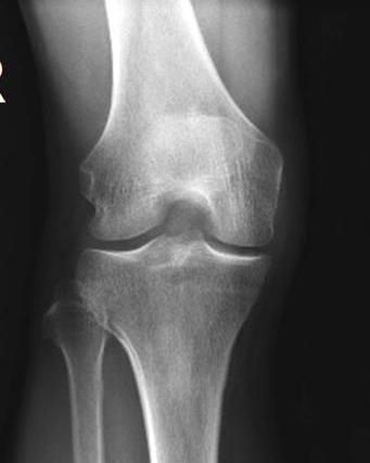
RADIOGRAPHY OF THE KNEE, PATELLA, and FEMUR
|
|
|
- Barnard Robbins
- 6 years ago
- Views:
Transcription
1 RADIOGRAPHY OF THE KNEE, PATELLA, and FEMUR


2 KNEE AP Projection Patient Position: Part Position: Leg in Center Femoral condyles
3 Central Ray: - Asthenic patient - if ASIS to tabletop is < 19 cm Sthenic patient if ASIS to tabletop is cm Hypersthenic patient if ASIS to tabletop is > 24 cm Note: CR is

4 Structures Shown:
5 Criteria of Evaluation: The femoral condyles should be The intercondylar eminences should be centered within the. The knee joint Intercondyloid fossa should be The head of the fibula should. The patella should be seen in the.
6 AP knee Error rotated causes lateral condyle to appear, intercondyloid fossa will be, and there will be. Correction - rotate leg. Good image

7 AP knee Error: Knee is this will cause the medial condyle to appear, the intercondyloid fossa will be, and there will be. Correction: rotate leg.

8 AP knee Error causes to be opened too much and the femur is. To correct:. Good image Correction - Fully extend knee if patient is able

9 KNEE AP Oblique Projection Medial (Internal) Rotation Patient Position: Part Position: Leg rotated Center
10 Central Ray: Asthenic patient - if ASIS to tabletop is < 19 cm Sthenic patient if ASIS to tabletop is cm Hypersthenic patient if ASIS to tabletop is > 24 cm


11 Structures Shown: Criteria of Evaluation The head of the fibula should be seen The lateral condyle should. The patella


12 Internal (medial) oblique knee- Error rotation causes the head of the fibula to be from the tibia. Correction -Oblique leg. (needs rotation) Good image

")
13 AP Oblique Projection Lateral (External) Rotation Structures shown;

14 External (lateral) oblique knee Error will cause the fibula to not be Good image Correction - Oblique leg. Needs more rotation.

15 Patient Position: KNEE Lateral Projection Part Position: Knee flexed Patella to IR Femoral condyles

 Center")
16 Central Ray: through knee joint (due to femoral condyle, plus it is at a lower level than the femoral condyle) Center epicondyle


17 Note: If patient unable to turn into lateral position, may be obtained


18 Structures Shown:

19 Criteria for Evaluation The distal articulating surfaces of the medial and lateral femoral condyles should be, and the knee joint space should. Determine if CR needs to be angled based on. Wide pelvis angle degrees. Narrow pelvis use angle.
20 Criteria for Evaluation The patella should be situated. The patellofemoral joint should. The tibia should be over the fibular head. Patella seen

21 Lateral Knee Error Knee is flexed the patellofemoral joint. Correction flex knee Good image


22 Lateral Knee Knee is the patellofemoral joint. Correction flex knee. Good image
.")
 to")
23 Lateral knee Error - the patella was situated too ( -rotated knee). The head of the fibula is than normal. Correction roll the patella ( ) to the IR.

.")

24 Lateral knee Error - the patella was situated too ( -rotated knee). There is. Correction - roll the patella ( ) from the IR and/or

25 Lateral knee Error. Note that medial femoral condyle. To correct.

26 Lateral knee, patella to IR, and. Correction: roll the patella the IR and have.

27 Case Study


28 Weight-Bearing Knees AP Projection For studies to show Note: CR is

29 Weight-Bearing Knees Lateral Projection For Note: CR is


30 PA Axial Bilateral Weight-Bearing Knees Rosenberg Method For studies to show narrowing of
31 INTERCONDYLAR FOSSA Tunnel Views Camp Coventry Method Homblad Method Beclere Method

32 PA Axial Projection Camp-Coventry Method Patient Position: Part Position: Knee of leg Foot

33 Central Ray: to through knee joint


34 Patient Position: PA Axial Projection Homblad Method Part Position: Knee Knee joint Femoral shafts Central Ray: to through ( knee joint)


35 Holmblad Method Modifications


36 Holmblad Method Modifications

37 AP Axial Projection Beclere Method Patient Position: Part Position: Knee flexed IR placed No Central Ray: Perpendicular to Angled through knee joint Enters

38 Structures Shown:

39 Criteria for Evaluation: Open Knee joint space Apex of patella of femoral condyles
40
 Central Ray: to IR Enters ;")
41 Patient Position: PATELLA PA Projection Part Position: Center IR to Rotate heel laterally to make patella to IR (or of anterior knee) Central Ray: to IR Enters ; exits

42 Structures Shown: NOTE: May be done, but detail better when done

43 Stellate Fracture of Patella

44 Patient Position: PATELLA Lateral Projection Part Position: Turn on to Knee flexed Patella is to IR Femoral condyles


45 Central Ray: to IR joint Structures Shown:


46 Transverse Fracture of Patella
47 PATELLA Sunrise or Skyline Views Merchant Method Hobbs Method Settegast Method Hughston Method


 of")
48 Bilateral Tangential Projection Merchant Method image ( projection) of both ; shows of patellae


49

50 Alternative method Hobbs Modification


51 Patient Position: Tangential Projections Settegast Methods Part Position: knee to place patella to IR Base of to IR


52 Settegast Method Modifications


53 Central Ray: to femoro-patellar joint space Angle depends on Structures Shown:


54 Tangential Projection Hughston Method Knee flexed from IR; CR angled Demonstrates

55 Tangential Patella error flexion of knee, patella superimposed on, joint space is. To correct flexion of knee or make the CR.
56
57 Trauma Radiography of the Patella Sequence of Radiographs shows shows Tangential shows * *Note: Acute flexion of knee should not be attempted
58 FEMUR AP Projection Patient Position: Part Position: Center of IR Rotate leg for distal femur (femoral condyles to IR); for proximal femur (femoral neck to IR)
59 Part Position (Cont d): For distal femur Bottom of IR is For proximal femur Top of IR at the Central Ray: to IR at midfemur


60 Structures Shown:

61 Criteria for Evaluation: Proximal femur Greater trochanter seen Femoral neck Lesser trochanter Distal femur No


62 AP s
63 Patient Position FEMUR Lateral Projection Femur Affected knee
64 Part Position: For distal femur Draw upper leg Pelvis true Patella and femoral condyles to IR Bottom of IR

65 Part Position: For proximal femur Upper leg drawn Pelvis rotated from lateral to prevent joint Top of IR at

66 Central Ray: to IR Structures Shown:
67 Criteria for Evaluation Proximal femur Lesser trochanter Greater trochanter Unaffected femur

68 Criteria for Evaluation Distal femur Anterior surface of femoral condyles Patella in Patellofemoral joint


69 Laterals

70 Trauma Lateromedial projection of Femur


71 Image Stitching

72 CT- Scanograms-Lower CT Scanograms Extremity Take CT Scout images of the femur/tibia For lower extremity measurements Place cursors over the respective hip, knee and ankle joints
73 Situation: A radiograph of a lateral projection of the patella reveals that the femoropatellar joint space is not open. The patella is within the intercondylar sulcus. Solution: The most likely cause of this is excessive of the knee.
74 Situation: A radiograph of an AP knee reveals rotation with almost total superimposition of the fibular head and the proximal tibia. Solution: To correct this positioning error on the repeat exposure, the technologist must rotate the knee slightly.
75 Situation: A radiograph of a PA axial projection for the intercondylar fossa does not demonstrate the fossa well. It is foreshortened. The following positioning factors were used: patient prone, knee flexed 40 to 45, CR angled to be perpendicular to the femur, 40-inch SID, and no rotation of the lower limb. Solution: To correct this positioning error on the repeat exposure, the technologist must direct the CR perpendicular to the to produce a more diagnostic image.
76 Situation: A patient comes to the radiology department for a knee study with special interest in the region of the proximal tibiofibular joint and the lateral condyle of the tibia. Solution: The positioning routines should include, oblique of the knee.
77 Situation: A geriatric patient comes to the radiology department for an intercondylar fossa study of the knee. The patient is unsteady and unsure of himself. Solution: If choosing between the Holmblad or the Camp-Coventry methods, the intercondylar fossa projection might provide the best results without risk of injury to the patient.
78 Situation: A patent enters the ER with a possible transverse fracture of the patella. Which projection(s) would safely confirm a transverse fracture without flexing the patient s knee? Solution: and
79 Situation: On a lateral knee radiograph, if there is increased separation of the tibia & fibula, the knee is -rotated. Solution: The patient must be rotated.
80 Situation: On a lateral knee radiograph, if the head of the fibula is more superimposed on the tibia than normal, the knee is -rotated. Solution: The patient must be rotated.
Multiple Choice Identify the letter of the choice that best completes the statement or answers the question.
 RA202 positioning class three- EXM Multiple Choice Identify the letter of the choice that best completes the statement or answers the question. 1. Which of the following hand projections would be used
Radiographic Positioning Summary (Basic Projections RAD 222)
") Lower Extremity Radiographic Positioning Summary (Basic Projections RAD 222) AP Pelvis AP Hip (Unilateral) (L or R) AP Femur Mid and distal AP Knee Lateral Knee Pt lies supine on table Align MSP to Center
Lower Extremity Radiographic Positioning Summary (Basic Projections RAD 222) AP Pelvis AP Hip (Unilateral) (L or R) AP Femur Mid and distal AP Knee Lateral Knee Pt lies supine on table Align MSP to Center
RADIOGRAPHY OF THE ANKLE and LOWER LEG
 RADIOGRAPHY OF THE ANKLE and LOWER LEG Patient Position: ANKLE AP Projection Part Position: True Slight to place foot s long axis Center to Central Ray: to IR Midway Note: Ankle joint is to tips of malleoli
RADIOGRAPHY OF THE ELBOW & HUMERUS
 RADIOGRAPHY OF THE ELBOW & HUMERUS Patient Position: ELBOW AP Projection in same plane Part Position: Hand in ; patient Centered to Humeral epicondyles Central Ray: Structures Shown: AP Elbow Criteria
... ON THE JOB. Holmblad Variations
 Holmblad Variations Dan L. Hobbs, M.S.R.S., R.T.(R)(CT)(MR), is an associate professor in the department of radiographic science at Idaho State University in Pocatello. In Chicago during the summer of
Copyright 2003 Pearson Education, Inc. publishing as Benjamin Cummings. Dr. Nabil Khouri MD, MSc, Ph.D
 Dr. Nabil Khouri MD, MSc, Ph.D Pelvic Girdle (Hip) Organization of the Lower Limb It is divided into: The Gluteal region The thigh The knee The leg The ankle The foot The thigh and the leg have compartments
Radiography Protocols
 Radiography Protocols Upper Limb Second through Fifth Digits (Standard 3 views) First Digit (Thumb) (Standard 3 views) Hand (Standard 3 views) Wrist (Standard 4 views) Forearm (Standard 2 views) Elbow
ORTHOSCAN MOBILE DI POSITIONING GUIDE
 ORTHOSCAN MOBILE DI POSITIONING GUIDE Table of Contents SHOULDER A/P of Shoulder... 4 Tangential (Y-View) of Shoulder... 5 Lateral of Proximal Humerus... 6 ELBOW A/P of Elbow... 7 Extended Elbow... 8 Lateral
 17.2 A-P Lower Leg Measure: A-P at mid-lower leg Protection: Apron draped over pelvis SID: 40 Table top No Tube Angle Film: 7 x17 I.D. down or diagonal 14 x 17 www.fisiokinesiterapia.biz A-P Lower Leg
Proteus XR/f Patient positioning guide
 Proteus XR/f Patient positioning guide PROTEUS XR/F Now a single digital x-ray room accommodates nearly all your radiographic studies. With extended tube coverage and wireless detectors, Proteus XR/f gives
The Lower Limb II. Anatomy RHS 241 Lecture 3 Dr. Einas Al-Eisa
 The Lower Limb II Anatomy RHS 241 Lecture 3 Dr. Einas Al-Eisa Tibia The larger & medial bone of the leg Functions: Attachment of muscles Transfer of weight from femur to skeleton of the foot Articulations
Radiology Positioning Practical Test #2 Table (By Jung Park):
:") Radiology Positioning Practical Test #2 Table (By Jung Park): (Lower Extremity): patient is fully gowned / no artifacts / properly shielded (exposure for femur and below : hold still, don t move ) (exposure
Radiology Positioning Practical Test #2 Table (By Jung Park): (Lower Extremity): patient is fully gowned / no artifacts / properly shielded (exposure for femur and below : hold still, don t move ) (exposure
RADIOGRAPHY OF THE WRIST
 RADIOGRAPHY OF THE WRIST Patient Position: WRIST PA Projection, elbow in same plane Part Position: Hand ; fingers centered to IR Central Ray: Structures Shown: NOTE: Optional AP projection best demonstrates
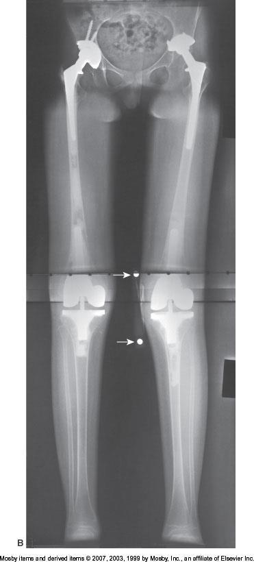
PRE & POST OPERATIVE RADIOLOGICAL ASSESSMENT IN TOTAL KNEE REPLACEMENT. Dr. Divya Rani K 2 nd Year Resident Dept. of Radiology
 PRE & POST OPERATIVE RADIOLOGICAL ASSESSMENT IN TOTAL KNEE REPLACEMENT Dr. Divya Rani K 2 nd Year Resident Dept. of Radiology PRE OPERATIVE ASSESSMENT RADIOGRAPHS Radiographs are used for assessment and
Country Health SA Medical Imaging
 Country Health SA Medical Imaging REMOTE OPERATORS POSITIONING GUIDE Contents Image Evaluation Page 4 Positioning Guides Section 1 - THORAX 1.1 Chest Page 5 1.2 Bedside Chest Page 7 1.3 Ribs Page 8 Section
The radiologist and the raiders of the lost image
 The radiologist and the raiders of the lost image Poster No.: P-0072 Congress: ESSR 2014 Type: Educational Poster Authors: M. J. Ereño Ealo, E. Montejo Rodrigo, B. Sancho, E. Pastor; Galdakao/ES Keywords:
Bones of Lower Limb. Dr. Heba Kalbouneh Associate Professor of Anatomy and Histology
 Bones of Lower Limb Dr. Heba Kalbouneh Associate Professor of Anatomy and Histology Bones of the lower limb Hip Bone Made up of 3 bones: 1) Ilium (flat), superior in position 2) Ischium (L), postero-inferior
Figure 7: Bones of the lower limb
 BONES OF THE APPENDICULAR SKELETON The appendicular skeleton is composed of the 126 bones of the appendages and the pectoral and pelvic girdles, which attach the limbs to the axial skeleton. Although the
Lower Limb CONTENTS CHAPTER. Radiographic Positioning cont d. Radiographic Anatomy. Radiographic Positioning. Radiographs for Critique, 257
 CHAPTER Lower Limb CONTRIBUTIONS BY Dan L. Hobbs, MSRS, RT(R)(CT)(MR) CONTRIBUTORS TO PAST EDITIONS Beth L. Vealé, MEd, RT(R)(QM), Jeannean Hall-Rollins, MRC, BS, RT(R)(CV) CONTENTS Radiographic Anatomy
Image C. A lateral projection of the right femur demonstrates a distal transverse fracture.
 Case # 2014A The ABCs of how to handle a trauma femur. While these images (6 in all) may never be nominated for inclusion into the hall of fame of imagery at the next international radiological conference
The Appendicular Skeleton
 8 The Appendicular Skeleton PowerPoint Lecture Presentations prepared by Jason LaPres Lone Star College North Harris 8-1 The Pectoral Girdle The Pectoral Girdle Also called shoulder girdle Connects the
Practical 1 Worksheet
 Practical 1 Worksheet ANATOMICAL TERMS 1. Use the word bank to fill in the missing words. reference side stand body arms palms anatomical forward All anatomical terms have a(n) point which is called the
2017 Resident Advanced Trauma Techniques Course COMPLICATIONS / CHALLENGES MALUNIONS/DEFORMITY
 2017 Resident Advanced Trauma Techniques Course COMPLICATIONS / CHALLENGES MALUNIONS/DEFORMITY What is a Malunion? Definition: a fracture that has healed in a nonanatomic (i.e. deformed) position Must
Administrative - Master Syllabus COVER SHEET
 Administrative - Master Syllabus COVER SHEET Purpose: It is the intention of this to provide a general description of the course, outline the required elements of the course and to lay the foundation for
INSTRUCTION. Course Package RAD 110 RADIOGRAPHIC POSITIONING I APPROVED 12/02/2011 EFFECTIVE FALL MCC Form EDU 0007 (rev.
 RAD 110 RADIOGRAPHIC POSITIONING I APPROVED 12/02/2011 EFFECTIVE FALL 2012-13 Prefix & Number RAD 110 Course Title: Radiographic Positioning I & Lab Purpose of this submission: New Change/Updated Retire
Knee Injury Assessment
 Knee Injury Assessment Clinical Anatomy p. 186 Femur Medial condyle Lateral condyle Femoral trochlea Tibia Intercondylar notch Tibial tuberosity Tibial plateau Fibula Fibular head Patella Clinical Anatomy
The Lower Limb. Anatomy RHS 241 Lecture 2 Dr. Einas Al-Eisa
 The Lower Limb Anatomy RHS 241 Lecture 2 Dr. Einas Al-Eisa The bony pelvis Protective osseofibrous ring for the pelvic viscera Transfer of forces to: acetabulum & head of femur (when standing) ischial
Lower Extremity Dislocations: Management and Triage on the Field
 Lower Extremity Dislocations: Management and Triage on the Field Scott J Tarantino, MD Towson Orthopaedic Associates, Towson, MD None Disclsures Purpose To provide you with knowledge which may guide you
Basic Principles of Fractures & Easily Missed Fractures. Mr Irfan Merchant Trauma & Orthopaedic Registrar Bedford Hospital, East of England
 Basic Principles of Fractures & Easily Missed Fractures Mr Irfan Merchant Trauma & Orthopaedic Registrar Bedford Hospital, East of England Objectives Types Fracture Patterns Fracture Healing Assessing
Knee Joint Anatomy 101
 Knee Joint Anatomy 101 Bone Basics There are three bones at the knee joint femur, tibia and patella commonly referred to as the thighbone, shinbone and kneecap. The fibula is not typically associated with
To describe he knee joint, ligaments, structure & To list the main features of other lower limb joints
 To describe he knee joint, ligaments, structure & neurovascular supply To demonstrate the ankle joint anatomy To list the main features of other lower limb joints To list main groups of lymph nodes in
Topic 7: Hip and pelvis. Parts of the hip. Parts of the femur
 Topic 7: Hip and pelvis Parts of the hip Parts of the femur Classifying the hip joint Ball and socket Synovial Multiaxial Movements of the hip: Abduction/adduction Flexion/extension Medial/lateral rotation
Balanced Body Movement Principles
 Balanced Body Movement Principles How the Body Works and How to Train it. Module 3: Lower Body Strength and Power Developing Strength, Endurance and Power The lower body is our primary source of strength,
In the name of god. Knee. By: Tofigh Bahraminia Graduate Student of the Pathology Sports and corrective actions. Heat: Dr. Babakhani. Nov.
 In the name of god Knee By: Tofigh Bahraminia Graduate Student of the Pathology Sports and corrective actions Heat: Dr. Babakhani Nov. 2014 1 Anatomy-Bones Bones Femur Medial/lateral femoral condyles articulate
Copyright 2003 Pearson Education, Inc. publishing as Benjamin Cummings. Dr. Nabil khouri
 Dr. Nabil khouri Appendicular Skeleton The appendicular skeleton is made up of the bones of the upper and lower limbs and their girdles Two girdles: Pectoral girdles attach the upper limbs to the body
PRE-LAB EXERCISES. Before we get started, look up the definitions of these common bone marking terms: Canal: Condyle: Facet: Fissure:
 1 PRE-LAB EXERCISES When studying the skeletal system, the bones are often sorted into two broad categories: the axial skeleton and the appendicular skeleton. This lab focuses on the appendicular skeleton,
Lab Activity 9. Appendicular Skeleton Martini Chapter 8. Portland Community College BI 231
 Lab Activity 9 Appendicular Skeleton Martini Chapter 8 Portland Community College BI 231 Appendicular Skeleton Upper & Lower extremities Shoulder Girdle Pelvic Girdle 2 Humerus 3 Humerus: Proximal End
RADIOGRAPHY OF THE HAND, FINGERS & THUMB
 RADIOGRAPHY OF THE HAND, FINGERS & THUMB FINGERS (2nd 5th) - PA Projection Patient Position: Seated; hand ; elbow on IR table top Part Position: Fingers centered to IR unless protocol is Central Ray: Perpendicular
Imaging assessment of Unicomp candidates!
 7th Advanced Course on Knee Surgery - 2018: Imaging assessment of Unicomp candidates! Presenter: Anders Troelsen, MD, ph.d., dr.med., Professor Distribution of the basic primary OA patterns Medial FT:
The Dance Hall by Vincent van Gogh,1888
 The Dance Hall by Vincent van Gogh,1888 Articulations of the pelvic girdle Lumbosacral joints, sacroiliac joints & pubic symphysis The remaining joints of the lower limb Hip joint Knee joint Tibiofibular
The Knee. Prof. Oluwadiya Kehinde
 The Knee Prof. Oluwadiya Kehinde www.oluwadiya.sitesled.com The Knee: Introduction 3 bones: femur, tibia and patella 2 separate joints: tibiofemoral and patellofemoral. Function: i. Primarily a hinge joint,
Hip joint Type: Articulating bones:
 Ana (242 ) Hip joint Type: Synovial, ball & socket Articulating bones: Formed between head of femur and lunate surface of acetabulum of hip bone. Capsule: it is a strong fibrous sleeve connecting the articulating
SKELETAL SYSTEM 206. AXIAL SKELETON 80 APPENDICULAR SKELETON 126 (see Figure 6.1) Clavicle. Clavicle. Pectoral girdles. Scapula. Scapula.
 Clavicle. Clavicle. Pectoral girdles. Scapula. Scapula.") SKELETAL SYSTEM 206 AXIAL SKELETON 80 APPENDICULAR SKELETON 126 (see Figure 6.1) Pectoral girdles 4 Clavicle Scapula 2 2 Clavicle Scapula Humerus 2 Humerus Upper limbs 60 Radius 2 Ulna Carpal bones Metacarpal
SKELETAL SYSTEM 206 AXIAL SKELETON 80 APPENDICULAR SKELETON 126 (see Figure 6.1) Pectoral girdles 4 Clavicle Scapula 2 2 Clavicle Scapula Humerus 2 Humerus Upper limbs 60 Radius 2 Ulna Carpal bones Metacarpal
Muscle Testing of Knee Extensors. Yasser Moh. Aneis, PhD, MSc., PT. Lecturer of Physical Therapy Basic Sciences Department
 Muscle Testing of Knee Extensors Yasser Moh. Aneis, PhD, MSc., PT. Lecturer of Physical Therapy Basic Sciences Department Muscle Testing of Knee Extensors othe Primary muscle Quadriceps Femoris -Rectus
Pectoral (Shoulder) Girdle
 Girdle") Chapter 8 Skeletal System: Appendicular Skeleton Pectoral girdle Pelvic girdle Upper limbs Lower limbs 8-1 Pectoral (Shoulder) Girdle Consists of scapula and clavicle Clavicle articulates with sternum
Chapter 8 Skeletal System: Appendicular Skeleton Pectoral girdle Pelvic girdle Upper limbs Lower limbs 8-1 Pectoral (Shoulder) Girdle Consists of scapula and clavicle Clavicle articulates with sternum
Biology 218 Human Anatomy
 Chapter 8 Adapted from Tortora 10 th ed. LECTURE OUTLINE A. Introduction (p. 203) 1. The appendicular skeleton contains 126 bones that form: i. two pectoral (shoulder) girdles two upper limbs i one pelvic
Anatomage Table Instructors Guide- Lower Limb
 The Lower Limb Anatomage Table Instructors Guide- Lower Limb Table of Contents Lower Limb 1- The Skeletal System...3 1: Hip Bone...3 2: Hip Joint and Femur...4 3: Patella and Knee Joint...7 4: Tibia, Fibula,
Small Animal radiography Stifle Joint and CruS
 Peer reviewed ImagIng EssEnTIals Small Animal radiography Stifle Joint and CruS Danielle Mauragis, CVT, and Clifford R. erry, DVM, Diplomate ACVR This is the fourth article in our Imaging Essentials series,
ESSEX COUNTY COLLEGE Nursing and Allied Health Division RTC 101 Radiographic Positioning Principles I Course Outline
 ESSEX COUNTY COLLEGE Nursing and Allied Health Division RTC 101 Radiographic Positioning Principles I Course Outline Course Number & Name: RTC 101 Radiographic Positioning Principles I Credit Hours: 4.0
Chapter 8. The Appendicular Skeleton. Lecture Presentation by Lee Ann Frederick University of Texas at Arlington Pearson Education, Inc.
 Chapter 8 The Appendicular Skeleton Lecture Presentation by Lee Ann Frederick University of Texas at Arlington An Introduction to the Appendicular Skeleton The Appendicular Skeleton 126 bones Allows us
It is formed by fusion of 3 bones: I. Ilium (superior bone). II. Pubis (antero-inferior bone). III. Ischium (postero-inferior bone).
. II. Pubis (antero-inferior bone). III. Ischium (postero-inferior bone).") It is formed by fusion of 3 bones: I. Ilium (superior bone). II. Pubis (antero-inferior bone). III. Ischium (postero-inferior bone). Pubis Acetabulum Ana (242 ) The three constituent of bones of the hip
It is formed by fusion of 3 bones: I. Ilium (superior bone). II. Pubis (antero-inferior bone). III. Ischium (postero-inferior bone). Pubis Acetabulum Ana (242 ) The three constituent of bones of the hip
Mr. S. Tanweer Ashraf MS, MRCS (Ed), FRCS (Eng). FRCS (Tr&Orth) MSc Ortho Engineering (Cardiff),
, FRCS (Eng). FRCS (Tr&Orth) MSc Ortho Engineering (Cardiff),") Mr. S. Tanweer Ashraf MS, MRCS (Ed), FRCS (Eng). FRCS (Tr&Orth) MSc Ortho Engineering (Cardiff), Consultant Knee Surgeon NHS: The Royal Orthopaedic Hospital & Queen Elizabeth Hospital, Birmingham MRI Knee:
Mr. S. Tanweer Ashraf MS, MRCS (Ed), FRCS (Eng). FRCS (Tr&Orth) MSc Ortho Engineering (Cardiff), Consultant Knee Surgeon NHS: The Royal Orthopaedic Hospital & Queen Elizabeth Hospital, Birmingham MRI Knee:
Chapter 7: Skeletal System: Gross Anatomy
 Chapter 7: Skeletal System: Gross Anatomy I. General Considerations A. How many bones in an average adult skeleton? B. Anatomic features of bones are based on II. Axial Skeleton A. Skull 1. Functionally
11/4/2018 SUBTLETIES OF LOWER EXTREMITY TRAUMA IMAGING SPEAKER DISCLOSURES
 SUBTLETIES OF LOWER EXTREMITY TRAUMA IMAGING Charles S. Resnik, M.D. Professor of Radiology University of Maryland School of Medicine Upon completion of this presentation, participants will be better able
NEW YORK CITY COLLEGE OF TECHNOLOGY THE CITY UNIVERSITY OF NEW YORK. Department of Radiologic Technology & Medical Imaging
 NEW YORK CITY COLLEGE OF TECHNOLOGY THE CITY UNIVERSITY OF NEW YORK Department of Radiologic Technology & Medical Imaging RAD 1125 (REVISED 9/1/15) Section D806: Monday, 10:00am to 11:15am Radiographic
Biology 218 Human Anatomy. Adapted from Martini Human Anatomy 7th ed. Chapter 7 The Skeletal System Appendicular Division
 Adapted from Martini Human Anatomy 7th ed. Chapter 7 The Skeletal System Appendicular Division Introduction The appendicular skeleton includes: Pectoral girdle Shoulder bones Upper limbs Pelvic girdle
Where to Draw the Line:
 Where to Draw the Line: Anatomical Measurements Used to Evaluate Patellofemoral Instability Murray Grissom, MD 1 Bao Do, MD 2 Kathryn Stevens, MD 2 1 Santa Clara Valley Medical Center, San Jose, CA 2 Stanford
Chapter 8 The Skeletal System: The Appendicular Skeleton. Copyright 2009 John Wiley & Sons, Inc.
 Chapter 8 The Skeletal System: The Appendicular Skeleton Appendicular Skeleton The primary function is movement It includes bones of the upper and lower limbs Girdles attach the limbs to the axial skeleton
10/12/2010. Upper Extremity. Pectoral (Shoulder) Girdle. Clavicle (collarbone) Skeletal System: Appendicular Skeleton
 Girdle. Clavicle (collarbone) Skeletal System: Appendicular Skeleton") Skeletal System: Appendicular Skeleton Pectoral girdle Pelvic girdle Upper limbs Lower limbs 8-1 Pectoral (Shoulder) Girdle Consists of scapula and clavicle Clavicle articulates with sternum (Sternoclavicular
Skeletal System: Appendicular Skeleton Pectoral girdle Pelvic girdle Upper limbs Lower limbs 8-1 Pectoral (Shoulder) Girdle Consists of scapula and clavicle Clavicle articulates with sternum (Sternoclavicular
Preoperative Planning. The primary objectives of preoperative planning are to:
 Preoperative Planning The primary objectives of preoperative planning are to: - Determine preoperative leg length discrepancy. - Assess acetabular component size and placement. - Determine femoral component
Overview. Acceptance criteria for all protocols
 X-Ray protocol Overview The Smith & Nephew VISIONAIRE X-Ray protocol is essentially an AP leg length image. The images are preferred to be done erect, but can be done supine if necessary due to the type
Barium Enema RD Sheet
 SIMS POSITION Instruct patient to turn onto the left side, lean forward and about 35 to 40 degree, and knees are flexed right knee on the table, above and in front of the slightly flexed left knee This
Chapter 8B. The Skeletal System: Appendicular Skeleton. The Appendicular Skeleton. Clavicle. Pectoral (Shoulder) Girdle
 Girdle") The Appendicular Skeleton Chapter 8B The Skeletal System: Appendicular Skeleton 126 bones Pectoral (shoulder) girdle Pelvic (hip) girdle Upper limbs Lower limbs Functions primarily to facilitate movement
The Appendicular Skeleton Chapter 8B The Skeletal System: Appendicular Skeleton 126 bones Pectoral (shoulder) girdle Pelvic (hip) girdle Upper limbs Lower limbs Functions primarily to facilitate movement
Revolution. Unicompartmental Knee System
 Revolution Unicompartmental Knee System While Total Knee Arthroplasty (TKA) is one of the most predictable procedures in orthopedic surgery, many patients undergoing TKA are in fact excellent candidates
Shoulder Position: Supine arm in the neutral position. Collateral arm above head Indication: fracture humerus, fracture scapula
 Shoulder Position: Supine arm in the neutral position. Collateral arm above head Indication: fracture humerus, fracture scapula No instrumentation With metal or cast KV/ Effective mas/rotation time 140/300/1.0
Objectives. The BIG Joint. Case 1. Boney Architecture. Presenter Disclosure Information. Common Knee Problems
 3:30 4:15 pm Common Knee Problems SPEAKER Christopher J. Visco, MD Presenter Disclosure Information The following relationships exist related to this presentation: Christopher J. Visco, MD: Speaker s Bureau
COURSE SYLLABUS RT 1145 RADIOGRAPHIC POSITIONING FILM CRITIQUE & MEDICAL TERMINOLOGY I
 COURSE SYLLABUS RT 1145 RADIOGRAPHIC POSITIONING FILM CRITIQUE & MEDICAL TERMINOLOGY I CLASS HOURS: 4 CREDIT HOURS: 4 LABORATORY HOURS: 5 CATALOG COURSE DESCRIPTION: This course is the first of a three-course
Myology of the Knee. PTA 105 Kinesiology
 Myology of the Knee PTA 105 Kinesiology Objectives Describe the planes of motion and axes of rotation of the knee joint Visualize the origins and insertions of the muscles about the knee List the innervations
Routine Guide EXAMINATION PROJECTION CASSETTE SIZE NOTES PRINT ORIENTATION. 14x17 CW* 14x17LW 14x17LW. 14x17LW 14x17LW 14x17LW
 EXAMINATION PROJECTION CASSETTE SIZE NOTES PRINT ORIENTATION A-C Joints without weights with weights 14x17 CW* One 14x17 divided; both shoulders on one exposure. *If part does not fit, do 10x12s CW. Both
ANATOMIC. Navigated Surgical Technique 4 in 1 TO.G.GB.016/1.0
 ANATOMIC Navigated Surgical Technique 4 in 1 TO.G.GB.016/1.0 SCREEN LAYOUT Take screenshot Surgical step Dynamic navigation zone Information area and buttons 2 SCREEN LAYOUT Indicates action when yellow
Contents of the Posterior Fascial Compartment of the Thigh
 Contents of the Posterior Fascial Compartment of the Thigh 1-Muscles: B i c e p s f e m o r i s S e m i t e n d i n o s u s S e m i m e m b r a n o s u s a small part of the adductor magnus (h a m s t
4/28/2010. Fractures. Normal Bone and Normal Ossification Bone Terms. Epiphysis Epiphyseal Plate (physis) Metaphysis
 Metaphysis") Fractures Normal Bone and Normal Ossification Bone Terms Epiphysis Epiphyseal Plate (physis) Metaphysis Diaphysis 1 Fracture Classifications A. Longitudinal B. Transverse C. Oblique D. Spiral E. Incomplete
Fractures Normal Bone and Normal Ossification Bone Terms Epiphysis Epiphyseal Plate (physis) Metaphysis Diaphysis 1 Fracture Classifications A. Longitudinal B. Transverse C. Oblique D. Spiral E. Incomplete
CHAPTER 8: THE BIOMECHANICS OF THE HUMAN LOWER EXTREMITY
 CHAPTER 8: THE BIOMECHANICS OF THE HUMAN LOWER EXTREMITY _ 1. The hip joint is the articulation between the and the. A. femur, acetabulum B. femur, spine C. femur, tibia _ 2. Which of the following is
Imaging the Athlete s Knee. Peter Lowry, MD Musculoskeletal Radiology University of Colorado
 Imaging the Athlete s Knee Peter Lowry, MD Musculoskeletal Radiology University of Colorado None Disclosures Knee Imaging: Radiographs Can be performed weight-bearing or non-weight-bearing View options
Axial alignment of the lower extremity in Chinese adults. Journal Of Bone And Joint Surgery - Series A, 2000, v. 82 n. 11, p.
 Title Axial alignment of the lower extremity in Chinese adults Author(s) Tang, WM; Zhu, YH; Chiu, KY Citation Journal Of Bone And Joint Surgery - Series A, 2000, v. 82 n. 11, p. 1603-1608 Issued Date 2000
HANDS ON: Knee Evaluation J. Scott Delaney MD, FRCPC, FACEP, CSPQ
 HANDS ON: Knee Evaluation J. Scott Delaney MD, FRCPC, FACEP, CSPQ FACULTY DISCLOSURE Dr. Delaney has no affiliation with the manufacturer of any commercial product or provider of any commercial service
ChiroCredit.com Presents Biomechanics: Focus on
 ChiroCredit.com Presents Biomechanics: Focus on the Knee Presented by: Ivo Waerlop, DC Shawn Allen, DC 1 Focus on The Knee 2 Pertinent Anatomy Femur Tibia Fibula Patella Prepatellar bursa Infrapatellar
The Knee. Tibio-Femoral
 The Knee Tibio-Femoral Osteology Distal Femur with Proximal Tibia Largest Joint Cavity in the Body A modified hinge joint with significant passive rotation Technically, one degree of freedom (Flexion/Extension)
radiologymasterclass.co.uk
 http://radiologymasterclass.co.uk Hip X-ray anatomy - Normal AP (anterior-posterior) Shenton's line is formed by the medial edge of the femoral neck and the inferior edge of the superior pubic ramus Loss
Practical Reduction Techniques: Diaphyseal Reduction. Philip Wolinsky University of California at Davis Medical Center
 OTA Specialty Day 2016 Practical Reduction Techniques: Diaphyseal Reduction Philip Wolinsky University of California at Davis Medical Center 8:55 am 9:55 am Tips and Tricks: Practical Reduction Techniques
The Lower Limb. Sevda LAFCI FAHRİOĞLU, MD.PhD.
 The Lower Limb Sevda LAFCI FAHRİOĞLU, MD.PhD. The Lower Limb The bones of the lower limb form the inferior part of the appendicular skeleton the organ of locomotion for bearing the weight of body stronger
An Introduction to Radiographic Views & Anatomy
 An Introduction to Radiographic Views & Anatomy Morey J. Kolber, PT, PhD, OCS, Cert MDT, CSCS*D An Introduction to Radiographic Views & Anatomy M.S.P.T. 1995-University of Miami Nova Southeastern University
The Knee. Clarification of Terms. Osteology of the Knee 7/28/2013. The knee consists of: The tibiofemoral joint Patellofemoral joint
 The Knee Clarification of Terms The knee consists of: The tibiofemoral joint Patellofemoral joint Mansfield, p273 Osteology of the Knee Distal Femur Proximal tibia and fibula Patella 1 Osteology of the
Pelvic Girdle
 ARTICULATIONS OF LOWER EXTREMITY Pages 429-437 Pelvic Girdle formed by connection of the hip bones and the sacrum Sacroiliac Joints compound joints synovial joint - anterior, between the auricular surfaces
Joints of the Lower Limb II
 Joints of the Lower Limb II Lecture Objectives Describe the components of the knee and ankle joint. List the ligaments associated with these joints and their attachments. List the muscles acting on these
Functional Movement Screen (Cook, 2001)
") Functional Movement Screen (Cook, 2001) TEST 1 DEEP SQUAT Purpose - The Deep Squat is used to assess bilateral, symmetrical, mobility of the hips, knees, and ankles. The dowel held overhead assesses bilateral,
Functional Movement Screen (Cook, 2001) TEST 1 DEEP SQUAT Purpose - The Deep Squat is used to assess bilateral, symmetrical, mobility of the hips, knees, and ankles. The dowel held overhead assesses bilateral,
VARIABILITY OF THE POSTERIOR CONDYLAR ANGLE
 VARIABILITY OF THE POSTERIOR CONDYLAR ANGLE Łukasz Cieliński, Damian Kusz, Michał Wójcik Department of Orthopedics Medical University of Silesia in Katowice Introduction Correct positioning of implants
BIOMECHANICAL EXAMINATION OF THE PEDIATRIC LOWER EXTREMITY 2017
 BIOMECHANICAL EXAMINATION OF THE PEDIATRIC LOWER EXTREMITY 2017 B. RESSEQUE, D.P.M., D.A.B.P.O. Professor, N.Y. College of Podiatric Medicine ARCH HEIGHT OFF WEIGHTBEARING Evaluate arch height by placing
Biology 152 Appendicular Skeleton Anatomy Objectives
 Biology 152 Appendicular Skeleton Anatomy Objectives We will learn proper bone names, left/right/medial, and the parts of bones in this exercise. Start by learning the names of the bones. As you gain comfort
Anatomy & Physiology. Muscles of the Lower Limbs.
 Anatomy & Physiology Muscles of the Lower Limbs http://www.ishapeup.com/musclecharts.html Muscles of the Lower Limbs Among the strongest muscles in the body. Because pelvic girdle is composed of heavy,
The thigh. Prof. Oluwadiya KS
 The thigh Prof. Oluwadiya KS www.oluwadiya.com The Thigh: Boundaries The thigh is the region of the lower limb that is approximately between the hip and knee joints Anteriorly, it is separated from the
Plaster-Wedging Technique:
 Plaster-Wedging Technique: An Appropriate, Safe, Quick, & Economical Method To Stretch Soft Tissue Contractures Of The Knee H.M. Steenbeek General Information. In the field of physical rehabilitation of
The Leg. Prof. Oluwadiya KS
 The Leg Prof. Oluwadiya KS www.oluwadiya.sitesled.com Compartments of the leg 4 Four Compartments: 1. Anterior compartment Deep fibular nerve Dorsiflexes the foot and toes 2. Lateral Compartment Superficial
BIOMECHANICAL EXAMINATION OF THE PEDIATRIC LOWER EXTREMITY
 BIOMECHANICAL EXAMINATION OF THE PEDIATRIC LOWER EXTREMITY B.Resseque, D.P.M. ARCH HEIGHT OFF WEIGHTBEARING Evaluate arch height by placing a ruler from the heel to the first metatarsal head Compare arch
Chapter 8 The Skeletal System: The Appendicular Skeleton. Copyright 2009 John Wiley & Sons, Inc.
 Chapter 8 The Skeletal System: The Appendicular Skeleton Appendicular Skeleton It includes bones of the upper and lower limbs Girdles attach the limbs to the axial skeleton The pectoral girdle consists
Human Anatomy Biology 351
 Human Anatomy Biology 351 Lower Limb Please place your name on the back of the last page of this exam. You must answer all questions on this exam. Because statistics demonstrate that, on average, between
5.1 Identify, describe the attachments of and deduce the actions of the muscles of the thigh:
 5.1 Identify, describe the attachments of and deduce the actions of the muscles of the thigh: Anterior group Proximal attachment Distal attachment Sartorius ASIS» Upper part of shaft tibia (middle surface)»
Overview. Acceptance criteria for all protocols
 X-Ray protocol Overview The Smith & Nephew VISIONAIRE X-Ray protocol is essentially an AP leg length image. The images are preferred to be done erect, but can be done supine if necessary due to the type
Institutional review board approval was obtained prior to the start of this study.
 Lower Limb Alignment and Length Measurements - Comparison of Computed Tomography, Upright Full-Length Conventional Radiography and Upright Biplanar Linear-Low Dose X-ray Scanner Poster No.: C-1382 Congress:
In-Depth Foundations: Anatomy Terms to Know
 Be familiar with / able to identify and define all the following parts. The Spine Cranium Vertebrae Cervical, Thoracic, Lumbar Sacrum Coccyx Bones of Upper Body Cranium Mastoid process; Occipital condyle,