EEG, ECG, EMG. Mitesh Shrestha
|
|
|
- Charity Caldwell
- 6 years ago
- Views:
Transcription
1 EEG, ECG, EMG Mitesh Shrestha
2 What is Signal? A signal is defined as a fluctuating quantity or impulse whose variations represent information. The amplitude or frequency of voltage, current, electric field strength, light, and sound can be varied as signals representing information. A signal can be simply defined as a function that conveys information. Signals are represented mathematically as functions of one or more independent variables. Examples: voltage, current, electric field strength, light, sound, etc. 2
3 Biomedical signals Biomedical signals means the bio-signals which are generated in biological systems only. Biomedical signals are observations of physiological activities of organisms, ranging from gene and protein sequences, to neural and cardiac rhythms, to tissue and organ images. Examples of biomedical signals: ECG (Electrocardiogram) signal, EEG (Electroencephalogram) signal, etc.
4 How biomedical signals are generated? Biomedical signals are electrical or magnetic signals generated by some biological activity in the human body. Human body is composed of living tissues that can be considered as a power station. Action of living tissues in terms of bioelectric potentials generate multiple electric signals from two internal sourcesmuscles and nerves system.
5

6 What are biopotentials An electric potential that is measured between points in living cells, tissues, and organisms, and which accompanies all biochemical processes. Also describes the transfer of information between and within cells
7 Classification of Biomedical Signals Bioelectric Signal Bioacoustics Signal Biomechanical Signal Biochemical Signal Bio-magnetic Signal Bio- optical signal
8 Bioelectric signal The electrical signals which we can measure mainly on the surface of the body is known as bioelectric signal. It is generated by muscle cells and nerve cells. Basic source is the cell membrane potential. Examples: ECG, EEG, EMG, EOG
 Very widely used method in clinical environment Very high diagnostic value 2. Ventricular depolarization 3. Ventricular repolarization 1.")
9 Electrocardiography (ECG) Measures galvanically the electric activity of the heart Well known and traditional, first measurements by Augustus Waller using capillary electrometer (year 1887) Very widely used method in clinical environment Very high diagnostic value 2. Ventricular depolarization 3. Ventricular repolarization 1. Atrial depolarization
10 Electrocardiography The heart is an electrical organ, and its activity can be measured non-invasively Wealth of information related to: The electrical patterns proper The geometry of the heart tissue The metabolic state of the heart Standard tool used in a wide-range of medical evaluations

11 ECG principle

12 Cardiac Electrical Activity


13 ECG basics Amplitude: 1-5 mv Bandwidth: Hz Largest measurement error sources: Motion artifacts 50/60 Hz powerline interference Typical applications: Diagnosis of ischemia Arrhythmia Conduction defects


14 12-Lead ECG measurement Most widely used ECG measurement setup in clinical environment Signal is measured non-invasively with 9 electrodes Lots of measurement data and international reference databases Well-known measurement and diagnosis practices This particular method was adopted due to historical reasons, now it is already rather obsolete Goldberger augmented leads: V R, V L & V F Precordial leads: V 1 -V 6 Einthoven leads: I, II & III

15 EINTHOVENS TRIANGLE 15


16 Why is 12-lead system obsolete? Over 90% of the heart s electric activity can be explained with a dipole source model Only 3 orthogonal components need to be measured, which makes 9 of the leads redundant The remaining percentage, i.e. nondipolar components, may have some clinical value 12-lead system does, to some extend, enhance pattern recognition and gives the clinician a few more projections to choose from but. If there was no legacy problem with current systems, 12-lead system would ve been discarded ages ago


17 Origination of the QRS - Signal
18 ECG - applications Diagnostics Functional analysis Implants (pace maker) Biofeedback (Heartrate variability, HRV) Peak Performacne Training, Monitoring


19 Normal sinus rhythm
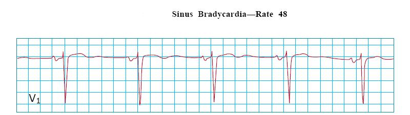
20 RATE P wave rate bpm with <10% variation - Normal Rate < 60 bpm = Sinus Bradycardia - Results from: - Excessive vagal stimulation - SA nodal ischemia (Inferior MI) Rate > 100 bpm = Sinus Tachycardia - Results from: - Pain / anxiety - CHF - Volume depletion - Pericarditis - Chronotropic Drugs (Dopamine)
21 Electroencephalography (EEG) An electrophysiological monitoring method to record electrical activity of the brain. Typically noninvasive, with the electrodes placed along the scalp, although invasive electrodes are sometimes used in specific applications. EEG measures voltage fluctuations resulting from ionic current within the neurons of the brain. In clinical contexts, EEG refers to the recording of the brain's spontaneous electrical activity over a period of time, as recorded from multiple electrodes placed on the scalp. Diagnostic applications generally focus on the spectral content of EEG, that is, the type of neural oscillations (popularly called "brain waves") that can be observed in EEG signals.

22 EEG Signal Fig.: EEG signal originating from the brain The Electroencephalogram (EEG) is a recording of electrical activity originating from the brain. It is recorded on the surface of the scalp using electrodes, thus the signal is retrievable non-invasively. Signal varies in terms of amplitude and frequency Normal frequency range: 0.5Hz to 50 Hz.

23 EEG Electrode cap locations of the 10/20 system
24 Electroencephalogram (EEG) The system of electrode placement for EEG recording.
25 Electroencephalogram (EEG) The commonly used terms for EEG frequency range: Delta (0.5-4 Hz): deep sleep Theta (4-8 Hz): beginning stages of sleep Alpha (8-13 Hz): principal resting rhythm Beta (>13 Hz): background activity in tense and anxious subjects
26 Electroencephalogram (EEG) a: delta, b: theta, c: alpha, d: beta, e: blocking of alpha rhythm by eye opening, f: marker 50 μv, 1 sec
27 EEG - applications Diagnostics (Epilepsy, Oncology,..) Cognitive Sciences Sleep Analysis Human Computer Interfaces (BCIs) Pharmacology Intensive Care, Monitoring
28 Electromyography (EMG) Electromyography (EMG) is a technique for evaluating and recording the activation signal of muscles. EMG is performed by an electromyograph, which records an electromyogram. Electromyograph detects the electrical potential generated by muscle cells when these cells contract and relax.


29 ELECTRODE TYPES Intramuscular - Needle Electrodes Extramuscular - Surface Electrodes

30 EMG PROCEDURE Clean the site of application of electrode; Insert needle/place surface electrodes at muscle belly; Record muscle activity at rest; Record muscle activity upon voluntary contraction of the muscle.

31 EMG Contd. Muscle Signals are Analog in nature. EMG signals are also collected over a specific period of time. Analog Signal

32 EMG Contd. EMG processing: Signal pick up Amplification & Filtering Computer Conversion of Analog signals to Digital signals
33 Factors Influencing Signal Measured Geometrical & Anatomical Factors Electrode size Electrode shape Electrode separation distance with respect to muscle tendon junctions Thickness of skin and subcutaneous fat Misalignment between electrodes and fiber alignment Physiological Factors Blood flow and temperature Type and level of contraction Muscle fiber conduction velocity Number of motor units (MU) Degree of MU synchronization
34 Factors That Influence the Signal Information Content of EMG Factor Neuroactivation Muscle fiber physiology Muscle anatomy Electrode size/orientation Influence - firing rate of motor unit AP s - no. of motor units recruited - synchronization of motor units - conduction velocity of fibers - orientation & distribution of fibers - diameter of muscle fibers - total no. of motor units - no. of fibers in pickup area
35 Factors That Influence the Signal Information Content of EMG Factor Electrode-electrolyte interface with Influence - type of material and site - electrode impedance decreases increasing frequency Bipolar electrode configuration - distance between electrodes - orientation of electrodes relative to the axis of muscle fibers

36 EMG signal
37 What can be learned from an EMG? Time course of muscle contraction Contraction force Coordination of several muscles in a movement sequence These parameters are DERIVED from the amplitude, frequency, and change of these over time of the EMG signal Field of Ergonomics: from the EMG conclusions about muscle strain and the occurrence of muscular fatigue can be derived as well
38 APPLICATION OF EMG EMG can be used for diagnosis of Neurogenic or Myogenic Diseases. Rehabilitation Functional analysis Active Prothetics, Orthesis Biomechanics, Sports medicine
39 Assignment Write short notes on ECG? [5] Write short notes on EEG? [5] Write short notes on EMG? [5]
40 Useful Links
PD233: Design of Biomedical Devices and Systems
 PD233: Design of Biomedical Devices and Systems (Lecture-7 Biopotentials- 2) Dr. Manish Arora CPDM, IISc Course Website: http://cpdm.iisc.ac.in/utsaah/courses/ Electromyogram (EMG) Skeletal muscles are
Biomedical Signal Processing
 DSP : Biomedical Signal Processing What is it? Biomedical Signal Processing: Application of signal processing methods, such as filtering, Fourier transform, spectral estimation and wavelet transform, to
Biomedical Signal Processing
 DSP : Biomedical Signal Processing Brain-Machine Interface Play games? Communicate? Assist disable? Brain-Machine Interface Brain-Machine Interface (By ATR-Honda) The Need for Biomedical Signal Processing
EKG. Danil Hammoudi.MD
 EKG Danil Hammoudi.MD What is an EKG? The electrocardiogram (EKG) is a representation of the electrical events of the cardiac cycle. Each event has a distinctive waveform, the study of which can lead to
ELEC ENG 4BD4 Lecture 1. Biomedical Instrumentation Instructor: Dr. Hubert de Bruin
 ELEC ENG 4BD4 Lecture 1 Biomedical Instrumentation Instructor: Dr. Hubert de Bruin 1 Cochlear Implant 2 Advances in Vision (Retinal Stimulation) 3 Argus II Implant 4 Mini Gastric Imaging 5 Taser 6 Shock
CHAPTER 6 INTERFERENCE CANCELLATION IN EEG SIGNAL
 116 CHAPTER 6 INTERFERENCE CANCELLATION IN EEG SIGNAL 6.1 INTRODUCTION Electrical impulses generated by nerve firings in the brain pass through the head and represent the electroencephalogram (EEG). Electrical
INTRODUCTION TO ECG. Dr. Tamara Alqudah
 INTRODUCTION TO ECG Dr. Tamara Alqudah Excitatory & conductive system of the heart + - The ECG The electrocardiogram, or ECG, is a simple & noninvasive diagnostic test which records the electrical
Electrocardiography Abnormalities (Arrhythmias) 7. Faisal I. Mohammed, MD, PhD
 7. Faisal I. Mohammed, MD, PhD") Electrocardiography Abnormalities (Arrhythmias) 7 Faisal I. Mohammed, MD, PhD 1 Causes of Cardiac Arrythmias Abnormal rhythmicity of the pacemaker Shift of pacemaker from sinus node Blocks at different
Electrocardiography Abnormalities (Arrhythmias) 7 Faisal I. Mohammed, MD, PhD 1 Causes of Cardiac Arrythmias Abnormal rhythmicity of the pacemaker Shift of pacemaker from sinus node Blocks at different
: Biomedical Signal Processing
 : Biomedical Signal Processing 0. Introduction: Biomedical signal processing refers to the applications of signal processing methods, such as Fourier transform, spectral estimation and wavelet transform,
DR QAZI IMTIAZ RASOOL OBJECTIVES
 PRACTICAL ELECTROCARDIOGRAPHY DR QAZI IMTIAZ RASOOL OBJECTIVES Recording of electrical events in heart Established electrode pattern results in specific tracing pattern Health of heart i. e. Anatomical
Birmingham Regional Emergency Medical Services System
 Birmingham Regional Emergency Medical Services System 2018 ALCTE Summer Conference EKG Basics Brian Gober, MAT, ATC, NRP, CSCS Education Services Manager ECC Training Center Coordinator Birmingham Regional
CASE 10. What would the ST segment of this ECG look like? On which leads would you see this ST segment change? What does the T wave represent?
 CASE 10 A 57-year-old man presents to the emergency center with complaints of chest pain with radiation to the left arm and jaw. He reports feeling anxious, diaphoretic, and short of breath. His past history
Introduc2on/Mo2va2on
 Introduc2on/Mo2va2on Today: many examples & applica2ons of signal & system analysis. Course will be much more mathema2cal than this intro! Remember the skill vs fame plot... Note about today s lecture:
Electroencephalography
 The electroencephalogram (EEG) is a measure of brain waves. It is a readily available test that provides evidence of how the brain functions over time. The EEG is used in the evaluation of brain disorders.
This presentation will deal with the basics of ECG description as well as the physiological basics of
 Snímka 1 Electrocardiography basics This presentation will deal with the basics of ECG description as well as the physiological basics of Snímka 2 Lecture overview 1. Cardiac conduction system functional
NATIONAL SCIENCE EDUCATION STANDARDS Levels 9-12: Correlation to Biopac Science Lab Lessons
 1 of 23 Detail options: Click the in the table to review the lesson correlation to the specified content standard. Click the content standard link (left column) to view all correlations for that standard.
Introduction to ECG Gary Martin, M.D.
 Brief review of basic concepts Introduction to ECG Gary Martin, M.D. The electrical activity of the heart is caused by a sequence of rapid ionic movements across cell membranes resulting first in depolarization
ECG INTERPRETATION MANUAL
 Lancashire & South Cumbria Cardiac Network ECG INTERPRETATION MANUAL THE NORMAL ECG Lancashire And South Cumbria Cardiac Physiologist Training Manual THE NORMAL ECG E.C.G CHECKLIST 1) Name, Paper Speed,
CIS 632 / EEE 687 Mobile Computing
 CIS 632 / EEE 687 Mobile Computing MC Platform #2: BioRadio Chansu Yu Body Signals Temperature, Skin humidity (dehydration) Lung sounds (LS), heart sounds (HS), and bowel sounds (BS) Blood pressure, Blood
11/18/13 ECG SIGNAL ACQUISITION HARDWARE DESIGN. Origin of Bioelectric Signals
 ECG SIGNAL ACQUISITION HARDWARE DESIGN Origin of Bioelectric Signals 1 Cell membrane, channel proteins Electrical and chemical gradients at the semi-permeable cell membrane As a result, we get a membrane
The Electrocardiogram
 The Electrocardiogram Chapters 11 and 13 AUTUMN WEDAN AND NATASHA MCDOUGAL The Normal Electrocardiogram P-wave Generated when the atria depolarizes QRS-Complex Ventricles depolarizing before a contraction
Electrocardiography Normal 5. Faisal I. Mohammed, MD, PhD
 Electrocardiography Normal 5 Faisal I. Mohammed, MD, PhD 1 Objectives 2 1. Describe the different waves in a normal electrocardiogram. 2. Recall the normal P-R and Q-T interval time of the QRS wave. 3.
Electrocardiogram ECG. Hilal Al Saffar FRCP FACC College of medicine,baghdad University
 Electrocardiogram ECG Hilal Al Saffar FRCP FACC College of medicine,baghdad University Tuesday 29 October 2013 ECG introduction Wednesday 30 October 2013 Abnormal ECG ( ischemia, chamber hypertrophy, heart
PSD Analysis of Neural Spectrum During Transition from Awake Stage to Sleep Stage
 PSD Analysis of Neural Spectrum During Transition from Stage to Stage Chintan Joshi #1 ; Dipesh Kamdar #2 #1 Student,; #2 Research Guide, #1,#2 Electronics and Communication Department, Vyavasayi Vidya
Interpreting Electrocardiograms (ECG) Physiology Name: Per:
 Physiology Name: Per:") Interpreting Electrocardiograms (ECG) Physiology Name: Per: Introduction The heart has its own system in place to create nerve impulses and does not actually require the brain to make it beat. This electrical
Interpreting Electrocardiograms (ECG) Physiology Name: Per: Introduction The heart has its own system in place to create nerve impulses and does not actually require the brain to make it beat. This electrical
Electrocardiography I Laboratory
 Introduction The body relies on the heart to circulate blood throughout the body. The heart is responsible for pumping oxygenated blood from the lungs out to the body through the arteries and also circulating
HTEC 91. Performing ECGs: Procedure. Normal Sinus Rhythm (NSR) Topic for Today: Sinus Rhythms. Characteristics of NSR. Conduction Pathway
 Topic for Today: Sinus Rhythms. Characteristics of NSR. Conduction Pathway") HTEC 91 Medical Office Diagnostic Tests Week 3 Performing ECGs: Procedure o ECG protocol: you may NOT do ECG if you have not signed up! If you are signed up and the room is occupied with people who did
HTEC 91 Medical Office Diagnostic Tests Week 3 Performing ECGs: Procedure o ECG protocol: you may NOT do ECG if you have not signed up! If you are signed up and the room is occupied with people who did
Testing the Accuracy of ECG Captured by Cronovo through Comparison of ECG Recording to a Standard 12-Lead ECG Recording Device
 Testing the Accuracy of ECG Captured by through Comparison of ECG Recording to a Standard 12-Lead ECG Recording Device Data Analysis a) R-wave Comparison: The mean and standard deviation of R-wave amplitudes
ECG Interpretation Cat Williams, DVM DACVIM (Cardiology)
") ECG Interpretation Cat Williams, DVM DACVIM (Cardiology) Providing the best quality care and service for the patient, the client, and the referring veterinarian. GOAL: Reduce Anxiety about ECGs Back to
ECG Interpretation Cat Williams, DVM DACVIM (Cardiology) Providing the best quality care and service for the patient, the client, and the referring veterinarian. GOAL: Reduce Anxiety about ECGs Back to
Diploma in Electrocardiography
 The Society for Cardiological Science and Technology Diploma in Electrocardiography The Society makes this award to candidates who can demonstrate the ability to accurately record a resting 12-lead electrocardiogram
Restoring Communication and Mobility
 Restoring Communication and Mobility What are they? Artificial devices connected to the body that substitute, restore or supplement a sensory, cognitive, or motive function of the nervous system that has
Practical 3 Nervous System Physiology 2 nd year English Module. Dept. of Physiology, Carol Davila University of Medicine and Pharmacy
 Electroencephalography l h (EEG) Practical 3 Nervous System Physiology 2 nd year English Module Dept. of Physiology, Carol Davila University of Medicine and Pharmacy What is EEG EEG noninvasively records
Please check your answers with correct statements in answer pages after the ECG cases.
 ECG Cases ECG Case 1 Springer International Publishing AG, part of Springer Nature 2018 S. Okutucu, A. Oto, Interpreting ECGs in Clinical Practice, In Clinical Practice, https://doi.org/10.1007/978-3-319-90557-0
Understanding basics of EKG
 Understanding basics of EKG By Alula A.(R III) www.le.ac.uk Topic for discussion Understanding of cellular electrophysiology Basics Rate Rhythm Axis Intervals P wave QRS ST/T wave Abnormal EKGs Understanding
Brain Computer Interface. Mina Mikhail
 Brain Computer Interface Mina Mikhail minamohebn@gmail.com Introduction Ways for controlling computers Keyboard Mouse Voice Gestures Ways for communicating with people Talking Writing Gestures Problem
CRC 431 ECG Basics. Bill Pruitt, MBA, RRT, CPFT, AE-C
 CRC 431 ECG Basics Bill Pruitt, MBA, RRT, CPFT, AE-C Resources White s 5 th ed. Ch 6 Electrocardiography Einthoven s Triangle Chest leads and limb leads Egan s 10 th ed. Ch 17 Interpreting the Electrocardiogram
CORONARY ARTERIES HEART
 CARDIAC/ECG MODULE THE HEART CORONARY ARTERIES FIBRILLATING HEART CORONARY ARTERIES HEART PRACTICE RHYTHMS PRACTICE RHYTHMS ELECTRICAL CONDUCTION SA Node (60 100) Primary pacemaker AV Node (40 60) ***Creates
EKG Abnormalities. Adapted from:
 EKG Abnormalities Adapted from: http://www.bem.fi/book/19/19.htm Some key terms: Arrhythmia-an abnormal rhythm or sequence of events in the EKG Flutter-rapid depolarizations (and therefore contractions)
Electrocardiography Biomedical Engineering Kaj-Åge Henneberg
 Electrocardiography 31650 Biomedical Engineering Kaj-Åge Henneberg Electrocardiography Plan Function of cardiovascular system Electrical activation of the heart Recording the ECG Arrhythmia Heart Rate
ABCs of ECGs. Shelby L. Durler
 ABCs of ECGs Shelby L. Durler Objectives Review the A&P of the cardiac conduction system Placement and obtaining 4-lead and 12-lead ECGs Overview of the basics of ECG rhythm interpretation Intrinsic
THE CARDIOVASCULAR SYSTEM. Heart 2
 THE CARDIOVASCULAR SYSTEM Heart 2 PROPERTIES OF CARDIAC MUSCLE Cardiac muscle Striated Short Wide Branched Interconnected Skeletal muscle Striated Long Narrow Cylindrical PROPERTIES OF CARDIAC MUSCLE Intercalated
ECG. Prepared by: Dr.Fatima Daoud Reference: Guyton and Hall Textbook of Medical Physiology,12 th edition Chapters: 11,12,13
 ECG Prepared by: Dr.Fatima Daoud Reference: Guyton and Hall Textbook of Medical Physiology,12 th edition Chapters: 11,12,13 The Concept When the cardiac impulse passes through the heart, electrical current
Outline. Electrical Activity of the Human Heart. What is the Heart? The Heart as a Pump. Anatomy of the Heart. The Hard Work
 Electrical Activity of the Human Heart Oguz Poroy, PhD Assistant Professor Department of Biomedical Engineering The University of Iowa Outline Basic Facts about the Heart Heart Chambers and Heart s The
Introduction (1) Nervous System & EEG. Introduction (2)
 Nervous System & EEG. Introduction (2)") Introduction () Nervous System & EEG Achmad Rizal BioSPIN Chapter 7, Biointrumentation, Webster Nervous system is defined as all cell, tissues, and organ that regulate the body s response to internal &
Introduction () Nervous System & EEG Achmad Rizal BioSPIN Chapter 7, Biointrumentation, Webster Nervous system is defined as all cell, tissues, and organ that regulate the body s response to internal &
Clinical and Electrocardiographic Characteristics of Patients with Brugada Syndrome: Report of Five Cases of Documented Ventricular Fibrillation
 J Arrhythmia Vol 25 No 1 2009 Original Article Clinical and Electrocardiographic Characteristics of Patients with Brugada Syndrome: Report of Five Cases of Documented Ventricular Fibrillation Seiji Takashio
ELECTROCARDIOGRAPHY (ECG)
") ELECTROCARDIOGRAPHY (ECG) The heart is a muscular organ, which pumps blood through the blood vessels of the circulatory system. Blood provides the body with oxygen and nutrients, as well as assists in
ELECTROCARDIOGRAPHY (ECG) The heart is a muscular organ, which pumps blood through the blood vessels of the circulatory system. Blood provides the body with oxygen and nutrients, as well as assists in
Introduction to Electrophysiology
 Introduction to Electrophysiology Dr. Kwangyeol Baek Martinos Center for Biomedical Imaging Massachusetts General Hospital Harvard Medical School 2018-05-31s Contents Principles in Electrophysiology Techniques
Removing ECG Artifact from the Surface EMG Signal Using Adaptive Subtraction Technique
 www.jbpe.org Removing ECG Artifact from the Surface EMG Signal Using Adaptive Subtraction Technique Original 1 Department of Biomedical Engineering, Amirkabir University of technology, Tehran, Iran Abbaspour
3/26/15 HTEC 91. EKG Sign-in Book. The Cardiac Cycle. Parts of the ECG. Waves. Waves. Review of protocol Review of placement of chest leads (V1, V2)
") EKG Sign-in Book HTEC 91 Review of protocol Review of placement of chest leads (V1, V2) Medical Office Diagnostic Tests Week 2 http://www.cvphysiology.com/arrhythmias/a013c.htm The Cardiac Cycle Represents
EKG Sign-in Book HTEC 91 Review of protocol Review of placement of chest leads (V1, V2) Medical Office Diagnostic Tests Week 2 http://www.cvphysiology.com/arrhythmias/a013c.htm The Cardiac Cycle Represents
Neurostyle. Medical Innovation for Better Life
 Neurostyle Medical Innovation for Better Life Neurostyle Pte Ltd is a company dedicated to design, develop, manufacture and distribute neurological and neuromuscular medical devices. Strategically located
ECG SENSOR ML84M USER S GUIDE. CENTRE FOR MICROCOMPUTER APPLICATIONS
 ECG SENSOR ML84M USER S GUIDE CENTRE FOR MICROCOMPUTER APPLICATIONS http://www.cma-science.nl Short description The ECG sensor measures electrical potentials produced by the heart (Electro- cardiogram).
BME 365 Website. Project Directions
 Lecture 17 EKG BME 365 Website Project Directions Heart rate Factors Affecting CO Parasympathetic activity decreases HR Sympathetic activity increases HR Stroke volume Depends on force generated by cardiac
Comparison of Different ECG Signals on MATLAB
 International Journal of Electronics and Computer Science Engineering 733 Available Online at www.ijecse.org ISSN- 2277-1956 Comparison of Different Signals on MATLAB Rajan Chaudhary 1, Anand Prakash 2,
ECG ABNORMALITIES D R. T AM A R A AL Q U D AH
 ECG ABNORMALITIES D R. T AM A R A AL Q U D AH When we interpret an ECG we compare it instantaneously with the normal ECG and normal variants stored in our memory; these memories are stored visually in
The Normal Electrocardiogram
 C H A P T E R 1 1 The Normal Electrocardiogram When the cardiac impulse passes through the heart, electrical current also spreads from the heart into the adjacent tissues surrounding the heart. A small
ECG and Cardiac Electrophysiology
 ECG and Cardiac Electrophysiology Simon Some very basic electrophysiology Intracellular fluid: 10 mm Na, 140 mm K, etc. K Na-K ATPase Extracellular fluid: 140mM Na, 4mM K, etc. Na Ion gradient plus selective
Electrocardiography for Healthcare Professionals
 Electrocardiography for Healthcare Professionals Kathryn A. Booth Thomas O Brien Chapter 5: Rhythm Strip Interpretation and Sinus Rhythms Learning Outcomes 5.1 Explain the process of evaluating ECG tracings
Presented By: Barbara Furry, RN-BC, MS, CCRN, FAHA Director The Center of Excellence in Education Director of HERO
 Presented By: Barbara Furry, RN-BC, MS, CCRN, FAHA Director The Center of Excellence in Education Director of HERO Follow me on Twitter! CEE Med Updates@BarbaraFurryRN Like me on Facebook! What is a
RN-BC, MS, CCRN, FAHA
 Presented By: Barbara Furry, RN-BC, MS, CCRN, FAHA Director The Center of Excellence in Education Director of HERO Follow me on Twitter! CEE Med Updates@BarbaraFurryRN Like me on Facebook! 1 A. Atropine
Chapter 12: Cardiovascular Physiology System Overview
 Chapter 12: Cardiovascular Physiology System Overview Components of the cardiovascular system: Heart Vascular system Blood Figure 12-1 Plasma includes water, ions, proteins, nutrients, hormones, wastes,
5- The normal electrocardiogram (ECG)
") 5- The (ECG) Introduction Electrocardiography is a process of recording electrical activities of heart muscle at skin surface. The electrical current spreads into the tissues surrounding the heart, a small
5- The (ECG) Introduction Electrocardiography is a process of recording electrical activities of heart muscle at skin surface. The electrical current spreads into the tissues surrounding the heart, a small
Cardiac Telemetry Self Study: Part One Cardiovascular Review 2017 THINGS TO REMEMBER
 Please review the above anatomy of the heart. THINGS TO REMEMBER There are 3 electrolytes that affect cardiac function o Sodium, Potassium, and Calcium When any of these electrolytes are out of the normal
Chapter 20 (2) The Heart
 The Heart") Chapter 20 (2) The Heart ----------------------------------------------------------------------------------------------------------------------------------------- Describe the component and function of
Chapter 20 (2) The Heart ----------------------------------------------------------------------------------------------------------------------------------------- Describe the component and function of
12-Lead ECG Interpretation. Kathy Kuznar, RN, ANP
 12-Lead ECG Interpretation Kathy Kuznar, RN, ANP The 12-Lead ECG Objectives Identify the normal morphology and features of the 12- lead ECG. Perform systematic analysis of the 12-lead ECG. Recognize abnormalities
Northeast Center for Special Care Grant Avenue Lake Katrine, NY
 300 Grant Avenue Lake Katrine, NY 12449 845-336-3500 Information Bulletin What is Brain Mapping? By Victor Zelek, Ph.D., Director of Neuropsychological Services Diplomate, National Registry of Neurofeedback
Long Term Monitoring of the Intrinsic Ventricular Monophasic Action Potential with an Implantable DDD Pacemaker
 May 1998 79 Long Term Monitoring of the Intrinsic Ventricular Monophasic Action Potential with an Implantable DDD Pacemaker T. LAWO, J. BARMEYER Abteilung für Kardiologie, Universitätsklinik Bergmannsheil,
Introduction to Electrocardiography
 Introduction to Electrocardiography Class Objectives: Introduction to ECG monitoring Discuss principles of interpretation Identify the components and measurements of the ECG ECG analysis ECG Monitoring
The Electrocardiogram part II. Dr. Adelina Vlad, MD PhD
 The Electrocardiogram part II Dr. Adelina Vlad, MD PhD Basic Interpretation of the ECG 1) Evaluate calibration 2) Calculate rate 3) Determine rhythm 4) Determine QRS axis 5) Measure intervals 6) Analyze
HST-582J/6.555J/16.456J-Biomedical Signal and Image Processing-Spring Laboratory Project 1 The Electrocardiogram
 HST-582J/6.555J/16.456J-Biomedical Signal and Image Processing-Spring 2007 DUE: 3/8/07 Laboratory Project 1 The Electrocardiogram 1 Introduction The electrocardiogram (ECG) is a recording of body surface
12 Lead ECG Interpretation: The Basics and Beyond
 12 Lead ECG Interpretation: The Basics and Beyond Cindy Weston, DNP, RN, CCRN, CNS-CC, FNP-BC Assistant Professor Texas A&M University College of Nursing cweston@tamhsc.edu Objectives Review the basics
BME 701 Examples of Biomedical Instrumentation. Hubert de Bruin Ph D, P Eng
 BME 701 Examples of Biomedical Instrumentation Hubert de Bruin Ph D, P Eng 1 Instrumentation in Cardiology The major cellular components of the heart are: working muscle of the atria & ventricles specialized
An electrocardiogram (ECG) is a recording of the electricity of the heart. Analysis of ECG
 is a recording of the electricity of the heart. Analysis of ECG") Introduction An electrocardiogram (ECG) is a recording of the electricity of the heart. Analysis of ECG data can give important information about the health of the heart and can help physicians to diagnose
Introduction An electrocardiogram (ECG) is a recording of the electricity of the heart. Analysis of ECG data can give important information about the health of the heart and can help physicians to diagnose
iworx Sample Lab Experiment HH-4: The Six-Lead Electrocardiogram
 Experiment HH-4: The Six-Lead Electrocardiogram Background The cardiac cycle involves a sequential contraction of the atria and the ventricles. These contractions are triggered by the coordinated electrical
CARDIOVASCULAR PHYSIOLOGY ECG. Dr. Ana-Maria Zagrean
 CARDIOVASCULAR PHYSIOLOGY ECG Dr. Ana-Maria Zagrean Electrocardiogram (ECG) ECG is a non-invasive method to record at the body surface the electrical activity of the heart. - the rate and regularity of
WIRELESS PATIENT HEALTH MONITORING SYSTEM
 WIRELESS PATIENT HEALTH MONITORING SYSTEM PADMAVATHI KAVURU 1, B.CHINNA RAO 2 & P.M FRANCIS 3 1 (VLSI&ES) 2 Electronics & Communication Engg, Gokul Institute of Technology & Sciences (JNTUK): Bobbili,
Ekg pra pr c a tice D.HAMMOUDI.MD
 Ekg practice D.HAMMOUDI.MD Anatomy Revisited RCA (Right Coronary Artery) Right ventricle Inferior wall of LV Posterior wall of LV (75%) SA Node (60%) AV Node (>80%) LCA (Left Coronary Artery) Septal wall
Introduction to Electromyography (EMG) Hands-On Research School Shanghai Jiao Tong University Daniel Goldman, Sarah Sharpe, Nick Gravish
 Hands-On Research School Shanghai Jiao Tong University Daniel Goldman, Sarah Sharpe, Nick Gravish") Introduction to Electromyography (EMG) Hands-On Research School Shanghai Jiao Tong University Daniel Goldman, Sarah Sharpe, Nick Gravish Muscles: Motors of the human body Act to generate force and produce
Introduction to Electromyography (EMG) Hands-On Research School Shanghai Jiao Tong University Daniel Goldman, Sarah Sharpe, Nick Gravish Muscles: Motors of the human body Act to generate force and produce
Where are the normal pacemaker and the backup pacemakers of the heart located?
 CASE 9 A 68-year-old woman presents to the emergency center with shortness of breath, light-headedness, and chest pain described as being like an elephant sitting on her chest. She is diagnosed with a
ECG (MCQs) In the fundamental rules of the ECG all the following are right EXCEP:
 In the fundamental rules of the ECG all the following are right EXCEP:") ECG (MCQs) 2010 1- In the fundamental rules of the ECG all the following are right EXCEP: a- It is a biphasic record of myocardial action potential fluctuations. b- Deflection record occurs only during
ECG (MCQs) 2010 1- In the fundamental rules of the ECG all the following are right EXCEP: a- It is a biphasic record of myocardial action potential fluctuations. b- Deflection record occurs only during
ECG UPDATE COURSE DESCRIPTION
 ECG UPDATE COURSE DESCRIPTION This CE course discusses techniques to improve ECG quality, interpretation of normal and abnormal ECG tracings, and troubleshooting ECG tracings. Note: An internet connection
International Journal of Engineering Science Invention Research & Development; Vol. III, Issue X, April e-issn:
 BRAIN COMPUTER INTERFACING FOR CONTROLLING HOME APPLIANCES ShubhamMankar, Sandeep Shriname, ChetanAtkare, Anjali R,Askhedkar BE Student, Asst.Professor Department of Electronics and Telecommunication,MITCOE,India
By the end of this lecture, you will be able to: Understand the 12 lead ECG in relation to the coronary circulation and myocardium Perform an ECG
 By the end of this lecture, you will be able to: Understand the 12 lead ECG in relation to the coronary circulation and myocardium Perform an ECG recording Identify the ECG changes that occur in the presence
ECG Interpretation. Introduction to Cardiac Telemetry. Michael Peters, RN, CCRN, CFRN CALSTAR Air Medical Services
 ECG Interpretation Introduction to Cardiac Telemetry Michael Peters, RN, CCRN, CFRN CALSTAR Air Medical Services Disclosures Nothing to disclose Objectives Describe the electrical conduction pathway in
Collin County Community College
 Collin County Community College BIOL. 2402 Anatomy & Physiology WEEK 5 The Heart 1 The Heart Beat and the EKG 2 1 The Heart Beat and the EKG P-wave = Atrial depolarization QRS-wave = Ventricular depolarization
Family Medicine for English language students of Medical University of Lodz ECG. Jakub Dorożyński
 Family Medicine for English language students of Medical University of Lodz ECG Jakub Dorożyński Parts of an ECG The standard ECG has 12 leads: six of them are considered limb leads because they are placed
Basic Dysrhythmia Interpretation
 Basic Dysrhythmia Interpretation Objectives 2 To understand the Basic ECG To understand the meaning of Dysrhythmia To describe the normal heart conduction system. To describe the normal impulse pathways.
Cardiovascular Physiology
 Cardiovascular Physiology The mammalian heart is a pump that pushes blood around the body and is made of four chambers: right and left atria and right and left ventricles. The two atria act as collecting
CHAPTER 13 Electrocardiography
 126 APTER Electrocardiography David M. Mirvis and Ary L. Goldberger FUNDAMENTAL PRINCIPLES, 126 Genesis of Cardiac Electrical Fields, 126 Recording Electrodes and Leads, 128 Electrocardiographic Processing
ECG Signal Characterization and Correlation To Heart Abnormalities
 ECG Signal Characterization and Correlation To Heart Abnormalities Keerthi G Reddy 1, Dr. P A Vijaya 2, Suhasini S 3 1PG Student, 2 Professor and Head, Department of Electronics and Communication, BNMIT,
Chapter 4: The Origin of. Biopotentials. Lecture 2. Biomedical Instrumentation JHU Applied Physics Lab. Dr. Nitish V. Thakor
 Lecture 2 Chapter 4: The Origin of Biopotentials Dr. Nitish V. Thakor Biomedical Instrumentation JHU Applied Physics Lab Introduction Biopotentials arise from cells, and more generally from organs. They
Understanding the 12-lead ECG, part II
 Bundle-branch blocks Understanding the 12-lead ECG, part II Most common electrocardiogram (ECG) abnormality Appears as a wider than normal S complex Occurs when one of the two bundle branches can t conduct
ECG CONVENTIONS AND INTERVALS
 1 ECG Waveforms and Intervals ECG waveforms labeled alphabetically P wave== represents atrial depolarization QRS complex=ventricular depolarization ST-T-U complex (ST segment, T wave, and U wave)== V repolarization.
ISSN: ISO 9001:2008 Certified International Journal of Engineering and Innovative Technology (IJEIT) Volume 2, Issue 10, April 2013
 Volume 2, Issue 10, April 2013") ECG Processing &Arrhythmia Detection: An Attempt M.R. Mhetre 1, Advait Vaishampayan 2, Madhav Raskar 3 Instrumentation Engineering Department 1, 2, 3, Vishwakarma Institute of Technology, Pune, India Abstract
ECG Processing &Arrhythmia Detection: An Attempt M.R. Mhetre 1, Advait Vaishampayan 2, Madhav Raskar 3 Instrumentation Engineering Department 1, 2, 3, Vishwakarma Institute of Technology, Pune, India Abstract
BASIC CONCEPT OF ECG
 BASIC CONCEPT OF ECG Electrocardiogram The electrocardiogram (ECG) is a recording of cardiac electrical activity. The electrical activity is readily detected by electrodes attached to the skin. After the
EI2311 BIOMEDICAL INSTRUMENTATION
 66 EI2311 BIOMEDICAL INSTRUMENTATION 1. What is meant by cell? UNIT I PHYSIOLOGY AND TRANSDUCERS The basic living unit of the body is cell. The function of organs and other structure of the body is understood
Biology 212: Anatomy and Physiology II Lab #4: CARDIOVASCULAR PHYSIOLOGY AND THE ELECTROCARDIOGRAM
 Biology 212: Anatomy and Physiology II Lab #4: CARDIOVASCULAR PHYSIOLOGY AND THE ELECTROCARDIOGRAM References: Saladin, KS: Anatomy and Physiology, The Unity of Form and Function 7 th (2015). Be sure you
12 Lead ECG Skills: Building Confidence for Clinical Practice. Presented By: Cynthia Webner, BSN, RN, CCRN-CMC. Karen Marzlin, BSN, RN,CCRN-CMC
 12 Lead ECG Skills: Building Confidence for Clinical Practice NTI 2009 Preconference Session 803 Presented By: Karen Marzlin, BSN, RN,CCRN-CMC 1 12 Lead ECG Fundamentals: The Starting Place for Linking
Cardiac arrhythmias. Janusz Witowski. Department of Pathophysiology Poznan University of Medical Sciences. J. Witowski
 Cardiac arrhythmias Janusz Witowski Department of Pathophysiology Poznan University of Medical Sciences A 68-year old man presents to the emergency department late one evening complaining of increasing
Μέθοδοι Εμβιομηχανικών μηχ Μετρήσεων
 MANAGING AUTHORITY OF THE OPERATIONAL PROGRAMME EDUCATION AND INITIAL VOCATIONAL TRAINING EUROPEAN COMMUNITY Co financing European Social Fund (E.S.F.) European Regional Development Fund (E.R.D.F.) MINISTRY
WHAT S THAT RHYTHM I AM HEARING? GUIDE TO AUSCULTATION OF ARRHYTHMIAS IN HORSES
 WHAT S THAT RHYTHM I AM HEARING? GUIDE TO AUSCULTATION OF ARRHYTHMIAS IN HORSES Michelle Henry Barton DVM, PhD, DACVIM University of Georgia, Athens, GA INTRODUCTION The purpose of this talk is to review