No Disclosures. Approach to Abdominal Radiographs
|
|
|
- Dwain Jefferson
- 6 years ago
- Views:
Transcription


 68% nonspecific 188 had abdomen")


1 Approach to Abdominal Radiographs Tapas K. Tejura, M.D. Assistant Professor of Clinical Radiology Keck Medical Center of USC No Disclosures 34-year-old male with acute abdominal pain Normal obstruction series Now what? Multiple studies have found the role of abdominal radiography to be limited in the adult emergency department setting Retrospective study of 1000 consecutive patients with abdominal pain in Emergency Department (ED) 871 had abdominal radiographs 23% normal 10% abnormal (bowel obstruction, urolithiasis, ileus, foreign body, gallstones) 68% nonspecific 188 had abdomen computed tomopgrahy (CT) scan 20% normal 80% had specific diagnosis Ahn, et al. Radiology 2002 Prospective study of 91 patients All had unenhanced CT and three-view abdominal series Abdominal Series Unenhanced CT 30.0% sensitivity % sensitivity 87.8% specificity % specificity 56.0% accuracy % accuracy MacKersie, et al. Radiology 2005 Retrospective study of 874 patients with abdominal pain in ED 34% normal, 46% nonspecific, and 19% abnormal Normal results led to additional imaging (CT, US, upper GI) in 42% of patients 72% of patients with normal abdominal radiographic findings had abnormal findings on further imaging Kellow, et al. Radiology 2008
 should not include radiographic imaging.")


 Supine Left lateral decubitus Sandström S, Ostensen H, Pettersson H et al. The WHO Manual of Diagnostic Imaging, Radiographic Technique and Projections.")
2 Role for abdominal radiography With the exception of catheter placement assessment, this study suggests that the appropriate work-up of a patient presenting to the emergency department with abdominal symptoms (without a history of trauma) should not include radiographic imaging. Rather, if the patient requires investigation beyond the clinical history, physical examination, and lab results, the emergency physician should be encouraged to immediately request more definitive imaging techniques. Kellow, et al. Radiology 2008 Abdominal radiographs frequently obtained as initial imaging examination for evaluation of acute abdominal pain Most common indications in ED: Bowel obstruction Renal colic General abdominal pain ED physicians did not seek advanced imaging following normal abdominal radiograph interpretation 20% of the time Kellow, et al. Radiology 2008 Role for abdominal radiography Technique Catheter placement Foreign Bodies Bowel perforation Acute bowel obstruction History of kidney stones, evaluate change in position Anterior-posterior (AP) Supine Left lateral decubitus Sandström S, Ostensen H, Pettersson H et al. The WHO Manual of Diagnostic Imaging, Radiographic Technique and Projections. Diamond Pocket Books (P) Ltd.; Radiation Dose Search Pattern Demographics Technical assessment Systematic review Implanted devices/catheters Stomach and bowel gas pattern Organs (liver, spleen, kidneys, urinary bladder) Abnormal calcifications Bones and soft tissues Mettler FA, Huda W, Yoshizumi TT, Mahesh M. Effective doses in radiology and diagnostic nuclear medicine: a catalog. Radiology. 2008;248(1):


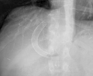
3 R Kidney L Kidney Spleen Liver Psoas muscles Nasogastric / Orogastric tube Implanted Devices/Catheters and Foreign Bodies Can be used for feeding, gastric sampling, gastric decompression, and medication administration Tip and sideport should be in stomach Sideport can extend up to 10 cm from tip of the tube Commonly malpositioned Can enter trachea or curl up in esophagus Indewlling tube can lead to gastroesophageal reflux and cause esophagitis and stricture


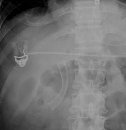
4 Dobhoff Tube Typically used for nutrition Weighted, radiodense tip Tip should be in 2 nd or 3 rd portion of the duodenum Most are in the stomach If tip located proximal to gastroesophageal junction, can lead to aspiration Gastrostomy tube Can be of varying length and appearance Should be overlying the expected location of the stomach An inflated balloon tip may be seen, preventing the tube from pulling out Intraluminal position can be confirmed with contrast administration


5 Other Implanted Devices


6 Foreign Bodies Many foreign bodies, including glass, metal, and stone are radiopaque and can be detected on plain film CT is more sensitive to detect foreign bodies surrounded by air (ie; in bowel) Bowel Gas Pattern


 Are there dilated segments of small bowel? 3) Are there dilated segments of large bowel?")
7 Normal Gas Pattern Distinguishing Large and Small Bowel Stomach Almost always has air Small Bowel Normal diameter ~ 3 cm Large Bowel Almost always has air in rectum or sigmoid Normal diameter ~ 6 cm Cecum up to ~ 10 cm Distention vs. Dilatation Bowel containing sufficient amount of air to fill lumen completely Bowel filled beyond normal size Large Bowel Peripheral Haustral folds usually do not extend across lumen Small Bowel Central Valvulae conniventes usually extend across lumen Spaced more closely Air-Fluid Levels Can be seen on upright views Stomach Almost always Small Bowel No more than 2 or 3 levels Should be < 3 cm long Large Bowel Usually none Abnormal Bowel Gas Patterns Ileus Small and large bowel obstruction Volvulus 3 Questions: 1) Is there gas in the rectum or sigmoid colon? 2) Are there dilated segments of small bowel? 3) Are there dilated segments of large bowel? Abnormal Bowel Gas Patterns Generalized ileus Dilated small AND large bowel Localized ileus (sentinel loop) Several persistently dilated segments of large or small bowel Gas in rectum/sigmoid Mechanical small bowel obstruction Dilated small bowel with little/no gas in large bowel Mechanical large bowel obstruction Dilated small and large bowel


8 Generalized Ileus Refers to disruption in the normal coordinated propulsive motor activity of the gastrointestinal tract in the absence of a mechanical bowel obstruction Suggests that the muscle of the bowel wall is transiently impaired and fails to transport intestinal contents Lack of coordinated propulsive action leads to the accumulation of both gas and fluids within the bowel Common causes: Surgery Inflammation Neural Metabolic Generalized Ileus Radiographic features Generalized, uniform, gaseous distension of both the large and small bowel No discrete transition point to decompressed distal segments of bowel Localized Ileus Can be the result of an adjacent inflammatory or infectious process Focal cluster of 1-3 distended and/or mildly dilated segments of small bowel Termed sentinel loops Location can help suggest underlying etiology Localized Ileus Cholecystitis Pancreatitis Ulcer Appendicitis Diverticulitis


9 Small Bowel Obstruction (SBO) Common clinical condition that occurs secondary to mechanical or functional obstruction of the small bowel Represents 20% of all surgical admissions for acute abdominal pain Proximal dilatation of the intestine due to accumulation of gastrointestinal secretions and swallowed air Bowel distal to the point of obstruction empties over time Small Bowel Obstruction Eventually leads to increased intraluminal pressures Causes compression of mucosal lymphatics Microvascular changes in the bowel wall allow translocation of gut bacteria to mesenteric lymph nodes Increase in incidence of bacteremia due to E. coli Mortality and morbidity are dependent on the etiology, the early recognition and correct diagnosis of obstruction Diagnosis of Small Bowel Obstruction Small bowel diameter > 2.5 cm (usually > 3 cm) KEY: Disproportionate dilatation of small bowel Gas-fluid levels > 2.5 cm wide and at different levels Small bowel feces sign - Often seen near transition point Relative paucity of gas in the colon Presence of residual colonic gas after 6-12 hours is suggestive of partial SBO Early SBO may resemble ileus need follow-up Lappas et al. AJR 2001; 176: May-Smith et al. Clin Radiol 1995; 50:


10 Causes of Small Bowel Obstruction Extraluminal Adhesions Hernias Volvulus Intraluminal Foreign bodies Gallstones Inspissated meconium Intramural Crohn s disease Tumor Radiation Hematoma SBO DDx: Adhesions, Bulges, Crohn s, Cancer


 Causes of")

11 Gasless Abdomen Refers to little or no bowel gas This is nonspecific and can be seen in a variety of etiologies Clinical history plays a key role in distinguishing between benign and threatening etiologies Diagnosis of Large Bowel Obstruction Dilated segments of colon to the point of obstruction Little or no gas in the rectum/sigmoid colon Little or no gas in the small bowel (assuming competent ileocecal valve) Causes of Large Bowel Obstruction Tumor, tumor, tumor. Diverticulitis/stricture Hernia Volvulus Intussusception


12 Volvulus Sigmoid More common in older patients Surgical emergency, as can lead to colonic strangulation and bowel necrosis Classic findings include coffee bean or inverted U appearance of sigmoid colon Distal large bowel obstruction Cecal Less common than sigmoid Displacement of massively dilated cecum away from right lower quadrant Proximal large bowel obstruction Can lead to small bowel dilatation


13 1 limb 1 limb Coffee Bean Ogilvie Syndrome / Acute colonic Pseduo-obstruction Refers to clinical picture of large bowel obstruction without any demonstrable evidence of mechanical obstruction Risk factors Medications which decrease motility Recent surgery Infection Debilitation High mortality rate if perforation occurs


14 Constipation and Fecal Impaction Clinical diagnosis that cannot be made on imaging alone Be aware of fecal impaction, typically referring to large obstructing mass of hardened stool in the distal colon or rectum that can occur in the setting of constipation Fecal impaction can lead to stercoral colitis and perforation Normal Haustral Folds Normally are ~3-4 mm in thickness Thickened haustral folds can be seen on radiographs Etiologies include inflammatory bowel disease, infectious colitis, hematoma, ischemia

 Most often seen in infectious or inflammatory bowel disease")

15 Lead pipe Colon Term used to describe complete loss of normal haustration Presumably due to alterations in muscle tone of the teniae coli from chronic inflammation Reflects burned-out disease Toxic Megacolon Life threatening condition characterized by severe colonic dilatation without obstruction in the setting of systemic toxicity (fever, tachycardia, leukocytosis) Most often seen in infectious or inflammatory bowel disease Pathologic Gas Pneumoperitoneum Pathologic Gas Pneumoretroperitoneum Pneumatosis Portal venous gas Emphysematous pyelpenphritis, cystitis, or cholecystitis Looking for Erect view Pneumopertioneum Air-fluid levels Substitute Left lateral decubitus


16 How Sensitive? Plain films can be 85% sensitive for free air Theoretical threshold is 1 ml CT is much more sensitive and is considered the Gold Standard Best views Erect chest and left lateral decubitus abdomen Supine abdomen is insensitive Signs for pneumoperitoneum Rigler s visualization of air on both sides of the bowel wall Flaciform ligament appearance of a linear opacity from the liver to the midabdomen Double bubble subdiaphragmatic gas outlining the wall of the stomach and diaphragm Miller, et al. Am J Roentgenol Radium Ther Nucl Med 197 Roh, et al. Am J Surg, 1983 Extraluminal Air Spontaneous Pneumoperitoneum Gastric or duodenal ulcer perforation Colonic perforation Diverticulitis Appendicitis Cancer Other causes Thoracic disease (pneumothorax, pneumomediastinum) Iatrogenic


17 Post-operative Pneumoperitoneum Usually presents 3-7 days When to worry? When volume of air increases over time Erect view can be used to evaluate quantity of air Pneumatosis Can occur in variety of conditions, including bowel ischemia, iatrogenic, chemotherapy, collage vascular disease, and chronic obstructive pulmonary disease Concomitant presence of gas in the portal venous circulation is suspicious for bowel ischemia Pneumoretroperitoneum Sites of origin Duodenum Ascending colon Descending colon Rectum Look for Linear air along margins of psoas muscle Gas surrounding kidneys Gas under medial surface of diaphragm


")


18 Abscess Small bubbles/collections of air Straight or triangular margins of air collections Unusually large collections of air Soft Tissue Masses Hepatosplenomegaly Mass (tumor, abscess, cyst, aneurysm, bladder) Bowel displacement Paucity of gas Focal region of increased density Extrinsic compression of bowel


19 Plain film diagnosis of ascites Gray abdomen: diffuse increase density Indistinct margins liver, spleen, psoas Medial displacement of colon, liver and spleen away from properitoneal stripe Bulging flanks Separation of gas-filled small bowel loops ASCITES NORMAL


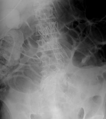
20 Calcification Patterns Rimlike Linear or track-like Lamellar Rimlike Calcification Hollow viscus wall Cysts Aneurysms Saccular organs Porcelain gallbladder Amorphous Walls of a tube Ureters Arterial walls Vas Deferens Linear or Track-like


21 Lamellar Formed in lumen of a hollow viscus Renal stones Gallstones Bladder stones Amorphous, Cloudlike Popcorn Formed in solid organ or solid mass Leiomyomas of uterus Chronic pancreatitis Lymph nodes Summary Abdominal radiographs are often the first imaging examination performed in patients with abdominal pain Understand role of abdominal radiographs in clinical management of patients Recognize range of abdominal radiograph findings, including Implanted devices/catheters, bowel gas patterns, pathologic gas, abdominal organs, and calcifications

22 Thank You
Abdominal radiology 腹部放射線學
 Abdominal radiology 腹部放射線學 台北醫學大學 - 市立萬芳醫院 留偉順 laowilson@hotmail.com The Normal Abdominal Series Chest Supine abdomen Erect abdomen Left lateral decubitus abdomen Learning objectives Understanding normal
Introduction and Definitions
 Bowel obstruction Introduction and Definitions Accounts for 5% of all acute surgical admissions Patients are often extremely ill requiring prompt assessment, resuscitation and intensive monitoring Obstruction
Plain abdomen The standard films are supine & erect AP views (alternative to erect, lateral decubitus film is used in ill patients).
.") Plain abdomen The standard films are supine & erect AP views (alternative to erect, lateral decubitus film is used in ill patients). The stomach can be readily identified by its location, gastric rugae
Plain abdomen The standard films are supine & erect AP views (alternative to erect, lateral decubitus film is used in ill patients). The stomach can be readily identified by its location, gastric rugae
LOOKING FOR AIR IN ALL THE WRONG PLACES Richard M. Gore, MD North Shore University Health System University of Chicago Evanston, IL
 SIGNIFICANCE OF EXTRALUMINAL ABDOMINAL GAS: LOOKING FOR AIR IN ALL THE WRONG PLACES Richard M. Gore, MD North Shore University Health System University of Chicago Evanston, IL SCBT/MR 2012 October 26,
Role of imaging in the evaluation of the acute abdomen
 Prof. András Palkó MD, PhD Role of imaging in the evaluation of the acute abdomen Faculty of General Medicine University of Szeged Hungary 1 Definition Sudden onset of severe symptoms requiring emergency
UNDERSTANDING X-RAYS: ABDOMINAL IMAGING THE ABDOMEN
 UNDERSTANDING X-RAYS: ABDOMINAL IMAGING THE ABDOMEN Radiology Enterprises radiologyenterprises@gmail.com www.radiologyenterprises.com STOMACH AND SMALL BOWEL STOMACH AND SMALL BOWEL Swallowed air is a
Nordic Forum - Trauma & Emergency Radiology. Bowel Obstruction: Imaging Update
 Nordic Forum - Trauma & Emergency Radiology Bowel Obstruction: Imaging Update Borut Marincek Institute of Diagnostic Radiology University Hospital Zurich, Switzerland Acute Abdomen Bowel Obstruction Bowel
Medical application of transabdominal ultrasound in gastrointestinal diseases
 Medical application of transabdominal ultrasound in gastrointestinal diseases Hsiu-Po Wang Department of Emergency Medicine National Taiwan University Hospital Real-time ultrasound has become a standard
ASSESSING THE PLAIN ABDOMINAL RADIOGRAPH M A A M E F O S U A A M P O F O
 ASSESSING THE PLAIN ABDOMINAL RADIOGRAPH M A A M E F O S U A A M P O F O Introduction The abdomen (less formally called the belly, stomach, is that part of the body between the thorax (chest) and pelvis,
X-ray Corner. Imaging of the Small Bowel. Pantongrag-Brown L. Case 1. A 63-year-old man presented with abdominal pain, nausea and vomiting.
 THAI J 42 Imaging of the Small Bowel GASTROENTEROL 2015 X-ray Corner Imaging of the Small Bowel Pantongrag-Brown L Small bowel is the longest tubular organ in the body, about 18-22 feet. It is anchored
Learning Radiology: Recognizing the Basics. Text with Student Consult Online Access Code
 Learning Radiology: Recognizing the Basics. Text with Student Consult Online Access Code Herring, W ISBN-13: 9780323074445 Table of Contents 1. Recognizing Anything The "colorful" world of radiology A
Radiology of the abdomen Lecture -1-
 Radiology of the abdomen Lecture -1- Objectives To know radiology modalities used in abdomen imaging mainly GI tract. To know advantages and disadvantages of each modality. To know indications and contraindications
General Data. 王 X 村 78 y/o 男性
 General Data 王 X 村 78 y/o 男性 Chief Complaint Vomiting twice this early morning Fever up to 38.9ºC was noted Present Illness (1) Old CVA with left side weakness for more than 10 years and with bed ridden
U Lecture Objectives. U Nordic Forum Trauma & Emergency Radiology. Bowel obstruction. U Bowel Obstruction: Etiologies
 Nordic Forum Trauma & Emergency Radiology Lecture Objectives Bowel Obstruction To illustrate the spectrum of acute obstruction of the small and the large bowel To explain how these bowel obstructions may
Abdominal Assessment
 Abdominal Assessment Mary Marian, MS,RD,CSO University of AZ, Tucson, AZ Neha Parekh, MS,RD,LD,CNSC Cleveland Clinic, Cleveland, OH Objectives: 1. Outline the steps in performing an abdominal examination.
Abdominal ultrasound:
 Abdominal ultrasound: Non-traumatic acute abdomen Wittanee Na-ChiangMai, MD Department of Radiology ChiangMai University 26/04/2017 Contents Technique of examination Normal anatomy Emergency conditions
Intestinal Obstruction Clinical Presentation & Causes
 Intestinal Obstruction Clinical Presentation & Causes V Chidambaram-Nathan Consultant Transplant and General Surgeon Sheffield Kidney Institute Northern General Hospital Intestinal Obstruction One of the
Pathology of Intestinal Obstruction. Dr. M. Madhavan, MBBS., MD., MIAC, Professor of Pathology Saveetha Medical College
 Pathology of Intestinal Obstruction Dr. M. Madhavan, MBBS., MD., MIAC, Professor of Pathology Saveetha Medical College Pathology of Intestinal Obstruction Objectives list the causes of intestinal obstruction
이희정. Plain Abdominal Radiography in Infants and Children. Hee Jung Lee, M.D.
 대한소아소화기영양학회지 : 제 14 권제 2 호 2011 DOI: 10.5223/kjpgn.2011.14.2.130 종설 영유아및소아의단순복부 X- 선사진 계명대학교의과대학영상의학교실 이희정 Plain Abdominal Radiography in Infants and Children Hee Jung Lee, M.D. Department of Radiology,
ACUTE ABDOMEN. Dr. M Asadi. Surgical Oncology Research Center MUMS. Assistant Professor of General Surgery
 ACUTE ABDOMEN Dr. M Asadi Assistant Professor of General Surgery Surgical Oncology Research Center MUMS Definition I. The term Acute Abdomen refers to signs & symptoms of abdominal pain and tenderness,
Lab Monitor Images Dissection of the Abdominal Vasculature + Lower Digestive System
 Lab Monitor Images Dissection of the Abdominal Vasculature + Lower Digestive System Stomach & Duodenum Frontal (AP) View Nasogastric tube 2 1 3 4 Stomach Pylorus Duodenum 1 Duodenum 2 Duodenum 3 Duodenum
Role of radiology and imaging in the daignosis of acute abdominal conditions
 Role of radiology and imaging in the daignosis of acute abdominal conditions Miah MAY Introduction In our day to day practice we have to face many of the acute abdominal conditions. As we know acute abdomen
Residents Section Pattern of the Month
 Residents Section Pattern of the Month Krajewski et al. olonic Dilation Residents Section Pattern of the Month Residents inradiology Katherine Krajewski 1 ettina Siewert Ronald L. Eisenberg Krajewski K,
Adult bowel obstruction with acute abdomen: spectrum of CT findings
 Adult bowel obstruction with acute abdomen: spectrum of CT findings Poster No.: C-1571 Congress: ECR 2013 Type: Educational Exhibit Authors: L. Turturici, G. Gherarducci, F. Bianchi, R. Pascale, M. Tonerini,
Hirschprung s. Meconium plug R/S >1 R/S <1
 NEONATAL ABDOMINAL EMERGENCIES LOW OBSTRUCTION HIGH OBSTRUCTION INTESTINAL OBSTRUCTION High obstruction - proximal to mid-ileumileum Few dilated, air filled bowel loops Complete obstruction diagnosed by
Safe Answers For The American Board of Surgery Certifying Exam & Recertifying Exam
 Safe Answers For The American Board of Surgery Certifying Exam & Recertifying Exam By Sarmad Aji, MD., FACS. A comprehensive review of the most commonly asked questions on the American Board of Surgery
Gastrointestinal Tract. Anatomy of GI Tract. Anatomy of GI Tract. (Effective February 2007) (1%-5%)
 (1%-5%)") Gastrointestinal Tract (Effective February 2007) (1%-5%) Anatomy of GI Tract Esophagus bulls-eye or target EG junction seen on sagittal scan posterior to left lobe of liver and anterior to aorta Anatomy
Gastrointestinal Tract (Effective February 2007) (1%-5%) Anatomy of GI Tract Esophagus bulls-eye or target EG junction seen on sagittal scan posterior to left lobe of liver and anterior to aorta Anatomy
Historical perspective
 Raj Santharam, MD GI Associates, LLC Clinical Assistant Professor of Medicine Medical College of Wisconsin Historical perspective FFS first widespread use in the early 1970 s Expansion of therapeutic techniques
Pneumatosis intestinalis, not always a surgical emergency
 Pneumatosis intestinalis, not always a surgical emergency Poster No.: C-2233 Congress: ECR 2012 Type: Educational Exhibit Authors: E. Vanhoutte, M. Lefere, R. Vanslembrouck, D. Bielen, G. De 1 1 2 1 1
DR JAIKISHOR JOTHIRAJ MD POST GRADUATE DEPT OF RADIODIAGNOSIS
 DR JAIKISHOR JOTHIRAJ MD POST GRADUATE DEPT OF RADIODIAGNOSIS YASHODAMMAL 70 YRS OD LADY had C/o diffuse lower abdominal pain 20 days h/o blood in stools 4 days h/o vomiting 2 days h/o burning micturation
Plain Radiographs in Non-Traumatic Abdominal Pain. Plain Radiographs in Non-Traumatic Abdominal Pain
 Jake Block, MD Associate Professor Associate Vice-Chairman for Clinical Operations Director, Musculoskeletal and Emergency Radiology Department of Radiology and Radiological Sciences Vanderbilt University
TOP 10 LIST OF INCIDENTAL GI PET PEEVES ON MDCT
 TOP 10 LIST OF INCIDENTAL GI PET PEEVES ON MDCT Richard M. Gore, MD North Shore University Medical Center University of Chicago Evanston, Illinois SCBT/MR 2011 Washington, DC October 23, 2011 4:30-4:40
Vomiting in children: The good coordination between radiologists and pediatricians is the key to success
 Vomiting in children: The good coordination between radiologists and pediatricians is the key to success C. Santos Montón 1, M. T. Garzon Guiteria 2, A. Hortal Benito-Sendín 1, K. El Karzazi 1, P. Sanchez
Strangulating Obstruction of the Bowel: A Reevaluation of Radiographic Criteria
 Strangulating Obstruction of the Bowel: A Reevaluation of Radiographic Criteria DAVID BRYK1 Fifty consecutive cases of strangulating obstruction were compared with 100 consecutive cases of surgically proven
Gastrointestinal Tract Imaging. Objectives. Reference. VMB 960 April 6, Stomach Small Intestine Colon. Radiography & Ultrasound
 Gastrointestinal Tract Imaging VMB 960 April 6, 2009 Stomach Small Intestine Colon Objectives Radiography & Ultrasound Contrast Examination of the Small Intestine Reference Chapters 45 47 Pages 750 805
Nasogastric tube. Stomach. Pylorus. Duodenum 1. Duodenum 2. Duodenum 3. Duodenum 4
 Esophagus Barium Swallow Stomach and Duodenum 4 year old Upper GI Nasogastric tube Stomach and Duodenum 4 year old Upper GI Nasogastric tube Stomach Pylorus Duodenum 1 Duodenum 2 Duodenum 3 Duodenum 4
Gastroenterology. Certification Examination Blueprint. Purpose of the exam
 Gastroenterology Certification Examination Blueprint Purpose of the exam The exam is designed to evaluate the knowledge, diagnostic reasoning, and clinical judgment skills expected of the certified gastroenterologist
Many of the disease processes that acutely affect the abdomen produce
 ABC of Emergency Radiology Radiographic signs oftrauma THE ABDOMEN-II D A Nicholson, P A Driscoll Many of the disease processes that acutely affect the abdomen produce radiographic signs, but most of these
Acquired pediatric esophageal diseases Imaging approaches and findings. M. Mearadji International Foundation for Pediatric Imaging Aid
 Acquired pediatric esophageal diseases Imaging approaches and findings M. Mearadji International Foundation for Pediatric Imaging Aid Acquired pediatric esophageal diseases The clinical signs of acquired
CHEST & ABDOMINAL X-RAYS MALIKA IBRAHIM CORE MEDICAL TRAINEE BLACKPOOL VICTORIA HOSPITAL DATA INTERPRETATION COURSE FEB 20, 2017
 CHEST & ABDOMINAL X-RAYS MALIKA IBRAHIM CORE MEDICAL TRAINEE BLACKPOOL VICTORIA HOSPITAL DATA INTERPRETATION COURSE FEB 20, 2017 1. Sample x-rays 2. Basic chest x-ray interpretation skills 3. Chest x-ray
ENTEROCOLITIDES CAN YOU TELL THEM APART ON MDCT? Richard M. Gore, MD North Shore University Medical Center University of Chicago Evanston, Illinois
 ENTEROCOLITIDES CAN YOU TELL THEM APART ON MDCT? Richard M. Gore, MD North Shore University Medical Center University of Chicago Evanston, Illinois SCBT/MR 2010 San Diego, California March 8, 2010 13:40-14:00
Back to Basics: What Imaging Test should I order? Jeanne G. Hill, M.D. Pediatric Radiology Medical University of South Carolina
 Back to Basics: What Imaging Test should I order? Jeanne G. Hill, M.D. Pediatric Radiology Medical University of South Carolina Disclosure Neither I nor any member of my immediate family has a relevant
Chapter 32 Gastroenterology General Pathophysiology General Risk Factors for GI emergencies: Excessive Consumption Excessive Smoking Increased
 1 2 3 4 5 6 7 Chapter 32 Gastroenterology General Pathophysiology General Risk Factors for GI emergencies: Excessive Consumption Excessive Smoking Increased Ingestion of Caustic Substances Poor Bowel Habits
ACUTE ABDOMEN IN OLDER CHILDREN. Carlos J. Sivit M.D.
 ACUTE ABDOMEN IN OLDER CHILDREN Carlos J. Sivit M.D. ACUTE ABDOMEN Clinical condition characterized by severe abdominal pain developing over several hours ACUTE ABDOMINAL PAIN Common childhood complaint
Summary and conclusions
 Summary and conclusions 7 Chapter 7 68 Summary and conclusions Chapter 1 provides a general introduction to this thesis focused on the use of ultrasound (US) in children with abdominal problems. The literature
Pitfalls in the CT diagnosis of appendicitis
 The British Journal of Radiology, 77 (2004), 792 799 DOI: 10.1259/bjr/95663370 E 2004 The British Institute of Radiology Pictorial review Pitfalls in the CT diagnosis of appendicitis 1 C D LEVINE, 2 O
Case Cholecystoduodenal fistula with migrated gallstone leading to gastric outlet obstruction: Bouveret's syndrome
 Case 14613 Cholecystoduodenal fistula with migrated gallstone leading to gastric outlet obstruction: Bouveret's syndrome Eva De Backer 1, Filip Vanhoenacker 2, 3, 4, Adelard De Backer5 1: Ghent University,
Abdominal Pain in Pediatric Patients Image Gently
 Abdominal Pain in Pediatric Patients Image Gently Susan D. John, M.D. Baptist Health Emergency Radiology 2017 Disclosure I have no financial relationships with a commercial entity producing healthcarerelated
Spleen indications of splenectomy complications OPSI
 Intestinal obstruction Differences between adynamic ileus and mechanical obstruction Aetiology Pathophysiology (Cluster contractions- bowel proximal to the obstruction dilate- wall of obstructed gut is
FHS Appendicitis US Protocol
 FHS Appendicitis US Protocol Reviewed By: Shireen Khan, MD; Sarah Farley, MD; Anna Ellermeier, MD Last Reviewed: May 2018 Contact: (866) 761-4200 **NOTE for all examinations: 1. If documenting possible
Gas patterns on plain abdominal radiographs: a pictorial review
 1 Royal Hallamshire Hospital, Sheffield Teaching Hospitals NHS Trust, Sheffield, South Yorkshire, UK 2 Barnsley Hospital NHS Foundation Trust, Barnsley, South Yorkshire, UK Correspondence to Dr R E Musson,
Plain Abdominal Radiography & GI series 腹部與腸胃道之放射線學
 Plain Abdominal Radiography & GI series 腹部與腸胃道之放射線學 陳潤秋台北市立聯合醫院 chenranchou@tpech.gov.tw Jen-Ai H. Standing abdomen Jen-Ai H. www.nlm.nih.gov Supine KUB Jen-Ai H. medicalcenter.osu.edu Jen-Ai H. Checklist:
GASTROENTEROLOGY Maintenance of Certification (MOC) Examination Blueprint
 Examination Blueprint") GASTROENTEROLOGY Maintenance of Certification (MOC) Examination Blueprint ABIM invites diplomates to help develop the Gastroenterology MOC exam blueprint Based on feedback from physicians that MOC assessments
GASTROENTEROLOGY Maintenance of Certification (MOC) Examination Blueprint ABIM invites diplomates to help develop the Gastroenterology MOC exam blueprint Based on feedback from physicians that MOC assessments
Anatomy of the Large Intestine
 Large intestine Anatomy of the Large Intestine 2 Large Intestine Extends from ileocecal valve to anus Length = 1.5-2.5m = 5 feet Regions Cecum = 2.5-3 inch Appendix= 3-5 inch Colon Ascending= 5 inch Transverse=
Computed tomography (CT) imaging review of small bowel obstruction
 imaging review of small bowel obstruction") Computed tomography (CT) imaging review of small bowel obstruction Poster No.: C-1602 Congress: ECR 2010 Type: Educational Exhibit Topic: GI Tract - Small Bowel Authors: A. Vousough, D. S. Prasad ; Aberdeen/UK,
Computed tomography (CT) imaging review of small bowel obstruction Poster No.: C-1602 Congress: ECR 2010 Type: Educational Exhibit Topic: GI Tract - Small Bowel Authors: A. Vousough, D. S. Prasad ; Aberdeen/UK,
GASTROINTESTINAL IMAGING STUDY GUIDE
 GASTROINTESTINAL IMAGING STUDY GUIDE Pharynx Diverticula Foreign bodies Trauma o Motility Disorders Esophagus Diverticula Trauma Esophagitis Barrett esophagus Rings, webs, and strictures Varices Benign
... Inflammatory disorder of the colon that occurs as a complication of antibiotic treatment.
 Definition Inflammatory disorder of the colon that occurs as a complication of antibiotic treatment. " Epidemiology Humans represent the main reservoir of Clostridium difficile, which is not part of the
The Value of Urgent Barium Enema and Computed Tomography in Acute Malignant Colonic Obstruction: Is Urgent Barium Enema Still Necessary?
 J Radiol Sci 2012; 37: 105-110 The Value of Urgent Barium Enema and Computed Tomography in Acute Malignant Colonic Obstruction: Is Urgent Barium Enema Still Necessary? Chun-Chao Huang 1,2 Fei-Shih Yang
Evidence Process for Abdominal Pain Guideline Research 11/16/2017. Guideline Review using ADAPTE method and AGREE II instrument 11/16/2017
 Evidence Process for Abdominal Pain Guideline Research Guideline Review using ADAPTE method and AGREE II instrument Approximately 139 Potentially relevant guidelines identified in various resources* 59
PROFESSIONAL SKILLS 1 3RD YEAR SEMESTER 6 RADIOGRAPHY. THE URINARY SYSTEM Uz. Fatema shmus aldeen Tel
 PROFESSIONAL SKILLS 1 3RD YEAR SEMESTER 6 RADIOGRAPHY THE URINARY SYSTEM Uz. Fatema shmus aldeen Tel. 0925111552 Professional skills-2 THE URINARY SYSTEM The urinary system (review anatomy and physiology)
Endoscopic Treatment of Luminal Perforations and Leaks
 Endoscopic Treatment of Luminal Perforations and Leaks Ali A. Siddiqui, MD Professor of Medicine Director of Interventional Endoscopy Jefferson Medical College Philadelphia, PA When Do You Suspect a Luminal
Perforation of a Duodenal Diverticulum. Elective Student S. C.
 Perforation of a Duodenal Diverticulum 2008 4 Elective Student S. C. Case History An elderly male presented to the Emergency Department with abdominal pain. Chief Complaint: Worsening, diffuse abdominal
Computed tomography (CT) imaging review of small bowel obstruction
 imaging review of small bowel obstruction") Computed tomography (CT) imaging review of small bowel obstruction Poster No.: C-1602 Congress: ECR 2010 Type: Educational Exhibit Topic: GI Tract Authors: A. Vousough, D. S. Prasad ; Aberdeen/UK, Leeds/UK
Computed tomography (CT) imaging review of small bowel obstruction Poster No.: C-1602 Congress: ECR 2010 Type: Educational Exhibit Topic: GI Tract Authors: A. Vousough, D. S. Prasad ; Aberdeen/UK, Leeds/UK
Cecal Volvulus: Case Presentation and Review of CT Findings
 August 2011 Cecal Volvulus: Case Presentation and Review of CT Findings Omar Pardesi, Harvard Medical School Year III Our Patient LD: History & Physical HPI: 28 y.o. female presents with diffuse abdominal
Close window to return to IVIS. in collaborazione con RICHIESTO ACCREDITAMENTO. organizzato da certificata ISO 9001:2000
 in collaborazione con Close window to return to IVIS RICHIESTO ACCREDITAMENTO SOCIETÀ CULTURALE ITALIANA VETERINARI PER ANIMALI DA COMPAGNIA SOCIETÀ FEDERATA ANMVI organizzato da certificata ISO 9001:2000
Surgical Education Series
 Surgical Education Series The Acute Abdomen Ahmad kachooei, MD MPH Assistant Professor Division of General Surgery Department of Surgery University of Qom Outline Definitions What causes an acute abdomen
CT Evaluation of Bowel Wall Thickening. Dr: Adel El Badrawy; M.D. Lecturer of Radio Diagnosis Faculty of Medicine Mansoura University.
 CT Evaluation of Bowel Wall Thickening By Dr: Adel El Badrawy; M.D. Lecturer of Radio Diagnosis Faculty of Medicine Mansoura University. The CT findings of bowel wall thickening includes 1 Degree of thickening.
Abdominal Ultrasonography
 Abdominal Ultrasonography David A. Masneri, DO, FACEP, FAAEM Assistant Professor of Emergency Medicine Assistant Director, Emergency Medicine Residency Medical Director, Operational Medicine Division Center
Gastrointestinal Disorders. Disorders of the Esophagus 3/7/2013. Congenital Abnormalities. Achalasia. Not an easy repair. Types
 Gastrointestinal Disorders Congenital Abnormalities Disorders of the Esophagus Types Stenosis Atresia Fistula Newborn aspirates while feeding. Pneumonia Not an easy repair Achalasia Lack of relaxation
Gastrointestinal Emergencies CEN REVIEW 2017 MARY RALEY, BSN, RN, CEN, TCRN, TNSCC
 Gastrointestinal Emergencies CEN REVIEW 2017 MARY RALEY, BSN, RN, CEN, TCRN, TNSCC Gastrointestinal Emergencies is 7% of the CEN A. Acute abdomen B. Bleeding C. Cholecystitis D. Cirrhosis E. Diverticulitis
GENERAL ABDOMINAL IMAGING PERITONEAL SPACE, PANCREAS, & SPLEEN. VMB 960 March 25, 2013
 GENERAL ABDOMINAL IMAGING PERITONEAL SPACE, PANCREAS, & SPLEEN VMB 960 March 25, 2013 REFERENCE Chapters 35-36 Pages 650-678 Chapter 37 Pages 694-701 Chapter 3 Pages 38-49 OBJECTIVES Radiography and Ultrasound
General Abdominal Radiography
 General Abdominal Radiography Tony Pease, DVM, MS Assistant Professor of Radiology North Carolina State University Objectives Acquisition of radiographs Abdominal radiographic anatomy Radiographic patterns
Radiology. Undergraduate Radiology Sample Questions
 Radiology Undergraduate Radiology Sample Questions April 2012 The following examples are offered of questions that might be used to assess undergraduate radiology. There are 3 different styles: An OSCE
elical CT plays an important role
 bdominal Imaging Yu et al. Helical CT of cute RLQ Pain Pictorial Essay Jinxing Yu 1 nn S. Fulcher Mary nn Turner Robert. Halvorsen Yu J, Fulcher S, Turner M, Halvorsen R Helical CT Evaluation of cute Right
Appendix 9: Endoscopic Ultrasound in Gastroenterology
 Appendix 9: Endoscopic Ultrasound in Gastroenterology This curriculum is intended for clinicians who perform endoscopic ultrasonography (EUS) in gastroenterology. It includes standards for theoretical
General Surgery Service
 General Surgery Service Patient Care Goals and Objectives Stomach/Duodenum and Bariatric assessed for a) Obesity surgery b) Treatment of i) Adenocarcinoma of the stomach ii) GIST iii) Carcinoid 2) Optimize
Appendix 5. EFSUMB Newsletter. Gastroenterological Ultrasound
 EFSUMB Newsletter 87 Examinations should encompass the full range of pathological conditions listed below A log book listing the types of examinations undertaken should be kept Training should usually
THE mainstay of the radiographic study of the upper gastrointestinal tract has
 BARIUM-SPRAY EXAMINATION OF THE STOMACH- PRELIMINARY REPORT OF A NEW ROENTGENOGRAPHIC TECHNIC EDWARD BUONOCORE, M.D., and THOMAS F. MEANEY, M.D. Department of Hospital Radiology THE mainstay of the radiographic
12 Blueprints Q&A Step 2 Surgery
 12 Blueprints Q&A Step 2 Surgery 34. A 40-year-old female has been referred to you for a recent ER and hospital admission, from which she was given a diagnosis of acute diverticulitis. Treatment at that
Comparative Study between Plain Radiography and Ultrasound Abdomen in Non Traumatic Surgical Acute Abdominal Conditions
 ORIGINAL ARTICLE Comparative Study between Plain Radiography and Ultrasound Abdomen in Non Traumatic Surgical Acute Sharma P 1, Sidharth 2, Singh BP 3, Singh D 3, Gupta A 4 1 Department of Radiology and
Volvulus of the Gastrointestinal Tract: x-ray and CT imaging
 Volvulus of the Gastrointestinal Tract: x-ray and CT imaging Poster No.: C-0076 Congress: ECR 2013 Type: Educational Exhibit Authors: E. Papadaki, S. Paschalidou, S. GIANNOU ; Rethymno, CR/ 1 2 2 3 1 3
Chapter 14: Training in Radiology. DDSEP Chapter 1: Question 12
 DDSEP Chapter 1: Question 12 A 52-year-old white male presents for evaluation of sudden onset of abdominal pain and shoulder pain. His past medical history is notable for a history of coronary artery disease,
CLINICAL PRESENTATION AND RADIOLOGY QUIZ QUESTION
 Donald L. Renfrew, MD Radiology Associates of the Fox Valley, 333 N. Commercial Street, Suite 100, Neenah, WI 54956 09/17/2011 Radiology Quiz of the Week # 38 Page 1 CLINICAL PRESENTATION AND RADIOLOGY
INVESTIGATIONS OF GASTROINTESTINAL DISEAS
 INVESTIGATIONS OF GASTROINTESTINAL DISEAS Lecture 1 and 2 دز اسماعيل داود فرع الطب كلية طب الموصل Radiological tests of structure (imaging) Plain X-ray: May shows soft tissue outlines like liver, spleen,
Basic Abdominal and Pelvic Imaging Concepts. David L. Smith, MD Assistant Professor of Radiology
 Basic Abdominal and Pelvic Imaging Concepts David L. Smith, MD Assistant Professor of Radiology Basic Imaging Concepts Contrast Resolution vs Spacial Resolution Spacial Resolution......refers to the ability
Preview from Notesale.co.uk Page 1 of 34
 Abdominal viscera and digestive tract Digestive tract Abdominal viscera comprise majority of the alimentary system o Terminal oesophagus, stomach, pancreas, spleen, liver, gallbladder, kidneys, suprarenal
Abdominal air is it in the right or in the wrong place?
 Abdominal air is it in the right or in the wrong place? Poster No.: C-1866 Congress: ECR 2014 Type: Educational Exhibit Authors: M. Drake Perez, M. Diez Blanco, E. Lopez Uzquiza, S. Sánchez 1 1 1 1 2 3
ABDOMEN - GI. Duodenum
 TALA SALEH ABDOMEN - GI Duodenum - Notice the shape of the duodenum, it looks like capital G shape tube which extends from the pyloroduodenal junction to the duodenojejunal junction. - It is 10 inches
Chest X rays and Case Studies. No disclosures. Outline 5/31/2018. Carlo Manalo, M.D. Department of Radiology Loma Linda University Children s Hospital
 Chest X rays and Case Studies Carlo Manalo, M.D. Department of Radiology Loma Linda University Children s Hospital No disclosures. Outline Importance of history Densities delineated on radiography An approach
Case Report A Case of Stercoral Perforation Detected on CT Requiring Proctocolectomy in a Heroin-Dependent Patient
 Case Reports in Surgery Volume 2016, Article ID 2893925, 4 pages http://dx.doi.org/10.1155/2016/2893925 Case Report A Case of Stercoral Perforation Detected on CT Requiring Proctocolectomy in a Heroin-Dependent
SWISS SOCIETY OF NEONATOLOGY. Spontaneous intestinal perforation or necrotizing enterocolitis?
 SWISS SOCIETY OF NEONATOLOGY Spontaneous intestinal perforation or necrotizing enterocolitis? June 2004 2 Stocker M, Berger TM, Neonatal and Pediatric Intensive Care Unit, Children s Hospital of Lucerne,
Abdominal extra-luminal gas - Is it always gastrointestinal perforation?
 Abdominal extra-luminal gas - Is it always gastrointestinal perforation? Poster No.: C-2526 Congress: ECR 2015 Type: Educational Exhibit Authors: M. Barros, L. A. Ferreira, I. Abreu, F. Caseiro Alves;
X-ray Corner. Imaging of The Colon. Pantongrag-Brown L
 110 Imaging of The Colon X-ray Corner Imaging of The Colon Pantongrag-Brown L Imaging modalities used in colon include plain radiographs, barium enema, US, CT, PET CT and MRI. Barium enema (BE) is declining
GASTROINTESTINAL SYSTEM
 GASTROINTESTINAL SYSTEM Topographic Anatomy of the Abdomen Surface Landmarks Xiphoid process T9/T10 Inferior costal margin L2/L3 Iliac Crest L4 level ASIS L5/S1 level Pubic symphysis level of greater trochanter
A rare case of intestinal obstruction due to internal hernia. Dr. Jayanth 3 rd year PG Dept. Of General Surgery
 A rare case of intestinal obstruction due to internal hernia Dr. Jayanth 3 rd year PG Dept. Of General Surgery One of the common cause of acute abdomen May lead to high morbidity and mortality if not treated
QUESTIONS for the examination in surgery for 4 th -year students of the Faculty of foreign students
 QUESTIONS for the examination in surgery for 4 th -year students of the Faculty of foreign students 1. The main principles of surgical deontology and its founders. 2. Acute appendicitis. Anatomico-physiological
Sonographic Appearances of Common Gut Pathology in Paediatric Patients: Comparison with Plain Abdominal Radiography
 3668 Radiographer Text 1/4/04 2:57 PM Page 11 The Radiographer vol. 51: 11-17 Sonographic Appearances of Common Gut Pathology in Paediatric Patients: Comparison with Plain Abdominal Radiography Lino Piotto
Proceedings of the 34th World Small Animal Veterinary Congress WSAVA 2009
 www.ivis.org Proceedings of the 34th World Small Animal Veterinary Congress WSAVA 2009 São Paulo, Brazil - 2009 Next WSAVA Congress : Reprinted in IVIS with the permission of the Congress Organizers IMAGING
Jhia Anjela D. Rivera 1 1. BS Biology, Department of Biology, College of Science, Polytechnic University of the Philippines
 DIGESTIVE SYSTEM Jhia Anjela D. Rivera 1 1 BS Biology, Department of Biology, College of Science, Polytechnic University of the Philippines DIGESTIVE SYSTEM Consists of the digestive tract (gastrointestinal
Original Report. Stercoral Colitis Leading to Fatal Peritonitis: CT Findings. Gastrointestinal Imaging Heffernan et al. CT of Stercoral Colitis
 Gastrointestinal Imaging Heffernan et al. CT of Stercoral Colitis Cathleen Heffernan 1 H. Leon Pachter 2 lec J. Megibow 1 Michael Macari 1 Hefferman C, Pachter HL, Megibow J, Macari M Received pril 21,