Abdominal radiology 腹部放射線學
|
|
|
- Allen Lambert
- 5 years ago
- Views:
Transcription
1 Abdominal radiology 腹部放射線學 台北醫學大學 - 市立萬芳醫院 留偉順 laowilson@hotmail.com
2 The Normal Abdominal Series Chest Supine abdomen Erect abdomen Left lateral decubitus abdomen
3 Learning objectives Understanding normal plain abdomen radiographs Understanding abnormal plain abdomen - Calcifications - Gas - Masses - Others

4 What to Examine l l l l Gas pattern Extraluminal air Soft tissue masses Calcifications
5 The Check List Bones and joints Calcifications Organs Fat Gas: In and outside of bowel
6 A memory aid First organs, bones, and stones, Then masses, fat, and gas. Don t forget the corner zones, And you ll always cover your subject
7 Abdominal Calcifications l l l Rimlike Linear or track-like walls of tubes such as ureter, arteries Lamellar gallstone, myoma
8 Rimlike Calcification Wall of a hollow viscus Cysts Renal cyst Aneurysms Aortic aneurysm Saccular organs e.g. GB Porcelain Gallbladder
9 AAA
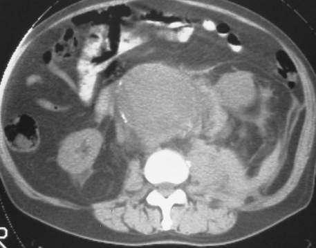
10 AAA: CT

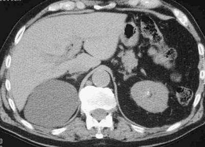
11 Renal Cyst Gallbladder Wall
12 Lamellar or Laminar l Formed in lumen of a hollow viscus n n n Renal stones Gallstones Bladder stones
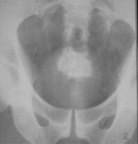
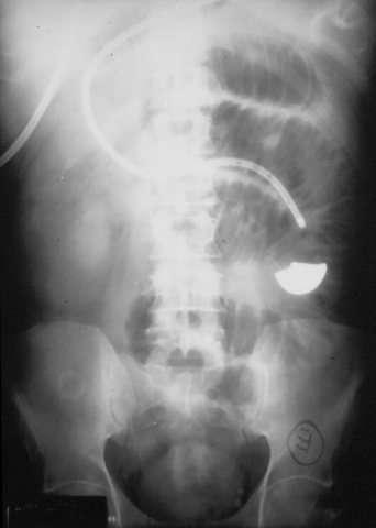
13 Stone in Ureterocoele

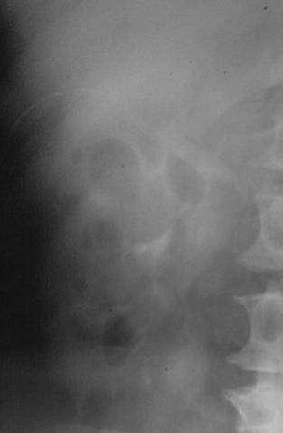
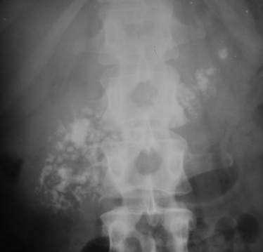
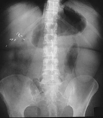
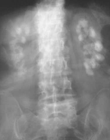
14 Staghorn Calculi
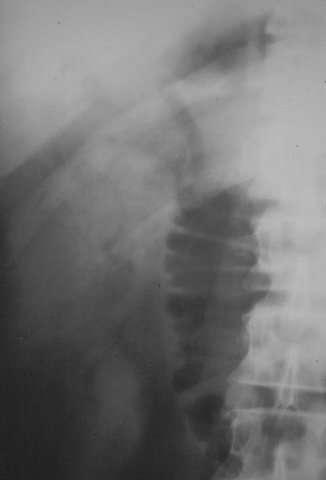
15 Chronic Pancreatitis
16 Normal Gas Pattern Gas in the stomach. Some gas in the small bowel. Gas in the rectum and sigmoid.

17 Gas in stomach Gas in a few loops of small bowel Gas in rectum or sigmoid
18 Abnormal Gas Patterns l l Functional Ileus n n Localized (Sentinel Loops) Generalized adynamic ileus Mechanical Obstruction n n SBO LBO

19 Large vs. Small Bowel l l Large Bowel n n Peripheral Haustral markings don't extend from wall to wall Small Bowel n n Central Valvulae extend across lumen n Maximum diameter of 2"
20 Sentinel Loops Cholecystitis Pancreatitis Ulcer Appendicitis Diverticulitis
21 Air in Rectum or sigmoid Air in Small Bowel Air in Large Bowel Localized Ileus Yes 2-3 distended loops Air in rectum or sigmoid Generalized Ileus Yes Multiple distended loops Yes- Distended SBO No Multiple dilated loops No LBO No None-unless ileocecal valve incompetent Yes- Dilated
22 Small Bowel Obstruction: Causes Adhesions 80% Hernia 15% Tumors, intussusception, midgut volvulus, etc.
23 COLON Obstruction Causes Carcinoma of the colon 80% Volvulus 5% Diverticulitis 5% Fecal impaction 5% Others 5%
24 Small Bowel Obstruction: Findings Step-ladder dilated bowel loops on supine view Step-ladder air-fluid levels on erect/decubitus views String-of-pearls sign on erect/decubitus views
25 Small Bowel Obstruction, Supine

26 Air Filled Small Bowel


27 Supine Prone Sentinel Loops
28 String-of-Pearls Sign: Erect

29 Fluid Filled Small Bowel
30 LBO
31

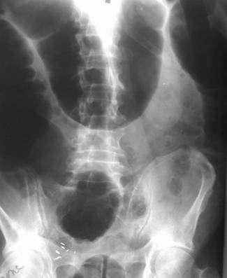
32 Sigmoid Volulus:
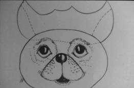
33 Air in Biliary System Usually secondary to surgery on bile ducts Can be due to biliary-bowel fistula from infection or neoplasm Rarely, can be due to infection

34 Air in Gall Bladder

35 Air in Bile Ducts
36
37 PNEUMOPERITONEUM FREE AIR: Perforated Gut
38 Free Air Best views: Erect chest and left lateral decubitus abdomen Erect abdomen is less sensitive Supine abdomen is insensitive
39 Free Air: Left Lateral Decubitus Right side up, left side down Patient who can t sit or stand Air under right abdominal wall
40 Imaging findings Large collection of gas Abdominal distension, no gastric air-fluid level "Football sign" = large pneumoperitoneum outlining entire abdominal cavity "Double wall sign" = "Rigler sign" = air on both sides of bowel as intraluminal gas and free air outside (usually requires >1,000 ml of free intraperitoneal gas + intraperitoneal fluid) "Telltale triangle sign" = triangular air pocket between 3 loops of bowel "Inverted V sign" = outline of both lateral umbilical ligaments (containing inferior epigastric vessels) Outline of medial umbilical ligaments (obliterated umbilical arteries) "Urachus sign" = outline of middle umbilical ligament

41 Free Air: Erect Chest

42 Crescent sign Free Intraperitoneal Air

43 Free Intraperitoneal Air Air on both sides of bowel wall Rigler s Sign

44 Falciform Ligament Sign

45 Air in Lesser Sac

46 lateral umbilicus sign (arrow)

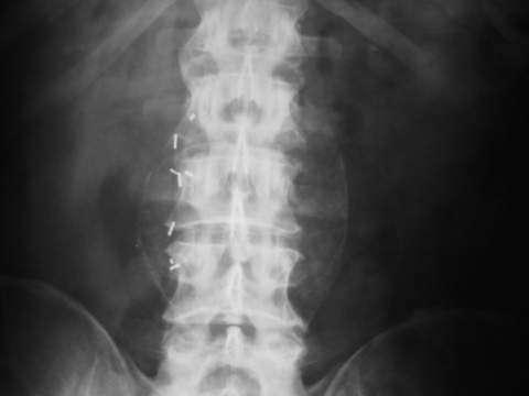
47 urachus sign

48 Falciform ligament sign

49 air in the Morison pouch
.")
50 bowel relief sign (arrow).
51 Extraperitoneal Air
52 Pneumoretroperitoneum From perforation of posterior extraperitoneal portions of duodenum or colon Extension from or through the mediastinum
53 Pneumoretroperitoneum

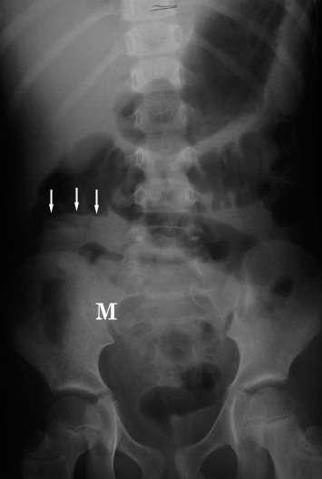
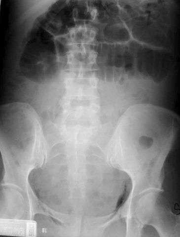
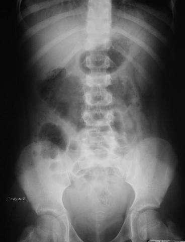
54 Supine anteroposterior abdominal radiograph shows a calcified appendicolith (white arrows) in the right lower quadrant. The bowel gas pattern is nonspecific, with mildly dilated, air-filled small bowel loops in the right upper quadrant. The left psoas muscle margin (black arrows) is normal and distinct; the right psoas muscle margin is not seen.
55 Soft Tissue Masses l l Hepatosplenomegaly n Plain films poor for judging liver size Tumor or cyst n Bowel displacement
56 Splenomegaly

57 Body Packer


58 Bladder Outlet Obstruction pre- and post- cath

59 Right Renal Cyst

60 Foreign Body: Battery
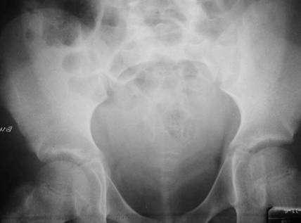
61 Free Peritoneal Fluid- Bladder Ears
62 Atherosclerosis Calcification Vas Deferens
63 Nephrocalcinosis Myomatous Uterus

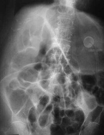
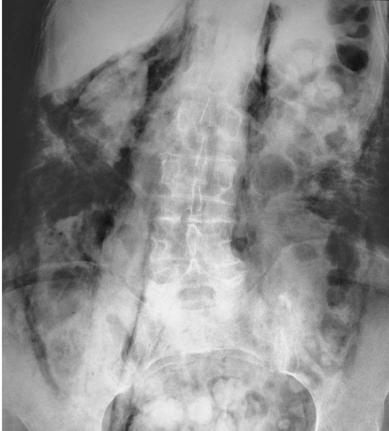
64 Supine Erect Generalized Adynamic Ileus
65 Summary Abdominal Series Abdominal Calcifications Normal and Abnormal Gas Patterns Large vs. Small Bowel Soft Tissue Masses
No Disclosures. Approach to Abdominal Radiographs
 Approach to Abdominal Radiographs Tapas K. Tejura, M.D. Assistant Professor of Clinical Radiology Keck Medical Center of USC tapas.tejura@med.usc.edu No Disclosures 34-year-old male with acute abdominal
Introduction and Definitions
 Bowel obstruction Introduction and Definitions Accounts for 5% of all acute surgical admissions Patients are often extremely ill requiring prompt assessment, resuscitation and intensive monitoring Obstruction
ASSESSING THE PLAIN ABDOMINAL RADIOGRAPH M A A M E F O S U A A M P O F O
 ASSESSING THE PLAIN ABDOMINAL RADIOGRAPH M A A M E F O S U A A M P O F O Introduction The abdomen (less formally called the belly, stomach, is that part of the body between the thorax (chest) and pelvis,
Plain abdomen The standard films are supine & erect AP views (alternative to erect, lateral decubitus film is used in ill patients).
.") Plain abdomen The standard films are supine & erect AP views (alternative to erect, lateral decubitus film is used in ill patients). The stomach can be readily identified by its location, gastric rugae
Plain abdomen The standard films are supine & erect AP views (alternative to erect, lateral decubitus film is used in ill patients). The stomach can be readily identified by its location, gastric rugae
Plain Abdominal Radiography & GI series 腹部與腸胃道之放射線學
 Plain Abdominal Radiography & GI series 腹部與腸胃道之放射線學 陳潤秋台北市立聯合醫院 chenranchou@tpech.gov.tw Jen-Ai H. Standing abdomen Jen-Ai H. www.nlm.nih.gov Supine KUB Jen-Ai H. medicalcenter.osu.edu Jen-Ai H. Checklist:
UNDERSTANDING X-RAYS: ABDOMINAL IMAGING THE ABDOMEN
 UNDERSTANDING X-RAYS: ABDOMINAL IMAGING THE ABDOMEN Radiology Enterprises radiologyenterprises@gmail.com www.radiologyenterprises.com STOMACH AND SMALL BOWEL STOMACH AND SMALL BOWEL Swallowed air is a
Role of radiology and imaging in the daignosis of acute abdominal conditions
 Role of radiology and imaging in the daignosis of acute abdominal conditions Miah MAY Introduction In our day to day practice we have to face many of the acute abdominal conditions. As we know acute abdomen
이희정. Plain Abdominal Radiography in Infants and Children. Hee Jung Lee, M.D.
 대한소아소화기영양학회지 : 제 14 권제 2 호 2011 DOI: 10.5223/kjpgn.2011.14.2.130 종설 영유아및소아의단순복부 X- 선사진 계명대학교의과대학영상의학교실 이희정 Plain Abdominal Radiography in Infants and Children Hee Jung Lee, M.D. Department of Radiology,
X-ray Corner. Imaging of the Small Bowel. Pantongrag-Brown L. Case 1. A 63-year-old man presented with abdominal pain, nausea and vomiting.
 THAI J 42 Imaging of the Small Bowel GASTROENTEROL 2015 X-ray Corner Imaging of the Small Bowel Pantongrag-Brown L Small bowel is the longest tubular organ in the body, about 18-22 feet. It is anchored
Learning Radiology: Recognizing the Basics. Text with Student Consult Online Access Code
 Learning Radiology: Recognizing the Basics. Text with Student Consult Online Access Code Herring, W ISBN-13: 9780323074445 Table of Contents 1. Recognizing Anything The "colorful" world of radiology A
Role of imaging in the evaluation of the acute abdomen
 Prof. András Palkó MD, PhD Role of imaging in the evaluation of the acute abdomen Faculty of General Medicine University of Szeged Hungary 1 Definition Sudden onset of severe symptoms requiring emergency
RADIOLOGY (SURGERY) BY MARYAM MALIK Rawalpindi Medical College
 BY MARYAM MALIK Rawalpindi Medical College") RADIOLOGY (SURGERY) BY MARYAM MALIK Rawalpindi Medical College NORMAL BOWEL GAS PATTERN Any part of the bowel may be visible if it contains gas/air within the lumen. Gas/air is of low density and forms
RADIOLOGY (SURGERY) BY MARYAM MALIK Rawalpindi Medical College NORMAL BOWEL GAS PATTERN Any part of the bowel may be visible if it contains gas/air within the lumen. Gas/air is of low density and forms
LOOKING FOR AIR IN ALL THE WRONG PLACES Richard M. Gore, MD North Shore University Health System University of Chicago Evanston, IL
 SIGNIFICANCE OF EXTRALUMINAL ABDOMINAL GAS: LOOKING FOR AIR IN ALL THE WRONG PLACES Richard M. Gore, MD North Shore University Health System University of Chicago Evanston, IL SCBT/MR 2012 October 26,
U Lecture Objectives. U Nordic Forum Trauma & Emergency Radiology. Bowel obstruction. U Bowel Obstruction: Etiologies
 Nordic Forum Trauma & Emergency Radiology Lecture Objectives Bowel Obstruction To illustrate the spectrum of acute obstruction of the small and the large bowel To explain how these bowel obstructions may
US in non-traumatic acute abdomen. Lalita, M.D. Radiologist Department of radiology Faculty of Medicine ChiangMai university
 US in non-traumatic acute abdomen Lalita, M.D. Radiologist Department of radiology Faculty of Medicine ChiangMai university Sagittal Orientation Transverse (Axial) Orientation Coronal Orientation Intercostal
Nordic Forum - Trauma & Emergency Radiology. Bowel Obstruction: Imaging Update
 Nordic Forum - Trauma & Emergency Radiology Bowel Obstruction: Imaging Update Borut Marincek Institute of Diagnostic Radiology University Hospital Zurich, Switzerland Acute Abdomen Bowel Obstruction Bowel
Plain Radiographs in Non-Traumatic Abdominal Pain. Plain Radiographs in Non-Traumatic Abdominal Pain
 Jake Block, MD Associate Professor Associate Vice-Chairman for Clinical Operations Director, Musculoskeletal and Emergency Radiology Department of Radiology and Radiological Sciences Vanderbilt University
Gas patterns on plain abdominal radiographs: a pictorial review
 1 Royal Hallamshire Hospital, Sheffield Teaching Hospitals NHS Trust, Sheffield, South Yorkshire, UK 2 Barnsley Hospital NHS Foundation Trust, Barnsley, South Yorkshire, UK Correspondence to Dr R E Musson,
ABDOMEN - GI. Duodenum
 TALA SALEH ABDOMEN - GI Duodenum - Notice the shape of the duodenum, it looks like capital G shape tube which extends from the pyloroduodenal junction to the duodenojejunal junction. - It is 10 inches
Abdominal Assessment
 Abdominal Assessment Mary Marian, MS,RD,CSO University of AZ, Tucson, AZ Neha Parekh, MS,RD,LD,CNSC Cleveland Clinic, Cleveland, OH Objectives: 1. Outline the steps in performing an abdominal examination.
Evidence Process for Abdominal Pain Guideline Research 11/16/2017. Guideline Review using ADAPTE method and AGREE II instrument 11/16/2017
 Evidence Process for Abdominal Pain Guideline Research Guideline Review using ADAPTE method and AGREE II instrument Approximately 139 Potentially relevant guidelines identified in various resources* 59
National Museum of Health and Medicine
 National Museum of Health and Medicine Otis Historical Archives Bower Photograph Collection Date of Records: 1910s-1920s Size: 1 box Finding Aid: by Eric W. Boyle (2012) Biographical Note: Col. Morris
Lab Monitor Images Dissection of the Abdominal Vasculature + Lower Digestive System
 Lab Monitor Images Dissection of the Abdominal Vasculature + Lower Digestive System Stomach & Duodenum Frontal (AP) View Nasogastric tube 2 1 3 4 Stomach Pylorus Duodenum 1 Duodenum 2 Duodenum 3 Duodenum
Pathology of Intestinal Obstruction. Dr. M. Madhavan, MBBS., MD., MIAC, Professor of Pathology Saveetha Medical College
 Pathology of Intestinal Obstruction Dr. M. Madhavan, MBBS., MD., MIAC, Professor of Pathology Saveetha Medical College Pathology of Intestinal Obstruction Objectives list the causes of intestinal obstruction
Radiology. Undergraduate Radiology Sample Questions
 Radiology Undergraduate Radiology Sample Questions April 2012 The following examples are offered of questions that might be used to assess undergraduate radiology. There are 3 different styles: An OSCE
Appendix 5. EFSUMB Newsletter. Gastroenterological Ultrasound
 EFSUMB Newsletter 87 Examinations should encompass the full range of pathological conditions listed below A log book listing the types of examinations undertaken should be kept Training should usually
Development of pancreas and Small Intestine. ANATOMY DEPARTMENT DR.SANAA AL-AlSHAARAWY DR.ESSAM Eldin Salama
 Development of pancreas and Small Intestine ANATOMY DEPARTMENT DR.SANAA AL-AlSHAARAWY DR.ESSAM Eldin Salama OBJECTIVES At the end of the lecture, the students should be able to : Describe the development
Many of the disease processes that acutely affect the abdomen produce
 ABC of Emergency Radiology Radiographic signs oftrauma THE ABDOMEN-II D A Nicholson, P A Driscoll Many of the disease processes that acutely affect the abdomen produce radiographic signs, but most of these
The peritoneum. Prof. Oluwadiya KS, MBBS, FMCS(Orthop) Website:
 Website:") The peritoneum Prof. Oluwadiya KS, MBBS, FMCS(Orthop) Website: http://oluwadiya.com The peritoneum Serous membrane that lines the abdominopelvic cavity and invests the viscera The largest serous membrane
The peritoneum Prof. Oluwadiya KS, MBBS, FMCS(Orthop) Website: http://oluwadiya.com The peritoneum Serous membrane that lines the abdominopelvic cavity and invests the viscera The largest serous membrane
Peritoneum: Def. : It is a thin serous membrane that lines the walls of the abdominal and pelvic cavities and clothes the viscera.
 Peritoneum: Def. : It is a thin serous membrane that lines the walls of the abdominal and pelvic cavities and clothes the viscera. Layers of the peritoneum: 1. Outer Layer ( Parietal Peritoneum) : lines
Guidelines, Policies and Statements D5 Statement on Abdominal Scanning
 Guidelines, Policies and Statements D5 Statement on Abdominal Scanning Disclaimer and Copyright The ASUM Standards of Practice Board have made every effort to ensure that this Guideline/Policy/Statement
Radiology of the abdomen Lecture -1-
 Radiology of the abdomen Lecture -1- Objectives To know radiology modalities used in abdomen imaging mainly GI tract. To know advantages and disadvantages of each modality. To know indications and contraindications
A rare case of intestinal obstruction due to internal hernia. Dr. Jayanth 3 rd year PG Dept. Of General Surgery
 A rare case of intestinal obstruction due to internal hernia Dr. Jayanth 3 rd year PG Dept. Of General Surgery One of the common cause of acute abdomen May lead to high morbidity and mortality if not treated
Intestinal Obstruction Clinical Presentation & Causes
 Intestinal Obstruction Clinical Presentation & Causes V Chidambaram-Nathan Consultant Transplant and General Surgeon Sheffield Kidney Institute Northern General Hospital Intestinal Obstruction One of the
GASTROINTESTINAL SYSTEM
 GASTROINTESTINAL SYSTEM Topographic Anatomy of the Abdomen Surface Landmarks Xiphoid process T9/T10 Inferior costal margin L2/L3 Iliac Crest L4 level ASIS L5/S1 level Pubic symphysis level of greater trochanter
Thoracic causes of pneumoperitoneum - it is not all about perforation
 Thoracic causes of pneumoperitoneum - it is not all about perforation Poster No.: C-2590 Congress: ECR 2013 Type: Educational Exhibit Authors: E. Ilieva; Sofia/BG Keywords: Education, Plain radiographic
GENERAL ABDOMINAL IMAGING PERITONEAL SPACE, PANCREAS, & SPLEEN. VMB 960 March 25, 2013
 GENERAL ABDOMINAL IMAGING PERITONEAL SPACE, PANCREAS, & SPLEEN VMB 960 March 25, 2013 REFERENCE Chapters 35-36 Pages 650-678 Chapter 37 Pages 694-701 Chapter 3 Pages 38-49 OBJECTIVES Radiography and Ultrasound
Clinical Anatomy of the Biliary Apparatus: Relations & Variations
 Clinical Anatomy of the Biliary Apparatus: Relations & Variations Handout download: http://www.oucom.ohiou.edu/dbms-witmer/gs-rpac.htm 27 March 2007 Lawrence M. Witmer, PhD Professor of Anatomy Department
Evolving Gallstone Ileus. SUNY Downstate Case Conference January 12, 2012
 Evolving Gallstone Ileus SUNY Downstate Case Conference January 12, 2012 Initial Presentation HPI: 90 yo F c 1wk h/o abdominal pain and N/V. Denied F/C. Passing flatus/bm. PMH: DM, HTN, CAD. PSH: C-sections
Adult bowel obstruction with acute abdomen: spectrum of CT findings
 Adult bowel obstruction with acute abdomen: spectrum of CT findings Poster No.: C-1571 Congress: ECR 2013 Type: Educational Exhibit Authors: L. Turturici, G. Gherarducci, F. Bianchi, R. Pascale, M. Tonerini,
FHS Appendicitis US Protocol
 FHS Appendicitis US Protocol Reviewed By: Shireen Khan, MD; Sarah Farley, MD; Anna Ellermeier, MD Last Reviewed: May 2018 Contact: (866) 761-4200 **NOTE for all examinations: 1. If documenting possible
Chapter 24 - Abdominal_Emergencies
 Introduction to Emergency Medical Care 1 OBJECTIVES 24.1 Define key terms introduced in this chapter. 13, 15, 18, 20 22 24.2 Describe the location, structure, and function of the organs in the abdominal
Abdominal ultrasound:
 Abdominal ultrasound: Non-traumatic acute abdomen Wittanee Na-ChiangMai, MD Department of Radiology ChiangMai University 26/04/2017 Contents Technique of examination Normal anatomy Emergency conditions
General Data. 王 X 村 78 y/o 男性
 General Data 王 X 村 78 y/o 男性 Chief Complaint Vomiting twice this early morning Fever up to 38.9ºC was noted Present Illness (1) Old CVA with left side weakness for more than 10 years and with bed ridden
DIAGNOSTIC VALUE OF PLAIN ABDOMINAL RADIOGRAPH IN NON TRAUMATIC CAUSES OF ACUTE ABDOMEN
 30 Original article DIAGNOSTIC VALUE OF PLAIN ABDOMINAL RADIOGRAPH IN NON TRAUMATIC CAUSES OF ACUTE ABDOMEN Dr Deepak Rajput ( Associate Professor ) Dr Aashaka Patel ( Resident ) Radio diagnosis Department,
USMLE Step 1 Problem Drill 17: Gastrointestinal System
 USMLE Step 1 Problem Drill 17: Gastrointestinal System Question No. 1 of 10 1. A surgeon is planning to remove a patient s gallbladder endoscopically. During the procedure, the endoscope will traverse
Duodenum retroperitoneal
 Duodenum retroperitoneal C shaped Initial region out of stomach into small intestine RETROperitoneal viscus Superior 1 st part duodenal cap ; moves upwards and backwards to lie on the R crura medial to
General Abdominal Radiography
 General Abdominal Radiography Tony Pease, DVM, MS Assistant Professor of Radiology North Carolina State University Objectives Acquisition of radiographs Abdominal radiographic anatomy Radiographic patterns
General Surgery Service
 General Surgery Service Patient Care Goals and Objectives Stomach/Duodenum and Bariatric assessed for a) Obesity surgery b) Treatment of i) Adenocarcinoma of the stomach ii) GIST iii) Carcinoid 2) Optimize
Medical application of transabdominal ultrasound in gastrointestinal diseases
 Medical application of transabdominal ultrasound in gastrointestinal diseases Hsiu-Po Wang Department of Emergency Medicine National Taiwan University Hospital Real-time ultrasound has become a standard
Perforation of a Duodenal Diverticulum. Elective Student S. C.
 Perforation of a Duodenal Diverticulum 2008 4 Elective Student S. C. Case History An elderly male presented to the Emergency Department with abdominal pain. Chief Complaint: Worsening, diffuse abdominal
Appendix 9: Endoscopic Ultrasound in Gastroenterology
 Appendix 9: Endoscopic Ultrasound in Gastroenterology This curriculum is intended for clinicians who perform endoscopic ultrasonography (EUS) in gastroenterology. It includes standards for theoretical
In any operation. Indications. Anaesthesia. Position of the patient. Incision. Steps of the operation. Complications.
 In any operation Indications. Anaesthesia. Position of the patient. Incision. Steps of the operation. Complications. Abdominal operation I position for operation Supine Abdominal operation I position for
Anatomy: Know Your Abdomen
 Anatomy: Know Your Abdomen Glossary Abdomen - part of the body below the thorax (chest cavity); separated by the diaphragm. Anterior - towards the front of the body. For example, the umbilicus is anterior
Exploring Anatomy: the Human Abdomen
 Exploring Anatomy: the Human Abdomen PERITONEUM AND PERITONEAL CAVITY PERITONEUM The peritoneum is a thin serous membrane that lines the abdominal cavity and covers, in variable amounts, the viscera within
In the name ofgod. Abdomen 3. Dr. Zahiri
 In the name ofgod Abdomen 3 Dr. Zahiri Peritoneum Peritoneum It is the serous membrane(a type of loose connective tissue and is covered by mesothelium) that lines the abdominal cavity. Extensions of the
Diagnostic value of pneumoperitoneum on plain abdominal film
 Diagnostic value of pneumoperitoneum on plain abdominal film Marija Frković, Tajana Klapan, Ines Moscatello, Marijan Frković Clinical Institute of Diagnostic and Interventional Radiology Rebro, Clinical
Development of the Digestive System. W.S. O The University of Hong Kong
 Development of the Digestive System W.S. O The University of Hong Kong Plan for the GI system Then GI system in the abdomen first develops as a tube suspended by dorsal and ventral mesenteries. Blood
Abdominal extra-luminal gas - Is it always gastrointestinal perforation?
 Abdominal extra-luminal gas - Is it always gastrointestinal perforation? Poster No.: C-2526 Congress: ECR 2015 Type: Educational Exhibit Authors: M. Barros, L. A. Ferreira, I. Abreu, F. Caseiro Alves;
Acute Abdomen 急腹症 钱黎俊. Radiology, Renji Hospital. Shanghai Jiaotong University School of Medicine
 Acute Abdomen 急腹症 Radiology, Renji Hospital Shanghai Jiaotong University School of Medicine 钱黎俊 Learning objectives To understand the normal anatomy of the erect abdominal plain film To understand and
Role of Ultrasound in Acute Non Traumatic Abdominal Emergencies
 ORIGINALARTICLE Role of Ultrasound in Acute Non Traumatic Abdominal Emergencies Kamlesh Gupta, Ramesh Chander, Arvinder Singh, Sohan Singh, Sandeep Singh Abstract The current study was undertaken to access
To describe the liver. To list main structures in porta hepatis.
 GI anatomy Lecture: 6 د. عصام طارق Objectives: To describe the liver. To list main structures in porta hepatis. To define portal system & portosystemic anastomosis. To list parts of biliary system. To
Development of the Digestive System. W.S. O School of Biomedical Sciences, University of Hong Kong.
 Development of the Digestive System W.S. O School of Biomedical Sciences, University of Hong Kong. Organization of the GI tract: Foregut (abdominal part) supplied by coeliac trunk; derivatives include
Approach to Pediatric Abdominal X-Rays
 Approach to Pediatric Abdominal X-Rays Ben Pi, Dr. J. Jaremko PedsCases University of Alberta Hello and welcome to a PedsCases podcast on a basic approach to interpreting pediatric abdominal x-rays. My
X-ray Corner. Imaging of The Colon. Pantongrag-Brown L
 110 Imaging of The Colon X-ray Corner Imaging of The Colon Pantongrag-Brown L Imaging modalities used in colon include plain radiographs, barium enema, US, CT, PET CT and MRI. Barium enema (BE) is declining
Lecture 01 Internal surface of anterolateral abdominal wall. BY Dr Farooq Khan Aurakzai
 Lecture 01 Internal surface of anterolateral abdominal wall BY Dr Farooq Khan Aurakzai Dated: 21.12.2017 Internal surface of the anterolateral abdominal wall The internal ( posterior ) surface of the anterolateral
STRUCTURAL BASIS OF MEDICAL PRACTICE EXAMINATION 3. October 16, 2015
 STRUCTURAL BASIS OF MEDICAL PRACTICE EXAMINATION 3 October 16, 2015 PART l. Answer in the space provided. (12 pts) 1. Identify the structures. (2 pts) A. B. A B C. D. C D 2. Identify the structures. (2
Comparative Study between Plain Radiography and Ultrasound Abdomen in Non Traumatic Surgical Acute Abdominal Conditions
 ORIGINAL ARTICLE Comparative Study between Plain Radiography and Ultrasound Abdomen in Non Traumatic Surgical Acute Sharma P 1, Sidharth 2, Singh BP 3, Singh D 3, Gupta A 4 1 Department of Radiology and
GIT RADIOLOGY. Water-soluble contrast media (e.g. gastrograffin) are the other available agents.which doesn t cause inflammatory peritonitis..
 are the other available agents.which doesn t cause inflammatory peritonitis..") GIT RADIOLOGY Imaging techniques-general principles: Contrast examinations: Barium sulphate is the best contrast for GIT (with good mucosal coating & excellent opacification & being inert); but is contraindicated
GIT RADIOLOGY Imaging techniques-general principles: Contrast examinations: Barium sulphate is the best contrast for GIT (with good mucosal coating & excellent opacification & being inert); but is contraindicated
CLINICAL PRESENTATION AND RADIOLOGY QUIZ QUESTION
 Donald L. Renfrew, MD Radiology Associates of the Fox Valley, 333 N. Commercial Street, Suite 100, Neenah, WI 54956 09/17/2011 Radiology Quiz of the Week # 38 Page 1 CLINICAL PRESENTATION AND RADIOLOGY
Body Regions Review. Anatomical Position. Anatomical Planes. Supine versus Prone 9/9/2009
 Body Regions Review The fundamental divisions of the human body Christine Sparks Anatomy / Physiology I Sept. 9, 2009 Anatomical Position Universal terms are used to describe the body accurately and result
Complicated Meckel`s diverticulum; to be considered as a differential diagnosis in the acute abdominal pain. Ultrasound and MDCT imaging finding
 Complicated Meckel`s diverticulum; to be considered as a differential diagnosis in the acute abdominal pain. Ultrasound and MDCT imaging finding Poster No.: C-0174 Congress: ECR 2013 Type: Educational
Abdominal & scrotal pain
 Abdominal & scrotal pain Junior Teach Emergency Department 1 Created by SR Bruijns 03/11/2010 Objectives Understanding of, and emergency management of Acute abdominal pain Undifferentiated abdominal pain
Gastrointestinal Tract. Anatomy of GI Tract. Anatomy of GI Tract. (Effective February 2007) (1%-5%)
 (1%-5%)") Gastrointestinal Tract (Effective February 2007) (1%-5%) Anatomy of GI Tract Esophagus bulls-eye or target EG junction seen on sagittal scan posterior to left lobe of liver and anterior to aorta Anatomy
Gastrointestinal Tract (Effective February 2007) (1%-5%) Anatomy of GI Tract Esophagus bulls-eye or target EG junction seen on sagittal scan posterior to left lobe of liver and anterior to aorta Anatomy
Lecture 02 Anatomy of the LIVER
 Lecture 02 Anatomy of the LIVER BY Dr Farooq Khan Aurakzai Dated: 02.01.2018 Introduction to Liver Largest gland in the body. 2 nd largest organ of the body. Weight approximately 1500 gm, and is roughly
SWISS SOCIETY OF NEONATOLOGY. Prenatal diagnosis and postnatal management of meconium pseudocysts
 SWISS SOCIETY OF NEONATOLOGY Prenatal diagnosis and postnatal management of meconium pseudocysts September 2007 2 Burch E, Caduff JH, Hodel M, Berger TM, Neonatal and Pediatric Intensive Care Unit (BE,
BLOCK IV: OFFICIAL BODY PARTS LIST FOR ANTERIOR ABDOMINAL WALL AND ABDOMINAL CONTENTS
 BLOCK IV: OFFICIAL BODY PARTS LIST FOR ANTERIOR ABDOMINAL WALL AND ABDOMINAL CONTENTS External oblique muscle Muscular portion Aponeurotic portion Superficial inguinal ring Lateral (inferior) crus Medial
Jhia Anjela D. Rivera 1 1. BS Biology, Department of Biology, College of Science, Polytechnic University of the Philippines
 DIGESTIVE SYSTEM Jhia Anjela D. Rivera 1 1 BS Biology, Department of Biology, College of Science, Polytechnic University of the Philippines DIGESTIVE SYSTEM Consists of the digestive tract (gastrointestinal
Radiology of hepatobiliary diseases
 GI cycle - Lecture 14 436 Teams Radiology of hepatobiliary diseases Objectives 1. To Interpret plan x-ray radiograph of abdomen with common pathologies. 2. To know the common pathologies presentation.
Gallstone ileus:diagnostic and therapeutic dilemma
 Saurabh et al. 1 CASE SERIES OPEN ACCESS Gallstone ileus:diagnostic and therapeutic dilemma Shireesh Saurabh, Andrew Camerota, Jeffrey Zavotsky ABSTRACT Introduction: Gallstone ileus is a rare complication
Back to Basics: What Imaging Test should I order? Jeanne G. Hill, M.D. Pediatric Radiology Medical University of South Carolina
 Back to Basics: What Imaging Test should I order? Jeanne G. Hill, M.D. Pediatric Radiology Medical University of South Carolina Disclosure Neither I nor any member of my immediate family has a relevant
EFSUMB EUROPEAN FEDERATION OF SOCIETIES FOR ULTRASOUND IN MEDICINE AND BIOLOGY Building a European Ultrasound Community
 MINIMUM TRAINING REQUIREMENTS FOR THE PRACTICE OF MEDICAL ULTRASOUND IN EUROPE Appendix 9: Endoscopic Ultrasound in Gastroenterology This curriculum is intended for clinicians who perform endoscopic ultrasonography
General'Surgery'Service'
 General'Surgery'Service' Patient Care Goals and Objectives 1)! Stomach/Duodenum and Bariatric 2)! Interpret the results of clinical evaluations (history, physical examination) performed on patients being
Gastroenterology. Certification Examination Blueprint. Purpose of the exam
 Gastroenterology Certification Examination Blueprint Purpose of the exam The exam is designed to evaluate the knowledge, diagnostic reasoning, and clinical judgment skills expected of the certified gastroenterologist
Fareed Khdair, MD Assistant Professor Chief, Section of Pediatric Gastroenterology, Hepatology, and Nutrition University of Jordan School of Medicine
 Fareed Khdair, MD Assistant Professor Chief, Section of Pediatric Gastroenterology, Hepatology, and Nutrition University of Jordan School of Medicine Outline Lecture one : Gut formation Foregut: esophagus,
Index. Note: Page numbers of article titles are in boldface type.
 Note: Page numbers of article titles are in boldface type. A Abdomen, surgery of, abdominal pain and, 163 vascular anatomy of, 253 255 Abdominal aortic aneurysm, 264 266 Abdominal emergencies, vascular,
OUTLINE ANATOMY, RADIOGRAPHY,
 16 ABDOMEN R OUTLINE SUMMARY OF PROJECTIONS, 84 ANATOMY, 85 Abdominopelvic cavity, 85 SUMMARY OF ANATOMY, 86 SUMMARY OF PATHOLOGY, 86 EXPOSURE TECHNIQUE CHART, 87 ABBREVIATIONS, 87 RADIOGRAPHY, 88 Abdominal
Abdominal Ultrasound. Diane Hallinen, MD. Bloodroot
 Abdominal Ultrasound Diane Hallinen, MD Bloodroot Abdominal Ultrasound Vasculature Hepatobiliary Spleen Kidney Bladder Bowel Where to put the probe? Vasculature We are going to talk about Celiac Trunk
د. عصام طارق. Objectives:
 GI anatomy Lecture: 5 د. عصام طارق Objectives: To describe anatomy of stomach, duodenum & pancreas. To list their main relations. To define their blood & nerve supply. To list their lymph drainage. To
Accessory Glands of Digestive System
 Accessory Glands of Digestive System The liver The liver is soft and pliable and occupies the upper part of the abdominal cavity just beneath the diaphragm. The greater part of the liver is situated under
Spleen indications of splenectomy complications OPSI
 Intestinal obstruction Differences between adynamic ileus and mechanical obstruction Aetiology Pathophysiology (Cluster contractions- bowel proximal to the obstruction dilate- wall of obstructed gut is
Policies, Standards, and Guidelines. Guidelines for Abdominal Ultrasound Examination
 Policies, Standards, and Guidelines Guidelines for Abdominal Ultrasound Examination Approved by Council Feb 2018 Disclaimer and Copyright The ASUM Standards of Practice Board have made every effort to
Excretory urography (EU) or IVP US CT & radionuclide imaging
 or IVP US CT & radionuclide imaging") Excretory urography (EU) or IVP US CT & radionuclide imaging MRI arteriography studies requiring catherization or direct puncture of collecting system EU & to a lesser extent CT provide both functional
Excretory urography (EU) or IVP US CT & radionuclide imaging MRI arteriography studies requiring catherization or direct puncture of collecting system EU & to a lesser extent CT provide both functional
STRUCTURAL BASIS OF MEDICAL PRACTICE EXAMINATION 3. October 17, 2014
 STRUCTURAL BASIS OF MEDICAL PRACTICE EXAMINATION 3 October 17, 2014 PART l. Answer in the space provided. (12 pts) 1. Identify the structures. (2 pts) A. B. A B C. D. C D 2. Identify the structures. (2
Basic Body Structure
 Basic Body Structure The Cell All life consists of microscopic living structures called cells. They perform various functions throughout the body. All cells are similar in structure, but not identical.
Pancreas & Biliary System. Dr. Vohra & Dr. Jamila
 Pancreas & Biliary System Dr. Vohra & Dr. Jamila 1 Objectives At the end of the lecture, the student should be able to describe the: Location, surface anatomy, parts, relations & peritoneal reflection
DATA INTERPRETATION FOR MEDICAL STUDENTS
 DATA INTERPRETATION FOR MEDICAL STUDENTS CASES Second Edition Paul K Hamilton BSc(Hons), MB BCh BAO(Hons) MRCP(UK) MD Consultant Physician Belfast Health and Social Care Trust Belfast United Kingdom Ian
The abdominal Esophagus, Stomach and the Duodenum. Prof. Oluwadiya KS
 The abdominal Esophagus, Stomach and the Duodenum Prof. Oluwadiya KS www.oluwadiya.com Viscera of the abdomen Abdominal esophagus: Terminal part of the esophagus The stomach Intestines: Small and Large
Abdominal Plain Radiograph in Neonatal Intestinal Obstruction. G Raghavendra Prasad*, Amtul Aziz
 Journal of Neonatal Surgery 2017; 6(1):6 Doi:10.21699/jns.v6i1.483 REVIEW ARTICLE Abdominal Plain Radiograph in Neonatal Intestinal Obstruction G Raghavendra Prasad*, Amtul Aziz Deccan College of Medical