Esophagus. Transport is achieved by peristaltic contractions and relaxation of the esophageal sphincters (upper and lower)
|
|
|
- Lambert Poole
- 5 years ago
- Views:
Transcription
1 GI Histology 2
 usually controlled by reflexes and by the autonomic nervous system.")
2 Esophagus is a muscular tube whose function is to transport foodstuffs from the mouth to the stomach and to prevent the retrograde flow of gastric contents Transport is achieved by peristaltic contractions and relaxation of the esophageal sphincters (upper and lower) usually controlled by reflexes and by the autonomic nervous system. In humans the esophagus is covered by nonkeratinized stratified squamous epithelium it has the same layers as the rest of the digestive tract.

3 In the submucosa are groups of small mucussecreting glands, the esophageal glands, whose secretion facilitates the transport of foodstuffs and protects the mucosa In the lamina propria of the region near the stomach are groups of glands, the esophageal cardiac glands, that also secrete mucus At the distal end of the esophagus, the muscular layer consists of only smooth muscle cells that, close to the stomach, form the lower esophageal sphincter in the mid portion, a mixture of striated and smooth muscle cells; and at the proximal end, only striated muscle cells. Only that portion of the esophagus that is in the peritoneal cavity is covered by serosa. The rest is covered by a layer of connective tissue, the adventitia, that blends into the surrounding tissue.
 and")
4 Stomach The stomach, like the small intestine, is a mixed exocrine endocrine organ that digests food and secretes hormones. main functions are to continue the digestion of carbohydrates initiated in the mouth, add an acidic fluid to the ingested food, transform it by muscular activity into a viscous mass (chyme) and promote the initial digestion of proteins with the enzyme pepsin It also produces a gastric lipase that digests triglycerides with the help of lingual lipase. Gross inspection reveals four regions: cardia, fundus, body, and pylorus the fundus and body are identical in microscopic structure The mucosa and submucosa of the undistended stomach lie in longitudinally directed folds known as rugae. When the stomach is filled with food, these folds flatten out.
5 Mucosa The gastric mucosa consists of a surface epithelium that invaginates to various extents into the lamina propria, forming gastric pits (can be seen by the magnifying glass). Emptying into the gastric pits are branched, tubular glands (cardiac, gastric, and pyloric) characteristic of each region of the stomach. The lamina propria of the stomach is composed of loose connective tissue interspersed with smooth muscle and lymphoid cells. Separating the mucosa from the underlying submucosa is a layer of smooth muscle, the muscularis mucosae. numerous small circular or ovoid invaginations of the epithelial lining are observed. These are the openings of the gastric pits \ The epithelium covering the surface and lining the pits is a simple columnar epithelium, and all the cells secrete an alkaline mucus
6 This mucus consists primarily of water (95%), lipids, and glycoproteins, which, in combination, form a hydrophobic protective gel Bicarbonate, secreted by the surface epithelial cells into the mucous gel, forms a ph gradient ranging from 1 at the gastric luminal surface to 7 along the epithelial cell surface Surface epithelial cells also form an important line of defense due to their function in mucus production, intracellular tight junctions, and the ionic transporters that maintain intracellular ph and bicarbonate production, important for gel alkalinization.

7 Cardia The cardia is a narrow circular band, cm in width, at the transition between the esophagus and the stomach Its mucosa contains simple or branched tubular cardiac glands The terminal portions of these glands are frequently coiled, often with large lumens. Most of the secretory cells produce mucus and lysozyme (an enzyme that attacks bacterial walls), but a few parietal cells secreting H+ and Cl (which will form HCl in the lumen) can be found These glands are similar in structure to the cardiac glands of the terminal portion of the esophagus.

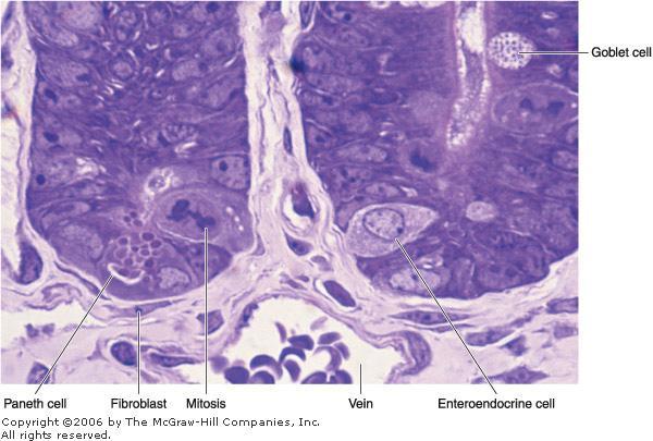
8 Fundus & Body The lamina propria of the fundus and body is filled with branched, tubular gastric (fundic) glands, three to seven of which open into the bottom of each gastric pit Each gastric gland has three distinct regions: the isthmus, neck, and base The distribution of epithelial cells in gastric glands is not uniform The isthmus, close to the gastric pit, contains differentiating mucous cells that will migrate and replace superficial mucous cells, undifferentiated stem cells, and oxyntic (parietal) cells the neck of the glands consists of stem, mucous neck (different from the mucous cells in the isthmus), and parietal cells the base of the glands primarily contains parietal and chief (zymogenic) cells Enteroendocrine cells are dispersed in the neck and base of the glands.

9 Stem Cells Found in the isthmus and neck regions but few in number, stem cells are low columnar cells with oval nuclei near the bases of the cells These cells have a high rate of mitosis; some of them move upward to replace the pit and surface mucous cells, which have a turnover time of 4 7 days Other daughter cells migrate more deeply into the glands and differentiate into mucous neck cells and parietal, chief, and enteroendocrine cells

10 Mucous Neck Cells Mucous neck cells are present in clusters or as single cells between parietal cells in the necks of gastric glands Their mucus secretion is quite different from that of the surface epithelial mucous cells They are irregular in shape, with the nucleus at the base of the cell and the secretory granules near the apical surface.

11 Oxyntic (Parietal) Cells Parietal cells are present mainly in the upper half of gastric glands; they are scarce in the base They are rounded or pyramidal cells, with one centrally placed spherical nucleus and intensely eosinophilic cytoplasm The most striking features of the active secreting cell seen in the electron microscope are an abundance of mitochondria and a deep, circular invagination of the apical plasma membrane, forming the intracellular canaliculus In the resting cell, a number of tubulovesicular structures can be seen in the apical region just below the plasmalemma, At this stage, the cell has few microvilli When stimulated to produce H+ and Cl, tubulovesicles fuse with the cell membrane to form the canaliculus and more microvilli, thus providing a generous increase in the surface of the cell membrane

12 Parietal cells secrete hydrochloric acid The ion H+ originates from the dissociation of the H2CO3 produced by the action of carbonic anhydrase, an enzyme abundant in oxyntic cells Once produced, H2CO3 dissociates in the cytoplasm into H+ and HCO32 The active cell also secretes K+ and Cl in the canaliculus; the K+ is exchanged for H+ by the action of the H+/K+ pump, while the Cl forms HCl. The presence of abundant mitochondria in the parietal cells indicates that their metabolic processes, particularly the pumping of H+/K+, are highly energy consuming The secretory activity of parietal cells is initiated by various mechanisms. One mechanism is through the cholinergic nerve endings (parasympathetic stimulation). Histamine and a polypeptide called gastrin, both secreted in the gastric mucosa, act strongly to stimulate the production of hydrochloric acid Gastrin also has a trophic effect on the gastric mucosa, stimulating growth.

13 Chief (Zymogenic) Cells Chief cells predominate in the lower region of the tubular glands characteristics of protein-synthesizing and - exporting cells Their basophilia is due to the abundant rough endoplasmic reticulum. The granules in their cytoplasm contain the inactive enzyme pepsinogen The precursor pepsinogen is rapidly converted into the highly active proteolytic enzyme pepsin after being released into the acid environment of the stomach There are seven different pepsins in the human gastric juice, which are aspartate endoproteinases of relatively broad specificity active at ph <5 In humans, chief cells also produce the enzyme lipase.

14 Enteroendocrine Cells are found in the neck and bases of gastric glands In the fundus of the stomach, 5-hydroxytryptamine (serotonin) is one of the principal secretory products In the stomach the G pylorus cells produces Gastrin that lead to the Stimulation of gastric acid secretion and Gastric mucosal growth

15 Pylorus has deep gastric pits into which the branched, tubular pyloric glands open. Compared with the glands in the cardiac region, pyloric glands have longer pits and shorter coiled secretory portions These glands secrete mucus as well as appreciable amounts of the enzyme lysozyme Gastrin (G) cells (which release gastrin) are enteroendocrine cells intercalated among the mucous cells of pyloric glands
 secrete somatostatin, which inhibits the release of some other hormones, including gastrin Secretion")
16 Parasympathetic stimulation, the presence of nutrients such as amino acids and amines in the stomach, and distention of the stomach wall directly stimulate the G cell to release gastrin, which in turn activates the parietal cell, increasing acid secretion Other enteroendocrine cells (D cells) secrete somatostatin, which inhibits the release of some other hormones, including gastrin Secretion of somatostatin is stimulated by HCl, counterbalancing the acid secretion.

17 Other layers The submucosa is composed of dense connective tissue containing blood and lymph vessels; it is infiltrated by lymphoid cells, macrophages, and mast cells. The muscularis is composed of smooth muscle fibers oriented in three main directions. The external layer is longitudinal, the middle layer is circular, and the internal layer is oblique At the pylorus, the middle layer is greatly thickened to form the pyloric sphincter. The stomach is covered by a thin serosa.
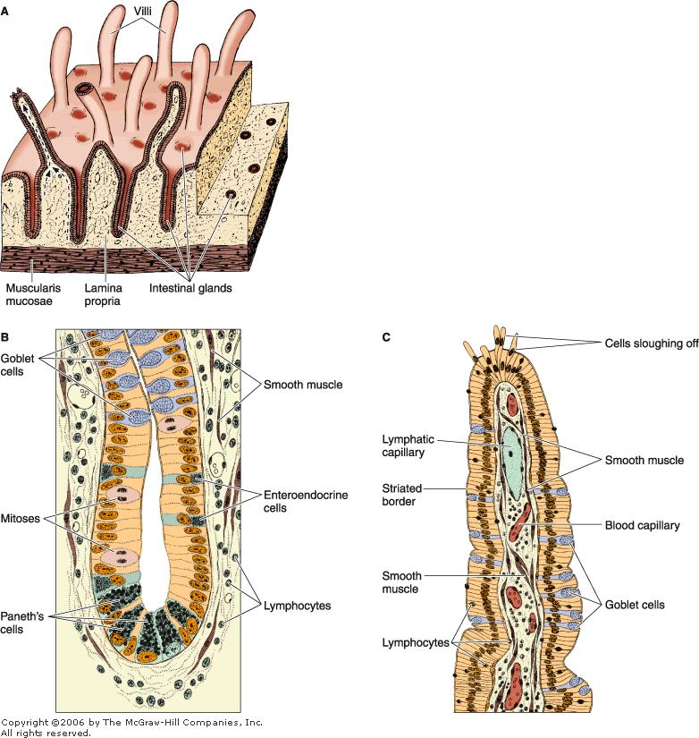
18 Small Intestine The small intestine is the site of terminal food digestion, nutrient absorption, and endocrine secretion processes of digestion are completed in the small intestine, where the nutrients (products of digestion) are absorbed by cells of the epithelial lining The small intestine is relatively long approximately 5 m and consists of three segments: the duodenum, jejunum, and ileum.

19 Mucous Membrane the lining of the small intestine shows a series of permanent folds, plicae circulares (Kerckring's valves), consisting of mucosa and submucosa and having a semilunar, circular, or spiral form The plicae are most developed in, and consequently a characteristic of, the jejunum. They do not constitute a significant feature of the duodenum and ileum, although they are frequently present. Intestinal villi are 0.5- to 1.5-mm-long outgrowths of the mucosa (epithelium plus lamina propria) projecting into the lumen of the small intestine In the duodenum they are leaf shaped, gradually assuming fingerlike shapes as they reach the ileum

20 Between the villi are small openings of simple tubular glands called intestinal glands (also called crypts), or glands of Lieberkühn The epithelium of the villi is continuous with that of the glands The intestinal glands contain stem cells, some absorptive cells, goblet cells, Paneth's cells, and enteroendocrine cells.
21

22 Absorptive cells or enterocytes are tall columnar cells, each with an oval nucleus in the basal half of the cell At the apex of each cell is a homogeneous layer called the striated (brush) border When viewed with the electron microscope, the striated border is seen to be a layer of densely packed microvilli Each absorptive cell is estimated to have an average of 3000 microvilli, and 1 mm2 of mucosa contains about 200 million of these structures

23 Each microvillus is a cylindrical protrusion of the apical cytoplasm that is approximately 1 m tall by 0.1 m in diameter consists of the cell membrane enclosing a core of actin microfilaments associated with other cytoskeletal proteins Microvilli have the important physiological function of increasing the area of contact between the intestinal surface and the nutrients. The presence of plicae, villi, and microvilli greatly increases the surface of the intestinal lining It has been calculated that plicae increase the intestinal surface 3-fold, the villi increase it 10-fold, and the microvilli increase it 20- fold. Together, these processes are responsible for a 600-fold increase in the intestinal surface, resulting in a total area of 200 m2

24 Goblet cells are interspersed between the absorptive cells They are less abundant in the duodenum and increase in number as they approach the ileum These cells produce acid glycoproteins of the mucin type to form mucus, whose main function is to protect and lubricate the lining of the intestine.

25 Paneth's cells in the basal portion of the intestinal glands are exocrine cells with secretory granules in their apical cytoplasm. lysozyme an enzyme that digests the cell walls of some bacteria was detected in the large eosinophilic secretory granules of these cells Lysozyme has antibacterial activity and may play a role in controlling the intestinal flora.
.")
26 M (microfold) cells are specialized epithelial cells overlying the lymphoid follicles of Peyer's patches the presence of numerous basal membrane invaginations that form pits containing many intraepithelial lymphocytes and antigenpresenting cells (macrophages). M cells can endocytose antigens and transport them to the underlying macrophages and lymphoid cells, which then migrate to other compartments of the lymphoid system (nodes), M cells represent an important link in the intestinal immunological system basement membrane under M cells is discontinuous, facilitating transit between the lamina propria and M cells
27

28 The very large mucosal surface of the gastrointestinal tract is exposed to many potentially invasive microorganisms Secretory immunoglobulins of the IgA are the first line of defense Another protective device is the intercellular tight junctions that make the epithelial cells a barrier to the penetration of microorganisms. In addition the gastrointestinal tract contains antibody-secreting plasma cells, macrophages, and a very large number of lymphocytes located in both the mucosa and the submucosa. Together, these cells are called the gut-associated lymphoid tissue (GALT).
29 The lamina propria of the small intestine is composed of loose connective tissue with blood and lymph vessels, nerve fibers, and smooth muscle cells. The lamina propria penetrates the core of the intestinal villi smooth muscle cells are responsible for the rhythmic movements of the villi, which are important for absorption
 glands The product of secretion of the glands is distinctly alkaline (ph 8.1 9.")
30 In the initial portion of the duodenum the submucosa contains clusters of ramified, coiled tubular glands that open into the intestinal glands. These are the duodenal (or Brunner's) glands The product of secretion of the glands is distinctly alkaline (ph ), acting to protect the duodenal mucous membrane from the effects of the acid gastric juice and to bring the intestinal contents to the optimum ph for pancreatic enzyme action.

31 The lamina propria and the submucosa of the small intestine contain aggregates of lymphoid nodules known as Peyer's patches, an important component of the GALT Each patch consists of nodules and is visible to the naked eye as an oval area on the antimesenteric side of the intestine There are about 30 patches in humans, most of them in the ileum each Peyer's patch appears as a dome-shaped area devoid of villi Instead of absorptive cells, its covering epithelium consists of M cells

32 The muscularis is well developed in the intestines, composed of an internal circular layer and an external longitudinal layer
33 Vessels & Nerves The blood vessels that nourish the intestine and remove absorbed products of digestion penetrate the muscularis and form a large plexus in the submucosa From the submucosa, branches extend through the muscularis mucosae and lamina propria and into the villi. Each villus receives, according to its size, one or more branches that form a capillary network just below its epithelium At the tips of the villi, one or more venules arise from these capillaries and run in the opposite direction, reaching the veins of the submucosal plexus These capillaries (lacteals), although larger than the blood capillaries, are difficult to observe because their walls are so close together that they appear to be collapsed Lacteals run to the region of lamina propria above the muscularis mucosae, where they form a plexus. From there they are directed to the submucosa, where they surround lymphoid nodules Lacteals anastomose repeatedly and leave the intestine along with the blood vessels. They are especially important for the absorption of lipids, because blood circulation does not easily accept the lipoproteins produced by the absorptive cells during this process
34 important for intestinal function is the rhythmic movement of the villi This movement is the result of the contraction of smooth muscle cells running vertically between the muscularis mucosae and the tip of the villi These contractions occur at the rate of several strokes per minute and have a pumping action on the villi that propels the lymph to the mesenteric lymphatics.
 plexus in the submucosa The plexuses contain some sensory neurons that receive")
35 The innervation of the intestines is formed by both an intrinsic component and an extrinsic component The intrinsic component comprises groups of neurons that form the myenteric (Auerbach's) nerve plexus between the outer longitudinal and inner circular layers of the muscularis and the submucosal (Meissner's) plexus in the submucosa The plexuses contain some sensory neurons that receive information from nerve endings near the epithelial layer and in the smooth muscle layer regarding the composition of the intestinal content (chemoreceptors) and the degree of expansion of the intestinal wall (mechanoreceptors) The other nerve cells are effectors and innervate the muscle layers and hormonesecreting cells

36
37 The intrinsic innervation formed by these plexuses is responsible for the intestinal contractions that occur in the total absence of the extrinsic innervation. The extrinsic innervation is formed by parasympathetic cholinergic nerve fibers that stimulate the activity of the intestinal smooth muscle and by sympathetic adrenergic nerve fibers that depress intestinal smooth muscle activity.
Dana Alrafaiah. Dareen Abu Shalbak. Mohammad Almuhtaseb. 1 P a g e
 2 Dana Alrafaiah Dareen Abu Shalbak Mohammad Almuhtaseb 1 P a g e Esophagus: A muscular tube that is 25 cm long, but if measured from the incisors it would be 45cm long. Extends from C6 of cervical vertebra,
General Structure of Digestive Tract
 Dr. Nabil Khouri General Structure of Digestive Tract Common Characteristics: Hollow tube composed of a lumen whose diameter varies. Surrounded by a wall made up of 4 principal layers: Mucosa Epithelial
The doctor mentioned a few things about the esophagus from the previous lecture:
 السالم عليكم [HISOLOGY 2] April 27, 2014 The doctor mentioned a few things about the esophagus from the previous lecture: Esophagus - It is about 25 cm in length (from the incisor it is 45 cm) Histological
Alimentary Canal (I)
") Alimentary Canal (I) Esophagus and Stomach (Objectives) By the end of this lecture, the student should be able to discuss the microscopic structure in correlation with the function of the following organs:
Alimentary Canal (I) Esophagus and Stomach (Objectives) By the end of this lecture, the student should be able to discuss the microscopic structure in correlation with the function of the following organs:
Digestive system L 2. Lecturer Dr. Firdous M. Jaafar Department of Anatomy/Histology section
 Digestive system L 2 Lecturer Dr. Firdous M. Jaafar Department of Anatomy/Histology section objectives 1-Describe the general structure of digestive tract: a-mucosa. b-submucosa. c-muscularis externa d-adventitia
(b) Stomach s function 1. Dilution of food materials 2. Acidification of food (absorption of dietary Fe in small intestine) 3. Partial chemical digest
 Stomach s function 1. Dilution of food materials 2. Acidification of food (absorption of dietary Fe in small intestine) 3. Partial chemical digest") (1) General features a) Stomach is widened portion of gut-tube: between tubular and spherical; Note arranged of smooth muscle tissue in muscularis externa. 1 (b) Stomach s function 1. Dilution of food
(1) General features a) Stomach is widened portion of gut-tube: between tubular and spherical; Note arranged of smooth muscle tissue in muscularis externa. 1 (b) Stomach s function 1. Dilution of food
Anatomy & Histology of The Small intestine
 Anatomy & Histology of The Small intestine Prof. Abdulameer Al-Nuaimi E-mail: a.al-nuaimi@sheffield.ac.uk E. mail: abdulameerh@yahoo.com Jejunum Ileum Histology: Duodenum, jejunum, and ileum
Small intestine. Small intestine
 General features Tubular organ longest part; 5-6 m most of chemical digestion absorption of nutrients reabsorption of H2O occurs. Two structural features; maximize the lumenal surface area villi microvilli
DIGESTIVE TRACT ESOPHAGUS
 DIGESTIVE TRACT From the lower esophagus to the lower rectum four fundamental layers comprise the wall of the digestive tube: mucosa, submucosa, muscularis propria (externa), and adventitia or serosa (see
Small Intestine, Large Intestine and anal cannel
 Small Intestine, Large Intestine and anal cannel 32409 Small intestine Large intestine Small intestine General Structure of the Digestive Tract rat 32409 Epithelium with goblet cells and absorptive cells
HISTOLOGY. GIT Block 432 Histology Team. Lecture 1: Alimentary Canal (1) (Esophagus & Stomach) Done by: Ethar Alqarni Reviewed by: Ibrahim Alfuraih
 (Esophagus & Stomach) Done by: Ethar Alqarni Reviewed by: Ibrahim Alfuraih") HISTOLOGY Lecture 1: Alimentary Canal (1) (Esophagus & Stomach) Done by: Ethar Alqarni Reviewed by: Ibrahim Alfuraih Color Guide: Black: Slides. Red: Important. Green: Doctor s notes. Blue: Explanation.
HISTOLOGY Lecture 1: Alimentary Canal (1) (Esophagus & Stomach) Done by: Ethar Alqarni Reviewed by: Ibrahim Alfuraih Color Guide: Black: Slides. Red: Important. Green: Doctor s notes. Blue: Explanation.
الله الر ح م ن الر ح يم مسب
 بسم رلا هللارلا هللا This is the second histology lecture in the GI system. In this lecture, we will discuss the histology of the esophagus, stomach, and small intestine so prepare yourself.this sheet
Dr Nadine Gravett School of Anatomical Sciences Room 2B10B
 Dr Nadine Gravett School of Anatomical Sciences Room 2B10B Nadine.Gravett@wits.ac.za Oral cavity Mechanical breakdown Formation of bolus Oesophagus Conduit from mouth to stomach Stomach Digestion Temporary
Gastrointestinal Anatomy and Physiology. Bio 219 Napa Valley College Dr. Adam Ross
 Gastrointestinal Anatomy and Physiology Bio 219 Napa Valley College Dr. Adam Ross Functions of digestive system Digestion Breakdown of food (chemically) using enzymes, acid, and water Absorption Nutrients,
The Digestive System Laboratory
 The Digestive System Laboratory 1 The Digestive Tract The alimentary canal is a continuous tube stretching from the mouth to the anus. Liver Gallbladder Small intestine Anus Parotid, sublingual, and submaxillary
Week 12 - Outline. Outline. Digestive System I Major Organs. Overview of Digestive System
 Outline Week 12 - Digestive System I Major Organs Copyright The McGraw-Hill Companies, Inc. Permission required for reproduction or display. Digestive Tract Function GI Tract Structure Regulation of the
Digestive System II - Lower tract Revised
 ANAT D502 Basic Histology Digestive System II - Lower tract Revised 10.12.12 Outline: I. Small intestine II. Enterocyte digestion II. Hepatic portal system IV. Large intestine V. Enteric nervous system
Lab activity manual - Histology of the digestive system. Lab activity 1: esophagus stomach - small intestines
 Lab activity manual - Histology of the digestive system Jeanne Adiwinata Pawitan Prerequisite: Histology of the 4 basic tissues In this module we learn about the histology of the digestive system, from
Soft palate elevates, closing off the nasopharynx. Hard palate Tongue Bolus Epiglottis. Glottis Larynx moves up and forward.
 The Cephalic Phase Chemical and mechanical digestion begins in the mouth Saliva is an exocrine secretion Salivary secretion is under autonomic control Softens and lubricates food Chemical digestion: salivary
Digestive System Module 4: The Stomach *
 OpenStax-CNX module: m49286 1 Digestive System Module 4: The * Donna Browne Based on The by OpenStax This work is produced by OpenStax-CNX and licensed under the Creative Commons Attribution License 4.0
Digestive System 7/15/2015. Outline Digestive System. Digestive System
 Digestive System Biology 105 Lecture 18 Chapter 15 Outline Digestive System I. Functions II. Layers of the GI tract III. Major parts: mouth, pharynx, esophagus, stomach, small intestine, large intestine,
Connective tissue The Digestive System
 Connective tissue The Digestive System Part 1 Structure of digestive system Functions Basic Structure of the Alimentary Canal Wall Tube is made up of four layers: 1. Mucosa 2. Submucosa 3. Muscularis externa
HISTOLOGY VIRTUAL LABORATORY GASTROINTESTINAL SYSTEM
 HISTOLOGY VIRTUAL LABORATORY GASTROINTESTINAL SYSTEM LIP (Slides GI 1, 2) Identify the outer portion lined by stratified squamous (keratinized) epithelium. Note the hair follicles and sebaceous glands
Slide 154: Pancreas, H&E
 Slide 154: Pancreas, H&E the pancreas, located adjacent to the duodenum, is a mixed exocrine and endocrine gland; it is usually readily identifiable by the presence of the interspersed endocrine pancreatic
Includes mouth, pharynx, esophagus, stomach, small intestine, large intestine, rectum, anus. Salivary glands, liver, gallbladder, pancreas
 Chapter 14 The Digestive System and Nutrition Digestive System Brings Nutrients Into the Body The digestive system includes Gastrointestinal (GI) tract (hollow tube) Lumen: space within this tube Includes
The Digestive System and Body Metabolism
 14 PART B The Digestive System and Body Metabolism PowerPoint Lecture Slide Presentation by Jerry L. Cook, Sam Houston University ESSENTIALS OF HUMAN ANATOMY & PHYSIOLOGY EIGHTH EDITION ELAINE N. MARIEB
Digestive Lecture Test Questions Set 4
 Digestive Lecture Test Questions Set 4 1. Which of the following is not associated directly with the small intestine: a. villi b. circular folds c. microvilli d. haustrae e. secretin 2. The largest (longest)
Gastrointestinal System!
 Gastrointestinal System! Assoc. Prof. Prasit Suwannalert, Ph.D. (Email: prasit.suw@mahidol.ac.th)! Objectives: After learning, student should be able to describe and discuss in topics of! 1. Anatomical
Physiological processes in the GI tract:
 Gastrointestinal physiology for medical students General principal of gastrointestinal function Motility, nervous control and blood circulation Physiological processes in the GI tract: Motility Secretion
Tissues and organs PART 1
 Tissues and organs PART 1 Animals and plants are multicellular (made of many cells). Cells become specialised according to their function Tissues: Many cells that perform one or several functions; they
Gastrointestinal Physiology
 Gastrointestinal Physiology Digestion and Absorption Definition Digestion: i Food dis dissolved d and broken down. Physical digestion: Propulsion and mixing of food by muscle in the alimentary tract. Chemical
Chapter 26 The Digestive System
 Chapter 26 The Digestive System Digestive System Gastroenterology is the study of the stomach and intestine. Digestion Catabolism Absorption Anabolism The actions of the digestive system are controlled
Tissue: The Living Fabric: Part A
 PowerPoint Lecture Slides prepared by Janice Meeking, Mount Royal College C H A P T E R 4 Tissue: The Living Fabric: Part A Tissues Groups of cells similar in structure and function Types of tissues Epithelial
Tissues. tissue = many cells w/ same structure and function. cell shape aids its function tissue shape aids its function
 Tissues tissue = many cells w/ same structure and function cell shape aids its function tissue shape aids its function Histology = study of tissues 4 types of tissues Epithelial coverings contact openings
University of Buea. Faculty of Health Sciences. Programme in Medicine
 Faculty of Health Sciences University of Buea Wednesday, 28 th January 2009 Time: 8 00-10 00 Programme in Medicine MED 303 (Gastrointestinal Physiology) EXAMS (2008-2009) Identify the letter of the choice
GASTROINTESTINAL PHYSIOLOGY PHYSIOLOGY DEPARTMENT KAMPALA INTERNATIONAL UNIVERSITY DAR ES SALAAM TANZANIA
 GASTROINTESTINAL PHYSIOLOGY PHYSIOLOGY DEPARTMENT KAMPALA INTERNATIONAL UNIVERSITY DAR ES SALAAM TANZANIA Anatomy of the GI Tract The GI tract is essentially a hollow tube connecting the mouth to the anus.
The Digestive System. Chapter 25
 The Digestive System Chapter 25 Introduction Structure of the digestive system A tube that extends from mouth to anus Accessory organs are attached Functions include Ingestion Movement Digestion Absorption
DIGESTIVE. CHAPTER 17 Lecture: Part 1 Part 2 BIO 212: ANATOMY & PHYSIOLOGY II
 BIO 212: ANATOMY & PHYSIOLOGY II CHAPTER 17 Lecture: DIGESTIVE Part 1 Part 2 Dr. Lawrence G. Altman www.lawrencegaltman.com Some illustrations are courtesy of McGraw-Hill. SMALL INTESTINE DUODENUM > JEJUNUM
Chapter 9. The digestive system. Glossary. Louise McErlean
 Chapter 9 The digestive system Louise McErlean Glossary Absorption Process whereby the products of digestion move into the blood or lymph fluid. Acini glands Produce pancreatic juice. Amylase Carbohydrate
DIGESTIVE SYSTEM ALIMENTARY CANAL / GI TRACT & ACCESSORY ORGANS. Mar 16 10:34 PM
 DIGESTIVE SYSTEM ALIMENTARY CANAL / GI TRACT & ACCESSORY ORGANS Mar 16 10:34 PM 1 I. Digestive System Functions > Ingestion the taking in of food > Propulsion movement caused by force > Digestion breakdown
The Digestive System and Body Metabolism Premedical Biology
 The Digestive System and Body Metabolism Premedical Biology Copyright 2003 Pearson Education, Inc. publishing as Benjamin Cummings The Digestive System and Body Digestion Metabolism Breakdown of ingested
Organs Histology D. Sahar AL-Sharqi. Digestive System
 Digestive System The digestive system consists of the digestive tract oral cavity, esophagus, stomach, small and large intestines, and anus and its associated glands salivary glands, liver, and pancreas.
consists of: Muscular, hollow tube (= digestive tract ) + Various accessory organs
 + Various accessory organs") DIGESTIVE SYSTEM consists of: Muscular, hollow tube (= digestive tract ) + Various accessory organs FUNCTION Individual parts function in: ingestion mechanical digestion chemical and enzymatic digestion
DIGESTIVE SYSTEM consists of: Muscular, hollow tube (= digestive tract ) + Various accessory organs FUNCTION Individual parts function in: ingestion mechanical digestion chemical and enzymatic digestion
The Stomach. Bởi: OpenStaxCollege
 Bởi: OpenStaxCollege Although a minimal amount of carbohydrate digestion occurs in the mouth, chemical digestion really gets underway in the stomach. An expansion of the alimentary canal that lies immediately
Two main groups Alimentary canal continuous coiled hollow tube Accessory digestive organs
 Digestion Breakdown of ingested food Absorption of nutrients into the blood Metabolism Production of cellular energy (ATP) Constructive and degradative cellular activities Two main groups Alimentary canal
Objectives. Describe the cells of the GI tract and their function. Differentiate between different parts of the GI tract
 GI Histology 1 Objectives Describe the cells of the GI tract and their function Describe the histological features of each part of the GI tract. Differentiate between different parts of the GI tract Appreciate
MCAT Biology Problem Drill 20: The Digestive System
 MCAT Biology Problem Drill 20: The Digestive System Question No. 1 of 10 Question 1. During the oral phase of swallowing,. Question #01 A. Initially, the food bolus is moved to the back of the tongue and
The Digestive System. What is the advantage of a one-way gut? If you swallow something, is it really inside you?
 The Digestive System What is the advantage of a one-way gut?! If you swallow something, is it really inside you? Functions and Processes of the Digestive System: Move nutrients, water, electrolytes from
MICROSTRUCTURES LIPS TOOTH TONGUE OESOPHAGUS STOMACH, CARDIAC, PYLORIC FUNDIC GLANDS
 MICROSTRUCTURES LIPS TOOTH TONGUE OESOPHAGUS STOMACH, CARDIAC, PYLORIC FUNDIC GLANDS HUMAN ANATOMY: MICROSTRUCTURES CLASSIFICATION: LOCATION AND BOUNDARIES, FORM, FUNCTION, MICROSCOPIC STRUCTURE: A hollow
Histology Lab. looking at microscopic pictures of tissues, for more information use Junqueira book and you can use BlueHistolgy website
 Done By: Aseel Twaijer & Laith Sorour Histology Lab *These notes help in differentiating tissues and you must read them while looking at microscopic pictures of tissues, for more information use Junqueira
Overview of the Digestive
 Overview of the Digestive System Bởi: OpenStaxCollege The function of the digestive system is to break down the foods you eat, release their nutrients, and absorb those nutrients into the body. Although
Chapter 14: The Digestive System
 Chapter 14: The Digestive System Digestive system consists of Muscular tube (digestive tract) alimentary canal Accessory organs teeth, tongue, glandular organs 6 essential activities 1. 2. 3. 4. 5. 6.
GI Secretion 1: Salivary and Gastric Secretion Jack Grider, Ph.D.
 GI Secretion 1: Salivary and Gastric Secretion Jack Grider, Ph.D. OBJECTIVES: 1. List the volumes of secretion by various regions. 2. Predict the components of salivary secretion at different flow rates.
Digestive System. - Food is ingested
 11 V. Digestive Processes in the Mouth - Food is ingested - Mechanical digestion begins (chewing) - Salivary amylase begins chemical breakdown of starch - Propulsion is initiated by Deglutition (Swallowing)
- Digestion occurs during periods of low activity - Produces more energy than it uses. - Mucosa
 Introduction Digestive System Chapter 29 Provides processes to break down molecules into a state easily used by cells - A disassembly line: Starts at the mouth and ends at the anus Digestive functions
Cell and Tissue Types. Epithelial, Connective, Muscle, Nerve
 Cell and Tissue Types Epithelial, Connective, Muscle, Nerve Objectives Explain the major stages of the cell cycle and cellular division (mitosis). Describe specific events occurring in each of the phases
Connective tissue The Digestive System
 Connective tissue The Digestive System Part 1 Structure of digestive system Functions Basic Structure of the Alimentary Canal Wall Tube is made up of four layers: 1. Mucosa 2. Submucosa 3. Muscularis externa
GI Histology Lab 1. Prepared by: Zeina Kalaji
 GI Histology Lab 1 Prepared by: Zeina Kalaji Lip ORAL MUCOSA -Arrow shows labial salivary glands in the submucosa. VERMILLION transitional zone. SKIN Stratified Squamous epithelium, keratinized -Arrow
General functions of digestive system. Ch. 15 The Digestive System. General histology of the wall of the digestive tract. Overview of digestive organs
 Overall idea: obtain nutrients from food (for energy and raw materials for synthesis), and defecate the leftover waste 2 types of organs involved: 1. Parts of the digestive tract (= a long muscular tube
Overview of digestion or, gut reactions - to food
 Key concepts in Digestion. Indigestion module Overview of digestion or, gut reactions - to food Prof. Barry Campbell Gastroenterology Cellular & Molecular Physiology e-mail: bjcampbl@liv.ac.uk http://pcwww.liv.ac.uk/~bjcampbl
Energy, Chemical Reactions and Enzymes
 Phosphorylation Hydrolysis Energy, Chemical Reactions and Enzymes Chapter 2 (selections) What is Energy? Energy is the capacity to do work Potential Energy Kinetic Energy Chemical Bond Energy Like a rechargeable
Paneth Cells. Road Map to the Finish. No Review this Friday. Today 11/29 Finish digestion/accessory organs. Wednesday 12/1 Immune System I
 Road Map to the Finish No Review this Friday Today 11/29 Finish digestion/accessory organs Wednesday 12/1 Immune System I Paneth Cells - base of intestinal glands -! large -! intense acidophilic granules
Physiology Unit 4 DIGESTIVE PHYSIOLOGY
 Physiology Unit 4 DIGESTIVE PHYSIOLOGY In Physiology Today Functions Motility Ingestion Mastication Deglutition Peristalsis Secretion 7 liters/day! Exocrine/endocrine Digestion Absorption Digestion of
Module 2 Heartburn Glossary
 Absorption Antacids Antibiotic Module 2 Heartburn Glossary Barrett s oesophagus Bloating Body mass index Burping Chief cells Colon Digestion Endoscopy Enteroendocrine cells Epiglottis Epithelium Absorption
NURSE-UP DIGESTIVE SYSTEM AKA G.I. SYSTEM
 NURSE-UP DIGESTIVE SYSTEM AKA G.I. SYSTEM The digestive system is used for breaking down food into nutrients which then pass into the circulatory system and are taken to where they are needed in the body.
Section 1.1: What is the function of digestion?
 Section 1.1: What is the function of digestion? When you have completed this section, you should be able to: Describe the overall function of the GI tract. Describe the processes involved in digestion.
Section Coordinator: Jerome W. Breslin, PhD, Assistant Professor of Physiology, MEB 7208, ,
 IDP Biological Systems Gastrointestinal System Section Coordinator: Jerome W. Breslin, PhD, Assistant Professor of Physiology, MEB 7208, 504-568-2669, jbresl@lsuhsc.edu Overall Learning Objectives 1. Characterize
Tissues. tissue = many cells w/ same structure and function. cell shape aids function tissue shape aids function. Histology = study of tissues
 Tissues tissue = many cells w/ same structure and function cell shape aids function tissue shape aids function Histology = study of tissues 4 types of tissues Epithelial coverings contact openings Connective
DIGESTIVE. CHAPTER 17 Lecture: Part 1 Part 2 BIO 212: ANATOMY & PHYSIOLOGY II
 BIO 212: ANATOMY & PHYSIOLOGY II 1 CHAPTER 17 Lecture: DIGESTIVE Part 1 Part 2 Dr. Lawrence G. Altman www.lawrencegaltman.com Some illustrations are courtesy of McGraw-Hill. Processes of DIGESTION Mechanical
1. Approximately 21 ft. long: duodenum (one ft.), jejunum (eight ft.), and ileum (twelve ft.)
, jejunum (eight ft.), and ileum (twelve ft.)") IV. Small Intestines A. General features and functions 1. Approximately 21 ft. long: duodenum (one ft.), jejunum (eight ft.), and ileum (twelve ft.) 2. Functions: move forward chyme, continue digestion,
IV. Small Intestines A. General features and functions 1. Approximately 21 ft. long: duodenum (one ft.), jejunum (eight ft.), and ileum (twelve ft.) 2. Functions: move forward chyme, continue digestion,
- Digestion occurs during periods of low activity - Produces more energy than it uses. 3 Copyright 2016 by Elsevier Inc. All rights reserved.
 Introduction Digestive System Chapter 29 Provides processes to break down molecules into a state easily used by cells - A disassembly line: Starts at the mouth and ends at the anus Digestive functions
Understandings, Applications & Skills
 D.2 Digestion Understandings, Applications & Skills Statement D.2.U1 Nervous and hormonal mechanisms control the secretion of digestive juices. D.2.U2 Exocrine glands secrete to the surface of the body
Anatomy of the Intes.ne: Epithelium, Lympha.cs, Vessels and Nerves
 Master Course Gastroenterology 2015 Anatomy of the Intes.ne: Epithelium, Lympha.cs, Vessels and Nerves Dr. Stephanie Ganal Department Klinische Forschung University of Bern stephanie.ganal@dkf.unibe.ch
Overview of digestion or, gut reactions - to food
 1 Key concepts in Digestion. Indigestion module Overview of digestion or, gut reactions to food Prof. Barry Campbell Gastroenterology Cellular & Molecular Physiology email: bjcampbl@liv.ac.uk http://pcwww.liv.ac.uk/~bjcampbl
The Digestive System. Basic process of digestion. Mouth and Teeth 10/30/2016
 The Digestive System Basic process of digestion 1. Ingestion: animal eats food. 2. Digestion: animal body breaks food down. Mechanical digestion: chewing (mastication). Chemical digestion: enzymes and
Unit I Problem 9 Histology: Basic Tissues of The Body
 Unit I Problem 9 Histology: Basic Tissues of The Body - What is the difference between cytology and histology? Cytology: it is the study of the structure and functions of cells and their contents. Histology:
Stomach. Stomach. Nerve supply. Blood supply. Sympathe0c and parasympathe0c fibers of the autonomic nervous system
 Stomach Nerve supply Sympathe0c and parasympathe0c fibers of the autonomic nervous system Blood supply Celiac trunk, and corresponding veins (part of the hepa0c portal system) Stomach Figure 23.14a Chapter
Human Structure and Function GI Tract Exercises
 GI Tract Exercises Study Exercises. Review of the Elements of the Alimentary Tube. On the following two pages is a chart or matrix of blank spaces. Each space is the intersection of a horizontal row and
/30/17 Ch 8: Muscular System 1. Table of Contents # Date Title Page # 03/13/17 Ch 10: Somatic and Special Senses 53
 Table of Contents # Date Title Page # 1. 01/30/17 Ch 8: Muscular System 1 2. 3. 4. 5. 6. 7. 02/14/17 Ch 9: Nervous System 12 03/13/17 Ch 10: Somatic and Special Senses 53 03/27/17 Ch 11: Endocrine System
Histology 3. We will continue talking about a few things from last lecture, starting with M cells:
 Histology 3 This is the last Histology lecture in the GI system. Enjoy! There are some extra notes listed as footnotes. We will continue talking about a few things from last lecture, starting with M cells:
Practical Histology o
 Practical Histology o 1.. Contents: Histology of the : Stomach Esophagus Small intestine Large intestine Liver Gallbladder Exocrine pancreas Spleen GNT Block Things you need to know before the exam : o
BIO 132 Anatomy and Physiology II Spring, 2016 Exam 1 Name: BIO 132 ID Number. Section 1 Answer questions 1 40 on the scan sheet.
 BIO 132 Anatomy and Physiology II Spring, 2016 Exam 1 Name: BIO 132 ID Number Section 1 Answer questions 1 40 on the scan sheet. 1. The homeostatic value of a particular controlled variable in the body
The Digestive System 1
 The Digestive System 1 Digestion Processing of food Types Mechanical (physical) Chew Tear Grind Mash Mix Chemical Catabolic reactions Enzymatic hydrolysis Carbohydrate Protein Lipid 2 Digestion Phases
1. Blood vessels that absorb the water and solutes leaving the filtrate of the nephron s tubules are called the
 Biology 2330: Lecture Test Five Multiple Choice. Read each question thoroughly before answering. From the choices available, choose the answer that is the most correct. Place all answers on the accompanying
The Digestive System. Chapter 16. Introduction. Overview of Digestive System. Histological Organization. Movement and Mixing of Digestive Materials
 The Digestive System Chapter 16 Introduction Structure of the digestive system A tube that extends from mouth to anus Accessory organs are attached Functions include Ingestion Movement Digestion Absorption
Histology Notes -Part 1: Epithelial Tissues
 Introduction Group of cells w/ similar structure & function = TISSUE Four Basic Tissue Types 1. Epithelial-covers 2. Connective-supports 3. Muscular*-produces movement (will discuss in the muscular system
Digestion and Absorption
 Digestion and Absorption General Considerations - No absorption in esophagus, little in the stomach and vast majority of absorption occurs in small intestine. - The small intestine has specialized structures
I. The Alimentary Canal (GI track)
") A. About 9 meters long B. Passes through the ventral cavity. C.Movements of the Tube 1. Mixing movements- smooth muscles contract rhythmically. 2. Propelling movements- a wavelike motion called peristalsis.
A. About 9 meters long B. Passes through the ventral cavity. C.Movements of the Tube 1. Mixing movements- smooth muscles contract rhythmically. 2. Propelling movements- a wavelike motion called peristalsis.
Epithelia will be discussed according to the following scheme: Type Number of layers Shape Line drawing. Squamous Cuboidal Columnar
 Epithelia Epithelia will be discussed according to the following scheme: Type Number of layers Shape Line drawing Simple Squamous Cuboidal Columnar Covering and Lining epithelium Pseudostratified Stratified
- Most nutrients are absorbed before reaching the ileum. - Colon is responsible for final removal of electrolytes and water.
 University of Jordan Department of physiology and Biochemistry Gastro-Intestinal physiology, Medical, Pt III. ---------------------------------------------------------------------------- Academic year:
Alimentary Canal (I) Salivatory Glands. (Esophagus and Stomach) Color index: Slides.. Important..Notes..Extra..
 Salivatory Glands. (Esophagus and Stomach) Color index: Slides.. Important..Notes..Extra..") Alimentary Canal (I) (Esophagus and Stomach) Salivatory Glands Color index: Slides.. Important..Notes..Extra.. Objectives: 1. By the end of this lecture, the student should be able to discuss the microscopic
Alimentary Canal (I) (Esophagus and Stomach) Salivatory Glands Color index: Slides.. Important..Notes..Extra.. Objectives: 1. By the end of this lecture, the student should be able to discuss the microscopic
Nutrition. Autotrophs. plants, some protists & bacteria producers
 Nutrition Autotrophs plants, some protists & bacteria producers Nutrition Heterotrophs animals, fungi, some protists & bacteria consumers Animal Nutrition Most obtain food by ingestion take in their food
BIOH122 Human Biological Science 2
 BIOH122 Human Biological Science 2 Session 13 Digestive System 1 Mouth to Stomach Bioscience Department Endeavour College of Natural Health endeavour.edu.au Session Plan o Functions of the digestive system
(A) Diarrhea. (B) Stomach cramps. (C) Dehydration due to excess fluid loss. (D) A, B, and C are correct. (E) Only answer B is correct.
 Diarrhea. (B) Stomach cramps. (C) Dehydration due to excess fluid loss. (D) A, B, and C are correct. (E) Only answer B is correct.") Human Anatomy - Problem Drill 21: The Digestive System Question No. 1 of 10 1. A 26-year-old male is treated in the emergency department for severe gastrointestinal disturbance. Which of the following
Human Anatomy - Problem Drill 21: The Digestive System Question No. 1 of 10 1. A 26-year-old male is treated in the emergency department for severe gastrointestinal disturbance. Which of the following
HUMAN NUTRITION: ABSORPTION & ASSIMILATION 14 MAY 2014
 HUMAN NUTRITION: ABSORPTION & ASSIMILATION 14 MAY 2014 In this lesson, we: Absorption Lesson Description Examine and understand absorption Define absorption and describe where it occurs Study the structure
Dr. Abeer.c.Yousif. Histology -2 nd stage. What is histology?
 What is histology? Histology is the science of microscopic anatomy of cells and tissues, in Greek language Histo= tissue and logos = study and it's tightly bounded to molecular biology, physiology, immunology
Tissues 10/21/2016. Epithelial Tissue
 Tissues This is a generalized cell diagram. It shows the anatomy of a cell, but most cells do not actually look like this. Cells can have a wide variety of shapes and sizes, depending on their function.
DIGESTIVE SYSTEM CLASS NOTES. tube along with several
 DIGESTIVE SYSTEM CLASS NOTES Digestion Breakdown of food and the of nutrients in the bloodstream. Metabolism Production of for and cellular activities. The digestive system is composed of the canal which
An overview of the digestive system. mouth pharynx esophagus stomach small intestine large intestine rectum anus
 An overview of the digestive system mouth pharynx esophagus stomach small intestine large intestine rectum anus Why GIT? What are the main steps in the digestive process? Ingestion intake of food via the
Gastric Contrac,le Ac,vity. Regula,on of Gastric Emptying
 Gastric Contrac,le Ac,vity Figure 23.18 Regula,on of Gastric Emptying Gastric emptying is regulated by: Neural enterogastric reflex Hormonal (enterogastrone) mechanisms In the presence of gastric gastrin