M2 microglia/ macrophages drive oligodendrocyte differentiation during CNS remyelination
|
|
|
- Mavis Crawford
- 5 years ago
- Views:
Transcription
1 Supplemental Information Title: M2 microglia/ macrophages drive oligodendrocyte differentiation during CNS remyelination Authors: Veronique E. Miron, Amanda Boyd, Jing-Wei Zhao, Tracy J. Yuen, Julia M. Ruckh, Jennifer L. Shadrach, Peter van Wijngaarden, Amy J. Wagers, Anna Williams, Robin J.M. Franklin & Charles ffrench-constant
2
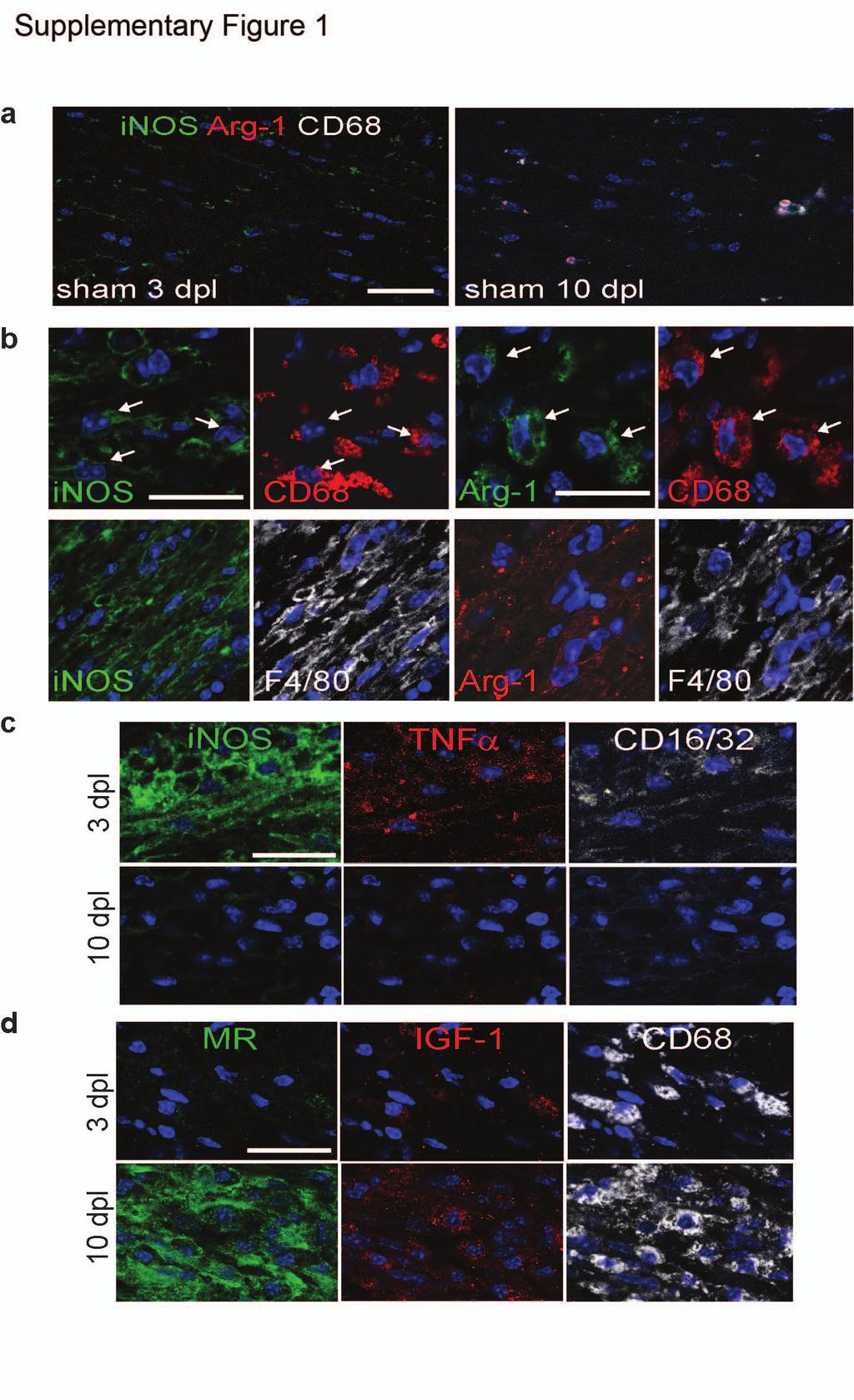
3 Supplementary Figure 1 Additional characterization of polarization in corpus callosum lesions. (a) Representative images of sham PBS-injected lesions at 3 and 10 dpl immunostained for inos (green), Arg-1 (red), and CD68 (white). (b) Co-localization of inos and Arg-1 in microglia/ macrophage cell bodies (CD68; arrows) and processes (F4/80). 3 and 10 dpl lesions immunostained for additional M1 markers (c; TNFα, CD16/32) and M2 markers (d; MR, IGF-1). All scale bars, 25 µm.
4
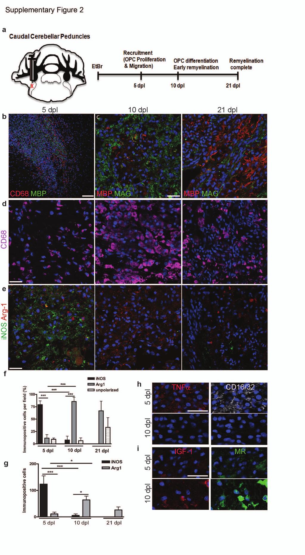
5 Supplementary Figure 2 Switch from M1- to M2-dominant response in remyelinating lesions of the rat caudal cerebellar peduncle. (a) Oligodendroglial lineage cell responses following induction of demyelination of the caudal cerebellar peduncle by stereotaxic injection of ethidium bromide (EtBr). Scale bar, 100 µm. (b) Injection of demyelinating toxin induces accumulation of microglia (CD68+; red) at 5 dpl. Recovery of myelin protein expression is observed with re-expression of MAG at 10 dpl (c, centre) and MBP at 21 dpl (c, right). (d) Microglia/ macrophages (CD68+; purple) are present at 5 dpl, increase in abundance at 10 dpl, and decrease in number by 21 dpl. (e) Representative images of lesions immunostained for inos (green) and Arg-1 (red). Percentage (f) and mean number (g) of inos+ M1, Arg-1+ M2, and unpolarized (inos Arg1 ) cells per field ± s.e.m. at 5, 10, and 21 dpl. One-way ANOVA and Newman-Keuls post test, *p<0.05, **p<0.01. (n=3, df=26, 17, respectively). (h) 5 and 10 dpl sections immunostained for additional M1 (TNFα, CD16/32) and M2 (IGF-1, MR) markers. All other scale bars, 25 µm.
6
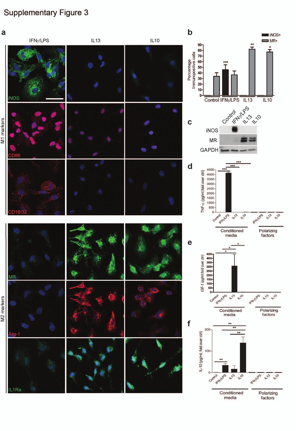
7 Supplementary Figure 3 Polarization of cultured microglia to M1 and M2 phenotypes. (a) Representative images of microglia immunostained against M1 markers inos, CD86, CD16/32 (top) and M2 markers MR, Arg-1, and interleukin-1 receptor antagonist (IL1Ra) (bottom). Scale bar, 25 µm. (b) Mean percentage of inos+ or MR+ cells of total CD68+ cells ± s.e.m.. Kruskal-Wallis test and Dunn s multiple comparison post-hoc test, *p<0.05, **p<0.01, ***p< (n=4) (c) Cropped Western blots showing expression of inos with IFNγ/LPS treatment and increase in MR expression with IL-13 or IL-10 treatment, with loading control GAPDH. ELISAs used to assay conditioned media for levels of TNFα (d; P=0.0004, n=5, df=8), IGF-1 (e; P=0.0330, n=3, df=4), and IL-10 (f; P=0.0051, n=5, df=8), presented as mean fold over M0 control ± s.e.m. demonstrating polarization to M1, M2a, and M2c, respectively. A small yet significant increase in IL-10 secretion was observed with IFNγ/ LPS treatment (P =0.0081). Polarizing factors alone were included in the assay as a control and did not show detectable levels relative to those measured in conditioned media (d-f). 2-tailed Student s t-test.
8
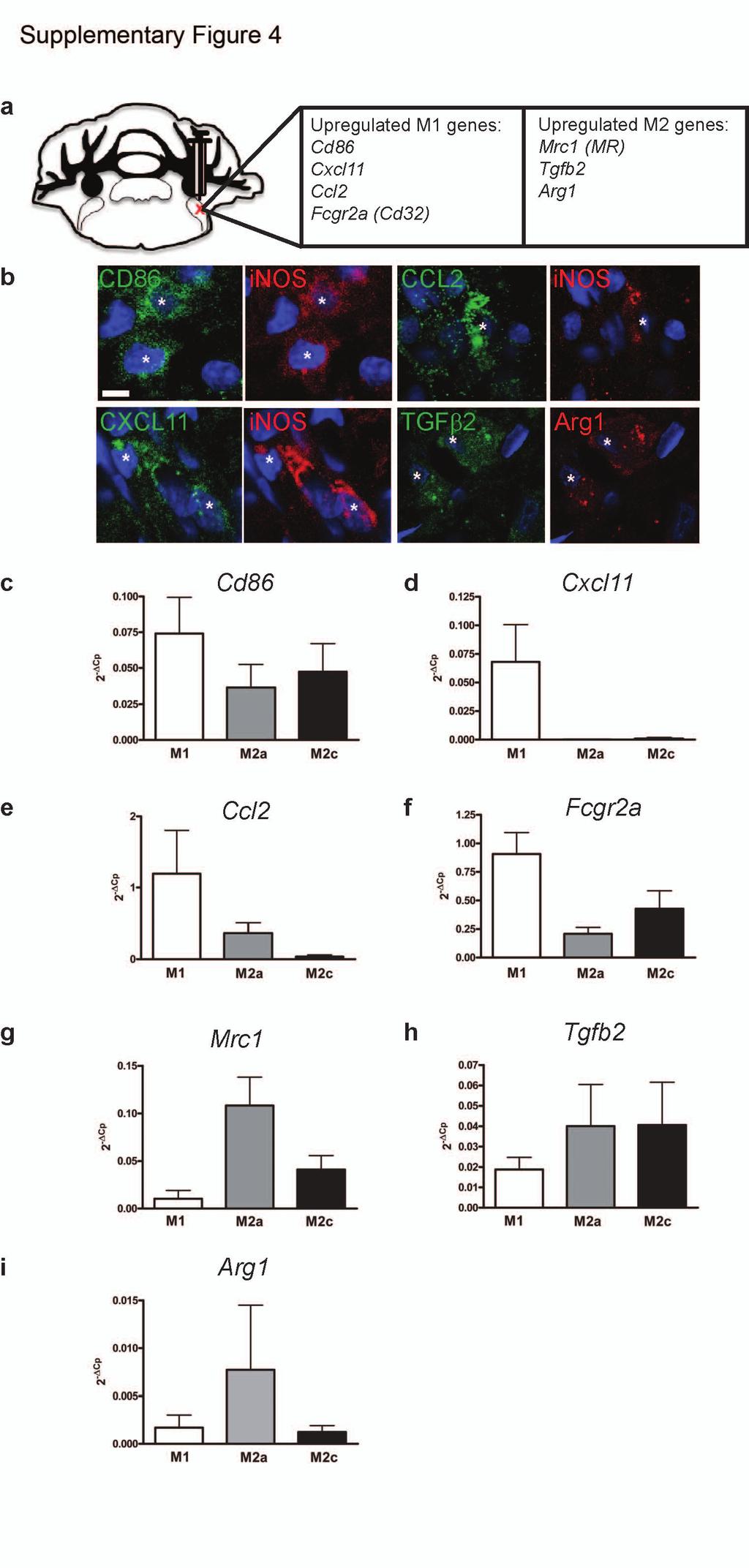
9 Supplementary Figure 4 M1 and M2 associated gene expression in cultured microglia. (a) M1- and M2-associated genes were identified from a previously performed microarray 18 as being significantly upregulated during remyelination of the rat caudal cerebellar peduncles (CCP). These genes were selected for a custom qpcr array to assess gene expression profiles of polarized microglia in vitro. (b) Confirmation of expression of additional polarization markers CD86, CCL2, CXCL11, and TGFβ2 in inos+ or Arg-1+ cells in the rat CCP. Scale bar, 5 µm. Microglia treated with IFNγ/LPS (M1), IL-13 (M2a), and IL-10 (M2c) were analyzed for gene expression levels of (c) Cd86, (d) Cxcl11, (e) Ccl2, (f) Fcgr2a (Cd32), (g) Mrc1(mannose receptor), (h) Tgfb2, and (i) Arg1, values are represented as 2 -ΔCp ± s.e.m.
10
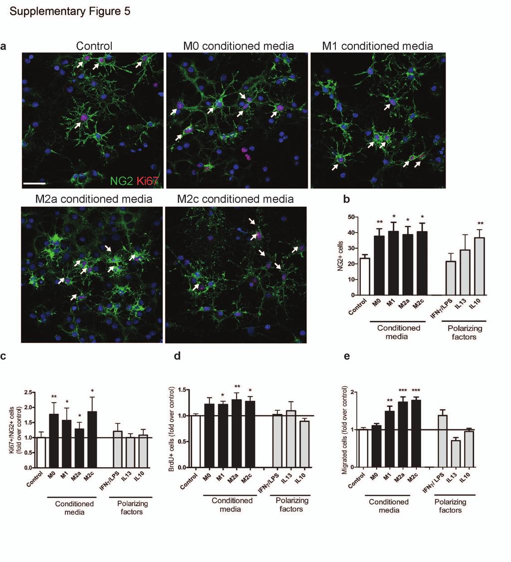
11 Supplementary Figure 5 M1 and M2 microglia conditioned media increase OPC proliferation and migration. OPCs were treated with microglia conditioned media for 3d. Polarizing factors present in the conditioned media (IFNγ/LPS, IL-13, and IL-10) were directly applied to OPCs as a control. (a) Representative images of OPCs immunostained against NG2 (green) and the proliferative marker Ki67 (red). Double-positive cells are highlighted by arrows. Scale bar, 25 µm. (b) Number of NG2+ cells per field was increased with M0 (P=0.005), M1 (P= ), M2a (P= ), M2c (P=0.0119) conditioned media (CM), and IL-10 (P=0.0029) compared to control (n=5, df=8). (c) Ki67+/NG2+ cells were increased with M0 (P=0.0084), M1 (P=0.0362), M2a (P=0.0194), M2c (P=0.0192) conditioned media (n=4, df=6) (d) BrdU+ cells were increased with M1 (P=0.042), M2a (P=0.0083) and M2c conditioned media (P=0.0423) (n=4, df=6). Values from conditioned media-treated conditions (black bars) and polarizing factors alone (grey bars) were normalized to control (unconditioned media, white bars) in (c) and (d), and are represented as mean ± s.e.m., 2-tailed Student s t-test. (e) Number of OPCs ± s.e.m. migrated towards microglia conditioned media (black bars), or polarizing factors alone (grey bars), normalized to values in control (unconditioned media, white bar) showing chemotactic properties of both M1 and M2 conditioned media. Kruskal-Wallis test and Dunn s multiple comparison post-test, **p<0.01, ***p< (n=3).
12
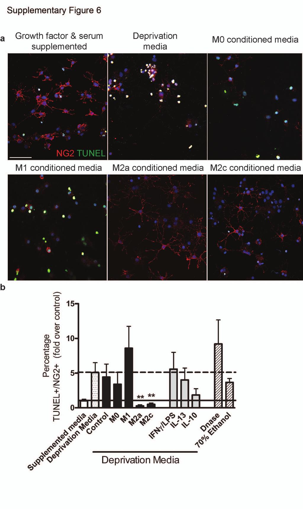
13 Supplementary Figure 6 M2 polarized microglia conditioned media promotes the survival of OPCs in a death-inducing environment. OPCs were treated with microglia conditioned media for 3d in media devoid of serum and growth factors. Polarizing factors present in the conditioned media (IFNγ/LPS, IL-13, and IL-10) were directly applied to OPCs as a control. (a) Representative images of OPCs under basal growth supplemented conditions, grown in deprivation media alone, or supplemented with microglia conditioned media. OPCs were immunostained against NG2 (red) and apoptotic OPCs were visualized by TUNEL assay (green). Scale bar, 50 µm. (b) Mean percentage of TUNEL+ NG2+ OPCs normalized to control supplemented with growth factors and media ± s.e.m.. DNase and ethanol treatment were positive controls for TUNEL positivity. Application of microglia conditioned media to OPCs under normal growth conditions did not induce apoptosis (data not shown). Kruskal-Wallis test and Dunn s multiple comparison post-test, *p<0.05, **p<0.01. (n=6).
14
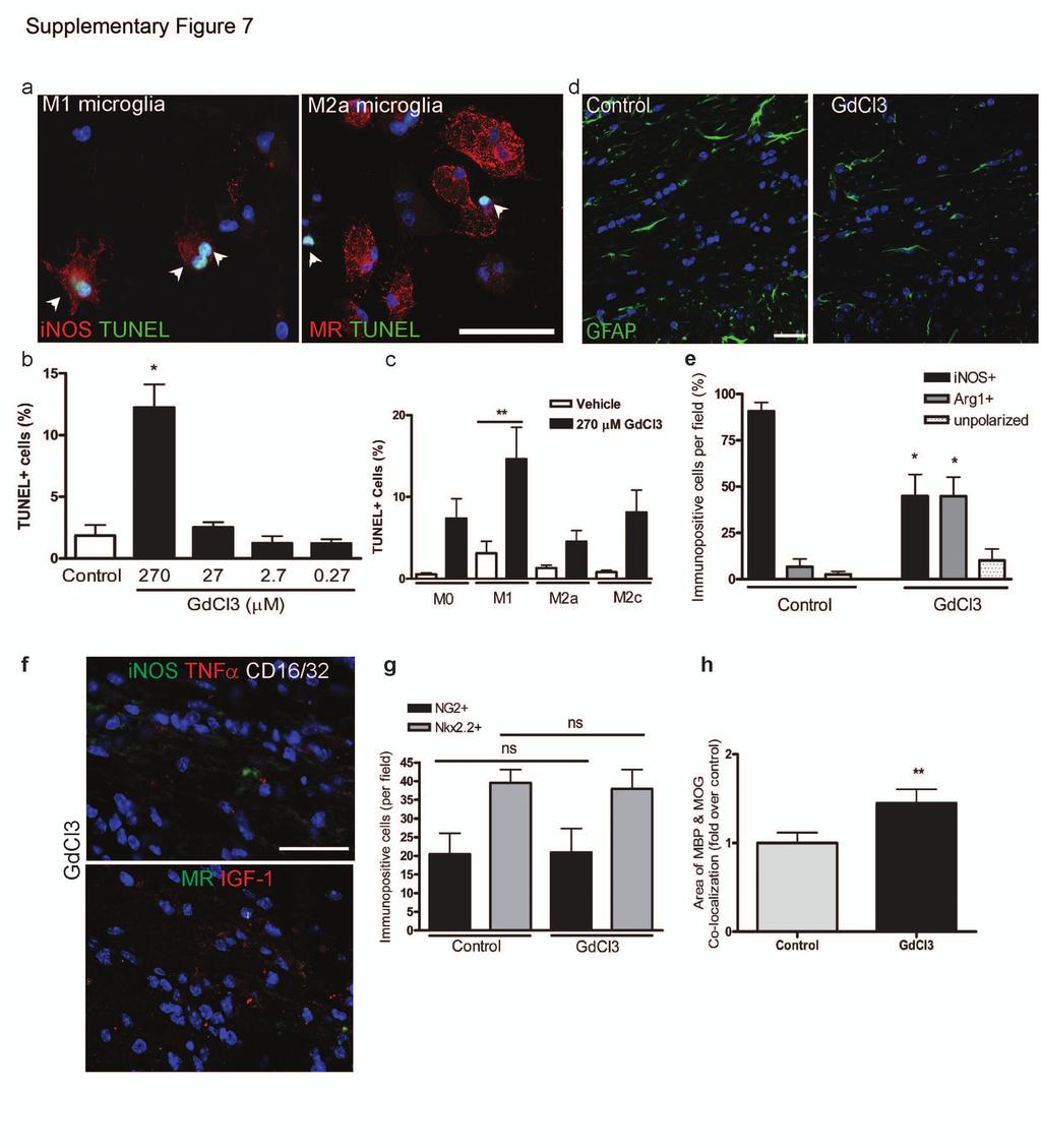
15 Supplementary Figure 7 Selective depletion of M1 polarized microglia/ macrophages using gadolinium chloride. (a) Representative images of M1 (inos+; left) and M2a (MR+; right) microglia treated with 270 µm GdCl3 and apoptosis assessed by TUNEL assay (green nuclei). Apoptotic cells are indicated by arrowheads, and were inos+ and MR. Scale bar, 25 µm. (b) Percentage of TUNEL+ M1 microglia treated with vehicle (ctrl), or GdCl3 ( µm). Mann-Whitney test, P= (n=4). (c) Mean percentage of TUNEL+ cells ± s.e.m. in M0, M1, M2a, and M2c polarized microglia treated with vehicle or 270 µm GdCl3. One-way ANOVA and Newman-Keuls post-hoc test, **p<0.01 (n=6, df=54). (d) GFAP immunostaining in representative control and GdCl3-injected lesions shows no difference in astrocyte reactivity. Scale bar, 25 µm. (e) Percentage of inos+ or Arg1+ cells in control or GdCl3-injected lesions, P= (inos), (Arg1), 2-tailed Student s t-test (n=5, df=8). (f) GdCl3- injected lesions immunostained for M1 markers (inos, TNFα, CD16/32) and M2 markers (MR, IGF-1) at 3 dpl. Scale bar, 25 µm. (g) NG2+ (n=4, df=6) and Nkx2.2+ (n=5, df=8) cells per field in control and GdCl3-treated lesions at 3 dpl. 2-tailed Student s t-test, p>0.05. (h) Mean area of MBP and MOG co-localization, fold over control ± s.e.m. in lesions at 21 dpl. P=0.081, 2-tailed Student s t-test (n=6, df=10).
16
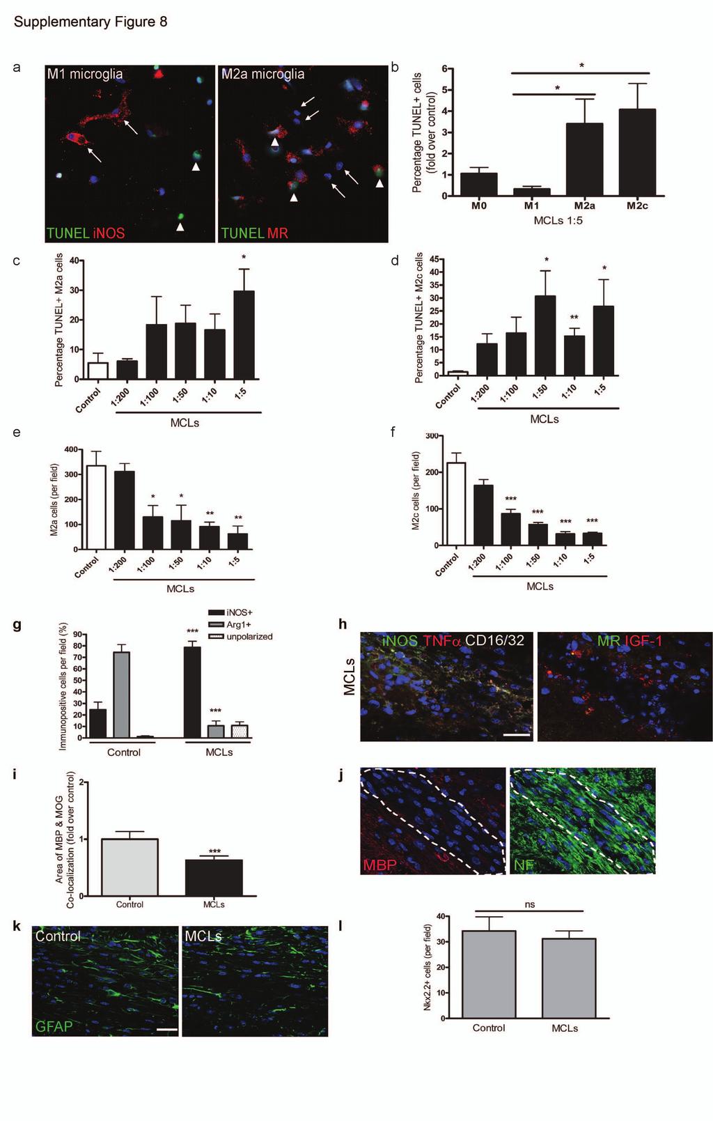
17 Supplementary Figure 8 Selective depletion of M2 polarized microglia/ macrophages using mannosylated clodronate liposomes. (a) Representative images of inos+ M1 and MR+ M2a microglia treated with MCLs (1:5) and assessed for apoptosis by TUNEL assay. In M1 polarizing conditions, inos+ cells were TUNEL (arrows); TUNEL+ cells were inos (arrowheads). In M2a polarizing conditions, TUNEL+ cells were MR+ (arrowheads), whereas MR cells were TUNEL (arrows). (b) Mean percentage of TUNEL+ cells ± s.e.m. in M0, M1, M2a, and M2c polarized microglia treated MCLs (1:5 dilution), normalized to values from M0 conditions. P=0.027 (M1 vs. M2a), P=0.013 (M1 vs. M2c), 2-tailed Student s t-test (n=6, df=10). Mean percentage of TUNEL+ cells ± s.e.m. in M2a (c, P=0.015, df=7) and M2c (d, P=0.0251, , 0.03 for 1:50, 1:10, 1:5, respectively, df=6) microglia treated with MCLs (1:200-1:5; black bars) compared to control (white bars) (n=4). Mean numbers of microglia per field ± s.e.m. for M2a (e, P=0.02, 0.03, 0.009, for 1:100, 1:50, 1:10, 1:5, respectively, df=6) and M2c (f; P=0.0003, <0.0001, , for 1:100, 1:50, 1:10, 1:5, respectively, df=6) polarized cells treated with MCLs (1:200-1:5; black bars) compared to control (white bars), 2-tailed Student s t-test (n=4). (g) Percentage of inos+ M1, Arg-1+ M2, or unpolarized (inos, Arg-1 ) cells per field ± s.e.m. in control and MCL-injected lesions at 10 dpl. P<0.0001, 2-tailed Student s t-test (n=5, df=8). (h) MCL-injected lesions immunostained for additional M1 (inos, TNFα, CD16/32) and M2 markers (MR, IGF-1). Scale bar, 25 µm. (i) Mean area of MBP and MOG colocalization fold over control ± s.e.m. in lesions at 21 dpl. 2-tailed Student s t-test, P= (n=6, df=10). (j) Axons (NF+) were detectable running through the lesion (MBP ) at 10 dpl following MCL injection. (k) MCL injection did not influence astrocytes (GFAP+)
18 within the lesion. Scale bar, 25 µm. (l) Total numbers of Nkx2.2+ OPCs ± s.e.m. in control and MCL injected lesions. 2-tailed Student s t-test, p>0.05 (n=4, df=6).
19
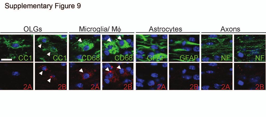
20 Supplementary Figure 9 Expression of activin-a receptors on cells in remyelinating lesions. Representative images of oligodendrocytes (CC1+), microglia/ macrophages (CD68+), astrocytes (GFAP+), and neurons (axons; NF+) with expression (arrows), or lack thereof, of activin-a receptors Acvr2A and Acvr2B. Scale bar, 10 µm.
21 Supplemental Table 1. Human brain tissue samples Multiple Sclerosis Patients Remyelinated Non- Neurological Controls Sex Age Disease duration (yrs) Block Active Lesions Analyzed Chronic Active Chronic Inactive SPMS M 46 8 P2E P3E SPMS F A3D SPMS M 40 9 P3B SPMS F P3C SPMS F A2D A2E PPMS M P4D SPMS F P3D SPMS F A2B P2C SPMS F A2C SPMS F P3C TOTAL Carcinoma of M 77 - P3B the lung metastasized Cardiac failure M 64 - P2D Carcinoma of M 35 - P3B the tongue Ovarian F 60 - P3D cancer Myelodysplast M 82 - A2C ic syndrome, Rheumatoid Arthritis Classification
Cord blood monocytes as a source of cell therapy products for treatment of brain injuries ISCT/CBA 2015 Cord Blood Workshop Wednesday, May 27, 2015
 Cord blood monocytes as a source of cell therapy products for treatment of brain injuries ISCT/CBA 2015 Cord Blood Workshop Wednesday, May 27, 2015 Andrew E. Balber, PhD Senior Scientific Advisor CT 2,
Supplementary Information
 Supplementary Information Title Degeneration and impaired regeneration of gray matter oligodendrocytes in amyotrophic lateral sclerosis Authors Shin H. Kang, Ying Li, Masahiro Fukaya, Ileana Lorenzini,
Nature Neuroscience: doi: /nn Supplementary Figure 1
 Supplementary Figure 1 Quantification of myelin fragments in the aging brain (a) Electron microscopy on corpus callosum is shown for a 18-month-old wild type mice. Myelin fragments (arrows) were detected
The anti-inflammatory enzyme A20 in the neuropathology of Multiple Sclerosis
 More Than Neurons, 1-3 December, Turin The anti-inflammatory enzyme A20 in the neuropathology of Multiple Sclerosis Dr. Simona Perga, PhD Neuroscience Institute Cavalieri Ottolenghi (NICO) & Multiple Sclerosis
Microglia-derived extracellular vesicles regulate the proliferation and differentiation of oligodendrocyte precursor cells
 University of Turin CNR Institute of Neuroscience Microglia-derived extracellular vesicles regulate the proliferation and differentiation of oligodendrocyte precursor cells Roberta Parolisi Turin, December
PREPARED FOR: U.S. Army Medical Research and Materiel Command Fort Detrick, Maryland
 AWARD NUMBER: W81XWH-14-1-0524 TITLE:Oligodendroglial MCT1 and Metabolic Support of Axons in Multiple Sclerosis PRINCIPAL INVESTIGATOR: Jeffrey D. Rothstein MD, PhD CONTRACTING ORGANIZATION: Johns Hopkins
Supplementary Figure 1
 Supplementary Figure 1 AAV-GFP injection in the MEC of the mouse brain C57Bl/6 mice at 4 months of age were injected with AAV-GFP into the MEC and sacrificed at 7 days post injection (dpi). (a) Brains
Fig 1 CD163. CD11b S100A9. Sirius Red. 100μm ** ** CD163. CD11b S100A9 ** Sirius Red (PL) Sirius Red SUM Mo.
 Sirius Red SUM Mo.") T47D T47D + o SU-59 Fig SU-59 + o IHC score (-3) IHC score (-2) CD63 3 2 IHC score (-3) CD63 3 ** 2 CDb CDb * * SA9 SA9 ** * 2 IHC score (-4) αsa αsa 4 ** ** 2 Sirius Red μm IHC score (%) Sirius Red 8
T47D T47D + o SU-59 Fig SU-59 + o IHC score (-3) IHC score (-2) CD63 3 2 IHC score (-3) CD63 3 ** 2 CDb CDb * * SA9 SA9 ** * 2 IHC score (-4) αsa αsa 4 ** ** 2 Sirius Red μm IHC score (%) Sirius Red 8
TGF-β Signaling Regulates Neuronal C1q Expression and Developmental Synaptic Refinement
 Supplementary Information Title: TGF-β Signaling Regulates Neuronal C1q Expression and Developmental Synaptic Refinement Authors: Allison R. Bialas and Beth Stevens Supplemental Figure 1. In vitro characterization
GFP/Iba1/GFAP. Brain. Liver. Kidney. Lung. Hoechst/Iba1/TLR9!
 Supplementary information a +KA Relative expression d! Tlr9 5!! 5! NSC Neuron Astrocyte Microglia! 5! Tlr7!!!! NSC Neuron Astrocyte! GFP/Sβ/! Iba/Hoechst Microglia e Hoechst/Iba/TLR9! GFP/Iba/GFAP f Brain
Contribution of microglia to tissue injury and repair in MS
 Contribution of microglia to tissue injury and repair in MS MS disease course histologic features Courtesy of Samuel Ludwin I ACUTE CHRONIC s ACTIVE CHRONIC Clinical Course Intra CNS Extra CNS Imaging
marker. DAPI labels nuclei. Flies were 20 days old. Scale bar is 5 µm. Ctrl is
 Supplementary Figure 1. (a) Nos is detected in glial cells in both control and GFAP R79H transgenic flies (arrows), but not in deletion mutant Nos Δ15 animals. Repo is a glial cell marker. DAPI labels
An unconventional role for mirna: let-7 activates Toll-like receptor 7 and causes neurodegeneration
 An unconventional role for mirna: let-7 activates Toll-like receptor 7 and causes neurodegeneration Sabrina M. Lehmann, Christina Krüger, Boyoun Park, Katja Derkow, Karen Rosenberger, Jan Baumgart, Thorsten
Neurodegeneration and macrophages; a beneficial or harmful role for macrophages and microglia in neuronal damage during multiple sclerosis
 Neurodegeneration and macrophages; a beneficial or harmful role for macrophages and microglia in neuronal damage during multiple sclerosis Marlijn van der Poel Writing assignment: literature review October
SUPPLEMENTARY FIG. S2. Representative counting fields used in quantification of the in vitro neural differentiation of pattern of dnscs.
 Supplementary Data SUPPLEMENTARY FIG. S1. Representative counting fields used in quantification of the in vitro neural differentiation of pattern of anpcs. A panel of lineage-specific markers were used
Supplementary Table 1. The primers used for quantitative RT-PCR. Gene name Forward (5 > 3 ) Reverse (5 > 3 )
 Reverse (5 > 3 )") 770 771 Supplementary Table 1. The primers used for quantitative RT-PCR. Gene name Forward (5 > 3 ) Reverse (5 > 3 ) Human CXCL1 GCGCCCAAACCGAAGTCATA ATGGGGGATGCAGGATTGAG PF4 CCCCACTGCCCAACTGATAG TTCTTGTACAGCGGGGCTTG
770 771 Supplementary Table 1. The primers used for quantitative RT-PCR. Gene name Forward (5 > 3 ) Reverse (5 > 3 ) Human CXCL1 GCGCCCAAACCGAAGTCATA ATGGGGGATGCAGGATTGAG PF4 CCCCACTGCCCAACTGATAG TTCTTGTACAGCGGGGCTTG
Primary oligodendropathy is not a trigger of CNS autoimmunity
 Primary oligodendropathy is not a trigger of CNS autoimmunity Ari Waisman Institute for Molecular Medicine University Medical Center, JGU Mainz 1 How is an anti-myelin immune response initiated? Secondary
Supplementary Figure 1
 Supplementary Figure 1 Global TeNT expression effectively impairs synaptic transmission. Injection of 100 pg tent mrna leads to a reduction of vesicle mediated synaptic transmission in the spinal cord
Supplemental Information. Menin Deficiency Leads to Depressive-like. Behaviors in Mice by Modulating. Astrocyte-Mediated Neuroinflammation
 Neuron, Volume 100 Supplemental Information Menin Deficiency Leads to Depressive-like Behaviors in Mice by Modulating Astrocyte-Mediated Neuroinflammation Lige Leng, Kai Zhuang, Zeyue Liu, Changquan Huang,
Targeting tumour associated macrophages in anti-cancer therapies. Annamaria Gal Seminar Series on Drug Discovery Budapest 5 January 2018
 Targeting tumour associated macrophages in anti-cancer therapies Annamaria Gal Seminar Series on Drug Discovery Budapest 5 January 2018 Macrophages: Professional phagocytes of the myeloid lineage APC,
Supplemental Figure 1. Intracranial transduction of a modified ptomo lentiviral vector in the mouse
 Supplemental figure legends Supplemental Figure 1. Intracranial transduction of a modified ptomo lentiviral vector in the mouse hippocampus targets GFAP-positive but not NeuN-positive cells. (A) Stereotaxic
Bezzi et al., Supplementary Figure 1 *** Nature Medicine: doi: /nm Pten pc-/- ;Zbtb7a pc-/- Pten pc-/- ;Pml pc-/- Pten pc-/- ;Trp53 pc-/-
 Gr-1 Gr-1 Gr-1 Bezzi et al., Supplementary Figure 1 a Gr1-CD11b 3 months Spleen T cells 3 months Spleen B cells 3 months Spleen Macrophages 3 months Spleen 15 4 8 6 c CD11b+/Gr1+ cells [%] 1 5 b T cells
IMMUNOTOOLS: EFFECT OF NOTCH-DEFICIENT MACROPHAGES TO AUTOIMMUNE DISEASE WIPAWEE WONGCHANA
 IMMUNOTOOLS: EFFECT OF NOTCH-DEFICIENT MACROPHAGES TO AUTOIMMUNE DISEASE 22-02-2017 WIPAWEE WONGCHANA WHAT DO YOU SEE? Allergy Ref: http://carrington.edu/blog/medical/vaccines/smallpox-andsmallpox-vaccine/
TITLE: Harnessing GPR17 Biology for Treating Demyelinating Disease
 AD Award Number: W81XWH-10-1-0721 TITLE: Harnessing GPR17 Biology for Treating Demyelinating Disease PRINCIPAL INVESTIGATOR: Nitin Karandikar, M.D., Ph.D. CONTRACTING ORGANIZATION: University of Texas
Supplementary Figure 1. Microglia do not show signs of classical immune activation following MD a-b. Images showing immunoreactivity for MHCII (a)
") 1 Supplementary Figure 1. Microglia do not show signs of classical immune activation following MD a-b. Images showing immunoreactivity for MHCII (a) and CD45 (b) in fixed sections of binocular visual cortex
1 Supplementary Figure 1. Microglia do not show signs of classical immune activation following MD a-b. Images showing immunoreactivity for MHCII (a) and CD45 (b) in fixed sections of binocular visual cortex
Remyelination in the CNS: from biology to therapy
 NeuroN Glia interactions Remyelination in the CNS: from biology to therapy Robin J. M. Franklin* and Charles ffrench-constant Abstract Remyelination involves reinvesting demyelinated axons with new myelin
SUPPLEMENTARY INFORMATION
 DOI:.38/ncb3399 a b c d FSP DAPI 5mm mm 5mm 5mm e Correspond to melanoma in-situ Figure a DCT FSP- f MITF mm mm MlanaA melanoma in-situ DCT 5mm FSP- mm mm mm mm mm g melanoma in-situ MITF MlanaA mm mm
Genesis of cerebellar interneurons and the prevention of neural DNA damage require XRCC1.
 Genesis of cerebellar interneurons and the prevention of neural DNA damage require XRCC1. Youngsoo Lee, Sachin Katyal, Yang Li, Sherif F. El-Khamisy, Helen R. Russell, Keith W. Caldecott and Peter J. McKinnon.
Supplementary Figure 1 Expression of Crb3 in mouse sciatic nerve: biochemical analysis (a) Schematic of Crb3 isoforms, ERLI and CLPI, indicating the
 Schematic of Crb3 isoforms, ERLI and CLPI, indicating the") Supplementary Figure 1 Expression of Crb3 in mouse sciatic nerve: biochemical analysis (a) Schematic of Crb3 isoforms, ERLI and CLPI, indicating the location of the transmembrane (TM), FRM binding (FB)
Supplementary Figure 1 Expression of Crb3 in mouse sciatic nerve: biochemical analysis (a) Schematic of Crb3 isoforms, ERLI and CLPI, indicating the location of the transmembrane (TM), FRM binding (FB)
T H E J O U R N A L O F C E L L B I O L O G Y
 T H E J O U R N A L O F C E L L B I O L O G Y Supplemental material Amelio et al., http://www.jcb.org/cgi/content/full/jcb.201203134/dc1 Figure S1. mir-24 regulates proliferation and by itself induces
Supplementary Figure S I: Effects of D4F on body weight and serum lipids in apoe -/- mice.
 Supplementary Figures: Supplementary Figure S I: Effects of D4F on body weight and serum lipids in apoe -/- mice. Male apoe -/- mice were fed a high-fat diet for 8 weeks, and given PBS (model group) or
a b c Esophageal eosinophilia
 TSLP-elicited basophil responses can mediate the pathogenesis of eosinophilic esophagitis. Mario Noti, Elia D. Tait Wojno, Brian S. Kim, Mark C. Siracusa, Paul R. Giacomin, Meera G. Nair, Alain J. Benitez,
Systemic 5-fluorouracil Treatment Causes a Syndrome of Delayed Myelin Destruction in the Central Nervous System
 Systemic 5-fluorouracil Treatment Causes a Syndrome of Delayed Myelin Destruction in the Central Nervous System The Harvard community has made this article openly available. Please share how this access
Supplementary Figure 1: TSLP receptor skin expression in dcssc. A: Healthy control (HC) skin with TSLP receptor expression in brown (10x
 skin with TSLP receptor expression in brown (10x") Supplementary Figure 1: TSLP receptor skin expression in dcssc. A: Healthy control (HC) skin with TSLP receptor expression in brown (10x magnification). B: Second HC skin stained for TSLP receptor in brown
Supplementary Figure 1: TSLP receptor skin expression in dcssc. A: Healthy control (HC) skin with TSLP receptor expression in brown (10x magnification). B: Second HC skin stained for TSLP receptor in brown
Supplemental Materials for. Effects of sphingosine-1-phosphate receptor 1 phosphorylation in response to. FTY720 during neuroinflammation
 Supplemental Materials for Effects of sphingosine-1-phosphate receptor 1 phosphorylation in response to FTY7 during neuroinflammation This file includes: Supplemental Table 1. EAE clinical parameters of
fl/+ KRas;Atg5 fl/+ KRas;Atg5 fl/fl KRas;Atg5 fl/fl KRas;Atg5 Supplementary Figure 1. Gene set enrichment analyses. (a) (b)
 (b)") KRas;At KRas;At KRas;At KRas;At a b Supplementary Figure 1. Gene set enrichment analyses. (a) GO gene sets (MSigDB v3. c5) enriched in KRas;Atg5 fl/+ as compared to KRas;Atg5 fl/fl tumors using gene set
KRas;At KRas;At KRas;At KRas;At a b Supplementary Figure 1. Gene set enrichment analyses. (a) GO gene sets (MSigDB v3. c5) enriched in KRas;Atg5 fl/+ as compared to KRas;Atg5 fl/fl tumors using gene set
Tcf21 MCM ; R26 mtmg Sham GFP Col 1/3 TAC 8W TAC 2W. Postn MCM ; R26 mtmg Sham GFP Col 1/3 TAC 8W TAC 2W
 A Tcf21 MCM ; R26 mtmg Sham GFP Col 1/3 Tcf21 MCM ; R26 mtmg TAC 2W Tcf21 MCM ; R26 mtmg TAC 8W B Postn MCM ; R26 mtmg Sham GFP Col 1/3 Postn MCM ; R26 mtmg TAC 2W Postn MCM ; R26 mtmg TAC 8W Supplementary
Edinburgh Research Explorer
 Edinburgh Research Explorer Fingolimod (FTY720) Enhances Remyelination Following Demyelination of Organotypic Cerebellar Slices Citation for published version: Miron, VE, Ludwin, SK, Darlington, PJ, Jarjour,
Neocortex Zbtb20 / NFIA / Sox9
 Neocortex / NFIA / Sox9 Supplementary Figure 1. Expression of, NFIA, and Sox9 in the mouse neocortex at. The lower panels are higher magnification views of the oxed area. Arrowheads indicate triple-positive
Supplementary information. Nkx2.1 regulates the generation of telencephalic astrocytes during embryonic
 Supplementary information Nkx2.1 regulates the generation of telencephalic astrocytes during embryonic development Shilpi Minocha 1*, Delphine Valloton 1*, Yvan Arsenijevic 2, Jean-René Cardinaux 3, Raffaella
Supplementary Figure 1. Antibiotic partially rescues mice from sepsis. (ab) BALB/c mice under CLP were treated with antibiotic or PBS.
 BALB/c mice under CLP were treated with antibiotic or PBS.") 1 Supplementary Figure 1. Antibiotic partially rescues mice from sepsis. (ab) BALB/c mice under CLP were treated with antibiotic or PBS. (a) Survival curves. WT Sham (n=5), WT CLP or WT CLP antibiotic
1 Supplementary Figure 1. Antibiotic partially rescues mice from sepsis. (ab) BALB/c mice under CLP were treated with antibiotic or PBS. (a) Survival curves. WT Sham (n=5), WT CLP or WT CLP antibiotic
SUPPLEMENTARY FIGURES
 SUPPLEMENTARY FIGURES 1 Supplementary Figure 1, Adult hippocampal QNPs and TAPs uniformly express REST a-b) Confocal images of adult hippocampal mouse sections showing GFAP (green), Sox2 (red), and REST
Rina Zilkha-Falb 3, Nathali Kaushansky 1, Naoto Kawakami 2 and Avraham Ben-Nun 1*
 Zilkha-Falb et al. Journal of Neuroinflammation (2016) 13:7 DOI 10.1186/s12974-015-0468-4 RESEARCH Post-CNS-inflammation expression of CXCL12 promotes the endogenous myelin/ neuronal repair capacity following
Supplementary Figure 1: GFAP positive nerves in patients with adenocarcinoma of
 SUPPLEMENTARY FIGURES AND MOVIE LEGENDS Supplementary Figure 1: GFAP positive nerves in patients with adenocarcinoma of the pancreas. (A) Images of nerves stained for GFAP (green), S100 (red) and DAPI
PhD thesis. The role of complement in Experimental Autoimmune Encephalomyelitis, the mouse modell of Multiple Sclerosis
 PhD thesis The role of complement in Experimental Autoimmune Encephalomyelitis, the mouse modell of Multiple Sclerosis Nóra Terényi Supervisor: Prof. Anna Erdei Biology Doctorate School Immunology Program
SUPPLEMENTARY INFORMATION
 doi:10.1038/nature10188 Supplementary Figure 1. Embryonic epicardial genes are down-regulated from midgestation stages and barely detectable post-natally. Real time qrt-pcr revealed a significant down-regulation
COPD lungs show an attached stratified mucus layer that separate. bacteria from the epithelial cells resembling the protective colonic
 COPD lungs show an attached stratified mucus layer that separate bacteria from the epithelial cells resembling the protective colonic mucus SUPPLEMENTARY TABLES AND FIGURES Tables S1 S8, page 1 and separate
Supplementary Table 1 Gene clone ID for ShRNA-mediated gene silencing TNFα downstream signals in in vitro Symbol Gene ID RefSeqID Clone ID
 1 2 3 4 5 6 7 8 9 10 11 12 13 14 15 16 17 18 19 20 21 22 23 24 25 26 27 28 29 30 31 32 33 34 35 36 37 38 39 40 41 42 43 44 Supplementary Table 1 Gene clone ID for ShRNA-mediated gene silencing TNFα downstream
Supplementary information CD4 T cells are required for both development and maintenance of disease in a new model of reversible colitis
 Supplementary information CD4 T cells are required for both development and maintenance of disease in a new model of reversible colitis rasseit and Steiner et al. .. Supplementary Figure 1 % of initial
SUPPLEMENTARY INFORMATION
 doi:1.138/nature1554 a TNF-α + in CD4 + cells [%] 1 GF SPF 6 b IL-1 + in CD4 + cells [%] 5 4 3 2 1 Supplementary Figure 1. Effect of microbiota on cytokine profiles of T cells in GALT. Frequencies of TNF-α
Address: Department of Biomedical Genetics, University of Rochester Medical Center, 601 Elmwood Avenue, Rochester, NY 14642, USA.
 Journal of Biology BioMed Central Research article CNS progenitor cells and oligodendrocytes are targets of chemotherapeutic agents in vitro and in vivo Joerg Dietrich, Ruolan Han, Yin Yang, Margot Mayer-Pröschel
Stewart et al. CD36 ligands promote sterile inflammation through assembly of a TLR 4 and 6 heterodimer
 NFκB (fold induction) Stewart et al. ligands promote sterile inflammation through assembly of a TLR 4 and 6 heterodimer a. mrna (fold induction) 5 4 3 2 1 LDL oxldl Gro1a MIP-2 RANTES mrna (fold induction)
Chronic variable stress activates hematopoietic stem cells
 SUPPLEMENTARY INFORMATION Chronic variable stress activates hematopoietic stem cells Timo Heidt *, Hendrik B. Sager *, Gabriel Courties, Partha Dutta, Yoshiko Iwamoto, Alex Zaltsman, Constantin von zur
SUPPLEMENTARY FIGURES AND TABLES
 SUPPLEMENTARY FIGURES AND TABLES Supplementary Figure S1: CaSR expression in neuroblastoma models. A. Proteins were isolated from three neuroblastoma cell lines and from the liver metastasis of a MYCN-non
TITLE: Harnessing GPR17 Biology for Treating Demyelinating Disease
 AD Award Number: W81XWH-10-1-0723 TITLE: Harnessing GPR17 Biology for Treating Demyelinating Disease PRINCIPAL INVESTIGATOR: Qing Lu, Ph.D. CONTRACTING ORGANIZATION: University of Texas Southwestern Medical
MicroRNAs Modulate the Noncanonical NF- B Pathway by Regulating IKK Expression During Macrophage Differentiation
 MicroRNAs Modulate the Noncanonical NF- B Pathway by Regulating IKK Expression During Macrophage Differentiation Tao Li 1 *, Michael J. Morgan 1 *, Swati Choksi 1, Yan Zhang 1, You-Sun Kim 2#, Zheng-gang
Figure S1 Generation of γ-gt DTR transgenic mice. (A) Schematic construct of the transgene. (B)
 Schematic construct of the transgene. (B)") Figure S1 Generation of γ-gt DTR transgenic mice. (A) Schematic construct of the transgene. (B) PCR identified expected hhb-egf band (left panel) and HA tag band (right) in kidneys of transgenic (TG) mice
Figure S1 Generation of γ-gt DTR transgenic mice. (A) Schematic construct of the transgene. (B) PCR identified expected hhb-egf band (left panel) and HA tag band (right) in kidneys of transgenic (TG) mice
Supplementary Figure 1
 Supplementary Figure 1 A B mir-141, human cell lines mir-2c, human cell lines mir-141, hepatocytes mir-2c, hepatocytes Relative RNA.1.8.6.4.2 Relative RNA.3.2.1 Relative RNA 1.5 1..5 Relative RNA 2. 1.5
Nerve Cells and Behavior
 Nerve Cells and Behavior 27 th September, 2016 Touqeer Ahmed Ph.D. Atta-ur-Rahman School of Applied Biosciences National University of Sciences and Technology Nervous System and Behavior Nervous system
Title of file for HTML: Supplementary Information Description: Supplementary Figures and Supplementary Table
 Title of file for HTML: Supplementary Information Description: Supplementary Figures and Supplementary Table Title of file for HTML: Peer Review File Description: Innate Scavenger Receptor-A regulates
c Ischemia (30 min) Reperfusion (8 w) Supplementary Figure bp 300 bp Ischemia (30 min) Reperfusion (4 h) Dox 20 mg/kg i.p.
 Reperfusion (8 w) Supplementary Figure bp 300 bp Ischemia (30 min) Reperfusion (4 h) Dox 20 mg/kg i.p.") a Marker Ripk3 +/ 5 bp 3 bp b Ischemia (3 min) Reperfusion (4 h) d 2 mg/kg i.p. 1 w 5 w Sacrifice for IF size A subset for echocardiography and morphological analysis c Ischemia (3 min) Reperfusion (8
a Marker Ripk3 +/ 5 bp 3 bp b Ischemia (3 min) Reperfusion (4 h) d 2 mg/kg i.p. 1 w 5 w Sacrifice for IF size A subset for echocardiography and morphological analysis c Ischemia (3 min) Reperfusion (8
Supporting Information. Calculation of the relative contributions of myocyte proliferation, stem cell. Supporting Information Fig 1 (page 9)
") Supporting Information Table of contents Calculation of the relative contributions of myocyte proliferation, stem cell differentiation and cardioprotection (page 2) Supporting Information Fig 1 (page 9)
Supporting Information Table of contents Calculation of the relative contributions of myocyte proliferation, stem cell differentiation and cardioprotection (page 2) Supporting Information Fig 1 (page 9)
Supplementary Materials:
 Supplementary Materials: Supplemental Figure 1: Profibrotic markers in wt/wt or ex/ex mouse kidneys 42 days post IRI. The levels of fibronectin and αsma were examined by immunostaining in ADAM17 wt/wt
Supplementary Information
 Nature Immunology doi:1.138/ni.2477 Supplementary Information Capillary and arteriolar pericytes attract innate leukocytes exiting through venules and instruct them with pattern recognition and motility
Nature Immunology: doi: /ni.3866
 Nature Immunology: doi:10.1038/ni.3866 Supplementary Figure 1 The effect of TIPE2 on chemotaxis. a, The expression of TIPE2 in dhl-60c, dhl-60t, TIPE2-expressing and 15/16Q-expressing dhl-60t neutrophils
Supplementary Figure 1:
 Supplementary Figure 1: (A) Whole aortic cross-sections stained with Hematoxylin and Eosin (H&E), 7 days after porcine-pancreatic-elastase (PPE)-induced AAA compared to untreated, healthy control aortas
SUPPLEMENTARY INFORMATION
 DOI: 10.1038/ncb2697 Figure S1 Cytokeratin 5 is a specific marker for basal and intermediate cells in all mouse prostate lobes. (a) Immunofluorescence staining showing co-localization of YFP with p63 in
Nature Neuroscience: doi: /nn Supplementary Figure 1
 Supplementary Figure 1 EGFR inhibition activates signaling pathways (a-b) EGFR inhibition activates signaling pathways (a) U251EGFR cells were treated with erlotinib (1µM) for the indicated times followed
Supplemental Figures Supplemental Figure 1:
 Supplemental Figures Supplemental Figure 1: Representative FACS data showing Concurrent Brain cell type Acquisition using either Percoll PLUS (top row) or myelin removal beads (bottom two rows). Debris
- 1 - Cell types Monocytes THP-1 cells Macrophages. LPS Treatment time (Hour) IL-6 level (pg/ml)
 IL-6 level (pg/ml)") Supplementary Table ST1: The dynamic effect of LPS on IL-6 production in monocytes and THP-1 cells after GdA treatment. Monocytes, THP-1 cells and macrophages (5x10 5 ) were incubated with 10 μg/ml of
Supplementary Table ST1: The dynamic effect of LPS on IL-6 production in monocytes and THP-1 cells after GdA treatment. Monocytes, THP-1 cells and macrophages (5x10 5 ) were incubated with 10 μg/ml of
Supporting Information
 Supporting Information M1 macrophage-derived nanovesicles potentiate the anticancer efficacy of immune checkpoint inhibitors Yeon Woong Choo, 1, Mikyung Kang, 2, Han Young Kim, 1 Jin Han, 1 Seokyung Kang,
SUPPLEMENTARY INFORMATION
 Figure S1. Loss of Ena/VASP proteins inhibits filopodia and neuritogenesis. (a) Bar graph of filopodia number per stage 1 control and mmvvee (Mena/ VASP/EVL-null) neurons at 40hrs in culture. Loss of all
Supplemental Information. Metabolic Maturation during Muscle Stem Cell. Differentiation Is Achieved by mir-1/133a-mediated
 Cell Metabolism, Volume 27 Supplemental Information Metabolic Maturation during Muscle Stem Cell Differentiation Is Achieved by mir-1/133a-mediated Inhibition of the Dlk1-Dio3 Mega Gene Cluster Stas Wüst,
Therapeutic effect of baicalin on experimental autoimmune encephalomyelitis. is mediated by SOCS3 regulatory pathway
 Therapeutic effect of baicalin on experimental autoimmune encephalomyelitis is mediated by SOCS3 regulatory pathway Yuan Zhang 1,2, Xing Li 1,2, Bogoljub Ciric 1, Cun-gen Ma 3, Bruno Gran 4, Abdolmohamad
Dietary cholesterol promotes repair of demyelinated lesions in the adult brain
 Received Apr 16 Accepted 1 Dec 16 Published Jan 17 DOI: 1.138/ncomms11 Dietary cholesterol promotes repair of demyelinated lesions in the adult brain OPEN Stefan A. Berghoff 1, Nina Gerndt 1, Jan Winchenbach
Nature Neuroscience: doi: /nn.2275
 Supplementary Figure S1. The presence of MeCP2 in enriched primary glial cultures from rat or mouse brains is not neuronal. Western blot analysis of protein extracts from (a) rat glial and neuronal cultures.
label the basement membrane). Different fixation methods of EB-perfused P8 mice to optimize the combination
. Different fixation methods of EB-perfused P8 mice to optimize the combination") Supplementary Figure 1 Optimization of the tissue fixation protocol to combine EB perfusion and IB4 endothelial tip cell staining in the postnatal mouse brain. a-l Labeling of EB-perfused P8 mice with
Supplementary Figure 1 Optimization of the tissue fixation protocol to combine EB perfusion and IB4 endothelial tip cell staining in the postnatal mouse brain. a-l Labeling of EB-perfused P8 mice with
Supplementary information. The proton-sensing G protein-coupled receptor T-cell death-associated gene 8
 1 Supplementary information 2 3 The proton-sensing G protein-coupled receptor T-cell death-associated gene 8 4 (TDAG8) shows cardioprotective effects against myocardial infarction 5 Akiomi Nagasaka 1+,
SOPten flox/flox (KO) Pten flox/flox (WT) flox allele 6.0 kb. Pten. Actin. ! allele 2.3 kb. Supplementary Figure S1. Yanagi, et al.
 Pten flox/flox (WT) flox allele 6.0 kb. Pten. Actin. ! allele 2.3 kb. Supplementary Figure S1. Yanagi, et al.") s1 A Pten flox/flox () SOPten flox/flox () flox allele 6. kb B Pten flox/flox () SOPten flox/flox () Pten Actin! allele 2.3 kb Supplementary Figure S1. Yanagi, et al. A B BrdU BrdU positive cells ( ) 3
s1 A Pten flox/flox () SOPten flox/flox () flox allele 6. kb B Pten flox/flox () SOPten flox/flox () Pten Actin! allele 2.3 kb Supplementary Figure S1. Yanagi, et al. A B BrdU BrdU positive cells ( ) 3
Supplemental Figure 1. Western blot analysis indicated that MIF was detected in the fractions of
 Supplemental Figure Legends Supplemental Figure 1. Western blot analysis indicated that was detected in the fractions of plasma membrane and cytosol but not in nuclear fraction isolated from Pkd1 null
3rd International Conference on Neurology & Therapeutics.
 3rd International Conference on Neurology & Therapeutics www.neuroimmunology.ca Multiple sclerosis is a devastating disease The first description of the disease was mentioned in 14th century In 1838 Dr.
Supplementary Figure 1 IMQ-Induced Mouse Model of Psoriasis. IMQ cream was
 Supplementary Figure 1 IMQ-Induced Mouse Model of Psoriasis. IMQ cream was painted on the shaved back skin of CBL/J and BALB/c mice for consecutive days. (a, b) Phenotypic presentation of mouse back skin
Nature Medicine doi: /nm.3957
 Supplementary Fig. 1. p38 alternative activation, IL-21 expression, and T helper cell transcription factors in PDAC tissue. (a) Tissue microarrays of pancreatic tissue from 192 patients with pancreatic
Stimulating healthy tissue regeneration by targeting the 5-HT 2B receptor in chronic liver disease.
 Stimulating healthy tissue regeneration by targeting the 5-HT 2B receptor in chronic liver disease. Mohammad R Ebrahimkhani, Fiona Oakley, Lindsay B Murphy, Jelena Mann, Anna Moles, Maria J Perugorria,
The Ying and Yang of IFN-γ in Autoimmunity
 The Ying and Yang of IFN-γ in Autoimmunity Chander Raman, Ph.D. Early Late Autoimmune neuroinflammation Experimental autoimmune encephalomyelitis (EAE) Rheumatoid arthritis (RA) Autoimmune neuroinflammation
Supplementary Figure 1. BMS enhances human T cell activation in vitro in a
 Supplementary Figure 1. BMS98662 enhances human T cell activation in vitro in a concentration-dependent manner. Jurkat T cells were activated with anti-cd3 and anti-cd28 antibody in the presence of titrated
Ghrelin Facilitates GLUT2-, SGLT1- and SGLT2-mediated Intestinal. Glucose Transport in Goldfish (Carassius auratus)
") Ghrelin Facilitates GLUT2-, SGLT1- and SGLT2-mediated Intestinal Glucose Transport in Goldfish (Carassius auratus) Ayelén Melisa Blanco 1,2, Juan Ignacio Bertucci 2,3, Naresh Ramesh 2, María Jesús Delgado
Ghrelin Facilitates GLUT2-, SGLT1- and SGLT2-mediated Intestinal Glucose Transport in Goldfish (Carassius auratus) Ayelén Melisa Blanco 1,2, Juan Ignacio Bertucci 2,3, Naresh Ramesh 2, María Jesús Delgado
CXCL13 drives spinal astrocyte activation and neuropathic pain via CXCR5
 The Journal of Clinical Investigation CXCL13 drives spinal astrocyte activation and neuropathic pain via CXCR5 Bao-Chun Jiang, 1 De-Li Cao, 1 Xin Zhang, 1 Zhi-Jun Zhang, 1,2 Li-Na He, 1 Chun-Hua Li, 1
Supplementary Figure 1 P53 is degraded following Chlamydia infection independent of the cell lysis and protein sample preparation procedure applied.
 Supplementary Figure 1 P53 is degraded following Chlamydia infection independent of the cell lysis and protein sample preparation procedure applied. (a) Western blotting analysis showing degradation of
Progress Report for NJCSCR (Yu-Wen Chang)
") Progress Report for NJCSCR (Yu-Wen Chang) Overall Plan Summary: Traumatic injury to the spinal cord initiates a cascade of degenerative processes, known as secondary injury, which include various inflammatory
Progress Report for NJCSCR (Yu-Wen Chang) Overall Plan Summary: Traumatic injury to the spinal cord initiates a cascade of degenerative processes, known as secondary injury, which include various inflammatory
Supplementary Figure 1. Nature Neuroscience: doi: /nn.4547
 Supplementary Figure 1 Characterization of the Microfetti mouse model. (a) Gating strategy for 8-color flow analysis of peripheral Ly-6C + monocytes from Microfetti mice 5-7 days after TAM treatment. Living
Supplemental Material
 Supplemental Material Supplementary Fig. 1. EETs stimulate primary tumor growth. a) Schematic presentation of genetic and pharmacological tools used to manipulate endogenous EET levels. b) Endothelial
Diabetic Complications Consortium
 Diabetic Complications Consortium Application Title: Cathepsin S inhibition and diabetic neuropathy Principal Investigator: Nigel A Calcutt 1. Project Accomplishments: We investigated the efficacy of cathepsin
Thomas HAIDER Journal Club
 Thomas HAIDER Journal Club 20.10.2014 Background Immunology of the CNS - History Ehrlich, 1885 & 1904 dye did not stain brain -> BBB Shirai, Y. (1921) On the transplantation of the rat sarcoma in adult
Supplementary Information
 Supplementary Information Astrocytes regulate adult hippocampal neurogenesis through ephrin-b signaling Randolph S. Ashton, Anthony Conway, Chinmay Pangarkar, Jamie Bergen, Kwang-Il Lim, Priya Shah, Mina
Supplementary fig. 1. Crystals induce necroptosis does not involve caspases, TNF receptor or NLRP3. A. Mouse tubular epithelial cells were pretreated
 Supplementary fig. 1. Crystals induce necroptosis does not involve caspases, TNF receptor or NLRP3. A. Mouse tubular epithelial cells were pretreated with zvad-fmk (10µM) and exposed to calcium oxalate
Supplementary Figures
 Supplementary Figures Supplementary Figure 1. Confirmation of Dnmt1 conditional knockout out mice. a, Representative images of sorted stem (Lin - CD49f high CD24 + ), luminal (Lin - CD49f low CD24 + )
Supplementary Fig. 1: TBR2+ cells in different brain regions.
 Hip SVZ OB Cere Hypo Supplementary Fig. 1: TBR2 + cells in different brain regions. Three weeks after the last tamoxifen injection, TBR2 immunostaining images reveal a large reduction of TBR2 + cells in
islets scored 1 week month months
 Supplementary Table 1. Sampling parameters for the morphometrical analyses Time (post- DT) Control mice (age-matched) α-cells mice pancreatic surface (mm 2 ) scored DT-treated mice islets scored mice pancreatic
Supplementary Figures
 Supplementary Figures Supplementary Figure 1 Characterization of stable expression of GlucB and sshbira in the CT26 cell line (a) Live cell imaging of stable CT26 cells expressing green fluorescent protein