Spinal Imaging. ssregypt.com. Mamdouh Mahfouz MD
|
|
|
- Posy Conley
- 5 years ago
- Views:
Transcription

1 Spinal Imaging Degenerative diseases ssregypt.com Mamdouh Mahfouz MD


2 MRI Open MRI Closed


3 Extremity MRI


4 Dynamic MRI Dynamic MRI The bed rotates from Upright to Recumbent, stopping at any angle in between


5 Patient with Low Back Pain After Surgery Dynamic MRI Does a Lie-Down-Only Scanner see the patient s problem? NO! Case courtesy of M. Rose, MD, Rose Radiology Centers


6 Dynamic MRI


7 Surface coils

8 Degenerative diseases 2 7 Spinal canal stenosis Disc lesions Bone marrow changes Ligamentous pathology Osseous changes Spondylolisthesis Cord pathology

9 Spinal canal stenosis Idiopathic type = Reduced sagittal diameter L2 L3 1.1 cm 1.2 cm


10 Spinal canal stenosis Developmental type = Hypertrophied laminae, articular facets and Ligamenta flava


11 Ligamenta Flava hypertrophy

12 Ligamenta Flava hypertrophy L4-5


13 Spinal canal stenosis L4-5 L4-5

14


15 Spinal canal stenosis Idiopathic type = Reduced sagittal diameter Developmental type = Hypertrophied laminae, articular facets and Ligamenta flava L cm
16 L5 Lateral recess spinal canal stenosis 1.4 cm


17 Lumbar spinal canal


18 Spinal canal stenosis Developmental type = Hypertrophied laminae, articular facets and Ligamenta flava


19 Developmental canal stenosis


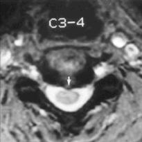
20 Cervical canal stenosis


21 Cervical canal stenosis


22 Cervical canal stenosis

23 Severe canal stenosis


24 Capacious spinal canal

25 L4-5 Capacious spinal canal L5-S1
26 Disc lesions Degeneration Bulge Herniation


27 Disc Lesions Normal lumbar disc CT L4-5

28 Normal cervical disc CT L4-5 C4-5 L3-4


29 MRI Normal lumbar disc


30 MRI Normal cervical disc


31 Disc lesions L4-5


32 Disc degeneration Loss of water T2 signal [MRI] Loss of height narrowing of disc space [X ray, MR, CT?] Intradiscal air ( vacuum phenomena) [CT]


33 Disc degeneration


34 Disc degeneration


35 Disc lesions


36 Vaccum phenomena


37 Disc lesions Disc bulge = Intact annulus = Diffuse pathology Disc herniation = Torn annulus= Focal pathology Normal L4-5 Bulge


38 Disc Herniation Normal herniation Bulge herniation protrusion Sequest. protrusion Seques t.


39 Disc Herniation

40 Bulge Disc Herniation


41 Bulge Disc Herniation L5-S1 L5-S1


42 Disc bulges


43 Bulge / Herniation


44 Protrusion / herniation


45 Disc Protrusion


46 Disc herniation


47 Disc herniation L4-5


48 Disc herniation


49 Disc herniation


50 Disc herniation


51 Disc herniation L R


52 Disc herniation L R


53 Disc herniation

54 Migration /Sequestration


55 Disc herniation with caudal migration


56 Disc migration


57 Disc migration


58 Disc Sequestration


59 Disc Sequestration


60 Disc and granulation


61 Disc and granulation

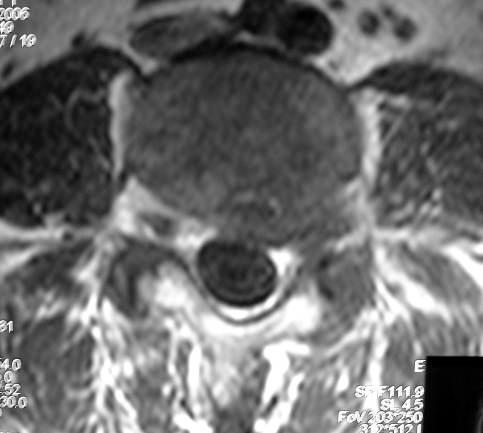
![types ] Type I Marrow](/docs-images/85/92214429/images/62-1.jpg "edema Type II Fatty")
62 Bone marrow changes [ Only seen on MRI][3 types ] Type I Marrow edema Type II Fatty marrow


63 Type I Marrow edema


64 Type II Fatty marrow
65 Type II Fatty marrow


66 Type III Bone sclerosis


67 Osseous pathology Osteophytes Anterior Posterior [Important] Osteoarthritis L4-5


68 Osteo -arthritis Narrowing of the joint space Subarticular bone sclerosis Osteophytic lippings Pseudo cystic changes Vacuum phenomena


69 Osteo - arthritis Normal Osteoarthritis


70 Disc /osteophyte complex


71 Ligament pathology Hypertrophy Calcification Ossification


72 Ligamenta flava hypertrophy [ Buckling ]


73 OPLL Ossification of Posterior Longitudinal Ligament


74 OPLL


75 Vertebral displacement Spondylolisthesis [Anterior displacement ] Lytic Degenerative Retrolisthesis [Posterior displacement ]

76 Spondylo - listhesis


77 Spondylo - listhesis Multi-level


78 Spondylo - listhesis [ Pseudo disc bulge ] L4-5 L4 L4 L5


79 Spondylo - listhesis [ Pseudo disc bulge ]


80 Spondylo - listhesis [ Pseudo disc bulge ] L5-S1


81 Spondylo - listhesis [ Multi-level ]


82 Spinal instability


83 Cord pathology Edema Myelomalacia [ early, late] Both produce focal area of low signal in T1 and high signal in T2 Differentiation by clinical presentation of the patient ± contrast injection.


84 Cord pathology Compressive myelomalacia Focal area of high signal in T2 WIs Decompression leads to regression or resolution of early lesions NB: Early lesion shows contrast enhancement

![fragments, [ Trauma]](/docs-images/85/92214429/images/85-2.jpg "Abscesses")
![[inflammatory lesions]](/docs-images/85/92214429/images/85-3.jpg "Neoplastic extra")
85 Para-vertebral shadows Hemorrhage, bone fragments, [ Trauma] Abscesses [inflammatory lesions] Neoplastic extra osseous masses [ Tumors]


86 Q.. Disc herniation, Right postro -lateral


87 Q.. Type II Fatty marrow


88 Q.. Type III Bone sclerosis Lumbar degenerative changes L3-4 disc herniation with cephalic migration L2-3 disc herniation


89 Q.. Disc herniation, left postro -lateral


90 Q.. Disc herniation, left postro -lateral


91 Q.. Lytic Spondylo listhesis with pseudo disc bulge


92 Q.. Type II Fatty marrow


93 Q.. L5-S1 disc herniation with cephalic migration


94 Q.. Disc herniation and granulation


95 Q.. Normal lumbar disc CT Myelography, osteophyte


96 Q.. Sequestrated L4-5 disc with cephalic migration


97 Q.. L5-S1 disc herniation with granulation


98 Q.. Foraminal disc herniation

99 سبحانك اللهم و بحمدك نشهد ان ال اله اال انت نستغفرك و نتوب اليك Thank you


100


101


102


103


104 Degenerative changes Disc lesion


105


106 Multiple disc lesions


107 Cervical discs Lumbar L.flava


108 Lumbar discs & L.flava


109 Canal stenosis Capacious canal,disc


110


111 Disc lesions


112


113

114 ?

115 ?


116 ?

117 ?


118 ?


119 ?


120 ?


121 ?

122 ?

123 ?


124 ? DISC AND GRANULATION


125 ?


126


127 Type II Fatty marrow
Brain Imaging. IC calcifications. Mamdouh mahfouz MD
 Brain Imaging IC calcifications www.ssregypt.com Mamdouh mahfouz MD mamdouh.m5@gmail.com CT Hyper dense [ more than100 HU ] MRI Low signal in T1 and T2 WIs [non mobile protons] Exceptions Minute calcifications
HEPATO-BILIARY IMAGING
 HEPATO-BILIARY IMAGING BY MAMDOUH MAHFOUZ MD PROF.OF RADIOLOGY CAIRO UNIVERSITY mamdouh.m5@gmail.com www.ssregypt.com CT ABDOMEN Indications Patient preparation Patient position Scanogram Fasting 4-6 hours
Spine. Neuroradiology. Spine. Spine Pathology. Distribution of fractures. Radiological algorithm. Role of radiology 18/11/2015
 Spine Neuroradiology Spine Prof.Dr.Nail Bulakbaşı X Ray: AP/L/Oblique Vertebra & disc spaces CT & CTA Vertebra, discs, vessels MRI & MRA Vertebra, disc, vessels, meninges Spinal cord & nerves Myelography
CERVICAL SPONDYLOSIS & CERVICAL DISC DISEASE
 CERVICAL SPONDYLOSIS & CERVICAL DISC DISEASE Cervical spondylosis l Cervical osteophytosis l Most common progressive disease in the aging cervical spine l Seen in 95% of the people by 65 years Pathophysiology
Hidayatullah Hamidi. MD Consultant Radiologist. Lumbar Spine MR Imaging Interpretation
 Hidayatullah Hamidi. MD Consultant Radiologist Lumbar Spine MR Imaging Interpretation 13/12/2018 Presenter Hidayatullah Hamidi Consultant Radiologist, Radiology PGME program director, FMIC, Kabul, Afghanistan
3D imaging reformation was obtained. The 3D color imaging reformation was reviewed in a different high resolution setting.
 POST OPERATIVE SPINE WITH CONTRAST CLINICAL INDICATION: Low back pain, Patient is post operative status for L4/5 diskectomy TECHNIQUE: MRI of the lumbosacral spine was performed with multiplanar imaging
Facet Joint Arthrosis Disc Degeneration and Lumbago. Dr.Ruchira Sethi Dr. Vishram Singh Department of Anatomy Santosh University, India
 Facet Joint Arthrosis Disc Degeneration and Lumbago Dr.Ruchira Sethi Dr. Vishram Singh Department of Anatomy Santosh University, India INTRODUCTION The initial division of spine into three columns for
Head&Neck Imaging. ssregypt.com. Parapharyngeal Spaces. Mamdouh mahfouz MD
 Head&Neck Imaging Parapharyngeal Spaces ssregypt.com Mamdouh mahfouz MD mamdouh.m5@gmail.com Definitio n Fat filled triangular space lateral the pharynx Extends from the skull base to the oropharynx Parapharyngeal
Degenerative Disc Disease. Nafi Aygun, MD. Associate Professor of Radiology
 Degenerative Disc Disease Nafi Aygun, MD. Associate Professor of Radiology Big Problem Great majority of adults suffer from at least one episode of acute low back pain during life time Disc degeneration
ESSENTIALS OF PLAIN FILM INTERPRETATION: SPINE DR ASIF SAIFUDDIN
 ESSENTIALS OF PLAIN FILM INTERPRETATION: SPINE DR ASIF SAIFUDDIN Consultant Musculoskeletal Radiologist Royal National Orthopaedic Hospital Stanmore,UK. INTRODUCTION 2 INTRODUCTION 3 INTRODUCTION Spinal
Spinal canal stenosis Degenerative diseases F 06
 What is spinal canal stenosis? The condition known as spinal canal stenosis is a narrowing (stenosis) of the spinal canal that in most cases develops due to the degenerative (wear-induced) deformation
CERVICAL SPINE: Radiographs and MRI Cases
 www.jprad.com Radiology reports with recommendations & clinical information - $30 per region, x-ray - $50 per MRI - Medpay Monthly Newsletter 700 East Redlands Blvd, Redlands CA 92373 909.353.9348 jpedley299@yahoo.com
RETROLISTHESIS. Retrolisthesis. is found mainly in the cervical spine and lumbar region but can also be often seen in the thoracic spine
 RETROLISTHESIS A retrolisthesis is a posterior displacement of one vertebral body with respect to adjacent vertebrae Typically a vertebra is to be in retrolisthesis position when it translates backward
102 Results RESULTS. Age Mean=S.D Range 42= years -84 years Number % <30 years years >50 years
 102 Results RESULTS A total of 50 cases were studied 39 males and 11females.Their age ranged between 16 years and 84 years (mean 42years). T1 and T2WI were acquired for all cases in sagittal and axial
Degenerative Disease of the Spine
 Degenerative Disease of the Spine Introduction: I. Anatomy Talk Overview II. Overview of Disease Processes: A. Spondylosis B. Intervertebral Disc Disease III. Diagnosis IV. Therapy Introduction: Myelopathy
DEGENERATIVE SPINAL DISEASE PRABIN SHRESTHA ANISH M SINGH B&B HOSPITAL
 SPINAL CHAPTER, NESON DEGENERATIVE SPINAL DISEASE PRABIN SHRESTHA ANISH M SINGH B&B HOSPITAL INTRODUCTION DEGENERATIVE SPINAL DISEASE Gradual loss of normal structure and function of spine with time Also
Properties of Purdue. Anatomy. Positioning AXIAL SKELETAL RADIOLOGY FOR PRIVATE PRACTITIONERS 11/30/2018
 AXIAL SKELETAL RADIOLOGY FOR PRIVATE PRACTITIONERS Anatomy Complex Text book is needed Species Contrast Positioning Painful/ non cooperative Sedation General anesthesia Species Contrast 1 Slightly oblique
Daniel J. Blizzard, MD, MS
 Daniel J. Blizzard, MD, MS None Common degenerative (usually) condition caused by compression on the spinal cord that is characterized by clumsiness and difficulty with fine motor tasks in the hands and
A third back injury on April 28, 2003, resulted in a two-week time loss from work.
 CLAIM HISTORY AND APPEAL PROCEEDINGS: The Worker injured his left knee in a workplace accident on July 28, 1989. Surgeries were performed in 1989 and 1994, and the Worker was awarded a 12 percent permanent
Imaging the Degenerative Diseases of the Lumbar Spine
 221 Imaging the Degenerative Diseases of the Lumbar Spine David Malfair, MD a, Douglas P. Beall, MD b,c, * MAGNETIC RESONANCE IMAGING CLINICS Magn Reson Imaging Clin N Am 15 (2007) 221 238 - Degenerative
Soccer causes degenerative changes in the cervical spine. European Spine Journal, February 2004, 13(1):76-82
:76-82") Soccer causes degenerative changes in the cervical spine European Spine Journal, February 2004, 13(1):76-82 Alparslan Kartal, Brahim Yldran, Alparslan Enköylü and Feza Korkusuz FROM ABSTRACT: Background
Soccer causes degenerative changes in the cervical spine European Spine Journal, February 2004, 13(1):76-82 Alparslan Kartal, Brahim Yldran, Alparslan Enköylü and Feza Korkusuz FROM ABSTRACT: Background
LUMBAR SPINAL STENOSIS
 LUMBAR SPINAL STENOSIS Always occurs in the mobile segment. Factors play role in Stenosis Pre existing congenital or developmental narrowing of the lumbar spinal canal Translation of one anatomic segment
Case Report: CASE REPORT OF FACET ARTHROPATHY INDUCED NERVE ROOT COMPRESSION RESULTING IN MOTOR WEAKNESS AND PAIN
 Cox Technic Case Report #100 published at www.coxtechnic.com (sent October 2011 on 10/11/11 ) 1 Case Report: CASE REPORT OF FACET ARTHROPATHY INDUCED NERVE ROOT COMPRESSION RESULTING IN MOTOR WEAKNESS
MRI Evaluation of Disc Lesions Causing Lumbar Canal Stenosis and Backache Y Aditya 1 *, Janardhan Reddy K 2 and MV Ramanappa 3
 Indian Journal of Mednodent and Allied Sciences Vol. 2, No. 3, November, 2014, pp- 242-247 DOI : 10.5958/2347-6206.2014.00022.3 Original Research MRI Evaluation of Disc Lesions Causing Lumbar Canal Stenosis
Key Primary CPT Codes: Refer to pages: 7-9 Last Review Date: October 2016 Medical Coverage Guideline Number:
 National Imaging Associates, Inc. Clinical guidelines CERVICAL SPINE SURGERY: ANTERI CERVICAL DECOMPRESSION WITH FUSION CERVICAL POSTERI DECOMPRESSION WITH FUSION CERVICAL ARTIFICIAL DISC CERVICAL POSTERI
Neuroimaging. spine / spinal cord
 Neuroimaging spine / spinal cord Spine & spinal cord imaging methodology Plain x-ray of spine Computed tomography CT - traditional ( normal CT) - reconstructions - myelo-ct Magnetic resonance MR - standard
Peggers Super Summaries: The Aging Spine
 Aging Spine: AGING PROCESS Osteopenia 10% of 50 year old males and 25% of 50 year females Disc dehydration Facet degeneration Soft tissue hypertrophy 2 0 deformity Leg pain worse than back pain from nerve
Bony framework of the vertebral column Structure of the vertebral column
 5.1: Vertebral column & back. Overview. Bones o vertebral column. o typical vertebra. o vertebral canal. o spinal nerves. Joints o Intervertebral disc. o Zygapophyseal (facet) joint. Muscles o 2 compartments:
Degenerative cervical myelopathy (DCM) encompasses. focus Neurosurg Focus 40 (6):E5, 2016
 encompasses. focus Neurosurg Focus 40 (6):E5, 2016") neurosurgical focus Neurosurg Focus 40 (6):E5, 2016 Magnetic resonance imaging assessment of degenerative cervical myelopathy: a review of structural changes and measurement techniques Aria Nouri, MD,
neurosurgical focus Neurosurg Focus 40 (6):E5, 2016 Magnetic resonance imaging assessment of degenerative cervical myelopathy: a review of structural changes and measurement techniques Aria Nouri, MD,
ABCs of the degenerative spine
 Insights into Imaging (2018) 9:253 274 https://doi.org/10.1007/s13244-017-0584-z PICTORIAL REVIEW ABCs of the degenerative spine Sergiy V. Kushchayev 1 & Tetiana Glushko 1 & Mohamed Jarraya 1 & Karl H.
2. The vertebral arch is composed of pedicles (projecting from the body) and laminae (uniting arch posteriorly).
 and laminae (uniting arch posteriorly).") VERTEBRAL COLUMN 2018zillmusom I. VERTEBRAL COLUMN - functions to support weight of body and protect spinal cord while permitting movements of trunk and providing for muscle attachments. A. Typical vertebra
VERTEBRAL COLUMN 2018zillmusom I. VERTEBRAL COLUMN - functions to support weight of body and protect spinal cord while permitting movements of trunk and providing for muscle attachments. A. Typical vertebra
8/4/2012. Causes and Cures. Nucleus pulposus. Annulus fibrosis. Vertebral end plate % water. Deforms under pressure
 Causes and Cures Intervertebral discs Facet (zygopophyseal) joints Inter body joints Spinal nerve roots Nerve compression Pathological conditions Video Causes of back pain Nucleus pulposus Annulus fibrosis
SPINAL CORD DISEASE IN DOGS PART TWO: MOST LIKELY CAUSES
 Vet Times The website for the veterinary profession https://www.vettimes.co.uk SPINAL CORD DISEASE IN DOGS PART TWO: MOST LIKELY CAUSES Author : RITA GONÇALVES Categories : Vets Date : April 7, 2014 RITA
SUBAXIAL CERVICAL SPINE TRAUMA- DIAGNOSIS AND MANAGEMENT
 SUBAXIAL CERVICAL SPINE TRAUMA- DIAGNOSIS AND MANAGEMENT 1 Anatomy 3 columns- Anterior, middle and Posterior Anterior- ALL, Anterior 2/3 rd body & disc. Middle- Posterior 1/3 rd of body & disc, PLL Posterior-
Orthopadic cors. Topic : -Cervical spondylitis. -Development disorders(spondylolysis and Spodylolsithesis)
") Orthopadic cors Topic : -Cervical spondylitis. -Development disorders(spondylolysis and Spodylolsithesis) Cervical spondylitis. Definition : - a painful condition of the cervical spine resulting from the
Orthopadic cors Topic : -Cervical spondylitis. -Development disorders(spondylolysis and Spodylolsithesis) Cervical spondylitis. Definition : - a painful condition of the cervical spine resulting from the
Cervical intervertebral disc disease Degenerative diseases F 04
 Cervical intervertebral disc disease Degenerative diseases F 04 How is a herniated cervical intervertebral disc treated? Conservative treatment is generally sufficient for mild symptoms not complicated
WORKPLACE SAFETY AND INSURANCE APPEALS TRIBUNAL DECISION NO. 2192/16
 WORKPLACE SAFETY AND INSURANCE APPEALS TRIBUNAL DECISION NO. 2192/16 BEFORE: E. Kosmidis: Vice-Chair HEARING: August 30, 2016 at Toronto Written DATE OF DECISION: October 25, 2016 NEUTRAL CITATION: 2016
PARADIGM SPINE. Patient Information. Treatment of a Narrow Lumbar Spinal Canal
 PARADIGM SPINE Patient Information Treatment of a Narrow Lumbar Spinal Canal Dear Patient, This brochure is intended to inform you of a possible treatment option for narrowing of the spinal canal, often
Imaging of Cervical Spine Trauma Tudor H Hughes, M.D.
 Imaging of Cervical Spine Trauma Tudor H Hughes, M.D. General Considerations Most spinal fractures are due to a single episode of major trauma. Fatigue fractures of the spine are unusual except in the
Gillian Wooldridge, DO Houston Methodist Willowbrook Hospital Primary Care Sports Medicine Fellowship May 3, 2018
 Gillian Wooldridge, DO Houston Methodist Willowbrook Hospital Primary Care Sports Medicine Fellowship May 3, 2018 Disclosures Neither I nor any family members have financial disclosures Special thanks
Lumbar Disc Prolapse. Dr. Ahmed Salah Eldin Hassan. Professor of Neurosurgery & Consultant spinal surgeon
 Lumbar Disc Prolapse By Dr. Ahmed Salah Eldin Hassan Professor of Neurosurgery & Consultant spinal surgeon 1-What are the Functions of the Spine Structural support for upright posture Protection of Spinal
Comprehension of the common spine disorder.
 Objectives Comprehension of the common spine disorder. Disc degeneration/hernia. Spinal stenosis. Common spinal deformity (Spondylolisthesis, Scoliosis). Osteoporotic fracture. Anatomy Anatomy Anatomy
CERVICAL SPONDYLOSIS AND CERVICAL SPONDYLOTIC MYELOPATHY
 CERVICAL SPONDYLOSIS AND CERVICAL SPONDYLOTIC MYELOPATHY A NEUROSURGEON S VIEW A Preventable Journey to a wheelchair bound-life Dr H. BOODHOO F.C.S (Neurosurgery) Cervical Spondylosis Spinal Osteoarthritis
NEURORADIOLOGY. Part III. Angela Csomor University of Szeged Department of Radiology
 NEURORADIOLOGY Part III Angela Csomor University of Szeged Department of Radiology DISEASES OF SPINE AND SPINAL CORD I. Non-tumourous diseases developmental anomalies vascular disorders inflammatory processes
Am I eligible for the TOPS study? Possibly, if you suffer from one or more of the following conditions:
 Am I eligible for the TOPS study? Possibly, if you suffer from one or more of the following conditions: Radiating leg pain Greater leg / buttock pain than back pain Severe pain sets in when walking as
SPONDYLOSIS Spin13 (1)
") SPONDYLOSIS Spin13 (1) Spondylosis Last updated: September 5, 2017 ETIOPATHOPHYSIOLOGY... 1 Mechanisms of damage / irritation to neural structures... 2 EPIDEMIOLOGY... 2 Cervical Spondylosis... 3 CLINICAL
SPONDYLOSIS Spin13 (1) Spondylosis Last updated: September 5, 2017 ETIOPATHOPHYSIOLOGY... 1 Mechanisms of damage / irritation to neural structures... 2 EPIDEMIOLOGY... 2 Cervical Spondylosis... 3 CLINICAL
Cox Technic Case Report #124 published at ( sent October 2013 ) 1
 1") Cox Technic Case Report #124 published at www.coxtechnic.com ( sent October 2013 ) 1 5 th Lumbar Disc Herniation with Spondylolisthesis Treated with Cox Technic Flexion Distraction by Travis Cross BS,
Cox Technic Case Report #124 published at www.coxtechnic.com ( sent October 2013 ) 1 5 th Lumbar Disc Herniation with Spondylolisthesis Treated with Cox Technic Flexion Distraction by Travis Cross BS,
ProDisc-L Total Disc Replacement. IDE Clinical Study
 Total Disc Replacement IDE Clinical Study Study Design TDR vs. circumferential fusion: Multi-center, prospective, randomized trial 17 centers, 292 patients 162 patients 80 fusion patients 50 non-randomized
Pathophysiology of lumbar disc degeneration: a review of the literature. Neurosurg Focus 13 (2): August, 2002
: August, 2002") Pathophysiology of lumbar disc degeneration: a review of the literature Neurosurg Focus 13 (2): August, 2002 MICHAEL D. MARTIN, M.D., CHRISTOPHER M. BOXELL, M.D., F.A.C.S., AND DAVID G. MALONE, M.D. FROM
Pathophysiology of lumbar disc degeneration: a review of the literature Neurosurg Focus 13 (2): August, 2002 MICHAEL D. MARTIN, M.D., CHRISTOPHER M. BOXELL, M.D., F.A.C.S., AND DAVID G. MALONE, M.D. FROM
How to interpret computed tomography of the lumbar spine
 REVIEW Ann R Coll Surg Engl 2014; 96: 502 507 doi 10.1308/003588414X13946184902361 How to interpret computed tomography of the lumbar spine Z Ahmad 1, R Mobasheri 2,TDas 3, S Vaidya 4, S Mallik 5, M El-Hussainy
VERTEBRAL COLUMN VERTEBRAL COLUMN
 VERTEBRAL COLUMN FUNCTIONS: 1) Support weight - transmits weight to pelvis and lower limbs 2) Houses and protects spinal cord - spinal nerves leave cord between vertebrae 3) Permits movements - *clinical
SpineFAQs. Neck Pain Diagnosis and Treatment
 SpineFAQs Neck Pain Diagnosis and Treatment Neck pain is a common reason people visit their doctor. Neck pain typically doesn't start from a single injury. Instead, the problem usually develops over time
Objectives. Comprehension of the common spine disorder
 Objectives Comprehension of the common spine disorder Disc degeneration/hernia Spinal stenosis Common spinal deformity (Spondylolisthesis, Scoliosis) Osteoporotic fracture Destructive spinal lesions Anatomy
Construction of Aged Patient Spine Database with Degenerative Diseases
 Construction of Aged Patient Spine Database with Degenerative Diseases Seungwoo Lee 1, Dongmin Seo 1, Soon-Chan Hong 1, Sang-Ho Lee 1, Hanmin Jung 1 1 Information and S/W Research Center, Korea Institute
Lumbar spinal canal stenosis Degenerative diseases F 08
 What is lumbar spinal canal stenosis? This condition involves the narrowing of the spinal canal, and of the lateral recesses (recesssus laterales) and exit openings (foramina intervertebralia) for the
The clinical features and surgical treatment of degenerative lumbar scoliosis: A review of 112 patientsos4_
 Orthopaedic Surgery (2009), Volume 1, No. 3, 176 183 ORIGINAL ARTICLE The clinical features and surgical treatment of degenerative lumbar scoliosis: A review of 112 patientsos4_030 176..183 Wei Liu MD,
Facet orientation in patients with lumbar degenerative spondylolisthesis
 35 J. Tokyo Med. Univ., 71 1 35 0 Facet orientation in patients with lumbar degenerative spondylolisthesis Wuqikun ALIMASI, Kenji ENDO, Hidekazu SUZUKI, Yasunobu SAWAJI, Hirosuke NISHIMURA, Hidetoshi TANAKA,
River North Pain Management Consultants, S.C., Axel Vargas, M.D., Regional Anesthesiology and Interventional Pain Management.
 River North Pain Management Consultants, S.C., Axel Vargas, M.D., Regional Anesthesiology and Interventional Pain Management. Chicago, Illinois, 60611 Phone: (888) 951-6471 Fax: (888) 961-6471 Clinical
Morphological changes of the cervical spinal canal and cord due to aging on MR imaging
 Morphological changes of the cervical spinal canal and cord due to aging on MR imaging Shigeru Kobayashi, MD,PhD 1, Katsuhiko Hayakawa, MD, PhD 2, Takashi Nakane, MD, PhD 2, Riya Kosaka MD,PhD 3. 1 Department
DEGENERATIVE SPONDYLOLISTHESIS
 AN INTRODUCTION TO DEGENERATIVE SPONDYLOLISTHESIS This booklet is designed to inform you about lumbar degenerative spondylolisthesis. It is not meant to replace any personal conversations that you might
University of Jordan. Professor Freih Abuhassan -
 Freih Odeh Abu Hassan F.R.C.S.(Eng.), F.R.C.S.(Tr.& Orth.). Professor of Orthopedics University of Jordan 1 A. Sacroiliitis History Trauma is very common Repetitive LS motion--lumbar rotation or axial
Cervical spondylarthrotic myelopathy with early onset in Down's syndrome: five cases and a review of the literature
 283 Journal of Intellectual Disability Research VOLUME 43 PART 4 pp 283±288 AUGUST 1999 Cervical spondylarthrotic myelopathy with early onset in Down's syndrome: five cases and a review of the literature
Digital Motion X-ray Cervical Spine
 NAME OF PATIENT: CASE STUDY 4 DATE OF REPORT: DATE OF EXAMINATION: REFERRING PHYSICIAN: TESTING FACILITY: Digital Motion X-ray Cervical Spine 1. In the neutral lateral projection: Shows reversal of the
Disclosures: T. Yoshii: None. T. Yamada: None. T. Taniyama: None. S. Sotome: None. T. Kato: None. S. Kawabata: None. A. Okawa: None.
 Dynamic Changes in Spinal Cord Compression by Cervical Ossification of the Posterior Longitudinal Ligament Evaluated by Kinematic Computed Tomography Myelogram Toshitaka Yoshii, Tsuyoshi Yamada, Takashi
Complex Spine Symposium January 12th, Balgrist University Hospital
 DEGENERATIVE CERVICAL MYELOPATHY CLINICAL DECISION MAKING Prof. Dr. Mazda Farshad Chair of Orthopedic Surgery Chief of Spine Surgery Medical Director CERVICAL MYELOPATHY - CAUSES degenerative cervical
POSTERIOR CERVICAL FUSION
 AN INTRODUCTION TO PCF POSTERIOR CERVICAL FUSION This booklet provides general information on the Posterior Cervical Fusion (PCF) surgical procedure for you to discuss with your physician. It is not meant
Degenerative Spinal Disorders. Gábor Nagy MD PhD Zoltán Papp MD
 Degenerative Spinal Disorders Gábor Nagy MD PhD Zoltán Papp MD Neurosurgery Surgical treatment of diseases of central and peripheral nervous system Surgery of nerves (brain, spine, peripheral nerves) Surgery
Incidence and Risk Factors for Late Neurologic Deterioration after C3-6 Laminoplasty in Patients with Cervical Spondylotic Myelopathy
 Incidence and Risk Factors for Late Neurologic Deterioration after C3-6 Laminoplasty in Patients with Cervical Spondylotic Myelopathy Sakaura H, Miwa T, Kuroda Y, Ohwada T Dept. of Orthop. Surg., Kansai
Positional Magnetic Resonance Imaging. Description
 Subject: Positional Magnetic Resonance Imaging Page: 1 of 6 Last Review Status/Date: June 2015 Positional Magnetic Resonance Imaging Description Positional magnetic resonance imaging (MRI) allows imaging
SpineFAQs. Cervical Disc Replacement
 SpineFAQs Cervical Disc Replacement Artificial disc replacement (ADR) is relatively new. In June 2004, the first ADR for the lumbar spine (low back) was approved by the FDA for use in the US. Replacing
Dynamic Mri of the Cervical Spine An Important Tool in Planning Surgical Treatment of Cervical Compressive Myelopathy
 International Journal of Neurosurgery 2018; 2(1): 17-22 http://www.sciencepublishinggroup.com/j/ijn doi: 10.11648/j.ijn.20180201.14 Dynamic Mri of the Cervical Spine An Important Tool in Planning Surgical
PROF. EPIMENIO RAMUNDO ORLANDO
 PROF. EPIMENIO RAMUNDO ORLANDO Lumbar Spinal Stenosis - Definition N.I.C. caused by lumbar stenosis was firstly described by Verbiest (1954)*1 and is characterized by contemporary single or multiple factors:
Spondylolysis. Lysis (Greek λύσις, lýsis from lýein "to separate") refers to the breaking down.
 refers to the breaking down.") Spondylolysis Lysis (Greek λύσις, lýsis from lýein "to separate") refers to the breaking down. Thomas J Kishen Spine Surgeon Sparsh Hospital for Advanced Surgeries Bangalore Spondylolysis Defect in the
Spondylolysis Lysis (Greek λύσις, lýsis from lýein "to separate") refers to the breaking down. Thomas J Kishen Spine Surgeon Sparsh Hospital for Advanced Surgeries Bangalore Spondylolysis Defect in the
New Magnetic Resonance Imaging Grading System for Lumbar Neural Foramina Stenosis
 Original Article New Magnetic Resonance Imaging Grading System for Lumbar Neural Foramina Stenosis DOI: 10.7860/IJARS/2018/30862:2366 Radiology Section BINOJ VARGHESE v, ARUN C BABU ABSTRACT Introduction:
Imaging and Management of the Charcot Spine Following Spinal Injury
 Imaging and Management of the Charcot Spine Following Spinal Injury Poster No.: P-0023 Congress: ESSR 2012 Type: Scientific Exhibit Authors: A. Isaac, P. A. Tyler; Stanmore/UK Keywords: Musculoskeletal
2/5/2019. Facet Joint Pain. Biomechanics
 Facet Arthropathy as a Pain Source Evaluation and Management Shelby Spine Jan 31 st Feb 2 nd, 2019 Kushagra Verma MD, MS Adult and Pediatric Scoliosis And Spine Deformity Beach Orthopaedics Specialty Institute
Bioactive glass S53P4 in spine surgery -results from a prospective 11-year-follow-up
 Bioactive glass S53P4 in spine surgery -results from a prospective 11-year-follow-up Janek Frantzén M.D., Neurosurgeon Turku University Hospital, Finland ROME SPINE 2011 7.12.2011 1 Clinical Development
Spectrum of magnetic resonance imaging findings in chronic low back pain
 Original article: Spectrum of magnetic resonance imaging findings in chronic low back pain Dr Sanjeev Sharma (1), Dr Monika Sharma (2), DR Bhardwaj (3), MD; Dr Asha Negi, (4) Department of Radiodiagnosis,
MR Imaging of the Degenerative Lumbar Spine. Acknowledgements 3/3/2016 MRI
 MR Imaging of the Degenerative Lumbar Spine Gina A. Ciavarra Assistant Professor of Radiology NYU-Langone Medical Center 4/1/2016 Acknowledgements Thank you to Leon Rybak, M.D. and Michael Mechlin, M.D.,
Coventry Pain Clinic - Spianl Pain - Sciatica and Brachalgia
 Coventry Pain Clinic - Spianl Pain - Sciatica and Brachalgia Copyright 2002-2005 Dr. Richard S. Walker Introduction Spinal Nerve Root Pain (Radiculopathy) can arise from the problems affecting the:- C4
Imaging Degeneration of the Lumbar Spine: What It All Means.
 Imaging Degeneration of the Lumbar Spine: What It All Means www.fisiokinesiterapia.biz Spine Nomenclature and Evidence for Imaging p, Talk Objectives Background Nomenclature for disc findings Who should
SPINAL MAGNETIC RESONANCE IMAGING INTERPRETATION
 CLINICAL VIGNETTE 2017; 3:2 SPINAL MAGNETIC RESONANCE IMAGING INTERPRETATION Editor-in-Chief: Idowu, Olufemi E. Neurological surgery Division, Department of Surgery, LASUCOM/LASUTH, Ikeja, Lagos, Nigeria.
ORIGINAL PAPER. Department of Orthopedic Surgery, Chubu Rosai Hospital, Nagoya, Japan ABSTRACT
 Nagoya J. Med. Sci. 77. 221 ~ 228, 2015 ORIGINAL PAPER RANGE OF MOTION DETERMINED BY MULTIDETECTOR-ROW COMPUTED TOMOGRAPHY IN PATIENTS WITH CERVICAL OSSIFICATION OF THE POSTERIOR LONGITUDINAL LIGAMENT
Alan H Daniels, MD. Spine Division, Department of Orthopaedics Warren Alpert School of Medicine of Brown University
 Spinal and Orthopaedic Surgery in the Elderly Alan H Daniels, MD Spine Division, Department of Orthopaedics Warren Alpert School of Medicine of Brown University As the population ages, and patients remain
Disclosures 5/27/2016. Narrowing of the spinal canal or neuroforamina causing a symptomatic compression of the neural element.
 Radiculopathy Saggital MRI view Contained disc extrusion uplifting PLL from bone Terrence Julien, MD, FACS, (blue FAANS arrows) Associate Professor of Neurosurgery System Director, Surgical Neuro-Oncology
Epidemiology of Low back pain
 Low Back Pain Definition Pain felt in your lower back may come from the spine, muscles, nerves, or other structures in that region. It may also radiate from other areas like the mid or upper back, a inguinal
Lumbar radiculopathy caused by foraminal stenosis in rheumatoid arthritis
 Upsala Journal of Medical Sciences. 2011; 116: 133 137 ORIGINL RTICLE Lumbar radiculopathy caused by foraminal stenosis in rheumatoid arthritis TOMOKI KOKUTSU, NOKI MOROZUMI, YUTK KOIZUMI & YUSHIN ISHII
PREPARED FOR. Marsha Eichhorn DATE OF INJURY : N/A DATE OF ANALYSIS : 12/14/2016 DATE OF IMAGES : 12/8/2016. REFERRING DOCTOR : Dr.
 Accent on Health Chiropractic 405 Firemans Ave PREPARED FOR Marsha Eichhorn DATE OF INJURY : N/A DATE OF ANALYSIS : 12/14/2016 DATE OF IMAGES : 12/8/2016 REFERRING DOCTOR : Dr. David Bohn This report contains
Outline. Epidemiology Indications for C-spine imaging Modalities Interpretation Types of fractures
 C-Spine Plain Films Outline Epidemiology Indications for C-spine imaging Modalities Interpretation Types of fractures Epidemiology 7000-10000 c-spine injuries treated each year Additional 5000 die at the
ProDisc-L Total Disc Replacement. IDE Clinical Study.
 ProDisc-L Total Disc Replacement. IDE Clinical Study. A multi-center, prospective, randomized clinical trial. Instruments and implants approved by the AO Foundation Table of Contents Indications, Contraindications
The imaging features of spondylolisthesis : what the clinician needs to know
 The imaging features of spondylolisthesis : what the clinician needs to know Poster No.: C-1018 Congress: ECR 2011 Type: Authors: Educational Exhibit D. Shah 1, C. J. Burke 1, A. C. andi 2, R. Houghton
We are IntechOpen, the world s leading publisher of Open Access books Built by scientists, for scientists. International authors and editors
 We are IntechOpen, the world s leading publisher of Open Access books Built by scientists, for scientists 4,000 116,000 120M Open access books available International authors and editors Downloads Our
Sinonasal Imaging. Mamdouh Mahfouz MD Professor of Radiology Cairo University. ssregypt.com
 Sinonasal Imaging Mamdouh Mahfouz MD Professor of Radiology Cairo University ssregypt.com Scanning Techniques Routine Study CORONAL Coronal 3-5mm sections from the posterior wall of the sphenoid sinus
Current Spine Procedures
 SPINE BOOT CAMP: WHAT YOU DON T KNOW MAY COST YOU! David Abraham, M.D. The Reading Neck and Spine Center Reading, PA Current Spine Procedures Epidural/Transforaminal Injections Lumbar Procedures Laminectomy
In-Space. Interspinous distraction through a mini-open, posterior, unilateral approach.
 In-Space. Interspinous distraction through a mini-open, posterior, unilateral approach. Surgical Technique Posterior Approach PRODUCT OBSOLETED 30th September 2017 DSEM/SPN/0915/0348(1) This publication
A minimally invasive surgical approach reduces cranial adjacent segment degeneration in patients undergoing posterior lumbar interbody fusion
 A minimally invasive surgical approach reduces cranial adjacent segment degeneration in patients undergoing posterior lumbar interbody fusion T. Tsutsumimoto, M. Yui, S. Ikegami, M. Uehara, H. Kosaku,
MUSCULOSKELETAL IMAGING FOR PHYSICAL THERAPISTS. COMBINED SECTIONS MEETING 2006 San Diego, CA February 1-5, 2006
 MUSCULOSKELETAL IMAGING FOR PHYSICAL THERAPISTS COMBINED SECTIONS MEETING 2006 San Diego, CA February 1-5, 2006 John Meyer, DPT, OCS University of Southern California Department of Athletic Medicine Los
A Patient s Guide to Neck Pain. William T. Grant, MD
 A Patient s Guide to Neck Pain Dr. Grant is a talented orthopedic surgeon with more than 30 years of experience helping people return to their quality of life. He and GM Pugh, PA-C pride themselves in
A Novel Classification and Minimally Invasive Treatment of Degenerative Lumbar Spinal Stenosis
 DOI: 10.5137/1019-5149.JTN.9173-13.2 Received: 28.08.2013 / Accepted: 11.02.2014 Original Investigation A Novel Classification and Minimally Invasive Treatment of Degenerative Lumbar Spinal Stenosis Guangfei
A Patient s Guide to Diffuse Idiopathic Skeletal Hyperostosis (DISH)
") A Patient s Guide to Diffuse Idiopathic Skeletal Hyperostosis (DISH) 6565 Fannin Street Houston, TX 77030 Phone: 713-790-3333 DISCLAIMER: The information in this booklet is compiled from a variety of sources.
A Patient s Guide to Diffuse Idiopathic Skeletal Hyperostosis (DISH) 6565 Fannin Street Houston, TX 77030 Phone: 713-790-3333 DISCLAIMER: The information in this booklet is compiled from a variety of sources.