Brain Imaging. IC calcifications. Mamdouh mahfouz MD
|
|
|
- Pearl Owen
- 5 years ago
- Views:
Transcription

1 Brain Imaging IC calcifications Mamdouh mahfouz MD

![HU ] MRI Low](/docs-images/81/84196671/images/2-1.jpg "signal in T1")

2 CT Hyper dense [ more than100 HU ] MRI Low signal in T1 and T2 WIs [non mobile protons]


3 Exceptions Minute calcifications are seen on CT but not on MRI High signal in T1 and low signal in T2 WIs DD Fat Early subacute blood (intracellular met Hb)


4 Physiological intracranial calcifications Choriod plexus Pineal body Falx Tentorium Diaphragma sellae Petroclinoid ligament


5 Pathological Intracranial Calcifications Diagnostic Non diagnostic Bilateral symmetrical basal ganglia calcification Periventricular calcification Gyral calcifications


6 Diagnostic Calcifications Bilateral symmetrical basal ganglia calcification Periventricular calcification Gyral calcifications
![Basal ganglia calcifications Idiopathic Hypoparathyroidism Pseudohypoparathyroidism Pseudo pseudohypoparathyroidism Fahr disease [Ferro calcinosis] Parkinsonism](/docs-images/81/84196671/images/7-0.jpg "Radiation therapy Encephalitis Carbon monoxide intoxication Familial Hyperparathyroidism Bilateral patchy almost symmetrical calcifications Mainly affecting the basal")
7 Basal ganglia calcifications Idiopathic Hypoparathyroidism Pseudohypoparathyroidism Pseudo pseudohypoparathyroidism Fahr disease [Ferro calcinosis] Parkinsonism Radiation therapy Encephalitis Carbon monoxide intoxication Familial Hyperparathyroidism Bilateral patchy almost symmetrical calcifications Mainly affecting the basal ganglia


8 Basal ganglia calcifications


9 Basal ganglia calcifications

![Congenital Intracranial infections] CT CT](/docs-images/81/84196671/images/10-4.jpg "Cytomegalovirus inclusion bodies Toxoplasmosis")
10 Periventricular calcifications Spotty non symmetrical periventricular calcific foci [ Congenital Intracranial infections] CT CT Cytomegalovirus inclusion bodies Toxoplasmosis Cysticercosis Tuberous sclerosis


11 Congenital brain infection


12 Cysticercosis

 Herpes simplex virus (HSV) type2")
13 Congenital and neonatal Infections The classic TORCH infections include: Toxoplasmosis Rublla Cytomegalovirus (CMV) Herpes simplex virus (HSV) type2 Toxoplasmosis
14 Transmission through three major routes Hematogenous / transplacental ( toxoplasmosis, most viruses except HSV type2) Ascending cervical infection ( most bacteria) During delivery through birth canal (HSV type2) The pattern of dystrophic calcification is not entirely specific, CMV calcifications are usually central with predilection for periventricular location, whereas toxoplasmosis calcifications are more widespread with slight predilection for basal ganglia


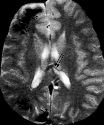
15 Tuberous sclerosis Neuro ectodermal disorder 1:150,000 live births Adenoma sebaceum 30% Seizures 80% Mental retardation 70%


16 Subependymal hamartomas Calcification with age progression 88% Subependymal Giant cell astrocytoma 5-15% F 35Y Tuberous sclerosis with Subependymal GCA


17

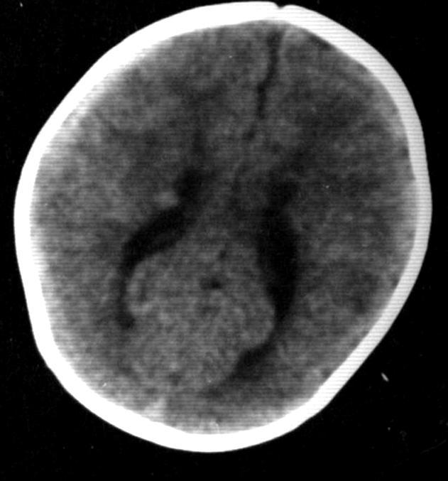
18 Tuberous sclerosis


19 Subependymal Giant Cell Astrocytoma Very rare 0.3% Develops from the nodules Usually in the frontal horn near the foramen of Monro Hydrocephalus Low grade lesion [ No recurrence ] CT Isodense or slightly hyper dense Calcification Homogenous enhancement


20 Tuberous sclerosis with giant cell astrocytoma


21 Tuberous sclerosis with giant cell astrocytoma operated upon with intraventricular hemorrhage


22 Sturge- Webber syndrome Vascular malformation with capillary venous angioma in the face, ocular choroid, leptomeninges Gyral calcification after 2 years of age Parietooccipital more than frontal Bilateral lesions in 20% of cases Cortical hemi atrophy Choroid plexus enlargement Thickened skull and orbits


23 Sturge- Webber syndrome


24 Sturge webber syndrome
25 Non diagnostic calcification Vascular Metabolic Inflammatory Neoplastic AVM, aneyrysm hemangioma arteriosclerosis Carbon monoxide poisoning Folic acid deficiency Hypervitaminosis D Homocystinuria Lead poisoning Wilson s disease healed brain abscess Fungal cerebritis Parasitic disease Cysticercosis, hydatid TB, Toxoplasma Traumatic Hematoma scarring and needle tracts Meningioma, glioma, deposits, neurofibromatosis, treated lymphoma and leukaemia

26 Multiple tuberculomata Multiple tuberculomata


27 Multiple tuberculomas


28 Oligodendroglioma


29 Oligodendroglioma M 50Y


30 Subfrontal meningioma


31 سبحانك اللهم و بحمدك نشهد ان ال اله اال انت نستغفرك و نتوب اليك ssr-eg.net Thank you Mamdouh Mahfouz MD
Spinal Imaging. ssregypt.com. Mamdouh Mahfouz MD
 Spinal Imaging Degenerative diseases ssregypt.com Mamdouh Mahfouz MD mamdouh.m5@gmail.com MRI Open MRI Closed Extremity MRI Dynamic MRI Dynamic MRI The bed rotates from Upright to Recumbent, stopping at
HEPATO-BILIARY IMAGING
 HEPATO-BILIARY IMAGING BY MAMDOUH MAHFOUZ MD PROF.OF RADIOLOGY CAIRO UNIVERSITY mamdouh.m5@gmail.com www.ssregypt.com CT ABDOMEN Indications Patient preparation Patient position Scanogram Fasting 4-6 hours
Head&Neck Imaging. ssregypt.com. Parapharyngeal Spaces. Mamdouh mahfouz MD
 Head&Neck Imaging Parapharyngeal Spaces ssregypt.com Mamdouh mahfouz MD mamdouh.m5@gmail.com Definitio n Fat filled triangular space lateral the pharynx Extends from the skull base to the oropharynx Parapharyngeal
Cerebro-vascular stroke
 Cerebro-vascular stroke CT Terminology Hypodense lesion = lesion of lower density than the normal brain tissue Hyperdense lesion = lesion of higher density than normal brain tissue Isodense lesion = lesion
Neuro - imaging. Sella. ssregypt.com
 Neuro - imaging Sella ssregypt.com Bony Sella AP diameter Depth Contents 16mm 14mm Pituitary gland, part of infundibular stalk, CSF CT Technique 5 mm slices Axial and coronal Contrast injection Bone and
Atypical Brain Calcifications Causing Seizure-Imaging Appearances
 ID: IJARS/2016/18508:2120 Radiology Section Original Article Atypical Brain Calcifications Causing Seizure-Imaging Appearances Nellaiappan Chelliah ABSTRACT Introduction: Brain calcification causing seizure
Non-Traumatic Neuro Emergencies
 Department of Radiology University of California San Diego Non-Traumatic Neuro Emergencies John R. Hesselink, M.D. Nontraumatic Neuroemergencies 1. Acute focal neurological deficit 2. Worst headache of
USCAP Neuropathology night panel CASE 4
 USCAP Neuropathology night panel CASE 4 B.K. Kleinschmidt-DeMasters MD University of Colorado Denver Denver, Colorado Sheep Mountain, Telluride, Colorado Clinical History The patient is a 46 year old male
Neuroimaging Core Curriculum
 Neuroimaging Core Curriculum Program Content The purpose of the training program is to prepare the physician for the independent practice of neuroimaging. Neuroimaging is the subspecialty of Neurology
Brain Pain Infections of the CNS
 FRIDAY, OCTOBER 28, 2016 Brain Pain Infections of the CNS Suyash Mohan MD, PDCC Assistant Professor of Radiology & Neurosurgery Division of Neuroradiology, Department of Radiology Perelman School of Medicine
NEURORADIOLOGY DIL part 5
 NEURORADIOLOGY DIL part 5 Masses and tumors K. Agyem MD, G. Hall MD, D. Palathinkal MD, Alexandre Menard March/April 2015 OVERVIEW Introduction to Neuroimaging - DIL part 1 Basic Brain Anatomy - DIL part
Imaging in Epilepsy. Nucharin Supakul, MD Ramathibodi Hospital, Mahidol University August 22, 2015
 Imaging in Epilepsy Nucharin Supakul, MD Ramathibodi Hospital, Mahidol University August 22, 2015 Nothing to disclose Outline Role of Imaging and pitfalls Imaging protocol Case scenarios Clinical & Electrophysiologic
Pathologic Analysis of CNS Surgical Specimens
 2015 Kenneth M. Earle Memorial Neuropathology Review Pathologic Analysis of CNS Surgical Specimens Peter C. Burger, MD Interdisciplinary Quality Control Familiarity with entities Use of diagnostic algorithm
CONTENTS. Section 1 Bilateral Predominantly Symmetric Abnormalities. Cases. Other Relevant Cases
 Edited by,, and List of contributors xi List of abbreviations xii Preface xv Section 1 Bilateral Predominantly Symmetric Abnormalities 1 Hepatic Encephalopathy 2 2 Neurofibromatosis Type 1 UBOs 4 3 Carbon
JMSCR Vol 04 Issue 12 Page December 2016
 www.jmscr.igmpublication.org Impact Factor 5.244 Index Copernicus Value: 83.27 ISSN (e)-2347-176x ISSN (p) 2455-0450 DOI: https://dx.doi.org/10.18535/jmscr/v4i12.113 Role of Computed Tomography in Evaluation
Torch Infections and Prenatal Ultrasound Findings
 Tutorial [1] August 09, 2011 By Eran Casiff, MD [2] TORCH INFECTIONS AND PRENATAL ULTRASOUND FINDINGS Eran Casiff M.D. Department of Obstetrics and Gynecology Kaplan Medical Center Rehovot 76100, Israel
Bacterial, viral, protoozal and fungal infections of the CNS
 Bacterial, viral, protoozal and fungal infections of the CNS Prof. Isidro Ferrer, Institut Neuropatologia, Servei Anatomia Patològica, IDIBELL-Hospital Universitari de Bellvitge, Universitat de Barcelona,
Benign brain lesions
 Benign brain lesions Diagnostic and Interventional Radiology Hung-Wen Kao Department of Radiology, Tri-Service General Hospital, National Defense Medical Center Computed tomography Hounsfield unit (HU)
DES 9 janvier P. David. Clinic of Neuroradiology Erasme Hospital Université Libre de Bruxelles Belgium
 DES 9 janvier 2015 P. David Clinic of Neuroradiology Erasme Hospital Université Libre de Bruxelles Belgium CNS Infections Early recognition in children, infants Longterm effects on the brain :devastating
Head CT Scan Interpretation: A Five-Step Approach to Seeing Inside the Head Lawrence B. Stack, MD
 Head CT Scan Interpretation: A Five-Step Approach to Seeing Inside the Head Lawrence B. Stack, MD Five Step Approach 1. Adequate study 2. Bone windows 3. Ventricles 4. Quadrigeminal cistern 5. Parenchyma
Sinonasal Imaging. Mamdouh Mahfouz MD Professor of Radiology Cairo University. ssregypt.com
 Sinonasal Imaging Mamdouh Mahfouz MD Professor of Radiology Cairo University ssregypt.com Scanning Techniques Routine Study CORONAL Coronal 3-5mm sections from the posterior wall of the sphenoid sinus
Neurocutaneous Syndromes. Phakomatoses
 Neurocutaneous Syndromes Phakomatoses Financial Disclosures I have NO SIGNIFICANT FINANCIAL, GENERAL, OR OBLIGATION INTERESTS TO REPORT Neurocutaneous Syndomes Definition Entities Diagnosis/ Presentation
For Emergency Doctors. Dr Suzanne Smallbane November 2011
 For Emergency Doctors Dr Suzanne Smallbane November 2011 A: Orbit B: Sphenoid Sinus C: Temporal Lobe D: EAC E: Mastoid air cells F: Cerebellar hemisphere A: Frontal lobe B: Frontal bone C: Dorsum sellae
SURGICAL MANAGEMENT OF BRAIN TUMORS
 SURGICAL MANAGEMENT OF BRAIN TUMORS LIGIA TATARANU, MD, Ph D NEUROSURGICAL CLINIC, BAGDASAR ARSENI CLINICAL HOSPITAL BUCHAREST, ROMANIA SURGICAL INDICATIONS CONFIRMING HISTOLOGIC DIAGNOSIS REDUCING TUMOR
EEG IN FOCAL ENCEPHALOPATHIES: CEREBROVASCULAR DISEASE, NEOPLASMS, AND INFECTIONS
 246 Figure 8.7: FIRDA. The patient has a history of nonspecific cognitive decline and multiple small WM changes on imaging. oligodendrocytic tumors of the cerebral hemispheres (11,12). Electroencephalogram
Case 7391 Intraventricular Lesion
 Case 7391 Intraventricular Lesion Bastos Lima P1, Marques C1, Cabrita F2, Barbosa M2, Rebelo O3, Rio F1. 1Neuroradiology, 2Neurosurgery, 3Neuropathology, Coimbra University Hospitals, Portugal. University
Imaging findings in CNS infections and differential diagnosis. M. Lequin
 Imaging findings in CNS infections and differential diagnosis M. Lequin OUTLINE Introduction and terminology Diagnosis & Differential diagnosis Pediatric brain infections viral infections Meningitis Encephalitis
Disclosure. + Outline. Case-based approach to neurological emergencies that might present to the ED
 Kathleen R. Fink, MD University of Washington 5 th Nordic Emergency Radiology Course May 21, 2015 Disclosure My spouse receives research salary support from: Bracco BayerHealthcare Guerbet Outline Case-based
List the conditions known as neurophakomatosis and demonstrate their clinical findings:
 Neurophakomatosis: List the conditions known as neurophakomatosis and demonstrate their clinical findings: Phacos (Greek): mole or freckle. Neurologic abnormalities combined with skin or retinal pigmented
RING ENCHANCING LESION BY M.S. HEMHNATH
 RING ENCHANCING LESION BY M.S. HEMHNATH A 21 YRS FEMALE CAME WITH H/O HEADACHE AND SEIZURE FOR THE PAST ONE MONTH. NO OTHER FOCAL NEUROLOGICAL DEFICIT. DIFFERENTIAL DIAGNOSIS For this case are Neurocysticerosis
PATHOPHYSIOLOGY OF THE NERVOUS SYSTEM. Peerayut Sitthichaiyakul, M.D. Department of Pathology, Faculty of Medicine, Naresuan University
 PATHOPHYSIOLOGY OF THE NERVOUS SYSTEM Peerayut Sitthichaiyakul, M.D. Department of Pathology, Faculty of Medicine, Naresuan University NERVOUS SYSTEM Central nervous system Brain Spinal cord Peripheral
The central nervous system
 Sectc.qxd 29/06/99 09:42 Page 81 Section C The central nervous system CNS haemorrhage Subarachnoid haemorrhage Cerebral infarction Brain atrophy Ring enhancing lesions MRI of the pituitary Multiple sclerosis
FIRST COAST SERVICE OPTIONS FLORIDA MEDICARE PART B LOCAL COVERAGE DETERMINATION
 FIRST COAST SERVICE OPTIONS FLORIDA MEDICARE PART B LOCAL COVERAGE DETERMINATION CPT/HCPCS Codes 70450 Computed tomography, head or brain; without contrast material 70460 with contrast material(s) 70470
Neuroradiology. J.Lisý
 Neuroradiology J.Lisý X-ray of skull/spine trauma (2 perpendicular projections) congenital developemental errors (scoliosis, spina bifida) Perimyelography (PMG) Lumbar puncture, isoosmolar iodine CM Dural
The Neurology of HIV Infection. Carolyn Barley Britton, MD, MS Associate Professor of Clinical Neurology Columbia University
 The Neurology of HIV Infection Carolyn Barley Britton, MD, MS Associate Professor of Clinical Neurology Columbia University HIV/AIDS Epidemiology World-wide pandemic, 40 million affected U.S.- Disproportionate
An Approach. to Brain. Infection. 37F found down. Disclosures. Approach to CNS Infection. Objectives. Parenchymal. None.
 An Approach Disclosures to Brain None. Infection Jason Shewchuk, MD Clinical Associate Professor Head of Neuroradiology UBC European Course in Neuroradiology 2018 Objectives Following this session the
Cerebral Toxoplasmosis in HIV-Infected Patients. Ahmed Saad,MD,FACP
 Cerebral Toxoplasmosis in HIV-Infected Patients Ahmed Saad,MD,FACP Introduction Toxoplasmosis: Caused by the intracellular protozoan, Toxoplasma gondii. Immunocompetent persons with primary infection
Attenuation value in HU From -500 To HU From -10 To HU From 60 To 90 HU. From 200 HU and above
 Brain Imaging Common CT attenuation values Structure Air Fat Water Brain tissue Recent hematoma Calcifications Bone Brain edema and infarction Normal liver parenchyma Attenuation value in HU From -500
CNS TUMORS. D r. Ali Eltayb ( U. of Omdurman. I ). M. Path (U. of Alexandria)
. M. Path (U. of Alexandria)") CNS TUMORS D r. Ali Eltayb ( U. of Omdurman. I ). M. Path (U. of Alexandria) CNS TUMORS The annual incidence of intracranial tumors of the CNS ISmore than intraspinal tumors May be Primary or Secondary
CNS TUMORS D r. Ali Eltayb ( U. of Omdurman. I ). M. Path (U. of Alexandria) CNS TUMORS The annual incidence of intracranial tumors of the CNS ISmore than intraspinal tumors May be Primary or Secondary
General History. Sex : female Birth Date : 68 / 02 /10 Date of Admission : 91 /08 / 04
 General History Sex : female Birth Date : 68 / 02 /10 Date of Admission : 91 /08 / 04 Chief Complain Epigastric pain with bloody vomitus for 1 day Present Illness This 22 year-old girl is a case of tuberous
NEURO IMAGING 2. Dr. Said Huwaijah Chairman of radiology Dep, Damascus Univercity
 NEURO IMAGING 2 Dr. Said Huwaijah Chairman of radiology Dep, Damascus Univercity I. EPIDURAL HEMATOMA (EDH) LOCATION Seventy to seventy-five percent occur in temporoparietal region. CAUSE Most likely caused
HEAD AND NECK IMAGING. James Chen (MS IV)
") HEAD AND NECK IMAGING James Chen (MS IV) Anatomy Course Johns Hopkins School of Medicine Sept. 27, 2011 OBJECTIVES Introduce cross sectional imaging of head and neck Computed tomography (CT) Review head
HEAD AND NECK IMAGING James Chen (MS IV) Anatomy Course Johns Hopkins School of Medicine Sept. 27, 2011 OBJECTIVES Introduce cross sectional imaging of head and neck Computed tomography (CT) Review head
Positive Analysis of Screening for TORCH Infection in Eugenic and Eugenic Children
 2018 4th International Symposium on Biomedical Science, Biotechnology and Healthcare (ISBSBH 2018) Positive Analysis of Screening for TORCH Infection in Eugenic and Eugenic Children Xu Qian1, Yang Genling*,
Brain Tumors. Medulloblastoma. Pilocytic astrocytoma: Ahmed Koriesh, MD. Pathological finding
 NeuroPathology Page 8 Brain Tumors Pathological finding Pseudorosette Rosenthal fibers Rosettes Wet Keratin Psammoma bodies Fried egg Tumor Ependymoma, SEGA Pilocytic astrocytoma Medulloblastoma Craniopharyngioma
Retro-bulbar visual anatomy Optic nerves carry. Normal left ocular fundus. Retinal nerve fiber layer anatomy
 The Patient with Visual Loss: Localization of Neuropathologic Disease and Select Diseases of Neuropathologic Interest Steven A. Kane, M.D., Ph.D. The Edward S. Harkness Eye Institute Shared embryology
Role of imaging (images) in my practice. Dr P Senthur Nambi Consultant Infectious Diseases
 in my practice. Dr P Senthur Nambi Consultant Infectious Diseases") Role of imaging (images) in my practice Dr P Senthur Nambi Consultant Infectious Diseases Medical images: My thoughts Images are just images Subject to the intellect of the interpreter View it in conjuction
Role of imaging (images) in my practice Dr P Senthur Nambi Consultant Infectious Diseases Medical images: My thoughts Images are just images Subject to the intellect of the interpreter View it in conjuction
IMAGING OF INTRACRANIAL INFECTIONS
 IMAGING OF INTRACRANIAL INFECTIONS Dr Carolina Kachramanoglou LYSHOLM DEPARTMENT OF NEURORADIOLOGY NATIONAL HOSPITAL FOR NEUROLOGY AND NEUROSURGERY Plan Introduce MR sequences that are useful in the diagnosis
PORT WINE STAINS AND STURGE-WEBER SYNDROME
 PORT WINE STAINS AND STURGE-WEBER SYNDROME Ong Hian Tat It is important for general practitioners to recognize cutaneous port-wine stains as these could signify important association with Sturge Weber
Shared embryology Eye and brain develop from neuro-ectoderm
 The Patient with Visual Loss: Localization of Neuropathologic Disease and Select Diseases of Neuropathologic Interest Steven A. Kane, M.D., Ph.D. The Edward S. Harkness Eye Institute Shared embryology
Index. aneurysm, 92 carotid occlusion, 94 ICA stenosis, 95 intracranial, 92 MCA, 94
 A ADC. See Apparent diffusion coefficient (ADC) Aneurysm cerebral artery aneurysm, 93 CT scan, 93 gadolinium, 93 Angiography, 13 Anoxic brain injury, 25 Apparent diffusion coefficient (ADC), 7 Arachnoid
Imaging the Spinal Cord & Intradural Disease
 Department of Radiology University of California San Diego Imaging the Spinal Cord & Intradural Disease John R. Hesselink, M.D. Spinal Cord Diseases Tumors Syringohydromyelia Trauma Ischemia / Infarction
Appendix 2 (as supplied by the authors): ICD codes to identify high-risk children
: ICD codes to identify high-risk children") Appendix 2 (as supplied by the authors): ICD codes to identify high-risk children ICD-9 codes to identify high risk children in physician claims database Category of condition Condition ICD-9 code Bacterial
Appendix 2 (as supplied by the authors): ICD codes to identify high-risk children ICD-9 codes to identify high risk children in physician claims database Category of condition Condition ICD-9 code Bacterial
Joana Ramalho, MD C. Ryan Miller, MD, PhD
 Joana Ramalho, MD C. Ryan Miller, MD, PhD Case 1 3 month old baby girl Presented with new onset of seizures Newborn. Questionable blurring of the gray-white junction within the right occipital lobe. Findings
Imaging for Epilepsy Diagnosis December 2, 2011
 Imaging for Epilepsy Diagnosis December 2, 2011 Samuel Wiebe, MD University of Calgary Canada American Epilepsy Society Annual Meeting Disclosure University of Calgary Hopewell Professorship of Clinical
RINGS N THINGS: Imaging Patterns in Differential Diagnosis. Anne G. Osborn, M.D.
 RINGS N THINGS: Imaging Patterns in Differential Diagnosis Anne G. Osborn, M.D. ExpDDxs: Intra-axial (Parenchymal) Lesions Ring-enhancing lesions, solitary 1 Ring-enhancing lesion crossing corpus callosum
PHAKOMATOSES SYNDROMES Michelle Willis, COMT, OSA
 PHAKOMATOSES SYNDROMES Michelle Willis, COMT, OSA This article and its accompanying quiz are worth.5 JCAHPO Group A continuing education credit. CONTINUING EDUCATION CREDITS ARE SUBJECT TO CHANGE. Internet
Neuroradiology. & Contrast Agents in Neuroradiology. & CAT Scan
 Neuroradiology & Contrast Agents in Neuroradiology 1. Complete the following for an iodinated contrast allergy prep. With regard to a prednisone i. pretest timing in hours ii. dose in mg iii. route b Benadryl
Petrous Bone Normal anatomy
 Petrous Bone Normal anatomy By Mamdouh Mahfouz MD Prof. of Radiology Cairo University ssregypt.com Axial Coronal Petrous bone External ear Middle ear Inner ear External ear Cartilaginous part Bony part
Neuroradiology of AIDS
 Neuroradiology of AIDS Frank Minja,, HMS IV Gillian Lieberman MD September 2002 AIDS 90% of HIV patients have CNS involvement 1 10% of AIDS patients present first with neurological symptoms 2 73-80% of
CNS infections (1 of 2)
") CNS infections (1 of 2) How can microbes enter the nervous system? Hematogenous the most common mostly arterial can be from facial veins (through anastomoses with venous sinuses of the skull) Direct implantation
CNS infections (1 of 2) How can microbes enter the nervous system? Hematogenous the most common mostly arterial can be from facial veins (through anastomoses with venous sinuses of the skull) Direct implantation
intracranial anomalies
 Chapter 5: Fetal Central Nervous System 84 intracranial anomalies Hydrocephaly Dilatation of ventricular system secondary to an increase in the amount of CSF. Effects of hydrocephalus include flattening
Cutoff.Guru. Radiology Sample papers. 1.The following features are true for Tetralogy of Fallot, except: a. Ventricular septal defect
 Radiology Sample papers 1.The following features are true for Tetralogy of Fallot, except: a. Ventricular septal defect b. Right ventricular hypertrophy c. Atrial septal defect d. Pulmonary stenosis. 2.
Neurocutaneous Disorders NEUROFIBROMATOSIS 11/1/2012 NEUROFIBROMATOSIS TYPE1 GENETICS. NEUOFIBROMATOSIS type 1 Cutaneous Manifestations
 Neurocutaneous Disorders M Ammar Katerji, MD NEUROFIBROMATOSIS STURGE WEBER SYNDROME INCONTINENTIA PIGMENTI INCONTINENTIA PIGMENTI ACHROMIANS LINEAR SEBACEOUS NEVUS NEVUS UNIS LATERIS KLIPPEL-TRENAUNAY-WEBER
Ultrasound examination of the neonatal brain
 Ultrasound examination of the neonatal brain Guideline for the performance and reporting of neonatal and preterm brain ultrasound examination, by the Finnish Perinatology Society and the Paediatric Radiology
Tumors of the Nervous System
 Tumors of the Nervous System Peter Canoll MD. PhD. What I want to cover What are the most common types of brain tumors? Who gets them? How do they present? What do they look like? How do they behave? 1
2. Subarachnoid Hemorrhage
 Causes: 2. Subarachnoid Hemorrhage A. Saccular (berry) aneurysm - Is the most frequent cause of clinically significant subarachnoid hemorrhage is rupture of a saccular (berry) aneurysm. B. Vascular malformation
Kathleen R. Fink, MD Virginia Mason Medical Center. 6 th Nordic Emergency Radiology Course 2017
 Kathleen R. Fink, MD Virginia Mason Medical Center 6 th Nordic Emergency Radiology Course 2017 Disclosure My spouse receives research salary support from: Guerbet Outline Indications for imaging CNS infections
Brain Meninges, Ventricles and CSF
 Brain Meninges, Ventricles and CSF Lecture Objectives Describe the arrangement of the meninges and their relationship to brain and spinal cord. Explain the occurrence of epidural, subdural and subarachnoid
Clinical History. 29 yo woman with polyhydramnios Cardiac mass at fetal ultrasound At 35 weeks, newborn died 30 minutes after delivery
 CASE 1 a Clinical History 29 yo woman with polyhydramnios Cardiac mass at fetal ultrasound At 35 weeks, newborn died 30 minutes after delivery Interface between tumor and normal myocardium Smaller well-demarcated
Contents 1 Normal Histology and Commonly Used Stains 2 Basic Pathologic Reactions
 Contents 1 Normal Histology and Commonly Used Stains.... 1 1.1 Cells of the Nervous System... 1 1.1.1 Neurons... 1 1.1.2 Astrocytes.... 2 1.1.3 Oligodendrocytes... 5 1.1.4 Schwann Cells... 5 1.1.5 Ependyma....
Tuberculous Meningitis Joseph Junewick, MD FACR
 Tuberculous Meningitis Joseph Junewick, MD FACR 08/11/2010 History 14 month old with fever and increasing lethargy. Diagnosis Tuberculous Meningitis Additional Clinical Grandmother with active tuberculosis.
A challenging neurological complication in a young HIV-infected woman
 A challenging neurological complication in a young HIV-infected woman Ianache Irina-Cristiana Vi tor Ba es Clini al Hospital for Infectious and Tropical Diseases Bucharest - HIV/AIDS department Assessment
Neuro-Ocular Grand Rounds
 Neuro-Ocular Grand Rounds Anthony B. Litwak,OD, FAAO VA Medical Center Baltimore, Maryland Dr. Litwak is on the speaker and advisory boards for Alcon and Zeiss Meditek COMMON OPTIC NEUROPATHIES THAT CAN
brain MRI for neuropsychiatrists: what do you need to know
 brain MRI for neuropsychiatrists: what do you need to know Christoforos Stoupis, MD, PhD Department of Radiology, Spital Maennedorf, Zurich & Inselspital, University of Bern, Switzerland c.stoupis@spitalmaennedorf.ch
NEURORADIOLOGY Part I
 NEURORADIOLOGY Part I Vörös Erika University of Szeged Department of Radiology SZEGED DISEASES OF CNS BRAIN Developmental anomalies Cerebrovascular disorders Tumours Inflammatory diseases Trauma DISEASES
Neuro-Ocular Grand Rounds Anthony B. Litwak,OD, FAAO VA Medical Center Baltimore, Maryland
 Neuro-Ocular Grand Rounds Anthony B. Litwak,OD, FAAO VA Medical Center Baltimore, Maryland Dr. Litwak is on the speaker and advisory boards for Alcon and Zeiss Meditek COMMON OPTIC NEUROPATHIES THAT CAN
Supra- and infratentorial brain tumors from childhood to maternity
 Supra- and infratentorial brain tumors from childhood to maternity What to expect? I am going to show you the characteristic imaging findings of following tumors: Thierry A.G.M. Huisman, MD, FICIS, EQNR
Vascular Malformations of the Brain: A Review of Imaging Features and Risks
 Vascular Malformations of the Brain: A Review of Imaging Features and Risks Comprehensive Neuroradiology: Best Practices October 27-30, 2016 Sudhakar R. Satti, MD Associate Director Neurointerventional
Pediatric TB Intensive Houston, Texas
 Pediatric TB Intensive Houston, Texas November 13, 2009 Radiographic Manifestations of Pediatric TB Susan D. John, MD, FACR November 13, 2009 Radiologic Presentation of Childhood TB Susan D. John, MD,
Metastasis. 57 year old with progressive Headache and Right Sided Visual Loss
 Metastasis 1% of sellar/parasellar masses Usually occurs with known primary Can involve third ventricle, hypothalamus, infundibular stalk May be both supra-, intrasellar 57 year old with progressive Headache
PULMONARY TUBERCULOSIS RADIOLOGY
 PULMONARY TUBERCULOSIS RADIOLOGY RADIOLOGICAL MODALITIES Medical radiophotography Radiography Fluoroscopy Linear (conventional) tomography Computed tomography Pulmonary angiography, bronchography Ultrasonography,
Neurosonography: State of the art
 Neurosonography: State of the art Lisa H Lowe, MD, FAAP Professor and Academic Chair, University MO-Kansas City Pediatric Radiologist, Children s Mercy Hospitals and Clinics Learning objectives After this
Vascular Malformations of the Brain. William A. Cox, M.D. Forensic Pathologist/Neuropathologist. September 8, 2014
 Vascular Malformations of the Brain William A. Cox, M.D. Forensic Pathologist/Neuropathologist September 8, 2014 Vascular malformations of the brain are classified into four principal groups: arteriovenous
PITUITARY PARASELLAR LESIONS. Kim Learned, MD
 PITUITARY PARASELLAR LESIONS Kim Learned, MD DIFFERENTIALS Pituitary Sella Clivus, Sphenoid Sinus Suprasellar Optic chiasm, Hypothalamus, Circle of Willis Parasellar Cavernous Sinus Case 1 17 YEAR-OLD
Diffuse Proliferative Cerebral Angiopathy: A case report and review of the literature
 Diffuse Proliferative Cerebral Angiopathy: A case report and review of the literature Rohit 1*, Poh Sun Goh 1 1. Department of Radiology, National University hospital, Singapore * Correspondence: Dr. Rohit,
Spontaneously T1-Hyperintense Lesions of the Brain on MRI: A Pictorial Review
 Spontaneously T1-Hyperintense Lesions of the Brain on MRI: A Pictorial Review Sinan Cakirer, MD, Ercan Karaarslan, MD, and Arzu Arslan, MD In this work, the brain lesions that cause spontaneously hyperintense
Imaging of Pediatric Epilepsy MRI. Epilepsy: Nonacute Situation
 Imaging of Pediatric Epilepsy Epilepsy: Nonacute Situation MR is the study of choice Tailor MR study to suspected epileptogenic zone Temporal lobe Extratemporal A. James Barkovich, MD University of California
Applicable Neuroradiology
 For the Clinical Neurology Clerkship LSU Medical School New Orleans Amy W Voigt, MD Clerkship Director Introduction The field of Radiology first developed following the discovery of X-Rays by Wilhelm Roentgen
CT - Brain Examination
 CT - Brain Examination Submitted by: Felemban 1 CT - Brain Examination The clinical indication of CT brain are: a) Chronic cases (e.g. headache - tumor - abscess) b) ER cases (e.g. trauma - RTA - child
Introduction to Neuroimaging spine. John J. McCormick MD
 Introduction to Neuroimaging spine John J. McCormick MD Neuroanatomy Netter drawings Radiographic Anatomy Cervical Spine Cervical Spine Oblique View Cervical Spine Dens View Thoracic Spine Lumbar Spine
Peter Canoll MD. PhD.
 Tumors of the Nervous System Peter Canoll MD. PhD. What I want to cover What are the most common types of brain tumors? Who gets them? How do they ypresent? What do they look like? How do they behave?
secondary effects and sequelae of head trauma.
 Neuroimaging of vascular/secondary secondary effects and sequelae of head trauma. Andrès Server Alonso Department of Neuroradiology Division of Radiology Ullevål University Hospital Oslo, Norway. Guidelines
Pediatric Neurointervention: Vein of Galen Malformations
 Pediatric Neurointervention: Vein of Galen Malformations Johanna T. Fifi, M.D. Assistant Professor of Neurology, Neurosurgery, and Radiology Icahn School of Medicine at Mount Sinai November 9 th, 2014
CT & MRI Evaluation of Brain Tumour & Tumour like Conditions
 CT & MRI Evaluation of Brain Tumour & Tumour like Conditions Dr. Anjana Trivedi 1, Dr. Jay Thakkar 2, Dr. Maulik Jethva 3, Dr. Ishita Virda 4 1 M.D. Radiology, Professor and Head, P.D.U. Medical College
Pediatric Ocular Sonography
 Pediatric Ocular Sonography Cicero J Torres A Silva, MD Associate Professor of Radiology 2016 SPR Pediatric Ultrasound Course Yale University School of Medicine None Disclosures Objectives of Presentation
Prenatal Prediction of The Neurologically Impaired Neonate By Ultrasound
 Prenatal Prediction of The Neurologically Impaired Neonate By Ultrasound Robert H. Debbs, D.O.,F.A.C.O.O.G. Professor of OB-GYN Perelman School of Medicine, University of Pennsylvania Director, Pennsylvania
Vascular Malformations
 Vascular Malformations LTC Robert Shih Chief of Neuroradiology Walter Reed Medical Center Special thanks to LTC Alice Smith (retired) Disclosures: None. This presentation reflects the personal views of
Intracranial spontaneous hemorrhage mechanisms, imaging and management
 Intracranial spontaneous hemorrhage mechanisms, imaging and management Dora Zlatareva Department of Diagnostic Imaging Medical University, Sofia, Bulgaria Intracranial hemorrhage (ICH) ICH 15% of strokes
Tuberous sclerosis: Evaluation of intracraneal lesions
 Tuberous sclerosis: Evaluation of intracraneal lesions Poster No: C-3394 Congress: ECR 2010 Type: Educational Exhibit Topic: Neuro Authors: J R Docampo, M Cabrini, C Morales, C Bruno; Buenos Aires/ AR