Sinonasal Imaging. Mamdouh Mahfouz MD Professor of Radiology Cairo University. ssregypt.com
|
|
|
- Georgia Ellis
- 5 years ago
- Views:
Transcription

1 Sinonasal Imaging Mamdouh Mahfouz MD Professor of Radiology Cairo University ssregypt.com


2


3 Scanning Techniques Routine Study CORONAL Coronal 3-5mm sections from the posterior wall of the sphenoid sinus to the anterior wall of the frontal sinus. No contrast injection


4 Scanning Techniques Axial Full Study Axial 3-5mm sections from the hard palate to the end of the frontal sinus after contrast injection. Coronal scanning of the sinuses Brain scanning as well


5 3D


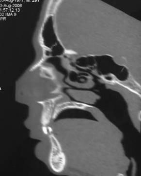
6 Anatomy-Axial


7 Anatomy-Axial


8 Anatomy-Axial

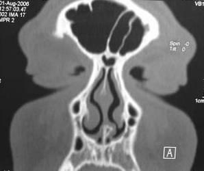
9 Anatomy- Coronal


10 Anatomy- Coronal


11 Anatomy- Coronal


12 Anatomy- Coronal


13 Anatomy- Coronal

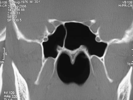
14 Anatomy details Midline nasal septum with two nasal cavities Three nasal conchae on each side The middle meatus [superolateral to the middle concha] drains the maxillary, frontal and anterior ethmoid sinuses

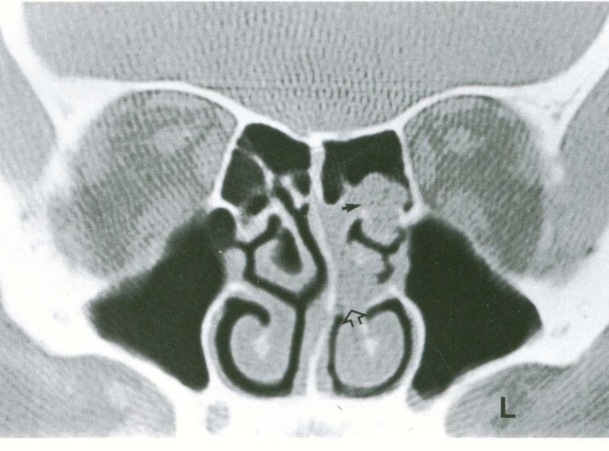
15 Maxillary sinus Largest PNS Pyramidal shaped with the apex pointing to zygoma Roof is formed by orbital floor. The infra-orbital canal contains infra-orbital N+A+V which exit through IOF Medial wall forms part of lateral wall of the nasal cavity IM

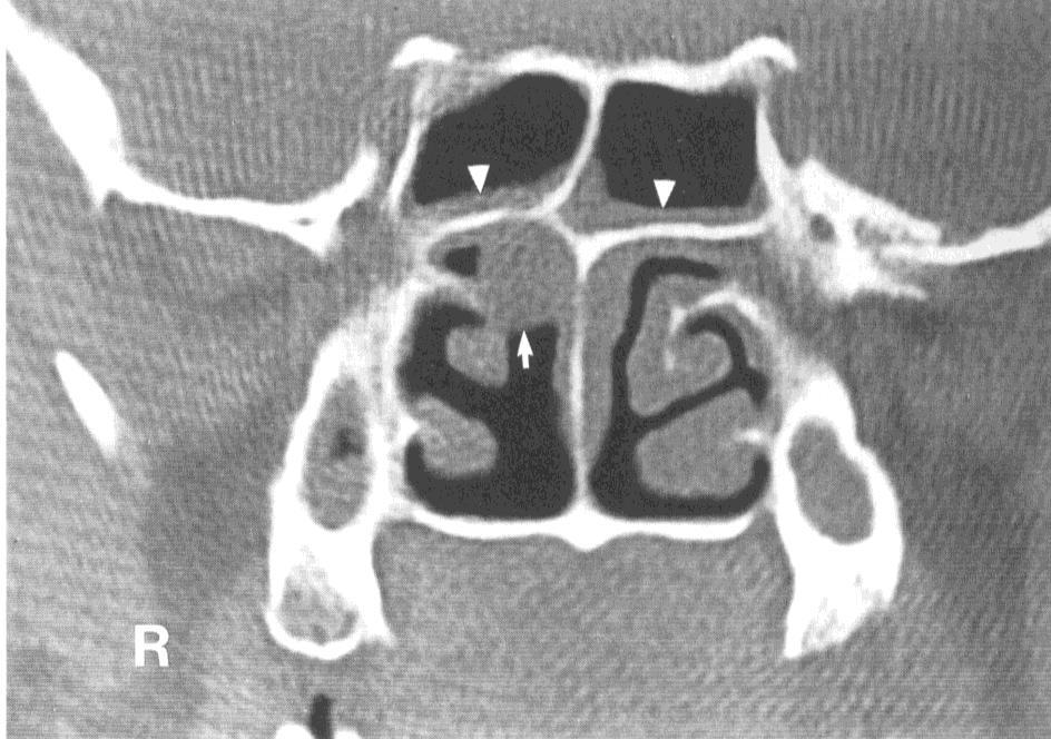
16 Maxillary sinus Ostium drains into the ethmoid infundibulum to the hiatus semilunaris to the middle meatus Uncinate process=the medial wall of the ethmoid infundibulum The lateral wall of the ethmoid infundibulum= orbital floor


17 Ethmoid Infundibulum Connects the maxillary sinus ostium to MM via semilunar hiatus

18 Ethmoid sinuses Gyrus rectus and olfactory bulb Ethmoid bone lies between the orbits. Horizontal plate: (Cribriform plate) Perforated for transmission of olfactory N. fibers from roof of nasal cavity Vertical plate: extends above the horizontal plate intracranially as the crista galli. The gyrus rectus + olfactory bulb rest upon the olfactory fossa on either sides of CG. Ethmoid Sinus : is separated from orbit by lamina papyracae

19 Ethmoid sinuses Anterior Group Smaller More numerous Drain in middle meatus Posterior Group Larger Fewer in no. Drain in superior meatus


20 Frontal sinus Best seen in the sagittal and coronal planes Drains into the anterior middle meatus via a canal which is an integral part of the ethmoid complex


21 Sphenoid sinus Relations Superior pituitary fossa + gland Inferior nasopharynx On both sides: cavernous sinus S


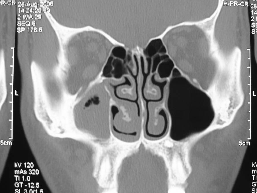
22 Anatomic variants Nasal septum Deviated to one side Nasal septum spur Pneumatized nasal septum drains into the sphenoid sinus

23 Middle turbinate Pneumatized turbinate = concha bullosa 4-15% Normal conchae Concha bullosa


24 Diagnostic role of plain Radiographs Screening for sinusitis, trauma, FB Preoperative planning Dental disease

25 adiographic signs of sinus disease Mucosal thickening Air- fluid level Total opacification Bone erosion Bone destruction Bone sclerosis Calcifications FB and fractures


26 Inflammatory lesions Sinusitis is a common medical problem Allergy, infection Inflammation swelling of the mucous membrane obstruction of the sinus drainage retained secretions secondary bacterial infection Acute, subacute and chronic types Air- fluid level= acute sinusitis Mucosal thickening= chronic sinusitis

27 Clinical note The imaging findings in sinusitis are non specific and should be clinically correlated 2-3mm mucosal thickening of the nasal cavity, anterior ethmoids and conchae is normal [a part of the nasal cycle] Opacified sinus in the childhood does not indicate sinusitis unless clinically correlated Ethmoid infundibulum and frontal recess are critical sites for mucosal thickening Reactive bone sclerosis of the sinus wall may suggest chronic sinusitis


28 Mucosal thickening compromising the osteomeatal units
29 Retention cyst Submucosal accumulation of serous fluid Smooth dome- shaped structure Usually in the inferior aspect of the maxillary sinus Polyp and cyst cannot be differentiated by imaging


30 Retention cysts Retention cyst + penumatized anterior clinoid Retention cyst + infraorbital foramen


31 Sinonasal polyposis Mucosal hypertrophy with sub mucosal fluid Allergy, inflammation, infection Imaging findings in Sinonasal polyposis Expanded nasal fossae filled with masses Opaque sinuses may be expanded Bone erosion may be seen


32 Sinonasal polyposis

![infection] Imaging findings of other](/docs-images/85/92777504/images/33-1.jpg "polyps in the nose Evidences of bone")
33 Sinus polyps versus cysts Clinical history [allergy, inflammation, infection] Imaging findings of other polyps in the nose Evidences of bone erosion MRI
34 Antro choanal polyp Polypoid lesion in the maxillary antrum The antrum is totally opaque The ostium in expanded The lesion extends into the nasal cavity May extend into the nasopharynx An expanding mass in the maxillary antrum and nasal fossa with characteristic widening of the ostium and absence of ethmoidal involvement to differentiate from inverted papilloma


35 Antro choanal polyp


36 Mucoceles Expanding lesions due to osteal obstruction Inflammation, trauma, tumors osteal obstruction Frontal sinuses 65%, ethmoids 25%, maxillary 10% Imaging findings Opaque expanded sinus Thin or eroded bony margins Marginal contrast enhancement Low signal in T1, high signal in T2 WIs


37 Maxillary mucocele


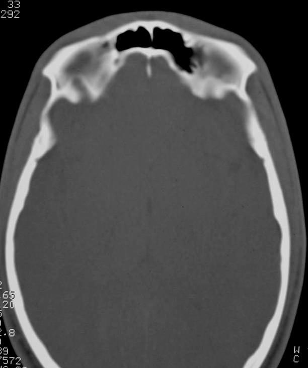
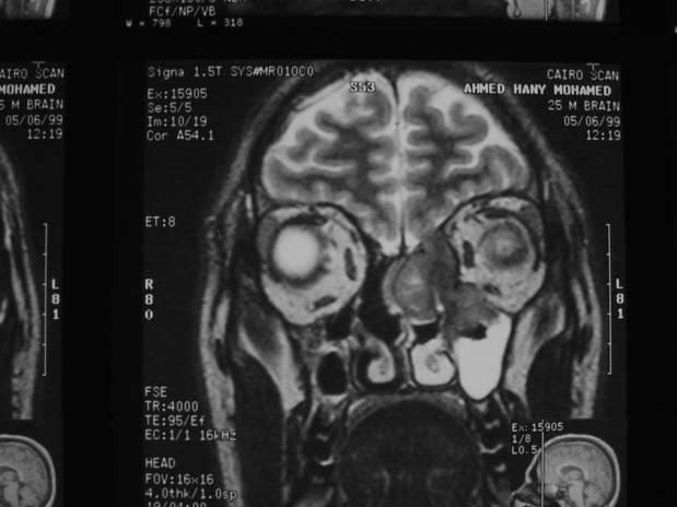
38 Fronto-ethmoidal mucocele F 30Y


39 Fungal sinusitis Non invasive Invasive Allergic Children & immune competent Unilateral or bilateral sinusitis Hyperdensities within the sinuses Surgical debridement + steroids Fungal ball Immune competent Sinusitis + calcifications Intact sinus walls Similar MR appearances Immune compromised Fulminate infection with bone erosion Intracranial & intraorbital extensions Cavernous sinus thrombosis M 13Y


40 Fungal sinusitis F 36Y


41 F 21Y Fungal sinusitis


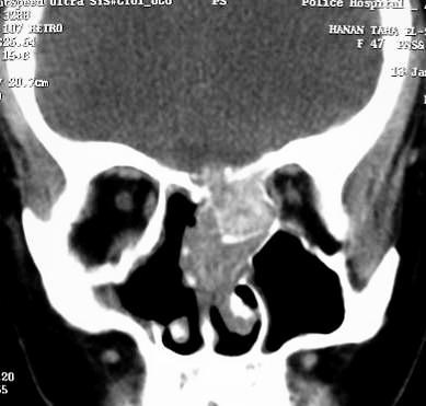

42 F 47Y Fungal sinusitis


43 Features suggestive of fungal sinusitis Hyper densities within the affected sinus on CT Intrasinus calcifications Marked signal loss in T2 WIs Sinusitis refractory to medical treatment Maxillary sinusitis extending to the cheek F 19Y


44 Allergic fungal sinusitis M 13Y


45 Intracranial complications of sinusitis Frontal sinuses are the most frequent source MRI is more sensitive than CT Meningitis Epidural, subdural, cerebral abscess Cavernous sinus thrombosis


46 Orbital complications of sinusitis Ethmoidal sinuses are the most frequent source M 48Y Preseptal and orbital cellulites Subperiosteal and orbital abscesses Cavernous sinus thrombosis may complicate sphenoid sinusitis


47 Sinonasal neoplastic lesions Osteoma F 8Y Most common in the frontal, then ethomid sinuses Usually asymptomatic Obstruction of sinus drainage sinusitis Dense bony mass with no enhancement Gardener s Syndrome = osteomas + colonic polyposis


48 Osteoma


49 Inverted papilloma 75% of sinus papillomas More in males years old Unilateral arising from the lateral nasal wall near MT May extend into any sinus, but maxillary is common Although histologically benign, they are locally aggressive Frequently multicentric 13% are associated with coexisting carcinoma


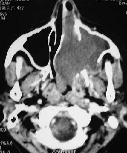
50 Inverted papilloma M 21Y


51 Inverted papilloma versus antro-choanal polyp


52 Nasopharyngeal Angiofibroma Hyer-vascular lesion Occurs almost exclusively in boys Arises near the sphenoplatine foramen Epistaxis is the main presentation


53 Nasopharyngeal Angiofibroma M 18Y

54 Sinonasal Malignancy Rare lesions representing 3% of head and neck neoplasms 65% in the maxillary antrum, 20% ethmoids, 15% nasal cavity 80% of all sinus malignancies are squamous cell type Other neoplasms include lymphoma, melanoma, fibrous histeocytoma,.. Clinical Findings Symptoms similar to chronic sinusitis Rhinorrhea, facial pain or paraesthesia Loosing of teeth, exophthalmos, epiphora Blood stained nasal discharge


55 CT Findings in malignancy M 43Y Soft tissue mass inside the affected sinus Homogenous or heterogenous enhancement Bone erosion or destruction Differentiation between tumor extension and retained secretions Matrix calcification in chondrosarcoma and osteosarcoma


56 M 40Y CHONDROSARCOMA OF THE HARD PALATE

57 Adenoid cystic carcinoma Commonly arise from the minor salivary glands Represent 10% of paranasal sinus malignancies Slow growing locally aggressive tumor Recurrence may occur years after treatment Perineural spread is the diagnostic hallmark of this lesion Distant blood born metastases to the lungs and bones Lymphatic spread is uncommon Adenoid cystic carcinoma of the maxillary sinus+ muscle atrophy secondary to 5 th nerve involvement


58 Ethmoidal carcinoma M 43Y


59 Nasopharyngeal Carcinoma Extensions Anteriorly nasal fossa, maxillary sinus, infratemporal fossa Posteriorly prevertebral muscles, carotid sheath Laterally parapharyngeal space, mastecator space
60 QUIZ? 2 3 1
61 QUIZ?


62 QUIZ? Sinusitis with concha bullosa

63 QUIZ? Retention cyst with deviated septum


64 QUIZ? Fungal sinusitis

65 QUIZ? Bilateral concha bullosa

66 QUIZ? Antro-choanal polyp


67 QUIZ? M 17Y Nasopharyngeal angiofibroma with sinusitis

68 QUIZ? Sino-nasal polyposis

69 QUIZ? Ethmoidal carcinoma invading the orbit
70 QUIZ? Retention cyst

71 QUIZ? Frontal sinusitis


72 QUIZ? Fungal sinusitis

73 Thank you سبحانك اللهم و بحمدك نشهد ان ال اله اال انت نستغفرك و نتوب اليك


74 Osteoma and sphenoid mucocele M 52Y


75 Sinonasal Malignancy CT Findings Assessment of metastatic lymphadenopathy in the submandibular, lateral retropharyngeal and upper deep cervical regions Post operative follow up Differentiation of tumor recurrence from postoperative sequlae Differentiation of bone destruction from surgical defects M 50Y RECURRENT FIBROSARCOMA OF THE MAXILLA


76 Sinonasal Malignancy CT diagnostic value Tumor extensions Orbit, infratemporal fossa, pterygopalatine fossa Other sinuses specially the sphenoid and frontal sinuses Intracranial extension CT guided needle biopsy


77 Fibrosarcoma of the maxilla


78 Olfactory neuroblastoma Arises from olfactory epithelium in the nasal cavity Males= Females and years old Epistaxis and nasal obstruction Metastatic spread to cervical lymph nodes, lung, liver, globe, parotid gland,..


79 Olfactory neuroblastoma


80


81


82


83

84


85


86
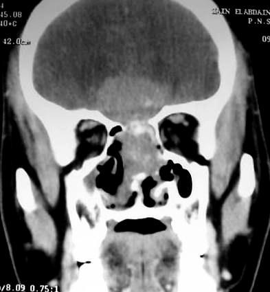
87


88


89 Blow out fracture

90


91


92

93 bullosa


94


95 F 43Y


96


97 4 cases of sinus carcinoma


98 aneamia Sinusitis and orbital changes TB sinusitis


99 Normal Variants


100 Fungus 2 cases


101 Fungus


102 Rahbdomyosarcoma biopsy Carcinoma ethmoid Ameloblastic fibrosarcoma


103 Fibrosarcoma maxilla NHL

104

105

106

107


108

109

110

111
112
113

114

115

116


117

118

119 SPHENOID SINUS The sphenoid bone has several processes A) Lesser wing + Ant. Clinoid process Optic strut Sphenoethmoidal recess Sphenopalatine foramen are attached anterioly to planum sphenoidal. ant. clinoid process houses optic canal and forms superior margin of SOF Sphenoethmoidal recess communicates with pterygopalatine Fossa through sphenopalatine foramen Optic Struct separates OF from SOF


120 SPHENOID SINUS B) Greater Wing: Forms part of orbital wall+ inf. margin SOF Gives origin pterygoid processes which forms post.border of pterygopalatine fossa. Makes a major contribution to anteromedial part of middle cranial fossa. Post. it is perforated by FO+ FS close to petrous apex

121

122

123

124

125


126

127


128


129 Anterior Group H Haller s cells: lat. extension of ant. ethmoid into Inferomedial margin of orbit. May narrow ostium of maxillary sinus. Ethmoid Bulla: largest most constantly present When enlarge may obstruct osteomeatal complex
130 Dentigerous cyst Cyst surrounding the crown of unerupted permanent tooth More in the mandible than in the maxilla Rapidly growing lesion in young adult Usually unilocular with sclerotic margin


131


132 Ethmomaxillary plate Anatomic Variants: Accessory ostia are small defects that open directly into MM. They do not normally assist in the drainage of the maxillary sinus. Ethmomaxillary Plate: Thin plate of bone separating superomed margin of maxillary sinus from post. ethmoid air cells

133 Posterior Group Lateral extension of post. Ethmoid into maxilla Is called the ethmomaxillary sinus


134 M 14 M Orbital cellulitis
Boundaries Septum Turbinates & Meati Lamellae Drainage Pathways Variants
 The Fastest 20 Minutes in Michelle A. Michel, MD Professor of Radiology and Otolaryngology Medical College of Wisconsin, Milwaukee Overview Nasal cavity Anterior skull base Ostiomeatal complex Frontal
Head&Neck Imaging. ssregypt.com. Parapharyngeal Spaces. Mamdouh mahfouz MD
 Head&Neck Imaging Parapharyngeal Spaces ssregypt.com Mamdouh mahfouz MD mamdouh.m5@gmail.com Definitio n Fat filled triangular space lateral the pharynx Extends from the skull base to the oropharynx Parapharyngeal
Radiological anatomy of frontal sinus By drtbalu
 2009 Radiological anatomy of frontal sinus By drtbalu Anatomy of frontal sinus is highly variable. Precise understanding of these variables will help a surgeon to avoid unnecessary complications during
Anatomic Relations Summary. Done by: Sohayyla Yasin Dababseh
 Anatomic Relations Summary Done by: Sohayyla Yasin Dababseh Anatomic Relations Lecture 1 Part-1 - The medial wall of the nose is the septum. - The vestibule lies directly inside the nostrils (Nares). -
*in general the blood supply of the nose comes from branches of the internal and external carotid arteries.
 In the previous lecture we talked about the anatomy of the nasal cavity, today we will talk about its blood supply, venous drainage, innervations, and finally about the paranasal sinuses. When we describe
Nasal region. cartilages: septal cartilage (l); lateral nasal cartilage (2); greater alar cartilages (2); lesser alar cartilages (?
; lateral nasal cartilage (2); greater alar cartilages (2); lesser alar cartilages (?") Nasal region skull bones: nasal and frontal processes of maxilla cartilages: septal cartilage (l); lateral nasal cartilage (2); greater alar cartilages (2); lesser alar cartilages (?) 1 Nasal cavity Roof
Nasal region skull bones: nasal and frontal processes of maxilla cartilages: septal cartilage (l); lateral nasal cartilage (2); greater alar cartilages (2); lesser alar cartilages (?) 1 Nasal cavity Roof
Bisection of Head & Nasal Cavity 頭部對切以及鼻腔. 解剖學科馮琮涵副教授 分機
 Bisection of Head & Nasal Cavity 頭部對切以及鼻腔 解剖學科馮琮涵副教授 分機 3250 E-mail: thfong@tmu.edu.tw Outline: The structure of nose The concha and meatus in nasal cavity The openings of paranasal sinuses Canals, foramens
Omran Saeed. Luma Taweel. Mohammad Almohtaseb. 1 P a g e
 2 Omran Saeed Luma Taweel Mohammad Almohtaseb 1 P a g e I didn t include all the photos in this sheet in order to keep it as small as possible so if you need more clarification please refer to slides In
Juvenile Angiofibroma
 Juvenile Angiofibroma Disclaimer The pictures used in this presentation have been obtained from a number of sources. Their use is purely for academic and teaching purposes. The contents of this presentation
Anatomy #1; Respiratory Nose and the Nasal Cavity December 1st, 2013
 Note #1: the doctor skipped some slides in the lecture. Those slides are not included in this sheet and so you will have to review the slides to study them. The reason they were not included is because
Dr. Sami Zaqout, IUG Medical School
 The skull The skull is composed of several separate bones united at immobile joints called sutures. Exceptions? Frontal bone Occipital bone Vault Cranium Sphenoid bone Zygomatic bones Base Ethmoid bone
FESS imaging - the role of MDCT
 FESS imaging - the role of MDCT Poster No.: C-0179 Congress: ECR 2013 Type: Educational Exhibit Authors: J. Plascak, K. Makaruha, B. Klasic, L. Kavur, V. Vidjak; Zagreb/HR Keywords: Image verification,
SINUS ANATOMY AND FUNCTION
 EMBRYOLOGY AND DEVELOPMENT SINUS ANATOMY AND FUNCTION -4 th week gestation: -frontonasal process identified, arises over developing forebrain -ectodermal -contributes to nasal capsule -9 th and 10 th week
PTERYGOPALATINE FOSSA
 PTERYGOPALATINE FOSSA Outline Anatomical Structure and Boundaries Foramina and Communications with other spaces and cavities Contents Pterygopalatine Ganglion Especial emphasis on certain arteries and
SINONASAL IMAGING. Kim O. Learned, MD. Assistant Professor Department of Radiology/Division of Neuroradiology University of Pennsylvania Health System
 SINONASAL IMAGING Kim O. Learned, MD Assistant Professor Department of Radiology/Division of Neuroradiology University of Pennsylvania Health System REVIEWS Key Anatomy: Sinus Drainage Pathways Practical
Tumors of the Paranasal Sinuses:
 Tumors of the Paranasal Sinuses: Approaches to Diagnostic Imaging Nir J. Harish September 2007 Head and Neck Cancers Oral cavity Pharynx Larynx Nasal cavity Paranasal sinuses Salivary glands Incidence
Skull-2. Norma Basalis Interna Norma Basalis Externa. Dr. Heba Kalbouneh Associate Professor of Anatomy and Histology
 Skull-2 Norma Basalis Interna Norma Basalis Externa Dr. Heba Kalbouneh Associate Professor of Anatomy and Histology Norma basalis interna Base of the skull- superior view The interior of the base of the
Communication issue - What should the radiologist report before functional endoscopic sinus surgery
 Communication issue - What should the radiologist report before functional endoscopic sinus surgery Poster No.: C-0509 Congress: ECR 2015 Type: Educational Exhibit Authors: A. M. Dobra 1, C. A. Badiu 1,
Chapter 7: Head & Neck
 Chapter 7: Head & Neck Osteology I. Overview A. Skull The cranium is composed of irregularly shaped bones that are fused together at unique joints called sutures The skull provides durable protection from
 www.oralradiologists.com CONE BEAM CT REPORT CASE ---- Case Information Referring Doctor: - Patient Name: - Scan Date: December 1, 2015 Patient DOB: - Reason for Exam: - Study Details: icat Flex, 160x160x112
Maxilla, ORBIT and infratemporal fossa. Neophytos C Demetriades MD, DDS, MSc Associate professor European University of Cyprus School of Medicine
 Maxilla, ORBIT and infratemporal fossa Neophytos C Demetriades MD, DDS, MSc Associate professor European University of Cyprus School of Medicine MAXILLA Superior, middle, and inferior meatus Frontal sinus
Chapter Five. 1 of 8 11/3/2008 2:52 PM.
 1 of 8 11/3/2008 2:52 PM Email : myousefmian@hotmail.com Chapter Five FRONT COVER Introduction Acknowledgement CHAPTERS Chapter One Chapter Two Chapter Three Chapter Four Chapter Five Chapter Six Chapter
Sinonasal Tumors. Objectives. Objectives. Incidence of Paranasal Sinus Tumors. Demographics of Paranasal Sinus Tumors. Paranasal Sinus Tumors
 Sinonasal Tumors Objectives Incidence and demographics of sinonasal tumors Separating tumors from inflammatory changes Common and notable histologic types of sinonasal tumors Staging of sinonasal tumors
AJCC Staging of Head & Neck Cancer (7 th edition, 2010) -LIP & ORAL CAVITY-
 -LIP & ORAL CAVITY-") TX: primary tumor cannot be assessed T0: no evidence of primary tumor Tis: carcinoma in situ. T1: tumor is 2 cm or smaller AJCC Staging of Head & Neck Cancer (7 th edition, 2010) -LIP & ORAL CAVITY- T2:
TX: primary tumor cannot be assessed T0: no evidence of primary tumor Tis: carcinoma in situ. T1: tumor is 2 cm or smaller AJCC Staging of Head & Neck Cancer (7 th edition, 2010) -LIP & ORAL CAVITY- T2:
Bones of the skull & face
 Bones of the skull & face Cranium= brain case or helmet Copyright The McGraw-Hill Companies, Inc. Permission required for reproduction or display. The cranium is composed of eight bones : frontal Occipital
MAXILLA, ORBIT & PTERYGOPALATINE FOSSA. Neophytos C Demetriades MD, DDS, MSc Associate professor European University of Cyprus School of Medicine
 MAXILLA, ORBIT & PTERYGOPALATINE FOSSA Neophytos C Demetriades MD, DDS, MSc Associate professor European University of Cyprus School of Medicine Maxilla MAXILLA Superior, middle, and inferior meatus Frontal
The Nose and Sinuses. Ophir Ilan, MD, PhD Department of Otolaryngology/Head&Neck surgery Hadassah University Hospital
 The Nose and Sinuses Ophir Ilan, MD, PhD Department of Otolaryngology/Head&Neck surgery Hadassah University Hospital Nasal Mucociliary System Function of the Nasal Mucosa warming and humidifying the
OPEN ACCESS ATLAS OF OTOLARYNGOLOGY, HEAD & NECK OPERATIVE SURGERY
 OPEN ACCESS ATLAS OF OTOLARYNGOLOGY, HEAD & NECK OPERATIVE SURGERY INFERIOR MAXILLECTOMY Tumours of the hard palate and superior alveolus may be resected by inferior maxillectomy (Figure 1). A Le Fort
Imaging of the Paranasal Sinuses
 14. Sommerschule Imaging of the Paranasal Sinuses Bettlach 24.08.2018 Christoph Schlegel Conventional Radiology NNH-Status: okzipito-frontal: frontal sinus, anterior ethmoid okzipito-nasal : maxillary
Professor Dr.Muhammad Ajmal Dr.Tehmina Nazir. HOLY FAMILY HOSPITAL Rawalpindi
 Professor Dr.Muhammad Ajmal Dr.Tehmina Nazir HOLY FAMILY HOSPITAL Rawalpindi SCHEME OF PRESENTATION PLAIN X-RAYS CT SCAN MRI CONCLUSION IMAGING MODALITIES PLAIN X-RAYS CT SCAN MRI OCCIPITOMENTAL/WATER
Dr.Ban I.S. head & neck anatomy 2 nd y جامعة تكريت كلية طب االسنان مادة التشريح املرحلة الثانية أ.م.د. بان امساعيل صديق 6102/6102
 جامعة تكريت كلية طب االسنان مادة التشريح املرحلة الثانية أ.م.د. بان امساعيل صديق 6102/6102 Pterygopalatine fossa: The pterygopalatine fossa is a cone-shaped depression, It is located between the maxilla,
Head & Neck Clinical Sub Group. Network Agreed Imaging Guidelines for UAT and Thyroid Cancer. Measure Nos: 11-1C-105i & 11-1C-106i
 Greater Manchester, Lancashire & South Cumbria Strategic Clinical Network & Senate Head & Neck Clinical Sub Group Network Agreed Imaging Guidelines for UAT and Thyroid Cancer Measure Nos: 11-1C-105i &
Case Studies in the Skull Base
 Case Studies in the Skull Base Amy C Tsai, MD Neuroradiology Fellow Department of Radiology and Imaging Sciences University of Utah Health Sciences Center Salt Lake City, Utah, USA No disclosures related
Skull-2. Norma Basalis Interna. Dr. Heba Kalbouneh Assistant Professor of Anatomy and Histology
 Skull-2 Norma Basalis Interna Dr. Heba Kalbouneh Assistant Professor of Anatomy and Histology Norma basalis interna Base of the skull- superior view The interior of the base of the skull is divided into
The orbit-1. Dr. Heba Kalbouneh Assistant Professor of Anatomy and Histology
 The orbit-1 Dr. Heba Kalbouneh Assistant Professor of Anatomy and Histology Orbital plate of frontal bone Orbital plate of ethmoid bone Lesser wing of sphenoid Greater wing of sphenoid Lacrimal bone Orbital
Clinical and imagistic correlations in the inflammatory pathology of nasosinusal cavities
 Romanian Journal of Rhinology, Volume 8, No. 29, January-March 2018 ORIGINAL STUDY DOI: 10.2478/rjr-2018-0003 Clinical and imagistic correlations in the inflammatory pathology of nasosinusal cavities Emilia
Nose & Mouth OUTLINE. Nose. - Nasal Cavity & Its Walls. - Paranasal Sinuses. - Neurovascular Structures. Mouth. - Oral Cavity & Its Contents
 Dept. of Human Anatomy, Si Chuan University Zhou hongying eaglezhyxzy@163.com Nose & Mouth OUTLINE Nose - Nasal Cavity & Its Walls - Paranasal Sinuses - Neurovascular Structures Mouth - Oral Cavity & Its
We are IntechOpen, the world s leading publisher of Open Access books Built by scientists, for scientists. International authors and editors
 We are IntechOpen, the world s leading publisher of Open Access books Built by scientists, for scientists 3,350 108,000 1.7 M Open access books available International authors and editors Downloads Our
Nasal Polyposis. DEPARTMENT OF ENT K.S.Hegde Medical Academy Deralakatte, Mangalore
 Nasal Polyposis DEPARTMENT OF ENT K.S.Hegde Medical Academy Deralakatte, Mangalore Def: INTRODUCTION Chronic inflammatory disease of the mucous membrane in the nose & PNS, presenting as pedunculated smooth
The cribriform plate. ethmoid bone. Ethmoid bone consists from: 1) A horizontal cribriform plate. 2) A perpendicular plate. 3) Two lateral labyrinths.
 A horizontal cribriform plate. 2) A perpendicular plate. 3) Two lateral labyrinths.") ethmoid bone Ethmoid bone consists from: 1) A horizontal cribriform plate. 2) A perpendicular plate. 3) Two lateral labyrinths. The cribriform plate 1) Connect the two labyrinths to the perpendicular plate.
ethmoid bone Ethmoid bone consists from: 1) A horizontal cribriform plate. 2) A perpendicular plate. 3) Two lateral labyrinths. The cribriform plate 1) Connect the two labyrinths to the perpendicular plate.
University of Palestine. Midterm Exam 2013/2014 Total Grade:
 Course No: DNTS2208 Course Title: Head and Neck Anatomy Date: 09/11/2013 No. of Questions: (50) Time: 1hour Using Calculator (No) University of Palestine Midterm Exam 2013/2014 Total Grade: Instructor
Bones Ethmoid bone Inferior nasal concha Lacrimal bone Maxilla Nasal bone Palatine bone Vomer Zygomatic bone Mandible
 splanchnocranium - Consists of part of skull that is derived from branchial arches - The facial bones are the bones of the anterior and lower human skull Bones Ethmoid bone Inferior nasal concha Lacrimal
Skull Base Danger Zones in FESS
 Skull Base Danger Zones in FESS Poster No.: C-2278 Congress: ECR 2014 Type: Educational Exhibit Authors: L. Renza Lozada, R. Carreño Gonzalez, G. Quintana Sanchez, 1 2 1 1 1 2 R. E. Figueroa ; Malaga/ES,
Neuroradiology Case of the Day
 Neuroradiology Case of the Day 76 th CAR Annual Meeting, Montreal, Quebec April 27, 2013 Eugene Yu, MD Assistant Professor of Radiology and Otolaryngology-Head and Neck Surgery Head and Neck Imaging Princess
Infratemporal fossa: Tikrit University college of Dentistry Dr.Ban I.S. head & neck Anatomy 2 nd y.
 Infratemporal fossa: This is a space lying beneath the base of the skull between the lateral wall of the pharynx and the ramus of the mandible. It is also referred to as the parapharyngeal or lateral pharyngeal
Skeletal System -Axial System. Chapter 7 Part A
 Skeletal System -Axial System Chapter 7 Part A Skeleton Learn: Names of the s. Identify specific landmarks that allow: Bones to fit into each other, Organs to fit into the cavities, Muscles to attach,
 www.oralradiologists.com CONE BEAM CT REPORT CASE XXXX Patient information Patient Name: - Referring Doctor: - Patient DOB: - Scan Date: [Start date] Reason for Exam: Maxillary facial pain Doctor Notes:
Paranasal Sinuses: Neoplastic Lesions
 Pravin Mundada Department of Radiology, Geneva University Hospital, Switzerland Paranasal Sinuses: Neoplastic Lesions ESHNR 2017 Lisbon, Portugal Layout of the presentation Clinical & imaging features
Mohammad Hisham Al-Mohtaseb. Lina Mansour. Reyad Jabiri. 0 P a g e
 2 Mohammad Hisham Al-Mohtaseb Lina Mansour Reyad Jabiri 0 P a g e This is only correction for the last year sheet according to our record. If you already studied this sheet just read the yellow notes which
Chapter 7 Part A The Skeleton
 Chapter 7 Part A The Skeleton Why This Matters Understanding the anatomy of the skeleton enables you to anticipate problems such as pelvic dimensions that may affect labor and delivery The Skeleton The
Temporal region. temporal & infratemporal fossae. Zhou Hong Ying Dept. of Anatomy
 Temporal region temporal & infratemporal fossae Zhou Hong Ying Dept. of Anatomy Temporal region is divided by zygomatic arch into temporal & infratemporal fossae. Temporal Fossa Infratemporal fossa Temporal
Dr. Sami Zaqout Faculty of Medicine IUG
 The Nose External Nose Nasal Cavity External Nose Blood and Nerve Supplies of the External Nose Blood Supply of the External Nose The skin of the external nose Branches of the ophthalmic and the maxillary
Major Anatomic Components of the Orbit
 Major Anatomic Components of the Orbit 1. Osseous Framework 2. Globe 3. Optic nerve and sheath 4. Extraocular muscles Bony Orbit Seven Bones Frontal bone Zygomatic bone Maxillary bone Ethmoid bone Sphenoid
Temporal fossa Infratemporal fossa Pterygopalatine fossa Terminal branches of external carotid artery Pterygoid venous plexus
 Outline of content Temporal fossa Infratemporal fossa Pterygopalatine fossa Terminal branches of external carotid artery Pterygoid venous plexus Boundary Content Communication Mandibular division of trigeminal
The sebaceous glands (glands of Zeis) open directly into the eyelash follicles, ciliary glands (glands of Moll) are modified sweat glands that open
 open directly into the eyelash follicles, ciliary glands (glands of Moll) are modified sweat glands that open") The Orbital Region The orbits are a pair of bony cavities that contain the eyeballs; their associated muscles, nerves, vessels, and fat; and most of the lacrimal apparatus upper eyelid is larger and more
The Orbital Region The orbits are a pair of bony cavities that contain the eyeballs; their associated muscles, nerves, vessels, and fat; and most of the lacrimal apparatus upper eyelid is larger and more
Commen Nose Diseases
 Commen Nose Diseases Symptoms List: Nasal obstruction. Nasal discharge: Anterior (Rhinorrhea). Posterior (Postnasal discharge). Epistaxis. Hyposmia and Anosmia. Headache. Snoring. Nasal Obstruction Definition:
Introduction to Local Anesthesia and Review of Anatomy
 5-Sep Introduction and Anatomy Review 12-Sep Neurophysiology and Pain 19-Sep Physiology and Pharmacology part 1 26-Sep Physiology and Pharmacology part 2 Introduction to Local Anesthesia and Review of
Destructive Giant Maxillary Sinus Mucocele: A Case Report
 Destructive Giant Maxillary Sinus Mucocele: A Vahit Mutlu 1, Ozgur Yoruk 1, Ozalkan Ozkan 2 1 Atatürk University Faculty of Medicine, Department of Ears, Nose and Throat, Erzurum, Turkey 2 Erzincan University
The Role of Computed Tomography in the Evaluation of Paranasal Sinuses Lesions
 ORIGINAL ARTICLE The Role of Computed Tomography in the Evaluation of Paranasal Sinuses Lesions Bhumikaben P. Suthar 1 *, Divya Vaidya 2, Pukhraj P. Suthar 3. 1 Assistant Professor, 2 Third Year Resident,
The Role of Computed Tomography in the Evaluation of Paranasal Sinuses Lesions
 ORIGINAL ARTICLE The Role of Computed Tomography in the Evaluation of Paranasal Sinuses Lesions Bhumikaben P. Suthar 1 *, Divya Vaid 2, Pukhraj P. Suthar 3. 1 Assistant Professor, 2 Third Year Resident,
TRAUMA TO THE FACE AND MOUTH
 Dr.Yahya A. Ali 3/10/2012 F.I.C.M.S TRAUMA TO THE FACE AND MOUTH Bailey & Love s 25 th edition Injuries to the orofacial region are common, but the majority are relatively minor in nature. A few are major
Cranium Facial bones. Sternum Rib
 Figure 7.1 The human skeleton. Skull Thoracic cage (ribs and sternum) Cranium Facial bones Sternum Rib Bones of pectoral girdle Vertebral column Sacrum Vertebra Bones of pelvic girdle (a) Anterior view
Bony orbit Roof The orbital plate of the frontal bone Lateral wall: the zygomatic bone and the greater wing of the sphenoid
 Bony orbit Roof: Formed by: The orbital plate of the frontal bone, which separates the orbital cavity from the anterior cranial fossa and the frontal lobe of the cerebral hemisphere Lateral wall: Formed
CT OF THE PARANASAL SINUSES : NORMAL ANATOMY, VARIANTS AND PATHOLOGY
 Journal of Optoelectronics and Biomedical Materials Vol.2 Issue 4, October-December 2010, p. 281 289 CT OF THE PARANASAL SINUSES : NORMAL ANATOMY, VARIANTS AND PATHOLOGY AMIT N D DWIVEDI *, KAPIL KUMAR
Three-Dimensional Volumetric Display of the Nasal Ostiomeatal Channels and Paranasal Sinuses
 Downloaded from www.ajronline.org by 37.44.202.192 on 12/22/17 from IP address 37.44.202.192. Copyright RRS. For personal use only; all rights reserved Three-Dimensional Volumetric Display of the Nasal
Conventional Sinus Surgery Vs Fess
 IOSR Journal of Dental and Medical Sciences (IOSR-JDMS) e-issn: 2279-0853, p-issn: 2279-0861.Volume 16, Issue 7 Ver. III (July. 2017), PP 44-51 www.iosrjournals.org Conventional Sinus Surgery Vs Fess *
SKULL AS A WHOLE + ANTERIOR CRANIAL FOSSA
 SKULL AS A WHOLE + ANTERIOR CRANIAL FOSSA LEARNING OBJECTIVES At the end of this lecture, the student should be able to know: Parts of skeleton (axial and appendicular) Parts of skull Sutures of skull
FRONTAL SINUPLASTY P R E P A R E D A N D P R E S E N T E D B Y : D R. Y A H Y A F A G E E H R 4 16/ 12/ 2013
 FRONTAL SINUPLASTY P R E P A R E D A N D P R E S E N T E D B Y : D R. Y A H Y A F A G E E H R 4 16/ 12/ 2013 ANATOMY: FRONTAL SINUS Not present at birth Starts developing at 4 years Radiographically visualized
Is there a role of CT in the evaluation of Proptosis
 International Journal of scientific research and management (IJSRM) Volume 3 Issue 4 Pages 2662-2666 2015 \ Website: www.ijsrm.in ISSN (e): 2321-3418 Is there a role of CT in the evaluation of Proptosis
Reasons for Failure and Surgical Revisions. Stil Kountakis, MD, PhD Professor and Chief, Division of Rhinology
 Reasons for Failure and Surgical Revisions Stil Kountakis, MD, PhD Professor and Chief, Division of Rhinology Medical College of Georgia of Georgia Regents University Department of Otolaryngology / Head
Anatomy and Physiology. Bones, Sutures, Teeth, Processes and Foramina of the Human Skull
 Anatomy and Physiology Chapter 6 DRO Bones, Sutures, Teeth, Processes and Foramina of the Human Skull Name: Period: Bones of the Human Skull Bones of the Cranium: Frontal bone: forms the forehead and the
Malignant growth Maxilla management an analysis
 ISSN: 2250-0359 Volume 3 Issue 2 2013 Malignant growth Maxilla management an analysis *Balasubramanian Thiagarajan *Geetha Ramamoorthy *Stanley Medical College Abstract: Malignant tumors involving maxilla
Transnasal Endoscopic Sinonasal Surgery
 Reda kamel, Cadaveric dissection 1 Transnasal Endoscopic Sinonasal Surgery Cadaver Dissection Guide For Endoscopic Sinus Surgery Cairo University Egypt Reda Kamel Professor of Rhinology Cairo University
Computed tomographic evaluation of anatomical variations of paranasal sinus region
 International Journal of Research in Medical Sciences Gupta S et al. Int J Res Med Sci. 2016 Jul;4(7):2909-2913 www.msjonline.org pissn 2320-6071 eissn 2320-6012 Research Article DOI: http://dx.doi.org/10.18203/2320-6012.ijrms20161975
CT of Maxillofacial Fracture Patterns. CT of Maxillofacial Fracture Patterns
 CT of Maxillofacial Fracture Patterns CT of Maxillofacial Fracture Patterns Stuart E. Mirvis, M.D., FACR Department of Radiology University of Maryland School of Medicine Viking 1 1976 MGS 2001 Technology
NASAL SEPTUM ADENOID CYSTIC CARCINOMA: A CASE REPORT
 NASAL SEPTUM ADENOID CYSTIC CARCINOMA: A CASE REPORT Shu-Yu Tai, 1 Chen-Yu Chien, 2 Chih-Feng Tai, 2,4 Wen-Rei Kuo, 2,4 Wan-Ting Huang, 3 and Ling-Feng Wang 2,4 Departments of 1 Family Medicine, 2 Otolaryngology
Histopathology of Nasal Masses
 ORIGINAL ARTICLE Histopathology of Nasal Masses 1 Hemant Chopra, 2 Kapil Dua, 3 Neha Chopra, 4 Vikrant Mittal AIJCR Histopathology of Nasal Masses 1 Professor and Head, Dayanand Medical College, Ludhiana,
Skull basic structures. Neurocranium
 Assoc. Prof. Květuše Lovásová, M.V.D., PhD. Skull basic structures Skull consists of two groups of bones: neurocranium (bones forming the brain box) splanchnocranium (bones forming the facial skeleton)
MALIGNANT TUMOURS OF THE JAWS
 MALIGNANT TUMOURS OF THE JAWS MALIGNANT TUMOURS OF THE JAWS Squamous cell carcinoma Osteogenic sarcoma Chondrosarcoma Fibrosarcoma Malignant lymphomas (incl. Burkitt s) Multiple myeloma Ameloblastoma Secondary
Biology 218 Human Anatomy. Adapted from Martini Human Anatomy 7th ed. Chapter 6 The Skeletal System: Axial Division
 Adapted from Martini Human Anatomy 7th ed. Chapter 6 The Skeletal System: Axial Division Introduction The axial skeleton: Composed of bones along the central axis of the body Divided into three regions:
The future of health is digital
 Dated: XX/XX/XXXX Name: XXXXXXXX XXXXXXXXXXX Birth Date: XX/XX/XXXX Date of scan: XX/XX/XXXX Examination of the anatomical volume: The following structures are reviewed and evaluated for bilateral symmetry,
Skull and Axial Skeleton
 Published on Second Faculty of Medicine, Charles University (http://www.lf2.cuni.cz ) Skull and Axial Skeleton Description of the test The examination of the skull skeleton is in oral format. It consists
ORIGINAL ARTICLE RELATIONSHIP OF CONCHA BULLOSA WITH OSTEOMEATAL UNIT BLOCKAGE. TOMOGRAPHIC STUDY IN 200 PATIENTS.
 RELATIONSHIP OF CONCHA BULLOSA WITH OSTEOMEATAL UNIT BLOCKAGE. TOMOGRAPHIC STUDY IN 200 PATIENTS. Shrikrishna B H 1, Jyothi A C 2, Sanjay G 3, Sandeep Samson G 4. 1. Associate Professor, Department of
Raneen Hamdan. Raghad Abu Jebbeh. Mohammad almuhtaseb
 1 Raneen Hamdan Raghad Abu Jebbeh Mohammad almuhtaseb Introduction Respiratory System Organs: 1) Starting from the Nose (nasal cavity). 2) Pharynx (Nasopharynx, oropharynx and laryngopharynx, previously
Diagnostic Role of CT in the Evaluation of Proptosis
 IOSR Journal of Dental and Medical Sciences (IOSR-JDMS) e-issn: 2279-0853, p-issn: 2279-0861.Volume 14, Issue 4 Ver. IX (Apr. 2015), PP 25-31 www.iosrjournals.org Diagnostic Role of CT in the Evaluation
International Journal of Biological & Medical Research
 Int J Biol Med Res.2015;6(1):4775-4781 Contents lists available at BioMedSciDirect Publications International Journal of Biological & Medical Research Journal homepage: www.biomedscidirect.com BioMedSciDirect
Trigeminal Nerve Anatomy. Dr. Mohamed Rahil Ali
 Trigeminal Nerve Anatomy Dr. Mohamed Rahil Ali Trigeminal nerve Largest cranial nerve Mixed nerve Small motor root and large sensory root Motor root Nucleus of motor root present in the pons and medulla
Computed tomography road map of the paranasal sinuses for treatment planning
 Computed tomography road map of the paranasal sinuses for treatment planning Poster No.: C-2607 Congress: ECR 2013 Type: Educational Exhibit Authors: N. Schembri, A. S. Gatt, D. Ellul, J. Brunton; Dundee/UK
PROBLEM RECOMMENDATION
 PREVENTION (MINIMIZING) IN ENDOSCOPIC Steven D. Schaefer, MD Professor and Chair Department of Otolaryngology PREVENTION AND Intraoperative Hemorrhage Loss of Orientation Inability to Identify/Preserve
REVIEW OF HEAD AND NECK CRANIAL NERVES AND EVERYTHING ELSE
 REVIEW OF HEAD AND NECK CRANIAL NERVES AND EVERYTHING ELSE OLFACTORY NERVE CN I ANTERIOR CRANIAL FOSSA CRISTA GALLI OF ETHMOID OLFACTORY FORAMINA IN CRIBIFORM PLATE OF ETHMOID BONE CN I OLFACTORY NERVE
often the opposing teeth will manifest symptoms as well, due to extrusion of the tooth from increased pressure from the cyst.
 Mucous Retention Cysts of the Maxillary Sinus and Superiority of 3D Cone Beam CT Scans versus Traditional Panoramic Imaging Rebecca L Griffiths, BS, DMD Mucous retention cysts of the maxillary sinus are
Katya A. Shpilberg 1 Simon C. Daniel 1 Amish H. Doshi 1 William Lawson 2 Peter M. Som 1. Neuroradiology/Head and Neck Imaging Original Research
 Neuroradiology/Head and Neck Imaging Original Research Shpilberg et al. CT of Paranasal Sinuses and Nasal Cavity Neuroradiology/Head and Neck Imaging Original Research Katya A. Shpilberg 1 Simon C. Daniel
Chapter 2: Radiology of the nose and paranasal sinuses. P. D. Phelps. Sinuses. Plain radiography. Views for the nose. Standard sinus views
 Chapter 2: Radiology of the nose and paranasal sinuses P. D. Phelps Sinuses Although computerized tomographic scanning is now the best means of imaging the sinuses, cost and rationing of scan time in the
Chapter 7. Skeletal System
 Chapter 7 Skeletal System 1 Skull A. The skull is made up of 22 bones: 8 cranial bones, 13 facial bones, and the mandible. B. The Cranium encloses and protects the brain, provides attachments for muscles,
Spinal Imaging. ssregypt.com. Mamdouh Mahfouz MD
 Spinal Imaging Degenerative diseases ssregypt.com Mamdouh Mahfouz MD mamdouh.m5@gmail.com MRI Open MRI Closed Extremity MRI Dynamic MRI Dynamic MRI The bed rotates from Upright to Recumbent, stopping at
Morphological Changes of the Ethmoid and Maxillary Cavities after Endoscopic Sinus Surgery, A Quantitative Digital Analysis.
 Morphological Changes of the Ethmoid and Maxillary Cavities after Endoscopic Sinus Surgery, A Quantitative Digital Analysis Thesis Submitted for fulfillment of M.D. degree in Otorhinolaryngology By: Hisham
Trigeminal Nerve Worksheets, Distributions Page 1
 Trigeminal Nerve Worksheet #1 Distribution by Nerve Dr. Darren Hoffmann Dental Gross Anatomy, Spring 2013 We have drawn out each of the branches of CN V in lecture and you have an idea now for their basic
SKULL / CRANIUM BONES OF THE NEUROCRANIUM (7) Occipital bone (1) Sphenoid bone (1) Temporal bone (2) Frontal bone (1) Parietal bone (2)
 Occipital bone (1) Sphenoid bone (1) Temporal bone (2) Frontal bone (1) Parietal bone (2)") Important! 1. Memorizing these pages only does not guarantee the succesfull passing of the midterm test or the semifinal exam. 2. The handout has not been supervised, and I can not guarantee, that these
Important! 1. Memorizing these pages only does not guarantee the succesfull passing of the midterm test or the semifinal exam. 2. The handout has not been supervised, and I can not guarantee, that these
HEPATO-BILIARY IMAGING
 HEPATO-BILIARY IMAGING BY MAMDOUH MAHFOUZ MD PROF.OF RADIOLOGY CAIRO UNIVERSITY mamdouh.m5@gmail.com www.ssregypt.com CT ABDOMEN Indications Patient preparation Patient position Scanogram Fasting 4-6 hours