DISCLOSURE OF RELEVANT RELATIONSHIPS WITH INDUSTRY
|
|
|
- Erin Adams
- 5 years ago
- Views:
Transcription
1 DISCLOSURE OF RELEVANT RELATIONSHIPS WITH INDUSTRY Ian A. Maher, MD, FAAD, FACMS Assistant Professor of Dermatology Saint Louis University I HAVE NO RELEVANT CONFLICTS OF INTEREST
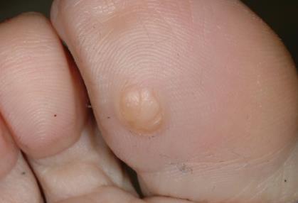
2 The most likely diagnosis is: A. Wart B. Malignant melanoma C. Basal cell carcinoma D. Verrucous carcinoma E. Corn
3
4 The most likely diagnosis is: A. Wart B. Malignant melanoma C. Basal cell carcinoma D. Verrucous carcinoma E. Corn
5 Verrucous carcinoma Low grade SCC Associated with high risk HPV subtypes (6b, 11, 16 and 18) Burrowed appearance Soles most common location Histology Minimal atypia Pushing border
6 Distractors *Wart Lesion very large, ulcerated and burrowed. Makes this diagnosis less likely *Melanoma Normally pigmented Can ulcerate *Basal cell carcinoma Abnormal on soles Normally pink, pearly Corn Hyperkeratotic papule at pressure point Accentuated skin lines
7 The most likely diagnosis is: A.Contact dermatitis B.Dermatomyositis C.Glomus tumor D.Hutchinson s sign E.Maffucci's syndrome Immersion Dermoscopy

8
9 The most likely diagnosis is: A.Contact dermatitis B.Dermatomyositis C.Glomus tumor D.Hutchinson s sign E.Maffucci's syndrome Immersion Dermoscopy

10 Associated with connective tissue diseases Dermatomyositis Systemic lupus erythematosus Scleroderma Dermatomyositis Cutaneous signs Heliotrope rash Shawl sign Gottron sign Proximal muscle weakness Dermatomyositis

11 Shawl like rash on chest and Gottron papules

12 Proximal Nailfold (Periungual) Telangiectasia Dermatomyositis Systemic lupus erythematosus Scleroderma Sausage-shaped loops of dilated capillaries and vascular dropout accentuated with oil immersion dermoscopy Immersion Dermoscopy

13 Distractors *Contact dermatitis erythema, hyperkeratosis, scaling and fissuring

14 Distractors Glomus tumor Painful distal or nailbed benign proliferation of glomus cells

15 Distractors Hutchinson s sign spread of pigmentation onto nailfold from subungual *melanoma
16 The patient presents for evaluation of these asymptomatic papules. He has a personal history of 3 episodes of pneumothorax. His family history is significant for renal carcinoma. What is the most likely diagnosis? a) Birt-Hogg-Dube b) Tuberous sclerosis c) Brooke-Spiegler d) Neurofibromatosis e) Cowden syndrome

17

18 The patient presents for evaluation of these asymptomatic papules. He has a personal history of 3 episodes of pneumothorax. His family history is significant for renal carcinoma. What is the most likely diagnosis? a) Birt-Hogg-Dube b) Tuberous sclerosis c) Brooke-Spiegler d) Neurofibromatosis e) Cowden syndrome
19 Birt-Hogg-Dube Mutation in folliculin, AD inheritance Skin manifestations Fibrofolliculomas Trichodiscomas Like many appendageal tumors these are non-descript flesh colored papules. Often around the nose Tend to be flat-topped Spontaneous pneumothorax Renal Carcinoma
20 *Tuberous sclerosis Distractors Skin findings Angiofibromas Renal findings angioleiomyomas Lung finding lymphangiomyomas CNS finding cortical tubers, seizures Brooke-Spiegler *Trichoepitheliomas Cylindromas
21 Distractors (cont.) *Type 1 Neurofibromatosis Neurofibromas *Cowden syndrome Skin findings tricholemmomas, mucosal papules Associated cancers breast, endometrium, thyroid
22 The most likely diagnosis is: A. Acne keloidalis nuchae B. Discoid lupus erythematosus C. Folliculitis decalvans D. Lichen planopilaris E. Trichotillomania Photo Courtesy Dr. M. Ioffreda

23 Photo Courtesy Dr. M. Ioffreda
24 The most likely diagnosis is: A. Acne keloidalis nuchae B. Discoid lupus erythematosus C. Folliculitis decalvans D. Lichen planopilaris E. Trichotillomania Photo Courtesy Dr. M. Ioffreda

25 Initially starts with chronic folliculitis and perifolliculitis of the posterior scalp and neck
 at")
26 Gradually develops into large plaques with alopecia and tufted hairs (like doll s hairs) at periphery
27 Acne keloidalis nuchae African Americans >>> Hispanics > Asians > Caucasians Men (20):Women (1), almost always after puberty Cause uncertain; possibly reaction to curling hairs penetrating skin Subcutaneous abscesses and draining sinuses may develop Treatment options Prevention: avoid close shave and irritating collars Topical retinoids or antibiotics Excision (may need serial excision)

28 Distractors *Discoid Lupus Erythematosus

29 Distractors *Folliculitis Decalvans

30 Distractors *Lichen Planopilaris

31 The most likely diagnosis is: A. Acquired digital fibrokeratoma B. Pyogenic granuloma C. Squamous cell carcinoma D. Supernumerary digit E. Verruca vulgaris

32
33 The most likely diagnosis is: A. Acquired digital fibrokeratoma B. Pyogenic granuloma C. Squamous cell carcinoma D. Supernumerary digit E. Verruca vulgaris
34 Acquired Digital Fibrokeratoma Usually a solitary, skin-colored to pink, slightly keratotic exophytic papulonodule with collarette of skin Location: fingers most common, but may be anywhere on acral skin
35

36 When periungual or subungual may be associated with tuberous sclerosus Koenen s Tumor


37 Distractors *Pyogenic Granuloma wet, red friable nodule


38 Distractors *Squamous cell carcinoma rare on the plantar foot or to not have any crusting or ulceration (would usually be verrucous carcinoma or Marjolin s ulcer)
 no thrombosed")
39 Distractors *Verruca Vulgaris (Wart) no thrombosed capillaries
40 The most likely diagnosis is: A. Berloque dermatitis B. Dermatomyositis C. Erythromelanosis follicularis faciei D. Poikiloderma of Civatte E. Subacute cutaneous lupus
41
42 The most likely diagnosis is: A. Berloque dermatitis B. Dermatomyositis C. Erythromelanosis follicularis faciei D. Poikiloderma of Civatte E. Subacute cutaneous lupus
43 Poikiloderma of Civatte Reticulated hyperpigmented patches associated with telangiectases and mild atrophy with sparing of perifollicular skin Most common on the lateral aspects of neck of fair-skinned people with chronic sun exposure Look for other signs of photoaging Histology: telangiectasias, dermal atrophy, irregular pigmentation of basal layer

44 Distractors Berloque Dermatitis


45 Distractors *Dermatomyositis

46 Distractors *Subacute Cutaneous Lupus
47 A. Actinic keratosis B. Atopic dermatitis C. Cutaneous T-cell lymphoma D. Psoriasis E. Tinea manuum The most likely diagnosis is:

48
49 A. Actinic keratosis B. Atopic dermatitis C. Cutaneous T-cell lymphoma D. Psoriasis E. Tinea manuum The most likely diagnosis is:


50 Look for similar lesions on sun-exposed areas Advanced actinic keratoses

51 Rough, sandpaper texture, sometimes better felt than seen

52 Many clinical variants with colors ranging from flesh-colored to pink to brown

53 Prominent horn may develop; AKs may progress to SCCs (biopsy may be necessary)


54 Destructive treatment options: cryotherapy, curettage

55 Topical therapy: 5-fluorouracil, imiquimod

56 Distractors: *Atopic Dermatitis

57 Distractors: *Cutaneous T-cell Lymphoma

58 Distractors: *Psoriasis

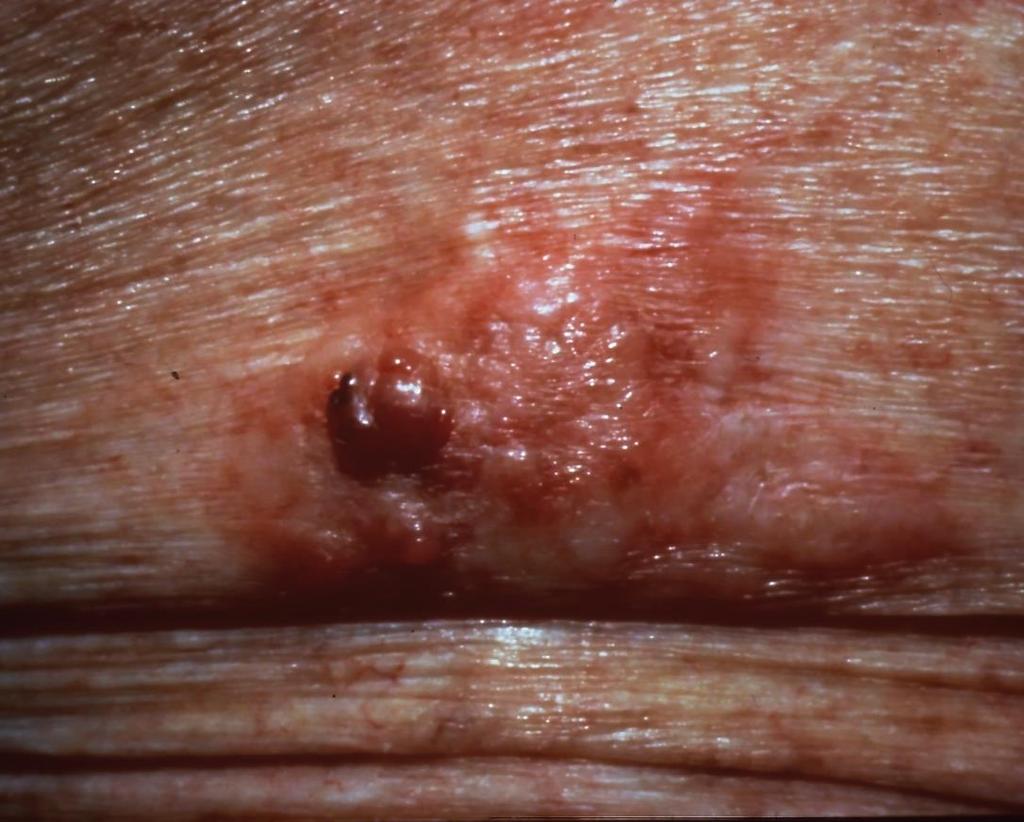
59 Distractors: *Tinea Manuum
60 This developmentally normal patient presents to you for cosmetic removal of these lesions. What is the most likely diagnosis?: A. Hidrocystoma B. Sebaceous carcinomas C. Syringomas D. Sebaceous hyperplasia E. Tricholemmomas

61

62 This developmentally normal patient presents to you for cosmetic removal of these lesions. What is the most likely diagnosis? A. Hidrocystoma B. Xanthelasma C. Syringomas D. Sebaceous hyperplasia E. Tricholemmomas

63 Syringomas Benign eccrine neoplasm May be a/c Down s syndrome Localized periorbital m/c Eruptive Familial

64 Distractors Sebaceous hyperplasia Colonoscopy Muir Torre Syndrome Sebaceous neoplasms, KAs GI, GU malignancies

65 Incorrect Choice- Xanthelasma Generally larger and planar Check lipid panel Associated with type II and IV familial hyperlipidemia
 risk breast, thyroid, uterine, GI, GU cancer Check mammogram, colonoscopy")
66 Incorrect Choice - *Tricholemmoma Fleshy papules favoring perinasal areas Associated with Cowden s syndrome (PTEN mutation) risk breast, thyroid, uterine, GI, GU cancer Check mammogram, colonoscopy

67 Distractor - Hidrocystoma Cystic papule Normally solitary Most common location is head and neck
68 The most likely diagnosis is: A. Angiosarcoma B. Masson lesion (intravascular endothelial hyperplasia) C. Minocycline hyperpigmentation D. Pigmented basal cell cancer E. Pyogenic granuloma
69
70 The most likely diagnosis is: A. Angiosarcoma B. Masson lesion (intravascular endothelial hyperplasia) C. Minocycline hyperpigmentation D. Pigmented basal cell cancer E. Pyogenic granuloma
71 Angiosarcoma Uncommon malignant neoplasm of endothelium (<1% of all sarcomas) Unlike most sarcomas, has a predilection for skin and superficial soft tissue Most commonly affects the scalp and face of elderly and areas of chronic lymphedema or radiodermatitis; rare in children Prognosis is extremely poor, with fewer than 15% surviving 5 years Treatment: surgical excision with wide margins (recurrence and metastases common) Chemotherapy and radical radiotherapy may be palliative, but do not improve survival

72

73 Angiosarcoma: 50% occur on head and neck; facial swelling and edema may be present

74 Advanced angiosarcoma may develop nodules and ulcerate

75 Distractors: Minocycline Hyperpigmentation

76 Distractors: Pigmented *Basal Cell Carcinoma
77 The most likely diagnosis is: A. Amelanotic melanoma B. Basal cell cancer C. Chondrodermatitis nodularis helicis D. Gouty tophus E. Squamous cell cancer

78

79 The most likely diagnosis is: A. Amelanotic melanoma B. Basal cell cancer C. Chondrodermatitis nodularis helicis D. Gouty tophus E. Squamous cell cancer
80 Chondrodermatitis nodularis helicis Exquisitely tender nodules on the helix and antihelix of ear Risk factors: cold exposure, significant sun damage, and trauma Treatment Excision of inflamed cartilage most effective Other options Steroids (topical and intralesional) Offloading of pressure Cryosurgery CO 2 laser ablation

81 Distractors: Amelanotic *Melanoma

82 Distractors: *Basal cell cancer

83 Distractors: Gouty tophus
84 The most likely diagnosis is: A. Amelanotic melanoma B. Basal cell cancer C. Dermal nevus D. Dermatofibroma E. Squamous cell cancer
85
86 The most likely diagnosis is: A. Amelanotic melanoma B. Basal cell cancer C. Dermal nevus D. Dermatofibroma E. Squamous cell cancer

87 Dermatofibroma More common on lower extremities, but may arise anywhere

88 Commonly hyperpigmented, but may appear pink in light-skinned individuals


89 Firm to palpation; pinching the lesion may result in dimple sign
90 Dermatofibromas Many histologic variants exist e.g., atrophic, palisading, clear cell, xanthomatous, with granular cells Treatment Observation is appropriate Excision if removal desired
91 This patient has other family members with similar lesions. A. Verruca plana B. Angiofibromas C. Molluscum contagiosum D. Trichoepitheliomas E. Fibrofolliculoma Larger image on next slide Vincent A. et al. JAAD 2003;49(4):

92

93 The most likely diagnosis is: A. Verruca plana B. Angiofibromas C. Molluscum contagiosum D. Trichoepitheliomas E. Fibrofolliculoma
94 Trichoepithelioma Benign adnexal neoplasm Associated with Familial trichoepithelioma Brooke-Spiegler syndrome mutation in cylindroma oncogene: cylindroma, trichoepithelioma, and spiradenoma Rombo syndrome: vermiculate atrophoderma, milia, trichoepithelioma, basal cell carcinoma Lesions are skin colored firm papules Located on the nasolabial folds, nose, and upper lip
95 *Angiofibroma Associated with *tuberous sclerosis Pinkish/telangiectatic papules More centrally located on the nose rather than nasolabial folds Fibrofolliculoma Small white dome-shaped papules Associated with *Birt-Hogg-Dube AD Recurrent spontaneous pneumothorax Increase risk of malignant renal tumor

: 698-705.")
96 Trichoepithelioma Brooke-Spiegler Skin colored, firm, on NLF *Angiofibroma *Tuberous Sclerosis Pink, telangiectatic papules Vincent A. et al. Birt-Hogg-Dubé syndrome: A review of the literature and the differential diagnosis of firm facial papules. JAAD 2003;49(4): Fibrofolliculoma *Birt-Hogg-Dube White smooth papules
97 Verruca Plana (*wart) less symmetrically distributed discrete slightly raised flap top papules with fine verrucous surface *Molluscum contagiosum less symmetrically distributed Discrete papules with central umbilication
98 The most likely diagnosis is: A. Acral melanoma B. Dysplastic nevus C. Pigmented actinic keratosis D. Pigmented purpuric dermatosis E. Talon noir Location: heel of foot

99

100 The most likely diagnosis is: A. Acral melanoma B. Dysplastic nevus C. Pigmented actinic keratosis D. Pigmented purpuric dermatosis E. Talon noir Location: heel of foot
101 Talon noir Also known as black heel Post-traumatic hemorrhage in the stratum corneum seen most often in athletes Thought to occur from shearing forces that rupture papillary dermal blood vessels with leakage into epidermis Paring of stratum corneum reveals re-brown specks of dried blood, distinguishing it from a melanocytic lesion

102 Distractors: Acral *melanoma

103 Distractors: Pigmented Purpuric Dermatosis
104 The patient shown has a history of seizures. What is the most likely diagnosis of the cutaneous lesions? a) Trichoepitheliomas b) Neurofibromas c) Trichodiscomas d) Angiofibromas e) Fibrofolliculomas

105

106 The patient shown has a history of seizures. What is the most likely diagnosis of the cutaneous lesions? a) Trichoepitheliomas b) Neurofibromas c) Trichodiscomas d) Angiofibromas e) Fibrofolliculomas
107 Angiofibromas Benign and common Commonly located on central face Path shows dermal fibrosis and dilated vessels Associated with tuberous sclerosis History of seizures provided suggests *TS
108 Distractors *Neurofibromas More fleshy Umbilicate with pressure Associated with NF *Trichoepitheliomas Also flesh colored papules frequently around nose Tend to be a little larger Associated several syndromes Trichodiscomas and fibrofolliculomas Flat topped flesh colored papules around nose Associated with *Birt-Hogg-Dube
109 The most likely diagnosis is: A. Dermatofibroma B. Dermatofibrosarcoma protuberans C. Epidermoid cyst D. Keloid E. Melanoma

110 The most likely diagnosis is
111 A. Dermatofibroma B. Dermatofibrosarcoma protuberans C. Epidermoid cyst D. Keloid E. Melanoma The most likely diagnosis is:
112 DFSP Soft tissue sarcoma, young to middle-aged adults Plaque/multinodular and locally aggressive Fibrosarcomatous transformation - bad Spindled cells in storiform pattern CD34(+), factor XIIIa(-) (differentiates from DF) Pigmented variant = Bednar tumor

113 The most likely diagnosis is
114 The most likely diagnosis is: A. Dermatofibroma B. Dermatofibrosarcoma protuberans C. Epidermoid cyst D. Keloid E. Melanoma
115
116 The most likely diagnosis is: A. Dermatofibroma B. Dermatofibrosarcoma protuberans C. Epidermoid cyst D. Keloid E. Melanoma

117 Keloid vs. Hypertrophic Scar Extend beyond the edge of the original site

118 Keloid vs. Hypertrophic Scar May regress centrally but maintain an active border

119 Keloid vs. *Hypertrophic Scar Do not extend beyond the surgical site or have central regression
120 The most likely diagnosis is: A. Keratoacanthoma B. Metastatic carcinoma C. Prurigo nodule D. Sporotrichosis E. Verrucous carcinoma Courtesy Dr. C. Travelute

121
122 The most likely diagnosis is: A. Keratoacanthoma B. Metastatic carcinoma C. Prurigo nodule D. Sporotrichosis E. Verrucous carcinoma Courtesy Dr. C. Travelute
123 SCC variant Keratoacanthoma Rapidly growing, spontaneous regression Solitary dome-shaped nodule with central crater or keratin plug Face, neck, upper extremity

124 Keratoacanthoma

125 Keratoacanthoma
126 Keratoacanthoma Ferguson-Smith syndrome Multiple familial KAs in early adulthood AD Grzybowski type Generalized eruptive KAs
127 Keratoacanthoma Can be part of Muir-Torre syndrome Sebaceous neoplasms, KAs, and GI cancers

128 Distractors: Metastatic Carcinoma

129 Distractors: *Prurigo Nodularis
130 Distractors: *Sporotrichosis
131 The patient presents for evaluation of the lesion shown below. Review of systems reveals a history of dysphagia as well as digital pain on cold exposure. Physical exam is significant for telangiectasias. What is the most likely diagnosis of the lesion shown? A) Calcinosis Cutis B) Pilomatrixoma C) Keratoacanthoma D) Pilar cyst E) Osteoma cutis Journal of the American Academy of Dermatology , 1-12DOI: ( /j.jaad )
132
133 The patient presents for evaluation of the lesion shown below. Review of systems reveals a history of dysphagia as well as digital pain on cold exposure. Physical exam is significant for telangiectasias. What is the most likely diagnosis of the lesion shown? A) Calcinosis Cutis B) Pilomatrixoma C) Keratoacanthoma D) Pilar cyst E) Osteoma cutis Journal of the American Academy of Dermatology , 1-12DOI: ( /j.jaad )
134 Calcinosis Cutis Pathogenesis deposition of insoluble calcium salts in skin Associated with variety of conditions Occurs in setting of several connective tissue diseases CREST syndrome (described in question stem) Systemic sclerosis Dermatomyositis Cutaneous calcium deposits appear, hard, white nodular. Frequently ulcerate Often present on fingers and elbows in CTD
135 Distractors Pilomatrixoma Firm cystic papule in hair bearing area + rocker bottom sign *Keratoacanthoma Rapidly growing keratotic nodule Favor head, neck, and extremities
136 Distractors Pilar cyst Soft cystic mass on scalp Osteoma cutis Most commonly presents with multiple small flesh colored papules on cheeks Can be milia-like
Benign versus Cancerous Lesions How to tell the difference FMF 2014 Christie Freeman MD, CCFP, DipPDerm, MSc
 1 Benign versus Cancerous Lesions How to tell the difference FMF 2014 Christie Freeman MD, CCFP, DipPDerm, MSc Benign lesions Seborrheic Keratoses: Warty, stuck-on Genetics and birthdays Can start in late
Skin lesions The Good and the Bad. Dr Virginia Hubbard Ipswich Hospital NHS Trust Barts and the London School of Medicine and Dentistry
 Skin lesions The Good and the Bad Dr Virginia Hubbard Ipswich Hospital NHS Trust Barts and the London School of Medicine and Dentistry Case 1 32 year old woman Australian Lesion on back New hair growing
Lid Lesions: Relax or Refer
 Lid Lesions: Relax or Refer Blair Lonsberry, MS, OD, MEd., FAAO Professor of Optometry Pacific University College of Optometry blonsberry@pacificu.edu Agenda Benign vs. Malignant lesions Benign Eyelid
LUMPS AND BUMPS: AN ORGANIZED APPROACH TO DIAGNOSIS AND MANAGEMENT
 LUMPS AND BUMPS: AN ORGANIZED APPROACH TO DIAGNOSIS AND MANAGEMENT Tammy P. Than, M.S., O.D., F.A.A.O. The University of Alabama at Birmingham / School of Optometry 1716 University Blvd. Birmingham, AL
Dermatopathology: The tumor is composed of keratinocytes which show atypia, increase mitoses and abnormal mitoses.
 Squamous cell carcinoma (SCC): A common malignant tumor of keratinocytes arising in the epidermis, usually from a precancerous condition: 1- UV induced actinic keratosis, usually of low grade malignancy.
Doctors of Optometry Course Notes
 Doctors of Optometry Course Notes OD19 1CE COPE: 43871-AS Eyelid Lumps and Bumps Sunday, February 26, 2017 2:40 pm 3:30 pm Regency C 3 rd Floor Presenter: Blair Lonsberry, OD, FAAO Dr. Lonsberry is a Full
Dermoscopy: Recognizing Top Five Common In- Office Diagnoses
 Dermoscopy: Recognizing Top Five Common In- Office Diagnoses Vu A. Ngo, DO Department of Family Medicine and Dermatology Choctaw Nation Health Services Authority Learning Objectives Introduction to dermoscopy
Appendageal skin tumors
 Appendageal skin tumors Ibrahim Khalifeh, M.D. Associate Professor Department of Pathology American University of Beirut Medical Center Beirut, Lebanon Appendageal tumors Neoplasms whose differentiation
Clinical characteristics
 Skin Cancer Fernando Vega, MD Seattle Healing Arts Clinical characteristics Precancerous lesions Common skin cancers ACTINIC KERATOSIS Precancerous skin lesions Actinic keratoses Dysplastic melanocytic
Table of Contents: Part 1 Medical Dermatology. Chapter 1 Acneiform Disorders. Acne. Acne Vulgaris. Pomade Acne. Steroid Acne
 Table of Contents: Part 1 Medical Dermatology Chapter 1 Acneiform Disorders Acne Acne Vulgaris Pomade Acne Steroid Acne Infantile Acne Pediatric Perspectives Neonatal Acne (Acne Neonatorum) Pediatric Perspectives
I have a skin lump doc! What s next? 12 th August 2017 Dr. Sue-Ann Ho Ju Ee
 I have a skin lump doc! What s next? 12 th August 2017 Dr. Sue-Ann Ho Ju Ee Some thoughts Is this skin cancer? How common is this? How likely is this in this patient? What happens next if it s something
المركب النموذج--- سبيتز وحمة = Type Spitz's Nevus, Compound SPITZ NEVUS 1 / 7
 SPITZ NEVUS 1 / 7 Epidemiology An annual incidence rate of 1.4 cases of Spitz nevus per 100,000 individuals has been estimated in Australia, compared with 25.4 per 100,000 individuals for cutaneous melanoma
Lumps and Bumps: An Organized Approach to Diagnosis and Management. Disclosure. Introduction. References. Structure of Skin.
 Lumps and Bumps: An Organized Approach to Diagnosis and Management Nothing to disclose Disclosure Tammy Pifer Than, MS, OD, FAAO Carl Vinson VAMC tammythan@bellsouth.net References Fitzpatrick's Color
Supplementary Online Content
 Supplementary Online Content Tschandl P, Rosendahl C, Akay BN, et al. Expert-level diagnosis of nonpigmented skin cancer by combined convolutional neural networks. JAMA Dermatol. Published online November
Living Beyond Cancer Skin Cancer Detection and Prevention
 Living Beyond Cancer Skin Cancer Detection and Prevention Cutaneous Skin Cancers Identification Diagnosis Treatment options Prevention What is the most common cancer in people? What is the most common
Basal cell carcinoma 5/28/2011
 Goal of this Presentation A practical approach to the diagnosis of cutaneous carcinomas and their mimics Thaddeus Mully, MD University of California San Francisco To review common non-melanoma skin cancers
IT S FUNDAMENTAL MY DEAR WATSON! A SHERLOCKIAN APPROACH TO DERMATOLOGY
 IT S FUNDAMENTAL MY DEAR WATSON! A SHERLOCKIAN APPROACH TO DERMATOLOGY Skin, Bones, and other Private Parts Symposium Dermatology Lectures by Debra Shelby, PhD, DNP, FNP-BC, FADNP, FAANP Debra Shelby,
Lumps and Bumps: The Dermatology of Lid Lesions
 Lumps and Bumps: The Dermatology of Lid Lesions Thomas J. Joly, MD, PhD Assistant Professor of Ophthalmology Eastern Virginia Medical School Ophthalmic Plastic Surgery Service Virginia Eye Consultants
Cutaneous Malignancies: A Primer COPYRIGHT. Marissa Heller, M.D.
 Cutaneous Malignancies: A Primer Marissa Heller, M.D. Associate Director of Dermatologic Surgery Department of Dermatology Beth Israel Deaconess Medical Center December 10, 2016 Skin Cancer Non-melanoma
أملس عضلي غرن = Leiomyosarcoma. Leiomyosarcoma 1 / 5
 Leiomyosarcoma 1 / 5 EPIDEMIOLOGY Exact incidence is unknown, but older studies suggest that leiomyosarcomas comprise approximately 3 percent of soft-tissue sarcomas. Superficial leiomyosarcoma occurs
Skin Malignancies Non - Melanoma & Melanoma Marilyn Ng, MD Dept. of Surgery M&M Conference Downstate Medical Center July 19, 2012
 Skin Malignancies Non - Melanoma & Melanoma Marilyn Ng, MD Dept. of Surgery M&M Conference Downstate Medical Center July 19, 2012 Case Presentation 57 yo man with 3 month hx of a nonhealing < 1 cm right
DISCLOSURE OF RELEVANT RELATIONSHIPS WITH INDUSTRY
 DISCLOSURE OF RELEVANT RELATIONSHIPS WITH INDUSTRY David R. Carr, MD I HAVE NO RELEVENT RELATIONSHIPS WITH ANY COMPANIES The most likely diagnosis is A. Actinic lentigo B. Eschar C. Blue nevus D. Melanoma
Identifying Benign and Malignant Skin Lesions. No Disclosures. Common Benign Lesions. Benign Lesions 2/25/2018. Stucco Keratoses.
 Dermatology in Primary Care Identifying Benign and Malignant Skin Lesions Christy Quire Baker, APRN, FNP-BC, DCNP Dermatology Certified Nurse Practitioner No Disclosures Common Benign Lesions Seborrheic
Dermatology for the PCP Deanna G. Brown, MD, FAAD Susong Dermatology Consulting Staff at CHI Memorial
 Dermatology for the PCP Deanna G. Brown, MD, FAAD Susong Dermatology Consulting Staff at CHI Memorial Cutaneous Oncology for the PCP Deanna G. Brown, MD, FAAD Susong Dermatology Consulting Staff at CHI
Lagophthalmos. Lagophthalmos: signs. Lagophthalmos: clinical tips. Lagophthalmos: treatment plan. Madarosis
 Lagophthalmos Def: incomplete closure of the eyelid SX: FBS, irritation, red, burn, dry, chronic morning corneal irritation Lagophthalmos: signs 2-5 mm lid separation with slit lamp during blink can force
Learning Objectives. Tanning. The Skin. Classic Features. Sun Reactive Skin Type Classification. Skin Cancers: Preventing, Screening and Treating
 Learning Objectives Skin Cancers: Preventing, Screening and Treating Robert A. Baldor, MD, FAAFP Professor, Family Medicine & Community Health University of Massachusetts Medical School Distinguish the
CONDITIONS OF THE SKIN
 CONDITIONS OF THE SKIN UCSF/SFGH Family & Community Medicine Residency Program Educational Objectives I. Knowledge The resident will be able to discuss the definition, diagnosis, and initial management
Common skin tumors. Benign Epidermal Tumors. Topic. Clinicopathologic Variants. Seborrheic keratoses
 Common skin tumors Topic Benign epidermal tumors Skin cyst and adnexal neoplasms Other common skin tumor Common skin malignancy สมศ กด ต นร ตนากร 26/02/2015 Benign Epidermal Tumors Seborrheic keratosis
Pathology of the skin. 2nd Department of Pathology, Semmelweis University
 Pathology of the skin 2nd Department of Pathology, Semmelweis University Histology of the skin Epidermis: Stratum corneum Stratum granulosum Stratum spinosum Stratum basale Dermis: papillary and reticular
Cutaneous reactions to targeted therapies. Stavonnie Patterson, MD, FAAD Northwestern University Feinberg School of Medicine March 6, 2017
 Cutaneous reactions to targeted therapies Stavonnie Patterson, MD, FAAD Northwestern University Feinberg School of Medicine March 6, 2017 Disclosures I have no relevant disclosures Papulopustular Eruption
Test your knowledge with multiple-choice cases. What are these speckled spots?
 Test your knowledge with multiple-choice cases Case 1 What are these speckled spots? A speckled, pigmented lesion is noticed on the upper arm of a 10-year-old girl. Her mother says the lesion has been
NEOPLASMS OF THE SURFACE EPITHELIUM (KERATINOCYTES)
") NEOPLASMS OF THE SURFACE EPITHELIUM (KERATINOCYTES) Papillary Lesions Precancerous Lesions Keratinocyte Proliferations Carcinomas Melanotic Lesions Melanomas Normal Mucosa Keratin layer Spinous layer Basal
NEOPLASMS OF THE SURFACE EPITHELIUM (KERATINOCYTES) Papillary Lesions Precancerous Lesions Keratinocyte Proliferations Carcinomas Melanotic Lesions Melanomas Normal Mucosa Keratin layer Spinous layer Basal
Objectives. 1. Recognizing benign skin lesions. 2.Know which patients will likely need surgical intervention.
 The Joy of Pediatric Skin Dr. Claire Sanger University of Kentucky Plastic & Reconstructive Surgery Objectives 1. Recognizing benign skin lesions 2.Know which patients will likely need surgical intervention.
Conflicts. Objectives. University of Texas Health Science Center at San Antonio. Pediatrics Grand Rounds 24 August Pediatric Dermatology 101
 Pediatric Dermatology 101 John C. Browning, MD, FAAD, FAAP Conflicts Investigator: ViroXis Advisor: ViroXis Advisory Board: TopMD Speaker: Galderma Objectives Understand the meaning and importance of cutaneous
Large majority caused by sun exposure Often sun exposure before age 20 Persons who burn easily and tan poorly are at greatest risk.
 Basics of Skin Cancer Detection and Treatment of Non- Melanoma Skin Cancers Large majority caused by sun exposure Often sun exposure before age 20 Persons who burn easily and tan poorly are at greatest
Disclosure. Objectives. PAFP CME Conference Lou Mancano MD, FAAFP Reading Health System November 18, 2016
 PAFP CME Conference Lou Mancano MD, FAAFP Reading Health System November 18, 2016 1 Disclosure The speaker has no conflict of interest, financial agreement, or working affiliation with any group or organization.
Rash Decisions Approach to the patient with a skin condition
 National Conference for Nurse Practitioners April 25, 2014 Rash Decisions Approach to the patient with a skin condition Margaret A. Bobonich, DNP, FNP C, DCNP, FAANP Assistant Professor, Case Western Reserve
Malignant non-melanocytic lesions
 Malignant non-melanocytic lesions Course C023: Fundamentals of Dermoscopy March 4, 2019, 11:20 AM - 11:50 PM Room: 146B Jason B. Lee, MD Professor & Vice Chair Director of Dermatopathology & Pigmented
Periocular Malignancies
 Periocular Malignancies Andrew Gurwood, O.D., F.A.A.O., Dipl. Marc Myers, O.D., F.A.A.O. Drs. Myers and Gurwood have no financial interests to disclose. Course Description Discussion of the most common
Samer Ghosn, MD Associate professor, Derpartment of Dermatology American University of Beirut Medical Center. Follicular lesions
 Samer Ghosn, MD Associate professor, Derpartment of Dermatology American University of Beirut Medical Center Follicular lesions Introduction Follicular lesions are important to recognize: For proper management
MECHANISMS OF HUMAN DISEASE: LABORATORY SESSION PATHOLOGY OF THE SKIN LAB. Friday, February 12, :30 am 11:00 am
 MECHANISMS OF HUMAN DISEASE: LABORATORY SESSION PATHOLOGY OF THE SKIN LAB Friday, February 12, 2012 9:30 am 11:00 am FACULTY COPY GOALS: Describe the basic clinical and morphologic features of various
Know who is at risk: LOOK! for ABCDs, rapidly changing lesions, do a biopsy when indicated
 Lindy P. Fox, MD Assistant Professor Director, Hospital Consultation Service Department of Dermatology University of California, San Francisco Applies to adults without history of malignancy or premalignant
Diploma examination. Dermatopathology: First paper. Tuesday 21 March Candidates must answer FOUR questions ONLY. Time allowed: Three hours
 Dermatopathology: First paper Tuesday 21 March 2017 1. Discuss the role of fluorescent in-situ hybridization (FISH) and emerging molecular techniques in the diagnosis of cutaneous melanocytic lesions,
Identifying Skin Cancer. Mary S. Stone MD Professor of Dermatology and Pathology University of Iowa Carver College of Medicine March, 2018
 Identifying Skin Cancer Mary S. Stone MD Professor of Dermatology and Pathology University of Iowa Carver College of Medicine March, 2018 American Cancer Society web site Skin Cancer Melanoma Non-Melanoma
DERMATOLOGY ROTATION: COMPETENCY-BASED GOALS AND OBJECTIVES
 UNC DIVISION OF PLASTIC AND RECONSTRUCTIVE SURGERY DERMATOLOGY ROTATION: COMPETENCY-BASED GOALS AND OBJECTIVES MEDICAL KNOWLEDGE A. Anatomy/Physiology/Embryology Goal: The resident will have knowledge
Common Benign Lesions and Skin Cancers. 22nd May 2015 Dr Mark Foley
 Common Benign Lesions and Skin Cancers 22nd May 2015 Dr Mark Foley Thank you for downloading this file. This intended to supplement the presentation given at the NZ Wound Care Conference, it is not intended
Chapter 6 Squamous Cell Carcinoma: Variants and Challenges
 Chapter 6 Squamous Cell Carcinoma: Variants and Challenges Michael B. Morgan EPIDEMIOLOGY: Second most common skin cancer, rare in the dark-skinned races. ETIOLOGY: Ultraviolet light, HPV infection. PATHOGENESIS:
SKIN SERVICES REVIEW Changes to Medicare Benefits Schedule for 1 November 2016
 Attachment A SKIN SERVICES REVIEW Changes to Medicare Benefits Schedule for 1 November 2016 Deleted items 31200-31215, 31230-31240 31255-31335 Colour Coding for new / updated items: MUCOSAL BIOPSY AND
Update on Cutaneous Mesenchymal Tumors. Thomas Brenn
 Update on Cutaneous Mesenchymal Tumors Thomas Brenn Cutaneous Mesenchymal Tumours Wide morphological and biological spectrum Myofibroblastic, smooth muscle, neural, vascular, apidocytic, undifferentiated;
DERMCASE. A Shiny, Pink, Nose Lesion. Case 1
 Test your knowledge with multiple-choice cases This month 5 cases: 1. A Shiny, Pink, Nose Lesion p.43 2. A Red Patch on the Forehead p.44 3. An Ulcerated Nodule on the Thigh p.45 4. A Large Lump on the
Thursday 21 st August Skin Problems
 Thursday 21 st August 2014 Skin Problems Skin Problems The Sun and the Skin Sun Damage Recognising the early signs of skin cancer The Big 3 inflammatory condi=ons Acne & Rosacea Eczema (Including Seborrhoeic
Benign and malignant epithelial lesions: Seborrheic keratosis: A common benign pigmented epidermal tumor occur in middle-aged or older persons more
 Benign and malignant epithelial lesions: Seborrheic keratosis: A common benign pigmented epidermal tumor occur in middle-aged or older persons more common on the trunk; but extremities, head and neck are
Skin Cancer. 5 Warning Signs. American Osteopathic College of Occupational and Preventive Medicine OMED 2012, San Diego, Monday, October 8, 2012 C-1
 Skin Cancer AMERICAN OSTEOPATHIC COLLEGE OF OCCUPATIONAL & PREVENTIVE MEDICINE OMED 2012 October 8, 2012 E. Robert Wanat II, D.O., M.P.H. Learning Objectives: Identify the 3 Basic Types of Skin Cancer
Things that go bump: Wart & Molluscum
 Things that go bump: Wart & Molluscum Raegan Hunt, MD, PhD Chief of Section, Pediatric Dermatology Texas Children s Hospital Disclosures Off label use of products may be discussed No relevant financial
Dermatological Manifestations in the Elderly. Sanjay Siddha Staff Dermatologist UHN & MSH
 Dermatological Manifestations in the Elderly Sanjay Siddha Staff Dermatologist UHN & MSH Disclosure No actual or potential conflicts of interest or commercial relationships to declare Objectives Recognize
Diploma Examination. Dermatopathology: First paper. Tuesday 20 March Candidates must answer FOUR questions. Time allowed: 3 hours
 Dermatopathology: First paper Tuesday 20 March 2018 Candidates must answer FOUR questions Time allowed: 3 hours 1. Give an account of the genetic aberrations encountered in Spitzoid neoplasms and how these
Know who is at risk: LOOK! for ABCDs, rapidly changing lesions, do a biopsy when indicated
 Lindy P. Fox, MD Associate Professor Director, Hospital Consultation Service Department of Dermatology University of California, San Francisco Applies to adults without history of malignancy or premalignant
Exenteration. Introduction. The skin. Epidermal malignancies 8/3/2017. Neglected basal cell carcinoma
 Jeremiah Tao, MD, FACS Director, Oculoplastic and Orbital Surgery Associate Professor, UC Irvine Neglected basal cell carcinoma Exenteration Introduction Chief question with any eyelid lesion: Suspicious
Emergent and Urgent Dermatology, Eruptions, and Wound Care
 Emergent and Urgent Dermatology, Eruptions, and Wound Care G. Scott Drew, DO, FAAD, FAOCD Smith Clinic Department of Dermatology Tucson Osteopathic Medical Foundation April 27, 2018 Acute Cutaneous Lupus
Atlas of Eyelid and Conjunctival Tumors
 Atlas of Eyelid and Conjunctival Tumors Jerry A. Shields, M.D. Director, Ocular Oncology Service Wills Eye Hospital Professor of Ophthalmology Thomas Jefferson University Philadelphia, Pennsylvania Carol
Glenn D. Goldman, MD. University of Vermont Medical Center. University of Vermont College of Medicine
 Glenn D. Goldman, MD University of Vermont Medical Center University of Vermont College of Medicine Recognize and identify the main types of skin cancer and their precursors Identify and understand new
Dermatology Procedure Coding
 Dermatology Procedure Coding Anatomy Two layers that make up human skin Epidermis most superficial layer Composed of four to five layers called stratum Anyone remember the mnemonic? Thickness varies based
The Integumentary System. Disorders, Conditions, and Diseases
 The Integumentary System Disorders, Conditions, and Diseases Definitions Disease- an abnormal condition of the body or the mind that causes dysfunction or discomfort. Disorder- a functional abnormality,
Service Specification: CPO Skin Lesions
 Service Specification: CPO Skin Lesions This pathway description needs to be read in conjunction with the CPO Admin Service Specifications. 1. Outcomes Framework The outcomes sought from this service are
- Selected Tumors of the Skin Appendages - Primary vs. Metastasis
 - Selected Tumors of the Skin Appendages - Primary vs. Metastasis Napa Valley 2018 Victor G. Prieto, MD, PhD Chair of Pathology UT MD Anderson Cancer Center vprieto@mdanderson.org Napa Valley in May Introduction
Skin cancer and benign lesions
 How to Treat PULL-OUT SECTION www.australiandoctor.com.au Complete How to Treat quizzes online (www.australiandoctor.com.au/cpd) to earn CPD or PDP points. Keratoacanthoma. inside Epidemiology and clinical
Treatments used Topical including cleansers and moisturizer Oral medications:
 Discipline: Dermatology Extended Topic: Acne & Rosacea : Onset: Location: Face Chest Back Menses if female: Regular Irregular PCOS Treatments used Topical including cleansers and moisturizer Oral medications:
DESCRIPTIONS FOR MED 3 ROTATIONS Dermatology A3S
 Regardless of your future field of practice, you will be exposed to a considerable amount of dermatology and this rotation provides you the chance to see a range of skin diseases. You will have the opportunity
Skin Cancer. Dr Elizabeth Ogden Associate Specialist in Dermatology East and North Herts Dr Elizabeth Ogden
 Skin Cancer Dr Elizabeth Ogden Associate Specialist in Dermatology East and North Herts 13.10.16 Skin Cancer Melanoma mole cancer - is a true cancer which can metastasize and kill Non Melanoma skin cancer
Contents. Part I Genodermatoses
 Contents Part I Genodermatoses 1 Hyperkeratotic Palms and Soles with Periorificial Keratosis............... 3 2 Indurated, Dark, Hairy Plaques, with Arthritis and Deafness.............. 9 3 Cleft Palate,
Eruptive Tumors of the Follicular Infundibulum: An Unexpected Diagnosis of Hypopigmented Macules
 Dermatol Ther (Heidelb) (2015) 5:207 211 DOI 10.1007/s13555-015-0079-0 CASE REPORT Eruptive Tumors of the Follicular Infundibulum: An Unexpected Diagnosis of Hypopigmented Macules Poonkiat Suchonwanit.
Pathology of the skin. Dr Fónyad László, 1sz. Patológiai és Kísérleti Rákkutató Intézet, SE
 Pathology of the skin Dr Fónyad László, 1sz. Patológiai és Kísérleti Rákkutató Intézet, SE The skin Biggest organ Kb. 1.8 nm Kb. 10 kg Most frequent site for tumor development (BCC) Pathology of the skin
Antonella Tosti Fredric Brandt Endowed Professor of Dermatology & Cutaneous Surgery
 Dermoscopy in the evaluation and treatment of hair loss Antonella Tosti Fredric Brandt Endowed Professor of Dermatology & Cutaneous Surgery DISCLOSURE OF RELATIONSHIPS WITH INDUSTRY Antonella Tosti, MD
MECHANISMS OF HUMAN DISEASE: LABORATORY SESSION PATHOLOGY OF THE SKIN LAB. Friday, February 13, :30 am 11:00 am
 MECHANISMS OF HUMAN DISEASE: LABORATORY SESSION PATHOLOGY OF THE SKIN LAB Friday, February 13, 2009 9:30 am 11:00 am FACULTY COPY GOALS: Describe the basic clinical and morphologic features of various
Common Cutaneous Signs of Medical Illnesses
 Common Cutaneous Signs of Medical Illnesses DR COLIN THENG MBBS, MMED (FAM. MED), MRCP(UK), FAMS SENIOR CONSULTANT DERMATOLOGIST THE SKIN SPECIALISTS & LASER CLINIC MOUNT ALVERNIA MEDICAL CENTRE D, #07-61
Actinic keratosis (AK): Dr Sarma s simple guide
: Dr Sarma s simple guide") Actinic keratosis (AK): Dr Sarma s simple guide Actinic keratosis is a very common lesion that you will see in your day-to-day practice. First, let me explain the name Actinic keratosis. It means keratosis
Actinic keratosis (AK): Dr Sarma s simple guide Actinic keratosis is a very common lesion that you will see in your day-to-day practice. First, let me explain the name Actinic keratosis. It means keratosis
Skin Cancer 101: Diagnosis and Management of the Most Common Cancer
 Skin Cancer 101: Diagnosis and Management of the Most Common Cancer Sarah Patton, PA-C, MSHS Skin Surgery Center www.skinsurgerycenter.com Seattle/Bellevue, WA Skin cancer Skin cancer is by far the most
Skin Cancer - Non-Melanoma
 Skin Cancer - Non-Melanoma Introduction Each year, millions of people find out that they have skin cancer. Skin cancer is almost 100% curable if found early and treated right away. It is possible to prevent
04/09/2018. Squamous Cell Neoplasia and Precursor Lesions. Agenda. Squamous Dysplasia. Squamo-proliferative lesions. Architectural features
 Squamous Cell Neoplasia and Precursor Lesions Jennifer L. Hunt, MD, MEd Aubrey J. Hough Jr, MD, Endowed Professor of Pathology Chair of Pathology and Laboratory Medicine University of Arkansas for Medical
Cutanous Manifestation of Lupus Erythematosus. Presented By: Dr. Naif S. Al Shahrani Salman Bin Abdaziz university
 Cutanous Manifestation of Lupus Erythematosus Presented By: Dr. Naif S. Al Shahrani Salman Bin Abdaziz university A 50-year old lady, who is otherwise healthy, presented to the dermatology clinic with
VACAVILLE DERMATOLOGY
 Connecting the Dots on those Spots NANDAN V. KAMATH, M.D. VACAVILLE DERMATOLOGY Sources All of the photos were taken with permission from the Dermnet NZ website - Dermnet New Zealand after communicating
FAMILY PRACTITIONERS! Beirut Medical Center
 LESIONS THAT MAY FOOL FAMILY PRACTITIONERS! Samer Ghosn American University of Beirut Medical Center DERMATOLOGY PATIENTS GP Diagnosis is in doubt Diagnosis and treatment DERMATOLOGIST Failure GPs are
Nonmelanoma skin cancers
 Skin cancer Philip Clarke Nonmelanoma skin cancers Treatment options Background Australia has one of the highest skin cancer rates in the world. Early detection and treatment of skin cancer is vital to
SKIN SONOGRAPHY IN CHILDREN. CRISTIAN J. GARCIA MD Santiago, Chile
 SKIN SONOGRAPHY IN CHILDREN CRISTIAN J. GARCIA MD Santiago, Chile I HAVE NO DISCLOSURES OBJECTIVES RELEVANCE OF SKIN LESIONS IN CHILDREN ROLEN OF THE RADIOLOGIST CLINICAL CORRELATION US TECHNIQUE NORMAL
Vision Health: Conditions, Disorders & Treatments EYELID DISORDERS
 Vision Health: Conditions, Disorders & Treatments EYELID DISORDERS There are a number of disorders that can affect the eyelid. Entropion Entropion is an inward turning of the eyelid and lashes toward the
Richard Turner Consultant Dermatologist
 Old Problems & New Treatments Photo Album by Administrator Richard Turner Consultant Dermatologist Plan for tonight? Refresher on SCC and solar keratosis How to distinguish the two Classic therapy than
Periocular skin cancer
 Periocular skin cancer Information for patients Skin cancer involving the skin of the eyelid or around the eye is called a periocular skin cancer. Eyelid skin cancers occur most often on the lower eyelid,
1 Assessment Techniques General Survey Skin, Hair, and Nails. 2 Cultivating Your Senses
 1 Assessment Techniques General Survey Skin, Hair, and Nails 2 Cultivating Your Senses Inspection Always performed first Palpation Purpose Use different parts of the hands Light vs. deep palpation 3 Cultivating
PATHOLOGY OF THE SKIN 2. Tumours of the skin
 PATHOLOGY OF THE SKIN 2. Tumours of the skin Máirín E. McMenamin MB MRCPI FRCPath Dip (Dermatopathol) RCPath St. James s Hospital and University of Dublin, Trinity College Tumour (Neoplasia) Benign or
DERMCASE. A Common Proliferation. Case 1
 Test your knowledge with multiple-choice cases This month 8 cases: 1. A Common Proliferation p.45 2. A Finger Nodule p.46 3. A Mysterious Mole p.47 4. A Painless Cystic Mass p.48 5. A Yellowish Scalp Lesion
Case-Based Approach to Common Dermatologic Neoplasms
 Case-Based Approach to Common Dermatologic Neoplasms Patrick Retterbush, MD, FAAD Mohs Surgery & Dermatologic Oncology Associate Member of the American College of Mohs Surgery Private Practice: Lockman
Cutaneous Adnexal Tumors
 Cutaneous Adnexal Tumors Lesions with Predominant Follicular Differentiation Special Emphasis on Basal Cell Carcinoma 2014-04-01 Prof. Dr. med. Katharina Glatz Pathologie Cutaneous Adnexal Tumors Hair
A 40-year old male with follicular papule and pustule at central face area for 3 months
 A 40-year old male with follicular papule and pustule at central face area for 3 months GMS- Neg AFB-Neg Fite stain - neg HISTOPATHOLOGICAL DIFFERENTIAL DIAGNOSIS CASEOUS GRANULOMA INFECTION -MYCOBACTERIUM
Hauora Maori. Map. Non-Melanoma Skin Lesion. Clinically Seborrhoeic Keratosis. Management Options. Punch Biopsy. Refer to Public Skin Lesion Service
 Care map information Information resources for patients and carers Updates to this Care Map Hauora Maori Pacific Skin lesion history Clinical examination and diagnostic test Suspicious of Melanoma Non-Melanoma
SKIN LESIONS. On behalf of Airedale, Wharfedale and Craven CCG, Bradford City CCG and Bradford Districts CCG. Bradford and Airedale CCGs.
 Bradford and Airedale CCGs SKIN LESIONS Version: 2 Ratified by: Date ratified: Author(s): Responsible Committee: Consultant in Public Health Individual Funding Request Panel Date issue: September 2013
Introduction. Results. Discussion. Histopathologic and immunohistochemical findings. Results. conclusions,
 1/5 2/5 Carcinoma distinctive carcinoma. form erysipeloides (CE), metastasis. which clinically Itfrom has resembles been termed erysipelas, is an uncommon, but may extend It164 toclassically back, presents
Skin and Body Membranes Body Membranes Function of body membranes Cover body surfaces Line body cavities Form protective sheets around organs
 Skin and Body Membranes Body Membranes Function of body membranes Cover body surfaces Line body cavities Form protective sheets around organs Classification of Body Membranes Epithelial membranes Cutaneous
Gross Appearance & Histology of Skin Cancer. Kyle Mannion M.D. January 21, 2005
 Gross Appearance & Histology of Skin Cancer Kyle Mannion M.D. January 21, 2005 Actinic Keratosis 5-20% will develop squamous/basal cell ca Almost solely from solar damage Usually develop during 4 th decade