NEOPLASMS OF THE SURFACE EPITHELIUM (KERATINOCYTES)
|
|
|
- Chad Mosley
- 5 years ago
- Views:
Transcription
1 NEOPLASMS OF THE SURFACE EPITHELIUM (KERATINOCYTES)

2 Papillary Lesions Precancerous Lesions Keratinocyte Proliferations Carcinomas Melanotic Lesions Melanomas

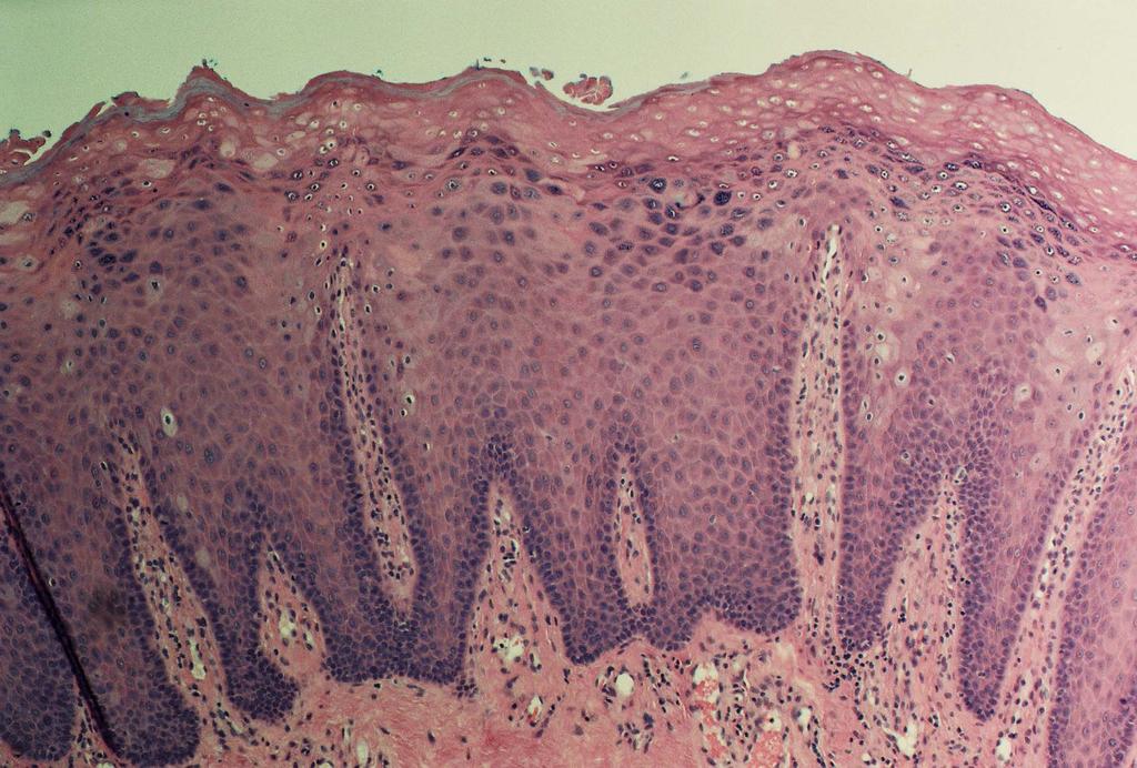
3 Normal Mucosa Keratin layer Spinous layer Basal layer Submucosal Connective Tissues
4 Epithelial Lesions Benign Surface Papillomas Premalignant Lesions Oral Skin Carcinoma Squamous Cell, Verrucous Basal Cell Benign Nevi Malignant Melanoma
5 Benign Oral Papillary and Verrucous Tumors Verruca vulgaris - HPV 2,4 Squamous papilloma - HPV 6,11 solitary Condyloma acuminatum - HPV 6,11 Multiple Keratoacanthoma HPV? Focal Epithelial Hyperplasia (Heck disease) HPV 13,32 Warty Dyskeratoma


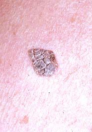
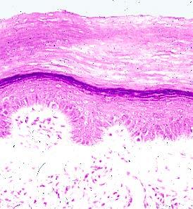
6 Verruca vulgaris Clinical Histopath and DNA


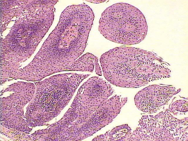
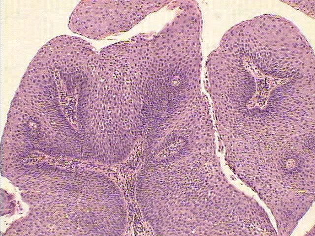
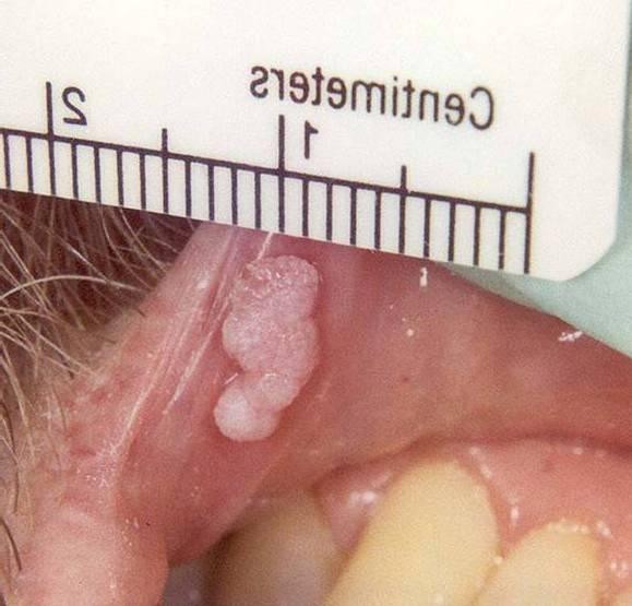
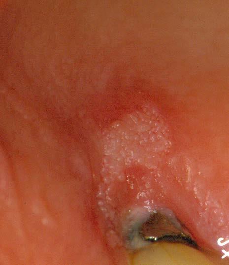
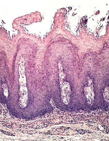
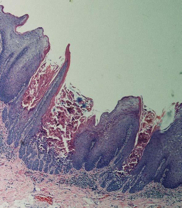
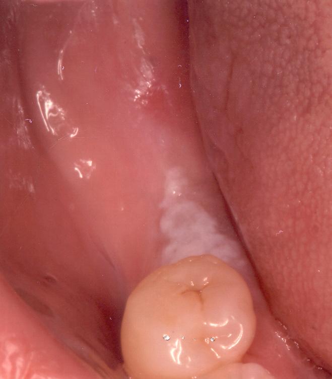
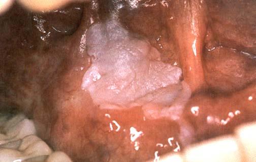
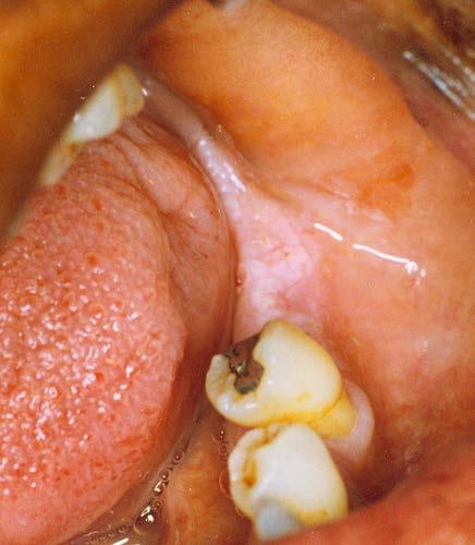
7 Clinical, Gross Squamous Papilloma Histopathology
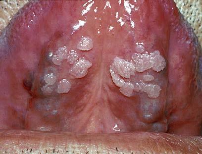
8 Condyloma Acuminatum Clinical Histopath & DNA
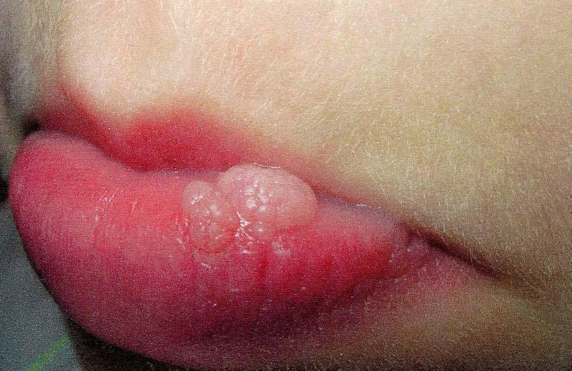
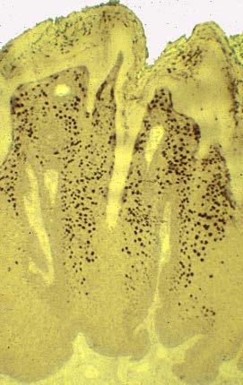
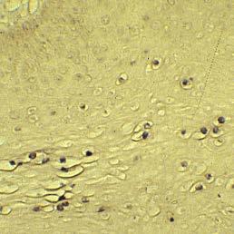
9 Condyloma
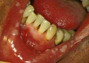
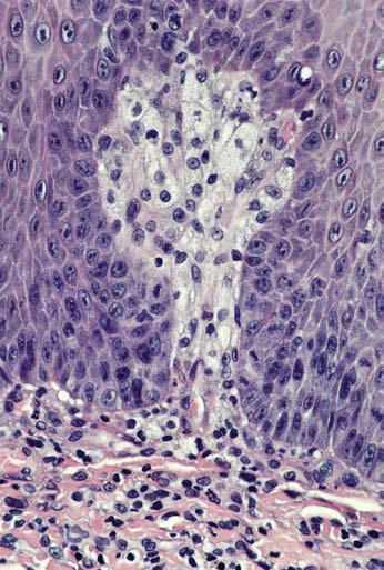
10 Focal Epithelial Hyperplasia Heck Disease Predominantly a childhood HPV disease Multifocal papules and nodules, lips and buccal mucosa HPV 13, 32, viruses that only cause oral mucosal flat warts The phenotype may be seen in HIV infected patients Spontaneous regression occurs in 6-12 months without treatment Microscopic: Dome shaped exophytic proliferations of SSE, marked acanthosis, mitosoid (mitotic-like) bodies found in the mid-spinous layer

11 FEH in HIV+ Subjects
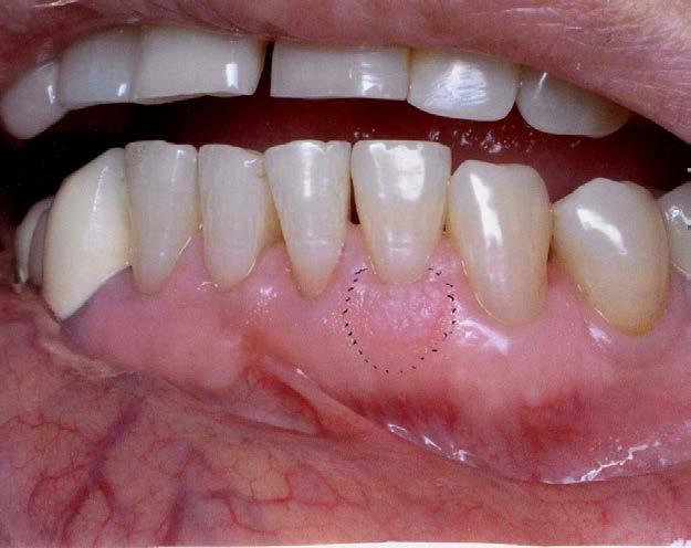
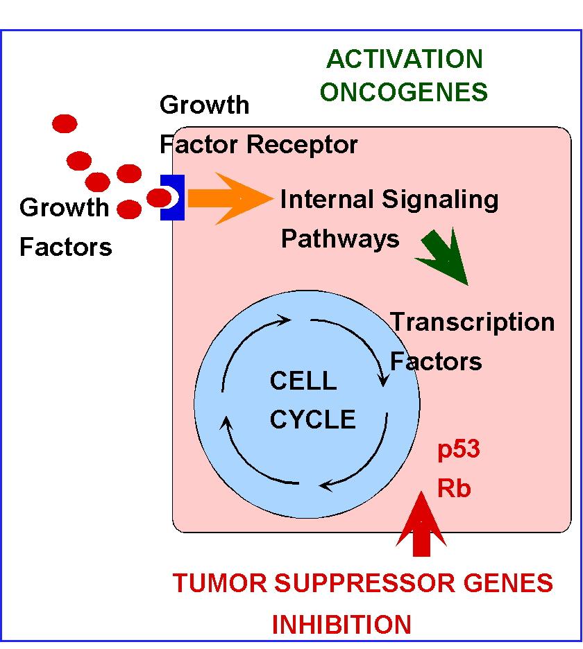
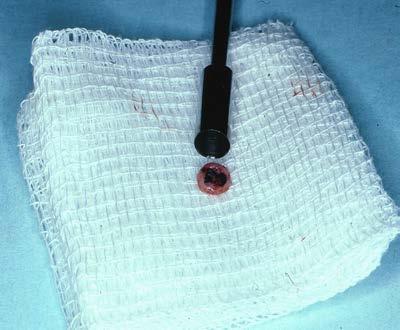
12 Keratoacanthoma A verrucous well circumscribed tumor of skin with self-limited growth Documented cases of spontaneous regression Microscopic: Abrupt cup-like borders, marked parakeratosis and acanthosis without cytologic atypia Treatment: simple excision Cases with atypia should be considered low grade squamous carcinomas
13 Keratoacanthoma
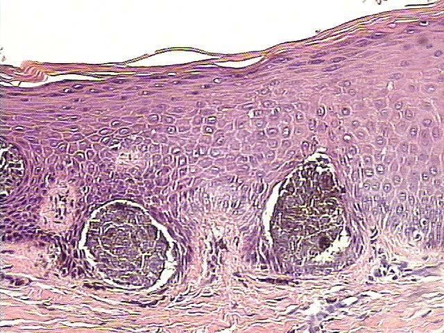
14 Verruciform Xanthoma A papillary lesions with keratosis Tends to occur on the gingiva and palate Benign lesion Equal sex distribution Microscopically: Hyperkeratosis, Papillary pattern, Xanthoma (foam cell histiocytes) within the submucosal papillae
15 Verruciform Xanthoma Xanthoma cells
16 Warty Dyskeratoma An epithelial wary proliferation of skin or mucosa with distinct microscopic features A focal counterpart to Darier White disease (keratosis follicularis) Multiple, yet limited lesions are referred to as focal acantholytic dyskeratosis (Grover s disease) Microscopic: Verrucous keratosis with villous rete pegs, acantholysis and dyskeratotic cells in spinous layer
17 Warty Dyskeratoma
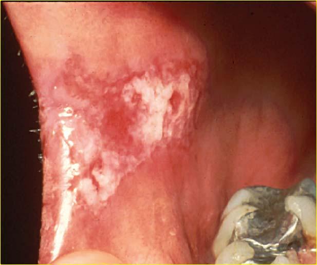
18 Carcinogenesis Oral Cancer Smoked Tobacco The Smokeless Tobacco Issue Alcohol Carcinogens Polycyclic Hydrocarbons Nitrosourias Human Papillomaviruses
19 Carcinogenesis Oncogenes Growth Factors Growth Factor Receptors Internal Signaling Pathway Mediators Tumor Suppressor Genes Apoptotic Pathway Mediators Cell Cycle Regulatory proteins
20
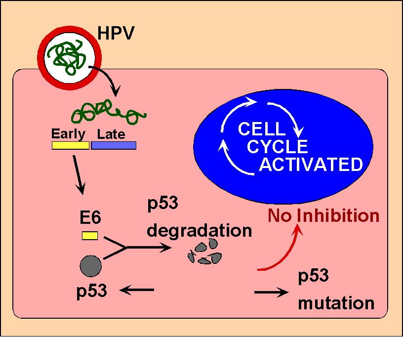
21 HPV and p53 HPV 16 Present in some leukoplakias and SCCA Present in >60% of tonsilar/tongue base SCCA P53 Mutated in >60% of oral SCCA Inactivated/nonmutated in HPV associated SCCA
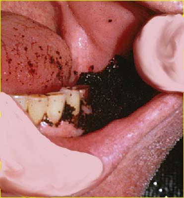
22
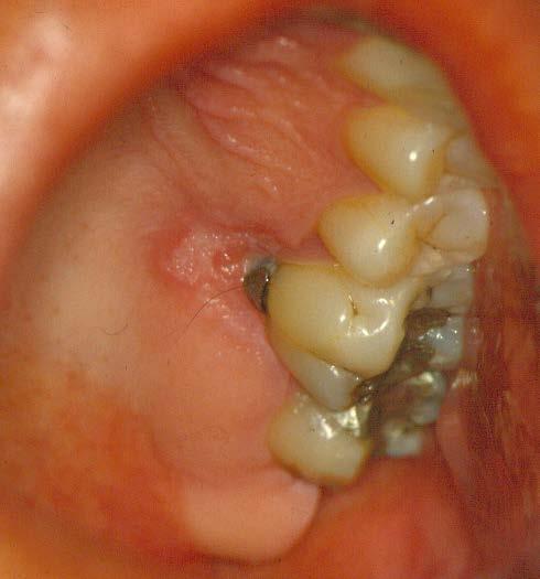
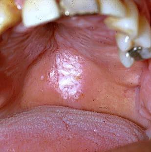
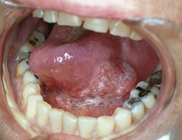
23 LEUKOPLAKIA A Clinical Term
24 Variants of Leukoplakia Homogeneous Verrucous Speckled
25 Leukoplakia 20% precancerous change histologically Floor of the Mouth 40% dysplastic 6% of all leukoplakias will progress to carcinoma within 5-7 years
26 Leukoplakia


27 Leukoplakia White lesions that cannot be rubbed away
28 Leukoplakia

29 Leukoplakia - Snuff Keratosis


30 Toluidine Blue, detection of dysplasia Application of dye Acetic Acid Dye retention


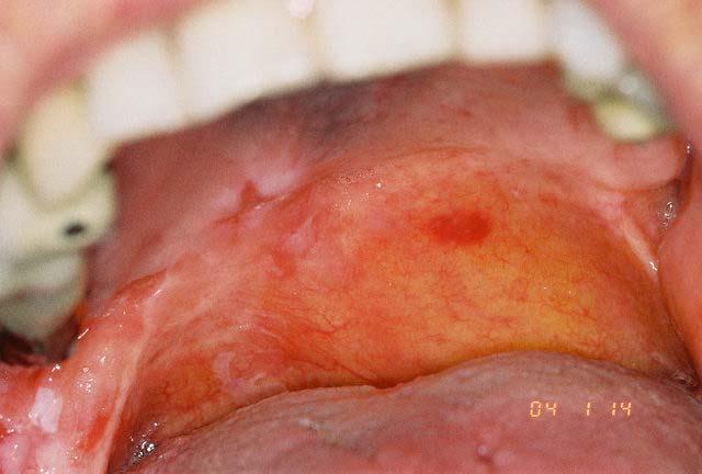
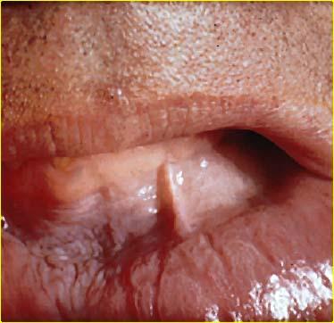
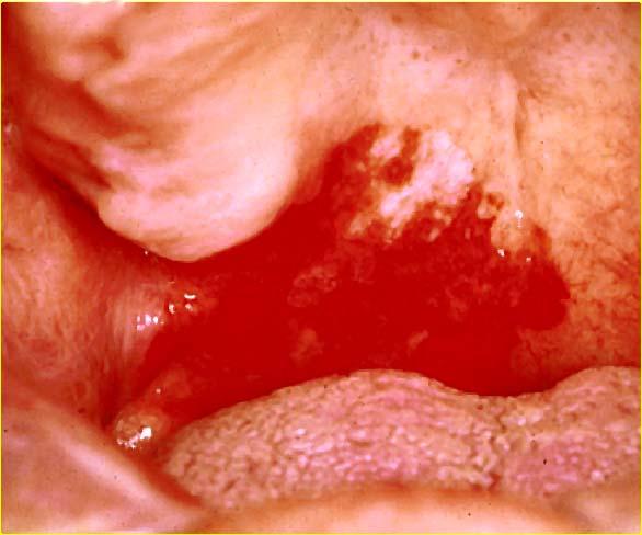
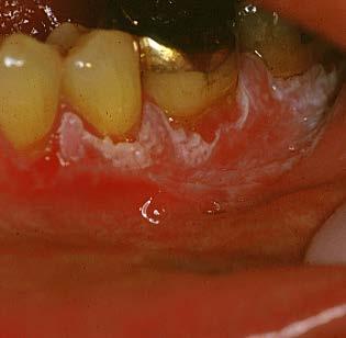
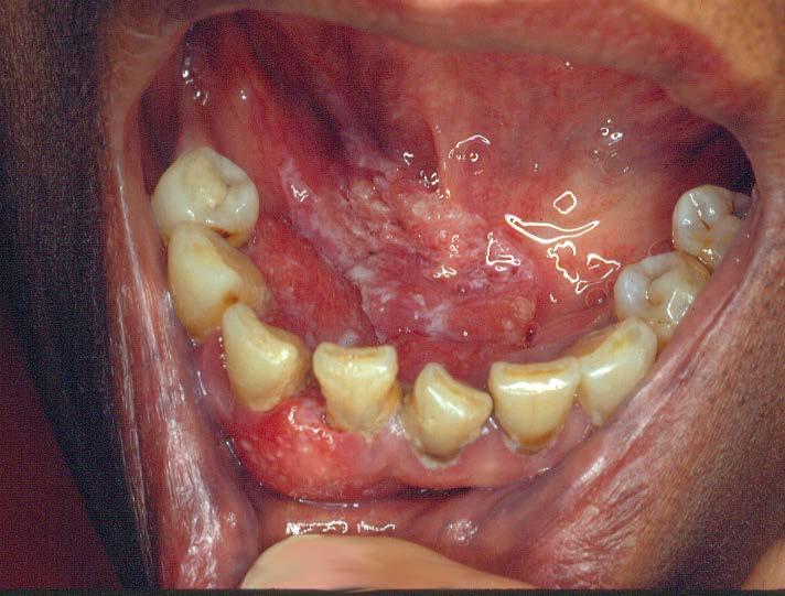
31 Tissue Sampling (BIOPSY) Brush Biopsy Punch Biopsy

32 Benign Keratosis hyperorthokeratosis hyperparakeratosis acanthosis

33 Histologic Spectrum of Leukoplakia Hyperorthokeratosis Hyperparakeratosis Acanthosis
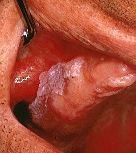
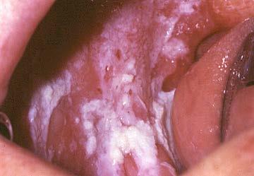
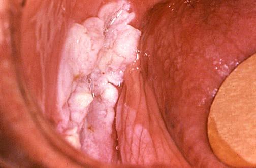
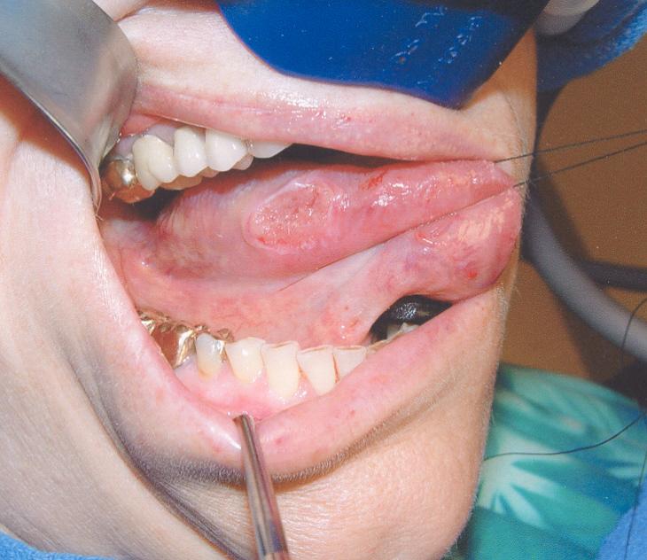
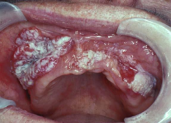
34 Hyperkeratosis/Acanthosis

35 Grades of Epithelial Dysplasia


36 Histologic Spectrum of Leukoplakia Mild Dysplasia Moderate Dysplasia Severe Dysplasia


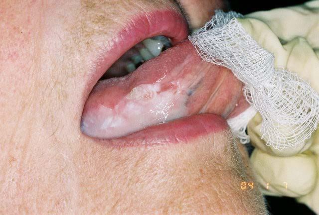
37 Histologic Spectrum of Leukoplakia Squamous Cell Carcinoma
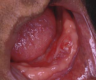
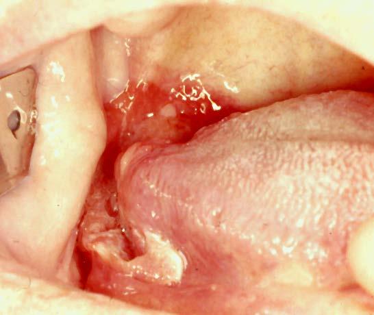
38 Erythroplakia Velvety red patch of unknown etiology More rare than leukoplakia Soft Palate, Floor of Mouth, Lateral Tongue 90% chance for dysplasia Often mixed with leukoplakic areas (Leukoerythroplakia) (Speckled leukoplakia)
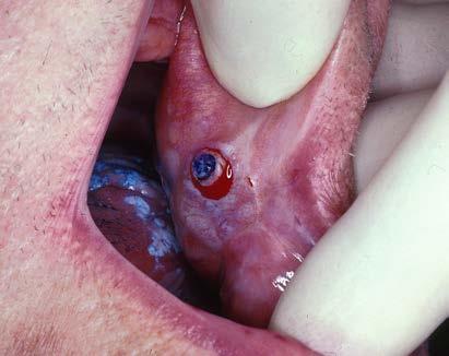
39 Erythroplakia
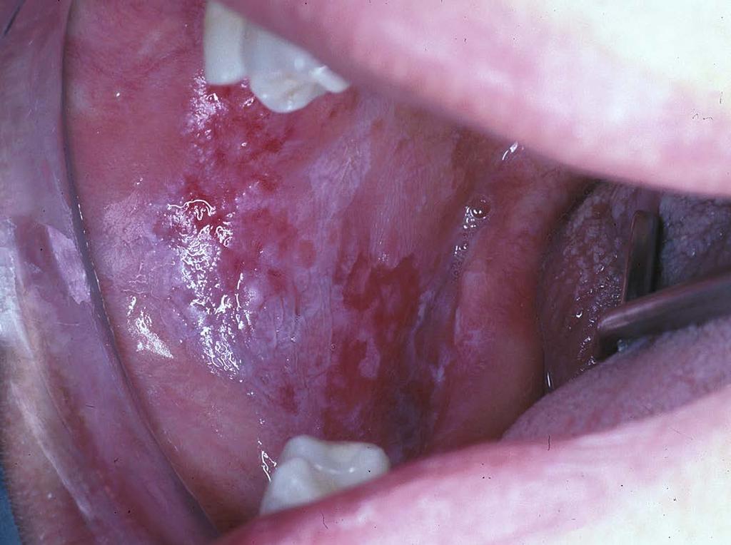
40 Erythroleukoplakia (Speckled)
41 Proliferative Verrucous Leukoplakia Predilection for elderly females Only 40% use tobacco Predilection for gingiva, mucobuccal fold Persistant and Diffuse High Recurrence
42 Proliferative Verrucous Leukoplakia
43 PVL

44 PVL - Histopathology Varies from verrucous hyperkeratosis to verrucous carcinoma, papillary squamous cancer and invasive carcinoma
45 Lichen Planus and Oral Cancer Oral LP occurs in.5% of the population PREVALENCE: 1-2% of patients with OLP develop oral cancer (1:100) over follow up periods of 5-10 years INDICIDENCE: Oral SCCA in US (35,000:298,000,000 or approximately 1.2/10,000 (.012%) Estimate over 10 year and 20 year follow up periods. ODDS RATIOS:* 1 year Follow up 1.0%:0.012% > year Follow up 1.0%:0.12% > year Follow up 1.0%:0..23% > 4.3 *Oral Ca over 10 year period 350,000/298,000,000 (.12%) *Oral CA over 20 year period 700,000/298,000,000 (.23%)
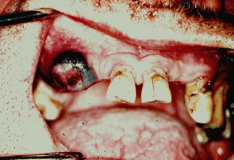
46 Squamous Cell Carcinoma Ulcerated, indurated, white/red, fixed tumefaction Anterior mouth: Well differentiated, good prognosis Posterior mouth: Less differentiated, poor prognosis Lateral tongue, floor of mouth are favored sites although SCCA can occur anywhere in the oral mucosa 70+% smoking and alcohol Tonsilar pillar, base of tongue: HPV 16 Tumor suppressor gene mutations (p16, p53) Prognosis: no nodes>70% 5 year survival; + nodes> 35% 5 year survival

47 Squamous Cell Carcinoma

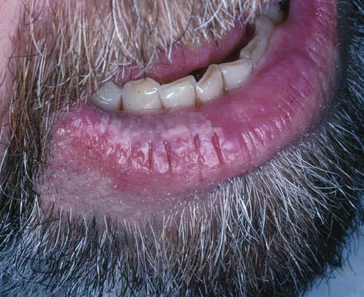
48 Squamous Cell Carcinoma

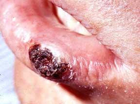
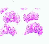
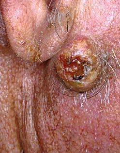
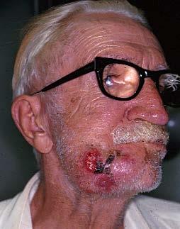
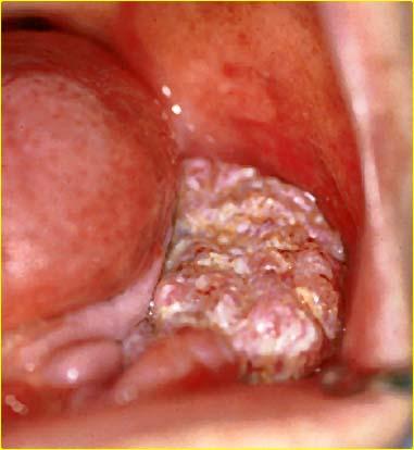
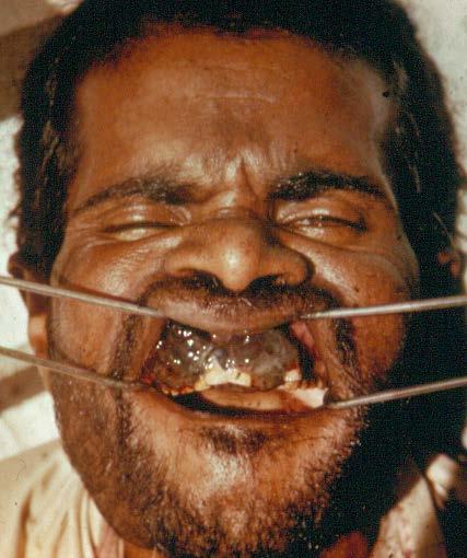
49 Papillary SCCA


50 Oral Squamous Cell Carcinoma Cervical Node Metastasis
51 TNM classification for Head and Neck Cancer T = size of Primary Tumor To: no evidence of tumor T1: carcinoma in situ T2: 2 cm or less T3: 2-4 cm T4: invasion of adjacent tissues N = regional lymph node involvement No: no palpable nodes N1: suspicous, palpable node ipsilateral N2: suspicous, palpable node contralateral N3: large fixed node M = distant metastasis Mo: no evidence of disease M1: distant metastases present Staging according to TNM classification Stage I: T1NoMo Stage II: T2NoMo Stage III: T3NoMo, T1N1Mo, T2N1M0, T3N1Mo Stage IV: T1N2Mo, T1N3Mo, T2N2Mo, T2N3Mo, T3N2Mo, T3N3Mo, any case with M1
52 Keratoses of the Face & Lips Seborrheic Keratosis Actinic Keratosis Actinic Cheilitis
53 Skin Keratoses Seborrheic Keratosis Elderly males, facial skin, brown oily Not precancerous Actinic Keratosis Elderly males, facial skin, red and scaley Precancerous, squamous cell CA Actinic Cheilitis Elderly males, lower lip, white lesion Precancerous, squamous cell CA

54 Seborrheic Keratosis Clinical Histopathology

55 Actinic Cheilitis
56 Basal Cell Carcinoma Facial Skin Keratosis, Ulcer with rolled borders Elderly Actinic Radiation Nonmetastasizing Other adnexa (sebaceous, sweat, hair)


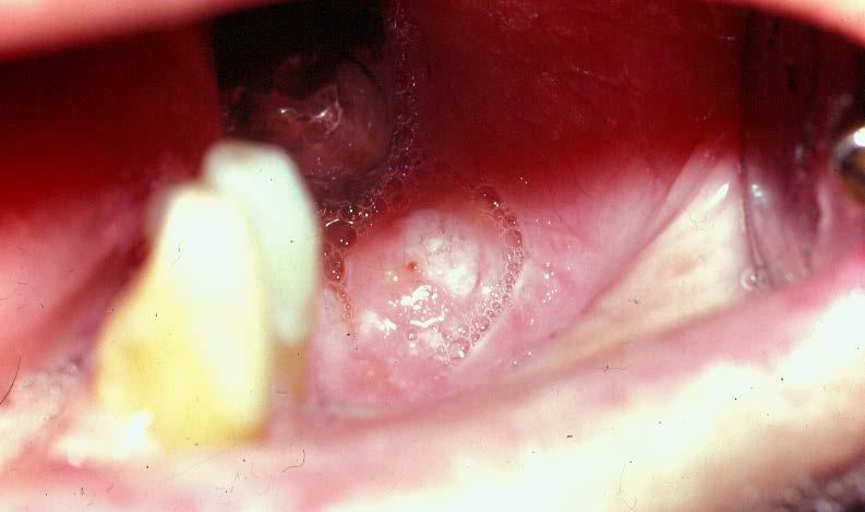
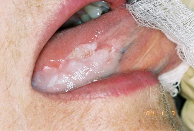
57 Basal Cell Carcinoma Cutaneous Ulcer Histopathology

58 Squamous Cell Carcinoma
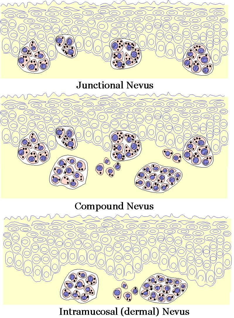
59 Variants of SCCA Histologic Grade Keratinizing Nonkeratinizing Verrucous Carcinoma Spindle Cell Carcinoma
60 Clinical Features SCCA >50 years In nonsmokers - >50 years >80% smoke cigarettes Alcohol is a risk cofactor Lateral tongue, Floor of mouth Prognosis: Anterior portion of mouth better Posterior portion of mouth - worse
61 Therapy for Oral Cancer Laser Ablation Surgical Excision Radiation Therapy Neck Node Management Partial Lymph Node Dissection Radical Lymph Node Dissection Radiation to the Neck

62 MRI Imaging for Head and Neck Cancer


63 Histopathology SCCA Well differentiated Poorly differentiated
64 Verrucous Carcinoma Elderly White keratotic Cauliflower or Verrucous Noninvasive, pushing margins Parakeratin crypts Nonmetastasizing

65 Verrucous Carcinoma Clinical Histopathology
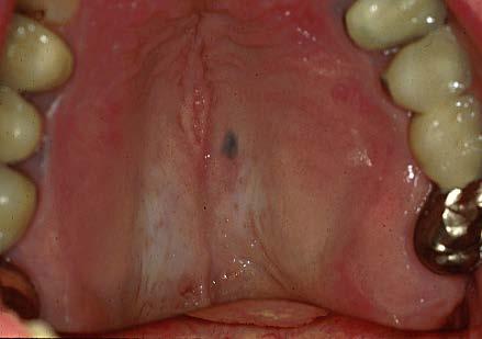
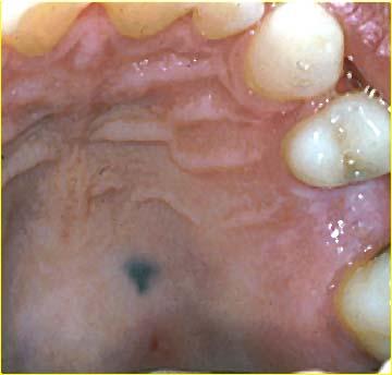
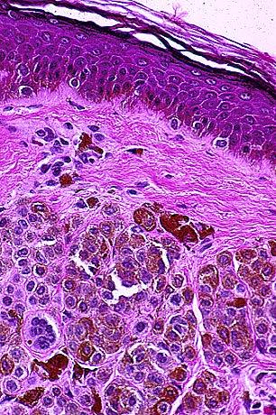
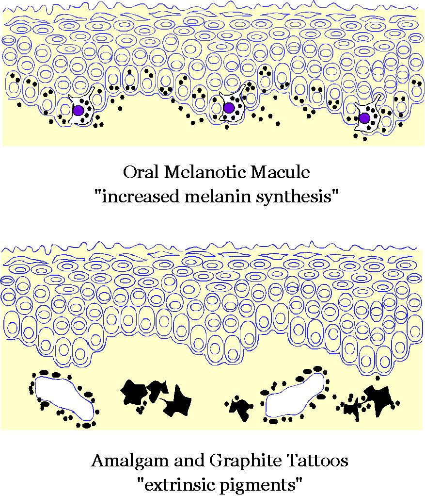
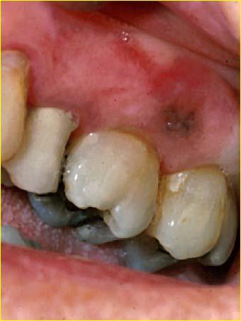
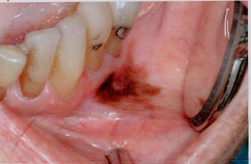
66 Benign Melanocytic Nevi Nevocellular Junctional Compound Intradermal/Intramucosal Specific Types (Ota, Ito) Blue Nevi Common Cellular
67

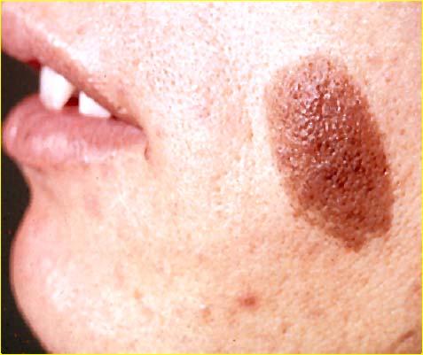
68 Cutaneous Nevi
69 ORAL NEVI All histologic types are seen Melanocytes are normally present in the basal layer Palate>Gingiva>Buccal Mucosa Malignant transformation is very rare
70 Oral Nevi
71 Nevi Junctional Intramucosal

72 Blue Nevus s100
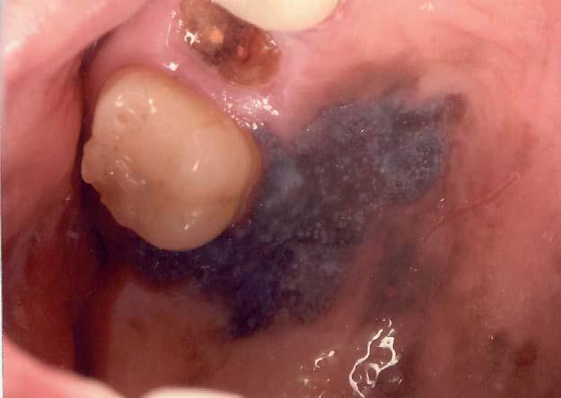
73 MELANOTIC MACULE Oral Freckle or Ephelis Lips>Gingiva>Palate Basilar Melanosis Melanin Incontinence No Malignant Potential
74

75 Melanotic Macule


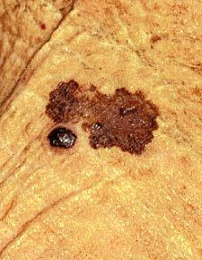
76 Melanotic Macule Basilar melanosis S100 protein
77 Melanoacanthoma Most common among African descent individuals Focal pigmented macule or plaque Basilar melanocytic hyperplasia with dendritic cells in spinous layer Not premalignant
78 Melanoacanthoma
79 Malignant Melanoma Melanoma in situ Superficial Spreading Nodular
80 ORAL MUCOSAL MELANOMA Anterior Maxillary Gingiva>Palate More common in Japan Highly lethal, metastasize widely Not classifiable by Clark levels


81 Superficial Spreading Melanoma Clinical Histopathology

82 Nodular Melanoma Clinical Histopathology
83 Superficial spreading Oral Melanoma

84 Nodular Oral Melanoma

85 Bresloe Scale is measured as depth of invasion in mm Thin Melanoma
Benign and malignant epithelial lesions: Seborrheic keratosis: A common benign pigmented epidermal tumor occur in middle-aged or older persons more
 Benign and malignant epithelial lesions: Seborrheic keratosis: A common benign pigmented epidermal tumor occur in middle-aged or older persons more common on the trunk; but extremities, head and neck are
Dermatopathology: The tumor is composed of keratinocytes which show atypia, increase mitoses and abnormal mitoses.
 Squamous cell carcinoma (SCC): A common malignant tumor of keratinocytes arising in the epidermis, usually from a precancerous condition: 1- UV induced actinic keratosis, usually of low grade malignancy.
Squamous papilloma Squamous acanthoma Keratoacanthoma Verruca vulgaris Condyloma acuminatum Focal epithelial hyperplasia Sino nasal papilloma
 Benign tumors Epithelial origin Squamous papilloma Squamous acanthoma Keratoacanthoma Verruca vulgaris Condyloma acuminatum Focal epithelial hyperplasia Sino nasal papilloma Squamous papilloma Exophytic
WHITE LESIONS OF THE UPPER AIRWAY
 WHITE LESIONS OF THE UPPER AIRWAY WHITE LESION CONFIGURATIONS Solitary vrs Multifocal Flat Plaque Verrucous/rippled Lacey White with red component Papular (curdled milk plaques) Pseudomembranous PLAQUES
Premalignant lesions may expose to a promoting. factor & may be induced to undergo malignant. Carcinoma in situ displays the cytologic features of
 بسم رلاهللا Def. Premalignant lesions may expose to a promoting factor & may be induced to undergo malignant transformation. Carcinoma in situ displays the cytologic features of malignancy without invasion
Lesions & Lifestyles
 Lesions & Lifestyles attended a 3 hour Continuing Education Seminar on Oral Pathology presented by Nancy Dewhirst, RDH,BS on (date) at (location):. Course material is directly related patient care. Notes:
Pathology of the skin. 2nd Department of Pathology, Semmelweis University
 Pathology of the skin 2nd Department of Pathology, Semmelweis University Histology of the skin Epidermis: Stratum corneum Stratum granulosum Stratum spinosum Stratum basale Dermis: papillary and reticular
Oral Epithelial Tumors, Melanocytic Nevi, and Melanoma (I)
") Introduction: Oral Epithelial Tumors, Melanocytic Nevi, and Melanoma (I) Oral Epithelial Tumors may be: Benign tumors Sequamous cell Papilloma Malignant tumors Sequamous cell carcinoma, Basal cell carcinoma
Introduction: Oral Epithelial Tumors, Melanocytic Nevi, and Melanoma (I) Oral Epithelial Tumors may be: Benign tumors Sequamous cell Papilloma Malignant tumors Sequamous cell carcinoma, Basal cell carcinoma
04/09/2018. Squamous Cell Neoplasia and Precursor Lesions. Agenda. Squamous Dysplasia. Squamo-proliferative lesions. Architectural features
 Squamous Cell Neoplasia and Precursor Lesions Jennifer L. Hunt, MD, MEd Aubrey J. Hough Jr, MD, Endowed Professor of Pathology Chair of Pathology and Laboratory Medicine University of Arkansas for Medical
Benign versus Cancerous Lesions How to tell the difference FMF 2014 Christie Freeman MD, CCFP, DipPDerm, MSc
 1 Benign versus Cancerous Lesions How to tell the difference FMF 2014 Christie Freeman MD, CCFP, DipPDerm, MSc Benign lesions Seborrheic Keratoses: Warty, stuck-on Genetics and birthdays Can start in late
Diagnostic difficulties with lesions of the oral mucosa
 BDIAP London, November 2010 School of Clinical Dentistry University of Sheffield Diagnostic difficulties with lesions of the oral mucosa Paul M Speight Dept Oral & Maxillofacial Pathology University of
IT S FUNDAMENTAL MY DEAR WATSON! A SHERLOCKIAN APPROACH TO DERMATOLOGY
 IT S FUNDAMENTAL MY DEAR WATSON! A SHERLOCKIAN APPROACH TO DERMATOLOGY Skin, Bones, and other Private Parts Symposium Dermatology Lectures by Debra Shelby, PhD, DNP, FNP-BC, FADNP, FAANP Debra Shelby,
A five year study on differential diagnosis of verruciform penile lesions
 Original Research Article A five year study on differential diagnosis of verruciform penile lesions S. Sujatha 1, V. Srinivas Kumar 2*, K. Durga 3 1 Associate Professor, 2 Assistant Professor, 3 Professor
Clinical characteristics
 Skin Cancer Fernando Vega, MD Seattle Healing Arts Clinical characteristics Precancerous lesions Common skin cancers ACTINIC KERATOSIS Precancerous skin lesions Actinic keratoses Dysplastic melanocytic
LEUKOPLAKIA Definition Epidemiology Clinical presentation
 LEUKOPLAKIA Definition Leukoplakia is the most common premalignant or "potentially malignant" lesion of the oral mucosa. Leukoplakia is a predominantly white lesion of the oral mucosa than cannot be clinicopathologically
Dysplasia, Mimics and Other Controversies
 Dysplasia, Mimics and Other Controversies Mary S. Richardson, MD Dept. of Pathology Medical University of South Carolina Charleston, SC Notice of Faculty Disclosure In accordance with ACGME guidelines,
Review Article- Leukoplakia: A mysterious white patch.
 International Journal Of Scientific Research And Education Volume 2 Issue 9 Pages 1824-1830 September-2014 ISSN (e): 2321-7545 Website: http://ijsae.in Review Article- Leukoplakia: A mysterious white patch.
Role of the Dental Hygienist in Oral Pathology. Role of the Dental Hygienist in Oral Pathology. Cancers of the Oral Cavity.
 Gum Gardeners Study Club April 25, 2016 Early Detection of Oral Cancer Cindy Kleinegger, DDS, MS NW Oral Pathology Tigard, OR nworalpathology.com Role of the Dental Hygienist in Oral Pathology Work closely
Diseases of oral cavity
 Diseases of oral cavity Diseases of Teeth and Supporting Structures Inflammatory/Reactive Lesions Infections Oral Manifestations of Systemic Disease Precancerous and Cancerous Lesions Odontogenic Cysts
Pigmented lesions of the Oral cavity
 Oral medicine أ.م.د احسان عبد هللا كميل Pigmented lesions of the Oral cavity Pigmented oral lesions are a large group of disorders in which the dark or brown color is the essential clinical characteristic.
DENIS P. LYNCH, DDS, PHD
 140 TH ANNUAL MEETING MAY 6 MAY 7, 2010 JEWEL OF THE GREAT LAKES DENIS P. LYNCH, DDS, PHD FRIDAY, MAY 7, 2010 9:00 A.M. TO 12:00 NOON ORAL CANCER AND RELATED PREMALIGNANCY Oral Cancer and Premalignancy
الطلاوة = Leukoplakia LEUKOPLAKIA
 LEUKOPLAKIA Leukoplakia is a clinical term that refers to a predominantly white lesion of the oral mucosa that cannot be rubbed off or characterized by any other definable lesion or known disease. 130
Chapter 6 Squamous Cell Carcinoma: Variants and Challenges
 Chapter 6 Squamous Cell Carcinoma: Variants and Challenges Michael B. Morgan EPIDEMIOLOGY: Second most common skin cancer, rare in the dark-skinned races. ETIOLOGY: Ultraviolet light, HPV infection. PATHOGENESIS:
Oral cavity cancer accounts for approximately 3% of all malignancies and is a significant worldwide health problem.
 Oral cavity cancer accounts for approximately 3% of all malignancies and is a significant worldwide health problem. Majority are SCC ( 5-year survival rate only about 50-60% ) Many SCC arrive from premalignant
Diseases of the vulva
 Diseases of the vulva 1. Bartholin Cyst - Infection of the Bartholin gland produces an acute inflammation within the gland (adenitis) and may result in an abscess. Bartholin duct cysts - Are relatively
LUMPS AND BUMPS: AN ORGANIZED APPROACH TO DIAGNOSIS AND MANAGEMENT
 LUMPS AND BUMPS: AN ORGANIZED APPROACH TO DIAGNOSIS AND MANAGEMENT Tammy P. Than, M.S., O.D., F.A.A.O. The University of Alabama at Birmingham / School of Optometry 1716 University Blvd. Birmingham, AL
Evaluation and Management of Head and Neck Cancer in Patients with Fanconi anemia David I. Kutler, M.D., F.A.C.S.
 Evaluation and Management of Head and Neck Cancer in Patients with Fanconi anemia David I. Kutler, M.D., F.A.C.S. Residency Site Director Weill Cornell Medical Center Associate Professor Division of Head
Periocular Malignancies
 Periocular Malignancies Andrew Gurwood, O.D., F.A.A.O., Dipl. Marc Myers, O.D., F.A.A.O. Drs. Myers and Gurwood have no financial interests to disclose. Course Description Discussion of the most common
Penile cancer teams in UK. Common variants. Penile cancer teams. Basaloid squamous carcinoma. The Pathology of Penile Tumours
 The Pathology of Penile Tumours Dr Jonathan H Shanks The Christie NHS Foundation Trust, Manchester, UK Penile cancer teams in UK 12 centres for penile cancer work (10 in England and Wales, 2 in Scotland)
Actinic keratosis (AK): Dr Sarma s simple guide
: Dr Sarma s simple guide") Actinic keratosis (AK): Dr Sarma s simple guide Actinic keratosis is a very common lesion that you will see in your day-to-day practice. First, let me explain the name Actinic keratosis. It means keratosis
Actinic keratosis (AK): Dr Sarma s simple guide Actinic keratosis is a very common lesion that you will see in your day-to-day practice. First, let me explain the name Actinic keratosis. It means keratosis
المركب النموذج--- سبيتز وحمة = Type Spitz's Nevus, Compound SPITZ NEVUS 1 / 7
 SPITZ NEVUS 1 / 7 Epidemiology An annual incidence rate of 1.4 cases of Spitz nevus per 100,000 individuals has been estimated in Australia, compared with 25.4 per 100,000 individuals for cutaneous melanoma
Oral Cancer and Common Oral Lesions seen in HIV Seropositive Patients. Gwen Cohen Brown DDS, FAAOMP Professor New York City College of Technology
 Oral Cancer and Common Oral Lesions seen in HIV Seropositive Patients Gwen Cohen Brown DDS, FAAOMP Professor New York City College of Technology Program Objectives Recognize the oral health needs of the
4/16/2018. Bumps & Lumps. What s HPV Got To Do With It? 2 hour Oral & Pharyngeal Pathology Review Nancy Dewhirst RDH,BS
 4/6/08 Bumps & Lumps. What s HPV Got To Do With It? hour Oral & Pharyngeal Pathology Review Nancy Dewhirst RDH,BS www.nancydewhirst.com Bumps & Lumps What s HPV Got To Do With It? Patient Assessment Clinical
Premalignant skin tumours
 Chapter 14: Premalignant skin tumours page: 434 Premalignant skin tumours page: 435 Solar keratoses (senile keratoses) Raised red and well-defined plaques with a rough surface covered in scales of varying
That. Name QUIZ. 60 SEPTEMBER 2017 // dentaltown.com
 QUIZ Name That General dentists are first in the line of practitioners that patients see for an oral lesion evaluation; therefore, a sound understanding of oral mucosal diseases and their clinical presentation
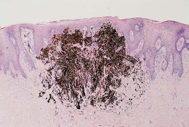
4Ps LUMPS AND BUMPS B.L.&T. BUMPS, LUMPS, AND TATTOOS. Most Common BUMP in the oral cavity Fibroma INTERDENTAL PAPILLAE LESIONS
 B.L.&T. BUMPS, LUMPS, AND TATTOOS LUMPS AND BUMPS DIFFERENTIAL DIAGNOSIS FOR LUMPS AND BUMPS Traumatic Fibroma Papilloma Epulis Fissuratum Inflammatory Papillary Hyperplasia Lesions of Attached Gingiva
Vulvar Carcinoma. Definition: Cases should be classified as carsinoma of the vulva when the primary site growth is in the vulva Malignant melanoma sho
 Carcinoma Vulva & Vagina Subdivisi Onkologi Ginekologi Bagian Obgin FK USU Vulvar Carcinoma. Definition: Cases should be classified as carsinoma of the vulva when the primary site growth is in the vulva
UNIVERSITY OF MEDICINE AND PHARMACY OF CRAIOVA FACULTY OF MEDICINE DOCTORAL THESIS SUMMARY
 UNIVERSITY OF MEDICINE AND PHARMACY OF CRAIOVA FACULTY OF MEDICINE DOCTORAL THESIS SUMMARY CLINICAL, HISTOPATHOLOGICAL AND IMMUNOHISTOCHEMICAL STUDY OF THE EPITHELIAL PRECANCEROUS LESIONS PRECURSORS OF
Finding Dangerous Mucosa
 Finding Dangerous Mucosa 2 Oral Cancer Squamous Cell Carcinoma Salivary Gland Adenocarcinoma Malignant Lymphoma Metastatic Carcinoma Sarcoma 4 Incidence of Cancer in the United States For Oral and Oropharyngeal
Basal cell carcinoma 5/28/2011
 Goal of this Presentation A practical approach to the diagnosis of cutaneous carcinomas and their mimics Thaddeus Mully, MD University of California San Francisco To review common non-melanoma skin cancers
Desmoplastic Melanoma R/O BCC. Clinical Information. 74 y.o. man with lesion on left side of neck r/o BCC
 R/O BCC Sabine Kohler, M.D. Professor of Pathology and Dermatology Dermatopathology Service Stanford University School of Medicine Clinical Information 74 y.o. man with lesion on left side of neck r/o
Gross Appearance & Histology of Skin Cancer. Kyle Mannion M.D. January 21, 2005
 Gross Appearance & Histology of Skin Cancer Kyle Mannion M.D. January 21, 2005 Actinic Keratosis 5-20% will develop squamous/basal cell ca Almost solely from solar damage Usually develop during 4 th decade
Lumps and Bumps: The Dermatology of Lid Lesions
 Lumps and Bumps: The Dermatology of Lid Lesions Thomas J. Joly, MD, PhD Assistant Professor of Ophthalmology Eastern Virginia Medical School Ophthalmic Plastic Surgery Service Virginia Eye Consultants
Pathology. Skin Tumor. Bayan N. Mohammad 15/10/2015. Mohammad al-orjani. Page 0 of 23
 #7 35 Pathology Skin Tumor Bayan N. Mohammad 15/10/2015 Mohammad al-orjani Page 0 of 23 بسم هللا الرحمن الرحيم GREETINGS This lecture is about skin tumors, all the slides are included and every slide will
Squamous Cell Carcinoma of the Head and Neck (SCCHN)
") Squamous Cell Carcinoma of the Head and Neck (SCCHN) Part 1 Bruce M. Wenig, M.D. Dept. of Pathology & Laboratory Medicine Continuum Health Partners New York, NY College of American Pathologists 2004. Materials
Squamous Cell Carcinoma of the Head and Neck (SCCHN) Part 1 Bruce M. Wenig, M.D. Dept. of Pathology & Laboratory Medicine Continuum Health Partners New York, NY College of American Pathologists 2004. Materials
Identifying Skin Cancer. Mary S. Stone MD Professor of Dermatology and Pathology University of Iowa Carver College of Medicine March, 2018
 Identifying Skin Cancer Mary S. Stone MD Professor of Dermatology and Pathology University of Iowa Carver College of Medicine March, 2018 American Cancer Society web site Skin Cancer Melanoma Non-Melanoma
Squamous Cell Neoplasia and Precursor Lesions
 Squamous Cell Neoplasia and Precursor Lesions Jennifer L. Hunt, MD, MEd Aubrey J. Hough Jr, MD, Endowed Professor of Pathology Chair of Pathology and Laboratory Medicine University of Arkansas for Medical
Leukoplakia is a white patch on the oral mucous membrane, which is undeliable and can not diagnose neither clinically nor pathologically as an other
 Leukoplakia Leukoplakia is a white patch on the oral mucous membrane, which is undeliable and can not diagnose neither clinically nor pathologically as an other disease. (Pindborg. 1978) Precancerous lesion
LARYNGEAL DYSPLASIA. Tomas Fernandez M; 3 rd year ENT resident, Son Espases University Hospital
 LARYNGEAL DYSPLASIA Tomas Fernandez M; 3 rd year ENT resident, Son Espases University Hospital INTRODUCTION Laryngeal cancer constitutes 1-2% of all malignancies diagnosed worldwide Survival is related
Oral Cancer. Online Course:
 Continuing Education Brought to you by Oral Cancer Course Author(s): Richard C. Jordan, DDS, PhD, FRCD(C) FRCPath CE Credits: 1 hour Intended Audience: Dentists, Dental Hygienists, Dental Assistants, Dental
Clinically Microscopically Pathogenesis: autoimmune not lifetime
 Vulvar Diseases: Can be divided to non-neoplastic and neoplastic diseases. The neoplastic diseases are much less common. Of those, squamous cell carcinoma is the most common. most common in postmenopausal
ORAL MELANOMA Definition Epidemiology Clinical Presentation
 ORAL MELANOMA Definition Melanoma is a highly malignant neoplasia, arising from melanocytes, the cells that produce the brownish pigment melanin. Melanin is the determinant in skin colour and protects
Oral Cancer and Precancerous Lesions Brad W. Neville and Terry A. Day. DOI: /canjclin This information is current as of June 15, 2011
 Oral Cancer and Precancerous Lesions Brad W. Neville and Terry A. Day CA Cancer J Clin 2002;52;195-215 DOI: 10.3322/canjclin.52.4.195 This information is current as of June 15, 2011 The online version
ACCURATE DIAGNOSIS IS THE ONLY TRUE CORNERSTONE ON WHICH RATIONAL TREATMENT CAN BE BUILT. C Noyek
 ACCURATE DIAGNOSIS IS THE ONLY TRUE CORNERSTONE ON WHICH RATIONAL TREATMENT CAN BE BUILT. C Noyek Oral diagnostics Definition of the discipline That area of dentistry, the which deals with gathering, recording
ORAL LEUKOPLAKIA IN A SOUTH AFRICAN SAMPLE: A CLINICOPATHOLOGICAL STUDY
 ORAL LEUKOPLAKIA IN A SOUTH AFRICAN SAMPLE: A CLINICOPATHOLOGICAL STUDY Rakesh Chandran A research report submitted to the Faculty of Health Sciences, University of Witwatersrand, Johannesburg, in partial
ORAL CANCER JOEL B. EPSTEIN, DMD, MSD, FRCD(C) EPIDEMIOLOGY ETIOLOGY AND RISK FACTORS Pathogenesis Molecular Changes in Oral Cancer and Premalignancy
 EPIDEMIOLOGY ETIOLOGY AND RISK FACTORS Pathogenesis Molecular Changes in Oral Cancer and Premalignancy") 8 ORAL CANCER JOEL B. EPSTEIN, DMD, MSD, FRCD(C) EPIDEMIOLOGY ETIOLOGY AND RISK FACTORS Pathogenesis Molecular Changes in Oral Cancer and Premalignancy PROGNOSIS PRECANCEROUS LESIONS Leukoplakia and Erythroplakia
8 ORAL CANCER JOEL B. EPSTEIN, DMD, MSD, FRCD(C) EPIDEMIOLOGY ETIOLOGY AND RISK FACTORS Pathogenesis Molecular Changes in Oral Cancer and Premalignancy PROGNOSIS PRECANCEROUS LESIONS Leukoplakia and Erythroplakia
Diagnostic aids of oral cancer
 Diagnostic aids of oral cancer The World Health Organization has clearly indentified prevention and early detection as major objectives in the control of the oral cancer. At the present time, screening
OROPHYRENGEAL CANCERS
 OROPHYRENGEAL CANCERS INTRODUCTION 2 % 4 % of all malignant Tumors in west Asia India 40% Men ^ Age :Over 60 yrs 90% of all oral cancers results from Tobacco and Alcohol Pan (Betel Leaf,Nut, Lime), Reverse
MECHANISMS OF HUMAN DISEASE: LABORATORY SESSION PATHOLOGY OF THE SKIN LAB. Friday, February 12, :30 am 11:00 am
 MECHANISMS OF HUMAN DISEASE: LABORATORY SESSION PATHOLOGY OF THE SKIN LAB Friday, February 12, 2012 9:30 am 11:00 am FACULTY COPY GOALS: Describe the basic clinical and morphologic features of various
HUMAN GENOME. Length of DNA/chromosome Six feet. 30,000 genes. 23 chromosomes
 HUMAN GENOME 30,000 genes. Length of DNA/chromosome Six feet 23 chromosomes Molecular Aspects of Carcinogenesis Human Papilloma Viruses Precancerous Oral Lesions Papillary\ Lesions Eversole LR, Leider
PRINCESS MARGARET CANCER CENTRE CLINICAL PRACTICE GUIDELINES GYNECOLOGIC CANCER VULVAR
 PRINCESS MARGARET CANCER CENTRE CLINICAL PRACTICE GUIDELINES GYNECOLOGIC CANCER VULVAR Last Revision Date July 2015 1 Site Group: Gynecologic Cancer Vulvar Author: Dr. Stephane Laframboise 1. INTRODUCTION
Original Research Article
 ASSESSMENT OF HUMAN PAPILLOMA VIRUS SUBTYPES BY POLYMERASE CHAIN REACTION AND THEIR IMPACT ON THE DEGREE OF DYSPLASIA IN ORAL LEUKOPLAKIA Submitted on: XXXX Dr. N. Kannan, Dr Teja Srinivas, Dr. Rakesh
Lid Lesions: Relax or Refer
 Lid Lesions: Relax or Refer Blair Lonsberry, MS, OD, MEd., FAAO Professor of Optometry Pacific University College of Optometry blonsberry@pacificu.edu Agenda Benign vs. Malignant lesions Benign Eyelid
Head and Neck Cancer How to recognize it in your office
 Head and Neck Cancer How to recognize it in your office Peter M Hunt, MD, FACS Associates in ENT/Head & Neck Surgery Director CHI Memorial Head & Neck and Melanoma Centers of Excellence September 8, 2018
CASE REPORT PLAQUE TYPE ORAL VERRUCOUS HYPERPLASIA AND IRRITATIONAL FIBROMA: A REPORT OF CONJOINT OCCURRENCE
 CASE REPORT PLAQUE TYPE ORAL VERRUCOUS HYPERPLASIA AND IRRITATIONAL FIBROMA: A REPORT OF CONJOINT OCCURRENCE Alphy Alphonsa Sebastian, Hasan Subhi 1. Phd student, Department of Oral Medicine and Oral Pathology,
Dermoscopy: Recognizing Top Five Common In- Office Diagnoses
 Dermoscopy: Recognizing Top Five Common In- Office Diagnoses Vu A. Ngo, DO Department of Family Medicine and Dermatology Choctaw Nation Health Services Authority Learning Objectives Introduction to dermoscopy
Contents. 3 Diagnostic Tests and Studies Introduction Examination... 27
 Contents 1 Normal Anatomy... 1 1.1 Introduction... 1 1.2 Surface Landmarks... 1 1.3 Oral Mucosa... 3 1.4 Tongue... 5 1.5 Floor of Mouth... 6 1.6 Palate... 6 1.7 Dentition... 7 1.8 Temporomandibular Joint...
Skin Malignancies Non - Melanoma & Melanoma Marilyn Ng, MD Dept. of Surgery M&M Conference Downstate Medical Center July 19, 2012
 Skin Malignancies Non - Melanoma & Melanoma Marilyn Ng, MD Dept. of Surgery M&M Conference Downstate Medical Center July 19, 2012 Case Presentation 57 yo man with 3 month hx of a nonhealing < 1 cm right
Head and Neck Cancer in FA: Risks, Prevention, Screening, & Treatment Options David I. Kutler, M.D., F.A.C.S.
 Head and Neck Cancer in FA: Risks, Prevention, Screening, & Treatment Options David I. Kutler, M.D., F.A.C.S. Associate Professor Division of Head and Neck Surgery Department of Otolaryngology-Head and
Proliferative Verrucous Leukoplakia of the Gingiva, Report of two Cases with Malignant Transformation
 Journal of Clinical and Anatomic Pathology Case Report Open Access Proliferative Verrucous Leukoplakia of the Gingiva, Report of two Cases with Malignant Transformation Nadereh Ghanee DMD, Selene Saraf
Large majority caused by sun exposure Often sun exposure before age 20 Persons who burn easily and tan poorly are at greatest risk.
 Basics of Skin Cancer Detection and Treatment of Non- Melanoma Skin Cancers Large majority caused by sun exposure Often sun exposure before age 20 Persons who burn easily and tan poorly are at greatest
Oral Manifestations of Dermatologic Disease: A Focus on Lichenoid Lesions. Proceedings of the NASHNP Companion Meeting, March, 2011, San Antonio, TX
 1 Oral Manifestations of Dermatologic Disease: A Focus on Lichenoid Lesions Proceedings of the NASHNP Companion Meeting, March, 2011, San Antonio, TX Susan Müller, DMD, MS Professor Department of Pathology
Vulvar squamous cell carcinoma
 The Clinical Significance of Stratifying Vulval Squamous Carcinoma into HPV and Non-HPV Related Variants C. BLAKE GILKS MD FRCPC Dept of Pathology, University of British Columbia Vulvar squamous cell carcinoma
WHITE LESIONS OF THE ORAL CAVITY - diagnostic appraisal & management strategies
 WHITE LESIONS OF THE ORAL CAVITY - diagnostic appraisal & management strategies * Joshy V.R ** Hari.S * Reader, Dept of Oral Pathology, Yenepoya Dental College, Yenepoya University, Mangalore 575 018.
MECHANISMS OF HUMAN DISEASE: LABORATORY SESSION PATHOLOGY OF THE SKIN LAB. Friday, February 13, :30 am 11:00 am
 MECHANISMS OF HUMAN DISEASE: LABORATORY SESSION PATHOLOGY OF THE SKIN LAB Friday, February 13, 2009 9:30 am 11:00 am FACULTY COPY GOALS: Describe the basic clinical and morphologic features of various
SEBACEOUS NEOPLASMS. Dr. Prachi Saraogi Clinical Fellow in Dermatology
 SEBACEOUS NEOPLASMS Dr. Prachi Saraogi Clinical Fellow in Dermatology Sebaceous neoplasms Sebaceous adenoma (Benign) Sebaceous carcinoma (Malignant) SEBACEOUS ADENOMA Benign tumours composed of incompletely
A clinical diagnosis of oral leukoplakia; A guide for dentists
 Journal section: Oral Medicine and Pathology Publication Types: Review doi:10.4317/medoral.22292 http://dx.doi.org/doi:10.4317/medoral.22292 ; A guide for dentists Vinicius C. Carrard 1, Isaäc van der
ISPUB.COM. Seborrheic Keratosis: A Pictorial Review of the Histopathologic Variations. D Sarma, S Repertinger
 ISPUB.COM The Internet Journal of Dermatology Volume 7 Number 2 Seborrheic Keratosis: A Pictorial Review of the Histopathologic Variations D Sarma, S Repertinger Citation D Sarma, S Repertinger.. The Internet
Differential Diagnosis of Oral Masses. Palatal Lesions
 Differential Diagnosis of Oral Masses Palatal Lesions Palatal Masses Periapical Abscess Torus Palatinus Mucocele Lymphoid Hyperplasia Adenomatous Hyperplasia Benign Salivary Neoplasms Malignant Salivary
Protocol applies to melanoma of cutaneous surfaces only.
 Melanoma of the Skin Protocol applies to melanoma of cutaneous surfaces only. Procedures Biopsy (No Accompanying Checklist) Excision Re-excision Protocol revision date: January 2005 Based on AJCC/UICC
 Page 1 of 15 Title Authored By Course No Contact Hours 2 Skin Cancer the Real Picture for Early Detection and Treatment Cheryl Sommer RN, MSN, ARNP SC120604 Purpose The purpose of this course is to provide
EVERYTHING YOU WANTED TO KNOW ABOUT. Robin Billet, MA, CTR, Head & Neck CTAP Member May 9, 2013
 EVERYTHING YOU WANTED TO KNOW ABOUT. Robin Billet, MA, CTR, Head & Neck CTAP Member May 9, 2013 Head and Neck Coding and Staging Head and Neck Coding and Staging Anatomy & Primary Site Sequencing and MPH
Pigmented Seborrheic Keratosis (Melanoacanthoma) of Nipple A case report with review of literature
 of Nipple A case report with review of literature") Case Report Pigmented Seborrheic Keratosis (Melanoacanthoma) of Nipple A case report with review of literature Aparna Narasimha, Harendra Kumar ML, Divyarani MN, Bhaskaran A* Department of Pathology and
Case Report Pigmented Seborrheic Keratosis (Melanoacanthoma) of Nipple A case report with review of literature Aparna Narasimha, Harendra Kumar ML, Divyarani MN, Bhaskaran A* Department of Pathology and
Histopathology: skin pathology
 Histopathology: skin pathology These presentations are to help you identify, and to test yourself on identifying, basic histopathological features. They do not contain the additional factual information
Papillary and verrucous lesions of the oral mucosa
 Papillary and verrucous lesions of the oral mucosa Gareth J Thomas A William Barrett Abstract A variety of verrucous and papillary lesions affect the oral mucosa. Those which are benign and reactive, for
Skin lesions The Good and the Bad. Dr Virginia Hubbard Ipswich Hospital NHS Trust Barts and the London School of Medicine and Dentistry
 Skin lesions The Good and the Bad Dr Virginia Hubbard Ipswich Hospital NHS Trust Barts and the London School of Medicine and Dentistry Case 1 32 year old woman Australian Lesion on back New hair growing
Pathology of the skin. Dr Fónyad László, 1sz. Patológiai és Kísérleti Rákkutató Intézet, SE
 Pathology of the skin Dr Fónyad László, 1sz. Patológiai és Kísérleti Rákkutató Intézet, SE The skin Biggest organ Kb. 1.8 nm Kb. 10 kg Most frequent site for tumor development (BCC) Pathology of the skin
Morsicatio Mucosae Oris A Chronic Oral Frictional Keratosis, Not a Leukoplakia
 J Oral Maxillofac Surg 67:140-146, 2009 Morsicatio Mucosae Oris A Chronic Oral Frictional Keratosis, Not a Leukoplakia Sook-Bin Woo, DMD,* and Dorothy Lin Purpose: Morsicatio mucosae oris (MMO) presents
A Speckled Lesion. Angela C. Chi, DMD; Michele Carter Ravenel, DMD
 A Speckled Lesion Angela C. Chi, DMD; Michele Carter Ravenel, DMD The following Case Challenge is provided in conjunction with the American Academy of Oral and Maxillofacial Pathology. Case Summary This
Malignant Melanoma Early Stage. A guide for patients
 This melanoma patient brochure is designed to help educate melanoma patients and their caregivers. It was developed under the guidance of Dr. Michael Smylie, Professor, Department of Oncology, University
Vascular. Extravasated blood. Melanocytic. Tattoo. Epidermolysis bullosa. Lichen planus. Pemphigoid Pemphigus Lupus. Candidosis. Surface Epithelial
 Oral Soft Tissue Pathology Epithelial Thickening (white) Combination Erythema migrans Epithelial atrophy (red) Surface Lesions Clinical Impression Enlargements Surface Debris Pigmented Vesicular Ulcerated
Head and Neck Case 1 PATIENT HISTORY
 Head and Neck Case 1 PATIENT HISTORY Patient History May 7, 2007 Otolaryngology Head & Neck Subjective: Patient was recently seen by a dentist, who noted a roughness in his lower alveolus, and wanted to
Chapter 5. Oxygenated Hemoglobin Diffuse Reflectance Ratio for In Vivo Detection of oral Pre-cancer
 Chapter 5 Oxygenated Hemoglobin Diffuse Reflectance Ratio for In Vivo Detection of oral Pre-cancer This work is published in: JB0 (SPIE) 13(4):041306 (1-10), 2008 Oxygenated Hemoglobin Diffuse Reflectance
VULVAR CARCINOMA. Page 1 of 5
 VULVAR CARCINOMA EXAMPLE OF A VULVAR CARCINOMA USING PROPOSED TEMPLATE Case: Invasive squamous cell carcinoma arising in D-VIN Tumor in left labia major Left partial vaginectomy and sentinel lymph node
Dermatopathology. Dr. Rafael Botella Estrada. Hospital La Fe de Valencia
 Dermatopathology Dr. Rafael Botella Estrada. Hospital La Fe de Valencia Melanoma and mimics Dr. Martin Mihm Malignant lesions result from the accumulation of mutations Class I lesions (benign) Class II
Head & Neck Squamous Carcinoma: Artifacts, Challenges, and Controversies. Agenda
 Head & Neck Squamous Carcinoma: Artifacts, Challenges, and Controversies Jennifer L. Hunt, MD, MEd Aubrey J. Hough Jr, MD, Endowed Professor of Pathology Chair of Pathology and Laboratory Medicine University
Oral cancer: Prognosis & Treatment. Dr. Hani Al Sheikh Radhi
 Oral cancer: Prognosis & Treatment Dr. Hani Al Sheikh Radhi Prognostic factors in Oral caner TNM staging T stage N stage M stage Site Histological Factors Vascular & Perineural Invasion Surgical Margins
External Neoplasms in Goats: A Clinicopathological Study on Five Types. Abu-Seida, A.M and Kawkab, A. Ahmed
 External Neoplasms in Goats: A Clinicopathological Study on Five Types By Abu-Seida, A.M and Kawkab, A. Ahmed Introduction Introduction Neoplasia is occasionally diagnosed in goats. A survey of 800000