Sarcoidosis with Skin Manifestations - Two Case Reports and Review of Literature
|
|
|
- Karin Ford
- 5 years ago
- Views:
Transcription
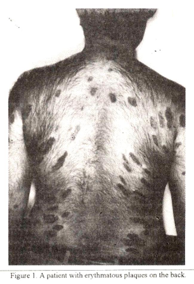
1 Sarcoidosis with Skin Manifestations - Two Case Reports and Review of Literature Pages with reference to book, From 162 To 164 Shamaila Siddiqi, Aleern Qureshi ( Departments of Dermatology, The Aga Khan University Medical College, Karachi. ) Shahid Pervaiz ( Departments of Pathology, The Aga Khan University Medical College, Karachi. ) Javaid Ahmed Khan ( Departments of Medicine, The Aga Khan University Medical College, Karachi. ) Sarcoidosis is a granulomatous disease of unknown etiology involving various organ systems. The skin involvement includes a variety of lesions. In this report two cases of sarcoidosis with different skin manifestations are described. Introduction Sarcoidosis is a multi-system granulomatous disease of unknown etiology, most commonly affecting young adults and presenting most frequently with bilateral hilarlymphadenopathy, pulmonary infiltration, eye or skin lesions 1. Cutaneous lesions are present in 20-35% of patients and are polymorphous 2. The diagnosis is established most securely when clinico-radiographic findings are supported by histologic evidence of non-caseating granulomas. Here we report two cases of sarcoidosis with skin involvement. Case 1 A 34 years old male presented with a two years histoty of multiple skin lesions all over the body. The lesions consisted of multiple erythmatous papules and plaques on back and chest with scaling and some indurated areas (Figures 1 and 2).
2

3 A skin biopsy revealed non-cascating granulomas and the patient was started on anli-tuberculous therapy, which he continued for seven months, but new lesions kept on appearing. He came to this hospital for second opinion. Besides skin lesions he gave a history of recentweight loss and low grade fever. General examination apart from skin lesions was unremarkable with no lymphadenopathy. Respiratory system examination was also normal. There were no other systemic findings, in particular liver and spleen were not palpable. His investigations revealed an ESR of 9 mm/hour, semm calcium of 8.9 mg%, angiotensin converting enzyme levels of 135 lu/l (normal 8-52 lu/l). His mantoux test was negative. Skin biopsy was repeated which once again showed non-caseating granulomas. Chest X-ray showed lymphadenopathy involving the mediastinum and paratracheal region. CT revealed bilateral mediastinal and paratracheallymphadenopathy. A repeat biopsy again showed non- casealing granulomatous inflanirnation without necrosis (Figure 3).

4 The patient was started on topical steroids, but because of the extensiveness of the lesions and lack of substantial improvement he was started on oral prednisolone 20mg bid. On follow-up remarkable improvement of the lesions was noted and the patient was continued on same treatment with a plan for gradual tapering of the steroids. Case 2 A 45 years old male originally presented in 1992 with complaints of dry cough and dvspnea. Chest x- ray revealed bilateral hilar lymphadenopathy and the CT scan showed bilateral hilar and mediastinal lymph node enlargement. He was advised to have a lymph node biopsy but he did not agree and was lost to follow-up. The patient again presented two years later with complaints of dry cough, progressively increasing dyspnea and weight loss. In the course of time he had also developed some skin lesions. His physical examination revealed presence of subcutaneous nodules on the back. Nodules were 0.5 to 1 cm in size, pale in colour and non-tender. They were scattered all over the back (Figure 4),
, serum Ca+ of 10.3 mg% and mantoux test was negative. A chest X-ray done in 1994 showed hilar lymphadenopathy and reticular pattern at the bases.")
5 there was no hepatosplenomegaly or any other findings and rest of systemic examination was normal. The investigations showed an ESR of 51 mm/hour, ACE levels of 91.5 lull (normal 8-52 lull), serum Ca+ of 10.3 mg% and mantoux test was negative. A chest X-ray done in 1994 showed hilar lymphadenopathy and reticular pattern at the bases. Biopsy of the skin lesions showed non-caseating granulomas. He was started on oral steroids 40 mg OD. On reviewing the patient few weeks later considerable improvement was seen in his skin lesions. His dyspnoea and cough also showed marked improvement. Discussion The skin involvement in sarcoidosis is polymorphous and the lesions may be specific or non-specific. The important specific lesions are lupus pcrnio. plaques and maculopapular eruptions. The important non-specific lesion is eiythema nodosurn. Other skin lesions include alopecia. erytherodermas, subcutaneous nodules. erythema multiforme, icthyosis, dystrophic calcifications and verucous outgrowths Nail involvement is rare in sarcoidosis 3. Lupus pemio, the most characteristic of all sarcoid lesions, is a chronic, violaceous, indurated skin lesion with a predilection for the nose, cheeks, ears and lips. It is more common in the females and those over forty. Compared with sarcoidosis overall, lupus pernio has a closer affinity with satcoidosis of the upper respiratory tract, bone cysts and lacnmal gland involvement 1. The maculopapular lesions are the most common skin manifestations of sarcoidosis in the black
6 patients. The waxy translucent lesions with a distinct flat top vaiy from 2-6 mm in diameter. They characteristically occur on the face, lids, around the orbits, in the nasolabial folds and on the nape and upper back 3. Plaques of sarcoidosis usually manifest as elevated, indurated purplish patches commonly located on the limbs, face, back and buttocks. The distribution is usually bilateral and symmetrical. The central part of the plaque is pale and atrophic, the periphery indurated elevated and dark. In the presence of telangiectatic vessels the lesions are called angiolupoid. Subcutaneous nodules, also called Darier-Roussy sarcoidosis are oval, firm, painless structures that anse deep in the dermis and subcutaneous tissue of the trunk and extremities. Scars from atrophy, trauma, surgery or venipuncture may become purple, swollen and tender either at the time the patient presents or during reactivation of the disease 4. Ulcerative sarcoidosis is rare, occurs more frequently in black women and usually involves the legs. Erythemia nodosum is a hypersensitivity reaction that is the most common non-specific cutaneous manifestation of sarcoidosis. Systemic manifestations such as fever, malaise and polyparthralgia occur in about fifty percent of patients with erythema nodosum 5. Because of the high rate of spontaneous resolution, they seldom require steroid therapy. The exact prevalence of sarcoidosis in Pakistan is not known. It is probably underdiagnosed and being overshadowed by other granulomatous diseases like tuberculosis. The criteria for establishing the diagnosis of sarcoidosis includes (1) a compatible clinical or radiological picture or both; (2) histological evidence of noncaseating granuloma and (3) negative special stains and cultures for other entities (e.g., acid fast bacilli or fungi in sputum or tissue specimen) 3. The serum calcium, ESR and ACE levels are elevated significantly in patients with sarcoidosis and serial measure- ments may prove useful in the momtoring of these patients, but theirvalue in the diagnosis of sarcoidosis is limited as they are non-specific. The definitive diagnosis still requires the demonsiration of non- caseatinggranuloma inthe involved tissue 1. The management of cutaneous sarcoidosis must take into account the seriousness of the accompanying internal involvement and the natural history of the particular type of lesion. Papular lesions are likely to fade without treatment, whereas lupus pernio does not. High potency topical steroids may sometimes prove helpful, as may intralesional triamcinalone injections. Cryotherapy and radiotherapy have occasionally been used. In systemic therapy at present, corticosteroids are the most effective treatment, given at first at a relatively higher dose and then tapering over a period of several weeks to a lower maintenance dose on alternate days 6. Cytostatic drugs may be tried if corticosteroids are contraindicated or have beenineffective. Methotraxate seems, so far, to have the best track record in the treatment of cutaneous as well as disseminated sarcoidosis. Chiorambucil and azathioprine may be used for their steroid sparing effect 7. Other drugs which have been tried with some success include allopurinol 8, antimalanals 10 and oxyphenabutazone 10. The chance of spontaneous remission favours a conservative approach to systemic therapy, which will usually carry the hazards of long term immunosuppression. At any time the pattern of the disease may change, but an expectant poiicy is best if the course is not progressive and if vital structures are not involved.the two cases discussed reiterate that sarcoidosis with its various clinical manifestations exists in Pakistan and poses a difficulty for the physician in diagnosis and management. References 1. Saboor, S. A. and Johnson, N. M. Review artcle on sarcoidosis. Br. J. Hosp. Med., 1992;48:6. 2. Kerdel, F. A. and Moschella S. L. Sarcoidosis: An updated review. J. Am. Acad. Dermatol., 1984;11: Sharma, 0. P. Cutaneous sarcoidosis. Chest 1972;61: Vien, N. K., Stahl, D. and Brodthagen, H. Cutaneous sarcoidsosis in Caucasians. J. Am. Acad. Dermatol., 1987; 16:
7 5. James, D. G. and Siltzbach, T E. A worldwide review ofsarcoidosis. Ann. N. Y. Acad. Sci., 1976;278: Israel, H. L. The treatment of sarcoidosis. Postgrad. Med. J., 1970;46: Lower, E. E. and Baughman, R. P. The useoflow dose methotraxate in refractory sarcoidosis. Am. J. Med. Sci., 1990;299: Samuel, M., Allen, 0. E., McMillan, S. C. et al. Sarcoidosis: Initial results on 6 patients treated with allopurinol. (Summary). Br. J. Dermatol., 1984; Barre, P. E., Gascon-Barre, M., Meakins, J. L. et al. Hydroxychloroquine treatment of hypercalcemia in a patient with sarcoidosis undergoing hemodia. lysis. Am. J. Med., 1987;82: James, D. 0., Carstairs, L. S. Trowell, 1. et al. Treatment of sarcoidosis: Report of a controlled therapeutic trial. Lancet, 1967;ii:526-8.
Pulmonary Sarcoidosis - Radiological Evaluation
 Original Research Article Pulmonary Sarcoidosis - Radiological Evaluation Jayesh Shah 1, Darshan Shah 2*, C. Raychaudhuri 3 1 Associate Professor, 2 1 st Year Resident, 3 Professor and HOD Radiology Department,
CLINICAL VIGNETTE Sarcoidosis: A Case Study Gloria Kim, M.D.
 CLINICAL VIGNETTE Sarcoidosis: A Case Study Gloria Kim, M.D. Case Report A 56-year-old female presented to her primary care physician with complaints of dyspnea on exertion and increasing cough. She reported
Case Report Pulmonary Sarcoidosis following Etanercept Treatment
 Case Reports in Rheumatology Volume 2012, Article ID 724013, 4 pages doi:10.1155/2012/724013 Case Report Pulmonary Sarcoidosis following Etanercept Treatment Kuljeet Bhamra and Richard Stevens Department
Role of Serum Angiotensin Converting Enzyme in Sarcoidosis
 Role of Serum Angiotensin Converting Enzyme in Sarcoidosis Pages with reference to book, From 131 To 133 Aysha H. Khan,Farooq Ghani,Mohammad Khurshid ( Departments of Pathology, The Aga Khan University
In our paper, we suggest that tuberculosis and sarcoidosis are two ends of the same spectrum. Given the pathophysiological and clinical link between
 In our paper, we suggest that tuberculosis and sarcoidosis are two ends of the same spectrum. Given the pathophysiological and clinical link between the two, we also propose a classification system for
Cutaneous Sarcoidosis Misdiagnosed as Leprosy. Report of Two Cases and Review of Literature
 Indian J Lepr 2016, 88 : 177-183 Hind Kusht Nivaran Sangh, New Delhi http://www.ijl.org.in Case Report Cutaneous Sarcoidosis Misdiagnosed as Leprosy. Report of Two Cases and Review of Literature 1 2 3
Sarcoidosis: the clinical problem
 Postgraduate Medical Journal (1988) 64, 531-535 Sarcoidosis: the clinical problem Edmund Neville St Mary's Hospital, Portsmouth P03 6AD, UK. Summary: This paper reviews the clinical manifestations of acute
manifestations are uncommon. Initial descriptions of the disease (Rosai and Dorfman, 1969) specifically
 specifically") Postgraduate Medical Journal (July 1980) 56, 521-525 Diffuse cutaneous involvement and sinus histiocytosis with massive lymphadenopathy A. A. WOODCOCK B.Sc., M.B., Ch.B., M.R.C.P. Summary Severe skin involvement
Postgraduate Medical Journal (July 1980) 56, 521-525 Diffuse cutaneous involvement and sinus histiocytosis with massive lymphadenopathy A. A. WOODCOCK B.Sc., M.B., Ch.B., M.R.C.P. Summary Severe skin involvement
A Case of Pulmonary Sarcoidosis with Endobronchial Nodular Involvement
 http://dx.doi.org/10.4046/trd.2013.74.6.274 ISSN: 1738-3536(Print)/2005-6184(Online) Tuberc Respir Dis 2013;74:274-279 CopyrightC2013. The Korean Academy of Tuberculosis and Respiratory Diseases. All rights
CPC. Chutika Srisuttiyakorn, M.D. Kobkul Aunhachoke, M.D. Phramongkutklao Hospital Bangkok, Thailand
 CPC Chutika Srisuttiyakorn, M.D. Kobkul Aunhachoke, M.D. Phramongkutklao Hospital Bangkok, Thailand A 53 year-old woman with fever, facial swelling and rashes on face, trunk and upper extremities for 3
CHRONIC INFLAMMATION
 CHRONIC INFLAMMATION Chronic inflammation is an inflammatory response of prolonged duration often for months, years or even indefinitely. Its prolonged course is proved by persistence of the causative
Arthritis and Rosai-Dorfman Disease of the Skin: A Diagnostic Dilemma
 Arthritis and Rosai-Dorfman Disease of the Skin: A Diagnostic Dilemma Introduction Pages with reference to book, From 280 To 282 Irshad Nabi Soomro ( Department of Pathology, The Aga Khan University Hospital,
Chest radiograph of an. asymptomatic man. Case report. Case history
 Eleftheria Chaini 1, Niki Giannakou 2, Dimitra Haini 3, Anna Maria Athanassiadou 4, Angelos Tsipis 4, Nikolaos D. Hainis 5 elhaini@otenet.gr 1 Pulmonary Dept, Corfu General Hospital, Kontokali, Greece.
Angiolupoid Sarcoidosis : A Clinicopathological Study of a Distinct Variant of Cutaneous Sarcoidosis
 Angiolupoid Sarcoidosis : A Clinicopathological Study of a Distinct Variant of Cutaneous Sarcoidosis Hsiang-Ju Tsai Yue-Zon Kuan Wen-Rou Wong Yea-Huey Chuang Wei-Ming Wu Sarcoidosis is a multisystem granulomatous
Sarcoidosis. S.K. Kabra A. Bagga Madhulika A. Chatterjee V. Seth. Sarcoidosis is a chronic multisystem disease of unkmbwn etiology, usually affecting
 INDIAN PEDIATRICS 9. Saigal S, Zunyk O, Larke B, Chernesky M. The outcome of children with congenital cytomegalovirus infection A long term follow up study. Am J Dis Child 1982,136: 896-901. 10. Deka RC.
Diagnosis of cutaneous sarcoidosis; clinical and the prognostic significance of skin lesions
 Yanardag et al. Multidisciplinary Respiratory Medicine 2013, 8:26 ORIGINAL RESEARCH ARTICLE Open Access Diagnosis of cutaneous sarcoidosis; clinical and the prognostic significance of skin lesions Halil
Tuberculosis. By: Shefaa Q aqa
 Tuberculosis By: Shefaa Q aqa Tuberculosis is a communicable chronic granulomatous disease caused by Mycobacterium tuberculosis. It usually involves the lungs but may affect any organ or tissue in the
Multiple bilateral pulmonary nodules masquerading as pulmonary metastasis; a case of nodular sarcoidosis
 Electronic Physician (ISSN: 2008-5842) August 2016, Volume: 8, Issue: 8, Pages: 2802-2806, DOI: http://dx.doi.org/10.19082/2802 Multiple bilateral pulmonary nodules masquerading as pulmonary metastasis;
Sarcoidosis. Sarcoidosis Care at National Jewish Health. Causes
 Sarcoidosis Sarcoidosis is a chronic disease that can affect any organ in the body, but most commonly affects the lungs. Very small (microscopic) clusters of inflammation or white cells, called granulomas,
Original Article Comparison of cutaneous sarcoidosis with systemic sarcoidosis: a retrospective analysis
 Int J Clin Exp Pathol 2014;7(1):372-377 www.ijcep.com /ISSN:1936-2625/IJCEP1310042 Original Article Comparison of cutaneous sarcoidosis with systemic sarcoidosis: a retrospective analysis Chunguang Tong,
Late-onset and Rare Far-advanced Pulmonary Involvement in Patients with Sarcoidosis in Taiwan
 Sarcoidosis in Taiwan ORIGINAL ARTICLE Late-onset and Rare Far-advanced Pulmonary Involvement in Patients with Sarcoidosis in Taiwan Chia-Wei Hsieh, 1 Der-Yuan Chen, 1 Joung-Liang Lan 1,2 * Background:
CHEILITIS GRANULOMATOSA
 CHEILITIS GRANULOMATOSA Report of two Cases with Clinical and Diagnostic Implications Presented By Dr. Amar Sholapurkar Under the guidance of Dr.Ausaf Ahsan Department of Oral Medicine & Radiology, MCODS,
Egyptian Dermatology Online Journal Vol. 6 No 1: 14, June 2010
 Wells Syndrome H. Gammaz, H. Amer, A. Adly and S. Mahmoud Egyptian Dermatology Online Journal 6 (1): 14 Al-Haud Al-Marsoud Hospital, Cairo, Egypt e-mail: hananderma@hotmail.com Submitted: April 15, 2010
This PDF is available for free download from a site hosted by Medknow Publications
 16 CMYK Original Article Cutaneous sarcoidosis: Clinical profile of 23 Indian patients Vikram K. Mahajan, Nand Lal Sharma, Ramesh Chander Sharma, Vikas C. Sharma Department of Dermatology, Venereology
Sarcoidosis. Julia Rhiannon Harborview/UW Rheumatology November 2010
 Sarcoidosis Julia Rhiannon Harborview/UW Rheumatology November 2010 julrhi@uw.edu outline Introduction/Epi/Genetics Clinical features Acute/Lofgren s vs chronic Pathogenesis Treatment sarc eidos osis flesh
Degos Disease: A Case Report and Review of Literature
 Degos Disease: A Case Report and Review of Literature Monira waked Egyptian Dermatology Online Journal 4 (1): 5, June 2008 Al Houd Al Marsod Hospital Submitted for publication: May 25 th, 2008 Accepted
Glistening, Skin-Colored Nodule
 To Print: Click your browser's PRINT button. NOTE: To view the article with Web enhancements, go to: http://www.medscape.com/viewarticle/436334 Medscape Dermatology Clinic Glistening, Skin-Colored Nodule
Collar stud abscess an interesting case report
 Volume 2 issue 2 2012 ISSN 2250-0359 Collar stud abscess an interesting case report Kameshwaran Kannappan Punniyakodi * Balasubramanian Thiagarajan* *Stanley Medical College Chennai, Tamilnadu Abstract
3/25/2012. numerous micro-organismsorganisms
 Congenital & Neonatal TB A Case of Tuberculosis Congenital or Acquired? Felicia Dworkin, MD NYC DOHMH Bureau TB Control World TB Day March 23, 2012 Congenital TB: acquired by the fetus during pregnancy
Elsevier B.V.; この論文は出版社版でありま Right 引用の際には出版社版をご確認ご利用ください This is
 Title Refractory cutaneous lichenoid sarc tranilast. Author(s) Nakahigashi, Kyoko; Kabashima, Kenj Utani, Atsushi; Miyachi, Yoshiki Citation Journal of the American Academy of 63(1): 171-172 Issue Date
Cutanous Manifestation of Lupus Erythematosus. Presented By: Dr. Naif S. Al Shahrani Salman Bin Abdaziz university
 Cutanous Manifestation of Lupus Erythematosus Presented By: Dr. Naif S. Al Shahrani Salman Bin Abdaziz university A 50-year old lady, who is otherwise healthy, presented to the dermatology clinic with
Chest Radiology Interpretation: Findings of Tuberculosis
 Chest Radiology Interpretation: Findings of Tuberculosis Get out your laptops, smart phones or other devices pollev.com/chestradiology Case #1 1 Plombage Pneumonia Cancer 2 Reading the TB CXR Be systematic!
Sarcoidosis. Sarcoidosis Alan J. Kanouff, DO. POMA District VIII 31 st Annual Educational Winter Seminar January 25 28, Disclosures.
 Sarcoidosis Alan J. Kanouff DO, FCCP Lung Disease Center of Central Pennsylvania Disclosures Speaker for AstraZeneca Symbicort Bevespi Speaker for Merck Belsomra Speaker for Sunovion Utibron Seebri Overview
Bilateral Chest X-Ray Shadowing and Bilateral leg lesions - A case of Pulmonary Kaposi Sarcoma
 Article ID: WMC005047 ISSN 2046-1690 Bilateral Chest X-Ray Shadowing and Bilateral leg lesions - A case of Pulmonary Kaposi Sarcoma Peer review status: No Corresponding Author: Dr. Mohammad Fawad Khattak,
INTERSTITIAL LUNG DISEASE. Radhika Reddy MD Pulmonary/Critical Care Long Beach VA Medical Center January 5, 2018
 INTERSTITIAL LUNG DISEASE Radhika Reddy MD Pulmonary/Critical Care Long Beach VA Medical Center January 5, 2018 Interstitial Lung Disease Interstitial Lung Disease Prevalence by Diagnosis: Idiopathic Interstitial
An Introduction to Radiology for TB Nurses
 An Introduction to Radiology for TB Nurses Garold O. Minns, MD September 14, 2017 TB Nurse Case Management September 12 14, 2017 EXCELLENCE EXPERTISE INNOVATION Garold O. Minns, MD has the following disclosures
Concise Review for Primary-Care Physicians
 Concise Review for Primary-Care Physicians Sarcoidosis RICHARD A. DEREMEE, M.D. Sarcoidosis is a systemic granulomatous process of unknown cause. Pathologically, it is characterized by noncaseous granuloma,
The exact cause of sarcoidosis is unknown. However, gender, race, and genetics can increase the risk of developing the condition:
 What is sarcoidosis? Sarcoidosis is an inflammatory disease in which granulomas, or clumps of inflammatory cells, form in various organs. This causes organ inflammation. Sarcoidosis may be triggered by
Acute miliary tuberculosis in a five-month-old boy
 Hong Kong J. Dermatol. Venereol. (2007) 15, 138-142 Case Report Acute miliary tuberculosis in a five-month-old boy FC Ip and KC Lee A five-month-old boy presented with fever and multiple cutaneous papular
Lofgren's syndrome A case report
 Open Journal of Clinical & Medical Case Reports Lofgren's syndrome A case report Volume 3 (2017) Issue 24 ISSN 2379-1039 Mohammad Iqbal KM; Thara Pratap; Muhammed Jasim Abdul Jalal*; Chippy Eldhose; Shani
Sarcoidosis Registry Proforma
 Patient Demographics Patient Data Has a patient consent form been completed? 1.1 Title Mr 1.2 1.3 Forename(s) PlainText Surname PlainText 1.4 Gender Male 1.5 1.6 1.7 1.8 1.9 Mrs Ms Miss Dr Other
Lymphoma co existing with Tuberculosis granulomatous
 Available online at www.worldscientificnews.com WSN 90 (2017) 265-270 EISSN 2392-2192 SHORT COMMUNICATION Lymphoma co existing with Tuberculosis granulomatous Madeeha Subhan 1, *, Waleed Sadiq 2 1 Ayub
TB Radiology for Nurses Garold O. Minns, MD
 TB Nurse Case Management Salina, Kansas March 31-April 1, 2010 TB Radiology for Nurses Garold O. Minns, MD April 1, 2010 TB Radiology for Nurses Highway Patrol Training Center Salina, KS April 1, 2010
MULTI-SYSTEM SARCOIDOSIS CAUSING PANHYPOPITUITARISM: RAPID IMPROVEMENT WITH CORTICOSTEROID THERAPY Rashid Mahboob, MD; Ali A.
 ENDOCRINE PRACTICE Rapid Electronic Article in Press Rapid Electronic Articles in Press are preprinted manuscripts that have been reviewed and accepted for publication, but have yet to be edited, typeset
Past Surgical History Unremarkable. case study [chemistry histology]
![Past Surgical History Unremarkable. case study [chemistry histology]](/thumbs/85/91357833.jpg "Past Surgical History Unremarkable. case study [chemistry histology]") case study [chemistry histology] Chronic Cough in an Older Female Ellen Sigauke, MD, Thompson T. Kamba, MD, Monte S. Willis, MD, PhD Department of Pathology, University of Texas Southwestern Medical Center,
case study [chemistry histology] Chronic Cough in an Older Female Ellen Sigauke, MD, Thompson T. Kamba, MD, Monte S. Willis, MD, PhD Department of Pathology, University of Texas Southwestern Medical Center,
CLINICAL FEATURES IN PULMONARY TUBERCULOSIS
 CLINICAL FEATURES IN PULMONARY TUBERCULOSIS Dr. Amitesh Aggarwal Department of Medicine Tuberculosis Captain of all the Men of Death Great White Plague devastating effect on society 100 years ago one in
Uncommon clinical presentations of leprosy: apropos of three cases
 Lepr Rev (2016) 87, 246 251 CASE REPORT Uncommon clinical presentations of leprosy: apropos of three cases RASHMI JINDAL* & NADIA SHIRAZI** *Department of Dermatology, Venereology & Leprosy, Himalayan
Annular elastolytic giant cell granuloma presented with annular erythematous patches over the face and cheek in a Chinese lady
 Hong Kong J. Dermatol. Venereol. (2009) 17, 151-155 Case Report Annular elastolytic giant cell granuloma presented with annular erythematous patches over the face and cheek in a Chinese lady SKF Loo, LY
Objectives. 1. Recognizing benign skin lesions. 2.Know which patients will likely need surgical intervention.
 The Joy of Pediatric Skin Dr. Claire Sanger University of Kentucky Plastic & Reconstructive Surgery Objectives 1. Recognizing benign skin lesions 2.Know which patients will likely need surgical intervention.
Langerhans Cell Histiocytosis with Anterior Mediastinum, Pulmonary and Liver Involvement: CT Demonstration
 Chin J Radiol 2002; 27: 191-195 191 Langerhans Cell Histiocytosis with Anterior Mediastinum, Pulmonary and Liver Involvement: CT Demonstration SIU-CHEUNG CHAN 1 MUN-CHING WONG 1 SHIU-FENG HUANG 2 WAN-CHAK
Infections and nonmicrobial inflammatory stimuli can cause leukocytosis (as seen in Lab 1) as well as lymph node enlargement (lymphadenopathy).
 as well as lymph node enlargement (lymphadenopathy).") LAB 5: LYMPHOID TISSUE AND SKIN The focus of this week s lab will be pathology of the lymphoid tissue and skin. The lymphoid organs include the thymus, spleen, and lymph nodes. Abnormalities in the lymph
LAB 5: LYMPHOID TISSUE AND SKIN The focus of this week s lab will be pathology of the lymphoid tissue and skin. The lymphoid organs include the thymus, spleen, and lymph nodes. Abnormalities in the lymph
A Rare case of Tubercular Gingivitis Case Report
 Case Report A Rare case of Tubercular Gingivitis Case Report *Dr. Ansh Chugh 1, Dr. Firoz A Hakkim 2, Dr. Rajesh. V 3, Dr. Raghava Sharma 4 1: JUNIOR RESIDENT IN GENERAL MEDICINE 2: SENIOR RESIDENT IN
CUTANEOUS SARCOIDOSIS AS AN EXPRESSION OF SYPHILIS*
 UTANEOUS SAROIDOSIS AS AN EXPRESSION OF SYPILIS* REPORT AND DISUSSION OF A ASE EUGENE TRAUGOTT BERNSTEIN, M.D. AND MORRIS LEIDER, M.D. The following case is reported as an instance of an eruption which
J of Evolution of Med and Dent Sci/ eissn , pissn / Vol. 3/ Issue 37/Aug 21, 2014 Page 9580
 THE SPECTRUM OF CERVICAL LYMPHADENOPATHY IN CHILDREN: A STUDY IN RURAL NORTH INDIA Pawan Tiwari 1, Satya Kiran Kapoor 2, Madhu Tiwari 3, Yogesh Yadav 4 HOW TO CITE THIS ARTICLE: Pawan Tiwari, Satya Kiran
The World of Dermatology
 The World of Dermatology, F.A.A.D. Assistant Professor of Dermatology University of Nevada School of Medicine Director of J. Woodson Dermatology & Associates, LTD Objectives Cite the advances in management
Bilateral multiple choroidal granulomas and systemic vasculitis as presenting features of tuberculosis in an immunocompetent patient
 Kumar et al. Journal of Ophthalmic Inflammation and Infection (2016) 6:40 DOI 10.1186/s12348-016-0109-9 Journal of Ophthalmic Inflammation and Infection BRIEF REPORT Open Access Bilateral multiple choroidal
Present Status of Lab oratory Diagnosis o f Sarcoidosis
 A n n a l s o f C l i n i c a l L a b o r a t o r y S c i e n c e, Vol. 3, No. 2 Copyright 1 9 7 3, Institute for Clinical Science Present Status of Lab oratory Diagnosis o f Sarcoidosis HAROLD L. ISRAEL,
Rare Dermatologic Condition That Belongs in the Differential of Granulomatous Cutaneous Disorders
 Rare Dermatologic Condition That Belongs in the Differential of Granulomatous Cutaneous Disorders 2013 Annual Meeting of the Medical Dermatology Society February 28, 2013 Amanda Champlain MD, Emily Keimig
Thoracic Sarcoidosis Imaging Updated: Jul 19, 2013
 Thoracic Sarcoidosis Imaging Updated: Jul 19, 2013 Overview Radiography Computed Tomography Magnetic Resonance Imaging Nuclear Imaging Show All Multimedia Library References Overview For patients with
Nodular Muscular Sarcoidosis Extending to All Limb Muscles
 47 1997 CD4/8 MRI T1 T2 74.6 IU/l 46.0 µg/ml MRI [ ] MRI Nodular Muscular Sarcoidosis Extending to All Limb Muscles Takahiro Nishitake, Eishi Miyazaki, Masaru Ando, Tetsujiro Fukami, Osamu Matsuno, Takuya
Pathology of pulmonary tuberculosis. Dr: Salah Ahmed
 Pathology of pulmonary tuberculosis Dr: Salah Ahmed Is a chronic granulomatous disease, caused by Mycobacterium tuberculosis (hominis) Usually it involves lungs but may affect any organ or tissue Transmission:
Dilemma of Thoracic Tuberculosis Vs. Sarcoidosis in TB Endemic Areas: An Imaging Approach
 Dilemma of Thoracic Tuberculosis Vs. Sarcoidosis in TB Endemic Areas: An Imaging Approach A. S. Bhalla, A. Das, A. GOYAL, P. NARANJE, R. GULERIA, G. C. KHILNANI ALL INDIA INSTITUTE OF MEDICAL SCIENCES
A Diagnostic Dilemma: Metastatic Testicular Cancer and Systemic Sarcoidosis A Review of the Literature
 118 This is an Open Access article licensed under the terms of the Creative Commons Attribution- NonCommercial-NoDerivs 3.0 License (www.karger.com/oa-license), applicable to the online version of the
PULMONARY TUBERCULOSIS RADIOLOGY
 PULMONARY TUBERCULOSIS RADIOLOGY RADIOLOGICAL MODALITIES Medical radiophotography Radiography Fluoroscopy Linear (conventional) tomography Computed tomography Pulmonary angiography, bronchography Ultrasonography,
Sarcoidosis of the upper respiratory tract
 Thorax (1976), 31, 66. Sarcoidosis of the upper respiratory tract and its association with lupus pernio E. NEVILLE, R. G. S. MILLS', D. K. JASH, D. M. MacKINNON, L. S. CARSTAIRS, and D. GERAINT JAMES Departments
Rheumatologic Emergencies It s not just swollen joints. Joanne Homik Rheumatologist University of Alberta
 Rheumatologic Emergencies It s not just swollen joints Joanne Homik Rheumatologist University of Alberta Or is it? Disclosures No relevant conflicts of interest regarding the content of this presentation
Thoracic Manifestations of Sarcoidosis Using Multi-Slice CT
 IOSR Journal of Dental and Medical Sciences (IOSR-JDMS) e-issn: 2279-0853, p-issn: 2279-0861.Volume 14, Issue 9 Ver. II (Sep. 2015), PP 63-68 www.iosrjournals.org Thoracic Manifestations of Sarcoidosis
Hiroyuki Kamiya 1), Soichiro Ikushima 1), Tetsu Sakamoto 1), Kozo Morimoto 1), Tsunehiro Ando 1), Masaru Oritsu 1), Atsuo Goto 2), Tamiko Takemura 3)
, Soichiro Ikushima 1), Tetsu Sakamoto 1), Kozo Morimoto 1), Tsunehiro Ando 1), Masaru Oritsu 1), Atsuo Goto 2), Tamiko Takemura 3)") 29 2001 ACE39.7IU/l X TBLB 2002 11 Langhans [ ] A Case of Granulomatous Interstitial Nephritis with Progressive Renal Impairment Due to Sarcoidosis in the Course of Spontaneous Improvement of Pulmonary
29 2001 ACE39.7IU/l X TBLB 2002 11 Langhans [ ] A Case of Granulomatous Interstitial Nephritis with Progressive Renal Impairment Due to Sarcoidosis in the Course of Spontaneous Improvement of Pulmonary
Imaging Features of Sarcoidosis on MDCT, FDG PET, and PET/CT
 AJR Integrative Imaging LIFELONG LEARNING FOR RADIOLOGY Imaging Features of Sarcoidosis on MDCT, FDG PET, and PET/CT Hima B. Prabhakar 1, Chad B. Rabinowitz 1, Fiona K. Gibbons 2, Walter J. O Donnell 2,
Fever in Lupus. 21 st April 2014
 Fever in Lupus 21 st April 2014 Fever in lupus Cause of fever N= 487 % SLE fever 206 42 Infection in SLE 265 54.5 Active SLE and infection 8 1.6 Tumor fever 4 0.8 Miscellaneous 4 0.8 Crucial Question Infection
PHYSIOLOGY AND MANAGEMENT OF HISTIOCYTIC DISEASE. Brant Ward, MD, PhD Division of Rheumatology, Allergy, and Immunology
 PHYSIOLOGY AND MANAGEMENT OF HISTIOCYTIC DISEASE Brant Ward, MD, PhD Division of Rheumatology, Allergy, and Immunology What do histiocytes do? Apoptotic body removal Phagocytosis Antigen presentation Types
Time to Learn. 6 th March 2018 Dr. Shirin Chakera GPwSI Integrated Dermatology Service
 Time to Learn 6 th March 2018 Dr. Shirin Chakera GPwSI Integrated Dermatology Service The Red Face Rosacea Acne Seborrhoeic eczema eczema Psoriasis Slapped cheek syndrome Fungal infection Erysipelas...
More Non-infectious Granulomatous Diseases! Karolyn Wanat, MD Assistant Professor, Dermatology & Pathology University of Iowa
 More Non-infectious Granulomatous Diseases! Karolyn Wanat, MD Assistant Professor, Dermatology & Pathology University of Iowa Conflicts of Interest/Disclosure None Classification/Overview 1) Necrobiotic/Palisading
COMMUNICATIONS OCULAR SARCOIDOSIS* it is the most troublesome and incapacitating. Its early recognition and treatment
 Brit. J. Ophthal. (1964) 48, 461. COMMUNICATIONS OCULAR SARCOIDOSIS* BY D. GERAINT JAMES Royal Northern Hospital and Royal Eye Medical Ophthalmology Unit, Lambeth Hospital R. ANDERSON AND D. LANGLEY Royal
1 yr old girl presented with Fever on and off 3 months H/o frequent semisolid bulky stools 3 months Progressive abdominal distension 3 months Failure
 Dr Rajasree S Dr Srinivas S, Dr Bagdi RK, Dr Satheesh C Apollo Childrens Hospital, Chennai 1 yr old girl presented with Fever on and off 3 months H/o frequent semisolid bulky stools 3 months Progressive
Egyptian Dermatology Online Journal Vol. 5 No 2:16, December Squamous Cell Carcinoma Arising on Extensive and Chronic Lupus Vulgaris
 Squamous Cell Carcinoma Arising on Extensive and Chronic Lupus Vulgaris Pathak D.* and Thapa A** Egyptian Dermatology Online Journal 5 (2): 16 * Consultant Dermatologist, Delhi Dermatology Group Kubba,
American College of Radiology ACR Appropriateness Criteria
 American College of Radiology ACR Criteria Radiologic Management of Thoracic Nodules and Masses Variant 1: Middle-aged patient (35 60 years old) with an incidental 1.5-cm lung nodule. The lesion was smooth.
Sarcoidosis Case. Robert P. Baughman Interstitial Lung Disease and Sarcoidosis Clinic University of Cincinnati, USA. WASOG: educational material
 Sarcoidosis Case Robert P. Baughman Interstitial Lung Disease and Sarcoidosis Clinic University of Cincinnati, USA WASOG: educational material Sarcoidosis Case patient is a Caucasian male age 46 was diagnosed
A 40-year old male with follicular papule and pustule at central face area for 3 months
 A 40-year old male with follicular papule and pustule at central face area for 3 months GMS- Neg AFB-Neg Fite stain - neg HISTOPATHOLOGICAL DIFFERENTIAL DIAGNOSIS CASEOUS GRANULOMA INFECTION -MYCOBACTERIUM
I have a skin lump doc! What s next? 12 th August 2017 Dr. Sue-Ann Ho Ju Ee
 I have a skin lump doc! What s next? 12 th August 2017 Dr. Sue-Ann Ho Ju Ee Some thoughts Is this skin cancer? How common is this? How likely is this in this patient? What happens next if it s something
Abscess. A abscess is a localized collection of pus in the skin and may occur on any skin surface and be formed in any part of body.
 Abscess A abscess is a localized collection of pus in the skin and may occur on any skin surface and be formed in any part of body. Ethyology Bacteria causing cutaneous abscesses are typically indigenous
Case presentation. Dr Rammohan Reddy 1 st year PG, Dept of DVL, Kamineni Institute of Medical Sciences, Narketpally.
 Case presentation Dr Rammohan Reddy 1 st year PG, Dept of DVL, Kamineni Institute of Medical Sciences, Narketpally. Name : XXX Age : 33 years Sex : Female Occupation : Farmer IP no : 201608905 DOA : 15-02-2016
Tuberculosis - clinical forms. Dr. A.Torossian,, M.D., Ph. D. Department of Respiratory Diseases
 Tuberculosis - clinical forms Dr. A.Torossian,, M.D., Ph. D. Department of Respiratory Diseases 1 TB DISEASE Primary Post-primary (Secondary) Common primary forms Primary complex Tuberculosis of the intrathoracic
Management of uveitis
 Management of uveitis DR. ANUPAMA KARANTH Anti-inflammatory agents -itis = inflammation Treatment : stop inflammation Use anti-inflammatory drugs Most potent of such agents : Corticosteroids Corticosteroids
Cutaneous Leishmaniasis with Unusual Clinical and Histological Presentation: Report of Four Cases
 CASE REPORT Cutaneous Leishmaniasis with Unusual Clinical and Histological Presentation: Report of Four Cases Hamideh Moravvej 1, Mohammadreza Barzegar 1, Soheila Nasiri 1, Ehsan Abolhasani 1, and Mehdi
BSD Self Assessment Workshop 7 th July 2013 CASE 27 RAC6123
 BSD Self Assessment Workshop 7 th July 2013 CASE 27 RAC6123 M55. 4/7 tender lesions on knee, legs and arms. Also iritis/ weight loss/headache, synovitis.?vasculitis. Sarcoidosis. Biopsy from left elbow
Case Report An Uncommon Cause of a Small-Bowel Obstruction
 Hindawi Case Reports in Gastrointestinal Medicine Volume 2017, Article ID 1628215, 4 pages https://doi.org/10.1155/2017/1628215 Case Report An Uncommon Cause of a Small-Bowel Obstruction Ali Zakaria, Bayan
* MILIARY MOTTLING --
 * MILIARY MOTTLING -- RARE CAUSE DR ARATHI SRINIVASAN FELLOW IN PEDIATRIC HEMATO ONCOLOGY DR A ANDAL DEPARTMENT OF PEDIATRICS DR JULIUS XAVIER SCOTT DEPARTMENT OF PEDIATRIC HEMATO ONCOLOGY KANCHI KAMAKOTI
RESEARCH ARTICLE. The Relationship between Sarcoidosis of Intrathoracic Lymph Nodes and Digestive System Diseases: A Comparative Observation
 ISSN 0975-8542 Journal of Global Pharma Technology Available Online at www.jgpt.co.in RESEARCH ARTICLE The Relationship between Sarcoidosis of Intrathoracic Lymph Nodes and Digestive System Diseases: A
Benign versus Cancerous Lesions How to tell the difference FMF 2014 Christie Freeman MD, CCFP, DipPDerm, MSc
 1 Benign versus Cancerous Lesions How to tell the difference FMF 2014 Christie Freeman MD, CCFP, DipPDerm, MSc Benign lesions Seborrheic Keratoses: Warty, stuck-on Genetics and birthdays Can start in late
Disseminated cutaneous BCG infection following BCG immunotherapy in patients with lepromatous leprosy
 Lepr Rev (2015) 86, 180 185 CASE REPORT Disseminated cutaneous BCG infection following BCG immunotherapy in patients with lepromatous leprosy GEETI KHULLAR*, TARUN NARANG*, KUSUM SHARMA**, UMA NAHAR SAIKIA***
Difficult Diagnosis: Case History. 7 months prior, she happened to have undergone a C-spine MRI after a car accident
 Relevant Disclosures: None Difficult Diagnosis: Recent Advances in Neurology 2013 Jeffrey M. Gelfand, MD Assistant Professor UCSF Neuroinflammation and MS Center UCSF Department of Neurology Case History
HEMORRHAGIC BULLOUS HENOCH- SCHONLEIN PURPURA: A CASE REPORT
 HEMORRHAGIC BULLOUS HENOCH- SCHONLEIN PURPURA: A CASE REPORT Nirmala Ponnuthurai, Sabeera Begum, Lee Bang Rom Paediatric Dermatology Unit, Institute of Paediatric, Hospital Kuala Lumpur, Malaysia Abstract
22 year old QH mare with regionally extensive alopecia and scaling on one front limb and ventral chest (Figure 1 and 2).
.") 22 year old QH mare with regionally extensive alopecia and scaling on one front limb and ventral chest (Figure 1 and 2). Which of the following is the most likely disease? a. Sterile granuloma complex
22 year old QH mare with regionally extensive alopecia and scaling on one front limb and ventral chest (Figure 1 and 2). Which of the following is the most likely disease? a. Sterile granuloma complex
The Imaging Analysis of Pulmonary Sarcodiosis
 www.cancercellresearch.org ISSN: 2161-2609 Article The Imaging Analysis of Pulmonary Sarcodiosis Xin He, Chuanyu Zhang* Department of Radiology, Affiliated Hospital of Qingdao University, Qingdao, China
How do I evaluate a patient with a swollen lip?
 How do I evaluate a patient with a swollen lip? Yang Gu, BDS, MSc; Michele Williams, BSN, DMD, FRCD(C); Catherine F. Poh, DDS, PhD, FRCD(C) Tags: diagnosis oral pathology Cite this as: J Can Dent Assoc
Diagnostic Value of EBUS-TBNA in Various Lung Diseases (Lymphoma, Tuberculosis, Sarcoidosis)
") Diagnostic Value of EBUS-TBNA in Various Lung Diseases (Lymphoma, Tuberculosis, Sarcoidosis) Sevda Sener Cömert, MD, FCCP. SBU, Kartal Dr.Lütfi Kırdar Training and Research Hospital Department of Pulmonary
Diagnostic Value of EBUS-TBNA in Various Lung Diseases (Lymphoma, Tuberculosis, Sarcoidosis) Sevda Sener Cömert, MD, FCCP. SBU, Kartal Dr.Lütfi Kırdar Training and Research Hospital Department of Pulmonary
Evaluation of Neck Mass. Disclosure. Learning Objectives 3/24/2014. Karen T. Pitman MD, FACS Banner MDACC, Gilbert AZ. Nothing to disclose
 Evaluation of Neck Mass Karen T. Pitman MD, FACS Banner MDACC, Gilbert AZ Nothing to disclose Disclosure Learning Objectives 1. Describe a systematic method to evaluate a patient with a neck mass 2. Select