The Pathologic Manifestations of Small Airway Disease. Samuel A. Yousem, MD. Small Airway Disease (SAD) SAD
|
|
|
- Jessie Powell
- 6 years ago
- Views:
Transcription
1 The Pathologic Manifestations of Small Airway Disease Samuel A. Yousem, MD Small Airway Disease (SAD) A clinicopathologic syndrome reflecting a CHRONIC inflammatory and cicatricial process primarily affecting the bronchioles (airways <2 mm) that results in obstructive lung disease. Synonyms: bronchiolitis obliterans, obliterative bronchiolitis, constrictive bronchiolitis. SAD As a CHRONIC clinical syndrome, it needs to be separated from acute forms of SAD e.g. acute neutrophilic bronchiolitis however... Acute SAD can cause such severe airway injury that the patient is left with a permanent airflow deficit/irreversibly scarred airways e.g. RSV in infants.
2 SAD There is a disconnect between the histologic presence of SA inflammation and the diagnosis of SAD. Bronchiolar inflammation is present in many diseases but it does not mean that the patient has clinical SAD e.g. many interstitial lung diseases have airway inflammation. To make a diagnosis of SAD, the clinical context, radiologic findings and PFTs must support a diagnosis of SAD- irreversible obstructive disease. SAD The likelihood of SAD is related to the # of airways affected (diffuse disease). Severity of airway damage. Irreversibility of injury scarring/ luminal compromise. * you must have widespread airway disease to fall into this CPC diagnosis. The classification of SAD is confusing.....sometimes the SAD is defined by the clinical scenario...other times by the histology...and of course, there is histologic and clinical overlap of many of these conditions.
3 Classification of SAD 1. Bronchiolar involvement in bronchial disease. Asthma. Bronchiectasis. Cystic fibrosis. Aspiration. Bronchial obstruction. 2. Bronchiolar involvement in parenchymal lung disease. Bronchopneumonia. Eosinophilic pneumonia. Cryptogenic organizing pneumonia (idiopathic BOOP). Extrinsic allergic alveolitis. Langerhans cell histiocytosis. 3. Disorders in which bronchiolitis is the predominant abnormality. Follicular bronchiolitis. Respiratory bronchiolitis. Diffuse panbronchiolitis. Mineral dust bronchiolitis. Constrictive obliterative bronchiolitis. Primary Bronchiolar Disorders Classification Bronchiolitis obliterans/obliterative br-itis Respiratory bronchiolitis Follicular bronchiolitis Diffuse panbronchiolitis Autoimmune bronchiolitis Mineral dust airway disease Aspiration bronchiolitis Bronchiolitis Obliterans Synonym: constrictive bronchiolitis/ obliterative bronchiolitis. Usually defined as a small airway inflammatory process with irreversible scarring of the airways (that correlates with fixed and progressive functional deficits); no unique histologic features.
. DISEASE AT THIS STAGE IS REVERSIBLE 2.")
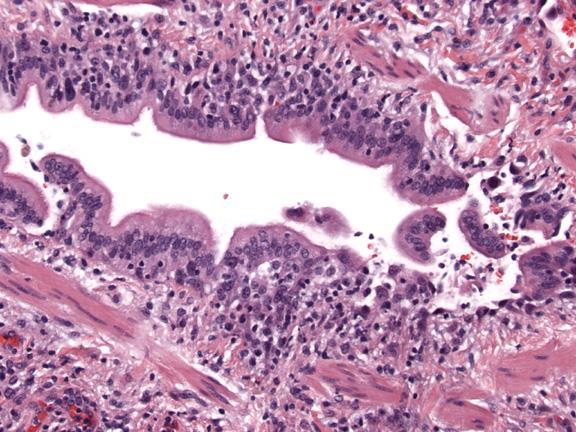
4 Bronchiolitis Obliterans/Obliterative Bronchiolitis Histopathology 1. Cellular bronchiolitis inflammatory infiltrate around airways; may include intraluminal granulation tissue polyps ( proliferative phase). DISEASE AT THIS STAGE IS REVERSIBLE 2. Constrictive bronchiolitis irreversible scarring of submucosa or obliteration of the airway lumen; may also have a peribronchiolar collar of collagen; usu. seen w/ active injury (#1) 3. Remember the functional impact of the inflammatory changes: Mucus/fibrosis physical obstruction. Epithelial hyperplasia, squamous and goblet metaplasia - luminal clearance. Cells cytokines/edema of airway wall. Smooth muscle spasm/constriction. Scar/metaplasia disrupted airflow dynamics.

5


6


7

8


9 Histology Etiology
10 Potential Causes of Constrictive/ Obliterative Bronchiolitis 1. Idiopathic 2. Collagen vascular diseases RA, SS, SLE, eosinophilic fasciitis. 3. Post infectious RSV, Adenovirus, influenza, parainfluenza, mycoplasma, chlamydia. 4. Toxic fume exposure NO 2, sulfur dioxide, ammonia, chlorine, nitrogen, mustard, phosgene, 9/11 lung. 5. Mineral dust inhalation asbestos, CWP, silicosis, nylon flock. 6. GVHD-bone marrow transplant recipients. 7. Lung allograft recipients. 8. Tobacco. 9. UC/CD. 10. Panbronchiolitis. 11. Drugs methotrexate, gold, penicillamine, lomustine. 12. Others. Respiratory Bronchiolitis RB is a ubiquitous finding in cigarette smokers when patients present with clinical ILD and no other cause is identified, we place these patients into the category of RB/ILD. Summary of findings in 156 patients Current smokers Exsmokers Never smokers Total RB present RB absent Total Ref: Fraig. AJSP, 2002


11 RB Histopathology Mild patchy lymphocytic bronchiolitis with luminal and centrilobular aggregates of finely pigmented macrophages, sparing of distal airspaces. Mild centrilobular alveolar septal collagenosis with lymphs. Associated changes mucous plugging, atelectasis. Excellent 10 year prognosis with smoking cessation with intermittent steroid therapy although recent studies question this benign course.


12

13 Diffuse Panbronchiolitis East Asians with predisposing HLA genotype (Japanese-B 54; Korean-A 11) with minority presence in Western world. 4 th -5 th decades; M>F; non-smokers. Subacute onset of productive cough with purulent sputum; dyspnea: repeated Hemophilus/Staph infections that change to Pseudomonas; clubbing. History of chronic sinusitis. Bilateral small nodular opacities in bases; bronchiectasis. Prognosis: 80% DOD (untreated), 30% (treated with macrolides), at ten years. Panbronchiolitis Diagnostic Criteria of Ministry of Health and Welfare of Japan Persistent cough, sputum and exertional dyspnea. Past history of/or current chronic sinusitis. Bilateral diffuse small nodular shadows on a plain chest film or centrilobular nodular shadows on chest CT images. Coarse crackles. FEV 1 /FVC <70% and PaO 2 <80 mm Hg. Titre of cold hemagglutinins equal to or higher than 64. Definite cases should fulfill the first three criteria and at least two of the last three criteria.


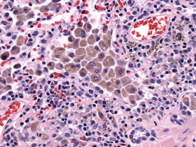
14 Diffuse Panbronchiolitis Gross: 4 mm yellow granular nodules. 1. Acute neutrophilic bronchiolitis with mucopurulent exudate. 2. Background chronic bronchiolitis involving RBs with peribronchiolar scarring, ectasia, lymphoid aggregates. 3. Luminal and interstitial aggregates of foamy macrophages.

15 Follicular Bronchiolitis A chronic bronchiolitis characterized histologically by the presence of hyperplastic lymphoid follicles with germinal centers distributed along the bronchioles. May be seen with diffuse lymphoid hyperplasia of the BALT, and as part of the LIP/DLH complex. Follicular Bronchiolitis Subacute onset of COE, SOB, occasional wheeze. HRCT small centrilobular nodules with patchy areas of ground glass opacity. Four clinical presentations. 1. Chronic infection mycoplasma, chlamydia, CF with bacterial colonization. 2. Autoimmune disease RA, SS. 3. Immunodeficiency states AIDS, CVID, B cell disorders. 4. Hypersensitivity reactions often associated with PB eosinophilia. Prognosis: poor in young; immunodeficiency group.


16 Bronchiolitis in Autoimmune Disease Rheumatoid arthritis/sjögren's: approximately 30%. Follicular bronchiolitis. Chronic bronchiolitis. Granulomatous bronchiolitis. SLE/PSS/Pemphigus. Chronic bronchiolitis. Cicatricial/constrictive bronchiolitis. Ulcerative colitis. Suppurative bronchitis/bronchiectasis with small airway damage. Crohn s disease. Chronic bronchiolitis with or without granulomas.

17 Bronchiolitis in Autoimmune Disease Many of the drugs used to treat CVD may independently cause or amplify the risk of developing SAD. Penicillamine Gold Methotrexate What if we classified SAD solely based on histopathologic changes......


18 Etiologies: Granulomatous SAD 1. Autoimmune RA, SS, ABPA. 2. Sarcoidosis. 3. Infections TB, fungus, M. Avium. 4. Aspiration. 5. Hypersensitivity pneumonitis. 6. Drugs methotrexate.
. 3.")

19 Eosinophilic SAD Etiologies: 1. Asthma. 2. Infections parasites, Cocci, fungus (Aspergillus). 3. Allergic drug induced, ABPA. 4. Churg-Strauss syndrome. 5. Leukemic infiltrates/hypereosinophilic syndrome. 6. NSHD.
20 Asthmatic Granulomatosis Definition: subset of severe steroid resistant asthmatics who manifest with asthmatic small airway disease with interstitial poorly formed granulomas. Wenzel S. et al; Am J Resp Crit Care Med 2012; 186: Asthmatic Granulomatosis F/M = 9; middle aged with severe nonsteroid responsive asthma; sinus disease; non-smokers. Cough, wheeze, chest tightness and SOB. PFTs reversible airflow limitation. HRCT 50% normal; dilation, bronchial wall thickening. PB eosinophilia; normal IgE, negative HSP panel; + atropy (60%). 60% self/family history of autoimmune disease. + response to non-steroid immunosuppressors Asthmatic Granulomatosis Asthmatic bronchiolitis mixed infiltrate with eosinophils with mucus plugging with eosinophils, Charcot Leyden crystals; BM thickening; smooth muscle hypertrophy. Interstitial pneumonitis patchy mild alveolar septal mononuclear infiltrate with poorly formed granulomas or giant cells.


21 AG WITH AIRWAY-CENTERED GRANUL

22 SAD SUMMARY 1. The diagnosis of SAD/bronchiolar disease is a clinicopathologic one where the histologic SAD is validated for significance by clinical S&S, PFTs and HRCT. 2. Classification of SAD is clinical and/or pathologic R/O large airway disease. 3. Remember histology etiology and consider the many possibilities. 4. Scarring usually indicates poor clinical response constrictive/cicatricial/obliterative bronchiolitis.
Imaging Small Airways Diseases: Not Just Air trapping. Eric J. Stern MD University of Washington
 Imaging Small Airways Diseases: Not Just Air trapping Eric J. Stern MD University of Washington What we are discussing SAD classification SAD imaging with MDCT emphasis What is a small airway? Airway with
Lung Allograft Dysfunction
 Lung Allograft Dysfunction Carlos S. Restrepo M.D. Ameya Baxi M.D. Department of Radiology University of Texas Health San Antonio Disclaimer: We do not have any conflict of interest or financial gain to
Outline Definition of Terms: Lexicon. Traction Bronchiectasis
 HRCT OF IDIOPATHIC INTERSTITIAL PNEUMONIAS Disclosures Genentech, Inc. Speakers Bureau Tadashi Allen, MD University of Minnesota Assistant Professor Diagnostic Radiology 10/29/2016 Outline Definition of
Asthma. - A chronic inflammatory disorder which causes recurrent episodes of wheezing, breathlessness, cough and chest tightness.
 Obstructive diseases Asthma - A chronic inflammatory disorder which causes recurrent episodes of wheezing, breathlessness, cough and chest tightness. - Characterized by Intermittent and reversible (the
Differential diagnosis
 Differential diagnosis The onset of COPD is insidious. Pathological changes may begin years before symptoms appear. The major differential diagnosis is asthma, and in some cases, a clear distinction between
Liebow and Carrington's original classification of IIP
 Liebow and Carrington's original classification of IIP-- 1969 Eric J. Stern MD University of Washington UIP Usual interstitial pneumonia DIP Desquamative interstitial pneumonia BIP Bronchiolitis obliterans
an inflammation of the bronchial tubes
 BRONCHITIS DEFINITION Bronchitis is an inflammation of the bronchial tubes (or bronchi), which are the air passages that extend from the trachea into the small airways and alveoli. Triggers may be infectious
Bronkhorst colloquium Interstitiële longziekten. Katrien Grünberg, klinisch patholoog
 Bronkhorst colloquium 2013-2014 Interstitiële longziekten De pathologie achter de CT Katrien Grünberg, klinisch patholoog K.grunberg@vumc.nl Preparing: introduction and 3 cases The introduction on microscopic
Respiratory Pathology. Kristine Krafts, M.D.
 Respiratory Pathology Kristine Krafts, M.D. Normal lung: alveolar spaces Respiratory Pathology Outline Acute respiratory distress syndrome Obstructive lung diseases Restrictive lung diseases Vascular
Financial disclosure COMMON DIAGNOSES IN HRCT. High Res Chest HRCT. HRCT Pre test. I have no financial relationships to disclose. Anatomy Nomenclature
 Financial disclosure I have no financial relationships to disclose. Douglas Johnson D.O. Cardiothoracic Imaging Gaston Radiology COMMON DIAGNOSES IN HRCT High Res Chest Anatomy Nomenclature HRCT Sampling
INTERSTITIAL LUNG DISEASE. Radhika Reddy MD Pulmonary/Critical Care Long Beach VA Medical Center January 5, 2018
 INTERSTITIAL LUNG DISEASE Radhika Reddy MD Pulmonary/Critical Care Long Beach VA Medical Center January 5, 2018 Interstitial Lung Disease Interstitial Lung Disease Prevalence by Diagnosis: Idiopathic Interstitial
Restrictive lung diseases
 Restrictive lung diseases Restrictive lung diseases are diseases that affect the interstitium of the lung. Interstitium of the lung is the very thin walls surrounding the alveoli, it s formed of epithelium
Differential diagnosis
 Differential diagnosis Idiopathic pulmonary fibrosis (IPF) is part of a large family of idiopathic interstitial pneumonias (IIP), one of four subgroups of interstitial lung disease (ILD). Differential
PULMONARY MEDICINE BOARD REVIEW. Financial Conflicts of Interest. Question #1: Question #1 (Cont.): None. Christopher H. Fanta, M.D.
: None. Christopher H. Fanta, M.D.") PULMONARY MEDICINE BOARD REVIEW Christopher H. Fanta, M.D. Pulmonary and Critical Care Division Brigham and Women s Hospital Partners Asthma Center Harvard Medical School Financial Conflicts of Interest
PULMONARY MEDICINE BOARD REVIEW Christopher H. Fanta, M.D. Pulmonary and Critical Care Division Brigham and Women s Hospital Partners Asthma Center Harvard Medical School Financial Conflicts of Interest
RESPIRATORY BLOCK. Bronchial Asthma. Dr. Maha Arafah Department of Pathology KSU
 RESPIRATORY BLOCK Bronchial Asthma Dr. Maha Arafah Department of Pathology KSU marafah@ksu.edu.sa Jan 2018 Objectives Define asthma (BA) Know the two types of asthma 1. Extrinsic or atopic allergic 2.
Vascular Lung Diseases
 Vascular Lung Diseases SESSION SPECIFIC OBJECTIVES List the major types of vascular lung disease Recognize and describe the pathology of vascular lung disease: Pulmonary embolism, thrombosis, hypertension,
Acute and Chronic Lung Disease
 KATHOLIEKE UNIVERSITEIT LEUVEN Faculty of Medicine Acute and Chronic Lung Disease W De Wever, JA Verschakelen Department of Radiology, University Hospitals Leuven, Belgium Clinical utility of HRCT To detect
I don t need you. Disclosure Statement. Pathology Approach to ILD 11/5/2016. Kirk D. Jones, MD UCSF Dept of Pathology
 Pathology Approach to ILD Disclosure Statement Relevant financial relationships with a commercial interest: Boeringer Ingleheim, speaker Kirk D. Jones, MD UCSF Dept of Pathology kirk.jones@ucsf.edu I don
HYPERSENSITIVITY PNEUMONITIS
 HYPERSENSITIVITY PNEUMONITIS A preventable fibrosis MOSAVIR ANSARIE MB., FCCP INTERSTITIAL LUNG DISEASES A heterogeneous group of non infectious, non malignant diffuse parenchymal disorders of the lower
INDEX. surgpath.theclinics.com. Note: Page numbers of article titles are in boldface type. diffuse pleural fibrosis, pleural plaques,
 INDEX Note: Page numbers of article titles are in boldface type. A Adenocarcinoma, minimally invasive. See Minimally invasive adenocarcinoma (MIA). Airway-centered interstitial fibrosis, 183 184 ALK (anaplastic
Manish Powari Regional Training Day 10/12/2014
 Manish Powari Regional Training Day 10/12/2014 Large number of different types of Interstitial Lung Disease (ILD). Most are very rare Most patients present with one of a smaller number of commoner diseases
11/10/2014. Multi-disciplinary Approach to Diffuse Lung Disease: The Imager s Perspective. Radiology
 Multi-disciplinary Approach to Diffuse Lung Disease: The Imager s Perspective Radiology Pathology Clinical 1 Role of HRCT Diagnosis Fibrosis vs. inflammation Next step in management Response to treatment
HRCT in Diffuse Interstitial Lung Disease Steps in High Resolution CT Diagnosis. Where are the lymphatics? Anatomic distribution
 Steps in High Resolution CT Diagnosis Pattern of abnormality Distribution of disease Associated findings Clinical history Tomás Franquet MD What is the diagnosis? Hospital de Sant Pau. Barcelona Secondary
Chronic Cough Due to Nonbronchiectatic Suppurative Airway Disease (Bronchiolitis) ACCP Evidence-Based Clinical Practice Guidelines
 ACCP Evidence-Based Clinical Practice Guidelines") Chronic Cough Due to Nonbronchiectatic Suppurative Airway Disease (Bronchiolitis) ACCP Evidence-Based Clinical Practice Guidelines Kevin K. Brown, MD, FCCP Objectives: To review the role of nonbronchiectatic
Chronic Cough Due to Nonbronchiectatic Suppurative Airway Disease (Bronchiolitis) ACCP Evidence-Based Clinical Practice Guidelines Kevin K. Brown, MD, FCCP Objectives: To review the role of nonbronchiectatic
Non-neoplastic Lung Disease II
 Pathobasic Non-neoplastic Lung Disease II Spasenija Savic Prince Pathology Program Systematic approach to surgical lung biopsies with ILD Examples (chronic ILD): Idiopathic interstitial pneumonias: UIP,
Case 1 : Question. 1.1 What is the intralobular distribution? 1. Centrilobular 2. Perilymphatic 3. Random
 Interesting case Case 1 Case 1 : Question 1.1 What is the intralobular distribution? 1. Centrilobular 2. Perilymphatic 3. Random Case 1: Answer 1.1 What is the intralobular distribution? 1. Centrilobular
Bronchiolar disorders: Current perspective on diagnosis & management. Puneet Malhotra Senior Resident, Dept. of Pulmonary Medicine, PGIMER
 Bronchiolar disorders: Current perspective on diagnosis & management Puneet Malhotra Senior Resident, Dept. of Pulmonary Medicine, PGIMER Anatomic considerations Classification Diagnostic approach Specific
Case Presentations in ILD. Harold R. Collard, MD Department of Medicine University of California San Francisco
 Case Presentations in ILD Harold R. Collard, MD Department of Medicine University of California San Francisco Outline Overview of diagnosis in ILD Definition/Classification High-resolution CT scan Multidisciplinary
Epidemiology and classification of smoking related interstitial lung diseases
 Epidemiology and classification of smoking related interstitial lung diseases Šterclová M. Department of Respiratory Diseases, Thomayer Hospital, Prague, Czech Republic Supported by an IGA Grant No G 1207
Radiologic Approach to Smoking Related Interstitial Lung Disease
 Radiologic Approach to Smoking Related Interstitial Lung Disease Poster No.: C-1854 Congress: ECR 2013 Type: Educational Exhibit Authors: K.-N. Lee, J.-Y. Han, E.-J. Kang, J. Kang; Busan/KR Keywords: Toxicity,
Follicular bronchiolitis in surgical lung biopsies: Clinical implications in 12 patients
 Respiratory Medicine (2008) 102, 307 312 Follicular bronchiolitis in surgical lung biopsies: Clinical implications in 12 patients Michelle R. Aerni a, Robert Vassallo a,, Jeffrey L. Myers b, Rebecca M.
5/9/2015. Multi-disciplinary Approach to Diffuse Lung Disease: The Imager s Perspective. No, I am not a pulmonologist! Radiology
 Multi-disciplinary Approach to Diffuse Lung Disease: The Imager s Perspective No, I am not a pulmonologist! Radiology Pathology Clinical 1 Everyone needs a CT Confidence in diagnosis Definitive HRCT +
4/17/2010 C ini n ca c l a Ev E a v l a ua u t a ion o n of o ILD U dat a e t e i n I LDs
 Update in ILDs Diagnosis 101: Clinical Evaluation April 17, 2010 Jay H. Ryu, MD Mayo Clinic, Rochester MN Clinical Evaluation of ILD Outline General aspects of ILDs Classification of ILDs Clinical evaluation
Bronchiolitis: A Schematic Diagnostic Approach with Radiologic-pathologic Correlation
 Bronchiolitis: A Schematic Diagnostic Approach with Radiologic-pathologic Correlation Mariana Benegas Urteaga 1, MD; M Sanchez 1, MD; J Ramirez 2, MD; D Barnes 1, MD; T de Caralt 1, MD; R J Perea 1, MD
NONE OVERVIEW FINANCIAL DISCLOSURES UPDATE ON IDIOPATHIC PULMONARY FIBROSIS/IPF (UIP) FOR PATHOLOGISTS. IPF = Idiopathic UIP Radiologic UIP Path UIP
 FOR PATHOLOGISTS. IPF = Idiopathic UIP Radiologic UIP Path UIP") UPDATE ON IDIOPATHIC PULMONARY FIBROSIS/IPF () FOR PATHOLOGISTS Thomas V. Colby, M.D. Professor of Pathology (Emeritus) Mayo Clinic Arizona FINANCIAL DISCLOSURES NONE OVERVIEW IPF Radiologic Dx Pathologic
UPDATE ON IDIOPATHIC PULMONARY FIBROSIS/IPF () FOR PATHOLOGISTS Thomas V. Colby, M.D. Professor of Pathology (Emeritus) Mayo Clinic Arizona FINANCIAL DISCLOSURES NONE OVERVIEW IPF Radiologic Dx Pathologic
Atopic Pulmonary Disease: Findings on Thoracic Imaging
 July 2003 Atopic Pulmonary Disease: Findings on Thoracic Imaging Rebecca G. Breslow Harvard Medical School Year IV Churg-Strauss Syndrome Hypersensitivity Pneumonitis Asthma Atopic Pulmonary Disease Allergic
Disease spectrum. IPA Invasive pulmonary aspergillosis
 Aspergillus & ABPA Disease spectrum IPA Invasive pulmonary aspergillosis ABPA ABPA pathophysiology conidia of Aspergillus trapped in mucous and narrowed airways of asthmatics/cf germinate to form hyphae
Lung Injury after HCT
 Lung Injury after HCT J. Douglas Rizzo, MD, MS Financial Disclosure None SCS06_1.ppt Background HCT an important therapeutic modality for malignant and non-malignant diseases Pulmonary Toxicity common
Dr.kassim.m.sultan F.R.C.P
 Dr.kassim.m.sultan F.R.C.P inflammatory disorder of the lung, involving alveolar walls and terminal airways, that is induced, in a susceptible host, by repeated inhalation of a variety of organic agents.
Usual Interstitial pneumonia and Nonspecific Interstitial Pneumonia. Nitra and the Gangs.
 Usual Interstitial pneumonia and Nonspecific Interstitial Pneumonia Nitra and the Gangs. บทน ำและบทท ๓, ๑๐, ๑๒, ๑๓, ๑๔, ๑๕, ๑๗ Usual Interstitial Pneumonia (UIP) Most common & basic pathologic pattern
Cryptococcosis Systemic infection involving Lungs CNS Skin Lymph nodes Usually an opportunistic infection in immunocompromised host Cryptococcal lymph
 Cryptococcosis Systemic infection involving Lungs CNS Skin Lymph nodes Usually an opportunistic infection in immunocompromised host Cryptococcal lymphadenitis is a rare AIDS defining illness Incidence
INTERSTITIAL LUNG DISEASE Dr. Zulqarnain Ashraf
 Indep Rev Jul-Dec 2018;20(7-12) Dr. Zulqarnain Ashraf IR-653 Abstract: ILD is a group of diseases affect interstitium of the lung. Repeated insult to the lung cause the interstitium to be damaged. Similarly
Bronchiectasis: An Imaging Approach
 Bronchiectasis: An Imaging Approach Travis S Henry, MD Associate Professor of Clinical Radiology Cardiac and Pulmonary Imaging Section University of California, San Francisco Large Middle Small 1 Bronchiectasis
Medicine Dr. Kawa Lecture 1 Asthma Obstructive & Restrictive Pulmonary Diseases Obstructive Pulmonary Disease Indicate obstruction to flow of air
 Medicine Dr. Kawa Lecture 1 Asthma Obstructive & Restrictive Pulmonary Diseases Obstructive Pulmonary Disease Indicate obstruction to flow of air through the airways. As asthma, COPD ( chronic bronchitis
Downloaded from by on 01/23/18 from IP address Copyright ARRS. For personal use only; all rights reserved
 Downloaded from www.ajronline.org by 46.3.194.46 on 01/23/18 from IP address 46.3.194.46. Copyright RRS. For personal use only; all rights reserved sthma is an inflammatory disease of the lungs characterized
DISEASES OF THE RESPIRATORY SYSTEM 2018 DR HEYAM AWAD LECTURE 3: CHRONIC BRNCHITIS AND BRONCHIECTASIS
 DISEASES OF THE RESPIRATORY SYSTEM 2018 DR HEYAM AWAD LECTURE 3: CHRONIC BRNCHITIS AND BRONCHIECTASIS INTRDUCTION In the last lecture we discussed the difference between restrictive and obstructive lung
International consensus statement on idiopathic pulmonary fibrosis
 Eur Respir J 2001; 17: 163 167 Printed in UK all rights reserved Copyright #ERS Journals Ltd 2001 European Respiratory Journal ISSN 0903-1936 PERSPECTIVE International consensus statement on idiopathic
Pulmonary Diseases. We Move A Lot of Air. Basic Categories. Alveolar Level. Developmental
 Pulmonary Diseases We Move A Lot of Air Alveolar Level Functions Oxygenation CO 2 & ph Basic defenses Nose hairs Cilia Mucus Cough reflex Immune system Basic Categories Congenital Infectious Neoplastic
The concept of small airways disease varies
 SMALL AIRWAYS DISEASE Timothy Craig Allen, MD, JD KEYWORDS Constrictive bronchiolitis Hypersensitivity pneumonitis Bronchiolitis Organizing pneumonia with intraluminal polyps Idiopathic bronchiolocentric
A Review of Interstitial Lung Diseases. Paul J. Wolters, MD Associate Professor Department of Medicine University of California San Francisco
 A Review of Interstitial Lung Diseases Paul J. Wolters, MD Associate Professor Department of Medicine University of California San Francisco Outline Overview of diagnosis in ILD Why it is important Definition/Classification
I have no relevant conflicts of interest to disclose
 I have no relevant conflicts of interest to disclose Diffuse parenchymal lung disease (DPLD) and its associations Secondary lobular anatomy DPLD History, clinical findings, temporal evolution, and exposures
Professor Rob Miller
 BHIVA AUTUMN CONFERENCE 2013 Including CHIVA Parallel Sessions Professor Rob Miller University College London Medical School COMPETING INTEREST OF FINANCIAL VALUE > 1,000: Speaker Name Prof Rob Miller
Small airways diseases, excluding asthma and COPD: an overview
 Eur Respir Rev 2013; 22: 128, 131 147 DOI: 10.1183/09059180.00001313 CopyrightßERS 2013 REVIEW Small airways diseases, excluding asthma and COPD: an overview Pierre-Régis Burgel, Anne Bergeron, Jacques
Residents Section Pattern of the Month
 Residents Section Pattern of the Month Gosset et al. Tree-In-Bud Pattern Residents Section Pattern of the Month Residents inradiology Natacha Gosset 1 Alexander A. Bankier Ronald L. Eisenberg Gosset N,
EOSINOPHLIC LUNG DISEASES
 EOSINOPHLIC LUNG DISEASES A wide spectrum of infiltrative lung diseases characterized by infiltration of lung parenchyma with eosinophils and/or peripheral blood eosinophilia. How is the diagnosis made?
Replacement of air with fluid, inflammatory. cells or cellular debris. Parenchymal, Interstitial (Restrictive) and Vascular Diseases.
 and Vascular Diseases.") Parenchymal, Interstitial (Restrictive) and Vascular Diseases Alain C. Borczuk, M.D. Dept of Pathology Replacement of air with fluid, inflammatory cells Pulmonary Edema Pneumonia Hemorrhage Diffuse alveolar
Parenchymal, Interstitial (Restrictive) and Vascular Diseases Alain C. Borczuk, M.D. Dept of Pathology Replacement of air with fluid, inflammatory cells Pulmonary Edema Pneumonia Hemorrhage Diffuse alveolar
Déjà vu all over again
 Disclosures Déjà vu all over again None Jonathan Singer MD MS University of California, San Francisco HPI 49 y/o woman presents for lung transplant evaluation for Hypersensitivity Pneumonitis Exposures:
Dr Rodney Itaki Lecturer Division of Pathology Anatomical Pathology Discipline
 Pathology of Asthma Dr Rodney Itaki Lecturer Division of Pathology Anatomical Pathology Discipline Bronchial Asthma Definition: chronic, relapsing inflammatory lung disorder characterised by reversible
Small airway disease: semiological and radiological evaluation. A pictorial review.
 Small airway disease: semiological and radiological evaluation. A pictorial review. Award: Magna Cum Laude Poster No.: C-3028 Congress: ECR 2018 Type: Educational Exhibit Authors: K. N. Nieto, A. Cerpa,
A Review of Interstitial Lung Diseases
 Outline A Review of Interstitial Lung Diseases Paul J. Wolters, MD Associate Professor Department of Medicine University of California San Francisco Overview of diagnosis in ILD Why it is important Definition/Classification
Bronchoalveolar Lavage and Histopathologic Diagnosis Based on Biopsy
 Idiopathic Pulmonary Fibrosis Bronchoalveolar Lavage and Histopathologic Diagnosis Based on Biopsy JMAJ 46(11): 469 474, 2003 Yukihiko SUGIYAMA Professor, Division of Pulmonary Medicine, Department of
Immune-mediated lung disease. Ian Sabroe
 Immune-mediated lung disease Ian Sabroe It s all immune? Diseases that doesn t have immune involvement? The processes of response to injury and tissue repair are key homeostatic pathways involved in all
Imaging Spectrum of Allergic Lung Disease: Hypersensitivity Reactions on the Lung Parenchyma
 Imaging Spectrum of Allergic Lung Disease: Hypersensitivity Reactions on the Lung Parenchyma Moon Sung Kim 1, Ki-Nam Lee 1, Won Jin Choi 1, Bo Ra Kim 1, Eun-Ju Kang 1 1 Department of Radiology, Dong-A
PATHOPHYSIOLOGY OF RESPIRATION 2
 PATHOPHYSIOLOGY OF RESPIRATION 2 Obstructive disorders R. Benacka Department of Pathophysiology, Medical faculty, P.J. Safarik University Košice Respiratory diseases Obstructive diseases (OPD) - restricted
Unit II Problem 2 Pathology: Pneumonia
 Unit II Problem 2 Pathology: Pneumonia - Definition: pneumonia is the infection of lung parenchyma which occurs especially when normal defenses are impaired such as: Cough reflex. Damage of cilia in respiratory
Chronic Interstitial (Restrictive) Lung Disease
 Lung Disease") Chronic Interstitial (Restrictive) Lung Disease Fibrosing Usual interstitial pneumonia (idiopathic pulmonary fibrosis) IPF/UIP Nonspecific interstitial pneumonia(nsip) Cryptogenic organizing pneumonia(cop)
Chronic Interstitial (Restrictive) Lung Disease Fibrosing Usual interstitial pneumonia (idiopathic pulmonary fibrosis) IPF/UIP Nonspecific interstitial pneumonia(nsip) Cryptogenic organizing pneumonia(cop)
Histopathology: pulmonary pathology
 Histopathology: pulmonary pathology These presentations are to help you identify basic histopathological features. They do not contain the additional factual information that you need to learn about these
Smoking-related Interstitial Lung Diseases: High-Resolution CT Findings
 Smoking-related Interstitial Lung Diseases: High-Resolution CT Findings Poster No.: C-2358 Congress: ECR 2013 Type: Educational Exhibit Authors: V. Cuartero Revilla, M. Nogueras Carrasco, P. Olmedilla
Bronchiectasis in Adults - Suspected
 Bronchiectasis in Adults - Suspected Clinical symptoms which may indicate bronchiectasis for patients Take full respiratory history including presenting symptoms, past medical & family history Factors
LUNG DISEASES DUE TO ORGANIC&INORGANIC DUSTS. Dr.kassim.m.sultan F.R.C.P
 LUNG DISEASES DUE TO ORGANIC&INORGANIC DUSTS Dr.kassim.m.sultan F.R.C.P efinition of hypersensitivity pneumonitis(extrinsic allergic alveolitis): Inflammatory disorder of the lung, involving alveolar walls
DISEASES OF THE RESPIRATORY SYSTEM LECTURE 5 DR HEYAM AWAD FRCPATH
 DISEASES OF THE RESPIRATORY SYSTEM LECTURE 5 DR HEYAM AWAD FRCPATH RESTRICTIVE, INTERSTITIAL LUNG DISESAES. FIROSING DISESES. GRANULOMATOUS DISEASES. EOSINOPHILIC. SMOKING RELATED. FIBROSING DISEASES
Diagnosis of Respiratory Bronchiolitis associated interstitial lung disease
 Monaldi Arch Chest Dis 2006; 65: 2, 96-101 ORIGINAL ARTICLE Diagnosis of Respiratory Bronchiolitis associated interstitial lung disease C. Robalo Cordeiro 1, S. Freitas 1, B. Rodrigues 1, A. Catarino 1,
P ulmonary fibrosis is usually taken to mean
 638 REVIEW SERIES Challenges in pulmonary fibrosis? 2 : Bronchiolocentric fibrosis Jean-François Cordier... Bronchiolocentric fibrosis is essentially represented by the pathological pattern of constrictive
CTD-related Lung Disease
 13 th Cambridge Chest Meeting King s College, Cambridge April 2015 Imaging of CTD-related Lung Disease Dr Sujal R Desai King s College Hospital, London Disclosure Statement No Disclosures / Conflicts of
* * APPROACH TO NON- NEOPLASTIC LUNG DISEASE IN TRANSBRONCHIAL AND SURGICAL BIOPSIES. Financial Disclosures: NONE. BIOPSY TECHNIQUES Bronchoscopic
 APPROACH TO NON- NEOPLASTIC LUNG DISEASE IN TRANSBRONCHIAL AND SURGICAL BIOPSIES Thomas V. Colby, M.D. Mayo Clinic Arizona Geraldine C. Zeiler Professor of Cytopathology Mayo Clinic Arizona Financial Disclosures:
Chronic Obstructive Pulmonary Disease (COPD Emphysema, Chronic Bronchitis) Bronchiectasis Smoking Related Problems Lung Cancer Screening
 Bronchiectasis Smoking Related Problems Lung Cancer Screening") Chest, Lung & COPD Chest, Lung & COPD Do you have difficulty breathing? Shortness of breath? Even COPD? Our experts can find the exact problem so you can breathe easier. Here s a guide if you would like
Chest, Lung & COPD Chest, Lung & COPD Do you have difficulty breathing? Shortness of breath? Even COPD? Our experts can find the exact problem so you can breathe easier. Here s a guide if you would like
CLEARING THE AIR ON DIFFUSE PARENCHYMAL (INTERSTITIAL) LUNG DISEASE (ILD)
 LUNG DISEASE (ILD)") CLEARING THE AIR ON DIFFUSE PARENCHYMAL (INTERSTITIAL) LUNG DISEASE (ILD) David Northrop MBA, RRT Assistant Director of Respiratory Therapy Services The University of Kansas Health System Clinical Assistant
CLEARING THE AIR ON DIFFUSE PARENCHYMAL (INTERSTITIAL) LUNG DISEASE (ILD) David Northrop MBA, RRT Assistant Director of Respiratory Therapy Services The University of Kansas Health System Clinical Assistant
Chapter 10 Respiratory System J00-J99. Presented by: Jesicca Andrews
 Chapter 10 Respiratory System J00-J99 Presented by: Jesicca Andrews 1 Respiratory System 2 Respiratory Infections A respiratory infection cannot be assumed from a laboratory report alone; physician concurrence
Pulmonary Test Brenda Shinar
 Pulmonary Test 2016 Brenda Shinar 1. What is a Renal Tubular Acidosis? What is the difference between the types of RTAs in terms of who gets them and how to diagnose them? Type 1, 2, and 4? Metabolic acidosis
BRONCHO ALVEOLAR LAVAGE IN INTERSTITIAL LUNG DISEASES A USEFUL TOOL OR AN OUT DATED CONCEPT?
 1 BRONCHO ALVEOLAR LAVAGE IN INTERSTITIAL LUNG DISEASES A USEFUL TOOL OR AN OUT DATED CONCEPT? Dr. M. V. Nagarjuna AIMS 1. Technical Considerations in performing BAL 2. Role of BAL in ILD 1. Diagnosis
Eosinophilia Associated Lung Diseases
 Eosinophilia Associated Lung Diseases Stephen P. Peters, MD, PhD, FAAAAI, FACP, FCCP, FCPP Thomas H. Davis Chair in Pulmonary Medicine Chief, Section on Pulmonary Critical Care, Allergy & Immunologic Diseases
Radiological Imaging of Drug-Induced Pulmonary Lesions
 Review Article imedpub Journals www.imedpub.com Journal of Clinical Radiology and Case Reports Radiological Imaging of Drug-Induced Pulmonary Lesions D souza M *, Rajiah P, Khan A and Irion K Department
Update on heterogeneity of COPD, evaluation of COPD severity and exacerbation
 Update on heterogeneity of COPD, evaluation of COPD severity and exacerbation Yung-Yang Liu, MD Taipei Veterans General Hospital Aug 29, 2015 G O lobal Initiative for Chronic bstructive L D ung isease
Hypersensitivity Pneumonitis: Spectrum of High-Resolution CT and Pathologic Findings
 CT of Hypersensitivity Pneumonitis Chest Imaging Pictorial Essay C. Isabela S. Silva 1 ndrew Churg 2 Nestor L. Müller 1 Silva CIS, Churg, Müller NL Keywords: high-resolution CT, hypersensitivity pneumonitis,
COPD. Dr.O.Paknejad Pulmonologist Shariati Hospital TUMS
 IN THE NAME OF GOD COPD Dr.O.Paknejad Pulmonologist Shariati Hospital TUMS Definition of COPD* COPD is a preventable and treatable chronic lung disease characterized by airflow limitation that is not fully
The Respiratory System
 The Respiratory System Respiratory Anatomy Upper respiratory tract Nose Nasal passages Pharynx Larynx Respiratory Anatomy Functions of the upper respiratory tract: Provide entry for inhaled air Respiratory
Definition, classification and epidemiology
 Interstitial Lung Diseases Definition, classification and epidemiology Haluk Türktaş Professor of Pulmonary Medicine Gazi University Ankara Interstitial Lung Diseases Definition of ILD A diverse group
Asthma COPD Overlap (ACO)
") Asthma COPD Overlap (ACO) Dr Thomas Brown Consultant Respiratory Physician Thomas.Brown@porthosp.nhs.uk Dr Hitasha Rupani Consultant Respiratory Physician Hitasha.rupani@porthosp.nhs.uk What is Asthma
Asthma COPD Overlap (ACO) Dr Thomas Brown Consultant Respiratory Physician Thomas.Brown@porthosp.nhs.uk Dr Hitasha Rupani Consultant Respiratory Physician Hitasha.rupani@porthosp.nhs.uk What is Asthma
Hypersensitivity Pneumonitis: Epidemiology and Classification
 Hypersensitivity Pneumonitis: Epidemiology and Classification Ulrich Costabel, MD University of Duisburg-Essen, Ruhrlandklinik Department of Pneumology/Allergy Objectives Definitions, Etiology Epidemiology
Obstructive Lung Diseases. By: Shefaa Qa qa
 Obstructive Lung Diseases By: Shefaa Qa qa Obstructive lung diseases (or airway diseases) are characterized by an increase in resistance to airflow due to partial or complete obstruction at any level from
The term bronchiolitis refers to a group of diverse
 C l i n i c a l R e v i e w A r t i c l e Syndromes of Bronchiolitis Sat Sharma, MD The term bronchiolitis refers to a group of diverse clinical disorders that share a common instigating mechanism: inflammatory
Parenchymal, Interstitial i (Restrictive) i and Vascular Diseases
 i and Vascular Diseases") Pulmonary Diseases: Structure-Function Correlation II Parenchymal, Interstitial i (Restrictive) i and Vascular Diseases Alain C. Borczuk, M.D. Dept of Pathology Pulmonary Diseases: Structure-Function Correlation
Pulmonary Diseases: Structure-Function Correlation II Parenchymal, Interstitial i (Restrictive) i and Vascular Diseases Alain C. Borczuk, M.D. Dept of Pathology Pulmonary Diseases: Structure-Function Correlation
DISEASES OF THE RESPIRATORY SYSTEM 2017 DR HEYAM AWAD LECTURE 5: restrictive lung diseases, part 1: fibrosing lung diseases
 DISEASES OF THE RESPIRATORY SYSTEM 2017 DR HEYAM AWAD LECTURE 5: restrictive lung diseases, part 1: fibrosing lung diseases Reference: Robbins, 9 th : 472-478, 10 th : 506-512 INTRODUCTION: RESTRICTIVE
Case 1: Question. 1.1 What is the main pattern of this HRCT? 1. Intralobular line 2. Groundglass opacity 3. Perilymphatic nodule
 HRCT WORK SHOP Case 1 Case 1: Question 1.1 What is the main pattern of this HRCT? 1. Intralobular line 2. Groundglass opacity 3. Perilymphatic nodule Case 1: Question 1.2 What is the diagnosis? 1. Hypersensitivity
Medical Policy An independent licensee of the Blue Cross Blue Shield Association
 Idiopathic Pulmonary Fibrosis Page 1 of 10 Medical Policy An independent licensee of the Blue Cross Blue Shield Association Title: Idiopathic Pulmonary Fibrosis (Esbriet /pirfenidone, Ofev /nintedanib)
Combined Unclassifiable Interstitial Pneumonia and Emphysema: A Report of Two Cases
 CASE REPORT Combined Unclassifiable Interstitial Pneumonia and Emphysema: A Report of Two Cases Nobuhiko Nagata 1, Kentaro Watanabe 2, Michihiro Yoshimi 3, Hiroshi Okabayashi 4, Katsuo Sueishi 5, Kentaro
Clinical significance of respiratory bronchiolitis on open lung biopsy and its relationship to smoking related interstitial lung disease
 Thorax 1999;54:1009 1014 1009 James Moon, Ronald M du Bois, Thomas V Colby, David M Hansell, Andrew G Nicholson Interstitial Lung Disease Unit J Moon R M du Bois Department of Radiology D M Hansell Department
Katerina M. Antoniou, MD, PhD As. Professor in Thoracic Medicine ERS ILD Group Secretary Medical School, University of Crete Prague, June 2014
 Hypersensitivity pneumonitis: Causes, clinical course, diagnosis and differential diagnosis, treatment Katerina M. Antoniou, MD, PhD As. Professor in Thoracic Medicine ERS ILD Group Secretary Medical School,