Title. CitationPathology International, 59(11): Issue Date Doc URL. Rights. Type. File Information.
|
|
|
- Brian Jacobs
- 5 years ago
- Views:
Transcription
1 Title Follicular dendritic cell sarcoma of small intestine Yamada, Yosuke; Haga, Hironori; Hernandez, Mako; Kub Author(s) Yoshihiro CitationPathology International, 59(11): Issue Date Doc URL Rights The definitive version is available at Type article (author version) File Information PI59-11_ pdf Instructions for use Hokkaido University Collection of Scholarly and Aca
2 <Title page> [The title] Follicular dendritic cell sarcoma of small intestine with aberrant T-cell marker expression. [Short running title] FDC sarcoma with T-cell marker (26 letters) [The full names of the authors] Yosuke Yamada 1, Hironori Haga 1, Mako Hernandez 1, Kanako Kubota 1, Fumika Orii 2, Kazuo Nagashima 3 and Yoshihiro Matsuno 1. [Corresponding author] Yoshihiro Matsuno, MD, PhD. Department of Surgical Pathology, Hokkaido University Hospital, Sapporo, Japan, 1 Nishi 5 chome, kita 14 jo, Kita-ku, Sapporo , Japan. Tel: , Fax: ymatsuno@med.hokudai.ac.jp. [The address of the institutions etc] 1 Department of Surgical Pathology, Hokkaido University Hospital, Sapporo, Japan Departments of 2 Internal Medicine and 3 Surgical Pathology, Sapporo Higashi Tokushukai Hospital, Sapporo, Japan 1
3 <Abstract> (no more than 200 words) Follicular dendritic cell sarcoma (FDCS) is an uncommon neoplasia usually occurring in lymphoid tissue. Herein we present a case of FDCS of the small intestine with positivity for T-cell antigen, simulating T-cell lymphoma. An 82-year-old man consulted a doctor for epigastric pain of one-week duration. Imaging study revealed a mass in the small intestine. Malignant lymphoma was suspected because of high serum levels of soluble interleukin-2 receptor, and resection of the tumor was performed. Microscopically, the tumor was comprised of large pleomorphic cells with reactive small lymphocytes. Most of the nuclei of the tumor cells were round or ovoid-shaped, and some of the tumor cells also showed spindle-shaped nuclei. Although the tumor cells were diffusely positive for CD45RO and CD4 immunohistochemically, negativity for pan-t-cell markers and CD56 were unusual for T-cell lymphoma of intestinal origin. Additional immunohistochemical study demonstrated that the tumor cells were positive for follicular dendritic cell markers including CD23, CD35 and CAN.42, and diagnosis of FDCS was made. To our knowledge, this is the first case of FDCS aberrantly expressing CD45RO, and FDCS expressing T-cell markers can be a pitfall for diagnosis of FDCS. (190 words) <Key words> Dendritic cell sarcoma, follicular Intestine, small Lymphoma, T-cell 2
4 <Text> [Introduction] Follicular dendritic cell sarcoma (FDCS) is a tumor composed of spindle or oval cells with follicular dendritic cell differentiation. Usually, their morphology and immunohistochemical features are distinct from those of non-hodgkin lymphomas. In this report, we present FDCS in an elderly man with pleomorphic histology and T-cell marker expression, simulating T-cell lymphoma. [Clinical summary] The patient was a previously healthy 82-year-old Japanese man. He visited a hospital for epigastric pain of one-week duration. Physical examination revealed a firm mass in the upper part of the abdomen. Abdominal CT scan showed focal obstruction of the small intestine by thickening of the intestinal wall. Contrast enema using an ileus pipe demonstrated severe stenosis at 50 cm distal to the Treitz ligament. The serum level of soluble interleukin-2 receptor was elevated to 1470 U/ml (normal range, U/ml). A malignant lymphoma of small intestine was suspected, and partial resection of the jejunum was performed. After the operation, he was received adjuvant chemotherapy (CHOP) for 3 months and there was no evidence of recurrence at 12-month follow-up. [Pathological findings] Macroscopically, a mass measuring 10 x 10 cm in greatest dimension was seen in the jejunum extending throughout the wall of the intestine. Cut surface demonstrated a well-circumscribed, gray-white solid tissue with focal hemorrhage (Figure 1). Microscopically, the tumor consisted of large pleomorphic cells, mainly forming diffuse sheets. Focal coagulative necrosis was found (Figure 2). The large cells had faintly eosinophilic cytoplasm and pleomorphic nuclei with coarse chromatin and distinct nucleoli. Nuclear pseudo-inclusion, binucleated and multinucleated nuclei were also present. Mitotic figures 3
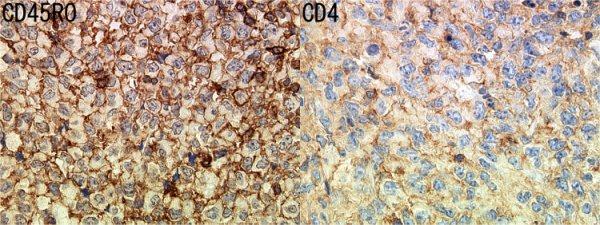
5 were numerous (50 counts / 10 high power field) (Figure 3). In the background of the tumor cells, interspersed small lymphocytes were evident, and some of them were seen around the blood vessels. Lymphoid follicles were recognizable in some areas of the tumor. Regional lymph node involvement of the tumor with sparing of some lymph follicles was also noted. The results of immunohistochemistry are summarized in Table 1. The tumor cells were focally (5%) positive for pan-b-cell marker CD20, and diffusely positive for CD45RO (Figure 4), a T-cell marker, as well as CD45RB. But the tumor cells did not show positivity for pan T-cell markers including CD3 and CD5. The tumor cells were diffusely positive for CD4 (Figure 4), CD68, and vimentin, and showed immunoreactivity for follicular dendritic cell markers including CD23, CD35, and CNA.42 (Figure 5). They were focally positive for S-100 protein and negative for CD21 (Figure 5) and D2-40. Ki-67 index was 90%. In situ hybridization for Epstein-Barr virus encodes small RNA (EBER) { INFORM EBER probe (CE) (Roche)} failed to show positive cells. In clonal analysis, genomic DNA was extracted from paraffin-embedded tissue and T-cell receptor (TCR) gene rearrangements were studied by PCR. The amplified DNA yielded a polyclonal smear pattern on electrophoresis. A final diagnosis of FDCS was made on these findings. [Discussion] FDCS is a neoplasm consisting of spindle to ovoid cells, which closely mimics various types of tumors and tumor-like lesions. Typically, they are nodal in origin, but extranodal involvement including the small intestine has been reported 1. In case of intestinal FDCS, the major differential diagnosis includes gastrointestinal stromal tumor (GIST) and primary intestinal lymphomas. In this case, GIST was easily excluded by a lack of expression of CD34 and c-kit in the tumor cells. Expression of T-cell markers, including CD4 and CD45RO, however, might have lead to a diagnosis of T-cell lymphoma of intestinal origin. Enteropathy-type T-cell lymphomas among Japanese are type 2 in most cases, and are phenotypically characterized by positivity of CD8 and CD56 2. Peripheral T-cell lymphoma, not otherwise specified, often 4
6 demonstrates CD4 with frequent loss of CD5 or CD7. However, complete loss of pan-t-cell markers is unusual for T-cell lymphoma. In our case, the possibility of aberrant expression of T-cell antigens in a non-lymphoid tumor was considered. Infiltration of small lymphocytes, especially around the blood vessels, suggested the possibility of FDCS 3). For investigation of undifferentiated neoplasm, including FDCS in the differential diagnosis is important since follicular dendritic cell markers are usually not included in the first panel of antibodies. To diagnose FDCS, at least one FDC marker (CD21, CD35, CD23, KiM4p and CNA.42) should be positive and CD1a should be negative 3). For lymphoid markers, CD20 and CD45(LCA) have been reported to be occasionally positive, while CD3, a pan-t-cell marker, is consistently negative 3. Our case suggests that even some T-cell associated antigens can be demonstrated in FDCS. Although CD4 is a co-receptor acting in the activation of T-helper cells by the MHC class II antigen restricted pathway, CD4 expression is not restricted to T lymphocytes. Langerhans' cells, plasmacytoid dendritic cells/monocytes and true histiocytic sarcomas are known to be positive for CD4 4,5. Likewise, CD45RO, recognized by antibody UCHL-1 and once widely used as a T-cell marker on paraffin sections 6, can be expressed in histiocytic tumors 7. Actually, both CD4 and CD45RO lack lineage specificity and are commonly expressed by histiocytic sarcoma 8. Additionally, our case expressed CD68, one of the most available histiocytic markers. For diagnosis of histiocytic sarcoma, however, dendritic cell markers should be negative and other histiocyte-specific markers such as lysozyme or CD163 are usually expressed. In this case, the tumor cells were negative for CD68, and CD163 was not available. Differential diagnosis from the CD4+ blastic plasmacytoid dendritic cell neoplasm would be straight forward, because of non-hematodermic distribution, more sarcomatous morphology, and negativity for immunoreactive CD56 in this case. Chan et al. reported that mitotic rate of FDCS is usually between 0 and 10/10HPF and Ki-67 labeling index are between 1 and 25% (mean 13%) 3. In contrast, our case showed high proliferative index (mitotic rate of 50/10 high power fields and Ki-67 labeling index of 90%). As poor prognostic factors of FDCS, significant cytologic atypia, extensive 5
7 coagulative necrosis, a high proliferative index, large tumor size (> 6cm) or intraabdominl location are documented 3, and our case meets the all factors. Moreover, to the best of our knowledge, there have been 7 reported cases of primary gastrointestinal FDCS 9-15, and 6 among the 7 showed recurrence and/or metastasis. In our case, regional lymph node metastasis is found. Considering those findings, we think that our case is a high-grade malignancy, and that careful follow-up is needed. In summary, we present a case of FDCS of small intestine origin. Expression of CD45RO was a feature that has not been previously reported. Including follicular dendritic cell markers into immunohistochemical panel and awareness of key histologic features such as perivascular distribution of small lymphocytes and residual lymphoid follicles in the tumor were crucial for diagnosis of FDCS. Possibility of FDCS with unusual phenotypic expression should be considered in undifferentiated neoplasms with a mixed population of lymphocytes. 6
8 <References> 1) Kevin H, Gordon S, Ioanna Z et al. Extranodal follicular dendritic cell sarcoma of the gastrointestinal tract: morphologic, immunohistochemical and ultrastructural analysis of two cases. Hematopathology 1994;103: ) Akiyama T, Okino T, Konishi H et al. CD8+, CD56+(natural killer-like) T-cell lymphoma involving the small intestine with no evidence of enteropathy: clinicopathology and molecular study of five Japanese patients. Pathol Int. 2008;58: ) JKC Chan, SA Pileri, G Delsol et al. Follicular dendritic cell sarcoma. In: SH Swerdlow, E Campo, NL Harris, ES Jaffe, SA Pileri, H Stein et al, editors. WHO classification of tumours of haematopoietic and lymphoid tissues. Switzerland: IARC;2008.p ) Pileri SA, Grogan TM, Harris NL et al. Tumours of histiocytes and accessory dendritic cells: an immunohistochemical approach to classification from the International Lymphoma Study Group based on 61 cases. Histopathology 2002;41: ) Herling M, Jones D. CD4+/CD56+ hematodermic tumor: the features of an evolving entity and its relationship to dendritic cells. Am J Clin Pathol 2007; 127: ) Steward M, Bishop R, Piggott NH et al. Production and characterization of a new monoclonal antibody effective in recognizing the CD3 T-cell associated antigen in formalin-fixed embedded tissue. Histopathology 1997;30: ) Zhang X, Kryston JJ, Michalak WA, et al. Histiocytic sarcoma in the small intestine: a case report with flow cytometry study and review of the literature. Pathol Res Pract 2008;204: Epub 2008 Jun 9. 8) Sun W, Nordberg ML, Fowler MR. Histiocytic sarcoma involving the central nervous system: clinical, immunohistochemical, and molecular genetic studies of a case with review of the literature. Am J Surg Pathol 2003;27: ) Hollowood K, Stamp G, Zouvani I, et al. Extranodal follicular dendritic cell sarcoma of the gastrointestinal tract. Morphologic, immunohistochemical and ultrastructural analysis of two cases. Am J Clin Pathol 1995;103:
9 10) Han JH, Kim SH, Noh SH et al. Follicular dendritic cell sarcoma presenting as a submucosal tumor of the stomach. Arch Pathol Lab Med 2000;124: ) Chang KC. Follicular dendritic cell sarcoma of the colon mimicking stromal tumour. Histopathology 2001;38: ) Geerts A, Lagae E, Dhaene K et al. Metastatic follicular dendritic cell sarcoma of the stomach: a case report and review of the literature. Acta Gastroenterol Belg 2004;67: ) D O Mally. Diagnosis: follicular dendritic cell tumor mimicking GI stromal tumor. 14) Shia J. Extranodal follicular dendritic cell sarcoma: clinical, pathologic, and histogenetic characteristics of an underrecognized disease entity. Virchows Arch 2006:449; ) Yang GC, Wang J, Yee HT. Interwoven dendritic processes of follicular dendritic cell sarcoma demonstrated on ultrafast papanicolaou-stained smears: a case report. Acta Cytol 2006;50:
10 <Figure legends> Figure 1: The cut surface of the tumor demonstrating a clear border and spreading throughout the wall of the small intestine. Figure 2: Diffuse proliferation of the tumor cells with focal necrosis. HE section, original magnification x10. Figure 3: Tumor cells showing abundant faint eosinophilic cytoplasm and pleomorphic nuclei. Occasional mitotic figures are seen. HE section, original magnification x40. Figure 4: The neoplastic cells are diffusely positive for CD45RO and CD4, original magnification x80 Figure 5: The neoplastic cells are positive for CD23, CD35 and FDC, and negative for CD21, original magnification x20. 9
11 Table 1 Immunohistochemistry Antigen Clone Source Dilution Results αsma 1A4 DAKO Glostrup, Denmark 1:1000 (-) Antichymotrypsin Polyclonal Zymed Carlsbad, California, USA 1:100 (-) Anaplastic lymphoma kinase-1 ALK-1 DAKO Glostrup, Denmark 1:50 (-) CD1a O10 DAKO Glostrup, Denmark 1:10 (-) CD3 2GV6 VENTANA Yokohama, Japan Prediluted (-) CD4 1F6 Novocastra Newcastle Upon Tyne, UK 1:15 (++) CD5 4C7 Novocastra Newcastle Upon Tyne, UK 1:50 (-) CD8 4B11 Novocastra Newcastle Upon Tyne, UK 1:40 (-) CD20 L26 DAKO Glostrup, Denmark 1:200 (+) CD21 2G9 Novocastra Newcastle Upon Tyne, UK 1:50 (-) CD23 1B12 Novocastra Newcastle Upon Tyne, UK 1:20 (++) CD30 Ber-H2 DAKO Glostrup, Denmark 1:20 (-) CD34 NU-4A1 NICHIREI Tokyo, Japan 1:50 (-) CD35 RLB25 Novocastra Newcastle Upon Tyne, UK 1:40 (++) CD45RB 2B11+PD7/26 DAKO Glostrup, Denmark 1:100 (++) CD45RO UCHL-1 DAKO Glostrup, Denmark 1:200 (++) CD56 123C3 Zymed Carlsbad, California, USA Prediluted (-) CD68 PGM-1 DAKO Glostrup, Denmark 1:100 (++) C-kit Polyclonal DAKO Glostrup, Denmark 1:400 (-) Pan-cytokeratin AE1/AE3 DAKO Glostrup, Denmark 1:70 (-) D2-40 D2-40 DAKO Glostrup, Denmark 1:50 (-) Follicular dendritic cell CNA42 DAKO Glostrup, Denmark 1:50 (++) Granzyme B GrB-7 MONOSAN Uden, Netherlands 1:25 (-) Ki-67 MIB-1 DAKO Glostrup, Denmark 1:20 (++: 90%) Lysozyme Polyclonal NICHIREI Tokyo, Japan Prediluted (-) Myeloperoxidase Polyclonal DAKO Glostrup, Denmark 1:10000 (-) S-100 protein Polyclonal DAKO Glostrup, Denmark 1:3000 (+) TIA-1 26gA10F5 IMMUNOTECH Marseiile, France 1:200 (-) Vimentin V9 DAKO Glostrup, Denmark 1:200 (++) (++), diffusely (5% or more) positive; (+), focally (< 5%) positive; (-), completely negative.

12

13

14
15

16
Case Presentation. Maha Akkawi, MD, Fatima Obeidat, MD, Tariq Aladily, MD. Department of Pathology Jordan University Hospital Amman, Jordan
 Case Presentation Maha Akkawi, MD, Fatima Obeidat, MD, Tariq Aladily, MD Department of Pathology Jordan University Hospital Amman, Jordan The 25th Annual Congress of the ADIAP The 8/11/2013 1 5th International
Follicular dendritic cell sarcoma of inguinal lymph node A case report
 Malaysian J Pathol 2008; 30(2) : 115 119 CASE REPORT Follicular dendritic cell sarcoma of inguinal lymph node A case report Jayalakshmi PAILOOR, MPath, FRCPath, Krishnan R IYENGAR, MD, DNB, CHAN KS, MPath*
3/24/2017 DENDRITIC CELL NEOPLASMS: HISTOLOGY, IMMUNOHISTOCHEMISTRY, AND MOLECULAR GENETICS. Disclosure of Relevant Financial Relationships
 DENDRITIC CELL NEOPLASMS: HISTOLOGY, IMMUNOHISTOCHEMISTRY, AND MOLECULAR GENETICS Jason L. Hornick, M.D., Ph.D. Director of Surgical Pathology and Immunohistochemistry Brigham and Women s Hospital Professor
Differential diagnosis of hematolymphoid tumors composed of medium-sized cells. Brian Skinnider B.C. Cancer Agency, Vancouver General Hospital
 Differential diagnosis of hematolymphoid tumors composed of medium-sized cells Brian Skinnider B.C. Cancer Agency, Vancouver General Hospital Lymphoma classification Lymphoma diagnosis starts with morphologic
Gastric Carcinoma with Lymphoid Stroma: Association with Epstein Virus Genome demonstrated by PCR
 Gastric Carcinoma with Lymphoid Stroma: Association with Epstein Virus Genome demonstrated by PCR Pages with reference to book, From 305 To 307 Irshad N. Soomro,Samina Noorali,Syed Abdul Aziz,Suhail Muzaffar,Shahid
, , 2011 HODGKIN LYMPHOMA
 European Federation of Cytology Societies 4tu Annual Tutorial in Cytopathology Trieste, June 6-10, 2011 HODGKIN LYMPHOMA Classification The World Health Organization Classification of Lymphomas (2001)
A Unique Case of Nasal NK/T Cell Lymphoma with Frequent Remission and Relapse Showing Different Histological Features During 12 Years of Follow Up
 J Clin Exp Hematopathol Vol. 50, No. 1, May 2010 Case Study A Unique Case of Nasal NK/T Cell Lymphoma with Frequent Remission and Relapse Showing Different Histological Features During 12 Years of Follow
Cytology of Follicular Dendritic Cell Sarcoma on Intraoperative Touch Imprint Smears
 The Korean Journal of Pathology 2009; 43: 589-93 DOI: 10.4132/KoreanJPathol.2009.43.6.589 Cytology of Follicular Dendritic Cell Sarcoma on Intraoperative Touch Imprint Smears - Case Report - Ju Young Song
Blastic Plasmacytoid Dendritic Cell Neoplasm : Report of Two Cases
 Case Study Blastic Plasmacytoid Dendritic Cell Neoplasm : Report of Two Cases J Clin Exp Hematopathol Vol. 52, No. 1, May 2012 Kanako Tsunoda, 1,2) Takashi Satoh, 2) Kiyomi Akasaka, 1,2) Yuichi Ishikawa,
From Morphology to Molecular Pathology: A Practical Approach for Cytopathologists Part 1-Cytomorphology. Songlin Zhang, MD, PhD LSUHSC-Shreveport
 From Morphology to Molecular Pathology: A Practical Approach for Cytopathologists Part 1-Cytomorphology Songlin Zhang, MD, PhD LSUHSC-Shreveport I have no Conflict of Interest. FNA on Lymphoproliferative
Case: The patient is a 62 year old woman with a history of renal cell carcinoma that was removed years ago. A 2.4 cm liver mass was found on CT
 Case: The patient is a 62 year old woman with a history of renal cell carcinoma that was removed years ago. A 2.4 cm liver mass was found on CT during follow- up. ALT, AST, Alk Phos and bilirubin were
Immunohistochemical Evaluation of Necrotic Malignant Melanomas
 Anatomic Pathology / EVALUATION OF NECROTIC MALIGNANT MELANOMAS Immunohistochemical Evaluation of Necrotic Malignant Melanomas Daisuke Nonaka, MD, Jordan Laser, MD, Rachel Tucker, HTL(ASCP), and Jonathan
Case Report A case of EBV positive diffuse large B-cell lymphoma of the adolescent
 Int J Clin Exp Med 2014;7(1):307-311 www.ijcem.com /ISSN:1940-5901/IJCEM1311029 Case Report A case of EBV positive diffuse large B-cell lymphoma of the adolescent Qilin Ao 2, Ying Wang 1, Sanpeng Xu 2,
Anaplastic Large Cell Lymphoma (of T cell lineage)
") Anaplastic Large Cell Lymphoma (of T cell lineage) Definition T-cell lymphoma comprised of large cells with abundant cytoplasm and pleomorphic, often horseshoe-shaped nuclei CD30+ Most express cytotoxic
Anaplastic Large Cell Lymphoma (of T cell lineage) Definition T-cell lymphoma comprised of large cells with abundant cytoplasm and pleomorphic, often horseshoe-shaped nuclei CD30+ Most express cytotoxic
Case Report Precursor B Lymphoblastic Lymphoma Involving the Stomach
 Volume 2013, Article ID 930918, 4 pages http://dx.doi.org/10.1155/2013/930918 Case Report Precursor B Lymphoblastic Lymphoma Involving the Stomach Masaya Iwamuro, 1,2 Yoshinari Kawai, 1 Yasuhide Yamawaki,
3/27/2017. Pulmonary Pathology Specialty Conference. Disclosure of Relevant Financial Relationships. Clinical History:
 Pulmonary Pathology Specialty Conference Saul Suster, M.D. Medical College of Wisconsin Disclosure of Relevant Financial Relationships USCAP requires that all planners (Education Committee) in a position
Case 3. Ann T. Moriarty,MD
 Case 3 Ann T. Moriarty,MD Case 3 59 year old male with asymptomatic cervical lymphadenopathy. These images are from a fine needle biopsy of a left cervical lymph node. Image 1 Papanicolaou Stained smear,100x.
A Report of a Rare Case of Anaplastic Large Cell Lymphoma of the Oral Cavity
 AJMS Al Ameen J Med Sci (2 0 1 2 )5 (1 ):9 8-1 0 2 (A US National Library of Medicine enlisted journal) I S S N 0 9 7 4-1 1 4 3 C O D E N : A A J M B G CASE REPORT A Report of a Rare Case of Anaplastic
Learn more about diffuse large B-cell lymphoma (DLBCL), the most common aggressive form of B-cell non-hodgkin s lymphoma 1
, the most common aggressive form of B-cell non-hodgkin s lymphoma 1") Learn more about diffuse large B-cell lymphoma (DLBCL), the most common aggressive form of B-cell non-hodgkin s lymphoma 1 Expression of B-cell surface antigens drives several non-hodgkin s lymphomas (NHLs)
Learn more about diffuse large B-cell lymphoma (DLBCL), the most common aggressive form of B-cell non-hodgkin s lymphoma 1 Expression of B-cell surface antigens drives several non-hodgkin s lymphomas (NHLs)
DETERMINATION OF A LYMPHOID PROCESS
 Chapter 2 Applications of Touch Preparation Cytology to Intraoperative Consultations: Lymph Nodes and Extranodal Tissues for Evaluation of Hematolymphoid Disorders INTRODUCTION As discussed in Chap. 1,
A 25 year old female with a palpable mass in the right lower quadrant of her abdomen
 May 2016 A 25 year old female with a palpable mass in the right lower quadrant of her abdomen Contributed by: Paul Ndekwe, MD, Resident Physician, Indiana University School of Department of Pathology and
ECP meeting, Lisbon, september 2012 Slide seminar New and old challenges in the diagnosis of peripheral T-cell lymphomas
 ECP meeting, Lisbon, september 2012 Slide seminar New and old challenges in the diagnosis of peripheral T-cell lymphomas Philippe Gaulard, Dept of Pathology, INSERM U955, Hôpital Henri Mondor, 94010 -
Case Report Anaplastic lymphoma kinase-positive large B-cell lymphoma: a case report with emphasis on the cytological features of the pleural effusion
 Int J Clin Exp Pathol 2013;6(11):2631-2635 www.ijcep.com /ISSN:1936-2625/IJCEP1308070 Case Report Anaplastic lymphoma kinase-positive large B-cell lymphoma: a case report with emphasis on the cytological
Hemangioendothelioma with a Prominent Lymphoid Infiltrate Mimicking Follicular Dendritic Cell Tumor: Report of a Case
 Journal of Cancer Research Updates, 2013, 2, 135-139 135 Hemangioendothelioma with a Prominent Lymphoid Infiltrate Mimicking Follicular Dendritic Cell Tumor: Report of a Case Justin Kerstetter 1, Mia Perez
Blastic NK-Cell Leukemia / Lymphoma
 * * Blastic NK-Cell Leukemia / Lymphoma A Case Report Chun-Ming Lin Shu-Hui Wang Tseng-tong Kuo* Ching-Chi Chi Hsin-Chun Ho Hong-Shang Hong Blastic natural killer (NK) cell lymphoma / leukemia is a rare
Synchronous squamous cell carcinoma of the breast. and invasive lobular carcinoma
 Sentani K et al. 1 Letter to the editor Synchronous squamous cell carcinoma of the breast and invasive lobular carcinoma Kazuhiro Sentani, 1 Takashi Tashiro, 2 Naohide Oue, 1 Wataru Yasui 1 1 Department
Mimics of Lymphoma in Routine Biopsies. I have nothing to disclose regarding the information to be reported in this talk.
 Mimics of Lymphoma in Routine Biopsies Patrick Treseler, MD, PhD Professor of Pathology University of California San Francisco I have nothing to disclose regarding the information to be reported in this
Images In Gastroenterology
 Images In Gastroenterology Thong-Ngam D, et al. THAI J GASTROENTEROL 2005 Vol. 6 No. 2 May - Aug. 2005 105 Imaging of Gastrointestinal Stromal Tumors Pornpim Fuangtharnthip, M.D. Narumol Hargroove, M.D.
Case year old female presented with asymmetric enlargement of the left lobe of the thyroid
 Case 4 22 year old female presented with asymmetric enlargement of the left lobe of the thyroid gland. No information available relative to a prior fine needle aspiration biopsy. A left lobectomy was performed.
Case 4 Diagnosis 2/21/2011 TGB
 Case 4 22 year old female presented with asymmetric enlargement of the left lobe of the thyroid gland. No information available relative to a prior fine needle aspiration biopsy. A left lobectomy was performed.
Case 2. Dr. Sathima Natarajan M.D. Kaiser Permanente Medical Center Sunset
 Case 2 Dr. Sathima Natarajan M.D. Kaiser Permanente Medical Center Sunset History 24 year old male presented with a 3 day history of right flank pain, sharp in nature Denies fever, chills, hematuria or
University Journal of Pre and Para Clinical Sciences
 ISSN 2455 2879 Volume 2 Issue 1 2016 Metaplastic carcinoma breast a rare case report Abstract : Metaplastic carcinoma of the breast is a rare malignancy with two distinct cell lines described as a breast
Hematopathology Specialty Conference Case #1
 Hematopathology Specialty Conference Case #1 Robert (Bob) Ohgami, MD, PhD Assistant Professor Stanford University Disclosure of Relevant Financial Relationships Disclosure of Relevant Financial Relationships
Original Article Sarcomatoid variant of anaplastic large cell lymphoma: a diagnostic challenge
 Int J Clin Exp Pathol 2016;9(3):3654-3659 www.ijcep.com /ISSN:1936-2625/IJCEP0021021 Original Article Sarcomatoid variant of anaplastic large cell lymphoma: a diagnostic challenge Hong Yu 1*, Wenchao Wang
A case of giant cell tumour of soft parts in a horse Francesco Cian 1, Sarah Whiteoak 2, Jennifer Stewart 1
 A case of giant cell tumour of soft parts in a horse Francesco Cian 1, Sarah Whiteoak 2, Jennifer Stewart 1 1 Animal Health Trust, Newmarket, UK 2 608 Equine and Farm Vets, Rowington, UK Signalment: Horse,
57th Annual HSCP Spring Symposium 4/16/2016
 An Unusual Malignant Spindle Cell Lesion to Involve the Breast Erinn Downs-Kelly, D.O. Associate Professor of Pathology University of Utah & ARUP Laboratories No disclosures Case 39 y/o female with no
Mimics of Lymphoma in Routine Biopsies. Mixed follicular and paracortical hyperplasia. Types of Lymphoid Hyperplasia
 Mimics of Lymphoma in Routine Biopsies Patrick Treseler, MD, PhD Professor of Pathology University of California San Francisco Types of Lymphoid Hyperplasia Follicular hyperplasia (B-cells) Paracortical
Applications of IHC. Determination of the primary site in metastatic tumors of unknown origin
 Applications of IHC Determination of the primary site in metastatic tumors of unknown origin Classification of tumors that appear 'undifferentiated' by standard light microscopy Precise classification
Primary Spinal T-Cell Rich B-Cell Lymphoma: A Case Report
 Primary Spinal T-Cell Rich B-Cell Lymphoma: A Case Report Pages with reference to book, From 148 To 149 Suhail Muzaffar,Irshad Nabi Soomro,Naila Kayani,Shahid Siddiqui ( Departments of Pathology, The Aga
ACCME/Disclosures ALK FUSION-POSITIVE MESENCHYMAL TUMORS. Tumor types with ALK rearrangements. Anaplastic Lymphoma Kinase. Jason L.
 Companion Meeting of the International Society of Bone and Soft Tissue Pathology The Evolving Concept of Mesenchymal Tumors ALK FUSION-POSITIVE MESENCHYMAL TUMORS Jason L. Hornick, MD, PhD March 13, 2016
Composite mantle cell and follicular lymphoma. A case report
 Human Pathology (2009) 40, 259 263 www.elsevier.com/locate/humpath Case study Composite mantle cell and follicular lymphoma. A case report Raquel B. Ilgenfritz MD a,, Agnès Le Tourneau MD a, Michel Arborio
Case Scenario 1: Thyroid
 Case Scenario 1: Thyroid History and Physical Patient is an otherwise healthy 80 year old female with the complaint of a neck mass first noticed two weeks ago. The mass has increased in size and is palpable.
Immunopathology of Lymphoma
 Immunopathology of Lymphoma Noraidah Masir MBBCh, M.Med (Pathology), D.Phil. Department of Pathology Faculty of Medicine Universiti Kebangsaan Malaysia Lymphoma classification has been challenging to pathologists.
Solitary Fibrous Tumor of the Kidney with Massive Retroperitoneal Recurrence. A Case Presentation
 246) Prague Medical Report / Vol. 113 (2012) No. 3, p. 246 250 Solitary Fibrous Tumor of the Kidney with Massive Retroperitoneal Recurrence. A Case Presentation Sfoungaristos S., Papatheodorou M., Kavouras
Squamous Cell Carcinoma of Thyroid: possible thymic origin, so-called ITET/CASTLE 2012/03/22
 Squamous Cell Carcinoma of Thyroid: possible thymic origin, so-called ITET/CASTLE 2012/03/22 History of ITET/CASTLE First Report Gross Appearance and Prognosis 1) Miyauchi A et al: Intrathyroidal epithelial
Primary Cutaneous CD30-Positive T-cell Lymphoproliferative Disorders
 Primary Cutaneous CD30-Positive T-cell Lymphoproliferative Disorders Definition A spectrum of related conditions originating from transformed or activated CD30-positive T-lymphocytes May coexist in individual
Mantle Cell Lymphoma
 HEMATOPATHOLOGY Original Article Mantle Cell Lymphoma Morphologic Findings in Bone Marrow Involvement JAY WASMAN, MD, 1 NANCY S. ROSENTHAL, MD,' AND DIANE C. FARHI, MD 2 Although mantle cell lymphoma (MCL),
Case Report A Rare Cutaneous Adnexal Tumor: Malignant Proliferating Trichilemmal Tumor
 Case Reports in Medicine Volume 2015, Article ID 742920, 4 pages http://dx.doi.org/10.1155/2015/742920 Case Report A Rare Cutaneous Adnexal Tumor: Malignant Proliferating Trichilemmal Tumor Omer Alici,
A case of primary cutaneous Langerhans cell sarcoma clinically mimicking pyogenic granuloma
 Hong Kong J. Dermatol. Venereol. (2017) 25, 187-191 Case Report A case of primary cutaneous Langerhans cell sarcoma clinically mimicking pyogenic granuloma O Kwon, KD Park, H Chung, JB Park, JY Park, DH
Atypical Palisaded Myofibroblastoma of Lymph Node: Report of a rare case.
 ISPUB.COM The Internet Journal of Pathology Volume 10 Number 1 Atypical Palisaded Myofibroblastoma of Lymph Node: Report of a rare case. V Kinnera, R Nandyala, M Yootla, K Mandyam Citation V Kinnera, R
Evaluating and Reporting Gastrointestinal Stromal Tumors after Imatinib Mesylate Treatment
 The Open Pathology Journal, 2009, 3, 53-57 53 Open Access Evaluating and Reporting Gastrointestinal Stromal Tumors after Imatinib Mesylate Treatment Katie L. Dennis * and Ivan Damjanov Department of Pathology
Classification of Hematologic Malignancies. Patricia Aoun MD MPH
 Classification of Hematologic Malignancies Patricia Aoun MD MPH Objectives Know the basic principles of the current classification system for hematopoietic and lymphoid malignancies Understand the differences
88-year-old Female with Lymphadenopathy. Faizi Ali, MD
 88-year-old Female with Lymphadenopathy Faizi Ali, MD Clinical History A 88-year-old caucasian female presented to our hospital with the complaints of nausea, vomiting,diarrhea, shortness of breath and
FOLLICULARITY in LYMPHOMA
 FOLLICULARITY in LYMPHOMA Reactive Follicular Hyperplasia Follicular Hyperplasia irregular follicles Follicular Hyperplasia dark and light zones Light Zone Dark Zone Follicular hyperplasia MIB1 Follicular
Lymphoid Neoplasms Associated With IgM Paraprotein A Study of 382 Patients
 Hematopathology / LYMPHOMAS WITH IGM PARAPROTEIN Lymphoid Neoplasms Associated With IgM Paraprotein A Study of 382 Patients Pei Lin, MD, 1 Suyang Hao, MD, 1* Beverly C. Handy, MD, 2 Carlos E. Bueso-Ramos,
VENTANA hematopathology solutions. Deliver diagnostic confidence
 VENTANA hematopathology solutions Deliver diagnostic confidence 2 Hematopathology diagnostic solutions Contents VENTANA hematopathology assays 3 Detecting and subtyping hematological cancers 4 The importance
Contents. vii. Preface... Acknowledgments... v xiii
 Contents Preface... Acknowledgments... v xiii SECTION I 1. Introduction... 3 Knowledge-Based Diagnosis... 4 Systematic Examination of the Lymph Node... 7 Cell Type Identification... 9 Cell Size and Cellularity...
Hemophagocytic Lymphohistiocytosis Secondary to T cell/histiocyte-rich Large B-cell Lymphoma
 Hemophagocytic Lymphohistiocytosis Secondary to T cell/histiocyte-rich Large B-cell Lymphoma Katherine Devitt, M.D., Benjamin Chen, M.D., Ph.D., Hongbo Yu, M.D., Ph.D., Bruce Woda, M.D. 1 1 Department
Unknown Case 6. Ann T. Moriarty, MD
 Unknown Case 6 Ann T. Moriarty, MD Unknown Case 6 61 year old male with an enlarged cervical lymph node. He has a history of lung carcinoma, renal cell carcinoma and lymphoma. Case 6 Image 1: Fine needle
GUT-C 11/30/2017. Debasmita Das, M.D. PGY-1 Danbury Hospital
 GUT-C 11/30/2017 Debasmita Das, M.D. PGY-1 Danbury Hospital CLINICAL SUMMARY 8/2017 59 year old female Presented to the ED with 1 month history of general malaise, fever and weight loss PMH: Significant
Case Report Pitfalls in the Diagnosis of Anaplastic Large Cell Lymphoma with a Small Cell Pattern
 Case Reports in Hematology Volume 23, Article ID 84253, 6 pages http://dx.doi.org/.55/23/84253 Case Report Pitfalls in the Diagnosis of Anaplastic Large Cell Lymphoma with a Small Cell Pattern Rowan L.
Division of Pathology
 Case 38 Adult woman with a 35mm right breast lump at the 10 o clock position. Excision performed. (Case contributed by Dr Mihir Gudi, KKH) Division of Pathology Merlion, One Fullerton Singapore Diagnosis
T cell lymphoma diagnostics and differential diagnosis to Hodgkin lymphoma
 T cell lymphoma diagnostics and differential diagnosis to Hodgkin lymphoma Sylvia Hartmann Dr. Senckenberg Institute of Pathology Goethe University Frankfurt Overview Borderline ALCL classical HL Borderline
Evening specialty conference: Liver
 Evening specialty conference: Liver Joseph Misdraji, M.D. Disclosure of Relevant Financial Relationships Disclosure of Relevant Financial Relationships USCAP requires that all planners (Education Committee)
Case Report Aggressive invasive micropapillary salivary duct carcinoma of the parotid gland
 Pathology International 2008; 58: 322 326 doi:10.1111/j.1440-1827.2008.02231.x Case Report Aggressive invasive micropapillary salivary duct carcinoma of the parotid gland Hidetaka Yamamoto, 1 Hideoki Uryu,
Primary enteric adenocarcinoma with predominantly signet ring features of the lung: A case report with clinicopathological and molecular findings
 CASE REPORT Primary enteric adenocarcinoma with predominantly signet ring features of the lung: A case report with clinicopathological and molecular findings Makoto Nagashima 1, Ayako Moriyama 1, Yasuo
DIAGNOSTIC DILEMMA. Case Reports Clinical history. Materials and Methods
 DIAGNOSTIC DILEMMA A Metastatic Renal Carcinoid Tumor Presenting as Breast Mass: A Diagnostic Dilemma Farnaz Hasteh, M.D., 1 Robert Pu, M.D., Ph.D., 2 and Claire W. Michael, M.D. 2 * We present clinicopathological
Ó Journal of Krishna Institute of Medical Sciences University 104
 ISSN 2231-4261 CASE REPORT Unusual Alveolar Pattern in Node Based Diffuse Large B-cell Lymphoma 1* 1 1 1 Archana C. Buch, Jay Y. Sheth, Sunita A Bamanikar, Aditi A. Pandey 1 Department of Pathology, Padmashri
Respiratory Tract Cytology
 Respiratory Tract Cytology 40 th European Congress of Cytology Liverpool, UK Momin T. Siddiqui M.D. Professor of Pathology and Laboratory Medicine Director of Cytopathology Emory University Hospital, Atlanta,
sarcomatoid carcinoma with small cell carcinoma component of the urinary bladder: a case report with review of the literature
 Int J Clin Exp Pathol 2013;6(8):1671-1676 www.ijcep.com /ISSN:1936-2625/IJCEP1306009 Case Report Sarcomatoid carcinoma with small cell carcinoma component of the urinary bladder: a case report with review
Case Report A Case of p63 Positive Diffuse Large B Cell Lymphoma of the Bladder
 Case Reports in Hematology Volume 2016, Article ID 4348208, 4 pages http://dx.doi.org/10.1155/2016/4348208 Case Report A Case of p63 Positive Diffuse Large B Cell Lymphoma of the Bladder Chelsey D. Deel,
Pearls and pitfalls in interpretation of lymphoid lesions in needle biopsies
 Pearls and pitfalls in interpretation of lymphoid lesions in needle biopsies Megan S. Lim MD PhD University of Pennsylvania October 8, 2018 Objectives To understand how the trend toward less invasive lymph
59 yo male with past medical history of prostate carcinoma, presented with upper abdominal pain
 December 2016 59 yo male with past medical history of prostate carcinoma, presented with upper abdominal pain Contributed by: Divya Sharma, MD. Fellow, Gastrointestinal Pathology, Department of Pathology
Uncommon pattern in soft tissues epithelioid sarcoma
 Romanian Journal of Morphology and Embryology 2005, 46(3):229 233 Uncommon pattern in soft tissues epithelioid sarcoma CARMEN ARDELEANU 1, 2), MARIA COMĂNESCU 3), VIOLETA COMĂNESCU 4), F. ANDREI 1) 1)
Case: The patient is a 24 year- old female who was found to have multiple mural nodules within the antrum. Solid and cystic components were noted on
 Case: The patient is a 24 year- old female who was found to have multiple mural nodules within the antrum. Solid and cystic components were noted on imaging. There is no significant past medical history.
Among the benign intraepithelial melanocytic proliferations, Inflamed Conjunctival Nevi. Histopathological Criteria. Resident Short Reviews
 Resident Short Reviews Inflamed conjunctival nevi (ICN) may suggest malignancy because of their rapid growth and atypical histology. The objective of this study was to characterize the diagnostic features
MULTISYSTEMIC LANGERHANS CELL HISTIOCYTOSIS IN ADULT AN UNCOMMON INCIDENCE POSING A DIAGNOSTIC CHALLENGE. Monal Trisal
 Pathology Case Report International Journal of Clinical And Diagnostic Research ISSN 2395-3403 Volume 4, Issue 4, July-Aug 2016. Glorigin Lifesciences Private Limited. MULTISYSTEMIC LANGERHANS CELL HISTIOCYTOSIS
Medullary Thyroid Carcinoma. This case was provided by Treant Hospital, Bethesda, Hoogeveen, The Netherlands
 Medullary Thyroid Carcinoma This case was provided by Treant Hospital, Bethesda, Hoogeveen, The Netherlands ADS-01504 Rev. 001 2016 Hologic, Inc. All rights reserved. Overview Medullary Thyroid Carcinoma
VENTANA hematopathology solutions Comprehensive aids for detecting and subtyping
 VENTANA hematopathology solutions Comprehensive aids for detecting and subtyping 1 12/4/2015 9:47:24 AM 2 Hematopathology diagnostic solutions Contents VENTANA hematopathology assays 3 Detecting and subtyping
Case Report PAX5-Negative Classical Hodgkin Lymphoma: A Case Report of a Rare Entity and Review of the Literature
 Hindawi Case Reports in Hematology Volume 2017, Article ID 7531729, 4 pages https://doi.org/10.1155/2017/7531729 Case Report PAX5-Negative Classical Hodgkin Lymphoma: A Case Report of a Rare Entity and
Newer soft tissue entities
 Newer soft tissue entities Examples among fibroblastic tumors Turku, May 6, 2010 Markku Miettinen, M.D. AFIP, Washington, DC Fibroblastic neoplasms Solitary fibrous tumor /Hemangiopericytoma Low-grade
No financial or other disclosures
 Case 2014-5 Esther N. Bit-Ivan, DO Northwestern University Jason Wang, MD Jason Park, MD Korgun Koral, MD Children s Medical Center Charles Timmons, MD Veena Rajaram, MD No financial or other disclosures
Epstein Barr virus-associated inflammatory pseudotumor of the spleen: report of two cases and review of the literature
 J Hematopathol (2009) 2:127 131 DOI 10.1007/s12308-009-0030-3 CASE REPORT Epstein Barr virus-associated inflammatory pseudotumor of the spleen: report of two cases and review of the literature Lizabeth
Autoimmune retinopathy associated with colonic adeno. The original publication is available at Instructions for use
 Title Autoimmune retinopathy associated with colonic adeno Author(s)Saito, Wataru; Kase, Satoru; Ohguro, Hiroshi; Ishida CitationGraefe's Archive for Clinical and Experimental Ophth Issue Date 2013-05
Granulomatous Slack Skin with an unusually aggressive course due to the subsequent development of a CD30-positive Large Cell Lymphoma
 Granulomatous Slack Skin with an unusually aggressive course due to the subsequent development of a CD30-positive Large Cell Lymphoma Alexandra Papoudou-Bai 1, Eleni Kapsali 2, Ioannis Kostas-Agnantis
International Journal of Pharma and Bio Sciences CHROMOPHOBE VARIANT OF RENAL CELL CARCINOMA MASQUARDING AS RENAL ONCOCYTOMA ON CYTOLOGY.
 Case Report Pathology International Journal of Pharma and Bio Sciences ISSN 0975-6299 CHROMOPHOBE VARIANT OF RENAL CELL CARCINOMA MASQUARDING AS RENAL ONCOCYTOMA ON CYTOLOGY. DR.MAMATHA K*, DR. ARAKERI
ADx Bone Marrow Report. Patient Information Referring Physician Specimen Information
 ADx Bone Marrow Report Patient Information Referring Physician Specimen Information Patient Name: Specimen: Bone Marrow Site: Left iliac Physician: Accession #: ID#: Reported: 08/19/2014 - CHRONIC MYELOGENOUS
ARTHUR PURDY STOUT SOCIETY COMPANION MEETING: DIFFICULT NEW DIFFERENTIAL DIAGNOSES IN PROSTATE PATHOLOGY. Jonathan I. Epstein.
 1 ARTHUR PURDY STOUT SOCIETY COMPANION MEETING: DIFFICULT NEW DIFFERENTIAL DIAGNOSES IN PROSTATE PATHOLOGY Jonathan I. Epstein Professor Pathology, Urology, Oncology The Reinhard Professor of Urological
7 Omar Abu Reesh. Dr. Ahmad Mansour Dr. Ahmad Mansour
 7 Omar Abu Reesh Dr. Ahmad Mansour Dr. Ahmad Mansour -Leukemia: neoplastic leukocytes circulating in the peripheral bloodstream. -Lymphoma: a neoplastic process in the lymph nodes, spleen or other lymphatic
A Case of G-CSF Producing Histiocytic Sarcoma of the Stomach
 Int Surg 2015;100:568 573 DOI: 10.9738/INTSURG-D-14-00023.1 Case Report A Case of G-CSF Producing Histiocytic Sarcoma of the Stomach Toshiharu Hanaoka 1, Kazuhiko Jingu 2, Toru Tochigi 2, Isamu Hoshino
Hepatic Lymphoma Diagnosis An Algorithmic Approach
 Hepatic Lymphoma Diagnosis An Algorithmic Approach Ryan M. Gill, M.D., Ph.D. University of California, San Francisco PLEASE TURN OFF YOUR CELL PHONES Disclosure of Relevant Financial Relationships USCAP
Disclosure of Relevant Financial Relationships
 Disclosure of Relevant Financial Relationships USCAP requires that all faculty in a position to influence or control the content of CME disclose any relevant financial relationship WITH COMMERCIAL INTERESTS
encapsulated thyroid nodule with a follicular architecture and some form of atypia. The problem is when to diagnose
 Histological Spectrum of Papillary Carcinoma of Thyroid A Two Years Study Gomathi Srinivasan 1, M. Vennila 2 1 Associate Professor Pathology, Government Medical College, Omandurar Estate, Chennai 600 002
Case Report Follicular lymphoma mimicking marginal zone lymphoma in lymph node: a case report
 Int J Clin Exp Pathol 2014;7(10):7076-7081 www.ijcep.com /ISSN:1936-2625/IJCEP0001940 Case Report Follicular lymphoma mimicking marginal zone lymphoma in lymph node: a case report Ikuo Matsuda 1, Yoshifumi
Case Report Osteoclastic Giant Cell Rich Squamous Cell Carcinoma of the Uterine Cervix: A Case Report and Review of the Literature
 Case Reports in Pathology, Article ID 415328, 4 pages http://dx.doi.org/10.1155/2014/415328 Case Report Osteoclastic Giant Cell Rich Squamous Cell Carcinoma of the Uterine Cervix: A Case Report and Review
Diplomate of the American Board of Pathology in Anatomic and Clinical Pathology
 A 33-year-old male with a left lower leg mass. Contributed by Shaoxiong Chen, MD, PhD Assistant Professor Indiana University School of Medicine/ IU Health Partners Department of Pathology and Laboratory
Presentation material is for education purposes only. All rights reserved URMC Radiology Page 1 of 98
 Presentation material is for education purposes only. All rights reserved. 2011 URMC Radiology Page 1 of 98 Radiology / Pathology Conference February 2011 Brooke Koltz, Cytopathology Resident Presentation
Department of Pathology, University of California San Diego Health Sciences, 3855 Health Sciences Drive, San Diego, CA 92093, USA.
 Int J Clin Exp Pathol 2011;4(2):190-196 www.ijcep.com /IJCEP1012012 Case Report Anaplastic lymphoma kinase-positive diffuse large B-cell lymphoma presenting as an isolated nasopharyngeal mass: a case report
Breast implant-associated anaplastic large cell lymphoma. (BIA-ALCL): a collaborative effort for diagnosis and treatment
: a collaborative effort for diagnosis and treatment") Case Report Page 1 of 5 Breast implant-associated anaplastic large cell lymphoma (BIA-ALCL): a collaborative effort for diagnosis and treatment Kerry E. Fine, Matthew Wi, Della C. Bennett Department of
Case Report Page 1 of 5 Breast implant-associated anaplastic large cell lymphoma (BIA-ALCL): a collaborative effort for diagnosis and treatment Kerry E. Fine, Matthew Wi, Della C. Bennett Department of
ABERRANT EXPRESSION OF CD19 AND CD43
 ABERRANT EXPRESSION OF CD19 AND CD43 IN A PATIENT WITH THERAPY-RELATED ACUTE MYELOID LEUKEMIA AND A HISTORY OF MANTLE CELL LYMPHOMA Yen-Chuan Hsieh, 1 Chien-Liang Lin, 2 Chao-Jung Tsao, 2 Pin-Pen Hsieh,