Primary Mouse Cerebral Cortex Neurons V: 80% TE: 70%
|
|
|
- Silas Craig
- 5 years ago
- Views:
Transcription


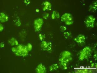
1 Primary Mouse Cerebral Cortex Neurons V: 80% TE: 70% Pictures: 9 days after electroporation Red: MAP2 Blue: GFAP Green: GFP The cells were from Embryonic Day 14 Mouse Cerebral Cortex

2 Primary Mouse Hippocampal Neurons Pictures: 3 days after electroporation Red: MAP2 Green: GFP

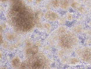
3 Primary Mouse Neural Progenitor Cells V: 80% TE: 60% Pictures: 7 days after electroporation Red: Phalloidin Blue: DAPI Green: GFP


4 Primary Mouse Cerebellar Granule Neurons Nucleofector (Lonza amaxa) result: Transfection Efficiency 24% TE: 65%


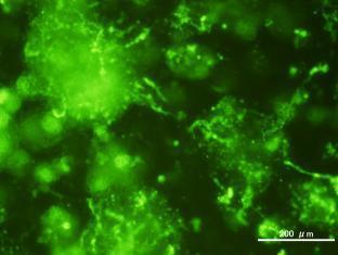
5 Primary Rat Cerebral Cortex Neurons V: 70% TE: 75% The cells were from Embryonic Day 16 Rat Cerebral Cortex. Pictures: 5 days after electroporation


6 Primary Rat Bulbar Neurons V: 80% TE: 75% 7 days after electroporation / The cells were from Embryonic Day 15 Rat Bulbar Neurons


7 Primary Rat Schwann cells V: 90% TE: 80%


8 Primary Rat Schwann cells V: 90% TE: 60% Pictures: 48 hours after electroporation


9 Primary Chick Dorsal Root Ganglia V: 90% TE: 40% The cells were from day 7-9 chick embryos


10 Primary Chick Dorsal Root Ganglia V: 90% TE: 40% The cells were from day 7-9 chick embryos


11 Mouse Neurospheres V: 90% TE: 75% The cells were from E13.5 mouse brain: ganglionic eminence


12 Mouse Neurospheres Neural stem cells derived from mouse SVZ 24 hours after electroporation 48 hours after electroporation 72 hours after electroporation


13 Primary Mouse Cerebral Cortex Neurons in Adherence The neurons were prepared from E15 mouse cerebral cortex. Electroporation: After 6 days in vitro (DIV) on 24-well plates Pictures: 2 days after electroporation Many robust EGFP signals suggest high transfection efficiency.


14 Primary Mouse Cerebral Cortex Neurons in Adherence The neurons were prepared from E15 mouse cerebral cortex. Electroporation: After 6 days in vitro (DIV) on 24-well plates Pictures: 2 days after electroporation *High magnification Neurites are shown clearly. Department of Neurochemistry, National Institute of Neuroscience, Japan


15 Primary Mouse Cerebral Cortex Neurons in Adherence The neurons were prepared from E15 mouse cerebral cortex. Electroporation: After 6 days in vitro (DIV) on 24-well plates Pictures: 2 days after electroporation *High magnification Neurites are shown clearly. Department of Neurochemistry, National Institute of Neuroscience, Japan

")
16 Primary Mouse Cerebral Cortex Neurons in Adherence V: 70% TE: 70% The neurons were prepared from E16 mouse cerebral cortex. Electroporation: After 2 days in vitro (DIV) on 24-well plates

")
17 Primary Mouse Cerebral Cortex Neurons in Adherence V: 60% TE: 65% The neurons were prepared from E16 mouse cerebral cortex. Electroporation: After 2 days in vitro (DIV) on 24-well plates

18 Primary Mouse Cerebral Cortex Neurons in Adherence V: 95% TE: 75% The neurons were prepared from E14 mouse cerebral cortex. Electroporation: After 5 days in vitro (DIV) on 24-well plates

19 Primary Mouse Hippocampal Neurons in Adherence V: 85% TE: 54% The neurons were prepared from E18 mouse cerebral cortex. Electroporation: After 2 days in vitro (DIV) on 24-well plates
 on 24-well plates, post 1-week co-culturing astrocyte and microglial")
20 Primary Mouse Microglial cells in Adherence V: 80% TE: 73% Electroporation: After 1 days in vitro (DIV) on 24-well plates, post 1-week co-culturing astrocyte and microglial cells
 V:")
21 Primary Mouse Glial cells in Adherence Electroporation: After 14 days in vitro (DIV) V: 80% TE: 50%

 on 24-well")
22 Primary Rat Cerebral Cortex Neurons in Adherence V: 70% TE: 60% The neurons were prepared from E17 rat cerebral cortex. Electroporation: After 2 days in vitro (DIV) on 24-well plates Pictures: 24 hours after electroporation

 on")
23 Primary Rat Hippocampal Neurons in Adherence V: 70% TE: 70% The neurons were prepared from E16 rat cerebral cortex. Electroporation: After 2 days in vitro (DIV) on 24-well plates Pictures: 2 days after electroporation

 on")
24 Primary Rat Hippocampal Neurons in Adherence V: 70% TE: 70% The neurons were prepared from E16 rat cerebral cortex. Electroporation: After 2 days in vitro (DIV) on 24-well plates Pictures: 4 days after electroporation


25 Primary Rat Hippocampal Neurons in Adherence V: 100% TE: 50% 48 hours after electroporation The neurons were prepared from P7 rat hippocampus. Electroporation: After 11 DIV


26 Primary Rat Hippocampal Neurons in Adherence V: 100% TE: 50% 48 hours after electroporation The neurons were prepared from P7 rat hippocampus. Electroporation: After 11 DIV
Genesis of cerebellar interneurons and the prevention of neural DNA damage require XRCC1.
 Genesis of cerebellar interneurons and the prevention of neural DNA damage require XRCC1. Youngsoo Lee, Sachin Katyal, Yang Li, Sherif F. El-Khamisy, Helen R. Russell, Keith W. Caldecott and Peter J. McKinnon.
mir-7a regulation of Pax6 in neural stem cells controls the spatial origin of forebrain dopaminergic neurons
 Supplemental Material mir-7a regulation of Pax6 in neural stem cells controls the spatial origin of forebrain dopaminergic neurons Antoine de Chevigny, Nathalie Coré, Philipp Follert, Marion Gaudin, Pascal
During Brain Development Final Destinations for Neurons and Glia Get Separated from Germinal Niches
 During Brain Development Final Destinations for Neurons and Glia Get Separated from Germinal Niches Progenitors are Contained within Unique Domains and Tangentially Fixed. EMBRYO ADULT Migratory Behavior
SUPPLEMENTARY FIG. S2. Representative counting fields used in quantification of the in vitro neural differentiation of pattern of dnscs.
 Supplementary Data SUPPLEMENTARY FIG. S1. Representative counting fields used in quantification of the in vitro neural differentiation of pattern of anpcs. A panel of lineage-specific markers were used
SUPPLEMENTARY FIGURES
 SUPPLEMENTARY FIGURES 1 Supplementary Figure 1, Adult hippocampal QNPs and TAPs uniformly express REST a-b) Confocal images of adult hippocampal mouse sections showing GFAP (green), Sox2 (red), and REST
ErbB4 migrazione II parte
 ErbB4 migrazione II parte Control SVZ cells prefer to migrate on the NRG1 type III substrate the substrate preference of the neuroblasts migrating out of the SVZ explant was evaluated SVZ cells had a strong
In vivo reprogramming reactive glia into ipscs to produce new neurons in the
 In vivo reprogramming reactive glia into ipscs to produce new neurons in the cortex following traumatic brain injury Xiang Gao 1, Xiaoting Wang 1, Wenhui Xiong 1, Jinhui Chen 1, * 1 Spinal Cord and Brain
Brain Development III
 Brain Development III Neural Development In the developing nervous system there must be: 1. The formation of different regions of the brain. 2. The ability of a neuron to differentiate. 3. The ability
Embryonic MGE Cells as a Treatment for Epilepsy December 1, 2012
 Embryonic MGE Cells as a Treatment for Epilepsy December 1, 2012 Scott C. Baraban, PhD University of California, San Francisco American Epilepsy Society Annual Meeting Disclosure Name of Commercial Interest
Supplementary Figure 1. Electroporation of a stable form of β-catenin causes masses protruding into the IV ventricle. HH12 chicken embryos were
 Supplementary Figure 1. Electroporation of a stable form of β-catenin causes masses protruding into the IV ventricle. HH12 chicken embryos were electroporated with β- Catenin S33Y in PiggyBac expression
Cell Birth and Death. Chapter Three
 Cell Birth and Death Chapter Three Neurogenesis All neurons and glial cells begin in the neural tube Differentiated into neurons rather than ectoderm based on factors we have already discussed If these
Top Peer Reviewed Journals Neuroscience & Behavior
 Top Peer Reviewed Journals Neuroscience & Behavior Presented to Iowa State University Presented by Thomson Reuters Neuroscience & Behavior The subject discipline for Neuroscience & Behavior is made of
An unconventional role for mirna: let-7 activates Toll-like receptor 7 and causes neurodegeneration
 An unconventional role for mirna: let-7 activates Toll-like receptor 7 and causes neurodegeneration Sabrina M. Lehmann, Christina Krüger, Boyoun Park, Katja Derkow, Karen Rosenberger, Jan Baumgart, Thorsten
Microglia, Inflammation, and FTD
 FTD Minicourse April, 2009 Microglia, Inflammation, and FTD Li Gan, Ph.D Gladstone Institute of Neurological Disease University of California, San Francisco Outline Why study inflammation in neurodegeneration?
Supplementary Figure S1: Tanycytes are restricted to the central/posterior hypothalamus
 Supplementary Figure S1: Tanycytes are restricted to the central/posterior hypothalamus a: Expression of Vimentin, GFAP, Sox2 and Nestin in anterior, central and posterior hypothalamus. In the anterior
Prss56, a novel marker of adult neurogenesis in the mouse brain. - Supplemental Figures 1 to 5- Brain Structure and Function
 Prss56, a novel marker of adult neurogenesis in the mouse brain - Supplemental Figures 1 to 5- Brain Structure and Function Alexandre Jourdon 1,2, Aurélie Gresset 1, Nathalie Spassky 1, Patrick Charnay
Neocortex Zbtb20 / NFIA / Sox9
 Neocortex / NFIA / Sox9 Supplementary Figure 1. Expression of, NFIA, and Sox9 in the mouse neocortex at. The lower panels are higher magnification views of the oxed area. Arrowheads indicate triple-positive
Contact: Course outline: Contact for other times.
 Contact: kdelaney@uvic.ca Course outline: http://web.uvic.ca/~kdelaney/b367 Scheduled office hours: 1:00-3:00, M&Th Cunn. 259A Contact kdelaney@uvic.ca for other times. Quiz (0.5 hrs) midterm (1.4 hrs)
CNS pathology Third year medical students,2019. Dr Heyam Awad Lecture 1: an introduction: Nervous system cells and their response to injury.
 CNS pathology Third year medical students,2019 Dr Heyam Awad Lecture 1: an introduction: Nervous system cells and their response to injury. CNS course This is a 7 hour course and the topics covered are
Supplementary information. Nkx2.1 regulates the generation of telencephalic astrocytes during embryonic
 Supplementary information Nkx2.1 regulates the generation of telencephalic astrocytes during embryonic development Shilpi Minocha 1*, Delphine Valloton 1*, Yvan Arsenijevic 2, Jean-René Cardinaux 3, Raffaella
Nature Neuroscience: doi: /nn.2275
 Supplementary Figure S1. The presence of MeCP2 in enriched primary glial cultures from rat or mouse brains is not neuronal. Western blot analysis of protein extracts from (a) rat glial and neuronal cultures.
Supplementary Figure 1
 Supplementary Figure 1 Kif1a RNAi effect on basal progenitor differentiation Related to Figure 2. Representative confocal images of the VZ and SVZ of rat cortices transfected at E16 with scrambled or Kif1a
Supplementary Figure 1. ACE robotic platform. A. Overview of the rig setup showing major hardware components of ACE (Automatic single Cell
 2 Supplementary Figure 1. ACE robotic platform. A. Overview of the rig setup showing major hardware components of ACE (Automatic single Cell Experimenter) including the MultiClamp 700B, Digidata 1440A,
Role of the Chondroitin Sulfate Proteoglycan, Neurocan, in Inhibition of Sensory Neurite Regeneration
 University of Kentucky UKnowledge Lewis Honors College Capstone Collection Lewis Honors College 2016 Role of the Chondroitin Sulfate Proteoglycan, Neurocan, in Inhibition of Sensory Neurite Regeneration
Cell Migration II: CNS Cell Migration. Steven McLoon Department of Neuroscience University of Minnesota
 Cell Migration II: CNS Cell Migration Steven McLoon Department of Neuroscience University of Minnesota 1 Course News Coffee Hour Wednesday (Oct 18) 9:00-10:00am Surdyk s Café in Northrop Auditorium Stop
Cell Migration II: CNS Cell Migration. Steven McLoon Department of Neuroscience University of Minnesota
 Cell Migration II: CNS Cell Migration Steven McLoon Department of Neuroscience University of Minnesota 1 Hey! The major concepts discussed relative to neural crest cell migration apply to cell migration
Neural progenitor cells - potent models of normal and disease neurobiology
 Neural progenitor cells - potent models of normal and disease neurobiology Brian Shapiro, Ph.D. Technical Writer, Cell Biology, ATCC November 19, 2015 About ATCC Founded in 1925, ATCC is a non-profit organization
A new role for meninges as a niche for stem/precursor cells with neural differentiation potential during development up to adulthood
 A new role for meninges as a niche for stem/precursor cells with neural differentiation potential during development up to adulthood Francesco Bifari, MD, PhD Mauro Krampera, MD, PhD Ilaria Decimo, PhD
What Cell Make Up the Brain and Spinal Cord
 What Cell Make Up the Brain and Spinal Cord Jennifer LaVail, Ph.D. (http://anatomy.ucsf.edu/pages/lavaillab/index.html) What kinds of cells are these?" Neuron?" Epithelial cell?" Glial cell?" What makes
Huntington s Disease & MARY ET BOYLE, PH.D. DEPARTMENT OF COGNITIVE SCIENCE
 Huntington s Disease & Early Nervous System Development MARY ET BOYLE, PH.D. DEPARTMENT OF COGNITIVE SCIENCE UCSD The cups fell to the floor with a crash. Was this the alarm signal? Or was it forgetting
Neurogenesis in Adult Central Nervous System: Death of a Dogma
 Aristotle University of Thessaloniki, Greece, Nov. 2007 Neurogenesis in Adult Central Nervous System: Death of a Dogma Anton B. Tonchev Division of Cell Biology, Varna University of Medicine, Bulgaria
Nature Neuroscience: doi: /nn Supplementary Figure 1. MADM labeling of thalamic clones.
 Supplementary Figure 1 MADM labeling of thalamic clones. (a) Confocal images of an E12 Nestin-CreERT2;Ai9-tdTomato brain treated with TM at E10 and stained for BLBP (green), a radial glial progenitor-specific
Visualization of embryonic neural stem cells using Hes promoters in transgenic mice
 www.elsevier.com/locate/ymcne Mol. Cell. Neurosci. 31 (2006) 109 122 Visualization of embryonic neural stem cells using Hes promoters in transgenic mice Toshiyuki Ohtsuka, a,b, * Itaru Imayoshi, b Hiromi
Neurogenic Potential of Clitoria ternatea Aqueous Root Extract A Basis for Enhancing Learning and Memory
 Neurogenic Potential of Clitoria ternatea Aqueous Root Extract A Basis for Enhancing Learning and Memory Kiranmai S.Rai* Abstract The neurogenic potential of many herbal extracts used in Indian medicine
During Brain Development Final Destinations for Neurons and Glia Get Separated from Germinal Niches
 During Brain Development Final Destinations for Neurons and Glia Get Separated from Germinal Niches Two mayor forms of neuronal migration: Radial and Tangential Leber & Sanes, 95 How do young neurons actually
Medial View of Cerebellum
 Meds 5371 System Neuroscience D. L. Oliver CEREBELLUM Anterior lobe (spinal) Posterior lobe (cerebral) Flocculonodular lobe (vestibular) Medial View of Cerebellum 1 Ventral View of Cerebellum Flocculus
Bio 3411 Midterm Review:
 Midterm Review: Structure/Development/Systems/ Plastics/Talents/Diseases/Genes Structure General Overview Wednesday 1( 2( THE BRAIN ATLAS 3 rd ed, p. 8! THE BRAIN ATLAS 3 rd ed, p. 9! Mid-line (sagittal)
Are Both Embryonic Migratory Pathways Preserved in the Adult Brain Cerebral Cortex?
 Prague Medical Report / Vol. 107 (2006) No. 1, p. 71 80 71) Are Both Embryonic Migratory Pathways Preserved in the Adult Brain Cerebral Cortex? Šimonová Z., Dutt J. Department of Neuroscience of the Institute
Control of CNS Cell-Fate Decisions by SHP-2 and Its Dysregulation in Noonan Syndrome
 Article Control of CNS Cell-Fate Decisions by SHP-2 and Its Dysregulation in Noonan Syndrome Andrée S. Gauthier, 1,2,3 Olivia Furstoss, 1,2 Toshiyuki Araki, 5 Richard Chan, 5 Benjamin G. Neel, 5 David
Bio 3411 Midterm Review:
 Bio 3411 Midterm Review: Structure/Development/Systems/ Plastics/Talents/Diseases/Genes Structure General Overview Wednesday October 26, 2011 1 2 THE BRAIN ATLAS 3 rd ed, p. 8! THE BRAIN ATLAS 3 rd ed,
Neurons vs. glia. Traditionally, glia have been viewed as passive cells that help to maintain the function of neurons.
 GLIA Neurons vs. glia The defining characteristic of a neuron is its ability to transmit rapid electrical signals in the form of action potentials. All other neural cells that lack this property are broadly
Nervous system. Dr. Rawaa Salim Hameed
 Nervous system Dr. Rawaa Salim Hameed Central nervous system (CNS) CNS consists of the brain (cerebrum, cerebellum, and brainstem) and spinal cord CNS is covered by connective tissue layers, the meninges
Rat Dlx5 is expressed in the subventricular zone and promotes neuronal differentiation
 ISSN 0100-879X Volume 43 (02) 124-225 February 2010 BIOMEDICAL SCIENCES AND CLINICAL INVESTIGATION Braz J Med Biol Res, February 2010, Volume 43(2) 176-185 Rat Dlx5 is expressed in the subventricular zone
Cephalization. Nervous Systems Chapter 49 11/10/2013. Nervous systems consist of circuits of neurons and supporting cells
 Nervous Systems Chapter 49 Cephalization Nervous systems consist of circuits of neurons and supporting cells Nervous system organization usually correlates with lifestyle Organization of the vertebrate
BRAIN REJUVENATION HOW TO KEEP BRAIN STEM CELLS HAPPY
 BRAIN REJUVENATION HOW TO KEEP BRAIN STEM CELLS HAPPY Natalia Surzenko, PhD Research Assistant Professor UNC Chapel Hill Nutrition Research Institute October 13, 2015 MY JOURNEY Tallinn, Estonia Aiken,
Citation for published version (APA): Martina-Mamber, C. E. (2014). GFAP as an understudy in adult neurogenesis. 's-hertogenbosch: Boxpress.
: Martina-Mamber, C. E. (2014). GFAP as an understudy in adult neurogenesis. 's-hertogenbosch: Boxpress.") UvA-DARE (Digital Academic Repository) GFAP as an understudy in adult neurogenesis Mamber, C.E. Link to publication Citation for published version (APA): Martina-Mamber, C. E. (2014). GFAP as an understudy
UvA-DARE (Digital Academic Repository) GFAP as an understudy in adult neurogenesis Mamber, C.E. Link to publication Citation for published version (APA): Martina-Mamber, C. E. (2014). GFAP as an understudy
Nervous System. Oct 15 10:00 AM
 Nervous System Oct 15 10:00 AM 1 Nerve net = series of interconnected nerve cells Nerve = axons of many nerve cells Oct 15 10:10 AM 2 bilateral organisms exhibit cephalization (evolutionary trend towards
Systems Neuroscience CISC 3250
 Systems Neuroscience CISC 325 Memory Types of Memory Declarative Non-declarative Episodic Semantic Professor Daniel Leeds dleeds@fordham.edu JMH 328A Hippocampus (MTL) Cerebral cortex Basal ganglia Motor
Supplementary Figure 1
 Supplementary Figure 1 AAV-GFP injection in the MEC of the mouse brain C57Bl/6 mice at 4 months of age were injected with AAV-GFP into the MEC and sacrificed at 7 days post injection (dpi). (a) Brains
The neurvous system senses, interprets, and responds to changes in the environment. Two types of cells makes this possible:
 NERVOUS SYSTEM The neurvous system senses, interprets, and responds to changes in the environment. Two types of cells makes this possible: the neuron and the supporting cells ("glial cells"). Neuron Neurons
Fig.9.2. Structure of embryonic brain
 T Chapter 9 Development of Ectodermal Organs he ectoderm gives rise to 3 separate cell populations: neural(plate) ectoderm, neural crest cells, and epiderm (general body ectoderm). A primordium (anlage)
Neuroepithelial Cells and Neural Differentiation
 Neuroepithelial Cells and Neural Differentiation Neurulation The cells of the neural tube are NEUROEPITHELIAL CELLS Neural crest cells migrate out of neural tube Neuroepithelial cells are embryonic stem
Electron micrograph of phosphotungstanic acid-stained exosomes derived from murine
 1 SUPPLEMENTARY INFORMATION SUPPLEMENTARY FIGURES Supplementary Figure 1. Physical properties of murine DC-derived exosomes. a, Electron micrograph of phosphotungstanic acid-stained exosomes derived from
Recent Findings from Analysis of HIV Clade C in India
 Recent Findings from Analysis of HIV Clade C in India Pankaj Seth, Ph.D Associate Professor, Molecular & Cellular Neuroscience National Brain Research Centre (NBRC) Manesar, INDIA pseth@nbrc.res.in NeuroAIDS
Metformin Activates an Atypical PKC-CBP Pathway to Promote Neurogenesis and Enhance Spatial Memory Formation
 rticle formin ctivates an typical PKC-CBP Pathway to Promote Neurogenesis and Enhance Spatial Memory Formation Jing Wang, 1,2 Denis Gallagher, 1,2,4,9 Loren M. DeVito, 3,9 Gonzalo I. Cancino, 1,2 David
Research Development: Bedside to Bench and Back
 Research Development: Bedside to Bench and Back Matt Bellizzi, MD PhD Department of Neurology University of Rochester School of Medicine and Dentistry Rochester, NY "I can walk down the hall just fine,
Neocortex. Cortical Structures in the Brain. Neocortex Facts. Laminar Organization. Bark-like (cortical) structures: Shepherd (2004) Chapter 12
 structures: Shepherd (2004) Chapter 12") Neocortex Shepherd (2004) Chapter 12 Rodney Douglas, Henry Markram, and Kevan Martin Instructor: Yoonsuck Choe; CPSC 644 Cortical Networks Cortical Structures in the Brain Bark-like (cortical) structures:
Neocortex Shepherd (2004) Chapter 12 Rodney Douglas, Henry Markram, and Kevan Martin Instructor: Yoonsuck Choe; CPSC 644 Cortical Networks Cortical Structures in the Brain Bark-like (cortical) structures:
Rapamycin Suppresses Astrocytic and Microglial Activation and Reduced Development of Neuropathic Pain after Spinal Cord Injury in Mice.
 Rapamycin Suppresses Astrocytic and Microglial Activation and Reduced Development of Neuropathic Pain after Spinal Cord Injury in Mice. Satoshi Tateda, M.D., Haruo Kanno, M.D., Ph.D., Hiroshi Ozawa, M.D.,
The neuron. Biocytin labeled pyramidal neuron recorded in piriform cortex
 The neuron Biocytin labeled pyramidal neuron recorded in piriform cortex Discovery of the neuron (A) Reticularist Doctrine (B) Neuron Doctrine Exception.GAP JUNCTIONS between neurons Neuronal shape Dendrites
Supplementary Materials
 Supplementary Materials Fig. S1. Weights of full-dose treatment groups comparing 1 st, 2 nd, and 3 rd generation gene replacement therapy. Mice were treated at p1 with 4x10 11 GC of the three different
THE CENTRAL NERVOUS SYSTE M
 THE CENTRAL NERVOUS SYSTE M Structure and Functio n THIRD EDITIO N PER BRODAL A Brief Survey, x i Studying the Structures and Function of the Nervous System, xii i Animal Experiments Crucial for Progress,
Achieving The Goals Of Toxicity Testing In the 21st Century: The TestSmart Developmental Neurotoxicology (DNT) Testing Program
 Testing Program") Achieving The Goals Of Toxicity Testing In the 21st Century: The TestSmart Developmental Neurotoxicology (DNT) Testing Program Joseph Bressler and Alan Goldberg Center In Alternatives To Animal Testing
Achieving The Goals Of Toxicity Testing In the 21st Century: The TestSmart Developmental Neurotoxicology (DNT) Testing Program Joseph Bressler and Alan Goldberg Center In Alternatives To Animal Testing
Neurodevelopment II Structure Formation. Reading: BCP Chapter 23
 Neurodevelopment II Structure Formation Reading: BCP Chapter 23 Phases of Development Ovum + Sperm = Zygote Cell division (multiplication) Neurogenesis Induction of the neural plate Neural proliferation
Neural plasticity in infants - relevance to baby swimming. Morten Overgaard
 Neural plasticity in infants - relevance to baby swimming Morten Overgaard Programme What is neuroscience? Totally superficial neuroanatomy Paradoxes of functional localization Mechanisms of neural plasticity
Index Note: Page numbers of article titles are in boldface type.
 Neurosurg Clin N Am 18 (2007) 191 198 Index Note: Page numbers of article titles are in boldface type. A AC133 antigen, in brain tumor cancer cells, 32 35 Activity-based restoration therapy, for spinal
Supplemental Information. Menin Deficiency Leads to Depressive-like. Behaviors in Mice by Modulating. Astrocyte-Mediated Neuroinflammation
 Neuron, Volume 100 Supplemental Information Menin Deficiency Leads to Depressive-like Behaviors in Mice by Modulating Astrocyte-Mediated Neuroinflammation Lige Leng, Kai Zhuang, Zeyue Liu, Changquan Huang,
Embryology of the Nervous System. Steven McLoon Department of Neuroscience University of Minnesota
 Embryology of the Nervous System Steven McLoon Department of Neuroscience University of Minnesota In the blastula stage embryo, the embryonic disk has two layers. During gastrulation, epiblast cells migrate
Embryonic Stem Cell Derived Neural Precursor Grafts for Treatment of Temporal Lobe Epilepsy
 Wesleyan University WesScholar Division III Faculty Publications Natural Sciences and Mathematics 4-2009 Embryonic Stem Cell Derived Neural Precursor Grafts for Treatment of Temporal Lobe Epilepsy Janice
Supplemental Figure 1. Intracranial transduction of a modified ptomo lentiviral vector in the mouse
 Supplemental figure legends Supplemental Figure 1. Intracranial transduction of a modified ptomo lentiviral vector in the mouse hippocampus targets GFAP-positive but not NeuN-positive cells. (A) Stereotaxic
CHAPTER 48: NERVOUS SYSTEMS
 CHAPTER 48: NERVOUS SYSTEMS Name I. AN OVERVIEW OF NERVOUS SYSTEMS A. Nervous systems perform the three overlapping functions of sensory input, integration, and motor output B. Networks of neurons with
BioMed Central. Jeannette E Davies*, Christoph Pröschel, Ningzhe Zhang, Mark Noble, Margot Mayer-Pröschel and Stephen JA Davies* Research article
 BioMed Central Open Access Research article Transplanted astrocytes derived from BMP- or CNTF-treated glialrestricted precursors have opposite effects on recovery and allodynia after spinal cord injury
Supplemental Materials. STK16 regulates actin dynamics to control Golgi organization and cell cycle
 Supplemental Materials STK16 regulates actin dynamics to control Golgi organization and cell cycle Juanjuan Liu 1,2,3, Xingxing Yang 1,3, Binhua Li 1, Junjun Wang 1,2, Wenchao Wang 1, Jing Liu 1, Qingsong
The cells of the nervous system
 The cells of the nervous system LESSON N.9 - PSYCHOBIOLOGY because of the location and volume as compared to our body, the brain has always been a matter of conjecture about its fundamental role in the
Nervous Tissue and Histology of CNS
 Nervous Tissue and Histology of CNS Functions of Nervous System Like the CPU of a computer, the nervous system is the master controlling system of the body. It is designed to constantly and rapidly adjust
The Predominant Neural Stem Cell Isolated from Postnatal and Adult Forebrain But Not Early Embryonic Forebrain Expresses GFAP
 2824 The Journal of Neuroscience, April 1, 2003 23(7):2824 2832 The Predominant Neural Stem Cell Isolated from Postnatal and Adult Forebrain But Not Early Embryonic Forebrain Expresses GFAP Tetsuya Imura,
Stem Cells and the Study of Neurodegeneration. Tracy Young-Pearse, PhD September 12, 2014!
 Stem Cells and the Study of Neurodegeneration Tracy Young-Pearse, PhD September 12, 2014! Techniques for studying mechanisms of neurological disease Animal models Human subjects Postmortem analyses, imaging
Investigating the role of EphAl ephrin-a signalling during trigeminal ganglion axon guidance
 Investigating the role of EphAl ephrin-a signalling during trigeminal ganglion axon guidance A thesis submitted for the degree of Doctor of Philosophy Molecular and Biomedical Science (Discipline of Genetics),
Department of Cognitive Science UCSD
 Department of Cognitive Science UCSD Verse 1: Neocortex, frontal lobe, Brain stem, brain stem, Hippocampus, neural node, Right hemisphere, Pons and cortex visual, Brain stem, brain stem, Sylvian fissure,
Developmental expression of the platelet-derived growth factor a-receptor gene in mammalian central nervous system
 Proc. Natl. Acad. Sci. USA Vol. 90, pp. 1952-1956, March 1993 Medical Sciences Developmental expression of the platelet-derived growth factor a-receptor gene in mammalian central nervous system (in situ
Rapamycin suppresses astrocytic and microglial activation and reduces development of neuropathic pain after spinal cord injury in mice.
 Rapamycin suppresses astrocytic and microglial activation and reduces development of neuropathic pain after spinal cord injury in mice. Satoshi Tateda, MD, Haruo Kanno, MD, PhD, Hiroshi Ozawa, MD, PhD,
The developmental history of a sensory ganglion cell
 (bozza v1, luglio 2016 per VC) The developmental history of a sensory ganglion cell by Brian Freeman, School of Medical Sciences, UNSW (b.freeman@unsw.edu.au) The sensory ganglion cell (e.g., in spinal
SUPPLEMENTARY INFORMATION
 SUPPLEMENTARY INFORMATION Human cerebral cortex development from pluripotent stem cells to functional excitatory synapses Yichen Shi 1,2, Peter Kirwan 1,2, James Smith 1,2, Hugh P.C. Robinson 3 and Frederick
Chemokine Regulation of Oligodendrocyte Development in the Spinal Cord. Bob Avino Saint Louis University Senior Honors Thesis April 19, 2011
 Chemokine Regulation of Oligodendrocyte Development in the Spinal Cord Bob Avino Saint Louis University Senior Honors Thesis April 19, 2011 Richard J. Miller, PhD Northwestern University Feinberg School
Multipolar Migration: The Third Mode of Radial Neuronal Migration in the Developing Cerebral Cortex
 9996 The Journal of Neuroscience, November 5, 2003 23(31):9996 10001 Brief Communication Multipolar Migration: The Third Mode of Radial Neuronal Migration in the Developing Cerebral Cortex Hidenori Tabata
NERVOUS SYSTEM. Academic Resource Center. Forskellen mellem oscillator og krystal
 NERVOUS SYSTEM Academic Resource Center Forskellen mellem oscillator og krystal Overview of the Nervous System Peripheral nervous system-pns cranial nerves spinal nerves ganglia peripheral nerves enteric
supplementary information
 DOI: 10.1038/ncb2153 Figure S1 Ectopic expression of HAUSP up-regulates REST protein. (a) Immunoblotting showed that ectopic expression of HAUSP increased REST protein levels in ENStemA NPCs. (b) Immunofluorescent
Supplementary Figure 1. Chimeric analysis of inner ears. (A-H) Chimeric inner ears with fluorescent ES cells and (I,J) Rainbow inner ears.
 Chimeric inner ears with fluorescent ES cells and (I,J) Rainbow inner ears.") Supplementary Figure 1. himeric analysis of inner ears. (A-H) himeric inner ears with fluorescent ES cells and (I,J) Rainbow inner ears. (A,B) omposite images showing three colors in different vestibular
Supplementary Figure 1. himeric analysis of inner ears. (A-H) himeric inner ears with fluorescent ES cells and (I,J) Rainbow inner ears. (A,B) omposite images showing three colors in different vestibular
Development of the Nervous System 1 st month
 Development of the Nervous System 1 st month day 1 - fertilization of egg day 6 - uterine implantation day 18 - trilaminar (3-layered) disc (blastoderm, embryo) ectoderm (dorsal) - nervous system and skin
Lecture 42: Final Review. Martin Wessendorf, Ph.D.
 Lecture 42: Final Review Martin Wessendorf, Ph.D. Lecture 33 cortex Heilbronner 5 lobes of the cortex Lateral view (left side) Mid-saggital view (right side) Cellular organization of cortex White matter
Recipes for Making Human Interneurons from Stem Cells Require Multiple Factors, Careful Timing, and Long Maturation Periods
 Current Literature In Basic Science Recipes for Making Human Interneurons from Stem Cells Require Multiple Factors, Careful Timing, and Long Maturation Periods Directed Differentiation and Functional Maturation
1. The basic anatomy of the Central Nervous System (CNS)
") Psyc 311A, fall 2008 Conference week 1 Sept 9 th to 11 th TA: Jürgen Germann; e-mail: jurgen.germann@mcgill.ca Overview: 1. The basic anatomy of the Central Nervous System (CNS) 2. Cells of the CNS 3.
Psyc 311A, fall 2008 Conference week 1 Sept 9 th to 11 th TA: Jürgen Germann; e-mail: jurgen.germann@mcgill.ca Overview: 1. The basic anatomy of the Central Nervous System (CNS) 2. Cells of the CNS 3.
1. Microfluidic device characteristics and cell compartmentalization
 Supplementary figures: 1. Microfluidic device characteristics and cell compartmentalization Microfluidic channels were molded in PDMS (Fig. S1 A and B) over silicon masters, which were structured with
Stem Cells. RapidCommunication. Transient Expression of Olig1 Initiates the Differentiation of Neural Stem Cells into Oligodendrocyte Progenitor Cells
 Stem Cells RapidCommunication Transient Expression of Olig1 Initiates the Differentiation of Neural Stem Cells into Oligodendrocyte Progenitor Cells Veerakumar Balasubramaniyan, Nienke Timmer, Britta Kust,
TITLE: Altered Astrocyte-Neuron Interactions and Epileptogenesis in Tuberous Sclerosis Complex Disorder
 AWARD NUMBER: W81XWH-12-1-0196 TITLE: Altered Astrocyte-Neuron Interactions and Epileptogenesis in Tuberous Sclerosis Complex Disorder PRINCIPAL INVESTIGATOR: Dr. David Sulzer, Ph.D. CONTRACTING ORGANIZATION:
Modeling of Hippocampal Behavior
 Modeling of Hippocampal Behavior Diana Ponce-Morado, Venmathi Gunasekaran and Varsha Vijayan Abstract The hippocampus is identified as an important structure in the cerebral cortex of mammals for forming
SUPPLEMENTARY INFORMATION
 DOI: 10.1038/ncb2566 Figure S1 CDKL5 protein expression pattern and localization in mouse brain. (a) Multiple-tissue western blot from a postnatal day (P) 21 mouse probed with an antibody against CDKL5.
Cerebellum- and forebrain-derived stem cells possess intrinsic regional character
 Research article 4497 Cerebellum- and forebrain-derived stem cells possess intrinsic regional character Corinna Klein 1, Simon J. B. Butt 1, Robert P. Machold 1, Jane E. Johnson 2 and Gord Fishell 1, *
The neuron. Biocytin labeled pyramidal neuron recorded in piriform cortex
 The neuron Biocytin labeled pyramidal neuron recorded in piriform cortex Discovery of the neuron (A) Reticularist Doctrine (B) Neuron Doctrine Exception.GAP JUNCTIONS between neurons Neuronal shape Dendrites
mm Distance (mm)
") b a Magnet Illumination Coverslips MPs Objective 2575 µm 1875 µm 1575 µm 1075 µm 875 µm 545 µm 20µm 2 3 0.5 0.3mm 1 1000 100 10 1 0.1 1000 100 10 1 0.1 Field Induction (Gauss) 1.5 0 5 10 15 20 Distance
b a Magnet Illumination Coverslips MPs Objective 2575 µm 1875 µm 1575 µm 1075 µm 875 µm 545 µm 20µm 2 3 0.5 0.3mm 1 1000 100 10 1 0.1 1000 100 10 1 0.1 Field Induction (Gauss) 1.5 0 5 10 15 20 Distance
Fig The C.S. of the Spinal Cord A ganglion is a term for a collection of perikarya located outside of the CNS. In certain regions of the spinal
 Chapter 9 Nervous System The nervous system is divided into two components: The CNS - the brain and spinal cord and the PNS - the nerves emanating from the spinal cord and brain that distribute to other