In vivo reprogramming reactive glia into ipscs to produce new neurons in the
|
|
|
- Percival Newman
- 5 years ago
- Views:
Transcription
1 In vivo reprogramming reactive glia into ipscs to produce new neurons in the cortex following traumatic brain injury Xiang Gao 1, Xiaoting Wang 1, Wenhui Xiong 1, Jinhui Chen 1, * 1 Spinal Cord and Brain Injury Research Group, Stark Neuroscience Research Institute, Department of Neurosurgery, Indiana University. 320 W 15th Street, Indianapolis, IN
2
3
4
5
6
7
8
9
10
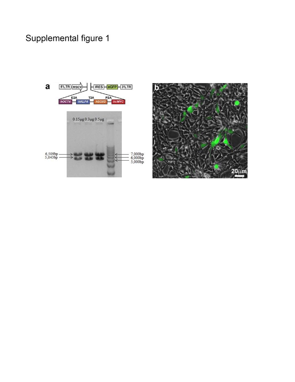
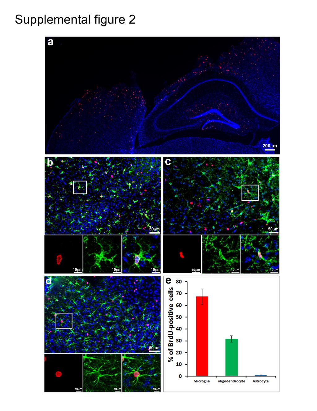
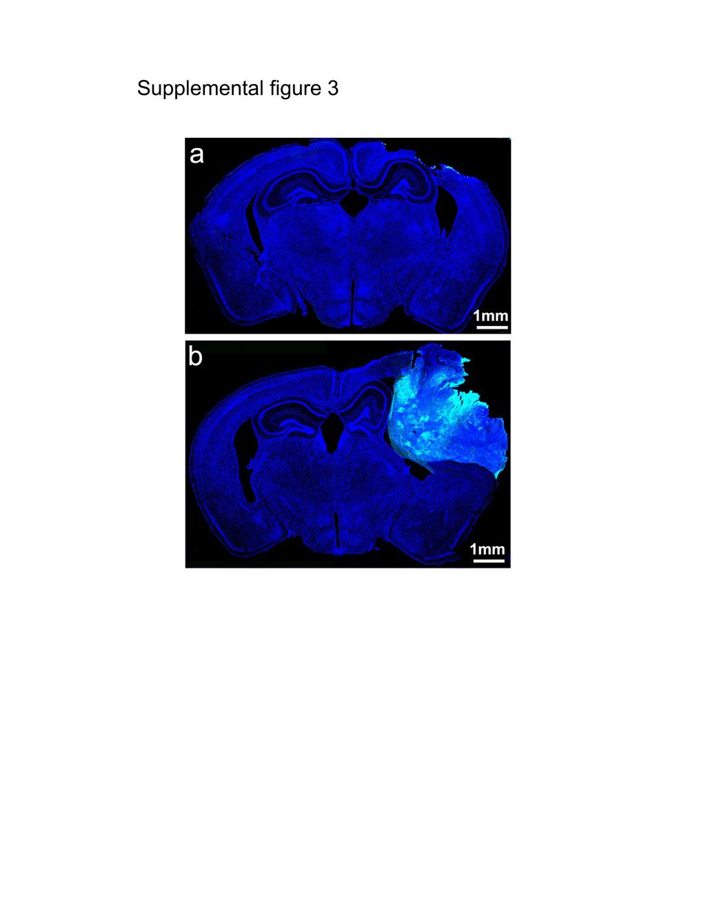
11 Supplemental figure legends Supplemental figure 1. Retroviral dual cassette vectors co-expressing four human transcriptional factors (hoct4, hklf4, hsox2 and hcmyc, hoksm) in combination with enhanced green fluorescent protein (EGFP). a. Expression vector used for generating retrovirus contained 4 hoksm factors, which can reprogram somatic cells into induced pluripotent stem cells and tagged by EGFP. b. Retrovirus infected proliferating cell and express EGFP (green) in vitro. The titration is about 1.6x10 7 pfu. Supplemental figure 2. Glial reactivation in the cortex following moderate traumatic brain injury (TBI). a. Immunostaining with antibody against bromo-2'-deoxyuridine (BrdU) (red) to detect proliferating cells in the cortex 3 days after moderate traumatic brain injury (TBI). b. Majority of proliferating cells was Iba1-positive reactive microglia (green). c. Some proliferating cells expressed oligodendrocyte precursor marker NG2 (green). d. Very few proliferating cells were double-labeled by astrocyte marker-gfap (green). e. Quantification data showed the ratio of different types of proliferating cells. Supplemental figure 3. Reprogrammed cells fill the tissue-lesion cavity induced by traumatic brain injury (TBI). a. TBI-injured brain received injection of controlled retroviruses expressing EGFP. b. TBI-injured brain received injection of retroviruses expressing defined transcriptional factors and EGFP. Green is EGFP, while blue is DAPI staining. 2
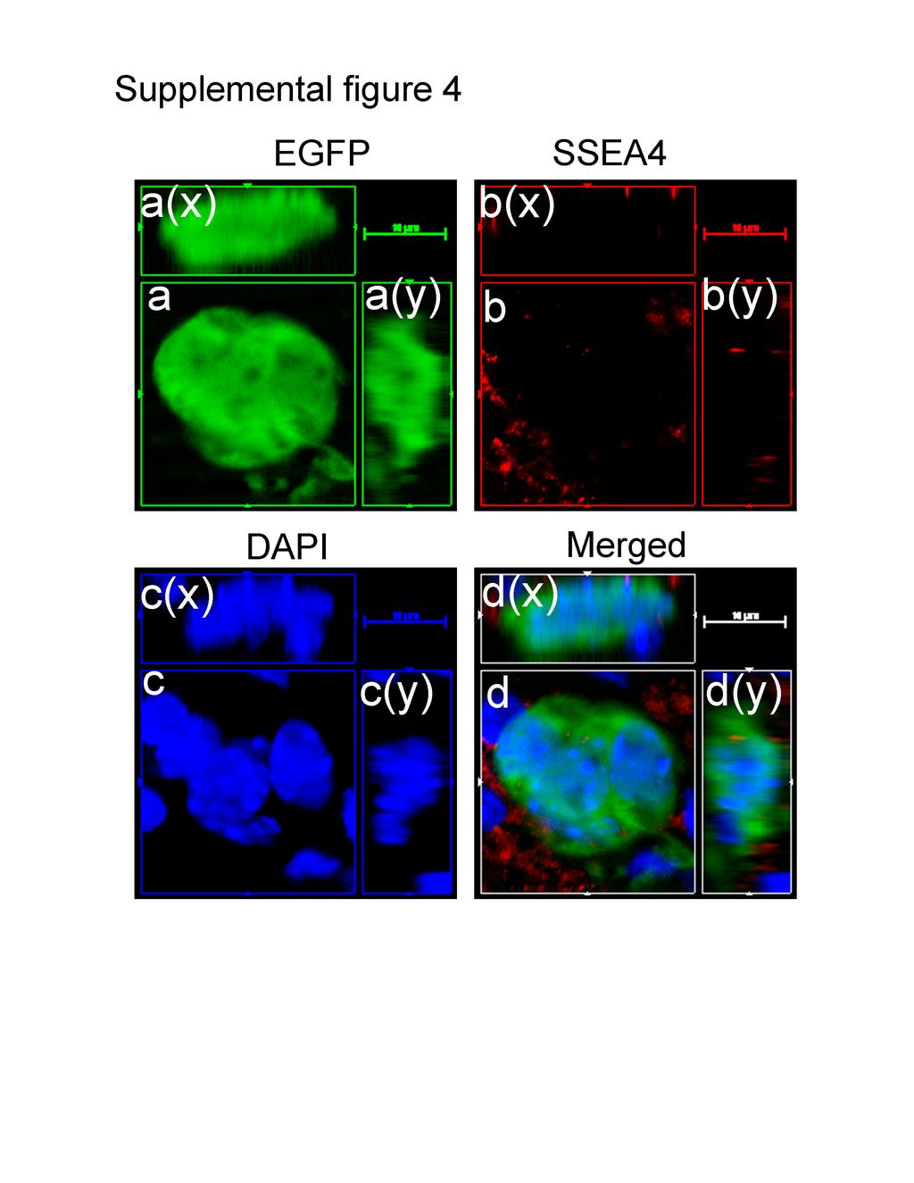
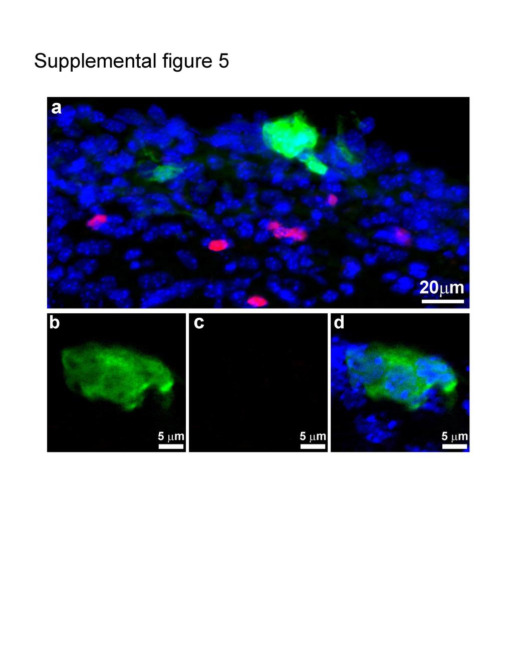
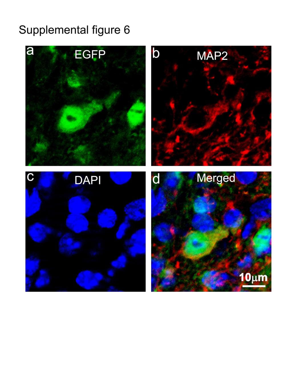
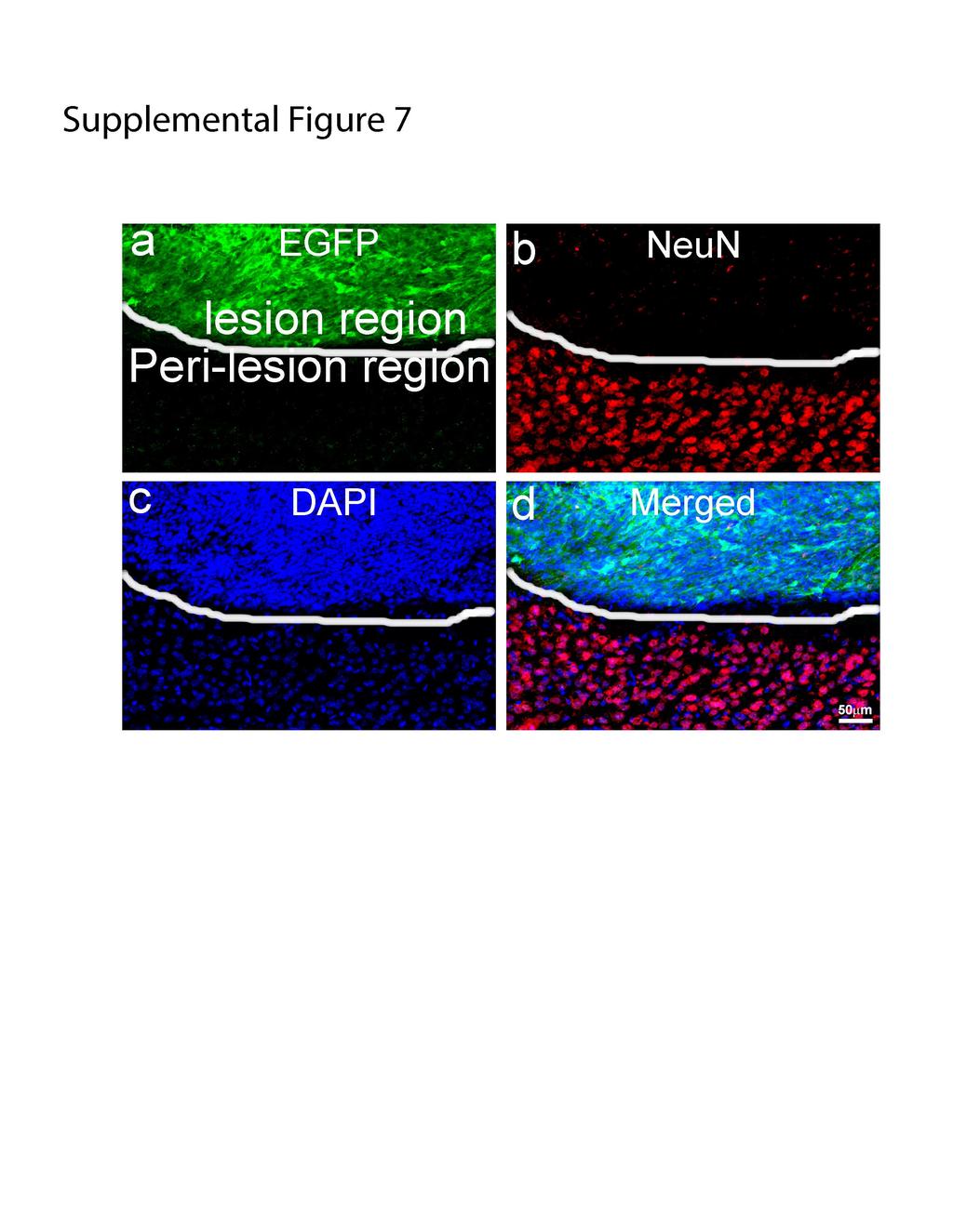
12 Supplemental figure 4. SSEA4 was not detectable in the cells expressing hoksm- EGFP at 2 weeks after traumatic brain injury (TBI). a. Three-dimensional images of cell cluster expressing hoksm-egfp 2 weeks after TBI. b. Three-dimensional image to show that the cell cluster did not express SSEA4 at 2 weeks after injury. c. 4,6-diamidino-2-phenylindole (DAPI) staining to exhibit nuclei. d. Merged image of a-c. Supplemental figure 5. SOX2 was not detectable in the cells expressing hoksm- EGFP at 2 weeks after traumatic brain injury (TBI). a. The hoksm-egfp-positive cell (green) clusters at the peri-lesion region did not express Sox2 at 2 weeks after TBI; instead, the Sox2 was expressed in cells that did not express EGFP, likely reactive glia, at the peri-lesion region of the injured cortex at 2 weeks after TBI. b-d, Enlarged images showed EGFP-positive cell aggregates without Sox2 expression 2 weeks after TBI. Supplemental figure 6. Reprogrammed cells developed into Map2-expressing mature neurons at 6 weeks after traumatic brain injury (TBI). a. The hoksm-egfp-expressing cells (green) in the TBI-injured cortex 6 weeks after reprogramming. b. The hoksm-egfp expressing cells differentiated into mature neurons expressing Map2 (red), a marker for mature neurons, in the injured cortex. c. DAPI staining. d. Merged image of a-c. Supplemental figure 7. Reprogramed cells did not express NeuN at 4 weeks after traumatic brain injury (TBI). a. Tissue lesion cavity in the cortex following TBI was filled with reprogramming cells with EGFP expression (green). White line indicates the boundary between the tissue lesion cavity and peri-lesion region in the cortex at 4 weeks 3
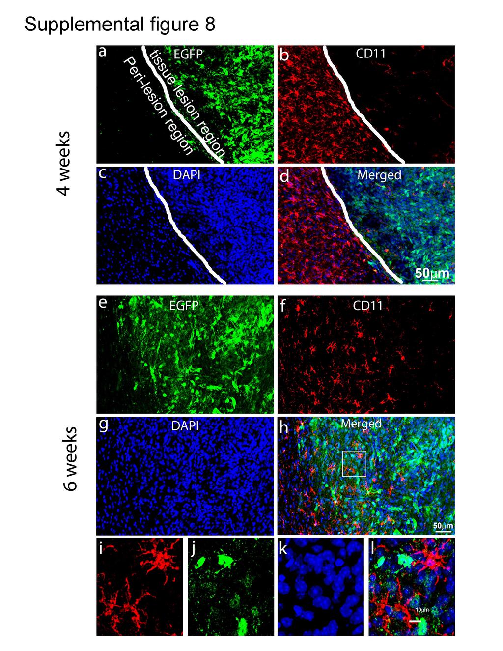
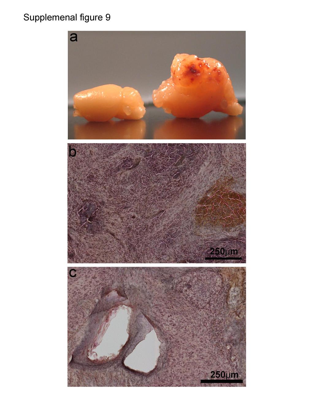
13 after moderate TBI. b. Spared endogenous mature neurons expressing NeuN (red) located at the peri-lesion region. c. DAPI staining to exhibit nuclei in the cortex. d. Merged image of a-c. Supplemental figure 8. Reprogramed cells did not differentiate into microglia. a, e. Tissue lesion cavity in the cortex following traumatic brain injury (TBI) was filled with reprogramming cells with EGFP expression (green) at 4 or 6 weeks after moderate TBI. White line indicates the boundary between the tissue lesion cavity and peri-lesion region in the cortex. b, f. CD-11b positive microglia at the peri-lesion region in the cortex at 4 or 6 weeks after TBI. c, g. DAPI staining to exhibit nuclei in the cortex. d, h. Merged image of a-c or e-g. i-l. Enlarged images to show cells express either CD11b or EGFP within the white box in the panel h. The EGFP-positive cells did not express CD11b. Supplemental figure 9. Reprogrammed cells developed into teratoma in the cortex. a. Brain of control without reprogramming (left) or brain with reprogramming (right) at 6 weeks after TBI. b and c. H&E staining of the brain region with reprogrammed cells. 4
SUPPLEMENTARY FIG. S2. Representative counting fields used in quantification of the in vitro neural differentiation of pattern of dnscs.
 Supplementary Data SUPPLEMENTARY FIG. S1. Representative counting fields used in quantification of the in vitro neural differentiation of pattern of anpcs. A panel of lineage-specific markers were used
Supplemental Figure 1. Intracranial transduction of a modified ptomo lentiviral vector in the mouse
 Supplemental figure legends Supplemental Figure 1. Intracranial transduction of a modified ptomo lentiviral vector in the mouse hippocampus targets GFAP-positive but not NeuN-positive cells. (A) Stereotaxic
Nature Neuroscience: doi: /nn Supplementary Figure 1. MADM labeling of thalamic clones.
 Supplementary Figure 1 MADM labeling of thalamic clones. (a) Confocal images of an E12 Nestin-CreERT2;Ai9-tdTomato brain treated with TM at E10 and stained for BLBP (green), a radial glial progenitor-specific
Primary Mouse Cerebral Cortex Neurons V: 80% TE: 70%
 Primary Mouse Cerebral Cortex Neurons V: 80% TE: 70% Pictures: 9 days after electroporation Red: MAP2 Blue: GFAP Green: GFP The cells were from Embryonic Day 14 Mouse Cerebral Cortex Primary Mouse Hippocampal
Microglia, Inflammation, and FTD
 FTD Minicourse April, 2009 Microglia, Inflammation, and FTD Li Gan, Ph.D Gladstone Institute of Neurological Disease University of California, San Francisco Outline Why study inflammation in neurodegeneration?
An unconventional role for mirna: let-7 activates Toll-like receptor 7 and causes neurodegeneration
 An unconventional role for mirna: let-7 activates Toll-like receptor 7 and causes neurodegeneration Sabrina M. Lehmann, Christina Krüger, Boyoun Park, Katja Derkow, Karen Rosenberger, Jan Baumgart, Thorsten
Supplementary Information
 Supplementary Information Astrocytes regulate adult hippocampal neurogenesis through ephrin-b signaling Randolph S. Ashton, Anthony Conway, Chinmay Pangarkar, Jamie Bergen, Kwang-Il Lim, Priya Shah, Mina
Supplementary Information
 Supplementary Information Title Degeneration and impaired regeneration of gray matter oligodendrocytes in amyotrophic lateral sclerosis Authors Shin H. Kang, Ying Li, Masahiro Fukaya, Ileana Lorenzini,
Supplemental Information. Menin Deficiency Leads to Depressive-like. Behaviors in Mice by Modulating. Astrocyte-Mediated Neuroinflammation
 Neuron, Volume 100 Supplemental Information Menin Deficiency Leads to Depressive-like Behaviors in Mice by Modulating Astrocyte-Mediated Neuroinflammation Lige Leng, Kai Zhuang, Zeyue Liu, Changquan Huang,
GFP/Iba1/GFAP. Brain. Liver. Kidney. Lung. Hoechst/Iba1/TLR9!
 Supplementary information a +KA Relative expression d! Tlr9 5!! 5! NSC Neuron Astrocyte Microglia! 5! Tlr7!!!! NSC Neuron Astrocyte! GFP/Sβ/! Iba/Hoechst Microglia e Hoechst/Iba/TLR9! GFP/Iba/GFAP f Brain
Supplementary Materials
 Supplementary Materials Fig. S1. Weights of full-dose treatment groups comparing 1 st, 2 nd, and 3 rd generation gene replacement therapy. Mice were treated at p1 with 4x10 11 GC of the three different
Supplementary Figure 1
 Supplementary Figure 1 Genetic labeling of microglia Male and female 2-3 month-old CreERT2;R26-tdTomato mice or CreERT2;R26-tdTomato;Iba1-eGFP transgenic mice were treated with 1x, 2x (48 h apart), or
Electron micrograph of phosphotungstanic acid-stained exosomes derived from murine
 1 SUPPLEMENTARY INFORMATION SUPPLEMENTARY FIGURES Supplementary Figure 1. Physical properties of murine DC-derived exosomes. a, Electron micrograph of phosphotungstanic acid-stained exosomes derived from
SUPPLEMENTARY FIGURES
 SUPPLEMENTARY FIGURES 1 Supplementary Figure 1, Adult hippocampal QNPs and TAPs uniformly express REST a-b) Confocal images of adult hippocampal mouse sections showing GFAP (green), Sox2 (red), and REST
ROCK/Cdc42-mediated microglial motility and gliapse formation lead to phagocytosis of degenerating dopaminergic neurons in vivo
 Supplementary Information ROCK/Cdc42-mediated microglial motility and gliapse formation lead to phagocytosis of degenerating dopaminergic neurons in vivo Carlos Barcia* 1,2, Carmen M Ros 1,2, Valentina
Neocortex Zbtb20 / NFIA / Sox9
 Neocortex / NFIA / Sox9 Supplementary Figure 1. Expression of, NFIA, and Sox9 in the mouse neocortex at. The lower panels are higher magnification views of the oxed area. Arrowheads indicate triple-positive
Supplementary Figure 1
 Supplementary Figure 1 The average sigmoid parametric curves of capillary dilation time courses and average time to 50% peak capillary diameter dilation computed from individual capillary responses averaged
SUPPLEMENTARY INFORMATION
 DOI: 10.1038/ncb2566 Figure S1 CDKL5 protein expression pattern and localization in mouse brain. (a) Multiple-tissue western blot from a postnatal day (P) 21 mouse probed with an antibody against CDKL5.
mir-7a regulation of Pax6 in neural stem cells controls the spatial origin of forebrain dopaminergic neurons
 Supplemental Material mir-7a regulation of Pax6 in neural stem cells controls the spatial origin of forebrain dopaminergic neurons Antoine de Chevigny, Nathalie Coré, Philipp Follert, Marion Gaudin, Pascal
Cell Birth and Death. Chapter Three
 Cell Birth and Death Chapter Three Neurogenesis All neurons and glial cells begin in the neural tube Differentiated into neurons rather than ectoderm based on factors we have already discussed If these
Supplemental Figures Supplemental Figure 1:
 Supplemental Figures Supplemental Figure 1: Representative FACS data showing Concurrent Brain cell type Acquisition using either Percoll PLUS (top row) or myelin removal beads (bottom two rows). Debris
Neurodevelopment II Structure Formation. Reading: BCP Chapter 23
 Neurodevelopment II Structure Formation Reading: BCP Chapter 23 Phases of Development Ovum + Sperm = Zygote Cell division (multiplication) Neurogenesis Induction of the neural plate Neural proliferation
Cells of the Nervous System
 Cells of the Nervous System Layout of the Nervous System Central Nervous System (CNS) Brain (in the skull) Spinal Cord (in the spine) Interprets sensory input, initiates movement, and mediates complex
Differential neuronal vulnerability identifies IGF-2 as a protective factor in
 Supplementary Information Differential neuronal vulnerability identifies IGF-2 as a protective factor in ALS Ilary Allodi 1,3, Laura Comley 1,3, Susanne Nichterwitz 1,3, Monica Nizzardo 2, Chiara Simone
Recent Findings from Analysis of HIV Clade C in India
 Recent Findings from Analysis of HIV Clade C in India Pankaj Seth, Ph.D Associate Professor, Molecular & Cellular Neuroscience National Brain Research Centre (NBRC) Manesar, INDIA pseth@nbrc.res.in NeuroAIDS
Supplementary Figure S1: Tanycytes are restricted to the central/posterior hypothalamus
 Supplementary Figure S1: Tanycytes are restricted to the central/posterior hypothalamus a: Expression of Vimentin, GFAP, Sox2 and Nestin in anterior, central and posterior hypothalamus. In the anterior
Supplementary Figure 1
 Supplementary Figure 1 3 3 3 1 1 Bregma -1.6mm 3 : Bregma Ref) Http://www.mbl.org/atlas165/atlas165_start.html Bregma -.18mm Supplementary Figure 1 Schematic representation of the utilized brain slice
Supplementary Figure 1. Nature Neuroscience: doi: /nn.4547
 Supplementary Figure 1 Characterization of the Microfetti mouse model. (a) Gating strategy for 8-color flow analysis of peripheral Ly-6C + monocytes from Microfetti mice 5-7 days after TAM treatment. Living
Supplemental Figure 1. Quantification of proliferation in thyroid of WT, Ctns -/- and grafted
 Supplemental Figure 1. Quantification of proliferation in thyroid of WT, Ctns -/- and grafted Ctns -/- mice. Cells immunolabeled for the proliferation marker (Ki-67) were counted in sections (n=3 WT, n=4
Expression of ALS-linked TDP-43 Mutant in Astrocytes Causes Noncell-autonomous
 Manuscript EMBO-2012-84256 Expression of ALS-linked TDP-43 Mutant in Astrocytes Causes Noncell-autonomous Motor Neuron Death in Rats Jianbin Tong, Cao Huang, Fangfang Bi, Qinxue Wu, Bo Huang, Xionghao
Supplementary Figure 1
 Supplementary Figure 1 AAV-GFP injection in the MEC of the mouse brain C57Bl/6 mice at 4 months of age were injected with AAV-GFP into the MEC and sacrificed at 7 days post injection (dpi). (a) Brains
Man and his environment
 Man and his environment Dr. Elriah M. Makie 0122858517 Nervous Tissue BSc.M.Sc.MBBS Introduction The nervous system is divided into two main parts: The central nervous system (CNS) comprising the brain
Progress Report for NJCSCR (Yu-Wen Chang)
") Progress Report for NJCSCR (Yu-Wen Chang) Overall Plan Summary: Traumatic injury to the spinal cord initiates a cascade of degenerative processes, known as secondary injury, which include various inflammatory
Progress Report for NJCSCR (Yu-Wen Chang) Overall Plan Summary: Traumatic injury to the spinal cord initiates a cascade of degenerative processes, known as secondary injury, which include various inflammatory
Microglia-derived extracellular vesicles regulate the proliferation and differentiation of oligodendrocyte precursor cells
 University of Turin CNR Institute of Neuroscience Microglia-derived extracellular vesicles regulate the proliferation and differentiation of oligodendrocyte precursor cells Roberta Parolisi Turin, December
SUPPLEMENTARY FIGURES
 SUPPLEMENTARY FIGURES 1 2 3 4 SUPPLEMENTARY TABLES Supplementary Table S1. Brain Tumors used in the study Code Tumor Classification Age Gender HuTuP51 Glioblastoma 57 Male HuTuP52 Glioblastoma 53 Male
regenerative medicine in the brain and the spinal cord spinal cord injuries
 regenerative medicine in the brain and the spinal cord spinal cord injuries primary and secondary events during SCI traumatic spinal cord injury (SCI) traumatic spinal cord injury (SCI) main goal is to
SUPPLEMENTARY INFORMATION
 doi:10.1038/nature10353 Supplementary Figure 1. Mutations of UBQLN2 in patients with ALS and ALS/dementia. (a) A mutation, c.1489c>t (p.p497s), was identified in F#9975. The pedigree is shown on the left
NG2 + CNS Glial Progenitors Remain Committed to the Oligodendrocyte Lineage in Postnatal Life and following Neurodegeneration
 Article NG2 + CNS Glial Progenitors Remain Committed to the Oligodendrocyte Lineage in Postnatal Life and following Neurodegeneration Shin H. Kang, 1 Masahiro Fukaya, 2,4 Jason K. Yang, 1 Jeffrey D. Rothstein,
Stem Cells and the Study of Neurodegeneration. Tracy Young-Pearse, PhD September 12, 2014!
 Stem Cells and the Study of Neurodegeneration Tracy Young-Pearse, PhD September 12, 2014! Techniques for studying mechanisms of neurological disease Animal models Human subjects Postmortem analyses, imaging
Kaul 1. Kaul et al _Inventory of Supplemental Materials. Supplemental Figures and Figure Legends. Figure S1, related to Figure 1
 Kaul 1 Kaul et al _Inventory of Supplemental Materials Supplemental Figures and Figure Legends Figure S1, related to Figure 1 Figure S2, related to Figure 2 Figure S3, related to Figure 3 Figure S4, related
What Cell Make Up the Brain and Spinal Cord
 What Cell Make Up the Brain and Spinal Cord Jennifer LaVail, Ph.D. (http://anatomy.ucsf.edu/pages/lavaillab/index.html) What kinds of cells are these?" Neuron?" Epithelial cell?" Glial cell?" What makes
Address: Department of Biomedical Genetics, University of Rochester Medical Center, 601 Elmwood Avenue, Rochester, NY 14642, USA.
 Journal of Biology BioMed Central Research article CNS progenitor cells and oligodendrocytes are targets of chemotherapeutic agents in vitro and in vivo Joerg Dietrich, Ruolan Han, Yin Yang, Margot Mayer-Pröschel
Cord blood monocytes as a source of cell therapy products for treatment of brain injuries ISCT/CBA 2015 Cord Blood Workshop Wednesday, May 27, 2015
 Cord blood monocytes as a source of cell therapy products for treatment of brain injuries ISCT/CBA 2015 Cord Blood Workshop Wednesday, May 27, 2015 Andrew E. Balber, PhD Senior Scientific Advisor CT 2,
Ophthalmology, Radiation Oncology,
 Supporting Online Material Journal: Nature Neuroscience Article Title: Corresponding Author: All Authors: Affiliations: Tanycytes of the Hypothalamic Median Eminence Form a Diet- Responsive Neurogenic
A Cxcl12-Cxcr4 Chemokine Signaling Pathway Defines
 Supplemental Data A Cxcl12-Cxcr4 Chemokine Signaling Pathway Defines the Initial Trajectory of Mammalian Motor Axons Ivo Lieberam, Dritan Agalliu, Takashi Nagasawa, Johan Ericson, and Thomas M. Jessell
Supplementary Figure 1: GFAP positive nerves in patients with adenocarcinoma of
 SUPPLEMENTARY FIGURES AND MOVIE LEGENDS Supplementary Figure 1: GFAP positive nerves in patients with adenocarcinoma of the pancreas. (A) Images of nerves stained for GFAP (green), S100 (red) and DAPI
Rapamycin suppresses astrocytic and microglial activation and reduces development of neuropathic pain after spinal cord injury in mice.
 Rapamycin suppresses astrocytic and microglial activation and reduces development of neuropathic pain after spinal cord injury in mice. Satoshi Tateda, MD, Haruo Kanno, MD, PhD, Hiroshi Ozawa, MD, PhD,
Astroglia induce neurogenesis from adult neural stem cells
 Astroglia induce neurogenesis from adult neural stem cells Hongjun Song*, Charles F. Stevens* & Fred H. Gage * Molecular Neurobiology Laboratory, Howard Hughes Medical Institute at the Salk Institute,
label the basement membrane). Different fixation methods of EB-perfused P8 mice to optimize the combination
. Different fixation methods of EB-perfused P8 mice to optimize the combination") Supplementary Figure 1 Optimization of the tissue fixation protocol to combine EB perfusion and IB4 endothelial tip cell staining in the postnatal mouse brain. a-l Labeling of EB-perfused P8 mice with
Supplementary Figure 1 Optimization of the tissue fixation protocol to combine EB perfusion and IB4 endothelial tip cell staining in the postnatal mouse brain. a-l Labeling of EB-perfused P8 mice with
B-cell. Astrocyte SCI SCI. T-cell
 RF #2015 P-01 PI: Azizul Haque, PhD Grant Title: Targeting Enolase in Spinal Cord Injury 12-month Technical Progress Report Progress Report (First Six Months): Enolase is one of the most abundantly expressed
Supplementary Information
 1 Supplementary Information A role for primary cilia in glutamatergic synaptic integration of adult-orn neurons Natsuko Kumamoto 1,4,5, Yan Gu 1,4, Jia Wang 1,4, Stephen Janoschka 1,2, Ken-Ichi Takemaru
Supplementary Materials for
 www.sciencetranslationalmedicine.org/cgi/content/full/4/117/117ra8/dc1 Supplementary Materials for Notch4 Normalization Reduces Blood Vessel Size in Arteriovenous Malformations Patrick A. Murphy, Tyson
marker. DAPI labels nuclei. Flies were 20 days old. Scale bar is 5 µm. Ctrl is
 Supplementary Figure 1. (a) Nos is detected in glial cells in both control and GFAP R79H transgenic flies (arrows), but not in deletion mutant Nos Δ15 animals. Repo is a glial cell marker. DAPI labels
SUPPLEMENTARY INFORMATION
 1. Supplementary Figures and Legends Supplementary Fig. 1. S1P-mediated transcriptional regulation of integrins expressed in OP/monocytoid cells. Real-time quantitative PCR analyses of mrna for two integrins,
Nature Neuroscience: doi: /nn.2275
 Supplementary Figure S1. The presence of MeCP2 in enriched primary glial cultures from rat or mouse brains is not neuronal. Western blot analysis of protein extracts from (a) rat glial and neuronal cultures.
Migration and Differentiation of Neural Progenitor Cells after Recurrent Laryngeal Nerve Avulsion in Rats
 Migration and Differentiation of Neural Progenitor Cells after Recurrent Laryngeal Nerve Avulsion in Rats Wan Zhao 1,2, Wen Xu 1 * 1 Department of Otorhinolaryngology-Head Neck Surgery, Beijing Tongren
USING LUHMES CELLS AS A MODEL SYSTEM TO STUDY DOPAMINERGIC NEURON CELL BIOLOGY. Tigwa H. Davis, Ph.D. Senior Scientist October 16, 2014
 USING LUHMES CELLS AS A MODEL SYSTEM TO STUDY DOPAMINERGIC NEURON CELL BIOLOGY Tigwa H. Davis, Ph.D. Senior Scientist October 16, 2014 About ATCC Founded in 1925, ATCC is a non-profit organization with
BioMed Central. Jeannette E Davies*, Christoph Pröschel, Ningzhe Zhang, Mark Noble, Margot Mayer-Pröschel and Stephen JA Davies* Research article
 BioMed Central Open Access Research article Transplanted astrocytes derived from BMP- or CNTF-treated glialrestricted precursors have opposite effects on recovery and allodynia after spinal cord injury
Supplementary information. Nkx2.1 regulates the generation of telencephalic astrocytes during embryonic
 Supplementary information Nkx2.1 regulates the generation of telencephalic astrocytes during embryonic development Shilpi Minocha 1*, Delphine Valloton 1*, Yvan Arsenijevic 2, Jean-René Cardinaux 3, Raffaella
Department of Orthopaedic Surgery, Tohoku University Graduate School of Medicine, Sendai, Japan, 2
 Low-energy Extracorporeal Shock Wave Therapy Promotes VEGF Expression and Angiogenesis and Improve Locomotor and Sensory Functions after spinal cord injury Kenichiro Yahata 1, Hiroshi Ozawa, M.D., Ph.D.
Engineering Microglia for Neural Cell Therapy LETHBRIDGE BRAINIACS:
 Engineering Microglia for Neural Cell Therapy LETHBRIDGE BRAINIACS: Dennis Bettenson Harland Brandon Evan Caton Rachael Chan Billy Cowitz Aubrey Demchuk Graeme Glaister Rhys Hakstol Suneet Kharey Kelsey
SUPPLEMENTARY INFORMATION
 doi:10.1038/nature12652 Supplementary Figure 1. PRDM16 interacts with endogenous EHMT1 in brown adipocytes. Immunoprecipitation of PRDM16 complex by flag antibody (M2) followed by Western blot analysis
Downregulation of angiotensin type 1 receptor and nuclear factor-κb. by sirtuin 1 contributes to renoprotection in unilateral ureteral
 Supplementary Information Downregulation of angiotensin type 1 receptor and nuclear factor-κb by sirtuin 1 contributes to renoprotection in unilateral ureteral obstruction Shao-Yu Yang 1,2, Shuei-Liong
Cellular components of CNS
 Cellular components of CNS Cellular components of CNS Neurons Glial cells: Astrocytes (including radial glia), oligodendrocytes, microglia, ependymal cells Epithelial cells of choroid plexus Endothelial
The Brain Symphony of Science
 The Brain Symphony of Science https://www.youtube.com/watch?t=2&v=jb7jsfevz1u!! Mosby items and derived items 2012 by Mosby, Inc., an affiliate of Elsevier Inc. 1 The Brain Symphony of Science https://www.youtube.com/watch?t=2&v=jb7jsfevz1u!!
Nerve tissue & the Nervous System
 Nerve tissue & the Nervous System The human nervous system, by far the most complex system in the body, is formed by a network of many billion nerve cells (neurons), all assisted by many more supporting
SUPPLEMENTARY INFORMATION
 DOI:.38/ncb3399 a b c d FSP DAPI 5mm mm 5mm 5mm e Correspond to melanoma in-situ Figure a DCT FSP- f MITF mm mm MlanaA melanoma in-situ DCT 5mm FSP- mm mm mm mm mm g melanoma in-situ MITF MlanaA mm mm
Nature Biotechnology: doi: /nbt Supplementary Figure 1. Diagram of BBB and brain chips.
 Supplementary Figure 1 Diagram of BBB and brain chips. (a) Schematic of the BBB Chip demonstrates the 3 parts of the chip, Top PDMS channel, membrane and Bottom PDMS channel; (b) Image of 2 BBB Chips,
GABA from reactive astrocytes impairs memory in mouse models of Alzheimer disease
 SUPPLEMENTARY INFORMATION from reactive astrocytes impairs memory in mouse models of Alzheimer disease Seonmi Jo *, Oleg Yarishkin *, Yu Jin Hwang, Ye Eun Chun, Mijeong Park, Dong Ho Woo, Jin Young Bae,
Supplemental Figure 1. Characterization of CNS lysosomal pathology in 2-month-old IDS-deficient mice. (A) Measurement of IDS activity in brain
 Measurement of IDS activity in brain") Supplemental Figure 1. Characterization of CNS lysosomal pathology in 2-month-old IDS-deficient mice. (A) Measurement of IDS activity in brain extracts from wild-type (WT) and IDS-deficient males. IDS
Supplemental Figure 1. Characterization of CNS lysosomal pathology in 2-month-old IDS-deficient mice. (A) Measurement of IDS activity in brain extracts from wild-type (WT) and IDS-deficient males. IDS
25/12/50. Delayed Transplantation of Adult Neural Precursor Cells Promotes Remyelination and Functional Neurological Recovery after Spinal Cord Injury
 2 Karimi-Abdolrezaee et al. J Neuroscience 26(13):3377-89; (2006) Delayed Transplantation of Adult Neural Precursor Cells Promotes Remyelination and Functional Neurological Recovery after Spinal Cord Injury
New Therapies for Spinal Cord Injury Ann M. Parr, MD, PhD, FAANS, FRCSC Director of Spinal Neurosurgery Assistant Professor University of Minnesota
 New Therapies for Spinal Cord Injury Ann M. Parr, MD, PhD, FAANS, FRCSC Director of Spinal Neurosurgery Assistant Professor University of Minnesota Neurological Conditions for Stem Cell Therapy Application
Receptor-interacting Protein Kinases Mediate Necroptosis In Neural Tissue Damage After Spinal Cord Injury
 Receptor-interacting Protein Kinases Mediate Necroptosis In Neural Tissue Damage After Spinal Cord Injury Haruo Kanno, M.D., Ph.D., Hiroshi Ozawa, M.D., Ph.D., Satoshi Tateda, M.D., Kenichiro Yahata, M.D.,
Application of induced pluripotent stem (ips) cells in intractable childhood disorders
 cells in intractable childhood disorders") 10th Annual World Congress on Pediatrics Application of induced pluripotent stem (ips) cells in intractable childhood disorders Lessons from Dravet synd. patient-derived ipscs Shinichi Hirose, MD, PhD
10th Annual World Congress on Pediatrics Application of induced pluripotent stem (ips) cells in intractable childhood disorders Lessons from Dravet synd. patient-derived ipscs Shinichi Hirose, MD, PhD
CNS pathology Third year medical students. Dr Heyam Awad 2018 Lecture 1: an introduction
 CNS pathology Third year medical students Dr Heyam Awad 2018 Lecture 1: an introduction CNS course This is a 7 hour course and the topics covered are important for your clinical years and your work as
10.1: Introduction. Cell types in neural tissue: Neurons Neuroglial cells (also known as neuroglia, glia, and glial cells) Dendrites.
 Dendrites.") 10.1: Introduction Copyright The McGraw-Hill Companies, Inc. Permission required for reproduction or display. Cell types in neural tissue: Neurons Neuroglial cells (also known as neuroglia, glia, and glial
10.1: Introduction Copyright The McGraw-Hill Companies, Inc. Permission required for reproduction or display. Cell types in neural tissue: Neurons Neuroglial cells (also known as neuroglia, glia, and glial
Silencing neurotransmission with membrane-tethered toxins
 nature methods Silencing neurotransmission with membrane-tethered toxins Sebastian Auer, Annika S Stürzebecher, René Jüttner, Julio Santos-Torres, Christina Hanack, Silke Frahm, Beate Liehl & Inés Ibañez-Tallon
Helen R Barbour 2, Christine D Plant 1, Alan R Harvey 3 and Giles W Plant 1,2*
 Barbour et al. BMC Neuroscience 2013, 14:106 RESEARCH ARTICLE Open Access Tissue sparing, behavioral recovery, supraspinal axonal sparing/regeneration following sub-acute glial transplantation in a model
Neural progenitor cells - potent models of normal and disease neurobiology
 Neural progenitor cells - potent models of normal and disease neurobiology Brian Shapiro, Ph.D. Technical Writer, Cell Biology, ATCC November 19, 2015 About ATCC Founded in 1925, ATCC is a non-profit organization
SUPPLEMENTARY INFORMATION
 Figure S1 Treatment with both Sema6D and Plexin-A1 sirnas induces the phenotype essentially identical to that induced by treatment with Sema6D sirna alone or Plexin-A1 sirna alone. (a,b) The cardiac tube
BIOL241 - Lecture 12a
 Cranial Nerves, source: training.seer.cancer.gov Nervous System Overview BIOL241 - Lecture 12a 1 Topics Divisions of the NS: CNS and PNS Structure and types of neurons Synapses Structure and function of
SUPPLEMENTARY INFORMATION
 SUPPLEMENTARY INFORMATION Human cerebral cortex development from pluripotent stem cells to functional excitatory synapses Yichen Shi 1,2, Peter Kirwan 1,2, James Smith 1,2, Hugh P.C. Robinson 3 and Frederick
Prss56, a novel marker of adult neurogenesis in the mouse brain. - Supplemental Figures 1 to 5- Brain Structure and Function
 Prss56, a novel marker of adult neurogenesis in the mouse brain - Supplemental Figures 1 to 5- Brain Structure and Function Alexandre Jourdon 1,2, Aurélie Gresset 1, Nathalie Spassky 1, Patrick Charnay
Supplementary Table I Blood pressure and heart rate measurements pre- and post-stroke
 SUPPLEMENTARY INFORMATION doi:10.1038/nature09511 Supplementary Table I Blood pressure and heart rate measurements pre- and post-stroke Pre Post 7-days Systolic Diastolic BPM Systolic Diastolic BPM Systolic
PHENOTYPIC DYNAMICS OF MICROGLIAL AND MONOCYTE-DERIVED CELLS IN GLIOBLASTOMA-BEARING MICE.
 SUPPLEMENTARY FIGURES, TABLES AND VIDEOS PHENOTYPIC DYNAMICS OF MICROGLIAL AND MONOCYTE-DERIVED CELLS IN GLIOBLASTOMA-BEARING MICE. Clément Ricard 1,2,3,4, Aurélie Tchoghandjian 2,4, Hervé Luche 5, Pierre
Rapamycin Suppresses Astrocytic and Microglial Activation and Reduced Development of Neuropathic Pain after Spinal Cord Injury in Mice.
 Rapamycin Suppresses Astrocytic and Microglial Activation and Reduced Development of Neuropathic Pain after Spinal Cord Injury in Mice. Satoshi Tateda, M.D., Haruo Kanno, M.D., Ph.D., Hiroshi Ozawa, M.D.,
Understanding general brain tumor pathology, Part I: The basics. Craig Horbinski, M.D., Ph.D. Department of Pathology University of Kentucky
 Understanding general brain tumor pathology, Part I: The basics Craig Horbinski, M.D., Ph.D. Department of Pathology University of Kentucky plan of attack what IS a pathologist, anyway? what s so special
Primer DNA sequence (5 to 3 )
") Supplemental Table 1 Oligonucleotide primers used in the study Primer DNA sequence (5 to 3 ) Plasmodium falciparum 18s forward Plasmodium falciparum 18s reverse Plasmodium falciparum MSP1 forward Plasmodium
Supplemental Table 1 Oligonucleotide primers used in the study Primer DNA sequence (5 to 3 ) Plasmodium falciparum 18s forward Plasmodium falciparum 18s reverse Plasmodium falciparum MSP1 forward Plasmodium
ADAMTS-4 promotes neurodegeneration in a mouse model of amyotrophic lateral sclerosis
 Lemarchant et al. Molecular Neurodegeneration (2016) 11:10 DOI 10.1186/s13024-016-0078-3 RESEARCH ARTICLE Open Access ADAMTS-4 promotes neurodegeneration in a mouse model of amyotrophic lateral sclerosis
Nature Neuroscience doi: /nn Supplementary Figure 1. Characterization of viral injections.
 Supplementary Figure 1 Characterization of viral injections. (a) Dorsal view of a mouse brain (dashed white outline) after receiving a large, unilateral thalamic injection (~100 nl); demonstrating that
SUPPLEMENTARY INFORMATION
 SUPPLEMENTARY INFORMATION doi:10.1038/nature11306 Supplementary Figures Supplementary Figure 1. Basic characterization of GFP+ RGLs in the dentate gyrus of adult nestin-gfp mice. a, Sample confocal images
Tuberous sclerosis complex (TSC) is a developmental disorder
 is a developmental disorder") Articles https://doi.org/1.138/s41591-18-139-y Genetically engineered human cortical spheroid models of tuberous sclerosis John D. Blair 1, Dirk Hockemeyer 1 and Helen S. Bateup 1,2 Tuberous sclerosis
Articles https://doi.org/1.138/s41591-18-139-y Genetically engineered human cortical spheroid models of tuberous sclerosis John D. Blair 1, Dirk Hockemeyer 1 and Helen S. Bateup 1,2 Tuberous sclerosis
Gene co-expression networks in the mouse, monkey, and human brain July 16, Jeremy Miller Scientist I
 Gene co-expression networks in the mouse, monkey, and human brain July 16, 2013 Jeremy Miller Scientist I jeremym@alleninstitute.org Outline 1. Brief introduction to previous WGCNA studies in brain 2.
The neuron. Biocytin labeled pyramidal neuron recorded in piriform cortex
 The neuron Biocytin labeled pyramidal neuron recorded in piriform cortex Discovery of the neuron (A) Reticularist Doctrine (B) Neuron Doctrine Exception.GAP JUNCTIONS between neurons Neuronal shape Dendrites
Contact: Course outline: Contact for other times.
 Contact: kdelaney@uvic.ca Course outline: http://web.uvic.ca/~kdelaney/b367 Scheduled office hours: 1:00-3:00, M&Th Cunn. 259A Contact kdelaney@uvic.ca for other times. Quiz (0.5 hrs) midterm (1.4 hrs)
CNS pathology Third year medical students,2019. Dr Heyam Awad Lecture 1: an introduction: Nervous system cells and their response to injury.
 CNS pathology Third year medical students,2019 Dr Heyam Awad Lecture 1: an introduction: Nervous system cells and their response to injury. CNS course This is a 7 hour course and the topics covered are
Neurons vs. glia. Traditionally, glia have been viewed as passive cells that help to maintain the function of neurons.
 GLIA Neurons vs. glia The defining characteristic of a neuron is its ability to transmit rapid electrical signals in the form of action potentials. All other neural cells that lack this property are broadly
Review Article Endogenous Proliferation after Spinal Cord Injury in Animal Models
 Stem Cells International Volume 2012, Article ID 387513, 16 pages doi:10.1155/2012/387513 Review Article Endogenous Proliferation after Spinal Cord Injury in Animal Models Ashley McDonough 1, 2, 3 and
Central Nervous System. January 7, 2016
 Central Nervous System January 7, 2016 Anatomy of a neuron Cell Body (soma) Receives information from the soma s extensions (dendrites) Passes on information away from the soma towards extensions (axons)
Central Nervous System
 Central Nervous System January 7, 2016 Anatomy of a neuron Cell Body (soma) Receives information from the soma s extensions (dendrites) Passes on information away from the soma towards extensions (axons)