Title in Children. Issue Date Copyright 2011 S. Karger AG, Base.
|
|
|
- Dortha Stone
- 5 years ago
- Views:
Transcription
, pp.1 Issue Date 2011-09 URL http://hdl.handle.net/10069/25474 Right Copyright 2011 S.")
1 NAOSITE: Nagasaki University's Ac Title Author(s) Clinical Features and Long-Term Fol in Children. Hayashi, Kentaro; Horie, Nobutaka; Citation Pediatric Neurosurgery, 47(1), pp.1 Issue Date URL Right Copyright 2011 S. Karger AG, Base This document is downloaded
2 Clinical features and long-term follow-up of quasi-moyamoya disease in children Kentaro Hayashi, Nobutaka Horie, Kazuhiko Suyama, Izumi Nagata Department of Neurosurgery, Nagasaki University School of Medicine, Nagasaki, Japan Correspondence to Kentaro Hayashi M.D Sakamoto, Nagasaki-city Japan Tel; Fax; ; Short title: Clinical features of quasi-moyamoya disease 1
3 Abstract Background: A inherited or acquired disorders and conditions may present in conjunction with moyamoya disease. This condition is known as quasi-moyamoya disease. Methods: A retrospective review of 69 moyamoya disease patients treated for the past 20 years identified seven patients of quasi-moyamoya disease and five of them were pediatric patients. Results: The mean age at initial diagnosis was 6.4 year-old (range 5 to 9). Associated disorders were as follows; craniosynostosis, dwarfism with coarctation of aorta, Proteus syndrome, cranial irradiation for brain tumor. Their clinical type included cerebral ischemia in three patients, cerebral bleeding with ischemia in one and epilepsy in one. Three patients with cerebral ischemia underwent bypass surgery and their ischemia was improved. One patient died of brain tumor recurrence and the activity of daily living was affected by mental retardation due to associated disorder. Conclusions: The clinical course and radiological finding of quasi-moyamoya disease are diverse because of associated disorders, distinguishing definite moyamoya disease. Key word: ischemic stroke, moyamoya disease, brain tumor, radiation therapy 2
4 Introduction Moyamoya disease is characterized by the progressive occlusion of the internal carotid artery (ICA) or its terminal branches, accompanied by the formation of extensive collateral vessels (moyamoya vessels) at the base of the brain. This disease has been reported in association with various disease entities including atherosclerosis, autoimmune disease, meningitis, brain tumor, neurofibromatosis type 1, Down syndrome, cranial irradiation and so on [1-3]. These conditions are distinguished from moyamoya disease according to the diagnostic criteria of the Research Committee on Moyamoya Disease (Spontaneous Occlusion of the Circle of Willis) of Ministry of Health and Welfare of Japan (RCMJ), and named as quasi-moyamoya disease [4, 5]. Since the incidence of quasi-moyamoya disease is quite rare, the clinical course has been unclear and the consensus for the treatment has not been established. We retrospectively reviewed our experience of quasi-moyamoya disease, and the clinical features and surgical management of quasi-moyamoya disease are described. 3
5 Materials and Methods Moyamoya disease patients diagnosed at Nagasaki University Hospital from 1985 to 2005 were retrospectively reviewed. Fifty-two patients were diagnosed as definite moyamoya disease based on the guidelines for the diagnosis of moyamoya disease set by RCMJ. Unilateral moyamoya disease (probable moyamoya disease) and quasi-moyamoya disease was defined angiographically and identified in 9 and 7 patients respectively. The associated disorders of quasi-moyamoya disease were craniosynostosis, neurofibromatosis type 1, dwarfism with coarctation of aorta, Proteus syndrome, cranial irradiation for brain tumor and atherosclerosis. Among 7 patients of quasi-moyamoya disease, 5 patients were less than 15-years-old. We analyzed medical history, age at onset, first sign at onset angiographic stage (Suzuki classification), ischemic or hemorrhagic lesions on CT or MRI, findings of hemodynamic hypoperfusion or decreased vascular reactivity to acetazolamide on single photon emission tomography (SPECT). Thus, surgical treatment was attempted when severe cerebral hypoperfusion was revealed. Patients were followed clinically and radiologically. When the progressing of the occlusive lesion in the major intracranial arteries was suspected, angiography was performed to verify it. 4
6 Results Baseline patient characteristics of quasi-moyamoya disease were summarized in table 1. The male-to-female ratio was 4 to 1, and the mean age at initial diagnosis was 6.4 year-old (range 5 to 9). Their clinical symptoms were complicated because preexisted neurological symptoms and acquired disorders were mixed. Clinical classification included ischemic type in three patients, bleeding with ischemia type in one, epilepsy in one. Mental retardation had been found in four patients and visual disturbance was identified in one case of brain tumor. Angiographically, all patient had bilateral lesion. The initial angiographic stages by Suzuki were grade III (n=4) or IV (n=1). Thus, stenosis was found in the terminal portion of the ICA or the horizontal portion of the middle cerebral artery (MCA). Moyamoya vessels were seen bilaterally in all patients. The collateral flow from external carotid artery was well developed in two cranial irradiation patients. Three patients underwent vascular reconstruction namely encephalo-duro-arterio-synostosis (EDAS) or superficial temporal artery (STA)-MCA anastomosis combination with encephalo-myo-synangiosis (EMS). Postoperative courses were uneventful and the ischemic attack has disappeared. Vascular reconstruction was not employed for two cases of brain tumor, since collateral flow from external carotid artery had been developed. The patients have been followed by noting clinical symptoms and by SPECT, MRI including MR angiography. Mean follow-up duration was 11.0 years, ranging 3 to 20 years. Angiographical disease 5
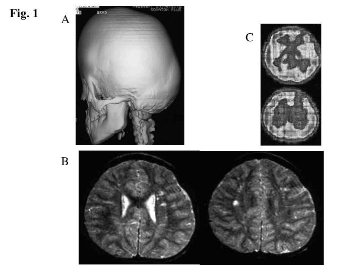
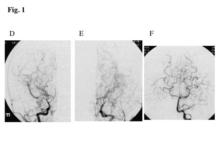
7 progression was observed in two cases of cranial irradiation for brain tumor. One patient died of brain tumor recurrence and the other 4 patients of daily life was influenced with mental retardation due to associated disorder. No one exhibited familial moyamoya disease. Illustrative Cases Patient 1 A 6-year-old boy who had craniosynostosis (Fig. 1A) and mental retardation, and had been diagnosed as Saethre-Chotzen syndrome presented because of gait disturbance. MRI showed multiple lacuna infarctions (Fig. 1B). Cerebral hypoperfusion was revealed by SPECT especially in the left cerebral hemisphere (Fig. 1C). Angiography revealed severe stenosis steno-occlusive lesion at the terminal portion of the ICA and horizontal portion of the middle cerebral artery (Fig. 1D-F). Moyamoya vessels were significantly developed bilaterally. He underwent EDAS bilaterally and disease progression has not been observed. However, he needs some supports on his daily life because of mental retardation. Patient 3 A 6-year-old boy visited our hospital because of right hemiparesis. He had been followed by pediatrician for left arm hypertrophy and mental retardation. Thus, his distinctive face indicated Proteus syndrome (Fig. 2A). CT demonstrated right thalamic hemorrhage with left frontal and right parietal infarction (Fig. 2B). The lesions 6
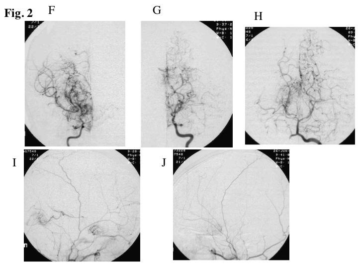
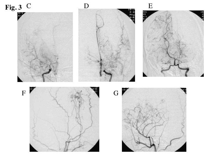
8 confirmed with MRI and left frontal lesion was revealed as fresh lesion (Fig. 2C, D). Cerebral perfusion was impaired bilaterally (Fig. 2E). Angiography showed steno-occlusive lesion at the terminal portion of the bilateral ICA (Fig. 2F, G). Moyamoya vessels were massively developed especially in the right side. Posterior cerebral arteries were also involved (Fig. 2H). Collateral flow from external carotid artery (ECA) was slightly seen (Fig. 2I, J). STA-MCA anastomosis combination with EMS was performed bilaterally. Postoperative course was uneventful. However, he need some supports on his daily life because of mental retardation. Patient 4 A-5-year-old boy, who has a history of partial removal of left temporal glioma with local irradiation, presented because of incidentally found cerebral infarction. MRI showed right frontal infarction as well as residual tumor in the left temporal lobe (Fig. 3A). SPECT revealed hypoperfusion in the right frontal and left parietal lobe (Fig. 3B). Steno-occlusive lesion was found at the terminal portion of the bilateral ICA and moyamoya vessels were developed (Fig. 3C-E). Collateral flow from ECA was significantly developed (Fig. 3F, G). Reoperation was performed for the recurrence of the tumor. He was followed clinically and radiologically. Additional ischemic lesion has not been demonstrated. However, his daily life was affected by mental retardation. 7
9 4. Discussion Patient Characteristics According to the diagnostic criteria of the RCMJ, the etiology of moyamoya disease is unknown and the condition associated with atherosclerosis, autoimmune disease, meningitis and so on, are distinguished from definite moyamoya disease [4, 5]. Since the terminology for this condition has not been defined, several words have been used to mean this condition as quasi-moyamoya disease, akin-moyamoya disease, moyamoya syndrome, moyamoya phenomenon, moyamoya-like vasculopathy and so on [1, 3, 6, 7]. We employed the term quasi-moyamoya disease throughout this report because it was given by Dr. Suzuki, who reported moyamoya disease at first [5]. This incidence of quasi-moyamoya disease (7/69=10.8%) was consistent with previous Japanese reports, which demonstrated approximately 10% of moyamoya disease [8]. The ratio of quasi-moyamoya disease is higher in western country [7]. Among seven patients of moyamoya syndrome, five were pediatric patients. Table 1 provides a summary of the clinical data obtained from the five quasi-moyamoya disease patients. The mean age was 6.4-year-old. It is well known that patients with moyamoya disease are predominantly female and we have demonstrated that the moyamoya syndrome affected patients are predominantly male. In terms of patient ages, it has been reported that there are two peaks, namely child type and adult type [9, 10]. In this series, child types are dominant because of congenital disorder or pediatric-onset brain tumor. 8
10 Symptoms and Sings The clinical manifestation of quasi-moyamoya disease are complicated since the aspect of cerebrovascular disorder and that of associated disorder are merged. Interestingly, mental retardation was seen in the most congenital disorder. Regarding with cerebrovascular disorder, four patients suffered ischemia and one patient had both hemorrhage and ischemia. As shown in Fig. 2B, the hemorrhage was seen in the right thalamus, which was unusual as moyamoya disease. Radiological findings We identified a wide variety of angiographical lesions in our series. All patients had arterial steno-occlusion with formation of moyamoya vessels bilaterally. Moyamoya disease associated with Down syndrome or neurofibromatosis type 1 occasionally involves only unilateral side [1, 11]. Moyamoya vessels were well developed in congenital disorders and were not significant in irradiated brain tumor patients. Collateral flow form ECA was significantly dominant in irradiated brain tumor patients. [12]. Posterior circulation was involved in three patients. Taken together, angiographical finding of quasi-moyamoya disease is widely varied. Treatment and prognosis In terms of treatment of quasi-moyamoya disease, vascular recanalization was performed for the patient with symptom or cerebral hypoperfusion revealed by SPECT. Two patients underwent STA-MCA direct bypass procedure. Indirect vascular 9
11 reconstruction was performed to two patients. Postoperative course was uneventful. Thus, vascular reconstruction was effective to prevent disease progression and cerebral hypoperfusion was improved postoperatively. Moyamoya disease, particularly in children, often exhibit progression during follow-up. Among our quasi-moyamoya disease patients, angiographical progression was seen in two irradiated brain tumor patients. In these two patients, vascular reconstruction was not employed for them, since collateral flow from ECA had been developed. Finally, activity of daily living of the patients was influenced with symptoms such as mental retardation due to associated disorder. And one patient died of brain tumor recurrence. Genetic analysis The pathologic and epidemiological facts suggest that genetic factors play a more important role in the pathogenesis of moyamoya disease than do acquired factors [13]. The familial occurrence was approximately 10% of moyamoya disease patients [14, 15]. Previous linkage studies have indicated that susceptibility loci for the moyamoya disease are located on chromosome 3p , 6q, 8q23 and 17q25 [13, 16-18]. Mineharu et al reported that the mode of inheritance of familial moyamoya disease is autosomal dominant with incomplete penetrance [19]. Several genetic factors in different loci cause the same disease (locus heterogeneity). Thus, the coincidence of unilateral and definite moyamoya disease within a single family indicates that they reflect different phenotypes cause by the same genetic defects [20]. Some of the congenital disorders namely neurofibromatosis type 1, of which responsible gene is 10
12 located on chromosome 17q11.2 may be related to the same gene with moyamoya disease. Others such as Down syndrome are cased by completely different gene, but manifest similar cerebral vascular disease. Limitation The incidence of moyamoya disease is estimated to be 0.54 per populations and that of quasi- moyamoya disease is as low as 10% of moyamoya disease [21]. Therefore, it is difficult to demonstrate clinical feature of quasi- moyamoya disease. Nation wide study is required to clarify the clinical feature of quasi- moyamoya disease and propose proper treatment. 11
13 5. Conclusions Among five cases of quasi-moyamoya disease, no one exhibited typical clinical course of moyamoya disease. Clinical manifestation and pathophysiology of quasi-moyamoya disease were diverse according to their associated disorders. Vascular reconstruction prevented disease progression and the prognosis of the quasi-moyamoya disease was influenced with associated disorders. 12
14 References 1) Horn P, Pfister S, Bueltmann E, Vajkoczy P, Schmiedek P. Moyamoya-like vasculopathy (moyamoya syndrome) in children. Childs Nerv Syst. 2004;20: ) Jea A, Smith ER, Robertson R, Scott RM. Moyamoya syndrome associated with Down syndrome: outcome after surgical revascularization. Pediatrics. 2005;116:e ) Kestle JR, Hoffman HJ, Mock AR. Moyamoya phenomenon after radiation for optic glioma. J Neurosurg. 1993;79: ) Fukui M. Guidelines for the diagnosis and treatment of spontaneous occlusion of the circle of Willis ('moyamoya' disease). Research Committee on Spontaneous Occlusion of the Circle of Willis (Moyamoya Disease) of the Ministry of Health and Welfare, Japan. Clin Neurol Neurosurg. 1997;99 Suppl 2:S238-S240 5) Suzuki J. Quasi-Moyamoya diseases. In: Suzuki J, eds. Moyamoya disease. New York: Springer-Verlag; 1986: ) Hallemeier CL, Rich KM, Grubb RL Jr, Chicoine MR, Moran CJ, Cross DT 3rd, Zipfel GJ, Dacey RG Jr, Derdeyn CP. Clinical features and outcome in North American 13
15 adults with moyamoya phenomenon. Stroke. 2006:37: ) Scott RM, Smith JL, Robertson RL, Madsen JR, Soriano SG, Rockoff MA. Long-term outcome in children with moyamoya syndrome after cranial revascularization by pial synangiosis. J Neurosurg. 2004;100(2 Suppl Pediatrics): ) Komiyama M, Nakajima H, Nishikawa M, Yasui T, Kitano S, Sakamoto H, Fu Y. High incidence of persistent primitive arteries in moyamoya and quasi-moyamoya diseases. Neurol Med Chir (Tokyo). 1999;39: ) Fukui M. Current state of study on moyamoya disease in Japan. Surg Neurol. 1997;47: ) Wakai K, Tamakoshi A, Ikezaki K, Fukui M, Kawamura T, Aoki R, Kojima M, Lin Y, Ohno Y. Epidemiological features of moyamoya disease in Japan: findings from a nationwide survey. Clin Neurol Neurosurg. 1997;99 Suppl 2:S1-S5 11) Rosser TL, Vezina G, Packer RJ. Cerebrovascular abnormalities in a population of children with neurofibromatosis type 1. Neurology. 2005;64: ) Ishikawa T, Houkin K, Yoshimoto T, Abe H. Vasoreconstructive surgery for 14
16 radiation-induced vasculopathy in childhood. Surg Neurol 1997;48: ) Yamauchi T, Tada M, Houkin K, Tanaka T, Nakamura Y, Kuroda S, Abe H, Inoue T, Ikezaki K, Matsushima T, Fukui M. Linkage of familial moyamoya disease (spontaneous occlusion of the circle of Willis) to chromosome 17q25. Stroke. 2000;31: ) Fukui M, Kono S, Sueishi K, Ikezaki K. Moyamoya disease. Neuropathology. 2000;20 Suppl:S61-S4 15) Nanba R, Kuroda S, Tada M, Ishikawa T, Houkin K, Iwasaki Y. Clinical features of familial moyamoya disease. Childs Nerv Syst. 2006;22: ) Ikeda H, Sasaki T, Yoshimoto T, Fukui M, Arinami T. Mapping of a familial moyamoya disease gene to chromosome 3p24.2-p26. Am J Hum Genet. 1999;64: ) Inoue TK, Ikezaki K, Sasazuki T, Matsushima T, Fukui M. Linkage analysis of moyamoya disease on chromosome 6. J Child Neurol. 2000;15: ) Sakurai K, Horiuchi Y, Ikeda H, Ikezaki K, Yoshimoto T, Fukui M, Arinami T. A novel susceptibility locus for moyamoya disease on chromosome 8q23. J Hum Genet. 15
17 2004;49: ) Mineharu Y, Takenaka K, Yamakawa H, Inoue K, Ikeda H, Kikuta KI, Takagi Y, Nozaki K, Hashimoto N, Koizumi A. Inheritance pattern of familial moyamoya disease: autosomal dominant mode and genomic imprinting. J Neurol Neurosurg Psychiatry. 2006;77: ) Kusaka N, Tamiya T, Adachi Y, Katayama S, Namba S, Tokunaga K, Sugiu K, Date I, Ohmoto T. Adult unilateral moyamoya disease with familial occurrence in two definite cases: a case report and review of the literature. Neurosurg Rev. 2006;29: ) Kuriyama S, Kusaka Y, Fujimura M, Wakai K, Tamakoshi A, Hashimoto S, Tsuji I, Inaba Y, Yoshimoto T. Prevalence and clinicoepidemiological features of moyamoya disease in Japan: findings from a nationwide epidemiological survey. Stroke. 2008;39:
18 Figure Legends Fig. 1 Patient 1 A 6-year-old boy with Saethre-Chotzen syndrome. Three-dimensional skull bone CT showed craniosynostosis (A). MRI (T2 weighted image) showed multiple lacuna infarctions (B). Cerebral hypoperfusion was revealed by single photon emission tomography (SPECT) especially in the left frontal lobe (C). Angiography reveal steno-occlusive lesion at the terminal portion of the bilateral internal carotid artery (ICA; D, E). Moyamoya vessels were also developed bilaterally. Posterior circulation was not involved (F). Fig. 2 Patient 3 A 6-year-old boy with Proteus syndrome. Distinctive face with left arm hypertrophy were seen (A). CT demonstrated right thalamic hemorrhage with left frontal and right parietal infarction (B), and lesions were confirmed by MRI (inversion recovery image; C). MRI (diffusion-weighted image) showed left frontal lesion as fresh lesion (D). Cerebral perfusion was impaired bilaterally (E). Angiography showed steno-occlusive lesion at the terminal portion of the bilateral ICA (F, G). Moyamoya vessels were developed especially in the right side. Pial anastomosis from the left posterior cerebral artery was developed (H). Collateral flow from external carotid artery was slightly seen (I, J). 17
19 Fig. 3 Patient 4 A-5-year-old boy with left temporal glioma and cranial irradiation. MRI (inversion recovery image) showed residual tumor in the left temporal lobe as well as right frontal infarction (A). SPECT revealed hypoperfusion in the right frontal and left parietal lobe (B). Steno-occlusive lesion was found at the terminal portion of the bilateral ICA and moyamoya vessels were developed (C, D). Pial anastomosis from the right posterior cerebral artery was developed (E). Collateral flow from ECA was significantly developed (F, G). 18
20 19
21 20

22 21
23 22
24 23
25 24
26 Table 1 patient characteristics of quasi-moyamoya disease Age, sex disease symptom imaging study treatment follow (years) 1) 6 M craniosynostois gate disturbance cerebral infarction Bil. EDAS 14 (mental returdation) 2) 9 M dwarfism epilepsy no abnormality Bil. EDAS 20 coarctation of aorta 3) 6 M Proteus syndrome TIA cerebral infarction Bil. STA-MCA 3 (mental returdation) intracerebral hemorrhge with EMS 4) 5 M BT/irradiation (mental returdation) cerebral infarction (removal) 13 (brin tumor) 5) 6 F BT/irradiation (visual disturbance) cerebral infarction (removal) 5 (dead) (mental returdation) (brain tumor) M: male, F: female, NF: neurofibromatosis type 1, BT: brain tumor, TIA: transient ischemic attack, Bil: bilateral EDAS: encephalo-duro-arterio-synostosis, STA-MCA: superficial temporal artery-middle cerebral artery anastomosis ( ) indicates findings due to associated disorder or treatment for the disorder. 25
Neurosurg Focus 5 (5):Article 4, 1998
:Article 4, 1998") Neurosurg Focus 5 (5):Article 4, 1998 Multiple combined indirect procedure for the surgical treatment of children with moyamoya disease. A comparison with single indirect anastomosis with direct anastomosis
Neurosurg Focus 5 (5):Article 4, 1998 Multiple combined indirect procedure for the surgical treatment of children with moyamoya disease. A comparison with single indirect anastomosis with direct anastomosis
Clinicoepidemiological Features of Asymptomatic Moyamoya Disease in Adult Patients
 Journal of Cerebrovascular and Endovascular Neurosurgery pissn 2234-8565, eissn 2287-3139, http://dx.doi.org/10.7461/jcen.2014.16.3.241 Original Article Clinicoepidemiological Features of Asymptomatic
Longitudinal anterior-to-posterior shift of collateral channels in patients with moyamoya disease: an implication for its hemorrhagic onset
 CLINICAL ARTICLE Longitudinal anterior-to-posterior shift of collateral channels in patients with moyamoya disease: an implication for its hemorrhagic onset Shusuke Yamamoto, MD, Satoshi Hori, MD, PhD,
Title. CitationWorld Neurosurgery, 80(5): Issue Date Doc URL. Rights. Rights(URL)
: Issue Date Doc URL. Rights. Rights(URL)") Title Effective Surgical Revascularization Improves Cerebr Moyamoya Disease Kawabori, Masahito; Kuroda, Satoshi; Nakayama, Naoki Author(s) Nagara CitationWorld Neurosurgery, 80(5): 612-619 Issue Date 2013-11
Title Effective Surgical Revascularization Improves Cerebr Moyamoya Disease Kawabori, Masahito; Kuroda, Satoshi; Nakayama, Naoki Author(s) Nagara CitationWorld Neurosurgery, 80(5): 612-619 Issue Date 2013-11
Moyamoya Syndrome with contra lateral DACA aneurysm: First Case report with review of literature
 Romanian Neurosurgery Volume XXXI Number 3 2017 July-September Article Moyamoya Syndrome with contra lateral DACA aneurysm: First Case report with review of literature Ashish Kumar Dwivedi, Pradeep Kumar,
MOYA Moya disease is a rare idiopathic
 Research Papers Moya Moya Cases Treated with Encephaloduroarteriosynangiosis Parimal Tripathi, Varsha Tripathi, Ronak J. Naik and Jaimin M. Patel From Gujarat Cancer & Research Institute, Ahmedabad; Sterling
Tohoku J. Exp. Med., 2015, 236, Current Management of Moyamoya Disease 45
 Tohoku J. Exp. Med., 2015, 236, 45-53 Current Management of Moyamoya Disease 45 Invited Review Current Status of Revascularization Surgery for Moyamoya Disease: Special Consideration for Its Internal Carotid-External
Moyamoya disease presenting as acute onset cortical blindness: a case report
 Romanian Neurosurgery Volume XXX Number 1 2016 January-March Article Moyamoya disease presenting as acute onset cortical blindness: a case report Dudi Maniram, Bansal Rajeev, Srivastava Trilochan, Sardana
INSTITUTE OF NEUROSURGERY & DEPARTMENT OF PICU
 CEREBRAL BYPASS An Innovative Treatment for Arteritis INSTITUTE OF NEUROSURGERY & DEPARTMENT OF PICU CASE 1 q 1 year old girl -recurrent seizure, right side limb weakness, excessive cry and irritability.
Moyamoya disease in the midwestern United States
 Neurosurg Focus 5 (5):Article 1, 1998 Moyamoya disease in the midwestern United States Nicholas M. Wetjen, B.S., P. Charles Garell, M.D., Nicholas V. Stence, and Christopher M. Loftus, M.D. Division of
Asymptomatic Moyamoya Disease: Literature Review and Ongoing AMORE Study
 REVIEW ARTICLE Neurol Med Chir (Tokyo) 55, 194 198, 2015 doi: 10.2176/nmc.ra.2014-0305 Online February 20, 2015 Asymptomatic Moyamoya Disease: Literature Review and Ongoing AMORE Study Satoshi Kuroda;
Case Report. Fumihiro MATANO 1, Yasuo MURAI 2, Asami KUBOTA 1, Takayuki MIZUNARI 1, Shiro KOBAYASHI 1, Akio MORITA 2
 DOI: 10.5137/1019-5149.JTN.19271-16.1 Received: 17.10.2016 / Accepted: 19.01.2017 Published Online: 07.02.2017 Case Report The Ivy Sign on Fluid Attenuated Inversion Recovery Images Related to Single-Photon
Moyamoya Disease A Vasculopathy and an Uncommon Cause of Recurrent Cerebrovascular Accidents
 Moyamoya Disease A Vasculopathy and an Uncommon Cause of Recurrent Cerebrovascular Accidents Yasmin S. Hamirani, Md 1 *, Mohammad Valikhani, Md 2, Allison Sweney, Ms Iii 2, Hafsa Khan, Md 2, Mohammad Pathan,
Direct Bypass Techniques for the Treatment of Pediatric Moyamoya Disease
 Direct Bypass Techniques for the Treatment of Pediatric Moyamoya Disease Raphael Guzman, MD a, Gary K. Steinberg, MD, PhD b, * KEYWORDS Moyamoya disease Pediatric Direct bypass INDICATIONS Moyamoya disease
Overview Blood supply of the brain What is moyamoya disease? > 1
 Moyamoya Disease Overview Moyamoya disease is caused by blocked arteries at the base of the brain. The name "moyamoya" means "puff of smoke" in Japanese and describes the appearance of tiny vessels that
In patients with moyamoya the clinical significance of. Preoperative transdural collateral vessels in moyamoya as radiographic biomarkers of disease
 CLINICAL ARTICLE J Neurosurg Pediatr 19:289 295, 2017 Preoperative transdural collateral vessels in moyamoya as radiographic biomarkers of disease Armide Storey, BS, 1 R. Michael Scott, MD, 1 Richard Robertson,
Moyamoya disease is an unusual form of chronic, occlusive
 Angiographic Dilatation and Branch Extension of the Anterior Choroidal and Posterior Communicating Arteries Are Predictors of Hemorrhage in Adult Moyamoya Patients Motohiro Morioka, MD; Jun-Ichiro Hamada,
Moyamoya disease is a progressive cerebrovascular. Pial synangiosis in patients with moyamoya younger than 2 years of age.
 J Neurosurg Pediatrics 13:420 425, 2014 AANS, 2014 Pial synangiosis in patients with moyamoya younger than 2 years of age Clinical article Eric M. Jackson, M.D., Ning Lin, M.D., Sunil Manjila, M.D., R.
Title. Author(s) 黒田, 敏 ; 川堀, 真人 ; 宮本, 倫行 ; 笹森, 徹 ; 遠藤, 将吾 ; 中山, 若樹 ; 石川, 達哉. Citation 脳卒中の外科, 37(5): Issue Date Doc URL.
 黒田, 敏 ; 川堀, 真人 ; 宮本, 倫行 ; 笹森, 徹 ; 遠藤, 将吾 ; 中山, 若樹 ; 石川, 達哉. Citation 脳卒中の外科, 37(5): Issue Date Doc URL.") Title 側頭葉から後頭葉にかけて高度の虚血を有するもやもや病に対する脳血行再建術 Author(s) 黒田, 敏 ; 川堀, 真人 ; 宮本, 倫行 ; 笹森, 徹 ; 遠藤, 将吾 ; 中山, 若樹 ; 石川, 達哉 Citation 脳卒中の外科, 37(5): 345-349 Issue Date 2009 Doc URL http://hdl.handle.net/2115/70929
Title 側頭葉から後頭葉にかけて高度の虚血を有するもやもや病に対する脳血行再建術 Author(s) 黒田, 敏 ; 川堀, 真人 ; 宮本, 倫行 ; 笹森, 徹 ; 遠藤, 将吾 ; 中山, 若樹 ; 石川, 達哉 Citation 脳卒中の外科, 37(5): 345-349 Issue Date 2009 Doc URL http://hdl.handle.net/2115/70929
Association of HLA-DR and -DQ Genes with Familial Moyamoya Disease in Koreans
 www.jkns.or.kr 10.3340/jkns.2009.46.6.558 J Korean Neurosurg Soc 46 : 558-563, 2009 Print ISSN 2005-3711 On-line ISSN 1598-7876 Copyright 2009 The Korean Neurosurgical Society Laboratory Investigation
Moyamoya Disease in China Its Clinical Features and Outcomes
 Moyamoya Disease in China Its Clinical Features and Outcomes Lian Duan, MD, PhD; Xiang-Yang Bao, MM; Wei-Zhong Yang, BS; Wan-Chao Shi, MD; De-Sheng Li, MD; Zheng-Shan Zhang, MM; Rui Zong, MM; Cong Han,
Thirteen-year Experience of 44 Patients with Adult Hemorrhagic Moyamoya Disease from a Single Institution: Clinical Analysis by Management Modality
 Journal of Cerebrovascular and Endovascular Neurosurgery ISSN 2234-8565, EISSN 2287-3139, http://dx.doi.org/10.7461/jcen.2013.15.3.191 Clinical Article Thirteen-year Experience of 44 Patients with Adult
Emergency EC-IC bypass for symptomatic atherosclerotic ischemic stroke
 Emergency EC-IC bypass for symptomatic atherosclerotic ischemic stroke Tetsuyoshi Horiuchi, Junpei Nitta, Shigetoshi Ishizaka, Kohei Kanaya, Takao Yanagawa, and Kazuhiro Hongo. Department of Neurosurgery,
This quiz is being published on behalf of the Education Committee of the SNACC.
 Quiz 48 Cerebrovascular Atherosclerotic Disease Shobana Rajan, M.D. Associate Director of Neuroanesthesia, Vice Chair of Education, Allegheny Health Network. Quiz team; Suneeta Gollapudy M.D, Angele Marie
Neurosurgical Treatment of Moyamoya Disease: Bypass Surgery for the Brain
 Neurosurgical Treatment of Moyamoya Disease: Bypass Surgery for the Brain Christopher Payne Currently, no medical treatment exists to prevent the progression of moyamoya disease, and neurosurgical treatment
 Impact of posterior cerebral artery Titleclinical and social outcome of pedi Dissertation_ 全文 ) Author(s) Funaki, Takeshi Citation Kyoto University ( 京都大学 ) Issue Date 2015-01-23 URL http://dx.doi.org/10.14989/doctor.r
Moyamoya disease (MMD) is a rare idiopathic occlusive
 is a rare idiopathic occlusive") Moyamoya Disease in Europeans Markus Kraemer, MD; Wilhelm Heienbrok, MD; Peter Berlit, MD Background and Purpose We describe the clinical, diagnostic, and outcome features of a cohort of white patients
Moyamoya Disease in Europeans Markus Kraemer, MD; Wilhelm Heienbrok, MD; Peter Berlit, MD Background and Purpose We describe the clinical, diagnostic, and outcome features of a cohort of white patients
Moyamoya disease (MMD) is a chronic, progressive cerebrovascular. Clinical and Angiographic Features and Stroke Types in Adult Moyamoya Disease
 is a chronic, progressive cerebrovascular. Clinical and Angiographic Features and Stroke Types in Adult Moyamoya Disease") ORIGINAL RESEARCH BRAIN Clinical and Angiographic Features and Stroke Types in Adult Moyamoya Disease D.-K. Jang, K.-S. Lee, H.K. Rha, P.-W. Huh, J.-H. Yang, I.S. Park, J.-G. Ahn, J.H. Sung, and Y.-M.
ORIGINAL RESEARCH BRAIN Clinical and Angiographic Features and Stroke Types in Adult Moyamoya Disease D.-K. Jang, K.-S. Lee, H.K. Rha, P.-W. Huh, J.-H. Yang, I.S. Park, J.-G. Ahn, J.H. Sung, and Y.-M.
Moyamoya disease. Critical Role of PDGFR-α in Angiogenesis through Indirect Bypass in Mouse Model of Moyamoya Disease.
 FPⅢ- Critical Role of PDGFR-α in Angiogenesis through Indirect Bypass in Mouse Model of Moyamoya Disease Tomohide Hayashi Departments of Neurosurgery, University of Toyama Background - is a unique cerebrovascular
Moyamoya syndrome associated with cocaine abuse Case report
 Neurosurg Focus 5 (5):Article 7, 1998 Moyamoya syndrome associated with cocaine abuse Case report Marc S. Schwartz, M.D., and R. Michael Scott, M.D. Division of Neurosurgery, Albany Medical College, Albany,
Although moyamoya disease, a rare cerebrovascular occlusive
 Renal Artery Lesions in Patients With Moyamoya Disease Angiographic Findings Ichiro Yamada, MD; Yoshiro Himeno, MD; Yoshiharu Matsushima, MD; Hitoshi Shibuya, MD Background and Purpose Renal artery lesions
Double STA-MCA Anatomosis for Bilateral Carotid Occlusion
 Double STA-MCA Anatomosis for Bilateral Carotid Occlusion -Case Report and Literature Review- Sandra vuignier 1, Kenji Kanamaru 2, Tomohiro Araki 2 1 Department of Neurosurgery, Nagoya University School
Cerebral Hemodynamic Change in the Child and the Adult With Moyamoya Disease
 272 Cerebral Hemodynamic Change in the Child and the Adult With Moyamoya Disease Yasuo Kuwabara, MD, Yuichi Ichiya, MD, Makoto Otsuka, MD, Takashi Tahara, MD, Ranjan Gunasekera, MD, Kanehiro Hasuo, MD,
Neuroscience. Journal. Moyamoya disease a review and case illustration. P A L M E T T O H E A L T H Vol. 2 Issue 3 Summer 2016
 Neuroscience P A L M E T T O H E A L T H Vol. 2 Issue 3 Summer 2016 Journal Moyamoya disease a review and case illustration pg. 5 Choroid Plexus Papilloma in adults pg. 8 As physician co-leaders of Palmetto
MMD is a rare cerebrovascular disease first described by
 ORIGINAL RESEARCH M.A. Mogensen P. Karzmark P.D. Zeifert J. Rosenberg M. Marks G.K. Steinberg L.J. Dorfman Neuroradiologic Correlates of Cognitive Impairment in Adult Moyamoya Disease BACKGROUND AND PURPOSE:
Young So, MD; Ho-Young Lee, MD; Seung-Ki Kim, MD; Jae Sung Lee, PhD; Kyu-Chang Wang, MD; Byung-Kyu Cho, MD; Eunjoo Kang, PhD; Dong Soo Lee, MD
 Prediction of the Clinical Outcome of Pediatric Moyamoya Disease With Postoperative Basal/Acetazolamide Stress Brain Perfusion SPECT After Revascularization Surgery Young So, MD; Ho-Young Lee, MD; Seung-Ki
Unstable moyamoya disease: clinical features and impact on perioperative ischemic complications
 clinical article J Neurosurg 122:400 407, 2015 Unstable moyamoya disease: clinical features and impact on perioperative ischemic complications Takeshi Funaki, MD, 1 Jun C. Takahashi, MD, PhD, 1 Yasushi
Assessment of Cerebrovascular Reserve before and after STA-MCA Bypass Surgery by SPECT and SPM Analysis
 Assessment of Cerebrovascular Reserve before and after STA-MCA Bypass Surgery by SPECT and SPM Analysis Joo-Hyun O, MD 1 Kyung-Sool Jang, MD 2 Ie-Ryung Yoo, MD 1 Sung-Hoon Kim, MD 1 Soo-Kyo Chung, MD 1
History of revascularization
 History of revascularization Author (year) Kredel, 1942 Woringer& Kunlin, 1963 Donaghy& Yasargil, 1968 Loughheed 1971 Kikuchini & Karasawa1973 Karasawa, 1977 Story, 1978 Sundt, 1982 EC/IC bypass study
Moyamoya disease (MMD) is a rare idiopathic disorder
 is a rare idiopathic disorder") Moyamoya: An Update for the Practicing Neurologist Distinguishing between definite MMD, probable MMD, and MMS may not have significant practical implications. By Timothy G. White, MD; Julia R. Schneider,
Moyamoya: An Update for the Practicing Neurologist Distinguishing between definite MMD, probable MMD, and MMS may not have significant practical implications. By Timothy G. White, MD; Julia R. Schneider,
Recent Advances in Neurology Difficult Cases
 Patient X: History Part 1 Recent Advances in Neurology Difficult Cases Heather J. Fullerton, MD, MAS Professor of Neurology & Pediatrics Director, Pediatric Brain Center Previously healthy 14-year old
010050,Inner Mongolia, China
 Possible factors influencing postoperative temporary neurologic deterioration following standard superficial temporal artery-middle cerebral artery (STA-MCA) bypass surgery: diameter of STA and MCA (M4)
A CASE OF RECURRENT ALTERNATING TRANSIENT HEMIPARESIS Dr. Shunmuga Arumugasamy.S DNB Resident Railway Hospital, Perambur.
 A CASE OF RECURRENT ALTERNATING TRANSIENT HEMIPARESIS Dr. Shunmuga Arumugasamy.S DNB Resident Railway Hospital, Perambur. 6 year old school going child. Apparently normal till 3 yrs when she developed
Occlusive cerebrovascular disease. A Novel Chronic Cerebral Hypoperfusion Model with Cognitive Impairment and Low Mortality Rate in Rats
 FPⅧ-1 A Novel Chronic Cerebral Hypoperfusion Model with Cognitive Impairment and Low Mortality Rate in Rats Ahmed Said Mansour 1, Kuniyasu Niizuma 2, Sherif Rashad 2, Hidenori Endo 2, Toshiki Endo 3, Kenichi
Moyamoya. Moyamoya Disease Double Trouble. Epidemiology 1/16/2015
 Moyamoya Moyamoya Disease Double Trouble Jan Boerke, ACNP AACN Brunch January 24, 2015 Moyamoya - puff of smoke in Japanese Describes the look of the tangle of tiny vessels formed to compensate for the
PTA 106 Unit 1 Lecture 3
 PTA 106 Unit 1 Lecture 3 The Basics Arteries: Carry blood away from the heart toward tissues. They typically have thicker vessels walls to handle increased pressure. Contain internal and external elastic
Moyamoya disease (MMD) is a rare steno-occlusive
 is a rare steno-occlusive") CLINICAL ARTICLE J Neurosurg 126:1573 1577, 2017 Low flow velocity in the middle cerebral artery predicting infarction after bypass surgery in adult moyamoya disease *Hoyeon Cho, MD, 1 Kyung Il Jo, MD,
CLINICAL ARTICLE J Neurosurg 126:1573 1577, 2017 Low flow velocity in the middle cerebral artery predicting infarction after bypass surgery in adult moyamoya disease *Hoyeon Cho, MD, 1 Kyung Il Jo, MD,
Imaging of Moya Moya Disease
 Abstract Imaging of Moya Moya Disease Pages with reference to book, From 181 To 185 Rashid Ahmed, Hurnera Ahsan ( Liaquat National Hospital, Karachi. ) Moya Moya disease is a rare disease causing occlusion
Moyamoya vasculopathy Patient demographics and characteristics in the Finnish population
 Research Moyamoya vasculopathy Patient demographics and characteristics in the Finnish population International Journal of Stroke 2017, Vol. 12(1) 90 95! 2016 World Stroke Organization Reprints and permissions:
Penetration of the Optic Nerve or Chiasm by Anterior Communicating Artery Aneurysms. - Three Case Reports-
 Penetration of the Optic Nerve or Chiasm by Anterior Communicating Artery Aneurysms. - Three Case Reports- Tetsuyoshi Horiuchi 1, Toshiya Uchiyama 1, Yoshikazu Kusano 1, Maki Okada 1, Kazuhiro Hongo 1,
Moyamoya disease (MMD), an idiopathic disease. Direct versus indirect revascularization in the treatment of moyamoya disease
, an idiopathic disease. Direct versus indirect revascularization in the treatment of moyamoya disease") CLINICAL ARTICLE J Neurosurg 129:480 489, 2018 Direct versus indirect revascularization in the treatment of moyamoya disease *Seong-eun Park, BSc, Ju-seong Kim, MD, Eun Kyung Park, MD, Kyu-Won Shim, MD,
CLINICAL ARTICLE J Neurosurg 129:480 489, 2018 Direct versus indirect revascularization in the treatment of moyamoya disease *Seong-eun Park, BSc, Ju-seong Kim, MD, Eun Kyung Park, MD, Kyu-Won Shim, MD,
Clinicoradiological Profile of Childhood Moyamoya Disease: Indian Study of 30 Children with Literature Review
 Original article Clinicoradiological Profile 10.5005/jp-journals-10066-0020 of Childhood Moyamoya Disease Clinicoradiological Profile of Childhood Moyamoya Disease: Indian Study of 30 Children with Literature
positron emission tomography PET
 020-8505 19-1 1-3) positron emission tomography PET PET PET single photon emission computed tomography SPECT acetazolamide 4-10) magnetic resonance imaging MRI MRI 11,12) MRI perfusion weighted imaging
Diagnosis of Middle Cerebral Artery Occlusion with Transcranial Color-Coded Real-Time Sonography
 Diagnosis of Middle Cerebral Artery Occlusion with Transcranial Color-Coded Real-Time Sonography Kazumi Kimura, Yoichiro Hashimoto, Teruyuki Hirano, Makoto Uchino, and Masayuki Ando PURPOSE: To determine
Surgical Treatment of Childhood Moyamoya Disease -Comparison of Reconstructive Surgery Centered on the Frontal Region and the Parietal Region-
 Surgical Treatment of Childhood Moyamoya Disease -Comparison of Reconstructive Surgery Centered on the Frontal Region and the Parietal Region- Akihiro TAKAHASHI, Hiroyasu KAMIYAMA, Kiyohiro HOUKIN, and
EXPERIMENTAL AND THERAPEUTIC MEDICINE 15: , 2018
 3570 Clinical and angiographic outcomes after combined direct and indirect bypass in adult patients with moyamoya disease: A retrospective study of 76 procedures JINBING ZHAO, HONGYI LIU, YUANJIE ZOU,
A Patient with Stenosis of the Cervical Internal Carotid Artery in Whom Hyperperfusion Syndrome Occurred after Staged Angioplasty
 DOI: 10.5797/jnet.cr.2017-0043 A Patient with Stenosis of the Cervical Internal Carotid Artery in Whom Hyperperfusion Syndrome Occurred after Staged Angioplasty Jun Niimi, Kenta Tasaka, Fumio Nemoto, Takuya
Ruptured Aneurysm of the Accessory Middle Cerebral Artery Associated with Moyamoya Disease A Case Report
 Case Report 541 Ruptured Aneurysm of the Accessory Middle Cerebral Artery Associated with Moyamoya Disease A Case Report Cheng-Chi Lee, MD; Zhuo-Hao Liu, MD; Shih-Ming Jung 1, MD; Tao-Chieh Yang, MD The
Moyamoya Disease: Epidemiology, Clinical Features, and Diagnosis
 Journal of Stroke 2016;18(1):2-11 http://dx.doi.org/10.5853/jos.2015.01627 Special Review Moyamoya Disease: Epidemiology, Clinical Features, and Diagnosis Jong S. Kim Stroke Center and Department of Neurology,
Chapter 62 Monitoring of Hemodynamic Change in Patients with Carotid Artery Stenosis During the Tilt Test Using Wearable Near-Infrared Spectroscopy
 Chapter 62 Monitoring of Hemodynamic Change in Patients with Carotid Artery Stenosis During the Tilt Test Using Wearable Near-Infrared Spectroscopy Takahiro Igarashi, Kaoru Sakatani, Norio Fujiwara, Yoshihiro
Intracranial vascular anastomosis using the microanastomotic system
 J Neurosurg 89:676 681, 1998 Intracranial vascular anastomosis using the microanastomotic system Technical note DAVID W. NEWELL, M.D., ANDREW T. DAILEY, M.D., AND STEPHEN L. SKIRBOLL, M.D. Department of
TABLES. Table 1 Terminal vessel aneurysms. Table. Aneurysm location. Bypass flow** Symptoms Strategy Bypass recipient. Age/ Sex.
 Table TABLES Table 1 Terminal vessel aneurysms Age/ Sex Aneurysm location Symptoms Strategy Bypass recipient Recipient territory Recipient territory flow* Cut flow Bypass flow** Graft Patent postop F/U
Subclavian artery Stenting
 Subclavian artery Stenting Etiology Atherosclerosis Takayasu s arteritis Fibromuscular dysplasia Giant Cell Arteritis Radiation-induced Vascular Injury Thoracic Outlet Syndrome Neurofibromatosis Incidence
Carotid artery stenting for long CTO and pseudo occlusion of carotid artery -2 case reports-
 Carotid artery stenting for long CTO and pseudo occlusion of carotid artery -2 case reports- Katsutoshi Takayama, MD, Ph.D Department of Radiology and Interventional Neuroradiology Ishinkai Yao General
The moyamoya syndrome is a cerebrovascular condition that
 The new england journal of medicine review article Medical Progress Moyamoya Disease and Moyamoya Syndrome R. Michael Scott, M.D., and Edward R. Smith, M.D. From the Department of Neurosurgery, Children
Anesthetic Management of Child with Moyamoya Disease for Pial Synangiosis
 Anesthetic Management of Child with Moyamoya Disease for Pial Synangiosis Craig D. McClain, MD, MPH Boston Children s Hospital and Harvard Medical School Case Presentation 14 year old male with bilateral
Moyamoya disease (MMD) is a chronic, progressive. Topical Reviews Section Editors: Guiseppe Lanzino, MD, and Michael Tymianski, MD, PhD
 is a chronic, progressive. Topical Reviews Section Editors: Guiseppe Lanzino, MD, and Michael Tymianski, MD, PhD") Topical Reviews Section Editors: Guiseppe Lanzino, MD, and Michael Tymianski, MD, PhD Neurosurgical Advances in the Treatment of Moyamoya Disease Paritosh Pandey, MD; Gary K. Steinberg, MD, PhD Background
Topical Reviews Section Editors: Guiseppe Lanzino, MD, and Michael Tymianski, MD, PhD Neurosurgical Advances in the Treatment of Moyamoya Disease Paritosh Pandey, MD; Gary K. Steinberg, MD, PhD Background
Angiographic features of hemorrhagic moyamoya disease with high recurrence risk: a supplementary analysis of the Japan Adult Moyamoya Trial
 CLINICAL ARTICLE J Neurosurg 128:777 784, 2018 Angiographic features of hemorrhagic moyamoya disease with high recurrence risk: a supplementary analysis of the Japan Adult Moyamoya Trial Takeshi Funaki,
Brain AVM with Accompanying Venous Aneurysm with Intracerebral and Intraventricular Hemorrhage
 Cronicon OPEN ACCESS EC PAEDIATRICS Case Report Brain AVM with Accompanying Venous Aneurysm with Intracerebral and Intraventricular Hemorrhage Dimitrios Panagopoulos* Neurosurgical Department, University
Disclosures. CREST Trial: Summary. Lecture Outline 4/16/2015. Cervical Atherosclerotic Disease
 Disclosures Your Patient Has Carotid Bulb Stenosis and a Tandem Intracranial Stenosis: How Do SAMMPRIS and Other Evidence Inform Your Treatment? UCSF Vascular Symposium 2015 Steven W. Hetts, MD Associate
Original Article Ischemic and hemorrhagic moyamoya disease in adults: CT findings
 Int J Clin Exp Med 2015;8(11):21351-21357 www.ijcem.com /ISSN:1940-5901/IJCEM0009621 Original Article Ischemic and hemorrhagic moyamoya disease in adults: CT findings Anming Xie 1*, Li Luo 2*, Yaojun Ding
Moyamoya disease (MMD) is a chronic, progressive. Topical Reviews Section Editors: Guiseppe Lanzino, MD, and Michael Tymianski, MD, PhD
 is a chronic, progressive. Topical Reviews Section Editors: Guiseppe Lanzino, MD, and Michael Tymianski, MD, PhD") Topical Reviews Section Editors: Guiseppe Lanzino, MD, and Michael Tymianski, MD, PhD Neurosurgical Advances in the Treatment of Moyamoya Disease Paritosh Pandey, MD; Gary K. Steinberg, MD, PhD Downloaded
Topical Reviews Section Editors: Guiseppe Lanzino, MD, and Michael Tymianski, MD, PhD Neurosurgical Advances in the Treatment of Moyamoya Disease Paritosh Pandey, MD; Gary K. Steinberg, MD, PhD Downloaded
Moyamoya disease (MMD) is a progressive stenoocclusive
 is a progressive stenoocclusive") J Neurosurg 121:432 440, 2014 AANS, 2014 The frequency of postoperative stroke in moyamoya disease following combined revascularization: a single-university series and systematic review Clinical article
J Neurosurg 121:432 440, 2014 AANS, 2014 The frequency of postoperative stroke in moyamoya disease following combined revascularization: a single-university series and systematic review Clinical article
Moyamoya disease (MMD) is a rare, chronic disorder
 is a rare, chronic disorder") LITERATURE REVIEW J Neurosurg 126:1523 1529, 2017 Direct versus indirect revascularization procedures for moyamoya disease: a comparative effectiveness study Luke Macyszyn, MD, MA, Mark Attiah, MD, MS,
LITERATURE REVIEW J Neurosurg 126:1523 1529, 2017 Direct versus indirect revascularization procedures for moyamoya disease: a comparative effectiveness study Luke Macyszyn, MD, MA, Mark Attiah, MD, MS,
with susceptibility-weighted imaging and computed tomography perfusion abnormalities in diagnosis of classic migraine
 Emerg Radiol (2012) 19:565 569 DOI 10.1007/s10140-012-1051-2 CASE REPORT Susceptibility-weighted imaging and computed tomography perfusion abnormalities in diagnosis of classic migraine Christopher Miller
Internal Carotid Artery Dissection
 May 2011 Internal Carotid Artery Dissection Carolyn April, HMS IV Agenda Presentation of a clinical case Discussion of the clinical features of ICA dissection Discussion of the imaging modalities used
CASE REPORT AIR VENT OF VEIN GRAFT IN EXTRACRANIAL-INTRACRANIAL BYPASS SURGERY
 Nagoya J. Med. Sci. 74. 339 ~ 345, 2012 CASE REPORT AIR VENT OF VEIN GRAFT IN EXTRACRANIAL-INTRACRANIAL BYPASS SURGERY HIROFUMI OYAMA, AKIRA KITO, HIDEKI MAKI, KENICHI HATTORI, TOMOYUKI NODA and KENTARO
Ruptured aberrant internal carotid artery pseudoaneurysm presenting with spontaneous massive ear bleeding following a single sneeze: a case report
 Case eport JNET 7:312-316, 2013 uptured aberrant internal carotid artery pseudoaneurysm presenting with spontaneous massive ear bleeding following a single sneeze: a case report Seiichiro HIONO 1) Eiichi
Intraoperative EC-IC Bypass Blood Flow Assessment With Indocyanine Green Angiography in Moyamoya and Non-Moyamoya Ischemic Stroke
 Intraoperative EC-IC Bypass Blood Flow Assessment With Indocyanine Green Angiography in Moyamoya and Non-Moyamoya Ischemic Stroke Takayuki Awano 1, Kaoru Sakatani 1,2, Noriaki Yokose 1, Yuko Kondo 1, Takahiro
Case Report INTRODUCTION
 Journal of Cerebrovascular and Endovascular Neurosurgery pissn 2234-8565, eissn 2287-3139, https//doi.org/10.7461/jcen.2018.20.2.127 Case Report Revision Superficial Temporal Artery-Middle Cerebral Artery
Vertebrobasilar Insufficiency
 Equilibrium Res Vol. (3) Vertebrobasilar Insufficiency Toshiaki Yamanaka Department of Otolaryngology-Head and Neck Surgery, Nara Medical University School of Medicine Vertebrobasilar insufficiency (VBI)
CT and MR findings of systemic lupus erythematosus involving the brain: Differential diagnosis based on lesion distribution
 CT and MR findings of systemic lupus erythematosus involving the brain: Differential diagnosis based on lesion distribution Poster No.: C-2723 Congress: ECR 2010 Type: Educational Exhibit Topic: Neuro
Epidemiology, diagnosis and treatment of moyamoya disease (Review)
") EXPERIMENTAL AND THERAPEUTIC MEDICINE 17: 1977-1984, 2019 Epidemiology, diagnosis and treatment of moyamoya disease (Review) HUI ZHANG, LIJIAN ZHENG and LEI FENG Department of Neurosurgery, The First People's
EXPERIMENTAL AND THERAPEUTIC MEDICINE 17: 1977-1984, 2019 Epidemiology, diagnosis and treatment of moyamoya disease (Review) HUI ZHANG, LIJIAN ZHENG and LEI FENG Department of Neurosurgery, The First People's
Chronic middle cerebral artery (MCA) occlusion as a cause
 occlusion as a cause") ORIGINAL RESEARCH M. Tanaka E. Shimosegawa K. Kajimoto Y. Kimura H. Kato N. Oku M. Hori K. Kitagawa J. Hatazawa Chronic Middle Cerebral Artery Occlusion: A Hemodynamic and Metabolic Study with Positron-Emission
ORIGINAL RESEARCH M. Tanaka E. Shimosegawa K. Kajimoto Y. Kimura H. Kato N. Oku M. Hori K. Kitagawa J. Hatazawa Chronic Middle Cerebral Artery Occlusion: A Hemodynamic and Metabolic Study with Positron-Emission
Vivek R. Deshmukh, MD Director, Cerebrovascular and Endovascular Neurosurgery Chairman, Department of Neurosurgery Providence Brain and Spine
 Vivek R. Deshmukh, MD Director, Cerebrovascular and Endovascular Neurosurgery Chairman, Department of Neurosurgery Providence Brain and Spine Institute The Oregon Clinic Disclosure I declare that neither
POSTOPERATIVE CHRONIC SUBDURAL HEMATOMA FOLLOWING CLIP- PING SURGERY
 Nagoya postoperative Med. J., chronic subdural hematoma after aneurysmal clipping 13 POSTOPERATIVE CHRONIC SUBDURAL HEMATOMA FOLLOWING CLIP- PING SURGERY TAKAYUKI OHNO, M.D., YUSUKE NISHIKAWA, M.D., KIMINORI
Blood Supply. Allen Chung, class of 2013
 Blood Supply Allen Chung, class of 2013 Objectives Understand the importance of the cerebral circulation. Understand stroke and the types of vascular problems that cause it. Understand ischemic penumbra
Moyamoya Disease: Comparison of Assessment with 3.0T MR Angiography and MR Imaging versus Conventional
 Moyamoya Disease: Comparison of Assessment with 3.0T MR Angiography and MR Imaging versus Conventional Angiography Poster No.: C-2771 Congress: ECR 2010 Type: Scientific Exhibit Topic: Neuro Authors: Q.
Postoperative Assessment of Extracranial Intracranial Bypass by Time- Resolved 3D Contrast-Enhanced MR Angiography Using Parallel Imaging
 AJNR Am J Neuroradiol 26:2243 2247, October 2005 Postoperative Assessment of Extracranial Intracranial Bypass by Time- Resolved 3D Contrast-Enhanced MR Angiography Using Parallel Imaging Kazuhiro Tsuchiya,
Subject Review. Moyamoya Disease: The Disorder and Surgical Treatment
 Subject Review Moyamoya Disease: The Disorder and Surgical Treatment KEISUKE UEKI, M.D.,* FREDRIC B. MEYER, M.D., AND JAMES F. MELLINGER, M.D. Objective: To discuss the clinical features of moyamoya disease,
Improvement of Cerebral Arterial Stenosis Associated With Basedow's Disease
 Neurol Med Chir (Tokyo) 45, 578 582, 2005 Improvement of Cerebral Arterial Stenosis Associated With Basedow's Disease Case Report Shiro YAMASHITA, Takashi TAMIYA, AtsushiSHINDO, KeisukeMIYAKE, Takehiro
The Importance of Middle Cerebral Artery Stenosis In Patients With A Lacunar Infarction In The Carotid Artery Territory
 The Importance of Middle Cerebral Artery Stenosis In Patients With A Lacunar Infarction In The Carotid Artery Territory Oh Young Bang, M.D., Jeong Hoon Cho, M.D., Ji Hoe Heo, M.D., Dong Ik Kim, M.D.* Department
10/19/12. Uncommon Causes of Stroke. José Biller, MD, FACP, FAAN, FAHA Disclosures. Dr. Biller has no disclosures to report
 10/19/12 Uncommon Causes of Stroke José Biller, MD, FACP, FAAN, FAHA Loyola University Chicago Stritch School of Medicine Chicago, IL José Biller, MD, FACP, FAAN, FAHA Disclosures Dr. Biller has no disclosures
Moyamoya disease (MMD) has been largely described by the
 has been largely described by the") Published March 20, 2014 as 10.3174/ajnr.A3883 ORIGINAL RESEARCH BRAIN Cerebrovascular Collaterals Correlate with Disease Severity in Adult North American Patients with Moyamoya Disease M.K. Strother,
Published March 20, 2014 as 10.3174/ajnr.A3883 ORIGINAL RESEARCH BRAIN Cerebrovascular Collaterals Correlate with Disease Severity in Adult North American Patients with Moyamoya Disease M.K. Strother,
Neurosurgical Management of Stroke
 Overview Hemorrhagic Stroke Ischemic Stroke Aneurysmal Subarachnoid hemorrhage Neurosurgical Management of Stroke Jesse Liu, MD Instructor, Neurological Surgery Initial management In hospital management
Nicolas Bianchi M.D. May 15th, 2012
 Nicolas Bianchi M.D. May 15th, 2012 New concepts in TIA Differential Diagnosis Stroke Syndromes To learn the new definitions and concepts on TIA as a condition of high risk for stroke. To recognize the
Posterior Cerebral Artery Aneurysms with Common Carotid Artery Occlusion: A Report of Two Cases
 Journal of Neuroendovascular Therapy 2017; 11: 371 375 Online March 3, 2017 DOI: 10.5797/jnet.cr.2016-0114 Posterior Cerebral Artery Aneurysms with Common Carotid Artery Occlusion: A Report of Two Cases
Acute stroke. Ischaemic stroke. Characteristics. Temporal classification. Clinical features. Interpretation of Emergency Head CT
 Ischaemic stroke Characteristics Stroke is the third most common cause of death in the UK, and the leading cause of disability. 80% of strokes are ischaemic Large vessel occlusive atheromatous disease
Anaesthesia recommendations for patients suffering from. Moyamoya disease
 orphananesthesia Anaesthesia recommendations for patients suffering from Disease name: Moyamoya disease ICD 10: I67.5 Moyamoya disease Synonyms: Moyamoya means something hazy, like a puff of cigarette
Recurring Extracranial Internal Carotid Artery Vasospasm Detected by Intravascular Ultrasound
 CSE EPOT ecurring Extracranial Internal Carotid rtery Vasospasm Detected by Intravascular Ultrasound Tomohisa Dembo 1,2 and Norio Tanahashi 2 bstract 24-year-old woman presented with headache and left-sided