Gastrointestinal stromal tumor
|
|
|
- William Robbins
- 5 years ago
- Views:
Transcription
1 Gastrointestinal stromal tumor 영남의대병리학교실 최준혁
2 Classification of gastrointestinal mesenchymal tumor Gastrointestinal stromal tumor(gist) Smooth muscle tumors : leiomyoma, leiomyosarcoma Neurogenic tumors : schwannoma, neurofibroma, granular cell tumor, ganglioneuroma, MPNST Vascular tumors : hemangioma, lymphangioma, glomus tumor, angiosarcoma, Kaposi sarcoma Lipomatous tumors : lipoma, liposarcoma
3 Historical overview : Gastrointestinal mesenchymal tumors 1941 Golden,et al Smooth muscle tumor (leiomyoma, leiomyosarcoma) 1983 Mazur,et al Gastric stromal tumor - lack of smooth muscle differentiation 1984 Herrera,et al Plexosarcoma(or GANT) - autonomic nerve differentiation 1998 Hirota,et al Gastrointestinal stromal tumor(gist) - interstitial cells of Cajal phenotype differentiation - c-kit gene mutation 1998 Kindblom,et al Gastrointestinal pacemaker tumor
4 Interstitial cells of Cajal (ICC) Origin Multipotential mesenchymal stem cells Function Control of gut motility Site Between autonomic nerve and muscle wall of GIT Morphology Fusiform, stellate Proto-oncogene c-kit gene expression Immunostain c-kit(cd117) protein

5 Interstitial cell of Cajal, small intestine
6 c-kit(cd117) gene Location Chromosome 4q Proto-oncogene encoding c-kit(cd117), a transmembrane tyrosine-kinase receptor Function c-kit + stem cell factor(scf) development of interstitial cell of Cajal (ICC) Mutation Proliferation of ICC, GIST
7 Gastrointestinal stromal tumor(gist) Definition Neoplasm showing differentiation toward ( or being derived from) the phenotype of interstitial cells of Cajal (ICC) C-kit(CD117) Diagnostic immunohistochemical marker Anatomic sites 50-60% stomach 20-30% small bowel 10% large bowel 5% esophagus 5 % mesentery, omentum, retroperitoneum
8 Gross features of GIST 3 gross types 1. polypoid submucosal growth 2. exophytic subserosal lesion 3. dumbbell growth Smooth or bosselated outer surface Gray to pink in color with rubbery consistency Hemorrhage, necrosis, cystic degeneration
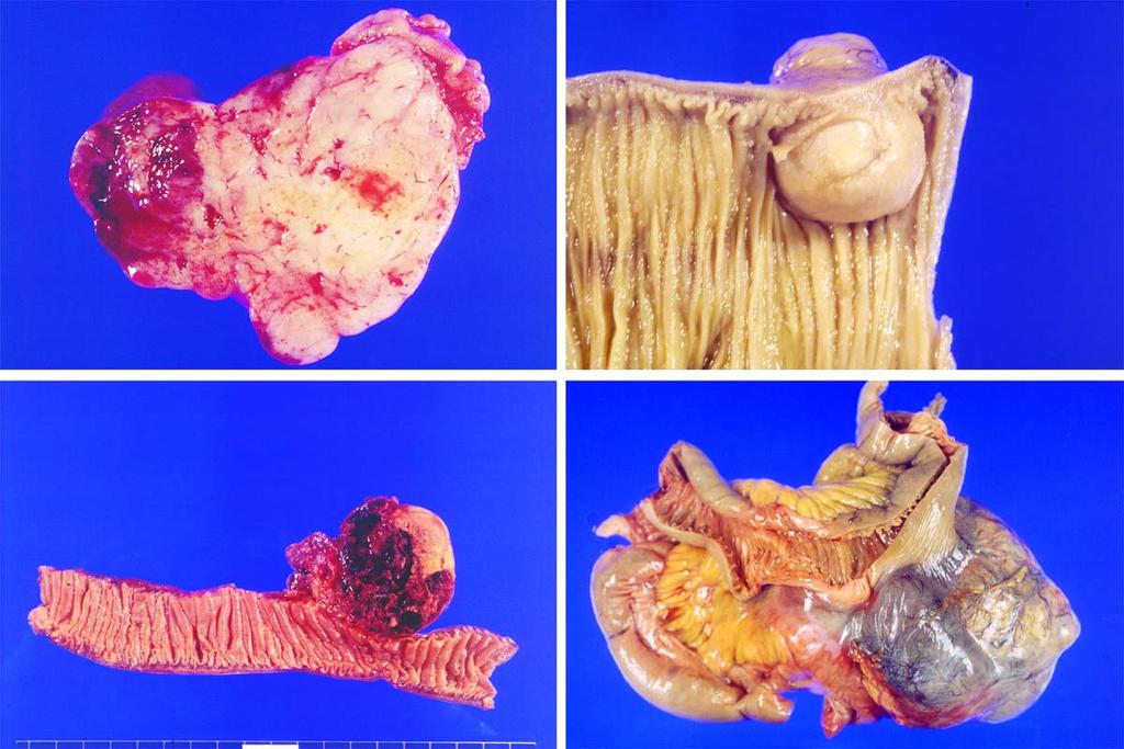
9
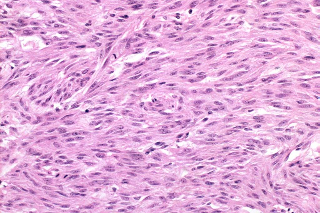
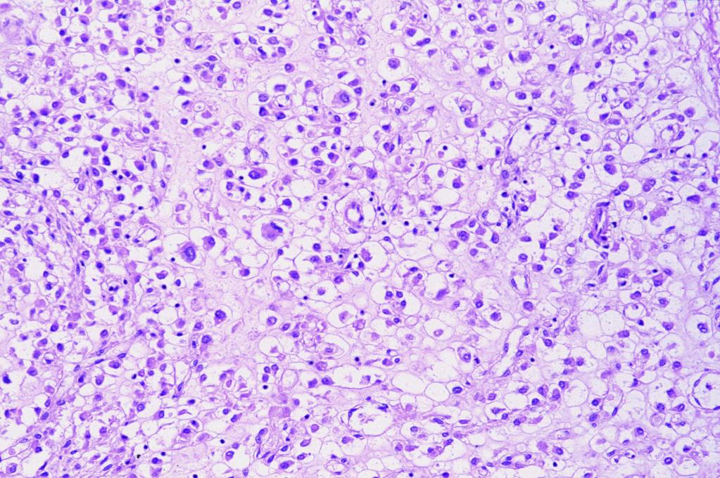
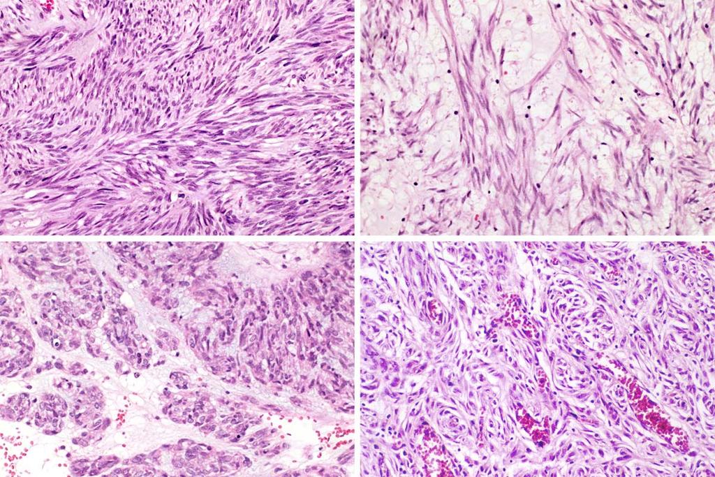
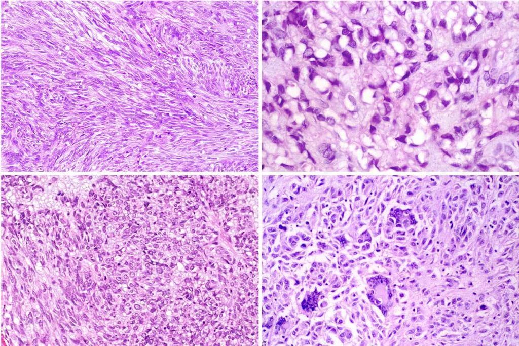
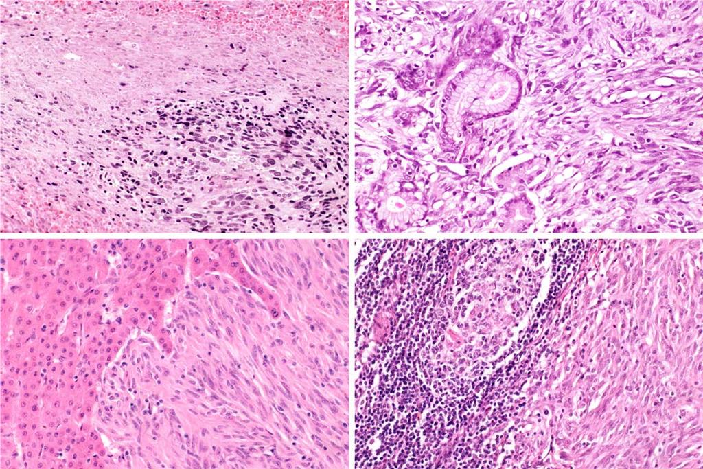
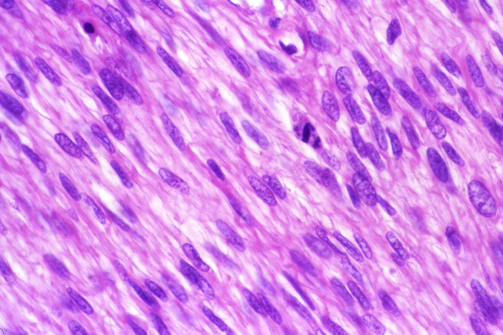
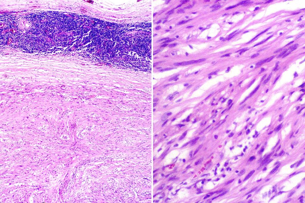
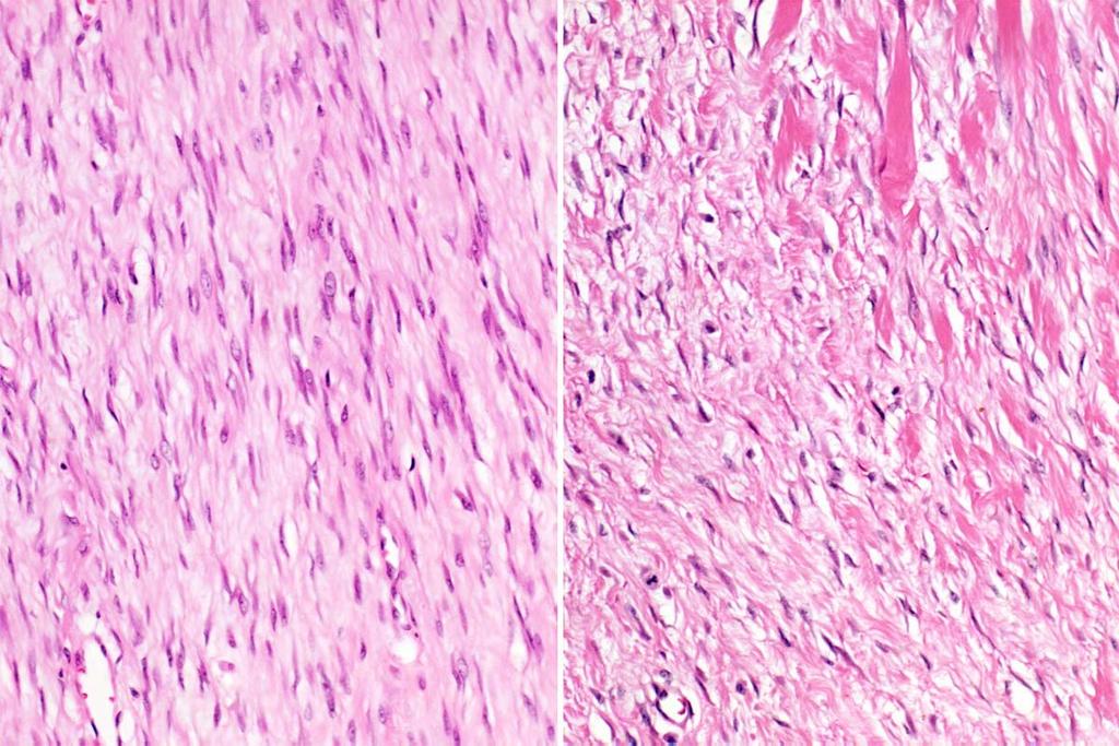
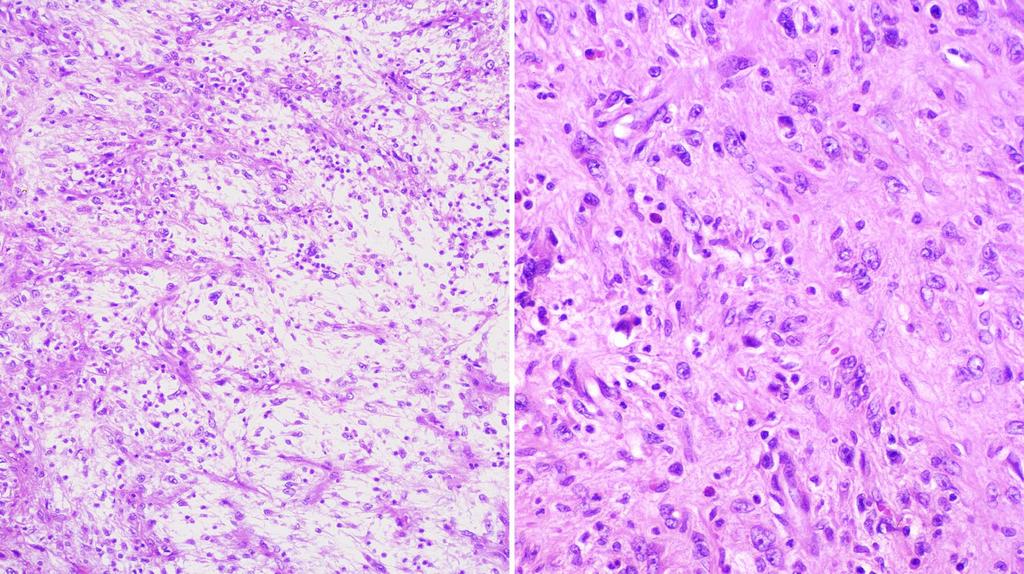
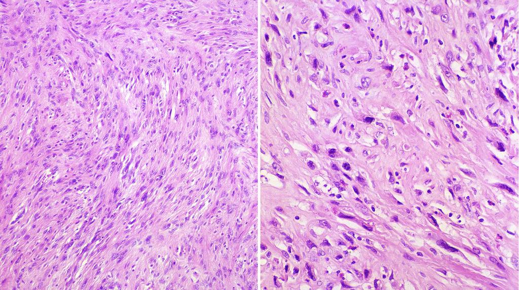
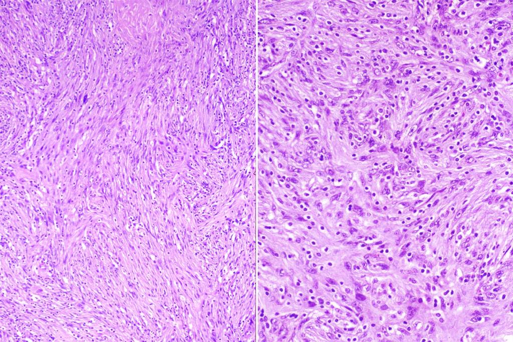
10 Microscopic features of GIST Spindle cell(70%) spindle, elongated nuclei paler eosinophilic cytoplasm sheets, fascicles, storiform, palisading Epithelioid cell(20%) round or polygonal cells abundant eosinophilic or clear cytoplasm Mixed(10%)
11
12
13
14
15
16 Skenoid fiber in GIST % of cases Hyaline or fibrillary eosinophilic structures, seemly composed of nodular tangles of collagen fiber No histogenetic significance

17 Skenoid fiber
18 Cytologic and growth patterns of GIST Cytologic types Growth patterns Spindle cell Fascicular Epithelioid Storiform Plasmacytoid cell Palisading Signet ring cell Diffuse, sheet-like Granular cell Organoid(nested) Multinucleated Myxoid Alveolar (Semin Diag Pathol 13: ,1996)
19 Immunophenotypes of GIST Immunostain Positivity c-kit(cd117) % CD % SMA 30-40% Desmin 1-2% S-100 protein 5%
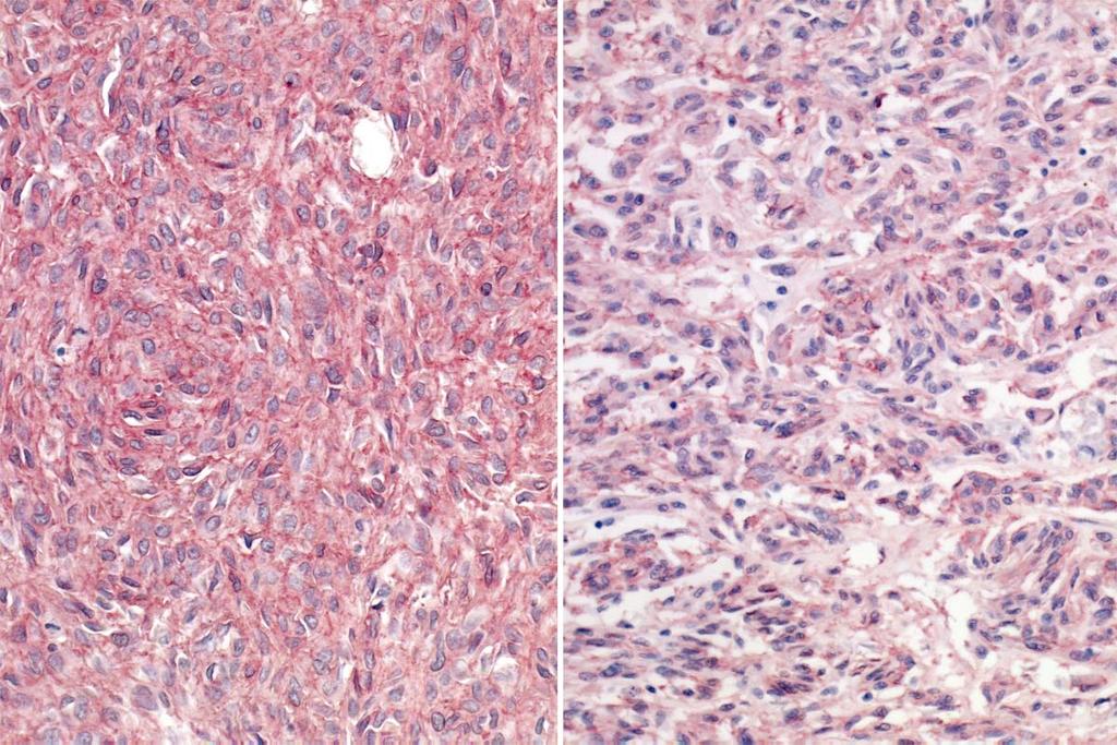
20 CD 117
21 Parameters used in evaluation of GIST malignancy reported in literatures Site Size Mitosis Histologic type Cytologic atypia Necrosis Amin Kindblom Panizo Trupiano Miettinen Fletcher Amin, et al: Am J Clin Pathol 100:428-32,1993. Kindblom, et al : Am J Clin Pathol 152: ,1998. Panizo, et al: Int J Surg Pathol 8:133-44, 2000 Trupiano, et al : AJSP 26:705-14, Miettinen, et al : Hum Pathol 33:478-83, Fletcher, et al : Sarcoma 2:133-41,1998.
22 Criteria of GIST malignancy by Miettinen Probably benign Uncertain or Probably malignant LMP Stomach 5 cm 5-10 cm >10 cm <5/50HPF <5/50HPF >5/50HPF Intestine 2 cm 2-5 cm >5 cm <5/50HPF <5/50HPF >5/50HPF (Hum Pathol 33:478-83,2002)
23 Criteria of GIST malignancy by Fletcher Benign Borderline Malignant Mitosis/30HPF Histologic type spindle cells, no atypia epithelioid cell spindle cells, mild pleorphism/hyperchromasia spindle cells, no atypia epithelioid cell spindle cells, no atypia spindle cells, frank pleomorphism/hyperchromasia epithelioid cell (Sarcoma 2 :133-41,1998)
24 Pathology report form of GIST in Fletcher s Institute Dx: Stomach, antrum, wedge resection : Gastrointestinal stromal sarcoma with 1) Cell type : spindle 2) Tumor size : 7 cm 3) Mitosis : 11/30 HPF 4) Necrosis : present 5) No vascular invasion 6) c-kit(cd117) : diffuse cytoplasmic strong positive 7) Clear resection margin
25 Histologic grading is not correlated strongly with metastatic risk or survival At least 10% of GIST (<5 cm and <5/50HPF) in more than 500 GISTs reviewed by Dr. Fletcher metastasis Any GIST can not be definitely regarded as benign. Distinction between benign and malignant appears to be a practically impossible at current time.
26 Diagnosis of Gastrointestinal Stromal Tumors: A Consensus Approach Fletcher CDM, Miettinen M, Weiss S, et al (Hum Pathol 33:459-65,2002) develop a scheme based on risk assessment rather than try to define strict criteria to separate benign and malignant
27 Proposed Approach for Defining Risk of Aggressive Behavior in GIST Risk Size(cm) Mitotic count(/50hpf) Very low <2 <5 Low 2-5 <5 Intermediate < <5 High >5 >5 >10 any mitoses Any size >10 (Hum Pathol 33:459-65,2002)
28 Approximate risk of metastasis/death Risk Metastasis/death Very low <5% Low 5-20% Intermediate 20-50% High >50% (Fletcher CDM, letter communication, 2002)
29 Diagnostic term recommended by Fletcher Risk Size(cm) Mitotic count(/50hpf) Pathology diagnosis Very low <2 cm <5 Gastrointestinal stromal tumor Low 2-5 <5 Gastrointestinal stromal sarcoma Intermediate < Gastrointestinal stromal sarcoma 5-10 <5 High >5 >5 Gastrointestinal stromal sarcoma >10 any mitoses Any size >10 (Fletcher CDM, letter communication, 2002)
30 Suggested pathology report form of GIST Dx: Stomach, antrum, wedge resection : Gastrointestinal stromal sarcoma with 1) Tumor size : 7 cm 2) Mitosis : 9/50 HPF 3) cell type : spindle 4) necrosis : present 5) no vascular invasion 6) c-kit(cd117) : diffuse cytoplasmic strong positive 7) Clear resection margin Note : According to risk assessment (Hum Pathol, 33:459-65, 2002), this tumor is considered as high risk
31 Frozen section of GIST Is the lesion stromal tumor? Frozen diagnosis Stromal tumor; determination of specific type and whether it is malignant must await permanent sections (AFIP, Fascicle 18,1996)
32 Procedure for pathologic examination 1. Measure greatest diameter 2. Note location, color, necrosis, hemorrhage on cross section 3. Check if tumor appears to obliterate mucosal-submucosal interface 4. Sample 1) area where most closely approaches the mucosa, including ulcers 2) areas of different consistency and color 3) both intramural and extramural component of large tumor 4) about one block per centimeter of maximum diameter 5. Micro. examination 1) count mitoses in contiguous fields in the highest mitotic activity area 6. Immunostain 1) routine : CD117, CD34, SMA, desmin, S-100 protein (AFIP, Fascicle 18,1996)
33 Pathology report of c-kit(cd117) negative GIST Dx: Spindle cell(or epithelioid) stromal neoplasm, most consistent with gastrointestinal stromal tumor(or gastrointestinal stromal sarcoma) (Hum Pathol 33:459-65, 2002)
34 Gastrointestinal autonomic nerve tumor(gant) Definition : Stromal tumor showing autonomic nerve differentiation ultrastructurally Site GI tract : small intestine Mesentery, retropritoneum, pelvis Microscopic features Similar to GIST Immunostain CD117, CD34, S-100 protein, NSE, PGP 9.5 Synaptophysin, chromogranin

35 GANT, mesentery(77/f) (Courtesy of Dr. Fletcher)
")
36 GANT(AJSP 17: , 1993)
37 EM study for diagnosis of GANT is not required now Exact same KIT mutation and same biological behavior as other GISTs No longer worth diagnosing
38 No longer talk about divergent differentiation in GIST Differentiation Neural No meaning at all GANT no longer exists as separate entity Myoid SMA (+) Nothing Present focally in most GIST Desmin (+) Extremely rare(< 2% of cases)
39 Leiomyoma in GIT Esophagus : most common Well circumscribed, non-encapsulated Mature smooth muscle cells with eosinophilic cytoplasm Low cellular No tumor necrosis Immunostain : SMA, desmin
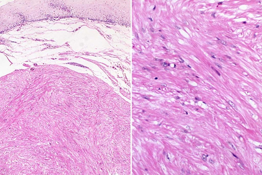
40 Leiomyoma, esophagus
41 Leiomyosarcoma in GIT Large bowel Spindle, cigar-shaped nuclei Epithelioid cells Large size : >5 cm Immunostain : SMA, desmin
42 Leiomyosarcoma
 (Courtesy of Dr.")
43 Epithelioid leiomyosarcoma, stomach(17/f) (Courtesy of Dr. Fletcher)
44 Epithelioid leiomyosarcoma vs Carcinoma Epithelioid leiomyosa. Carcinoma Cytokeratin + + EMA + Desmin + Caldesmon +
45 Criteria of leiomyosarcoma in GIT Ranchod Evans Cunningham <5 cm and <5/10HPF Watanabe Leiomyoma Leiomyosarcoma Ranchod, et al. Cancer 39:255-62,1977. Evans. Cancer 56: , Cunningham, et al. AJSP 17:588-94,1993. Watanabe, et al : WHO, 2 nd ed, /10HPF 1/10 HPF 10/10 HPF 10/50HPF
46 No any really meaningful criteria until now between leiomyoma vs leiomyosarcoma in GIT smooth muscle tumor Esophagus Large bowel most are benign submucosal small lesions are benign others malignant
47 Schwannoma in GIT Schwannoma in GIT Schwannoma in soft tissue Capsule - + Nuclear palisading - + Vascular hyalin. - + Verocay body - + Xanthoma cells -/+ + Lymphoid cuff + - Antoni A and B - + S-100 protein + +
48 Schwannoma, stomach
49 Soft tissue tumors histologically resembling GIST Intra-abdominal fibromatosis Solitary fibrous tumor Inflammatory myofibroblastic tumor Follicular dendritic cell tumor True histiocytic lymphoma
50 Intra-abdominal fibromatosis Pelvic, mesenteric, retroperitoneum Circumscribed or infiltrative, firm, homogenous Long fascicles of bland-looking spindle cells Collagenous or myxoid stroma Infiltrative margin Immunostain : Positive : CD117(focal weak), SMA, desmin Negative : CD34, S-100 protein
51 Intra-abdominal fibromatosis
52 Solitary fibrous tumor(sft) Pleura, intraabdomen, pelvis, retroperitoneum, peritoneum Well circumscribed Alternating hypercellular and hypocellular area Bland-looking short spindle or oval cells Haphazard, storiform or fascicular pattern of spindle cells Intimate intertwining of thin or thick collagen fibrils with spindle cells Hemangiopericytoma-like vascular pattern Immunostain : CD34, CD99, bcl-2 Malignant SFT: > 4/10 HPF, hypercellularity, pleomorphism

53 Solitary fibrous tumor, retroperitoneum

54 Solitary fibrous tumor
55 Inflammatory myofibroblastic tumor Abdominal cavity, gastrointestinal 10-25% local recur, 5% metastasis ALK(anaplastic lymphoma kinase) gene rearrangements Myofibroblastic proliferation with atypical nuclei 3 patterns : hypocelluar fibrous, cellular, myxoid/vascular Inflammatory cells, plasma cell, lymphocytes Immunostain : Vimentin(100%), SMA(78%), desmin(50%), ALK(36%), CD34(18%), EMA(16%)

56 IMT, mesentery (49/M) (Courtesy of Dr. Fletcher)
57 IMT, retroperitoneum (46/M)
58 IMT, retroperitoneum (46/M)
59 Follicular dendritic cell tumor Gastrointestinal tract, intra-abdominal Tumor cells are spindle, oval or polygonal shape Eosinophilic cytoplasm, poorly defined cell border Syncytial appearance Whorls, fascicles, storiform pattern, diffuse sheets Diffusely small lymphocytes Immunostain : CD21, CD35
 (Courtesy of Dr.")
60 FDRCT, small intestine (33/F) (Courtesy of Dr. Fletcher)
61 FDRCT
62 True histiocytic lymphoma Lymphoma showing true histiocytic differentiation : 0.5% of NHL Gastrointestinal tract, skin, soft tissue Spindle, round, irregular or grooved nuclei Abundant eosinophilic cytoplasm Diffuse, non-cohesive, sarcomatoid appearance Immunostain : CD68, lysozyme, CD4, CD11c, CD14,
 (Courtesy of Dr.")
63 True histiocytic lymphoma, mesentery (58/M) (Courtesy of Dr. Fletcher)
The Relevance of Cytologic Atypia in Cutaneous Neural Tumors
 The Relevance of Cytologic Atypia in Cutaneous Neural Tumors Recent Findings - New Developments New Problems Zsolt B. Argenyi, M.D. Professor of Pathology & Dermatology Director of Dermatopathology Department
A 25 year old female with a palpable mass in the right lower quadrant of her abdomen
 May 2016 A 25 year old female with a palpable mass in the right lower quadrant of her abdomen Contributed by: Paul Ndekwe, MD, Resident Physician, Indiana University School of Department of Pathology and
Newer soft tissue entities
 Newer soft tissue entities Examples among fibroblastic tumors Turku, May 6, 2010 Markku Miettinen, M.D. AFIP, Washington, DC Fibroblastic neoplasms Solitary fibrous tumor /Hemangiopericytoma Low-grade
Classification (1) Classification (3) Classification (2) Spindle cell lesions. Spindle cell lesions of bladder (Mills et al.
 Classification (3) Classification (2) Spindle cell lesions. Spindle cell lesions of bladder (Mills et al.") Non-epithelial tumours and nonepithelial tumour-like lesions of the bladder Dr Jonathan H Shanks The Christie NHS Foundation Trust, Manchester, UK Classification (1) Myofibroblastic proliferations and
Non-epithelial tumours and nonepithelial tumour-like lesions of the bladder Dr Jonathan H Shanks The Christie NHS Foundation Trust, Manchester, UK Classification (1) Myofibroblastic proliferations and
Spindle Cell Lesions Of The Breast. Emad Rakha Professor of Breast Pathology and Consultant Pathologist
 Spindle Cell Lesions Of The Breast Emad Rakha Professor of Breast Pathology and Consultant Pathologist * SCLs comprise a wide spectrum of diseases, ranging from reactive processes to aggressive malignant
ACCME/Disclosures ALK FUSION-POSITIVE MESENCHYMAL TUMORS. Tumor types with ALK rearrangements. Anaplastic Lymphoma Kinase. Jason L.
 Companion Meeting of the International Society of Bone and Soft Tissue Pathology The Evolving Concept of Mesenchymal Tumors ALK FUSION-POSITIVE MESENCHYMAL TUMORS Jason L. Hornick, MD, PhD March 13, 2016
Gastrointestinal Stromal Tumours - Report of Three Cases and Review of Literature
 jc6906 7joc207-3rd proof Case Report Gastrointestinal Stromal Tumours - Report of Three Cases and Review of Literature Deshpande A *, Munshi MM ** Abstract Three cases of GIST were diagnosed on guided
Gross appearance of peritoneal cysts. They have a thin, translucent wall and contain a clear fluid.
 Gross appearance of peritoneal cysts. They have a thin, translucent wall and contain a clear fluid. So-called multicystic benign mesothelioma. A, Gross appearance. So-called multicystic benign mesothelioma.
Normal endometrium: A, proliferative. B, secretory.
 Normal endometrium: A, proliferative. B, secretory. Nội mạc tử cung Nội mạc tử cung Cyclic changes in endometrium.. Approximate relationship of useful microscopic changes. Arias-Stella reaction in endometrial
A 9cm mass was excised from the jejunal wall and mesentery of a 33 year old woman.
 A Few Observations on Gastrointestinal Stromal Tumors and Their Differential Diagnosis E. Montgomery A 9cm mass was excised from the jejunal wall and mesentery of a 33 year old woman. 1 2 3 CD117/c-kit
3/27/2017. Disclosure of Relevant Financial Relationships
 Ophthalmic Pathology Evening Specialty Conference USCAP 2017 5 th March, 2017 Mukul K. Divatia, MD Assistant Professor Department of Pathology & Genomic Medicine Weill Cornell Medical College Houston Methodist
GUT-C 11/30/2017. Debasmita Das, M.D. PGY-1 Danbury Hospital
 GUT-C 11/30/2017 Debasmita Das, M.D. PGY-1 Danbury Hospital CLINICAL SUMMARY 8/2017 59 year old female Presented to the ED with 1 month history of general malaise, fever and weight loss PMH: Significant
Case Presentation. Maha Akkawi, MD, Fatima Obeidat, MD, Tariq Aladily, MD. Department of Pathology Jordan University Hospital Amman, Jordan
 Case Presentation Maha Akkawi, MD, Fatima Obeidat, MD, Tariq Aladily, MD Department of Pathology Jordan University Hospital Amman, Jordan The 25th Annual Congress of the ADIAP The 8/11/2013 1 5th International
59 yo male with past medical history of prostate carcinoma, presented with upper abdominal pain
 December 2016 59 yo male with past medical history of prostate carcinoma, presented with upper abdominal pain Contributed by: Divya Sharma, MD. Fellow, Gastrointestinal Pathology, Department of Pathology
Diplomate of the American Board of Pathology in Anatomic and Clinical Pathology
 A 33-year-old male with a left lower leg mass. Contributed by Shaoxiong Chen, MD, PhD Assistant Professor Indiana University School of Medicine/ IU Health Partners Department of Pathology and Laboratory
Malignant Peripheral Nerve Sheath Tumor
 C H A P T E R 120 Malignant Peripheral Nerve Sheath Tumor Currently, malignant peripheral nerve sheath tumor (MPNST) is the most commonly used generic name for the neoplasms known in the past as neurosarcoma,
Case: The patient is a 24 year- old female who was found to have multiple mural nodules within the antrum. Solid and cystic components were noted on
 Case: The patient is a 24 year- old female who was found to have multiple mural nodules within the antrum. Solid and cystic components were noted on imaging. There is no significant past medical history.
International Journal of Health Sciences and Research ISSN:
 International Journal of Health Sciences and Research www.ijhsr.org ISSN: 2249-9571 Case Report Malignant Gastrointestinal Stromal tumor of the Sigmoid Colon with Perforation and Peritonitis - an Unusual
3/24/2017 DENDRITIC CELL NEOPLASMS: HISTOLOGY, IMMUNOHISTOCHEMISTRY, AND MOLECULAR GENETICS. Disclosure of Relevant Financial Relationships
 DENDRITIC CELL NEOPLASMS: HISTOLOGY, IMMUNOHISTOCHEMISTRY, AND MOLECULAR GENETICS Jason L. Hornick, M.D., Ph.D. Director of Surgical Pathology and Immunohistochemistry Brigham and Women s Hospital Professor
أملس عضلي غرن = Leiomyosarcoma. Leiomyosarcoma 1 / 5
 Leiomyosarcoma 1 / 5 EPIDEMIOLOGY Exact incidence is unknown, but older studies suggest that leiomyosarcomas comprise approximately 3 percent of soft-tissue sarcomas. Superficial leiomyosarcoma occurs
Mody. AIS vs. Invasive Adenocarcinoma of the Cervix
 Common Problems in Gynecologic Pathology Michael T. Deavers, M.D. Houston Methodist Hospital, Houston, Texas Common Problems in Gynecologic Pathology Adenocarcinoma in-situ (AIS) of the Cervix vs. Invasive
Mesenchymal neoplasms of the gastrointestinal tract what s new? Newton ACS Wong Department of Histopathology Bristol Royal Infirmary
 Mesenchymal neoplasms of the gastrointestinal tract what s new? Newton ACS Wong Department of Histopathology Bristol Royal Infirmary Talk plan Summary from 2010 talk. What s happened since 2010. GISTs
CASE REPORT Benign epithelioid peripheral nerve sheath tumour resembling schwannoma
 Malaysian J Pathol 2014; 36(3) : 217 221 CASE REPORT Benign epithelioid peripheral nerve sheath tumour resembling schwannoma Thejasvi KRISHNAMURTHY MD and SR NIVEDITHA MD, DNB Department of Pathology,
Intussuception due to gastrointestinal stromal tumor with neural differentiation in a patient with. Von Recklinghausen Neurofibromatosis,
 Turkish Journal of Cancer Vol 31/ No.4 /2001 Intussuception due to gastrointestinal stromal tumor with neural differentiation in a patient with Von Recklinghausen Neurofibromatosis (NF-1): A case report
No financial or other disclosures
 Case 2014-5 Esther N. Bit-Ivan, DO Northwestern University Jason Wang, MD Jason Park, MD Korgun Koral, MD Children s Medical Center Charles Timmons, MD Veena Rajaram, MD No financial or other disclosures
DOI: /jemds/2014/1921 ORIGINAL ARTICLE
 CYTODIAGNOSIS OF GASTROINTESTINAL STROMAL TUMOURS (GISTs) ON ROMANOWSKY STAINED SMEARS Priyanka Agrawal 1, Subhash Agrawal 2, Atul Gupta 3, Anurag Gupta 4 HOW TO CITE THIS ARTICLE: Priyanka Agrawal, Subhash
Malignant gastrointestinal stromal (GISTs) of the duodenum A rare occurrence: Case report
 of the duodenum A rare occurrence: Case report") Malignant gastrointestinal stromal tumors (GISTs) of the duodenum Case Report ISSN: 2394-0026 (P) Malignant gastrointestinal stromal tumors (GISTs) of the duodenum A rare occurrence: Case report Kandukuri
Malignant gastrointestinal stromal tumors (GISTs) of the duodenum Case Report ISSN: 2394-0026 (P) Malignant gastrointestinal stromal tumors (GISTs) of the duodenum A rare occurrence: Case report Kandukuri
5/10. Pathology Soft tissue tumors. Farah Bhani. Mohammed Alorjani
 5/10 Pathology Soft tissue tumors Mohammed Alorjani Farah Bhani Slides are included in this sheet. Objectives: Soft tissue tumors 1. Describe soft tissue tumors. 2. Understand the classification of soft
57th Annual HSCP Spring Symposium 4/16/2016
 An Unusual Malignant Spindle Cell Lesion to Involve the Breast Erinn Downs-Kelly, D.O. Associate Professor of Pathology University of Utah & ARUP Laboratories No disclosures Case 39 y/o female with no
RADIOFREQUENCY ABLATION
 RADIOFREQUENCY ABLATION ELIZABETH DAVID M D FRCPC VASCULAR A ND INTERVENTIONAL RADIOLOGIST SUNNYBROOK HEALTH SCIENCES CENTRE GIST GASTROINTESTINAL STROMAL TUMORS Stromal or mesenchymal neoplasms affecting
Imaging of Gastrointestinal Stromal Tumors (GIST) Amir Reza Radmard, MD Assistant Professor Shariati hospital Tehran University of Medical Sciences
 Amir Reza Radmard, MD Assistant Professor Shariati hospital Tehran University of Medical Sciences") Imaging of Gastrointestinal Stromal Tumors (GIST) Amir Reza Radmard, MD Assistant Professor Shariati hospital Tehran University of Medical Sciences Describe the typical imaging findings of GIST at initial
Imaging of Gastrointestinal Stromal Tumors (GIST) Amir Reza Radmard, MD Assistant Professor Shariati hospital Tehran University of Medical Sciences Describe the typical imaging findings of GIST at initial
Endometrial Stromal Tumors
 Endometrial Stromal Tumors WHO Categories: Endometrial Stromal Nodule (ESN) Endometrial Stromal Sarcoma, low grade (LGESS) Endometrial Stromal Sarcoma, high grade (HGESS) Undifferentiated Uterine Sarcoma
SOFT TISSUE TUMOR PATHOLOGY: AN UPDATE
 SOFT TISSUE TUMOR PATHOLOGY: AN UPDATE Jason L. Hornick, MD, PhD July 18, 2013 Department of Pathology Brigham and Women s Hospital Harvard Medical School Boston, MA, USA I have no disclosures. New Soft
Understanding Your GIST Pathology Report
 Gastrointestinal Stromal Tumor Understanding Your GIST Pathology Report Jason L. Hornick, MD PhD Harvard Medical School Brigham and Women's Hospital Alexander J.F. Lazar, MD PhD Sarcoma Research Center
An Overview of Genital Stromal Tumors
 An Overview of Genital Stromal Tumors By Konstantinos Linos MD, FCAP, FASDP Bone, Soft Tissue and Dermatopathology Assistant Professor of Pathology Dartmouth-Hitchcock Medical Center Geisel School of Medicine
1/10/2018. Soft Tissue Tumors Showing Melanocytic Differentiation. Overview. Desmoplastic/ Spindle Cell Melanoma
 2016 MFMER slide-1 2016 MFMER slide-2 2016 MFMER slide-3 Soft Tissue Tumors Showing Melanocytic Differentiation Andrew L. Folpe, M.D. Professor of Laboratory Medicine and Pathology Mayo Clinic, Rochester,
Enterprise Interest Nothing to declare
 Enterprise Interest Nothing to declare Diagnoses one would not like to miss in soft tissue pathology early in your career Marta Sbaraglia, MD Department of Pathology Hospital of Treviso University of Padua
IN THE NAME OF GOD Dr. Kheirandish Oral and maxillofacial pathology
 IN THE NAME OF GOD Dr. Kheirandish Oral and maxillofacial pathology ORAL FOCAL MUCINOSIS Uncommon Tumorlike Cutaneous myxoid cyst Overproduction of hyaluronic acid by firoblasts Young adults Female Gingiva
Desmoplastic Melanoma R/O BCC. Clinical Information. 74 y.o. man with lesion on left side of neck r/o BCC
 R/O BCC Sabine Kohler, M.D. Professor of Pathology and Dermatology Dermatopathology Service Stanford University School of Medicine Clinical Information 74 y.o. man with lesion on left side of neck r/o
Morphological Features of Metastatic Gastrointestinal Stromal Tumors after Gleevec Treatment
 The Korean Journal of Pathology 2009; 43: 368-73 DOI: 10.4132/KoreanJPathol.2009.43.4.368 Morphological Features of Metastatic Gastrointestinal Stromal Tumors after Gleevec Treatment - Two Cases Report
What really matters When and Why. Pathology of Uterine Mesenchymal Lesions. Nafisa Wilkinson London
 What really matters When and Why Pathology of Uterine Mesenchymal Lesions Nafisa Wilkinson London Patient centred approach immunohistochemistry Histological diagnosis Next generation sequencing Genetic
Cellular Neurothekeoma
 Cellular Neurothekeoma Scott W Binder, MD Pritzker Professor of Pathology & Dermatology Sr. Vice Chair Director, Pathology Clinical Services Chief, Dermatopathology Geffen/UCLA School of Medicine Clinical
Brief History. Identification : Past History : HTN without regular treatment.
 Brief History Identification : Name : 陳 x - Admission : 94/10/06 Gender : male Age : 75 y/o Chief Complaint : Urinary difficulty for months. Past History : HTN without regular treatment. Brief History
From Morphology to Molecular Pathology: A Practical Approach for Cytopathologists Part 1-Cytomorphology. Songlin Zhang, MD, PhD LSUHSC-Shreveport
 From Morphology to Molecular Pathology: A Practical Approach for Cytopathologists Part 1-Cytomorphology Songlin Zhang, MD, PhD LSUHSC-Shreveport I have no Conflict of Interest. FNA on Lymphoproliferative
Special slide seminar
 Special slide seminar Tomáš Rozkoš The Fingerland Department of Pathology Charles University Medical Faculty and Faculty Hospital in Hradec Králové Czech Republic Case history, 33 years old resistance
Malignant Epithelioid Gastrointestinal Stromal Tumors. Report of a Case with Cytologic and Immunohistochemical Studies DO NOT DUPLICATE
 Malignant Epithelioid Gastrointestinal Stromal Tumors Report of a Case with Cytologic and Immunohistochemical Studies Juan B. Laforga, M.D. Background Gastrointestinal stromal tumors (GISTs) may exhibit
Inflammatory pseudotumor
 Inflammatory pseudotumor Inflammatory pseudotumor (IPT) Heterogeneous group of lesions of obscure etiology On physical and radiographic examination often confused with malignancy Synonyms Plasma cell granuloma
Selected Pseudomalignant Soft Tissue Tumors of the Skin and Subcutis
 Selected Pseudomalignant Soft Tissue Tumors of the Skin and Subcutis Andrew L. Folpe, M.D. Professor of Laboratory Medicine and Pathology Mayo Clinic, Rochester, MN folpe.andrew@mayo.edu 2016 MFMER slide-1
Pancreas. Atrophy, acinar cell. Pathogenesis: Diagnostic key features:
 Pancreas Atrophy, acinar cell Pathogenesis: Decrease in number and/or size of acinar cells may be due to spontaneous or experimentally induced degenerative changes, apoptosis, or a sequel of chronic inflammation.
Financial disclosures
 Mesenchymal Neoplasms with Melanocytic Differentiation By Konstantinos Linos MD, FCAP, FASDP Bone, Soft Tissue and Dermatopathology Assistant Professor of Pathology Dartmouth-Hitchcock Medical Center Geisel
Solitary Fibrous Tumor of the Kidney with Massive Retroperitoneal Recurrence. A Case Presentation
 246) Prague Medical Report / Vol. 113 (2012) No. 3, p. 246 250 Solitary Fibrous Tumor of the Kidney with Massive Retroperitoneal Recurrence. A Case Presentation Sfoungaristos S., Papatheodorou M., Kavouras
SMOOTH MUSCLE TUMOURS
 SMOOTH MUSCLE TUMOURS NORMAL SMOOTH MUSCLE Cytology Immunohistochemistry Ultrastructure Masson Trichrome Smooth Muscle Ultrastructure Many myofilaments running parallel to the long axis of the smooth
05/07/2018. Types of challenges. Challenging cases in uterine pathology. Case 1 ` 65 year old female Post menopausal bleeding Uterine Polyp
 Types of challenges Challenging cases in uterine pathology Nafisa Wilkinson Gynaecological Pathologist UCLH London Lack of complete history often, NO clinical history at all! Cases from other centres often
Images In Gastroenterology
 Images In Gastroenterology Thong-Ngam D, et al. THAI J GASTROENTEROL 2005 Vol. 6 No. 2 May - Aug. 2005 105 Imaging of Gastrointestinal Stromal Tumors Pornpim Fuangtharnthip, M.D. Narumol Hargroove, M.D.
A case of pedunculated intraperitoneal leiomyoma
 Jichi Medical University Journal Chio Shuto Kuniyasu Soda Takayoshi Yoshida Fumio Konishi Abstract We report a very rare case of a pedunculated intraperitoneal leiomyoma in the parietal peritoneum of the
Atypical Palisaded Myofibroblastoma of Lymph Node: Report of a rare case.
 ISPUB.COM The Internet Journal of Pathology Volume 10 Number 1 Atypical Palisaded Myofibroblastoma of Lymph Node: Report of a rare case. V Kinnera, R Nandyala, M Yootla, K Mandyam Citation V Kinnera, R
Soft Tissue Perineurioma
 The Korean Journal of Pathology 2009; 43: 266-70 DOI: 10.4132/KoreanJPathol.2009.43.3.266 Soft Tissue Perineurioma - A Case Report - Jun Mo Kim Joon Hyuk Choi Department of Pathology, Yeungnam University
Affiliazione autori0. Riccardo Ricci Journal Club GIPAD, settore GIST Anatomia Patologica, Università Cattolica, Roma
 GIST Manifesting as a Retroperitoneal Tumor: Clinicopathologic Immunohistochemical, and Molecular Genetic Study of 112 Cases American Journal of Surgical Pathology, 2017, 41:577-585 Miettinen M*; Felisiak-Golabek
ESS: Pathologic Insights
 GEIS XVI INTERNATIONAL SYMPOSIUM Seville 4th October 2018 ESS: Pathologic Insights Sílvia Bagué The Royal Marsden Hospital London (United Kingdom) I have no conflicts of interest Endometrial stromal sarcoma
Clinicopathologic Spectrum of Gastrointestinal Stromal Tumours; Six Years Experience at King Hussein Medical Center
 Clinicopathologic Spectrum of Gastrointestinal Stromal Tumours; Six Years Experience at King Hussein Medical Center Sahem T. Alqusous MD*, Osama J. Rabadi MD, MRCS*, Ala D. Al Omari MD*, Nabeeha N.Abbasi
Grading of Bone Tumors
 Grading of Bone Tumors Joon Hyuk Choi, M.D. Department of Pathology College of Medicine, Yeungnam University Introduction to grading system of bone tumor used at Mayo Clinic WHO Histologic Classification
Keywords solitary fibrous tumor, dedifferentiation, dedifferentiated solitary fibrous tumor, STAT6, GRIA2, cytokeratin, rhabdomyosarcomatous
 758452IJSXXX10.1177/1066896918758452International Journal of Surgical PathologyCreytens et al research-article2018 Pitfalls in Pathology Multifocal Cytokeratin Expression in a Dedifferentiated Solitary
Salivary Glands 3/7/2017
 Salivary Glands 3/7/2017 Goals and objectives Focus on the entities unique to H&N Common board type facts Information for your future practice Salivary Glands Salivary Glands Major gland. Paratid. Submandibular.
Lung Tumor Cases: Common Problems and Helpful Hints
 Lung Tumor Cases: Common Problems and Helpful Hints Brandon T. Larsen, MD, PhD Senior Associate Consultant Department of Laboratory Medicine and Pathology Mayo Clinic Arizona Arizona Society of Pathologists
Fine Needle Aspiration Biopsy of a Myxoid Leiomyosarcoma with Epithelioid Features and It Metastasized to the Abdominal Wall - A Case Report -
 The Korean Journal of Pathology 2010; 44: 220-4 DOI: 10.4132/KoreanJPathol.2010.44.2.220 Fine Needle Aspiration Biopsy of a Myxoid Leiomyosarcoma with Epithelioid Features and It Metastasized to the Abdominal
International Journal of Scientific & Engineering Research, Volume 7, Issue 5, May ISSN Case Report Rare Case Of Small Bowel GIST
 International Journal of Scientific & Engineering Research, Volume 7, Issue 5, May-2016 282 Case Report Rare Case Of Small Bowel GIST Shahaji G. Chavan, Sagar R. Ambre, Vinayak kshirsagar, Ashish Vashistha
Case Report Glomus tumor of uncertain malignant potential of the lung: a case report and review of literature
 Int J Clin Exp Pathol 2015;8(11):15402-15406 www.ijcep.com /ISSN:1936-2625/IJCEP0015761 Case Report Glomus tumor of uncertain malignant potential of the lung: a case report and review of literature Peng-Zhi
Fine Needle Aspiration Cytology of Gastric Glomus Tumor
 The Korean Journal of Pathology 2010; 44: 448-52 DOI: 10.4132/KoreanJPathol.2010.44.4.448 Fine Needle Aspiration Cytology of Gastric Glomus Tumor - A Case Report - Dong Geun Lee Kyu Yun Jang Myoung Ja
Contents Part I Introduction 1 General Description 2 Natural History: Importance of Size, Site, Histopathology
 Contents Part I Introduction 1 General Description... 3 1.1 Introduction... 3 1.2 Incidence and Prevalence... 5 1.3 Predisposing and Genetic Factors... 8 References... 16 2 Natural History: Importance
Diagnostic problems in uterine smooth muscle tumors
 Diagnostic problems in uterine smooth muscle tumors Marina Kos Ljudevit Jurak Clinical Department of Pathology, Clinical Hospital Center Sestre milosrdnice, Zagreb Institute of Pathology, University of
Subepithelial Lesions of the Gut: When Should I Worry?
 Subepithelial Lesions of the Gut: When Should I Worry? President, ASGE Chairman, GI & Hepatology Scottsdale, AZ Faigel.douglas@mayo.edu Case 55 yo male with reflux EGD for Barrett s Screening SET, mucosal
All authors abide by the Association for Medical Ethics (AME) ethical rules of disclosure.
 ethical rules of disclosure.") Longo F, Musumeci G, Parenti R, Vecchio G, Magro G. Atypical cell leiomyoma of the uterus with amianthoid-like fibers: A case report. OA Case Reports 2013 Nov 15;2(14):137. Licensee OA Publishing London
Longo F, Musumeci G, Parenti R, Vecchio G, Magro G. Atypical cell leiomyoma of the uterus with amianthoid-like fibers: A case report. OA Case Reports 2013 Nov 15;2(14):137. Licensee OA Publishing London
Pathology of Sarcoma ELEANOR CHEN, MD, PHD, ASSISTANT PROFESSOR DEPARTMENT OF PATHOLOGY UNIVERSITY OF WASHINGTON
 Pathology of Sarcoma ELEANOR CHEN, MD, PHD, ASSISTANT PROFESSOR DEPARTMENT OF PATHOLOGY UNIVERSITY OF WASHINGTON Presentation outline Background and epidemiology of sarcomas Sarcoma classification Sarcoma
Tumors of Adipose Tissue Tumors Epidemiology Clinical Features. Morphology. Mature Adipocytes Separated by delicate fibrous septa
 Tumors of Adipose Tissue Lipoma Liposarcoma Most commonly happens in female The most common soft tissue tumor o Originates from matured Adipocytes Most commonly happes at the 4 th and 5 th decade of life
Extragastrointestinal (soft tissue) stromal tumors. Analysis of 48 Cases with Emphasis on Histologic Predictors of Outcome
 stromal tumors. Analysis of 48 Cases with Emphasis on Histologic Predictors of Outcome") Extragastrointestinal (Soft Tissue) Stromal Tumors: An Analysis of 48 Cases with Emphasis on Histologic Predictors of Outcome John D. Reith, M.D., John R. Goldblum, M.D., Robert H. Lyles, Ph.D., Sharon
Extragastrointestinal (Soft Tissue) Stromal Tumors: An Analysis of 48 Cases with Emphasis on Histologic Predictors of Outcome John D. Reith, M.D., John R. Goldblum, M.D., Robert H. Lyles, Ph.D., Sharon
Case 27 Male 42. Painless, static, well-circumscribed, subcutaneous nodule right lower leg,?lipoma. The best diagnosis is:
 Case 27 Male 42. Painless, static, well-circumscribed, subcutaneous nodule right lower leg,?lipoma. The best diagnosis is: A. Angiosarcoma B. Haemangiopericytoma C.Myopericytoma D.Myofibroma E. Angioleiomyoma
Rhabdomyomas and Rhabdomyosarcomas (RMS) David M. Parham, MD Chief of Anatomic Pathology
 David M. Parham, MD Chief of Anatomic Pathology") Rhabdomyomas and Rhabdomyosarcomas (RMS) David M. Parham, MD Chief of Anatomic Pathology Tumors of skeletal muscle: Rhabdomyomas and rhabdomyosarcomas Embryonal muscle 2 3 4 5 6 7 8 Rhabdomyoma Benign
Rhabdomyomas and Rhabdomyosarcomas (RMS) David M. Parham, MD Chief of Anatomic Pathology Tumors of skeletal muscle: Rhabdomyomas and rhabdomyosarcomas Embryonal muscle 2 3 4 5 6 7 8 Rhabdomyoma Benign
4/12/2018. MUSC Pathology Symposium Kiawah Island April 18, Jesse K. McKenney, MD
 MUSC Pathology Symposium Kiawah Island April 18, 2018 Jesse K. McKenney, MD 1 Urothelial Carcinoma with Alternative Differentiation 2 Urothelial Carcinoma with Alternative Differentiation Recognition as
Case year female. Routine Pap smear
 Case 1 57 year female Routine Pap smear Diagnosis? 1. Atypical glandular cells of unknown significance (AGUS) 2. Endocervical AIS 3. Endocervical adenocarcinoma 4. Endometrial adenocarcinoma 5. Adenocarcinoma
Respiratory Tract Cytology
 Respiratory Tract Cytology 40 th European Congress of Cytology Liverpool, UK Momin T. Siddiqui M.D. Professor of Pathology and Laboratory Medicine Director of Cytopathology Emory University Hospital, Atlanta,
A Gastric Schwannoma Misdiagnosed as B Cell Lymphoma
 계명의대학술지제 31 권 2 호 Keimyung Med J Vol. 31, No. 2, December, 2012 A Gastric Schwannoma Misdiagnosed as B Cell Lymphoma Kyung lim Koo, Yu Na Kang 1, M.D. Undergraduate Student, Department of Pathology 1,
Gross appearance of nodular hyperplasia in material obtained from suprapubic prostatectomy. Note the multinodular appearance and the admixture of
 Tiền liệt tuyến Tiền liệt tuyến Gross appearance of nodular hyperplasia in material obtained from suprapubic prostatectomy. Note the multinodular appearance and the admixture of solid and microcystic areas.
Smooth Muscle and Similar Lesions. Disclosure Statement. Smooth Muscle Tumors. Elizabeth Montgomery
 Smooth Muscle and Similar Lesions Elizabeth Montgomery Disclosure Statement Dr. Montgomery reports no relevant financial relationships with commercial interests. Smooth Muscle Tumors Most are leiomyomas
Update on Cutaneous Mesenchymal Tumors. Thomas Brenn
 Update on Cutaneous Mesenchymal Tumors Thomas Brenn Cutaneous Mesenchymal Tumours Wide morphological and biological spectrum Myofibroblastic, smooth muscle, neural, vascular, apidocytic, undifferentiated;
21/07/2017. Hobnail endothelial cells are not the same as epithelioid endothelial cells
 UPDATE IN CUTANEOUS VASCULAR S DERMATOPATHOLOGY SESSION BELFAST PATHOLOGY JUNE 21/2017 Dr E Calonje St John s Institute of Dermatology, London, United Kingdom THE FAMILY OF VASCULAR S WITH EPITHELIOID
Update On Lipomatous Tumors: Old Standbys and New Concepts
 Update On Lipomatous Tumors: Old Standbys and New Concepts John R. Goldblum, M.D. Chairman, Department of Anatomic Pathology Cleveland Clinic Professor of Pathology Cleveland Clinic Lerner College of Medicine
Slide Seminar Spanish Society of Pathology
 Slide Seminar Spanish Society of Pathology John R. Goldblum, M.D. Chairman, Department of Anatomic Pathology Cleveland Clinic Professor of Pathology Cleveland Clinic Lerner College of Medicine 1921 Original
IMMUNOHISTOCHEMISTRY IN THE DIAGNOSIS OF SOFT TISSUE TUMORS
 IMMUNOHISTOCHEMISTRY IN THE DIAGNOSIS OF SOFT TISSUE TUMORS Nicolas de Saint Aubain Somerhausen Institut Jules Bordet / Hôpital Erasme nicolas.desaintaubain@synet.be ForPath 2005 1 I. Ancillary techniques
Evaluating and Reporting Gastrointestinal Stromal Tumors after Imatinib Mesylate Treatment
 The Open Pathology Journal, 2009, 3, 53-57 53 Open Access Evaluating and Reporting Gastrointestinal Stromal Tumors after Imatinib Mesylate Treatment Katie L. Dennis * and Ivan Damjanov Department of Pathology
I have nothing to disclose
 A 47 year old female with multiple lung nodules Disclosure of Relevant Financial Relationships Tamar Giorgadze, MD, PhD Professor of Pathology Medical College of Wisconsin Milwaukee, Wisconsin USCAP requires
Inflammatory Myofibroblastic Tumor of the Stomach Presenting as an Exophytic Mass - A Diagnostic Dilemma
 Case Report doi: 10.516/tjpath.017.0188 Inflammatory Myofibroblastic Tumor of the Stomach Presenting as an Exophytic Mass - A Diagnostic Dilemma Meena JADHAV, Rekha HARVI, Rashmi PATIL, Shreekant KITTUR
ACCME/Disclosures. Everything is spindle - how far can we go with limited FNA material? Everything is spindle how far can we go? Everything is spindle
 ACCME/Disclosures The USCAP requires that anyone in a position to influence or control the content of CME disclose any relevant financial relationship WITH COMMERCIAL INTERESTS which they or their spouse/partner
Disclosure. Case. Mixed Tumors of the Uterine Corpus and Cervix. I have nothing to disclose
 Mixed Tumors of the Uterine Corpus and Cervix Marisa R. Nucci, M.D. Division of Women s and Perinatal Pathology Department of Pathology Brigham and Women s Hospital Boston, MA UCSF Current Issues in Anatomic
Gastrointestinal stromal tumours - clinicopathological study
 Original research article Gastrointestinal stromal tumours - clinicopathological study *Dr. Putrevu Venkata Gurunadha Raju, ** Dr. Kanwaljit Kaur *Professor, **Assistant Professor Department of Pathology,
GIST. Keys for a fast radiologic identification
 GIST. Keys for a fast radiologic identification Poster No.: C-0210 Congress: ECR 2013 Type: Educational Exhibit Authors: R. Castañón Martinez, I. bañales arnaiz, F. Alonso Avalos, 1 2 2 2 1 1 J. Sanchez
Angiomatoid Fibrous Histiocytoma : A Case Report
 영남의대학술지제24권제2호 Yeungnam Univ. J. of Med. Vol.24 No.2 p315-321, Dec. 2007 증례 Angiomatoid Fibrous Histiocytoma : A Case Report Joon Hyuk Choi, Woo Jung Sung, Nam Hyuk Lee* Department of Pathology, and *Department
Case 2. Dr. Sathima Natarajan M.D. Kaiser Permanente Medical Center Sunset
 Case 2 Dr. Sathima Natarajan M.D. Kaiser Permanente Medical Center Sunset History 24 year old male presented with a 3 day history of right flank pain, sharp in nature Denies fever, chills, hematuria or
CHAPTER 4. Smooth Muscle Tumours
 CHAPTER 4 Smooth Muscle Tumours Smooth muscle tumours arising at non-cutaneous, non-uterine locations have been the focus of a considerable conceptual shift in recent years and this is ongoing. Specifically,
case report Oman Medical Journal [2016], Vol. 31, No. 1: 60 64
![case report Oman Medical Journal [2016], Vol. 31, No. 1: 60 64](/thumbs/90/102852192.jpg "case report Oman Medical Journal [2016], Vol. 31, No. 1: 60 64") case report Oman Medical Journal [2016], Vol. 31, No. 1: 60 64 Malignant Gastric Glomus Tumor: A Case Report and Literature Review of a Rare Entity Shaesta Zaidi * and Maha Arafah Department of Histopathology,
case report Oman Medical Journal [2016], Vol. 31, No. 1: 60 64 Malignant Gastric Glomus Tumor: A Case Report and Literature Review of a Rare Entity Shaesta Zaidi * and Maha Arafah Department of Histopathology,