Spindle Cell Lesions Of The Breast. Emad Rakha Professor of Breast Pathology and Consultant Pathologist
|
|
|
- Christina Sullivan
- 5 years ago
- Views:
Transcription


1 Spindle Cell Lesions Of The Breast Emad Rakha Professor of Breast Pathology and Consultant Pathologist


2 * SCLs comprise a wide spectrum of diseases, ranging from reactive processes to aggressive malignant tumours * Overlapping clinical and morphological features, making accurate diagnosis a challenging task, particularly in biopsies * Important to maintain a wide differential diagnosis * All SCLs occurring in the soft tissue and skin can happen in the breast in addition to breast specific SCL


3


4 Spindle cell breast lesions Spindle cells of MBC Spindle cell DCIS Spindle cells SPC Spindle cells of UDH Myoepithelial cell Spindle cell adenomyoepithelioma Myoepithelial carcinoma Mesenchymal cell Fibrous scar Fibromatosis, PASH Myofibroblastoma Nodular Fasciitis SFT and SC lipoma Reactive SC nodule Neural / muscle lesions Vascular lesions Sarcoma Others: Spindle cell component of fibroepithelial lesions, DFSP, SC melanoma and metastatic Spindle cell carcinoma
5 Diagnosis of Breast SCL: Approach * Cytonuclear atypia: Low grade (bland) Vs high grade (malignant looking) * Cellularity: low, moderate, or high * Component cells: Pure spindle cells, or mixed with other cells * Growth pattern: Fascicular, storiform, diffuse, or whirling * Margins: infiltrating or well defined * Immunohistochemistry (IHC) * Other features: Such as presence of necrosis, associated lesions, entrapped normal tissue, location, patient age
6 Low grade (bland-looking) Fibrous scar Fibromatosis Myofibroblastoma PASH Reactive spindle cell nodule Nodular Fasciitis, IMT Benign neural lesions DFSP Fibromatosis-like metaplastic carcinoma Benign/borderline phyllodes tumours (stroma) Myofibroblasts High grade (malignant-looking) SC metaplastic carcinoma Malignant phyllodes tumour Sarcoma Metastases /melanoma
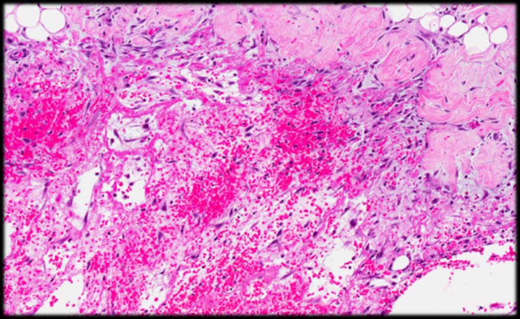
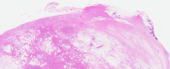
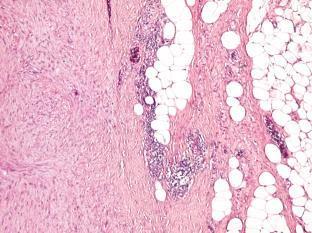
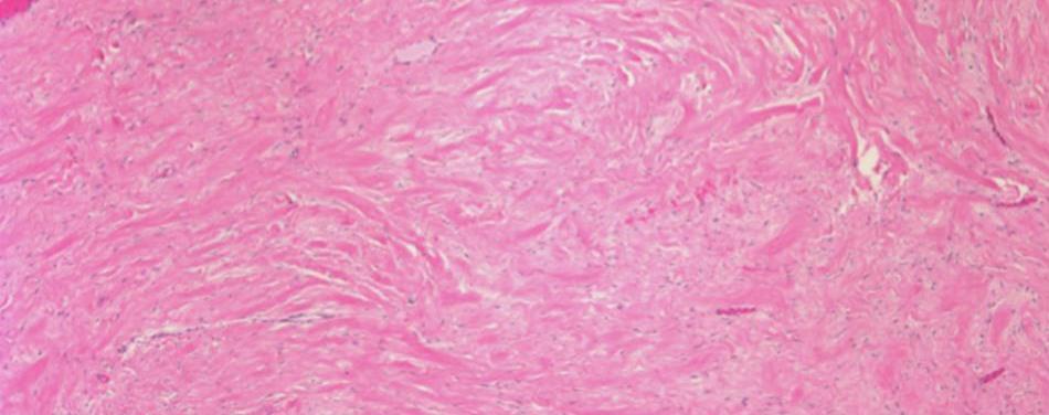
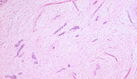
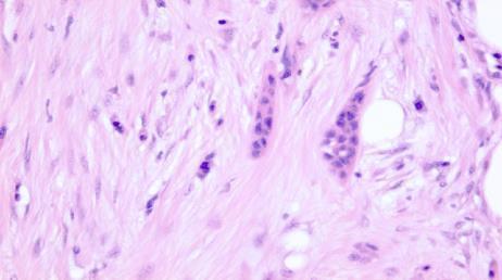
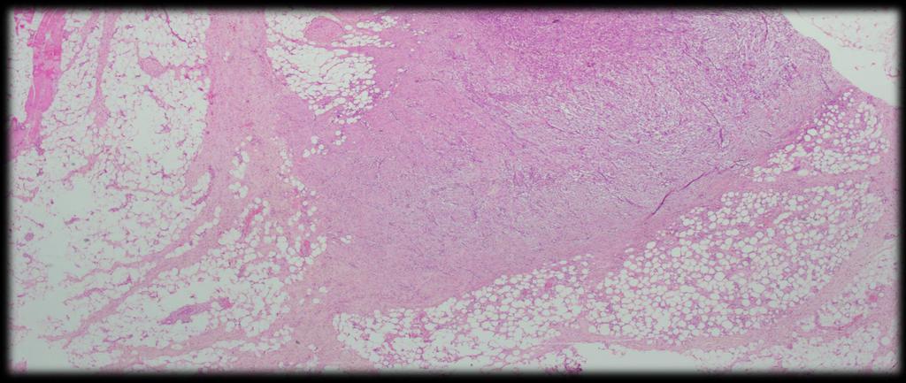
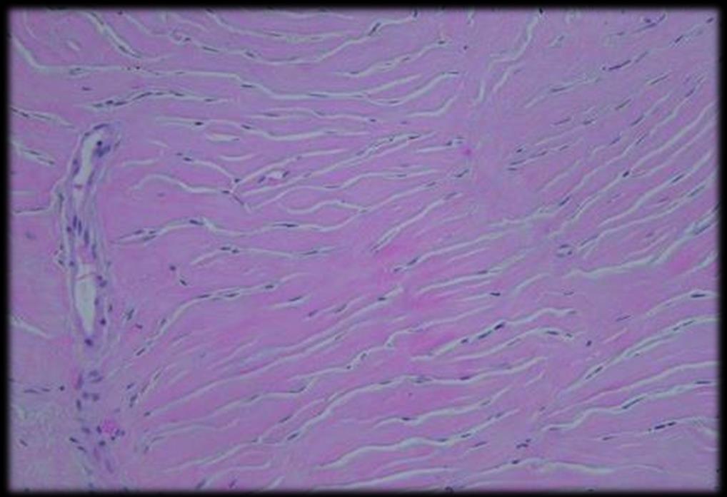
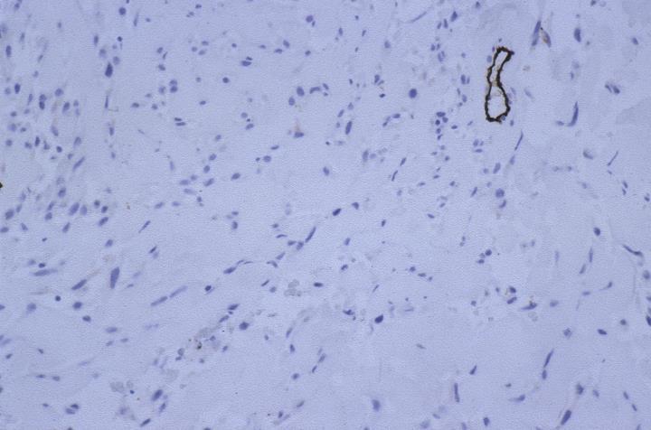
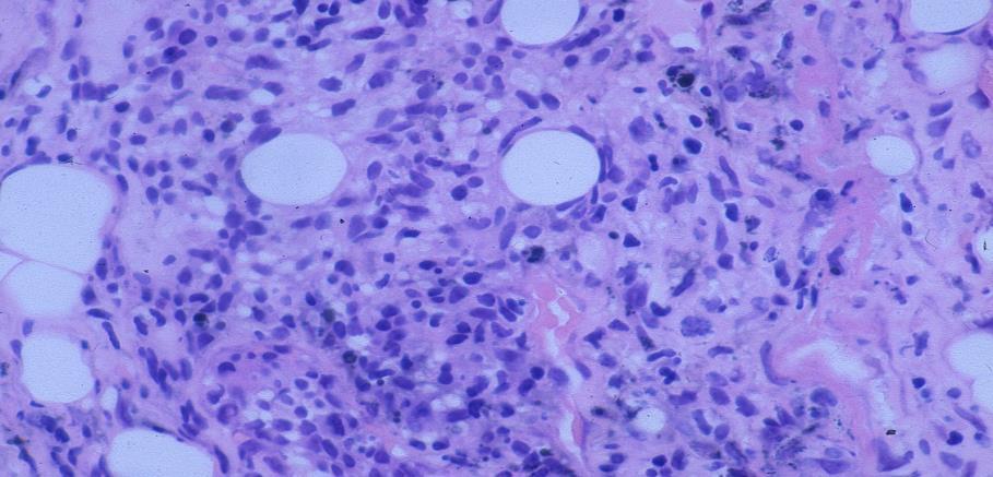
7 Fibrous scar More common than other SCLs of the breast Tissue injury (history of prior instrumentation biopsy, surgery) Histological appearance depends on the elapsed time between injury and microscopy Early: Granulation, fat necrosis, hemosiderin deposition some fibrosis, Late: fibrosis and sclerosis
8 Early
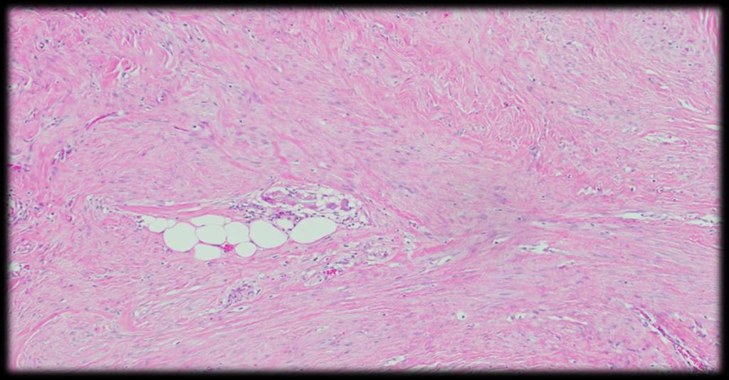
9 Late
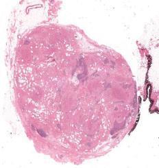
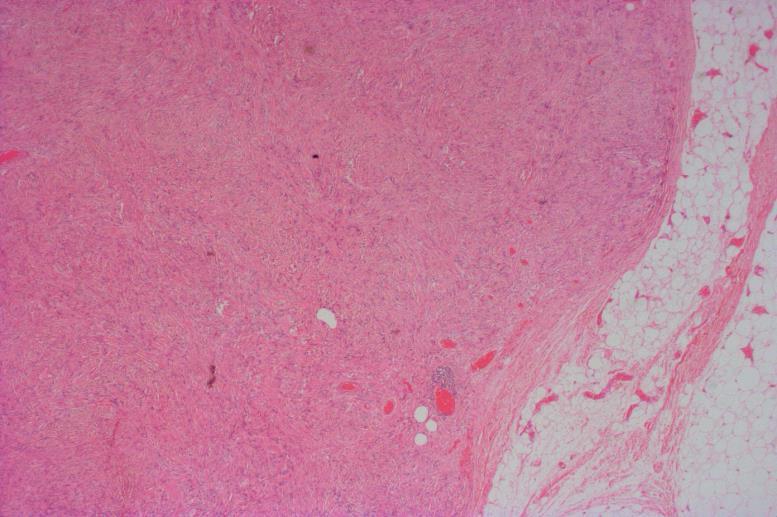
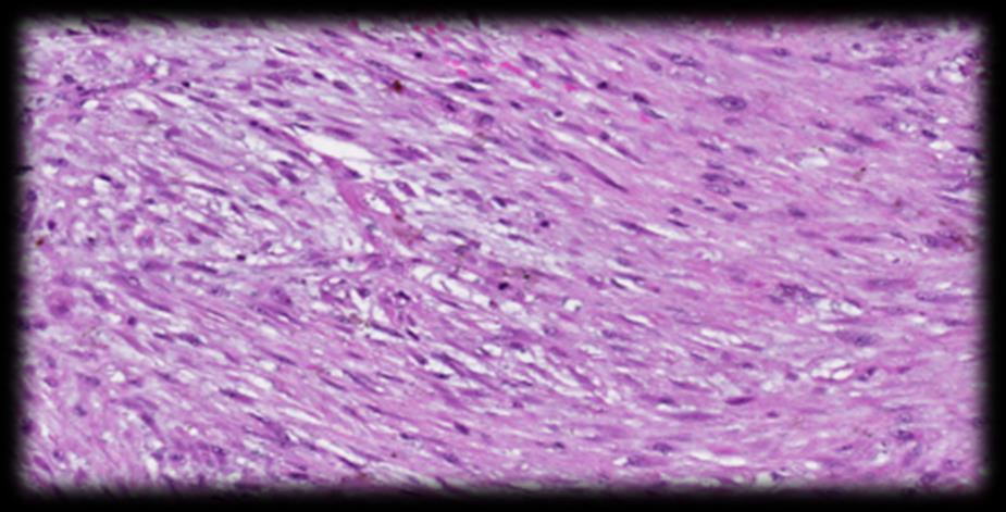
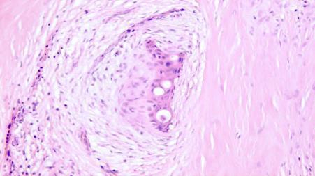
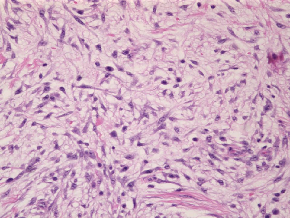
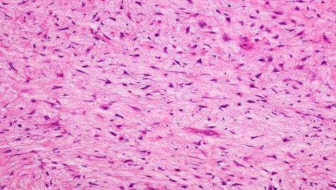
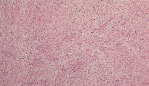
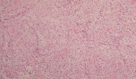
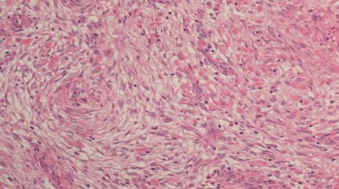
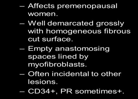
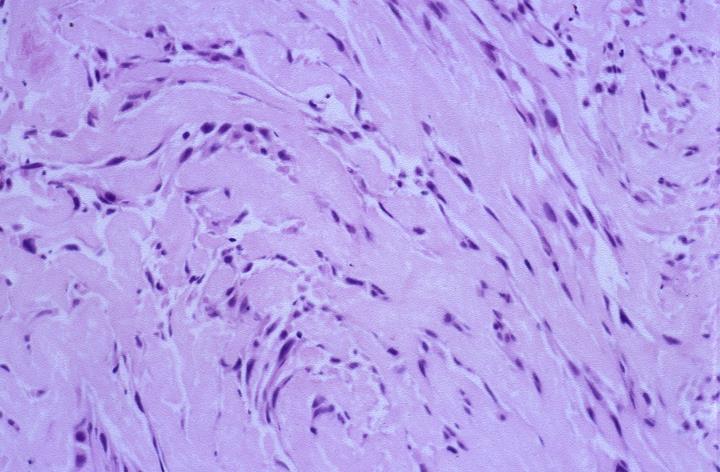
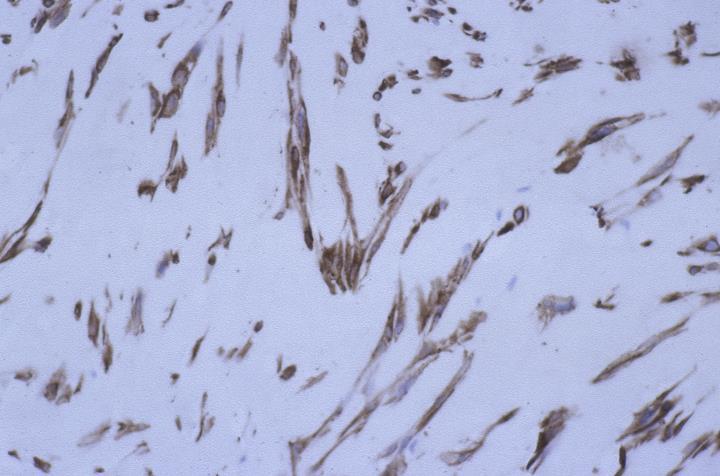
10 Myofibroblastoma Benign myofibroblastic neoplasm Genetically related to spindle cell lipoma & solitary fibrous tumour (monosomy 13q, 16q) Positive expression for CD34, ER, desmin, SMA, caldesmon, vimentin, Bcl2 and CD99. Negative: CKs, S100 and p63, RB, STAT6 Hormone receptor positivity may lead to resemblance of invasive lobular carcinoma (epithelioid variant)
11
12
13 Epithelioiid
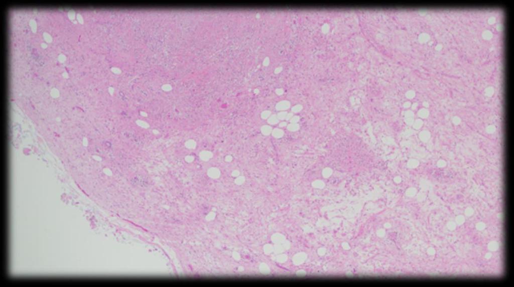
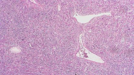
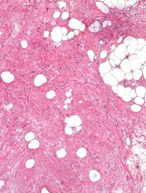
14 Myogenic differentiation Fat component Hemangiopericytomatous pattern

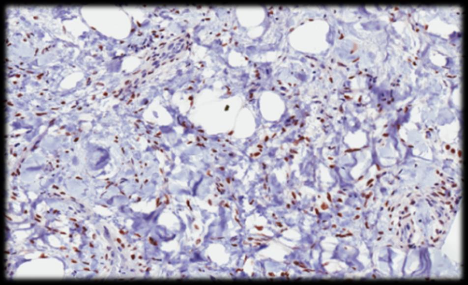
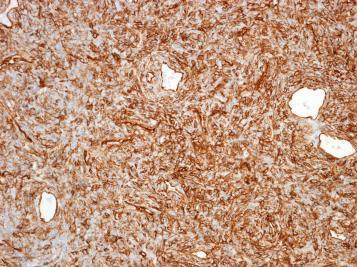
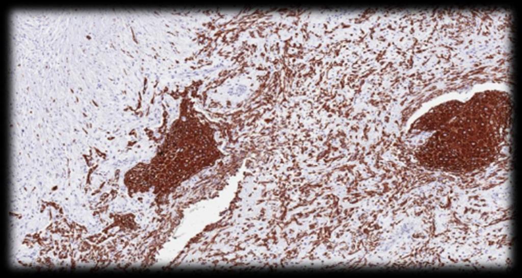
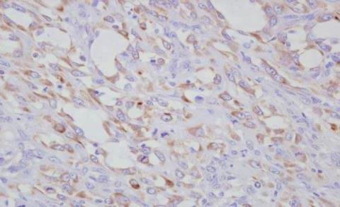
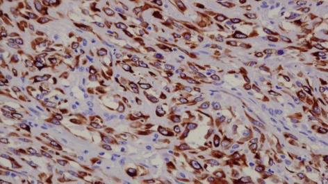
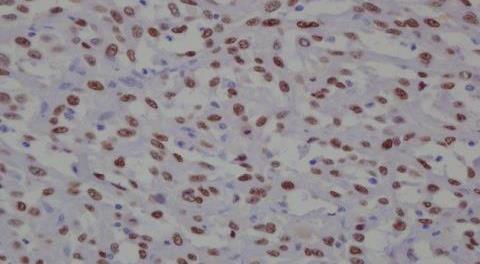
15 CD34 ACTIN desmin ER
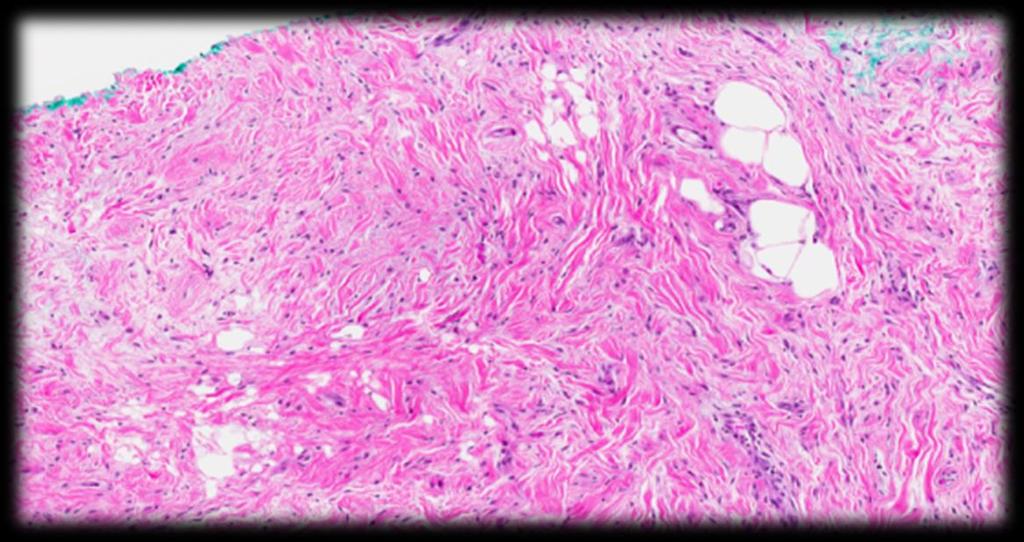
16 Pleomorphic Myofibroblastoma Histological variants Classic Collagenous Cellular Infiltrative (DD: MBC) Myxoid Epithelioid (DD: ILC)
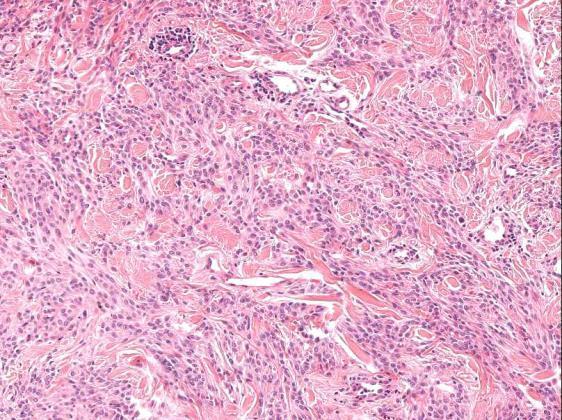
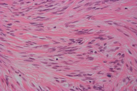
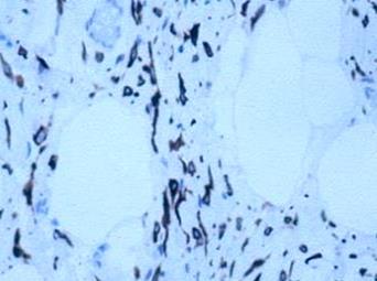
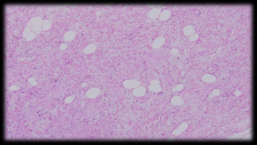
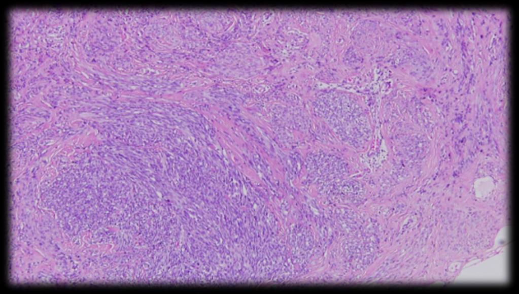
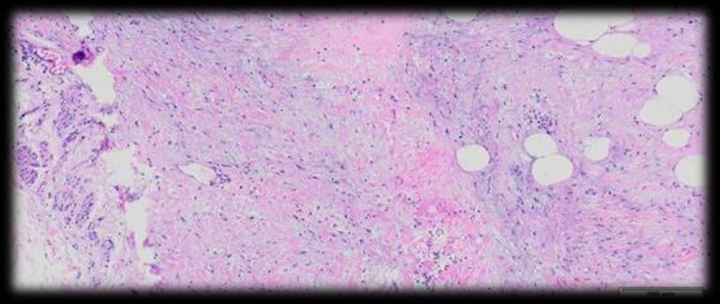
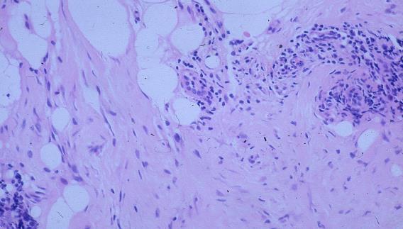
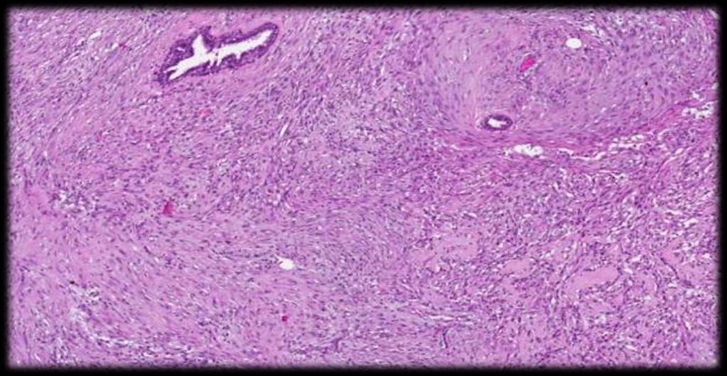
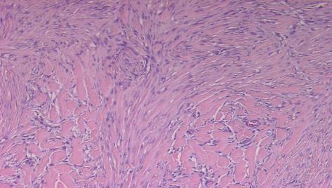
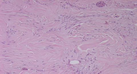
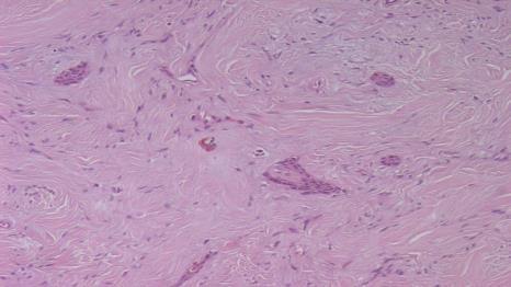
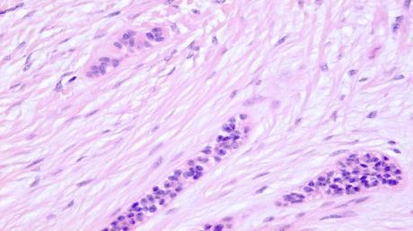
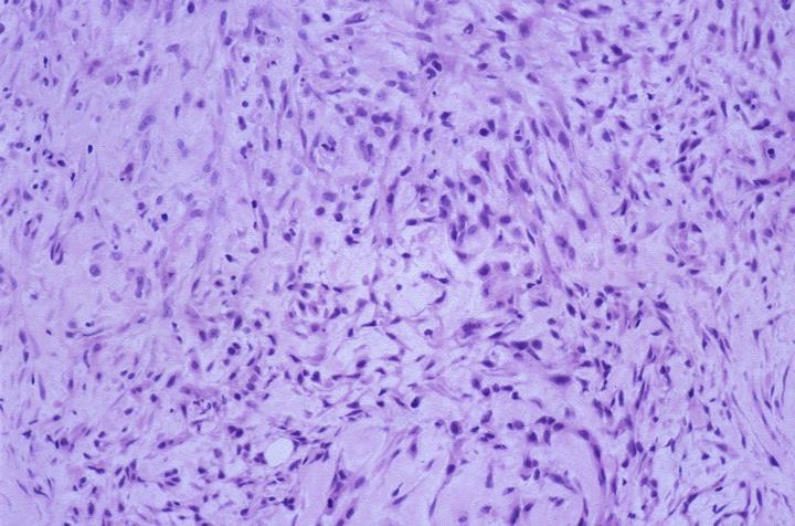
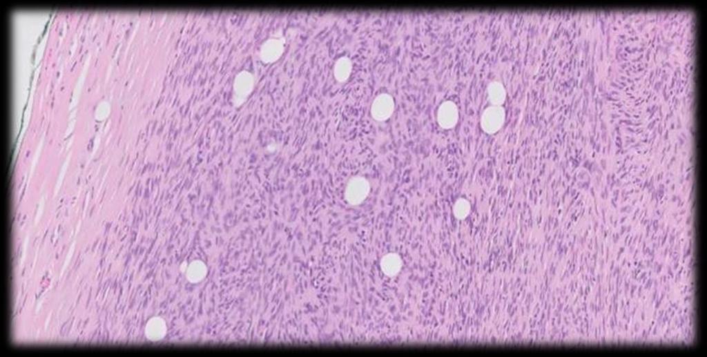
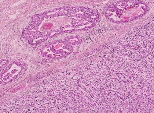
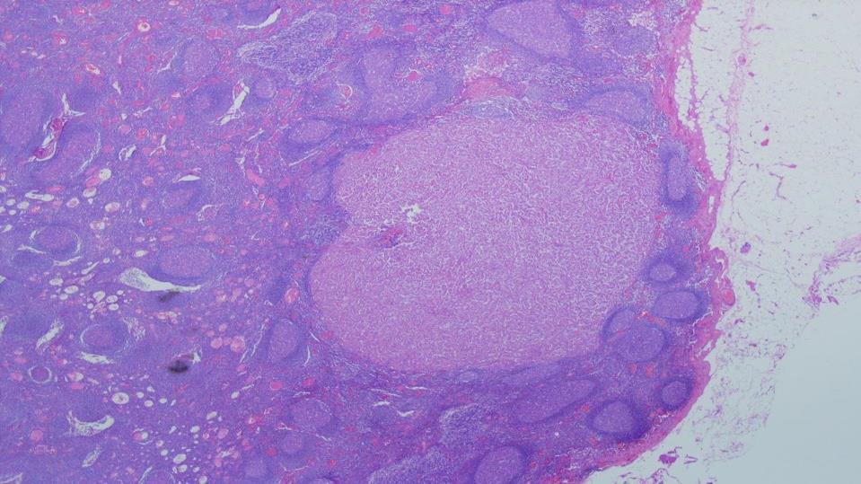
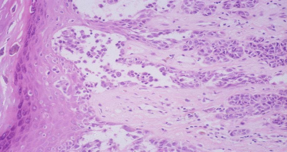
17 25-50 years old Fibromatosis Palpable mass or suspicious mammographic lesion mimics breast malignancy Can arise within the breast or chest wall Locally aggressive but non-metastasizing lesion composed of fibroblasts and myofibroblasts. May arise in patients with familial adenomatous polyposis, Gardner s syndrome
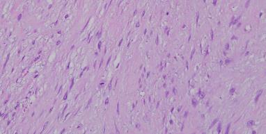
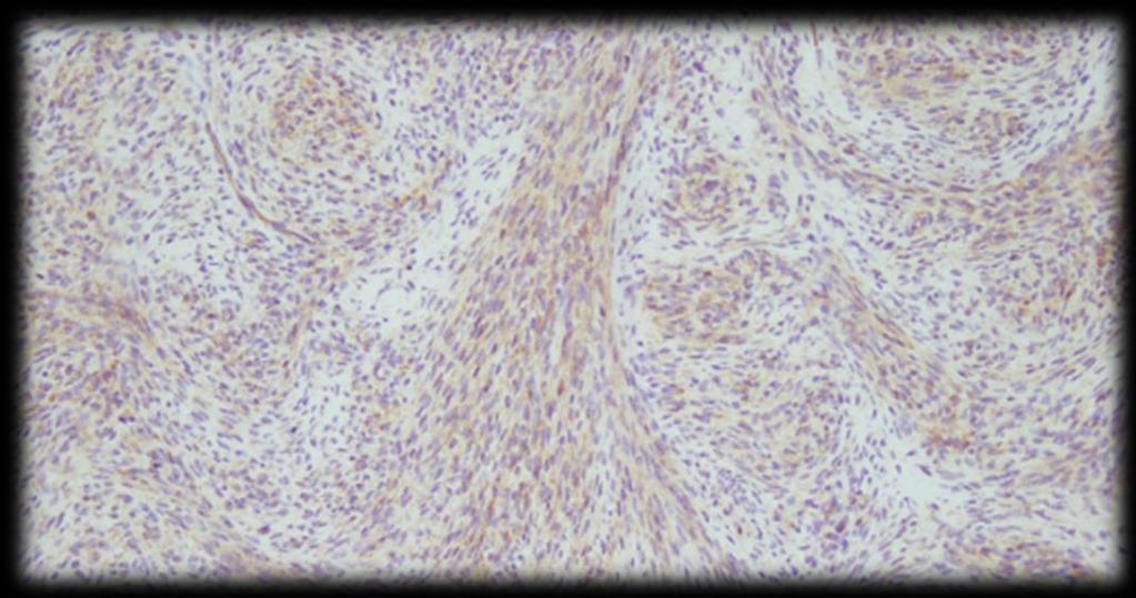
18 Fibromatosis Average size 2 to 4 cm Entraps fat and epithelial structures Variably low cellularity, Variably scant mitoses Bland tapering nuclei and little pink cytoplasm Long fascicles, thick bands of collagen Thin-walled venules, Lymphoid infiltrates perivascular and peripheral
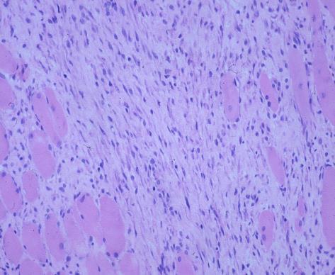
19 Infiltrative margins
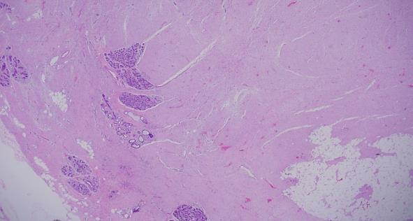
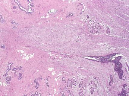
20 Entrapped parenchyma
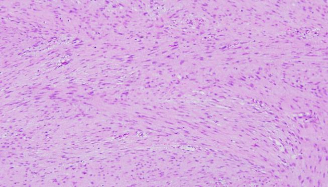
21 Fascicular pattern Spaced nuclei
22 Collagenised areas


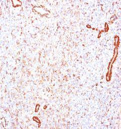
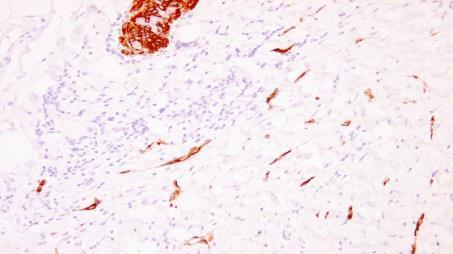
23 Immunohistochemistry Beta catenin 80% (nuclear) CD 34 negative CKs, p63, S100 and desmin- negative + CTNNB1 mutations in ~70% CD34 Beta-catenin
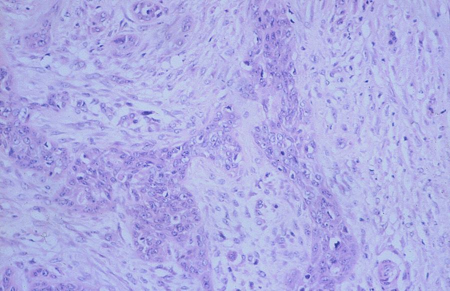
24 Low grade spindle cell metaplastic breast carcinoma (Fibromatosis-like) Radiologically spiculated or well-defined Low grade SCL resembling fibromatosis Slender/wavy nuclei. Atypia: mild/focal. Low mitotic activity Stroma usually shows regressive changes at least focally (hyalinization, myxoid degeneration and inflammatory infiltrate). +/- Scattered epithelioid/glandular elements (cords/clusters)
25 Low-grade spindle cell metaplastic breast carcinoma (Fibromatosis-like) May disclose focal squamous differentiation Expresses epithelial diff markers: keratins (HMWCKs and to less extent LMWCKs) and p63. Triple negative phenotype, CD34 negative May be associated with papilloma and radial sclerosing lesion Diagnosis change management either on core biopsy or excision: SLN biopsy. Complete excision +/- radiotherapy
26
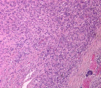
27
28
29

30 CK7 CK14 p63


31 P63 MNF116
 Local recurrence can occur Metastases are very infrequent Low genomic instability compared to other metaplastic")
32 Low grade Fibromatosis-like MBC Excellent prognosis despite being a variant of triple negative metaplastic carcinoma (LOW GRADE) Local recurrence can occur Metastases are very infrequent Low genomic instability compared to other metaplastic carcinomas

33 Fibromatosis like MBC is not high grade
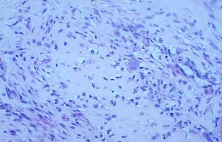
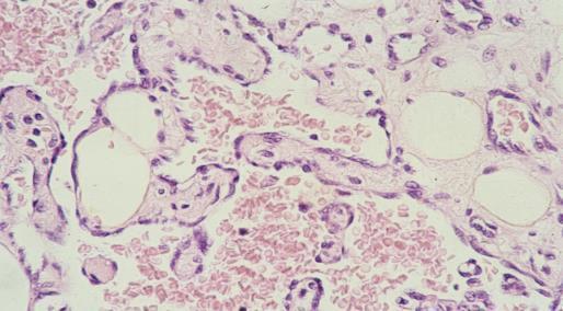
34 Translocation t(17;22) / MYH9-USP6 gene fusion Nodular fasciitis Young adults, Small (<4 cm) More often occurs in the subcutis and deeper in the chest wall. Rare in the breast parenchyma. Rapid growth, may be painful / tender Self-limited clonal proliferation of fibroblasts and myofibroblasts Spontaneous regression without recurrent potential
35 IHC: Actin Nodular fasciitis Well-defined non-encapsulated but can be infiltrative Can be cellular with mitoses (no atypical mitosis) Plump stellate myofibroblasts in small whorls or fascicles and in loose matrix (tissue culture appearance) Red cell extravasation, may be mxoid or fibrotic No significant atypia or necrosis. No entrapped parenchyma or epithelioid structures
36

37


38 RBC Tissue culture SMA
39 MBC NF
40 Pseudoangiomatous stromal hyperplasia (PASH)
41
42
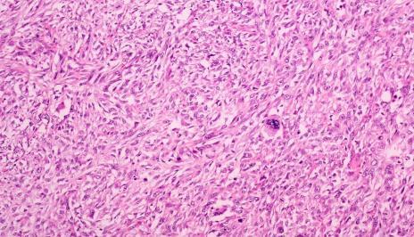
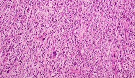
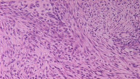
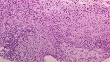
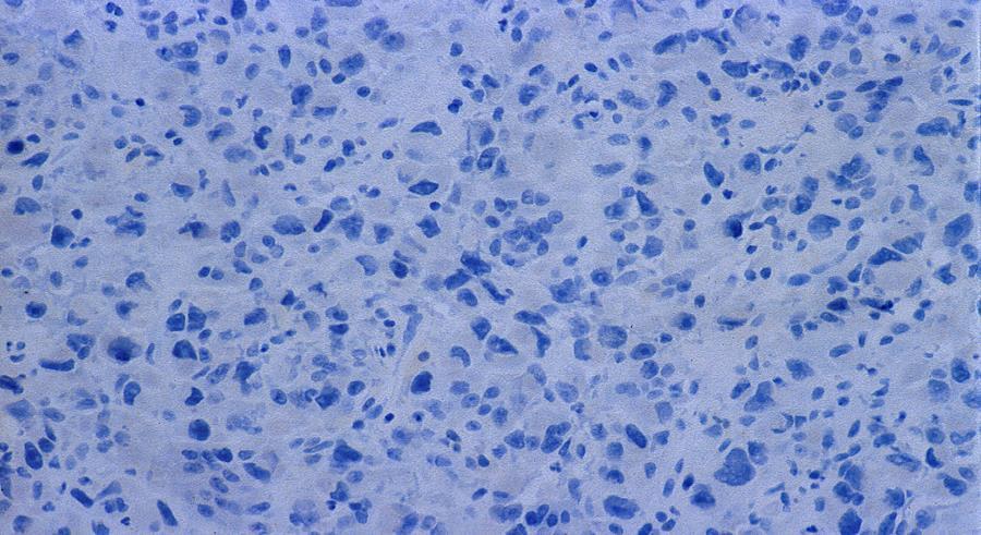
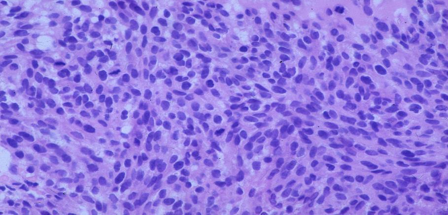
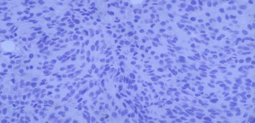
43 CD34
44 AE1/AE3


45 Benign neural tumours Schwannoma, neurofibroma Usually encountered in the skin of the breast May present as a breast lump S100+ Schwannoma


,")
46 Dermatofibrosarcoma protuberans Arises in dermal skin Sometimes may not manifest skin changes, presenting as a primary breast mass Translocation t(17;22), COL1A1 / PDGFB fusion gene
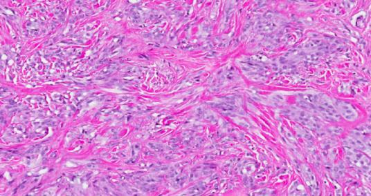
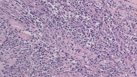
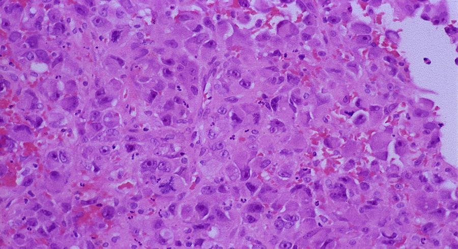
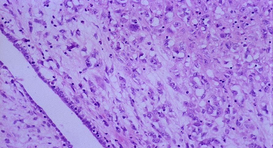
47 CD34
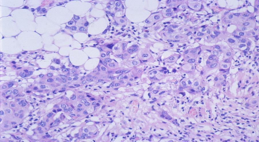
48 Benign & borderline phyllodes tumours Not usually a diagnostic problem on excision specimens as the epithelial component is visible May be a challenge on core biopsy that samples only stromal elements, or in recurrent lesions that are devoid of epithelium
49 Bland spindle cell lesion Spindle cell carcinoma: epithelioid islands, IHC+ Scar: Fat necrosis, gran tissue, haemosiderin, IHC- Fibromatosis: b-catenin Myofibroblastoma: ER, Desmin, CD34 Nodular fasciitis: Look for features, IHC (Actin) Adenomyoepithelioma and fibroepithelial lesions: Biphasic Remember other entities: Hamartoma, SFT (STAT6+), IMT (ALK1), Leiomyoma, Hemangioma, DFSP, etc
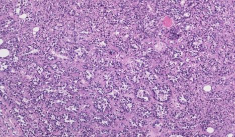
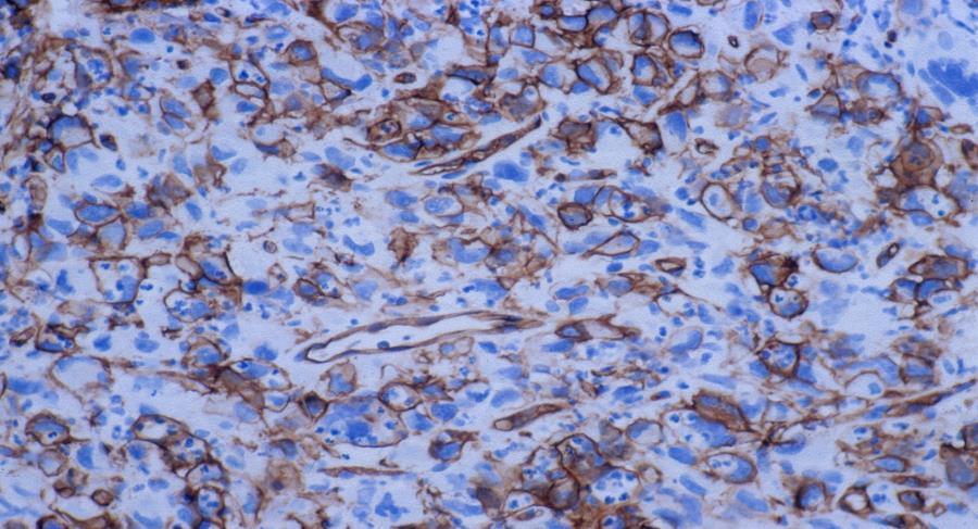
50 High grade spindle cell lesions High grade spindle cell metaplastic breast carcinoma Malignant phyllodes tumour Sarcoma Metastases /melanoma
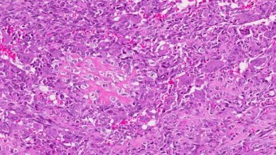
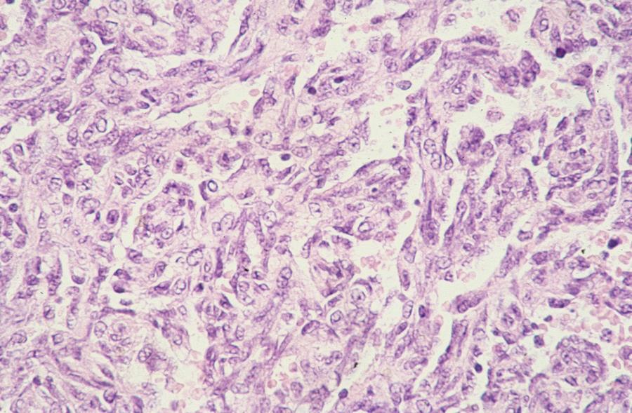
51 High-grade Spindle cell Carcinoma Sarcoma like / sarcomatoid carcinoma Pure or often coexist with high grade conventional carcinoma (glandular or squamous) Variable pattern and architecture, includes carcinomas with pleomorphic -like appearances Typically triple negative (basal-like) Poor prognosis
52
53 High-grade Spindle cell Carcinoma
54
55
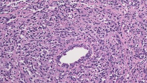
56 High-grade Spindle cell Carcinoma


57 High-grade Spindle cell Carcinoma Giant cells

58

59 AE1/3 Cam 5.2 CK7 p63

60 Diagnosis of high grade SC MBC 1- Coexistence of conventional type mammary carcinoma 2- Coexistence of an in-situ component (DCIS) And in the absence of both 3- Molecular demonstration of epithelial differentiation using IHC with epithelial specific markers (ie CKs and EMA)
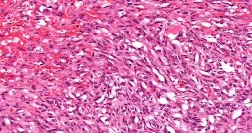
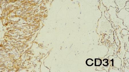
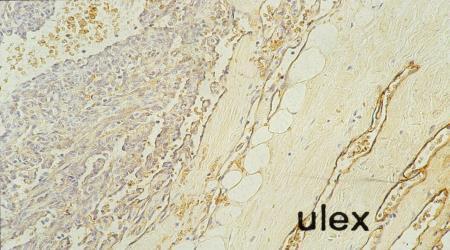
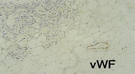
61 Diagnosis of MBC: Absence of phyllodes tumour architecture (CD34-) Thorough sampling Clinical / previous history, Review of the previous core biopsy

62 Department of Pathology, SGH
63
64 CAM5.2
65 CD34
66
 2- Primary skin (ie SCC) or chest wall tumours 3- Specific types of soft tissue tumours such as leiomyosarcoma or myofibroblastic")
67 Diagnosis of MBC: In the rare cases that lack of these feature, MBC can be considered after exclusion of: 1- Being part of a metastatic process (ie metastatic RCC) 2- Primary skin (ie SCC) or chest wall tumours 3- Specific types of soft tissue tumours such as leiomyosarcoma or myofibroblastic sarcoma
68
69
70 High grade spindle cell breast lesions Sarcoma * Angiosarcoma (commonest) * Liposarcoma (typically part of PT) * Chondrosarcoma and osteosarcoma (typically part of MBC but metastatic and primary chest wall should be excluded) * Leiomyosracoma, MPNST, myofibrobalstic & synovial sarcoma
71
72
73

74 CD34 vwf CD31 D2-40
75 Metastases to the breast Unusual histology, absence of in situ carcinoma Clinical history is crucial High index of suspicion Adjunctive immunohistochemistry
76
77
78

79 S100 AE1/AE3
80
81 Spindle cell lesions on core biopsy Keep a broad range of differential diagnoses Clinicoradiological findings are important Benign can look malignant and malignant can look benign IHC is helpful, but be aware of pitfalls and limitations Thorough sampling Final diagnosis can be rendered on excision Important to rule out metaplastic carcinoma.
82 Breast SCL: additional questions Best immunohistochemical marker? Panel approach Keratins broad spectrum, basal p63 Markers for specific diagnoses (CD34, Alk1, B-catenin, STAT6, S100, desmin, ER, SMA,..etc) Exercise caution when staining is focal, or on limited material Be aware of cross reactivities and pitfalls Consult a colleague with soft tissue pathology expertise


83 Spindle cells lesions of the breast
84 CD34 in breast lesions Any >10% of cells Myofibroblastoma, & DFSP 100% 100% Fibroadenoma 100% 100% Phyllodes Benign 100% 100% Borderline 92% 92% Malignant 84% 63% Spindle cell carcinoma 0% 0% Fibromatosis, NF & Scar 0% 0%
57th Annual HSCP Spring Symposium 4/16/2016
 An Unusual Malignant Spindle Cell Lesion to Involve the Breast Erinn Downs-Kelly, D.O. Associate Professor of Pathology University of Utah & ARUP Laboratories No disclosures Case 39 y/o female with no
Classification (1) Classification (3) Classification (2) Spindle cell lesions. Spindle cell lesions of bladder (Mills et al.
 Classification (3) Classification (2) Spindle cell lesions. Spindle cell lesions of bladder (Mills et al.") Non-epithelial tumours and nonepithelial tumour-like lesions of the bladder Dr Jonathan H Shanks The Christie NHS Foundation Trust, Manchester, UK Classification (1) Myofibroblastic proliferations and
Non-epithelial tumours and nonepithelial tumour-like lesions of the bladder Dr Jonathan H Shanks The Christie NHS Foundation Trust, Manchester, UK Classification (1) Myofibroblastic proliferations and
Papillary Lesions of the breast
 Papillary Lesions of the breast Emad Rakha Professor of Breast Pathology The University of Nottingham Papillary lesions of the breast are a heterogeneous group of disease, which are characterised by neoplastic
Evening Specialty Conference Bone and Soft Tissue Pathology. Diagnostic pitfalls in bone and soft tissue pathology
 Evening Specialty Conference Bone and Soft Tissue Pathology. Case 1 Elizabeth G Demicco, MD, PhD Mount Sinai Hospital, New York Disclosure of Relevant Financial Relationships USCAP requires that all planners
Update on Cutaneous Mesenchymal Tumors. Thomas Brenn
 Update on Cutaneous Mesenchymal Tumors Thomas Brenn Cutaneous Mesenchymal Tumours Wide morphological and biological spectrum Myofibroblastic, smooth muscle, neural, vascular, apidocytic, undifferentiated;
This case presentation reviews a challenging case of. Metaplastic Carcinomas of the Breast: Diagnostic Challenges and New Translational Insights
 Metaplastic Carcinomas of the Breast: Diagnostic Challenges and New Translational Insights Comprising less than 1% of invasive carcinomas of the breast, metaplastic carcinomas are a heterogeneous group
Desmoplastic Melanoma R/O BCC. Clinical Information. 74 y.o. man with lesion on left side of neck r/o BCC
 R/O BCC Sabine Kohler, M.D. Professor of Pathology and Dermatology Dermatopathology Service Stanford University School of Medicine Clinical Information 74 y.o. man with lesion on left side of neck r/o
3/27/2017. Disclosure of Relevant Financial Relationships
 Ophthalmic Pathology Evening Specialty Conference USCAP 2017 5 th March, 2017 Mukul K. Divatia, MD Assistant Professor Department of Pathology & Genomic Medicine Weill Cornell Medical College Houston Methodist
An Overview of Genital Stromal Tumors
 An Overview of Genital Stromal Tumors By Konstantinos Linos MD, FCAP, FASDP Bone, Soft Tissue and Dermatopathology Assistant Professor of Pathology Dartmouth-Hitchcock Medical Center Geisel School of Medicine
Lung Tumor Cases: Common Problems and Helpful Hints
 Lung Tumor Cases: Common Problems and Helpful Hints Brandon T. Larsen, MD, PhD Senior Associate Consultant Department of Laboratory Medicine and Pathology Mayo Clinic Arizona Arizona Society of Pathologists
The Relevance of Cytologic Atypia in Cutaneous Neural Tumors
 The Relevance of Cytologic Atypia in Cutaneous Neural Tumors Recent Findings - New Developments New Problems Zsolt B. Argenyi, M.D. Professor of Pathology & Dermatology Director of Dermatopathology Department
Enterprise Interest Nothing to declare
 Enterprise Interest Nothing to declare Diagnoses one would not like to miss in soft tissue pathology early in your career Marta Sbaraglia, MD Department of Pathology Hospital of Treviso University of Padua
Diseases of the breast (1 of 2)
") Diseases of the breast (1 of 2) Introduction A histology introduction Normal ducts and lobules of the breast are lined by two layers of cells a layer of luminal cells overlying a second layer of myoepithelial
Diseases of the breast (1 of 2) Introduction A histology introduction Normal ducts and lobules of the breast are lined by two layers of cells a layer of luminal cells overlying a second layer of myoepithelial
Diagnosis of Fibroepithelial and Mesenchymal Lesions on Core Needle Biopsy
 Diagnosis of Fibroepithelial and Mesenchymal Lesions on Core Needle Biopsy Emmanuel Agosto-Arroyo, MD Assistant Member Department of Anatomic Pathology 3/3/2018 Disclosure There are no conflicts of interest.
Mousa. Israa Ayed. Abdullah AlZibdeh. 0 P a g e
 1 Mousa Israa Ayed Abdullah AlZibdeh 0 P a g e Breast pathology The basic histological units of the breast are called lobules, which are composed of glandular epithelial cells (luminal cells) resting on
5/10. Pathology Soft tissue tumors. Farah Bhani. Mohammed Alorjani
 5/10 Pathology Soft tissue tumors Mohammed Alorjani Farah Bhani Slides are included in this sheet. Objectives: Soft tissue tumors 1. Describe soft tissue tumors. 2. Understand the classification of soft
Disclosures. An update on ancillary techniques in the diagnosis of soft tissue tumors. Ancillary techniques. Introduction
 Disclosures An update on ancillary techniques in the diagnosis of soft tissue tumors. I have nothing to disclose. Andrew Horvai, MD, PhD Clinical Professor, Pathology Introduction Ancillary techniques
Papillary Lesions of the Breast A Practical Approach to Diagnosis. (Arch Pathol Lab Med. 2016;140: ; doi: /arpa.
 Papillary Lesions of the Breast A Practical Approach to Diagnosis (Arch Pathol Lab Med. 2016;140:1052 1059; doi: 10.5858/arpa.2016-0219-RA) Papillary lesions of the breast Span the spectrum of benign,
GUT-C 11/30/2017. Debasmita Das, M.D. PGY-1 Danbury Hospital
 GUT-C 11/30/2017 Debasmita Das, M.D. PGY-1 Danbury Hospital CLINICAL SUMMARY 8/2017 59 year old female Presented to the ED with 1 month history of general malaise, fever and weight loss PMH: Significant
Diplomate of the American Board of Pathology in Anatomic and Clinical Pathology
 A 33-year-old male with a left lower leg mass. Contributed by Shaoxiong Chen, MD, PhD Assistant Professor Indiana University School of Medicine/ IU Health Partners Department of Pathology and Laboratory
Selected Pseudomalignant Soft Tissue Tumors of the Skin and Subcutis
 Selected Pseudomalignant Soft Tissue Tumors of the Skin and Subcutis Andrew L. Folpe, M.D. Professor of Laboratory Medicine and Pathology Mayo Clinic, Rochester, MN folpe.andrew@mayo.edu 2016 MFMER slide-1
Low-Grade Periductal Stromal of Breast: a case report
 Low-Grade Periductal Stromal of Breast: a case report Rosanna Nenna 1 Cosimo Damiano Inchingolo 1 Domenico Palmieri 2 Annalisa De Lucia 1 Giusy Elicio 1 Pina Miscioscia 1 ( 1 ) U.O.C. di Anatomia Patologica,
Abid Irshad, MD Director Breast Imaging. Medical University of South Carolina Charleston
 Abid Irshad, MD Director Breast Imaging Medical University of South Carolina Charleston Cases Financial disclosure: I or my family have no financial interest related to the material discussed in this presentation
Case year female. Routine Pap smear
 Case 1 57 year female Routine Pap smear Diagnosis? 1. Atypical glandular cells of unknown significance (AGUS) 2. Endocervical AIS 3. Endocervical adenocarcinoma 4. Endometrial adenocarcinoma 5. Adenocarcinoma
أملس عضلي غرن = Leiomyosarcoma. Leiomyosarcoma 1 / 5
 Leiomyosarcoma 1 / 5 EPIDEMIOLOGY Exact incidence is unknown, but older studies suggest that leiomyosarcomas comprise approximately 3 percent of soft-tissue sarcomas. Superficial leiomyosarcoma occurs
History A 89 year old gentleman presenting with a scalp/forehead nodule. Patient had squamous cell carcinoma 18 m at same site, excised. Outside diagn
 Case III History A 89 year old gentleman presenting with a scalp/forehead nodule. Patient had squamous cell carcinoma 18 m at same site, excised. Outside diagnoses: Squamous cell carcinoma. R/O: SCC, Melanoma,
University of Washington Radiology Review Course: Strange and Specific Diagnoses. Case #1
 University of Washington Radiology Review Course: Strange and Specific Diagnoses Katherine E. Dee, MD Seattle Breast Center Via Radiology 2014 Case #1 37 year old presents with bilateral palpable lumps.
COMMON CONSULTATION CONUNDRUMS IN BREAST PATHOLOGY
 COMMON CONSULTATION CONUNDRUMS IN BREAST PATHOLOGY SANDRA J. SHIN CHIEF OF BREAST PATHOLOGY ASOCIATE PROFESSOR OF PATHOLOGY AND LABORATORY MEDICINE NEW YORK PRESBYTERIAN HOSPITAL-WEILL CORNELL MEDICAL
Treatment options for the precancerous Atypical Breast lesions. Prof. YOUNG-JIN SUH The Catholic University of Korea
 Treatment options for the precancerous Atypical Breast lesions Prof. YOUNG-JIN SUH The Catholic University of Korea Not so benign lesions? Imaging abnormalities(10% recall) lead to diagnostic evaluation,
Mesothelioma: diagnostic challenges from a pathological perspective. Naseema Vorajee August 2016
 Mesothelioma: diagnostic challenges from a pathological perspective Naseema Vorajee August 2016 Naseema.vorajee@nhls.ac.za Pleural diseases (whether neoplastic, reactive or infective) may have similar
Disclosures. An update on ancillary techniques in the diagnosis of soft tissue tumors. Ancillary techniques. Introduction
 Disclosures An update on ancillary techniques in the diagnosis of soft tissue tumors. I have nothing to disclose. Andrew Horvai, MD, PhD Clinical Professor, Pathology Introduction Ancillary techniques
CURRICULUM FOR THE BREAST PATHOLOGY ROTATION UNIVERSITY OF FLORIDA DEPARTMENT OF PATHOLOGY
 CURRICULUM FOR THE BREAST PATHOLOGY ROTATION UNIVERSITY OF FLORIDA DEPARTMENT OF PATHOLOGY JULY, 2003 The following is a conceptual curriculum and set of guidelines for Pathology Residents on the Breast
Slide seminar. Asist. Prof. Jože Pižem, MD, PhD Institute of Pathology Medical Faculty, University of Ljubljana
 Slide seminar Asist. Prof. Jože Pižem, MD, PhD Institute of Pathology Medical Faculty, University of Ljubljana Case 5 A 57-year-old man with a dermal/subcutaneous lesion on the scalp, which was interpreted
3/27/2017. Disclosure of Relevant Financial Relationships. Papilloma???
 Management of Papillary Lesions Diagnosed at Rad Path Concordant Core Biopsy (CNB) Disclosure of Relevant Financial Relationships USCAP requires that all planners (Education Committee) in a position to
1/10/2018. Soft Tissue Tumors Showing Melanocytic Differentiation. Overview. Desmoplastic/ Spindle Cell Melanoma
 2016 MFMER slide-1 2016 MFMER slide-2 2016 MFMER slide-3 Soft Tissue Tumors Showing Melanocytic Differentiation Andrew L. Folpe, M.D. Professor of Laboratory Medicine and Pathology Mayo Clinic, Rochester,
CASE year old male with a PET avid nodule in the left adrenal gland
 CASE 1 55 year old male with a PET avid nodule in the left adrenal gland Case 1 Adrenal gland parenchyma partly replaced by a spindle cell tumour with mild nuclear pleomorphism Atypical mitoses present
Breast pathology. 2nd Department of Pathology Semmelweis University
 Breast pathology 2nd Department of Pathology Semmelweis University Breast pathology - Summary - Benign lesions - Acute mastitis - Plasma cell mastitis / duct ectasia - Fat necrosis - Fibrocystic change/
A Practical Approach to the Evaluation of Fibroepithelial Lesions. Edi Brogi MD PhD Attending Pathologist Director of Breast Pathology
 A Practical Approach to the Evaluation of Fibroepithelial Lesions Edi Brogi MD PhD Attending Pathologist Director of Breast Pathology Overview Fibroadenomas (FAs) Phyllodes Tumors (PTs) Morphology and
A 25 year old female with a palpable mass in the right lower quadrant of her abdomen
 May 2016 A 25 year old female with a palpable mass in the right lower quadrant of her abdomen Contributed by: Paul Ndekwe, MD, Resident Physician, Indiana University School of Department of Pathology and
Atypical Palisaded Myofibroblastoma of Lymph Node: Report of a rare case.
 ISPUB.COM The Internet Journal of Pathology Volume 10 Number 1 Atypical Palisaded Myofibroblastoma of Lymph Node: Report of a rare case. V Kinnera, R Nandyala, M Yootla, K Mandyam Citation V Kinnera, R
SOFT TISSUE TUMOR PATHOLOGY: AN UPDATE
 SOFT TISSUE TUMOR PATHOLOGY: AN UPDATE Jason L. Hornick, MD, PhD July 18, 2013 Department of Pathology Brigham and Women s Hospital Harvard Medical School Boston, MA, USA I have no disclosures. New Soft
04/10/2018. Intraductal Papillary Neoplasms Of Breast INTRADUCTAL PAPILLOMA
 Intraductal Papillary Neoplasms Of Breast Savitri Krishnamurthy MD Professor of Pathology Deputy Division Head The University of Texas MD Anderson Cancer Center 25 th Annual Seminar in Pathology Pittsburgh,
Gastrointestinal stromal tumor
 Gastrointestinal stromal tumor 영남의대병리학교실 최준혁 Classification of gastrointestinal mesenchymal tumor Gastrointestinal stromal tumor(gist) Smooth muscle tumors : leiomyoma, leiomyosarcoma Neurogenic tumors
Immunohistochemistry in Bone and Soft Tissue Tumors. Sahar Rassi Zankoul, MD
 Immunohistochemistry in Bone and Soft Tissue Tumors Sahar Rassi Zankoul, MD Introduction Bone tumors represent a wide variety of tumors of various origins and malignant potentials. These different tumor
Benign Mimics of Malignancy in Breast Pathology
 Arthur Purdy Stout Society of Surgical Pathologists Companion Meeting Benign Mimics of Malignancy in Breast Pathology Stuart J. Schnitt, M.D. Beth Israel Deaconess Medical Center and Harvard Medical School,
CUTANEOUS PSEUDONEOPLASTIC MESENCHYMAL LESIONS ( PSEUDOSARCOMAS )
") CUTANEOUS PSEUDONEOPLASTIC MESENCHYMAL LESIONS ( PSEUDOSARCOMAS ) Mark R. Wick, M.D. University of Virginia Health System Charlottesville, VA www.markwickmd.com; mrw9c@virginia.edu PSEUDONEOPLASMS & PSEUDOTUMORS
CUTANEOUS PSEUDONEOPLASTIC MESENCHYMAL LESIONS ( PSEUDOSARCOMAS ) Mark R. Wick, M.D. University of Virginia Health System Charlottesville, VA www.markwickmd.com; mrw9c@virginia.edu PSEUDONEOPLASMS & PSEUDOTUMORS
ACCME/Disclosures ALK FUSION-POSITIVE MESENCHYMAL TUMORS. Tumor types with ALK rearrangements. Anaplastic Lymphoma Kinase. Jason L.
 Companion Meeting of the International Society of Bone and Soft Tissue Pathology The Evolving Concept of Mesenchymal Tumors ALK FUSION-POSITIVE MESENCHYMAL TUMORS Jason L. Hornick, MD, PhD March 13, 2016
A neoplasm is defined as "an abnormal tissue proliferation, which exceeds that of adjacent normal tissue. This proliferation continues even after
 NEOPLASIA Neoplasia is a very important topic in pathology because neoplasms are both common and serious diseases. A neoplasm literally means a new growth, and this term is used interchangeably with a
Review of the AP Part II Practical Examination. Dr David Clift Co Chief Examiner
 Review of the AP Part II Practical Examination Dr David Clift Co Chief Examiner General Remarks The part II practical examination involved 15 cases which were presented with sufficient clinical data to
OUTLINE FIBROADENOMA FIBROADENOMA. FIBROEPITHELIAL LESIONS OF THE BREAST UCSF Current Issues in Anatomic Pathology 2015 FIBROADENOMA PHYLLODES TUMOR
 OUTLINE FIBROADENOMA FIBROEPITHELIAL LESIONS OF THE BREAST UCSF Current Issues in Anatomic Pathology 2015 Gregor Krings, MD PhD Assistant Professor PHYLLODES TUMOR DIFFERENTIAL DIAGNOSIS CELLULAR FIBROEPITHELIAL
Division of Pathology
 Case 38 Adult woman with a 35mm right breast lump at the 10 o clock position. Excision performed. (Case contributed by Dr Mihir Gudi, KKH) Division of Pathology Merlion, One Fullerton Singapore Diagnosis
Gross appearance of nodular hyperplasia in material obtained from suprapubic prostatectomy. Note the multinodular appearance and the admixture of
 Tiền liệt tuyến Tiền liệt tuyến Gross appearance of nodular hyperplasia in material obtained from suprapubic prostatectomy. Note the multinodular appearance and the admixture of solid and microcystic areas.
Overview of Pathology Evaluation of Breast Lesions and Quality Assurance
 Overview of Pathology Evaluation of Breast Lesions and Quality Assurance 2 Michael O. Idowu, Jaime A. Singh, and Margaret M. Grimes Masses/Densities/Distortions: General Considerations Radiologic evaluation
Papillary Lesions of the Breast
 Papillary Lesions of the Breast Laura C. Collins, M.D. Associate Professor of Pathology Associate Director, Division of Anatomic Pathology Beth Israel Deaconess Medical Center and Harvard Medical School
ACCME/Disclosures. Everything is spindle - how far can we go with limited FNA material? Everything is spindle how far can we go? Everything is spindle
 ACCME/Disclosures The USCAP requires that anyone in a position to influence or control the content of CME disclose any relevant financial relationship WITH COMMERCIAL INTERESTS which they or their spouse/partner
An approach to the diagnosis of spindle cell lesions of the breast
 Histopathology 2016, 68, 33 44. DOI: 10.1111/his.12865 REVIEW An approach to the diagnosis of spindle cell lesions of the breast Emad A Rakha, Mohammed A Aleskandarany, Andrew H S Lee & Ian O Ellis Department
Financial disclosures
 Mesenchymal Neoplasms with Melanocytic Differentiation By Konstantinos Linos MD, FCAP, FASDP Bone, Soft Tissue and Dermatopathology Assistant Professor of Pathology Dartmouth-Hitchcock Medical Center Geisel
Disclosures. Parathyroid Pathology. Objectives. The normal parathyroid 11/10/2012
 Disclosures Parathyroid Pathology I have nothing to disclose Annemieke van Zante MD/PhD Assistant Professor of Clinical Pathology Associate Chief of Cytopathology Objectives 1. Review the pathologic features
RSNA, /radiol Appendix E1. Methods
 RSNA, 2016 10.1148/radiol.2016151097 Appendix E1 Methods US and Near-infrared Data Acquisition Four optical wavelengths (740 nm, 780 nm, 808 nm, and 830 nm) were used to sequentially deliver the light
MALIGNANT PHYLLODES TUMOUR OF BREAST WITH LIPOSARCOMATOUS DIFFERENTIATION PRESENTING CLINICALLY AS FIBROADENOMA REPORT OF A RARE CASE
 IJCRR Vol 06 issue 10 Section: Healthcare Category: Case Report Received on: 13/04/14 Revised on: 28/04/14 Accepted on: 13/05/14 MALIGNANT PHYLLODES TUMOUR OF BREAST WITH LIPOSARCOMATOUS DIFFERENTIATION
Basement membrane in lobule.
 Bahram Memar, MD Basement membrane in lobule. Normal lobule-luteal phase Normal lobule-follicular phase Lactating breast Greater than 95% are adenocarcinomas in situ carcinomas and invasive carcinomas.
Financial disclosures
 An update on immunohistochemical markers in mesenchymal neoplasms By Konstantinos Linos MD, FCAP, FASDP Assistant Professor of Pathology Geisel School of Medicine at Dartmouth Dartmouth-Hitchcock Medical
21/07/2017. Hobnail endothelial cells are not the same as epithelioid endothelial cells
 UPDATE IN CUTANEOUS VASCULAR S DERMATOPATHOLOGY SESSION BELFAST PATHOLOGY JUNE 21/2017 Dr E Calonje St John s Institute of Dermatology, London, United Kingdom THE FAMILY OF VASCULAR S WITH EPITHELIOID
SESSION 1: GENERAL (BASIC) PATHOLOGY CONCEPTS Thursday, October 16, :30am - 11:30am FACULTY COPY
 PATHOLOGY CONCEPTS Thursday, October 16, :30am - 11:30am FACULTY COPY") SESSION 1: GENERAL (BASIC) PATHOLOGY CONCEPTS Thursday, October 16, 2008 9:30am - 11:30am FACULTY COPY GOAL: Describe the basic morphologic (structural) changes which occur in various pathologic conditions.
SESSION 1: GENERAL (BASIC) PATHOLOGY CONCEPTS Thursday, October 16, 2008 9:30am - 11:30am FACULTY COPY GOAL: Describe the basic morphologic (structural) changes which occur in various pathologic conditions.
Biopsy Interpretation of Spindle cell proliferations of the Serosa
 Biopsy Interpretation of Spindle cell proliferations of the Serosa Richard Attanoos, Cardiff. U.K. Disclosure of Relevant Financial Relationships USCAP requires that all planners (Education Committee)
SMOOTH MUSCLE TUMOURS
 SMOOTH MUSCLE TUMOURS NORMAL SMOOTH MUSCLE Cytology Immunohistochemistry Ultrastructure Masson Trichrome Smooth Muscle Ultrastructure Many myofilaments running parallel to the long axis of the smooth
Contents Part I Introduction 1 General Description 2 Natural History: Importance of Size, Site, Histopathology
 Contents Part I Introduction 1 General Description... 3 1.1 Introduction... 3 1.2 Incidence and Prevalence... 5 1.3 Predisposing and Genetic Factors... 8 References... 16 2 Natural History: Importance
Lesion Imaging Characteristics Mass, Favoring Benign Circumscribed Margins Intramammary Lymph Node
 Lesion Imaging Characteristics Mass, Favoring Benign Circumscribed Margins Intramammary Lymph Node Oil Cyst Mass, Intermediate Concern Microlobulated Margins Obscured Margins Mass, Favoring Malignant Indistinct
Special slide seminar
 Special slide seminar Tomáš Rozkoš The Fingerland Department of Pathology Charles University Medical Faculty and Faculty Hospital in Hradec Králové Czech Republic Case history, 33 years old resistance
Malignant Peripheral Nerve Sheath Tumor
 C H A P T E R 120 Malignant Peripheral Nerve Sheath Tumor Currently, malignant peripheral nerve sheath tumor (MPNST) is the most commonly used generic name for the neoplasms known in the past as neurosarcoma,
4/12/2018. MUSC Pathology Symposium Kiawah Island April 18, Jesse K. McKenney, MD
 MUSC Pathology Symposium Kiawah Island April 18, 2018 Jesse K. McKenney, MD 1 Urothelial Carcinoma with Alternative Differentiation 2 Urothelial Carcinoma with Alternative Differentiation Recognition as
Breast Pathology. Breast Development
 Breast Pathology Lecturer: Hanina Hibshoosh, M.D. Reading: Kumar, Cotran, Robbins, Basic Pathology, 6th Edition, pages 623-635 Breast Development 5th week - thickening of the epidermis - milk line 5th
Salivary Glands 3/7/2017
 Salivary Glands 3/7/2017 Goals and objectives Focus on the entities unique to H&N Common board type facts Information for your future practice Salivary Glands Salivary Glands Major gland. Paratid. Submandibular.
Klinisch belang van chromosomale translocatie detectie in sarcomen
 Translocations in sarcomas Klinisch belang van chromosomale translocatie detectie in sarcomen Judith V.M.G. Bovée, M.D., Ph.D. Department of Pathology Leiden University Medical Center RNA binding DNA binding
What is New in the 2015 WHO Lung Cancer Classification? Zhaolin Xu, MD, FRCPC, FCAP
 What is New in the 2015 WHO Lung Cancer Classification? Zhaolin Xu, MD, FRCPC, FCAP Professor, Dept of Pathology, Dalhousie University, Canada Pulmonary Pathologist and Cytopathologist, QEII HSC Senior
Newer soft tissue entities
 Newer soft tissue entities Examples among fibroblastic tumors Turku, May 6, 2010 Markku Miettinen, M.D. AFIP, Washington, DC Fibroblastic neoplasms Solitary fibrous tumor /Hemangiopericytoma Low-grade
Pathology of Sarcoma ELEANOR CHEN, MD, PHD, ASSISTANT PROFESSOR DEPARTMENT OF PATHOLOGY UNIVERSITY OF WASHINGTON
 Pathology of Sarcoma ELEANOR CHEN, MD, PHD, ASSISTANT PROFESSOR DEPARTMENT OF PATHOLOGY UNIVERSITY OF WASHINGTON Presentation outline Background and epidemiology of sarcomas Sarcoma classification Sarcoma
IBCM 2, April 2009, Sarajevo, Bosnia and Herzegovina
 Preoperative diagnosis and treatment planning in breast cancer The pathologist s perspective L. Mazzucchelli Istituto Cantonale di Patologia Locarno, Switzerland IBCM 2, 23-25 April 2009, Sarajevo, Bosnia
I have nothing to disclose
 A 47 year old female with multiple lung nodules Disclosure of Relevant Financial Relationships Tamar Giorgadze, MD, PhD Professor of Pathology Medical College of Wisconsin Milwaukee, Wisconsin USCAP requires
University Journal of Pre and Para Clinical Sciences
 ISSN 2455 2879 Volume 2 Issue 1 2016 Metaplastic carcinoma breast a rare case report Abstract : Metaplastic carcinoma of the breast is a rare malignancy with two distinct cell lines described as a breast
Mesothelioma Pathobasic. Lukas Bubendorf Pathology
 Mesothelioma Pathobasic Lukas Bubendorf Pathology Mechanisms of Asbestos Carcinogenesis in Mesothelioma Asprin High-mobility group protein B1 master switch HMGB1 Initiation/ perpetuation of inflamm. response
Mody. AIS vs. Invasive Adenocarcinoma of the Cervix
 Common Problems in Gynecologic Pathology Michael T. Deavers, M.D. Houston Methodist Hospital, Houston, Texas Common Problems in Gynecologic Pathology Adenocarcinoma in-situ (AIS) of the Cervix vs. Invasive
Keywords solitary fibrous tumor, dedifferentiation, dedifferentiated solitary fibrous tumor, STAT6, GRIA2, cytokeratin, rhabdomyosarcomatous
 758452IJSXXX10.1177/1066896918758452International Journal of Surgical PathologyCreytens et al research-article2018 Pitfalls in Pathology Multifocal Cytokeratin Expression in a Dedifferentiated Solitary
Disclosure. Relevant Financial Relationship(s) None. Off Label Usage None MFMER slide-1
 None. Off Label Usage None MFMER slide-1") Disclosure Relevant Financial Relationship(s) None Off Label Usage None 2013 MFMER slide-1 Case Presentation A 43 year old male, with partial nephrectomy for a right kidney mass 2013 MFMER slide-2 2013
Disclosure Relevant Financial Relationship(s) None Off Label Usage None 2013 MFMER slide-1 Case Presentation A 43 year old male, with partial nephrectomy for a right kidney mass 2013 MFMER slide-2 2013
IN THE NAME OF GOD Dr. Kheirandish Oral and maxillofacial pathology
 IN THE NAME OF GOD Dr. Kheirandish Oral and maxillofacial pathology ORAL FOCAL MUCINOSIS Uncommon Tumorlike Cutaneous myxoid cyst Overproduction of hyaluronic acid by firoblasts Young adults Female Gingiva
Triple Negative Breast Cancer
 Triple Negative Breast Cancer Prof. Dr. Pornchai O-charoenrat Division of Head-Neck & Breast Surgery Department of Surgery Faculty of Medicine Siriraj Hospital Breast Cancer Classification Traditional
EQA Circulation 43 Educational Cases
 EQA Circulation 43 Educational Cases E1-E2 Monica Agarwal Monklands Hospital E1 38 yrs male Submandibular gland tumour E1 Formal excision following diagnosis of poorly differentiated carcinoma on core
3/24/2017 DENDRITIC CELL NEOPLASMS: HISTOLOGY, IMMUNOHISTOCHEMISTRY, AND MOLECULAR GENETICS. Disclosure of Relevant Financial Relationships
 DENDRITIC CELL NEOPLASMS: HISTOLOGY, IMMUNOHISTOCHEMISTRY, AND MOLECULAR GENETICS Jason L. Hornick, M.D., Ph.D. Director of Surgical Pathology and Immunohistochemistry Brigham and Women s Hospital Professor
Case 1 10/2/17. Myxoid Soft Tissue Tumors & Tumor-like Lesions. Myxofibro- or Fibromyxo-?: Myxoid Soft Tissue Tumours We Are All Mixed Up About
 Myxoid Soft Tissue Tumors & Tumor-like Lesions Myxofibro- or Fibromyxo-?: Myxoid Soft Tissue Tumours We Are All Mixed Up About Rajiv M. Patel, M.D. RCPA NZ ASM 2017 (4:15-5:00pm, Saturday, 23-09-17) Heterogenous
Diagnostic problems in uterine smooth muscle tumors
 Diagnostic problems in uterine smooth muscle tumors Marina Kos Ljudevit Jurak Clinical Department of Pathology, Clinical Hospital Center Sestre milosrdnice, Zagreb Institute of Pathology, University of
Update in Salivary Gland Pathology. Benjamin L. Witt University of Utah/ARUP Laboratories February 9, 2016
 Update in Salivary Gland Pathology Benjamin L. Witt University of Utah/ARUP Laboratories February 9, 2016 Objectives Review the different appearances of a selection of salivary gland tumor types Establish
Mimics of Sarcoma. I have no conflict of interest to declare
 Maastricht 2018 Mimics of Sarcoma Cyril Fisher MA MD DSc FRCPath Consultant Pathologist, Royal Orthopaedic Hospital, Birmingham, UK Emeritus Professor of Tumour Pathology Institute of Cancer Research,
Slide Seminar Spanish Society of Pathology
 Slide Seminar Spanish Society of Pathology John R. Goldblum, M.D. Chairman, Department of Anatomic Pathology Cleveland Clinic Professor of Pathology Cleveland Clinic Lerner College of Medicine 1921 Original
ESS: Pathologic Insights
 GEIS XVI INTERNATIONAL SYMPOSIUM Seville 4th October 2018 ESS: Pathologic Insights Sílvia Bagué The Royal Marsden Hospital London (United Kingdom) I have no conflicts of interest Endometrial stromal sarcoma
Solitary Fibrous Tumor of the Kidney with Massive Retroperitoneal Recurrence. A Case Presentation
 246) Prague Medical Report / Vol. 113 (2012) No. 3, p. 246 250 Solitary Fibrous Tumor of the Kidney with Massive Retroperitoneal Recurrence. A Case Presentation Sfoungaristos S., Papatheodorou M., Kavouras
Pathology Mystery and Surprise
 Pathology Mystery and Surprise Tim Smith, MD Director Anatomic Pathology Medical University of South Carolina Disclosures No conflicts to declare Some problem cases Kidney tumor Scalp tumor Bladder tumor
Neoplasia 2018 Lecture 2. Dr Heyam Awad MD, FRCPath
 Neoplasia 2018 Lecture 2 Dr Heyam Awad MD, FRCPath ILOS 1. List the differences between benign and malignant tumors. 2. Recognize the histological features of malignancy. 3. Define dysplasia and understand
Objectives. Salivary Gland FNA: The Milan System. Role of Salivary Gland FNA 04/26/2018
 Salivary Gland FNA: The Milan System Dr. Jennifer Brainard Section Head Cytopathology Cleveland Clinic Objectives Introduce the Milan System for reporting salivary gland cytopathology Define cytologic
Diagnostic Value of Immunohistochemistry in Soft Tissue Tumors
 Original Article DOI: 10.21276/APALM.1637 Diagnostic Value of Immunohistochemistry in Soft Tissue Tumors Sridevi. V*., Susruthan Muralitharan., and Thanka. J Dept of Pathology, SriMuthukumaran Medical
Case Presentation. Maha Akkawi, MD, Fatima Obeidat, MD, Tariq Aladily, MD. Department of Pathology Jordan University Hospital Amman, Jordan
 Case Presentation Maha Akkawi, MD, Fatima Obeidat, MD, Tariq Aladily, MD Department of Pathology Jordan University Hospital Amman, Jordan The 25th Annual Congress of the ADIAP The 8/11/2013 1 5th International