THE NATURAL HISTORY OF TUMORS PECULIAR TO THE SALIVARY GLANDS
|
|
|
- Stanley O’Neal’
- 6 years ago
- Views:
Transcription
1 THE NATURAL HISTORY OF TUMORS PECULIAR TO THE SALIVARY GLANDS FRANK VELLIOS, M.D., AND DALE DAVIDSON, M.D. Departments of Clinical Pathology and Surgery, Indiana University School of Medicine Tumors of the salivary glands because of their bizarre histologic appearances and relative infrequency continue to present problems in diagnosis pathologically. These neoplasms arise not only in the major salivary glands but also in accessory salivary gland tissue of the lips, palate, tongue and cheeks. These tumors were the subject of extensive studies by McFarland 7 who, after many years' experience with such lesions, believed that he was unable to distinguish histologically between benign and malignant neoplasms. Stewart, Foote and Becker helped clarify the classification of salivary gland neoplasms with their classical study of the mucoepidermoid tumors. Quattlebaum, Dockerty and Mayo 9 reviewed a group of cylindromas which they pointed out are adenocarcinomas with definite invasive and metastasizing propensities. These cylindromas have a long clinical course and have most frequently been confused with pleomorphic adenomas. Recent studies of salivary gland tumors by various groups have resulted in classifications of salivary gland tumors that are similar, variations in most instances being minor ones that reflect the types of cases seen in the particular study., - 4> 8 - " Further proof of the truly epithelial nature of mixed tumors by tissue culture methods has simplified the classification of these neoplasms. A review of the lesions of the major salivary glands and oral cavity studied in the Surgical Pathology Laboratory of the Indiana University Medical Center prior to July, 954, was undertaken in order to determine the relative frequency and the natural course of the various histologic types of salivary gland tumors. The cases were studied especially for evidence of multiple foci of origin, malignant change in a previously benign tumor and the appearance of metastases in cases with malignant tumor. The tumors were studied initially without benefit of history, and a classification similar to that proposed by others was evolved. There were 9 tumors of the salivary gland type in this series (Table ). In addition, whose exact histologic type was not determined and 6 tumors of supporting tissues were encountered in the parotid gland. An attempt was made to determine the present status of each patient and in 80 cases such information was obtained. On the basis of this study these tumors may be grouped as suggested by Byars and Ackerman as follows: (I) benign epithelial tumors () malignant epithelial tumors with a long clinical course; () malignant epithelial tumors with a rapid clinical course; (4) tumors occurring incidentally in the major salivary glands. Received for publication August 0, 954; accepted, November, 954. Dr. Vcllios is Assistant Professor of Clinical Pathology and Dr. Davidson is Assistant.Resident, Department of Surgery. 47 Downloaded from on 0 January 08
2 48 VELLIOS AND DAVIDSON Vol. 5 TABLE DISTRIBUTION OF SALIVARY GLAND TUMORS Parotid Submaxillary Sublingual Lip Palate Other, in Oral Cavity Total Benign epithelial tumors Pleomorphic adenoma Papillary cystadenoma lymphomatosum Intraductal papilloma Adenolymphoid lesion Slowly growing malignant epithelial tumors Malignant mixed tumor Cylindromatous carcinoma Low-grade mucoepidermoid carcinoma Malignant mucoepidermoid carcinoma Rapidly growing malignant epithelial tumors Epidermoid carcinoma Adenocarcinoma Undifferentiated carcinoma Totals Incidental tumors Neurofibroma Hemangioendothelioma Undiagnosed tumor Fibrosarcoma BENIGN EPITHELIAL TUMORS Pleomorphic adenoma or mixed tumor (myxochondrosarcoma, chrondroma, myxoma, adenoma). The mixed tumor of salivary gland origin has been given various names. Because we believe all of these tumors are adenomas, we prefer the term suggested by Willis of pleomorphic adenoma. There were 80 instances of this type of tumor in this series. This neoplasm is always encapsulated. The thickness of the capsule varies in different areas, and bosselation is frequent. The cut surface frequently has a glary mucoid appearance with areas resembling hyaline cartilage. Occasionally, cysts are present. There is no tendency for this tumor to invade surrounding structures, but extension of tumor into the capsule may be present. In our material we encountered no evidence of multicentric origin. Histologically, this tumor is characterized by the presence of tubes, or ducts, lined by small, uniform, cuboidal cells. These ducts usually contain an eosinophilic coagulum. They are frequently surrounded by small sheets of similar cells that at the periphery trail off into a mucoid stroma. The mucoid stroma is characteristic of almost all Downloaded from on 0 January 08
3 Feb. 955 TUMORS OF SALIVARY GLANDS 49 these tumors, and in nodules of recurrent tumor may be the only element seen in a given section. The cartilage-like areas (Fig. ) seen in mixed tumors almost always blend with mucoid zones, and no perichrondrium is present. In several instances the tumors were almost completely cellular, with only small foci of mucoid or cartilage-like areas present. In 4 cases, in the sections which were available, only epithelial areas were present. In areas of some of the mixed tumors other features may be noted. In our series these have included foci of squamous epithelium with formation of keratin pearls (Fig. ), collections of myoepithelial cells and foci of calcification and ossification. In instances there were areas resembling onkocytoma (oxyphil adenoma) (Fig. ), similar to that reported by others, 4 ' " although the remainder of the tumors were of the typical mixed-tumor type. There was no example of pure onkocytoma in our material. No tumor of the sebaceous gland of the adenoma type 0 was observed. These tumors were more common in female patients, 46 occurring in women and 4 in men. The ages of the patients ranged from 4 to 90 years, the majority being in the fourth to sixth decades. In 0 cases the patients noted their tumors more than 0 years before they sought therapy, but in most instances the duration was 5 years or less. The most common presenting symptom was that of a slowly growing tumor, but in several instances the patient complained of a rapidly growing mass. The size of the tumors ranged from to 8 cm. In 48 cases subsequent course of the patients is known for periods ranging from a few months to 4 years. Seventeen of these patients had recurrences. Eight of the latter group had their initial therapy at other institutions. Most of the recurrences appeared within years after initial surgery, but in some instances the patients waited many years before seeking further therapy. One patient developed 5 recurrences in a period of 5 years, but has now been symptomless for more than years. In none of the recurrences was there histologic evidence of a malignant change, and all of these patients have continued to have a benign clinical course. Papillary cystadenoma lymphomatosum (Warthin's tumor). The tumors of this type in our series occurred in the parotid gland. In instance the tumor was large, measuring 7 by 4.5 by 4 cm. The other tumors each measured about cm. All were encapsulated. The papillary projections were seen grossly in the largest tumor, and these were separated by coagulated secretions. Histologically, the tumors were composed of papillary projections (Fig. 4) lined by tall columnar, pseudociliated, eosinophilic cells with small round nuclei. In the largest tumor some regions showed metaplasia of the epithelium to stratified squamous type. The stroma of the papillary projections was occupied by numerous lymphocytes frequently forming follicles. These patients were men 5, 5 and 56 years old. The duration of the largest tumor was years. One of the other tumors had been present for 6 months. The presenting symptoms were those of a slowly growing mass in the parotid region. The patients have been followed for months, year and 9 years following excision without recurrence. Downloaded from on 0 January 08
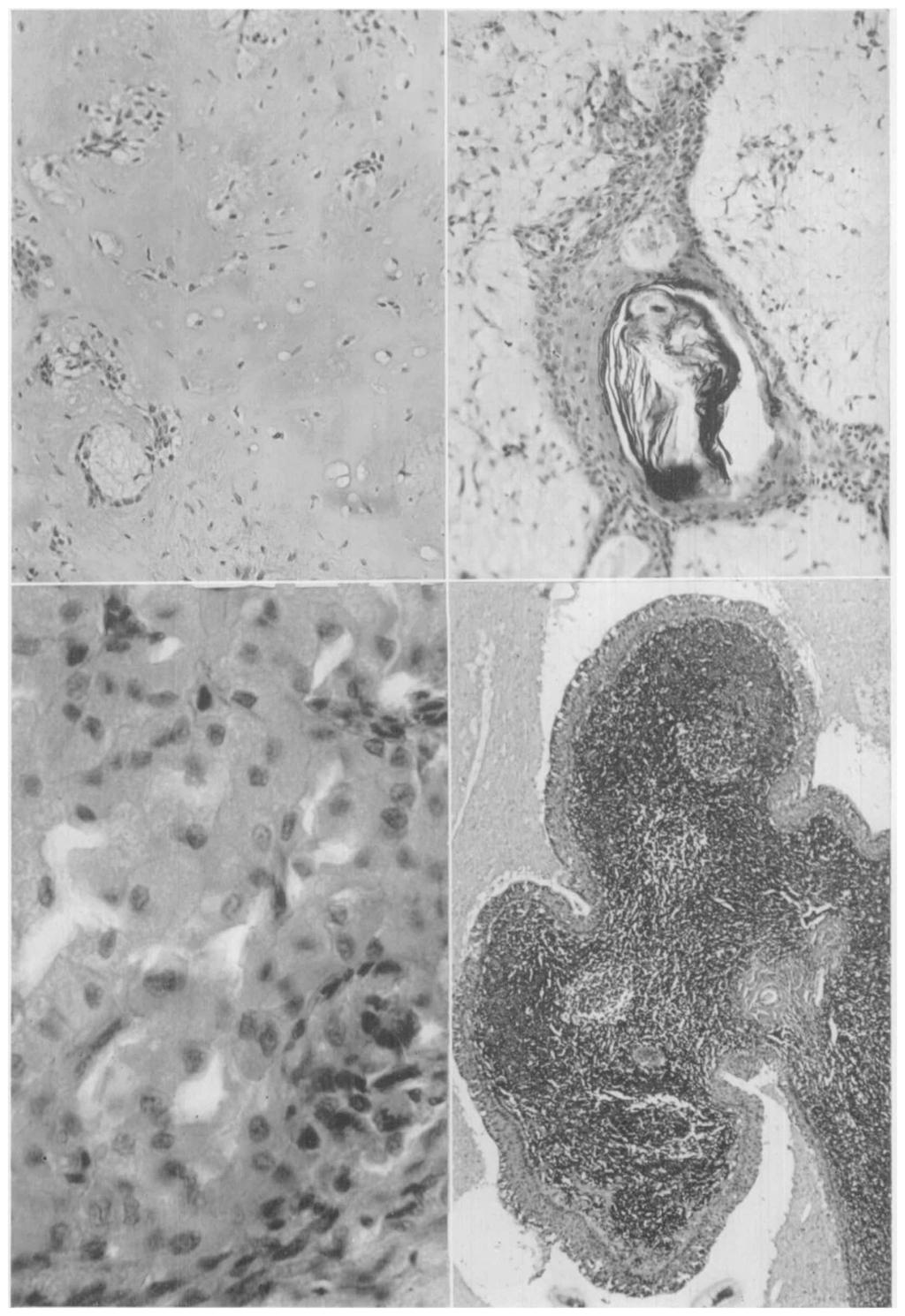
4 50 VELLIOS AND DAVIDSON Vol. 5 Intraductal papilloma. There was case of intraductal papillary adenoma of the palate in this series. Histologically, the lesion was characterized by the presence of intracystic papillary projections (Fig. 5) similar to those seen in intraductal papillomas of the breast. No clinical data were available in this case. Benign lymphoepithelial lesion (Sjogren's syndrome, Mikulicz's disease, 8 sicca syndrome, adenolymphoma, lymphoepithelioma, adenoma lymphomatosum, lymphocytic tumor). There were instances of benign lymphoepithelial lesion 5 involving the parotid glands in our series. In both cases the disease was bilateral. Biopsies of the glands revealed encapsulated nodules of lymphoid tissue, the nodules resembling lobules of salivary gland. Within the lymphoid tissue there were tubules and islands of epithelial cells. These cells superficially resembled reticulum cells, but study elucidated their epithelial nature and their origin from ducts. In some areas there was partial or complete replacement of the epithelial islands by hyalinized connective tissue. We do not regard the lesion as neoplastic but include the cases here because they may be confused with tumors. Both of our patients were men, 9 and 7 years old. Peripheral blood counts in both instances were within normal limits. In the younger patient the masses in the parotid regions were associated with dryness of the mouth and eyes, and long bone and joint pain. In both instances the lesions, following biopsy, were treated with external radiation. There has been no recurrence after and years. MALIGNANT EPITHELIAL TUMORS WITH A LONG CLINICAL COURSE Malignant mixed tumor. AVe have classified case in this series as a malignant mixed tumor. Histologically, this tumor resembled the benign mixed tumors or pleomorphic adenomas, contained sheets of small epithelial cells (Fig. 6), mucoid and cartilaginous tissue, and the epithelial cells showed moderate variation and occasional mitoses. This patient first noted a mass in the parotid region at the age of years. Subsequently, there were attempts to excise it. In addition, he was treated at various times with external radiation that appeared to retard the growth. The tumor continued to invade locally, extending to the base of the skull. At the time of the patient's death, 5 years after onset, it was thought, on the basis of radiograms of the chest, that metastases were present in the lungs. No autopsy was performed, however. Cylindromatous carcinoma (cylindroma, basal-cell carcinoma with hyaline stroma, adenocystic carcinoma, adenoid cystic epithelioma, adenomyo-epithelioma). This type of carcinoma was seen in 7 cases and was primary in acessory salivary gland tissue as well as the parotid and submaxillary glands. The tumor was not encapsulated and had a great tendency to invade perineural lymphatics. Histologically, the tumor cells formed small plugs that were occasion- FIG. (left upper). Typical cartilage-like focus in a pleomorphic adenoma. X 75. FIG. (right, upper). Squamous metaplasia with a keratin pearl in a pleomorphic adenoma. X 75 FIG. (left lower). Area of typical onkocytes occurring in a pleomorphic adenoma. X 450. FIG. 4 (right lower). Papillary structure in a papillary cystadenoma lymphomatosum. Approximately X 50. Downloaded from on 0 January 08

5 >*. HO.'. V /I ^*ag Downloaded from on 0 January 08
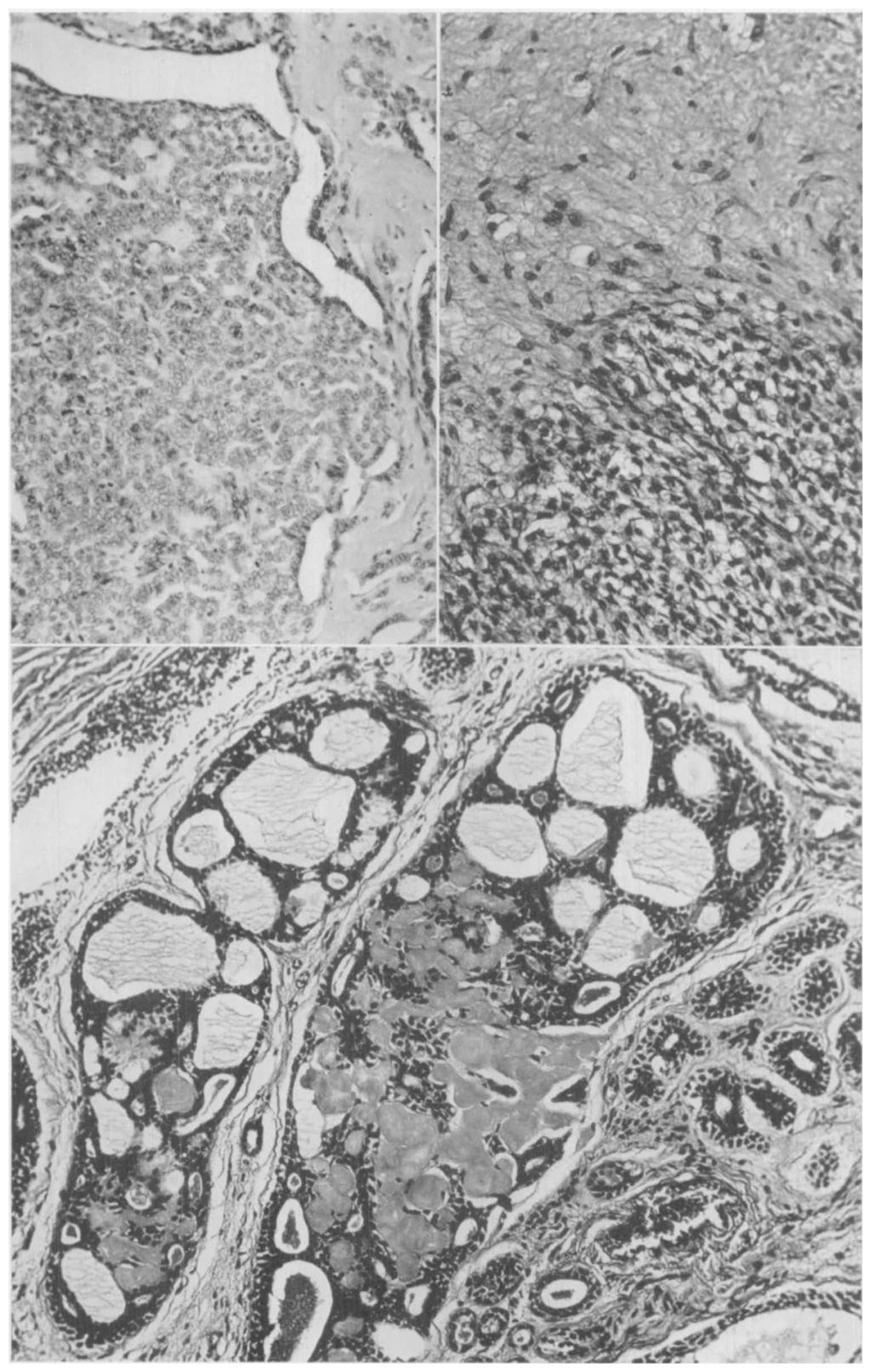
6 5 VELLIOS AND DAVIDSON Vol. 5 ally honeycombed. The cells were small, dark and relatively uniform. Mitoses were rare. The tumor cells were frequently embedded in a hyalinized stroma (Fig. 7), but in some areas the stroma showed a myxomatous change. This stromal change has led to confusion between these tumors and pleomorphic adenomas. In some instances the association of this tumor with salivary gland ducts may be detected in the sections. Seven of these tumors occurred in men and 0 in women. The ages of the patients varied from 8 to 8 years. In instance the tumor was found during a routine physical examination. The longest stated duration before initial therapy was 4 years, most patients seeking medical attention within years. The presenting symptom was usually that of a slowly growing mass although in several instances, in which the lesion was primary in the oral cavity, the patient entered complaining of an ulcer within the mouth. There were 0 patients who had been followed for more than years or who had developed recurrences. All were treated with surgery or radiation therapy, or both. In instances metastases to the lungs were present and of these patients also had evidence of intracranial metastases at the time of his death. Two patients have died with extensive local recurrences. Four patients are alive with persistent tumor locally for periods ranging up to years. Two patients are alive and free of disease clinically after and 4 years, respectively. Mucoepidermoid carcinoma. This type of carcinoma may be divided into a group with relatively low malignancy and a group with relatively high malignancy on histologic criteria, as suggested by Stewart, Foote and Becker. In both types both mucus-secreting epithelial cells and epidermoid cells are found although sometimes only with difficulty. Mucin may be readily demonstrated with the aid of the mucicarmine stain. The relatively benign group is locally invasive histologically and is justifiably classified as carcinoma. In this type, the tumor is generally composed of large cystic spaces lined in part by mucussecreting epithelium and in part by squamous epithelium (Fig. 8). The stroma is abundant so that the epithelial components of the tumor are widely separated. In the malignant form the stroma is relatively slight. The tumor cells form large sheets and usually the epidermoid pattern is readily apparent (Fig. 9). Cysts lined by mucus-secreting cells are usually small, but single cells containing mucin-filled vacuoles are present. Occasionally papillary projections are seen. Invasion of surrounding structures and perineural lymphatics and metastases to regional lymph nodes may be demonstrated. There were cases of the relatively benign form of mucoepidermoid carcinoma in this series, 4 in women, 6 in men and in the sex was not stated. The patients' ages ranged from 5 to 74 years, and the duration of symptoms before initial therapy, from months to year. All patients complained of a lump or "cyst" FIG. 5 (left upper). Intraductal papillary projections in a papillary adenoma of the palate. X 75. FIG. 6 (right upper). Myxoid and cellular area in a malignant mixed tumor of the parotid gland. X 50. FIG. 7 (lower). Plugs of epithelial cells arranged in cribriform pattern in a cylindromatous carcinoma. Foci of hyalinization within tumor are apparent. X 50. Downloaded from on 0 January 08

7 Downloaded from on 0 January 08
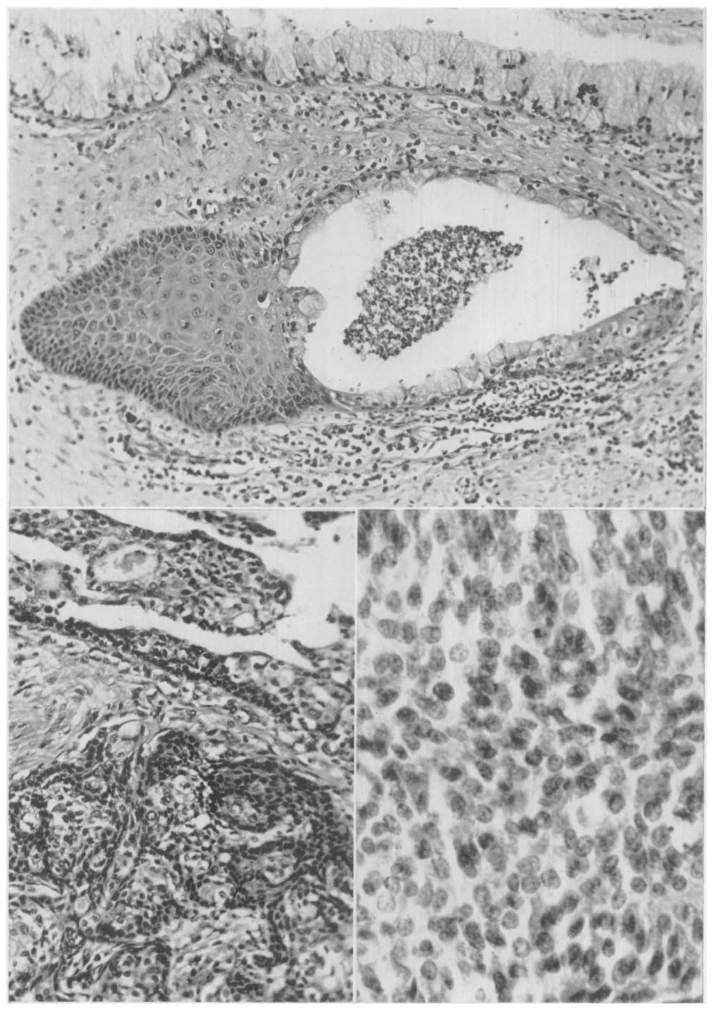
8 54 VELLIOS AND DAVIDSON Vol. 5 in the involved area, and in this was associated with pain. Seven of these tumorsoccurred in the parotid gland and 4 in the oral cavity. Seven- of these patients have been followed years or longer. Two have developed extensive local recurrences and of these patients died with his disease- In other patient a recurrent lesion was excised but the patient is now free of disease, as are the other 4 patients. In none of the patients with this form of tumor have regional or distant metastases been demonstrated. Four patients with the malignant form of mucoepidermoid carcinoma hav& been seen. Three were women and a man; their ages were 8, 4, 58 and 74 years. All complained of a mass at the involved site. One patient was treated by local excision 5 years previously and has had no recurrence. The second patient also had excision of the mass from the parotid gland with dissection of the regional lymph nodes. Histologically, the lymph nodes revealed metastases of tumor and the patient has recurrent disease after year. The other patients have been, followed for only a few months. MALIGNANT EPITHELIAL TUMORS WITH RAPID CLINICAL COURSE This group includes epidermoid carcinoma ( case), adenocarcinoma ( cases) and undifferentiated carcinoma (5 cases) primary in the parotid gland (Fig. 0), and case of undifferentiated carcinoma of the sublingual gland. In all instances the tumor was removed locally and subsequently 5 were treated with external radiation. Seven of these 9 patients had distant metastases or have died with disease within periods of months to 6 years (average, ^ years). INCIDENTAL TUMORS Tumors of supporting tissues included hemangioendothelioma of infancy, neurofibromas and fibrosarcomas occurring in the parotid gland. Histologically and clinically they are similar to such lesions occurring in other sites. DISCUSSION Although tumors of the salivary glands assume diverse histologic forms, they are classifiable on histologic grounds. As suggested by Byars and Ackerman. these tumors may be divided into clinical groups depending upon their usual clinical course: () benign; () malignant with a long clinical course; () malignant with a rapid clinical course. The benign group includes about two thirds of the salivary gland neoplasms, mostly pleomorphic adenomas. Because of its bosselation the tumor may be cut across when excised so that recurrences have been frequent, especially when the surgeon was not familiar with its gross characteristics. In our series there has been no evidence of multicentric origin, and well-authenticated instances of such FIG. 8 (upper). Low-grade mucoepidermoid carcinoma. The layer of mucus-secreting cells in the upper portion of the field is part of the lining of a large cyst. X 0. FIG. 9 (left lower). Relatively malignant mucoepidermoid carcinoma. Many of the cells contain mucin droplets. Part of a cyst is seen in the upper portion of the field. Much of the tumor is composed of pavement epithelium. X 0. FIG. 0 (right lower). Undifferentiated carcinomaof sublingual gland. Mitoses are relatively frequent. X 40. Downloaded from on 0 January 08

9 Downloaded from on 0 January 08
10 56 VELLIOS AND DAVIDSON Vol. 5 an occurrence are rare. It is difficult to prove that malignant tumors develop in benign pleomorphic adenomas but some suggestive evidence has been reported, such as sudden rapid growth in a previously slowly growing tumor, or areas resembling pleomorphic adenoma in an otherwise malignant lesion. The malignant tumors with a protracted clinical course include malignant mixed tumors, cylindromatous carcinoma and mucoepidermoid carcinoma. The true malignant mixed tumor is a relatively rare lesion accounting for less than per cent of all mixed tumors. It may perhaps result from a malignant transformation of the benign tumor. There was no histologic evidence for such a change in our case. This tumor is capable of metastasizing distantly as well as invading locally. The cylindromatous type of adenocarcinoma invades locally and along lymphatics, and metastasizes to regional lymph nodes and distantly. This type of tumor has a long clinical course even when distant metastases are evident. In of our cases the lesions in the lungs appeared to regress with radiation therapy and the patient remained active 8 months after lung metastases were first noted. Mucoepidermoid carcinoma also has a protracted clinical course. In our series, the "benign" form invaded locally but no lymph node or distant metastases developed. Metastases to regional lymph nodes were demonstrated in instance at the original operation for the "malignant" form of mucoepidermoid carcinoma. The malignant tumors with a rapid clinical course include adenocarcinoma, epidermoid carcinoma and undifferentiated carcinoma. These primary lesions are usually small when the patient first seeks medical advice. Metastases to regional lymph nodes and distant sites are the rule. Unfortunately, in most instances the true nature of the neoplasm is not appreciated until it is examined histologically. The role of the biopsy in surgery of neoplasms of the salivary gland should be appreciated. Histologic examination is the only method of ascertaining the benignancy or malignancy of salivary gland neoplasms. A properly planned initial surgical approach should enhance the "cure rate" of malignant salivary gland tumors. SUMMARY In a review of 9 salivary gland tumors 85 were classified as benign and 44 as malignant. These may be subdivided into various histologic types as suggested by many authors. For clinical purposes, they may be grouped into 4 categories:. Benign lesions: pleomorphic adenoma (mixed tumor), papillary cystadenoma lymphomatosum, intraductal papilloma, benign lymphoid lesions.. Malignant tumors with a slow clinical course: malignant mixed tumor, cylindromatous carcinoma, mucoepidermoid carcinoma.. Malignant tumors with a rapid clinical course: epidermoid carcinoma, adenocarcinoma, undifferentiated carcinoma. 4. Incidental tumors not peculiar to salivary glands. Downloaded from on 0 January 08
11 Feb. 955 TUMORS OF SALIVARY GLANDS 57 It is suggested that biopsy of the lesion be performed before therapy is undertaken in all persons with salivary gland tumors. Acknowledgment. We are indebted to Miss Dorothy Dickens, Registrar of the Tumor Registry, for her aid in obtaining follow-up data on many of the patients, and to Mr. James G. Glore and Mr. Paris Johnson of the Department of Illustration for preparation of the photographs. REFERENCES. BAUER, W. H., AND BAUER, J. D.: Classification of glandular tumors of salivary glands. Arch. Path., 55: 8-46, 95.. BUXTON, R. W., MAXWELL, J. H., AND FRENCH, A. J.: Surgical treatment of epithelial tumors of the parotid gland. Surg., Gynec. & Obst., 97: 40-46, 95.. BYARS, L. T., AND ACKERMAN, L. V.: Letter to the editor. J. A. M. A., 5: 54-55, FOOTE, F. W., AND FRAZELL, E. L.: Tumors of the major salivary glands. Cancer, 6: 065-, GODWIN, J. T.: Benign lymphoepithelial lesion of the parotid gland. Cancer, 5: 0S9-0, KIRKLIN, J. W., MCDONALD, J. R., HARRINGTON, S. W., AND NEW, G. B.: Parotid tumors: Histopathology, clinical behavior, and end results. Surg., Gvnec. & Obst., 9: 7-7, MCFARLAND, J.: The mysterious mixed tumors of the salivary glands. Surg., Gynec. & Obst., 76: -4, MORGAN, W. S., AND CASTLEMAN. B.: A clinicopathologic study of "Mikulicz's Disease." Am. J. Path., 9: 47-50, QUATTLEBAUM, F. W., DOCKERTV, M. B., AND MAYO, C. W.: Adenocarcinoma, cylindroma type, of the parotid gland. Surg., Gynec. & Obst., 8: 4-46, RAWSON, A. J., AND HORN, R. C: Sebaceous glands and sebaceous gland-containing tumors of the parotid salivary gland. Surgery, 7: 9-0, RAWSON, A. J., HOWARD, J. M., ROYSTER, H. P., AND HORN, R. C.: Tumors of the salivary glands. Cancer, : , STEWART, F. W., FOOTE, F. W., AND BECKER, W. F.: Mucoepidermoid tumors of salivary glands. Ann. Surg., : , WILLIS, R. A.: Pathology of Tumours. St. Louis: C. V. Mosby Co., 95, 997 pp. Downloaded from on 0 January 08
Salivary Glands 3/7/2017
 Salivary Glands 3/7/2017 Goals and objectives Focus on the entities unique to H&N Common board type facts Information for your future practice Salivary Glands Salivary Glands Major gland. Paratid. Submandibular.
DISORDERS OF THE SALIVARY GLANDS Neoplasms Dr.M.Baskaran Selvapathy S IV
 DISORDERS OF THE SALIVARY GLANDS Neoplasms Dr.M.Baskaran Selvapathy S IV NEOPLASMS A) Epithelial I. Benign Pleomorphic adenoma( Mixed tumour) Adenolymphoma (Warthin s tumour) Oxyphil adenoma (Oncocytoma)
My Journey into the World of Salivary Gland Sebaceous Neoplasms
 My Journey into the World of Salivary Gland Sebaceous Neoplasms Douglas R. Gnepp Warren Alpert Medical School at Brown University Rhode Island Hospital Pathology Department Providence RI Asked to present
SALIVARY GLAND DISEASES. Omar alnoubani MD,MRCS
 SALIVARY GLAND DISEASES Omar alnoubani MD,MRCS Salivary Glands Overview Parotid gland Sublingual gland Submandibular gland Salivary glands - Types 3 Major Salivary Glands Parotid Submandibular Sublingual
(CYLINDROMA) ATLAS OF HEAD AND NECK PATHOLOGY ADENOID CYSTIC CARCINOMA
 ATLAS OF HEAD AND NECK PATHOLOGY ADENOID CYSTIC CARCINOMA") (CYLINDROMA) This malignant tumor is poorly encapsulated and while seemingly well defined within the affected gland, there is usually infiltration of surrounding tissue on closer examination. The cut surface
(CYLINDROMA) This malignant tumor is poorly encapsulated and while seemingly well defined within the affected gland, there is usually infiltration of surrounding tissue on closer examination. The cut surface
Lesions Mimicking Adenoid Cystic Carcinoma. Diagnostic Problems in Salivary Gland Pathology An Update 5/29/2009
 Diagnostic Problems in Salivary Gland Pathology An Update Lesions Mimicking Adenoid Cystic Carcinoma Stacey E. Mills, M.D. W.S. Royster Professor of Pathology Director of Surgical and Cytopathology University
PLEOMORPHIC ADENOMA ( BENIGN MIXED TUMOR )
") ( BENIGN MIXED TUMOR ) Grossly, the tumor is freely movable, solid, sometimes lobulated and occasionally cystic. If recurrent, multinodular masses are common. Histologically, within a fibrous capsule,
( BENIGN MIXED TUMOR ) Grossly, the tumor is freely movable, solid, sometimes lobulated and occasionally cystic. If recurrent, multinodular masses are common. Histologically, within a fibrous capsule,
Note: The cause of testicular neoplasms remains unknown
 - In the 15- to 34-year-old age group, they are the most common tumors of men. - Tumors of the testis are a heterogeneous group of neoplasms that include: I. Germ cell tumors : 95%; all are malignant.
Neoplasia literally means "new growth.
 NEOPLASIA Neoplasia literally means "new growth. A neoplasm, defined as "an abnormal mass of tissue the growth of which exceeds and is uncoordinated with that of the normal tissues and persists in the
See the latest estimates for new cases of salivary gland cancers in the US and what research is currently being done.
 About Salivary Gland Cancer Overview and Types If you have been diagnosed with salivary gland cancer or are worried about it, you likely have a lot of questions. Learning some basics is a good place to
Differential Diagnosis of Oral Masses. Palatal Lesions
 Differential Diagnosis of Oral Masses Palatal Lesions Palatal Masses Periapical Abscess Torus Palatinus Mucocele Lymphoid Hyperplasia Adenomatous Hyperplasia Benign Salivary Neoplasms Malignant Salivary
Salivary Gland Cytology
 Salivary Gland Cytology Diagnostic challenges and potential pitfalls Tarik M. Elsheikh, MD Professor and Medical Director Anatomic Pathology Cleveland Clinic FNA Salivary Gland Lesions Indications Distinguish
doi: /j.anl
 doi: 10.1016/j.anl.2006.07.001 Synchronous unilateral parotid gland neoplasms of three different histological types Shuho Tanaka 1, Keiji Tabuchi 1, Keiko Oikawa 1, Rika Kohanawa 1, Hideki Okubo 1, Dai
Oncocytic-Appearing Salivary Gland Tumors. Oncocytic, Cystic, Mucinous, and High Grade Salivary Gland Tumors SALIVARY GLAND FNA: PART II
 William C. Faquin, MD, PhD Professor of Pathology Harvard Medical School Director of Head and Neck Pathology Massachusetts Eye and Ear Massachusetts General Hospital SALIVARY GLAND FNA: PART II Oncocytic,
Salivary Gland Pathology
 IN THE NAME OF GOD Salivary Gland Pathology CHAPTER 11 Dr.kheirandish Oral and maxillofacial pathology Sialadenosis Adenomatoid Hyperplasia of the Minor Salivary Glands Necrotizing Sialometaplasia Pleomorphic
FNA OF SALIVARY GLANDS: A PRACTICAL APPROACH
 FNA OF SALIVARY GLANDS: A PRACTICAL APPROACH FNA of Salivary Glands: Challenges Wide range of neoplastic and non-neoplastic lesions Cytological overlap between the different benign and malignant tumors
Papillary Lesions of the breast
 Papillary Lesions of the breast Emad Rakha Professor of Breast Pathology The University of Nottingham Papillary lesions of the breast are a heterogeneous group of disease, which are characterised by neoplastic
Los Angeles Society Of Pathologists Dr. Shobha Castelino Prabhu
 Los Angeles Society Of Pathologists Dr. Shobha Castelino Prabhu Loma Linda University Medical Center June 12, 2007 CASE 1 76 year-old gentleman Status post right parotidectomy 1 year ago for a rare tumor
TYPES and FREQUENCY of SALIVARY GLAND TUMORS in MAJOR and MINOR. Karl Donath Department of Oral Pathology (Director:Prof. Dṛ Dr.
 TYPES and FREQUENCY of SALIVARY GLAND TUMORS in MAJOR and MINOR SALIVARY GLANDS Karl Donath Department of Oral Pathology (Director:Prof. Dṛ Dr. Karl Donath) University of Hamburg, Salivary gland tumors
Salivary Gland Imaging. Mary Scanlon MD FACR October 2016
 Salivary Gland Imaging Mary Scanlon MD FACR October 2016 Objectives Recognize normal and abnormal anatomy Discuss work up, management and differential diagnosis of commonly referred clinical scenarios
Epithelial tumors. Dr. F.F. Khuzin, PhD Dr. M.O. Mavlikeev
 Epithelial tumors Dr. F.F. Khuzin, PhD Dr. M.O. Mavlikeev Epithelial tumors Tumors from the epithelium are the most frequent among tumors. There are 2 group features of these tumors: The presence in most
Muco-epidermoid tumours of the anal canal
 J. clin. Path. (1963), 16, 200 Muco-epidermoid tumours of the anal canal B. C. MORSON AND H. VOLKSTADT From the Research Department, St. Mark's Hospital, London SYNOPSIS The pathology of 21 cases of muco-epidermoid
Diseases of the breast (1 of 2)
") Diseases of the breast (1 of 2) Introduction A histology introduction Normal ducts and lobules of the breast are lined by two layers of cells a layer of luminal cells overlying a second layer of myoepithelial
Diseases of the breast (1 of 2) Introduction A histology introduction Normal ducts and lobules of the breast are lined by two layers of cells a layer of luminal cells overlying a second layer of myoepithelial
Salivary gland tumor cytologic and histologic correlation: Algorithmic and risk stratification based approaches
 Salivary gland tumor cytologic and histologic correlation: Algorithmic and risk stratification based approaches Christopher C. Griffith, MD, PhD Raja R. Seethala, MD 1. Salivary gland tumor cytology: A
Salivary gland Workshop Trondheim 31th may 2012
 Salivary gland Workshop Trondheim 31th may 2012 Peter Jebsen cytopathologist Oslo University Hospital Rikshospitalet Anna Bofin ass. Professor St. Olavs Hospital, Trondheim Drying artifacts Lymfocytes
Proliferative Epithelial lesions of the Breast. Sami Shousha, MD, FRCPath Charing Cross Hospital & Imperial College, London
 Proliferative Epithelial lesions of the Breast Sami Shousha, MD, FRCPath Charing Cross Hospital & Imperial College, London Amman, November2013 Proliferative Epithelial Lesions of the Breast Usual type
Update in Salivary Gland Pathology. Benjamin L. Witt University of Utah/ARUP Laboratories February 9, 2016
 Update in Salivary Gland Pathology Benjamin L. Witt University of Utah/ARUP Laboratories February 9, 2016 Objectives Review the different appearances of a selection of salivary gland tumor types Establish
NEOPLASIA-I CANCER. Nam Deuk Kim, Ph.D.
 NEOPLASIA-I CANCER Nam Deuk Kim, Ph.D. 1 2 Tumor in the hieroglyphics of the Edwin Smith papyrus (1,600 B.C., Breasted s translation 1930) 3 War on Cancer (National Cancer Act, 1971) 4 Cancer Acts in Korea
Mixed glandular and squamous-cell carcinoma
 Thorax (1967), 22, 431. Mixed glandular and squamous-cell carcinoma of the bronchus DAVID J. B. ASHLEY AND H. DUNCAN DAVIES From the Pathology Department, Morriston Hospital, Swansea A group of 26 examples
XX. Tumours of the nasal cavity *
 XX. Tumours of the nasal cavity * H. STONZI 1 & B. HAUSER2 Tumours of the nasal cavity are rare in domestic animals, most cases occurring in the dog. Epithelial tumours are the most common type in carnivores
Basaloid carcinoma of the anal canal
 J. clin. Path. (1967), 0, 18 Basaloid carcinoma of the anal canal LILLIAN S. C. PANG AND B. C. MORSON From the Research Department, St. Mark's Hospital, London SYNOPSIS The pathology and results of treatment
Objectives. Salivary Gland FNA: The Milan System. Role of Salivary Gland FNA 04/26/2018
 Salivary Gland FNA: The Milan System Dr. Jennifer Brainard Section Head Cytopathology Cleveland Clinic Objectives Introduce the Milan System for reporting salivary gland cytopathology Define cytologic
Disorders of Cell Growth & Neoplasia. Histopathology Lab
 Disorders of Cell Growth & Neoplasia Histopathology Lab Paul Hanna April 2010 Case #84 Clinical History: 5 yr-old, West Highland White terrier. skin mass from axillary region. has been present for the
Normal thyroid tissue
 Thyroid Pathology Overview Normal thyroid tissue Normal thyroid tissue with follicles filled with colloid. Thyroid cells form follicles, spheres of epithelial cells (always single layered in health, usually
Pleomorphic adenoma of breast - a case report and distinction with metaplastic carcinoma D Gupta, S Agrawal, N Trivedi, A Tewari
 of breast - a case report and distinction with metaplastic carcinoma D Gupta, S Agrawal, N Trivedi, A Tewari Introduction, also known as mixed tumour, is a benign tumour which typically presents as a painless,
Neoplasia 2018 Lecture 2. Dr Heyam Awad MD, FRCPath
 Neoplasia 2018 Lecture 2 Dr Heyam Awad MD, FRCPath ILOS 1. List the differences between benign and malignant tumors. 2. Recognize the histological features of malignancy. 3. Define dysplasia and understand
Polymorphous Low-Grade. December 5 th, 2008
 Polymorphous Low-Grade Adenocarcinoma December 5 th, 2008 Epidemiology Represents 2 nd or 3 rd most common minor salivary gland malignancy (17-26%) 1 st mucoepidermoid carcinoma Rare in reported Asian
PRELIMINARY CYTOLOGIC DIAGNOSIS: Suspicious for Acinic Cell Carcinoma. Cell Block: Immunohistochemical Studies CYTOLOGIC DIAGNOSIS:
 1 PRELIMINARY CYTOLOGIC DIAGNOSIS: Suspicious for Acinic Cell Carcinoma. Cell Block: Immunohistochemical Studies GCDFP-15 S-100 CYTOLOGIC DIAGNOSIS: Consistent with mammary analogue secretory carcinoma.
Papillary tumours of the minor salivary glands
 J. clin. Path., 1976, 29, 795-805 Papillary tumours of the minor salivary glands J. S. WHITTAKER AND E. P. TURNER The departments of Pathology, Withington Hospital, Manchester, and the Dental Hospital
ARIZONA SOCIETY OF PATHOLOGISTS 13 TH APRIL 2013 HEAD AND NECK CYTOPATHOLOGY. F ZAHRA ALY, MD, PhD
 ARIZONA SOCIETY OF PATHOLOGISTS 13 TH APRIL 2013 HEAD AND NECK CYTOPATHOLOGY F ZAHRA ALY, MD, PhD The main areas sites amenable for cytopathology include lymph nodes, thyroid, major salivary glands especially
Histopathological Study of Lacrimal Gland Tumors
 ORIGINAL ARTICLE Pratikkumar B. Desai 1, Ami Shah 2 1 4 th Year Resident, Pathology Department, B.J.Medical College, Civil Hospital, Ahmedabad 2 Associate Professor, M. J. Institute of Ophthalmology, Civil
On 180 Biopsies of Oral Carcinomas in Our Department of Pathology. Yasuyuki AWAZAWA * and Itaru MORO * Introduction
 On 180 Biopsies of Oral Carcinomas in Our Department of Pathology by Yasuyuki AWAZAWA * and Itaru MORO * Introduction Carcinomas in the oral region, like those found in other regions of human body, have
ONCOLOGY. Csaba Bödör. Department of Pathology and Experimental Cancer Research november 19., ÁOK, III.
 ONCOLOGY Csaba Bödör Department of Pathology and Experimental Cancer Research 2018. november 19., ÁOK, III. bodor.csaba1@med.semmelweis-univ.hu ONCOLOGY Characteristics of Benign and Malignant Neoplasms
DISCUSSION: PLGA accounts for about 2% of all salivary gland tumours and occurs almost exclusively in the minor salivary glands.
 SWELLING ON THE HARD PALATE PRESENTING AS POLYMORPHOUS LOW GRADE ADENOCARCINOMA: A AND REVIEW OF LITERATURE Swapnil D. Chandekar 1, Sunita S. Dantkale 2, Rahul R. Narkhede 3, Snehal V. Chavhan 4, Khushboo
-The cause of testicular neoplasms remains unknown
 - In the 15- to 34-year-old age group, they are the most common tumors of men. - include: I. Germ cell tumors : (95%); all are malignant. II. Sex cord-stromal tumors: from Sertoli or Leydig cells; usually
Diseases of oral cavity
 Diseases of oral cavity Diseases of Teeth and Supporting Structures Inflammatory/Reactive Lesions Infections Oral Manifestations of Systemic Disease Precancerous and Cancerous Lesions Odontogenic Cysts
Section of Laryngology
 15 Volume 59 May 1966 429 Section of Laryngology President G H Bateman FRcs Meeting December 31965 Tumours of the Parotid Gland Dr Carl-Magnus Eneroth (Department ofotolaryngology, Karolinska Sjukhuset,
04/09/2018. Salivary Gland Pathology in the Molecular Era Old Friends, Old Foes, & New Acquaintances
 Salivary Gland Pathology in the Molecular Era Old Friends, Old Foes, & New Acquaintances Jennifer L. Hunt, MD, MEd Aubrey J. Hough Jr, MD, Endowed Professor of Pathology Chair of Pathology and Laboratory
Carcinoma of Unknown Primary site (CUP) in HEAD & NECK SURGERY
 in HEAD & NECK SURGERY") Carcinoma of Unknown Primary site (CUP) in HEAD & NECK SURGERY SEARCHING FOR THE PRIMARY? P r o f J P P r e t o r i u s H e a d : C l i n i c a l U n i t C r i t i c a l C a r e U n i v e r s i t y O f
Carcinoma of Unknown Primary site (CUP) in HEAD & NECK SURGERY SEARCHING FOR THE PRIMARY? P r o f J P P r e t o r i u s H e a d : C l i n i c a l U n i t C r i t i c a l C a r e U n i v e r s i t y O f
Salivary gland cytology. Salivary gland cytology. Triage helps the clinician. Salivary gland tumors. Diagnostic difficulties
 Salivary gland cytology Salivary Gland Cytology Pınar Fırat, MD Professor of Pathology İ.U. İstanbul Faculty of Medicine Çapa, İstanbul It is a reliable diagnostic test However, definitive subclassification
Papillary Lesions of the Breast A Practical Approach to Diagnosis. (Arch Pathol Lab Med. 2016;140: ; doi: /arpa.
 Papillary Lesions of the Breast A Practical Approach to Diagnosis (Arch Pathol Lab Med. 2016;140:1052 1059; doi: 10.5858/arpa.2016-0219-RA) Papillary lesions of the breast Span the spectrum of benign,
A CASE OF A Huge Submandibular Pleomorphic Adenoma
 ISPUB.COM The Internet Journal of Head and Neck Surgery Volume 4 Number 2 S VERMA Citation S VERMA.. The Internet Journal of Head and Neck Surgery. 2009 Volume 4 Number 2. Abstract Pleomorphic adenoma
Carcinoma ex Pleomorphic Adenoma (CXPA)-A rare parotid malignancy
-A rare parotid malignancy") Indian Journal of Mednodent and Allied Sciences, pp- 54-58 Indian journals.com Case Report Carcinoma ex Pleomorphic Adenoma (CXPA)-A rare parotid malignancy Vani Padmaja GJ 1 *, Sireesha A 2, Sunderi Devi
Indian Journal of Mednodent and Allied Sciences, pp- 54-58 Indian journals.com Case Report Carcinoma ex Pleomorphic Adenoma (CXPA)-A rare parotid malignancy Vani Padmaja GJ 1 *, Sireesha A 2, Sunderi Devi
Histopathology: Cervical HPV and neoplasia
 Histopathology: Cervical HPV and neoplasia These presentations are to help you identify basic histopathological features. They do not contain the additional factual information that you need to learn about
SESSION 1: GENERAL (BASIC) PATHOLOGY CONCEPTS Thursday, October 16, :30am - 11:30am FACULTY COPY
 PATHOLOGY CONCEPTS Thursday, October 16, :30am - 11:30am FACULTY COPY") SESSION 1: GENERAL (BASIC) PATHOLOGY CONCEPTS Thursday, October 16, 2008 9:30am - 11:30am FACULTY COPY GOAL: Describe the basic morphologic (structural) changes which occur in various pathologic conditions.
SESSION 1: GENERAL (BASIC) PATHOLOGY CONCEPTS Thursday, October 16, 2008 9:30am - 11:30am FACULTY COPY GOAL: Describe the basic morphologic (structural) changes which occur in various pathologic conditions.
Trichofolliculoma of the Guinea Pig 1,2
 Trichofolliculoma of the Guinea Pig 1,2 Raymond D. Ediger, Garrett S. Dill, Jr., and Robert M. Kovatch, Aerobiology and Evaluation Laboratories and Medical Sciences Laboratories, Fort Detrick, Frederick,
APOCRINE SWEAT GLAND CARCINOMA OF THE VULVA* JOHN R. McDONALD, M.D. Section on Surgical Pathology, The Mayo Clinic, Rochester, Minnesota
 APOCRINE SWEAT GLAND CARCINOMA OF THE VULVA* JOHN R. McDONALD, M.D. Section on Surgical Pathology, The Mayo Clinic, Rochester, Minnesota The wide variety of neoplasms, both benign and malignant, originates
EQA Circulation 43 Educational Cases
 EQA Circulation 43 Educational Cases E1-E2 Monica Agarwal Monklands Hospital E1 38 yrs male Submandibular gland tumour E1 Formal excision following diagnosis of poorly differentiated carcinoma on core
Educational Cases EQA November T.J. Palmer Raigmore Hospital Inverness
 Educational Cases EQA November 2013 T.J. Palmer Raigmore Hospital Inverness Case 2 Clinical Details Dob 11 February 1951 PMH: 1964 Extraction of 45 aet 13 yr 1966 Cyst between 44 and 46 enucleated 1973
Pathology Slides. [Pathology]
![Pathology Slides. [Pathology]](/thumbs/94/120604575.jpg "Pathology Slides. [Pathology]") Pathology Slides MedicoNotes provides real laboratory pathological slides to aid you to differentiate between different pathological structures under microscope. www.mediconotes.com Histology slides example
Pathology Slides MedicoNotes provides real laboratory pathological slides to aid you to differentiate between different pathological structures under microscope. www.mediconotes.com Histology slides example
Gross appearance of nodular hyperplasia in material obtained from suprapubic prostatectomy. Note the multinodular appearance and the admixture of
 Tiền liệt tuyến Tiền liệt tuyến Gross appearance of nodular hyperplasia in material obtained from suprapubic prostatectomy. Note the multinodular appearance and the admixture of solid and microcystic areas.
Histopathologic spectrum of salivary gland neoplasms
 Original article Histopathologic spectrum of salivary gland neoplasms Dr. Dipkana Das 1, Dr.Subhasish Saha 1, Dr V Satyanarayana 2 Name & Address of Institution: Department of Pathology, Kamineni Institute
Maram Abdaljaleel, MD Dermatopathologist and Neuropathologist University of Jordan, School of Medicine
 Maram Abdaljaleel, MD Dermatopathologist and Neuropathologist University of Jordan, School of Medicine The most common non-skin malignancy of women 2 nd most common cause of cancer deaths in women, following
HISTOPATHOLOGICAL EVALUATION OF BENIGN PROLIFERATIVE BREAST LESIONS
 7 ORIGINAL ARTICLE HISTOPATHOLOGICAL EVALUATION OF BENIGN PROLIFERATIVE BREAST LESIONS DR. VIBHUTI H. CHIHLA*, DR. N N. JAGRIT **, DR. JAYASHREE M. SHAH*** *3 rd year Pathology Resident, **Associate Professor,
The International Federation of Head and Neck Oncologic Societies. Current Concepts in Head and Neck Surgery and Oncology
 The International Federation of Head and Neck Oncologic Societies Current Concepts in Head and Neck Surgery and Oncology www.ifhnos.net The International Federation of Head and Neck Oncologic Societies
What is ACC? (Adenoid Cystic Carcinoma)
") What is ACC? (Adenoid Cystic Carcinoma) 10-9-10 Where ACC Occurs ACC (Adenoid Cystic Carcinoma) is a rare and unique form of cancer that is known to be unpredictable in nature, with a typical growth pattern
What is ACC? (Adenoid Cystic Carcinoma) 10-9-10 Where ACC Occurs ACC (Adenoid Cystic Carcinoma) is a rare and unique form of cancer that is known to be unpredictable in nature, with a typical growth pattern
Papillary Lesions of the Breast
 Papillary Lesions of the Breast Laura C. Collins, M.D. Associate Professor of Pathology Associate Director, Division of Anatomic Pathology Beth Israel Deaconess Medical Center and Harvard Medical School
PLEOMORPHIC ADENOMA OF LATERAL WALL OF NOSE A RARE PRESENTATION
 ISSN: 2250-0359 Volume 4 Issue 1 2014 PLEOMORPHIC ADENOMA OF LATERAL WALL OF NOSE A RARE PRESENTATION *USHA KUMAR MAHESH *RATNAKAR MADHAVARAO POTEKAR * B.L.D.E UNIVERSITY ABSTRACT: The aim of the article
CLINICAL SIGNIFICANCE OF BENIGN EPITHELIAL CHANGES
 Papillomas. Papillomas are composed of multiple branching fibrovascular cores, each having a connective tissue axis lined by luminal and myoepithelial cells ( Fig. 23-11 ). Growth occurs within a dilated
LYMPHATIC DRAINAGE AXILLARY (MOSTLY) INTERNAL MAMMARY SUPRACLAVICULAR
 INTERNAL MAMMARY SUPRACLAVICULAR") BREAST LYMPHATIC DRAINAGE AXILLARY (MOSTLY) INTERNAL MAMMARY SUPRACLAVICULAR HISTOLOGY LOBE: (10 in whole breast) LOBULE: (many per lobe) ACINUS/I, aka ALVEOLUS/I: (many per lobule) DUCT(S): INTRA- or
BREAST LYMPHATIC DRAINAGE AXILLARY (MOSTLY) INTERNAL MAMMARY SUPRACLAVICULAR HISTOLOGY LOBE: (10 in whole breast) LOBULE: (many per lobe) ACINUS/I, aka ALVEOLUS/I: (many per lobule) DUCT(S): INTRA- or
XIII. Tumours of the liver and biliary system
 XIII. Tumours of the liver and biliary system V. PONOMARKOV 1 & L. J. MACKEY 2 In this histological classification of liver and gall bladder tumours the tumour types largely correspond to those found in
Journal of International Academy of Forensic Science & Pathology (JIAFP)
") Journal of International Academy of Forensic Science & Pathology (JIAFP) ISSN 2395-0722 MICROCYSTIC ADNEXAL CARCINOMA-A CASE REPORT WITH REVIEW OF LITERATURE Case Report Sulakshana M S 1,Natarajan M 2
Journal of International Academy of Forensic Science & Pathology (JIAFP) ISSN 2395-0722 MICROCYSTIC ADNEXAL CARCINOMA-A CASE REPORT WITH REVIEW OF LITERATURE Case Report Sulakshana M S 1,Natarajan M 2
They cells can not function death.
 Jenna Hellack Jan 2001 Tissues What do you think happens when the cells use up their food and oxygen before there is time to replenish it? They cells can not function death. Blood Cell Cancer cell Plant
Fine-needle aspiration (FNA) has been used increasingly
 has been used increasingly") Worrisome Histologic Alterations Following Fine-Needle Aspiration of Benign Parotid Lesions Shiyong Li, MD, PhD; Zubair W. Baloch, MD, PhD; John E. Tomaszewski, MD; Virginia A. LiVolsi, MD Objective. To
Worrisome Histologic Alterations Following Fine-Needle Aspiration of Benign Parotid Lesions Shiyong Li, MD, PhD; Zubair W. Baloch, MD, PhD; John E. Tomaszewski, MD; Virginia A. LiVolsi, MD Objective. To
Rare Presentation Of Adenoidcystic Carcinoma Of External Auditory Canal With Subcutaneous Metastasis In Temporal Region
 ISPUB.COM The Internet Journal of Otorhinolaryngology Volume 13 Number 2 Rare Presentation Of Adenoidcystic Carcinoma Of External Auditory Canal With Subcutaneous Metastasis In Temporal Region S Kaushik,
There are 3 pairs of major salivary glands, namely
 Kathmandu University Medical Journal (2008), Vol. 6, No. 2, Issue 22, 204-208 Original Article Role of FNAC in the diagnosis of salivary gland swellings Akhter J 1, Hirachand S 1, Lakhey M 2 1 Lecturer,
Slide seminar. Asist. Prof. Jože Pižem, MD, PhD Institute of Pathology Medical Faculty, University of Ljubljana
 Slide seminar Asist. Prof. Jože Pižem, MD, PhD Institute of Pathology Medical Faculty, University of Ljubljana Case 5 A 57-year-old man with a dermal/subcutaneous lesion on the scalp, which was interpreted
4Ps LUMPS AND BUMPS B.L.&T. BUMPS, LUMPS, AND TATTOOS. Most Common BUMP in the oral cavity Fibroma INTERDENTAL PAPILLAE LESIONS
 B.L.&T. BUMPS, LUMPS, AND TATTOOS LUMPS AND BUMPS DIFFERENTIAL DIAGNOSIS FOR LUMPS AND BUMPS Traumatic Fibroma Papilloma Epulis Fissuratum Inflammatory Papillary Hyperplasia Lesions of Attached Gingiva
PSA. HMCK, p63, Racemase. HMCK, p63, Racemase
 Case 1 67 year old male presented with gross hematuria H/o acute prostatitis & BPH Urethroscopy: small, polypoid growth with a broad base emanating from the left side of the verumontanum Serum PSA :7 ng/ml
Basement membrane in lobule.
 Bahram Memar, MD Basement membrane in lobule. Normal lobule-luteal phase Normal lobule-follicular phase Lactating breast Greater than 95% are adenocarcinomas in situ carcinomas and invasive carcinomas.
ATLAS OF HEAD AND NECK PATHOLOGY METAPLASIA
 Metaplasia is the conversion of one adult differentiated cell type to another. Generally it is the result of persistent cellular trauma and serves as a protective mechanism. Thus anteriorly along the nasal
4/17/2015. Case 1. A 37 year old man with a 2.2 cm solitary left thyroid mass.
 Case 1 A 37 year old man with a 2.2 cm solitary left thyroid mass. Case 1 Case 1 1 Case 1: Diagnosis? A. Benign B. Atypia of undetermined significance/follicular lesion of undetermined significance C.
Synchronous squamous cell carcinoma of the breast. and invasive lobular carcinoma
 Sentani K et al. 1 Letter to the editor Synchronous squamous cell carcinoma of the breast and invasive lobular carcinoma Kazuhiro Sentani, 1 Takashi Tashiro, 2 Naohide Oue, 1 Wataru Yasui 1 1 Department
Papillary Cystadenoma Lymphomatosum (Warthin's Tumor) in Patients in a General Hospital over a 24-year Period
 in Patients in a General Hospital over a 24-year Period") Papillary Cystadenoma Lymphomatosum (Warthin's Tumor) in Patients in a General Hospital over a 24-year Period SCOTT E. DIETERT, M.D. Eli Lilly and Company, Greenfield Laboratories, Toxicology Division,
Papillary Cystadenoma Lymphomatosum (Warthin's Tumor) in Patients in a General Hospital over a 24-year Period SCOTT E. DIETERT, M.D. Eli Lilly and Company, Greenfield Laboratories, Toxicology Division,
Papillary Lesions of the Breast
 Papillary Lesions of the Breast Texas Society of Pathologists 2013 Laura C. Collins, M.D. Associate Professor of Pathology Associate Director, Division of Anatomic Pathology Beth Israel Deaconess Medical
Breast pathology. 2nd Department of Pathology Semmelweis University
 Breast pathology 2nd Department of Pathology Semmelweis University Breast pathology - Summary - Benign lesions - Acute mastitis - Plasma cell mastitis / duct ectasia - Fat necrosis - Fibrocystic change/
SOME ESSENTIAL FACTORS IN THE PATHOLOGY AND TREATMENT OF CANCER OF THE SKIN LOUIS H. JORSTAD, M.D.
 SOME ESSENTIAL FACTORS IN THE PATHOLOGY AND TREATMENT OF CANCER OF THE SKIN LOUIS H. JORSTAD, M.D. (From the Department of Pathology, the Barnard Free Skin and Cancer Hospital, St. Louis, Missouri) The
Central Poorly Differentiated Adenocarcinoma of the Maxilla: Report of a Case
 Kobe J. Med. Sci., Vol. 49, No. 2, pp. 45-49, 2003 Central Poorly Differentiated Adenocarcinoma of the Maxilla: Report of a Case MASAHIRO UMEDA 1), SATOSHI YOKOO 1), YASUYUKI SHIBUYA 1), TAKAHIDE KOMORI
A 60-year old Man with Left Jaw Mass. Simon Chiosea, MD University of Pittsburgh medical Center 3/15/2016
 ACCME/Disclosures The USCAP requires that anyone in a position to influence or control the content of CME disclose any relevant financial relationship WITH COMMERCIAL INTERESTS which they or their spouse/partner
encapsulated thyroid nodule with a follicular architecture and some form of atypia. The problem is when to diagnose
 Histological Spectrum of Papillary Carcinoma of Thyroid A Two Years Study Gomathi Srinivasan 1, M. Vennila 2 1 Associate Professor Pathology, Government Medical College, Omandurar Estate, Chennai 600 002
Oncocytic carcinoma: A rare malignancy of the parotid gland
 ISPUB.COM The Internet Journal of Pathology Volume 8 Number 2 Oncocytic carcinoma: A rare malignancy of the parotid gland K Mardi, J Sharma Citation K Mardi, J Sharma.. The Internet Journal of Pathology.
Rare Breast Tumours. 1. Breast Tumours. 1.1 General Results. 1.2 Incidence
 Rare Breast Tumours 1. Breast Tumours 1.1 General Results Table 1. Epithelial Tumours of Breast: Incidence, Trends, Survival Flemish Region 2001-2010 Incidence Trend Survival Females EAPC Relative survival
BREAST PATHOLOGY. Fibrocystic Changes
 BREAST PATHOLOGY Lesions of the breast are very common, and they present as palpable, sometimes painful, nodules or masses. Most of these lesions are benign. Breast cancer is the 2 nd most common cause
A neoplasm is defined as "an abnormal tissue proliferation, which exceeds that of adjacent normal tissue. This proliferation continues even after
 NEOPLASIA Neoplasia is a very important topic in pathology because neoplasms are both common and serious diseases. A neoplasm literally means a new growth, and this term is used interchangeably with a
Catholic University of Louvain, St - Luc University Hospital Head and Neck Oncology Programme. Anatomopathology. Pathology 1 Sept.
 Anatomopathology Pathology 1 Anatomopathology Biopsies Frozen section Surgical specimen Peculiarities for various tumor site References Pathology 2 Biopsies Minimum data, which should be given by the pathologist
TUMOR,NEOPLASM. Pathology Department, Zhejiang University School of Medicine,
 TUMOR,NEOPLASM Pathology Department, Zhejiang University School of Medicine, 马丽琴,maliqin198@zju.edu.cn The points in this chapter What is a neoplasm (conception) Morphology of neoplasm Macroscopy of Neoplasm
04/10/2018. Intraductal Papillary Neoplasms Of Breast INTRADUCTAL PAPILLOMA
 Intraductal Papillary Neoplasms Of Breast Savitri Krishnamurthy MD Professor of Pathology Deputy Division Head The University of Texas MD Anderson Cancer Center 25 th Annual Seminar in Pathology Pittsburgh,
Diagnostically Challenging Cases in Gynecologic Pathology
 Diagnostically Challenging Cases in Gynecologic Pathology Eric C. Huang, M.D., Ph.D. Department of Pathology and Laboratory Medicine University of California, Davis Medical Center Case 1 Presentation 38
Synonyms. Nephrogenic metaplasia Mesonephric adenoma
 Nephrogenic Adenoma Synonyms Nephrogenic metaplasia Mesonephric adenoma Definition Benign epithelial lesion of urinary tract with tubular, glandular, papillary growth pattern Most frequently in the urinary
* Read before the New Tork Pathological Society at the Academy of Medicine, CANCER OF THE THYROID GLAND'
 CANCER OF THE THYROID GLAND' HOWARD M. CLUTE, M.D., AND SHIELDS WARREN, M.D. (From the Lahey Clinic and the Pathology Lnboratorirs of the New England Deaconess and the New England Baptzst Hospatals, and