Papillary Lesions of the Breast
|
|
|
- Benjamin Blankenship
- 5 years ago
- Views:
Transcription
1 Papillary Lesions of the Breast Laura C. Collins, M.D. Associate Professor of Pathology Associate Director, Division of Anatomic Pathology Beth Israel Deaconess Medical Center and Harvard Medical School Boston, MA
2 Papillary Lesions of the Breast Intraductal papilloma Papilloma with atypia (atypical papilloma) Papilloma with DCIS Papillary DCIS Encapsulated papillary carcinoma Solid papillary carcinoma
3 Papillary Lesions of the Breast Intraductal papilloma Papilloma with atypia (atypical papilloma) Implications of papillary lesions Papilloma with DCIS on core needle biopsy Papillary DCIS Encapsulated papillary carcinoma Insights into encapsulated and Solid papillary carcinoma solid papillary carcinomas Lymph node papillomas
4 Papillary Lesions of the Breast The importance of myoepithelial cells and myoepithelial cell immunohistochemical markers in assessment of papillary lesions
5 Intraductal Papilloma Solitary (central) or multiple (peripheral) Any age; most common yrs Nipple discharge and/or palpable mass Subsequent breast cancer risk similar for other proliferative lesions without atypia (~1.5-2x)
6 Diagnostic Criteria for Papillary Lesions adapted from Kraus and Neubecker, 1962 Papilloma Cell types Epithelial and myoepithelial Cell orientation Haphazard Nuclei Stroma of papillae Apocrine metaplasia Proliferation in adjacent ducts Normochromatic Prominent; fibrosis with epithelial entrapment Present Hyperplasia

7

8

9

10


11
12
13 Atypical Papillary Lesions Papilloma with atypia (atypical papilloma) Papilloma with DCIS Papillary DCIS


14 Atypical Papillary Lesions The importance of myoepithelial cells and MEC markers in assessment of papillary lesions
15 Papilloma with Atypia Papilloma with foci of ADH Papilloma with limited area(s) of non-high grade DCIS
16 Papilloma with Atypia DCIS engrafted on pre-existing benign papilloma
17 Papilloma with Atypia vs. Papilloma with DCIS Page et al: Size Atypical area > 3mm (Cancer 1996;78:258) Tavassoli: Proportion WHO Working Group Recommendation 2012 Atypical area > 1/3 (Pathol of the Breast 2 nd Ed,1999) Elston, Ellis & Pinder: Qualitative Use size/extent criteria <3mm ADH >3mm LG DCIS Overt features of malignancy, no matter what the proportion (The Breast, 1998)
18

19

20

21
22 SMMHC


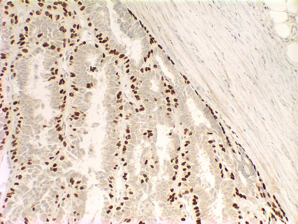
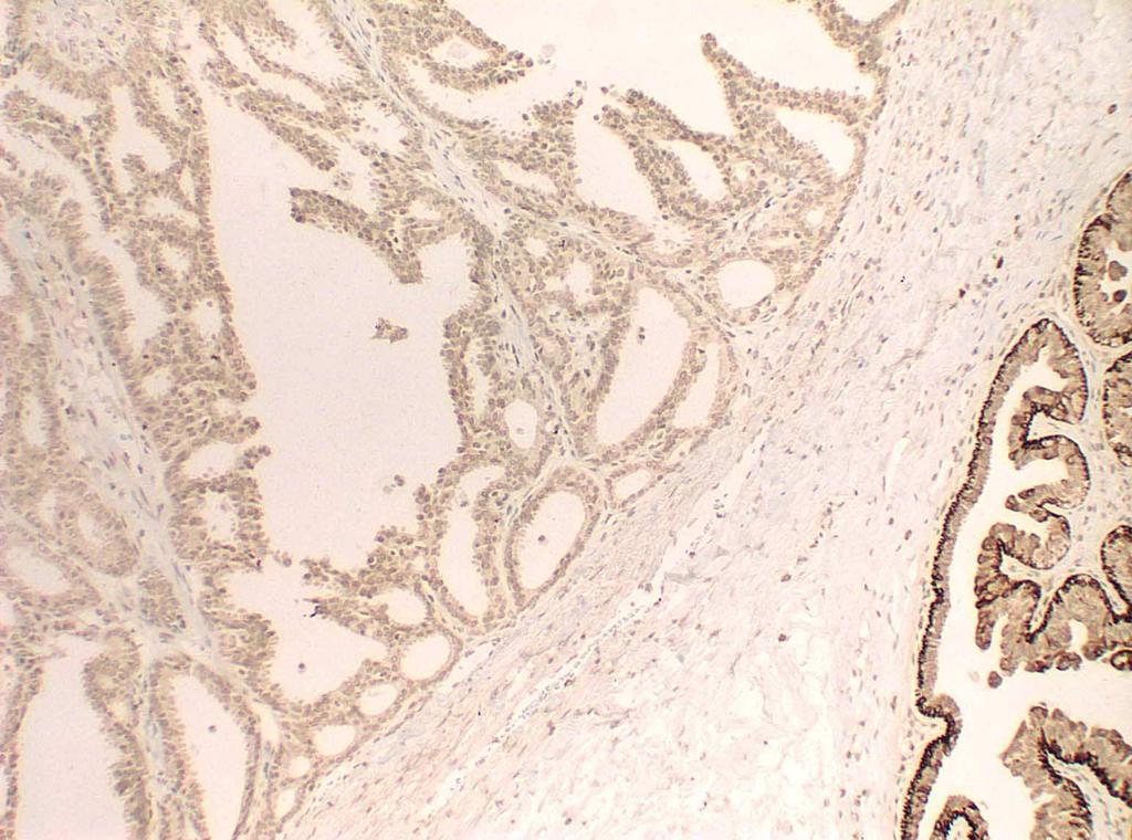
23 The combination of an absence of CK5/6 staining and the presence of strong, diffuse ER staining is helpful in defining extent of atypia CK5/6 ER
24 ER-high/CK5-low profile predicts atypia ER-low/CK5-high profile characterizes non-atypical papillary lesions Pitfalls: Apocrine cells Basal-like DCIS Grin, Am J Surg Pathol, 2009 Columnar cell change Tse, 2010, questioned accuracy of this approach

25 Br Cancer Res Treat, 2013
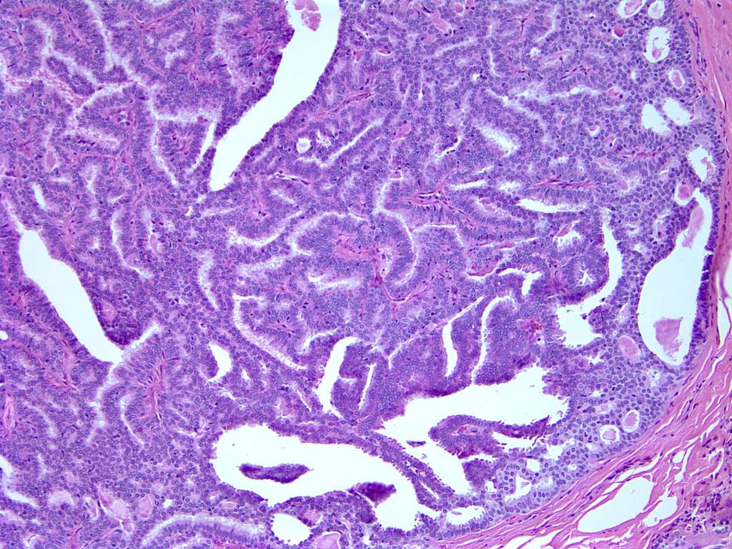
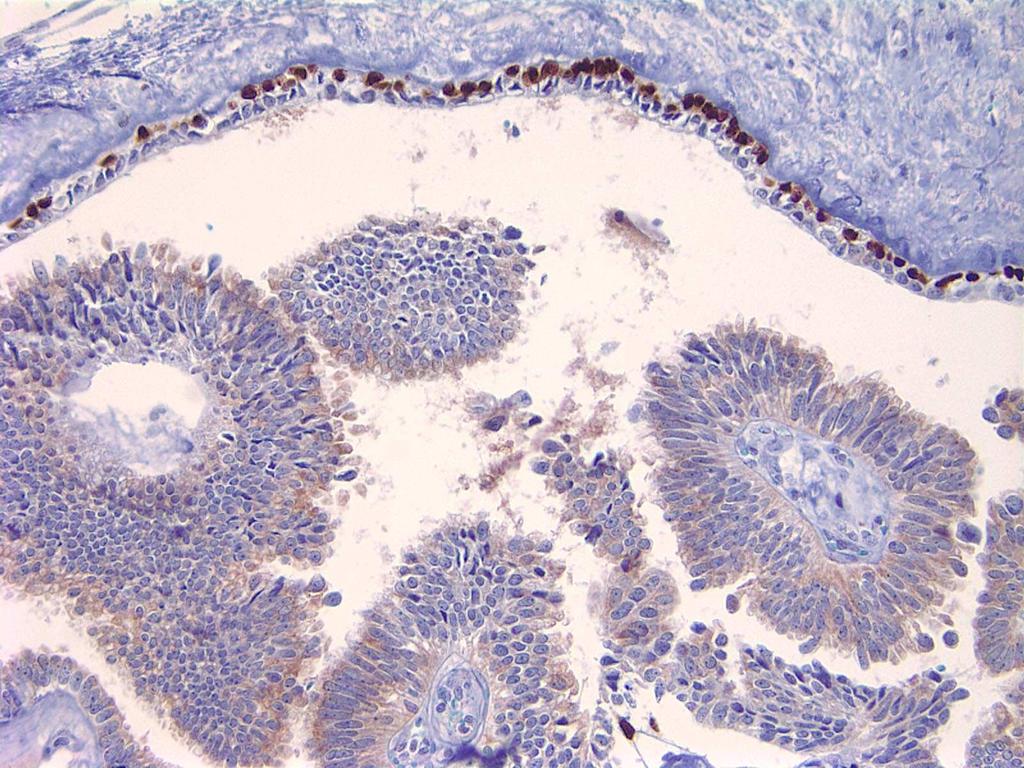
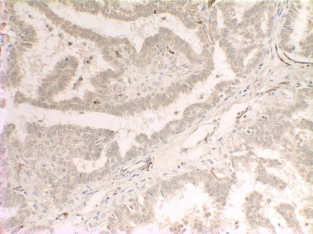
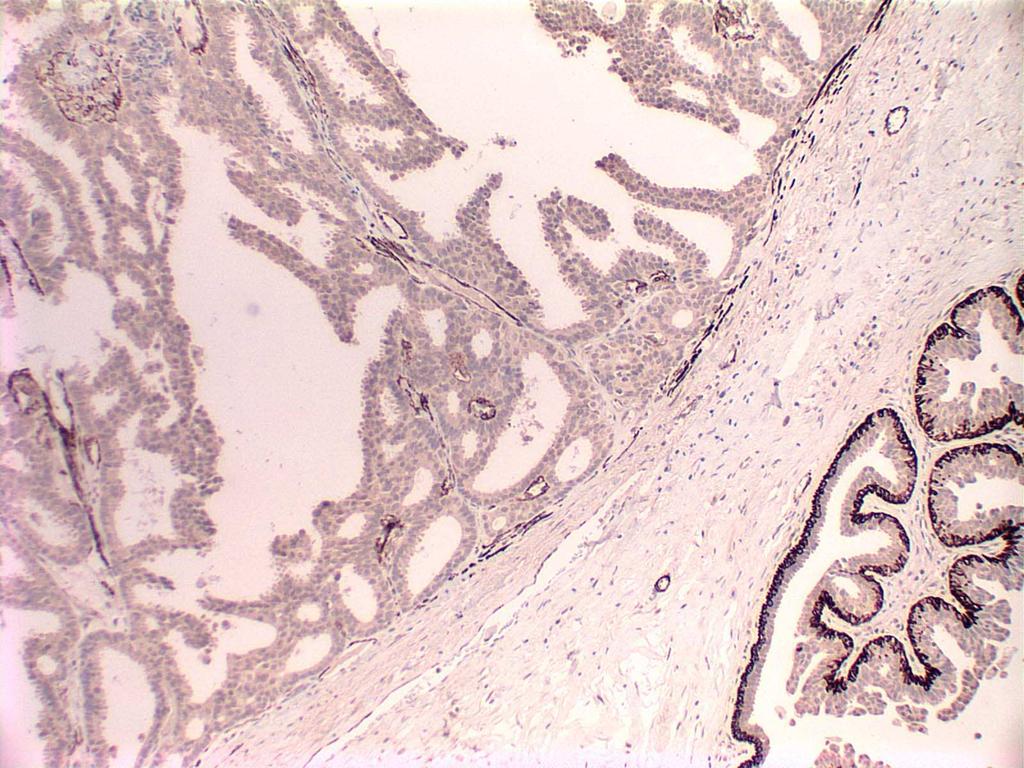
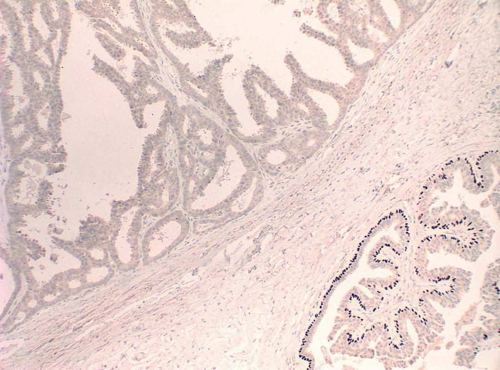
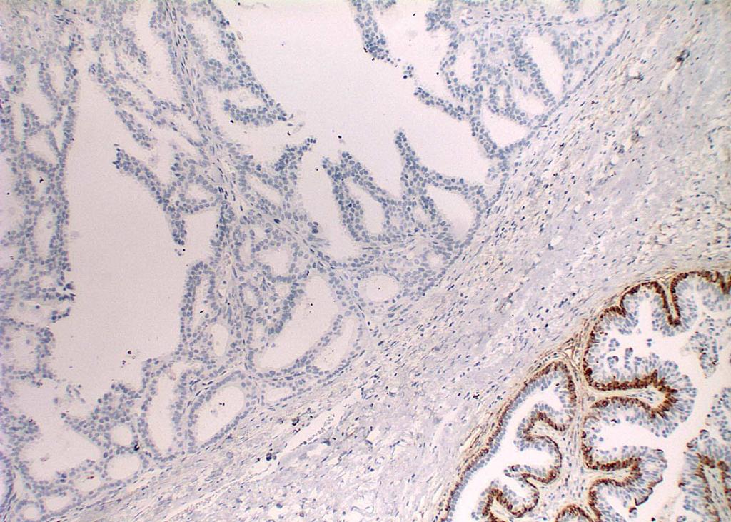
26 Diagnostic Criteria for Papillary Lesions adapted from Kraus and Neubecker, 1962 Papilloma Papillary DCIS Cell types Epithelial and myoepithelial Epithelial; myoepithelial cells absent or scant Cell orientation Haphazard Uniform, perpendicular to fibrovascular stalks Nuclei Normochromatic Hyperchromatic Stroma of papillae Apocrine metaplasia Proliferation in adjacent ducts Prominent; fibrosis with epithelial entrapment Present Hyperplasia Delicate Absent DCIS
27 Papilloma with DCIS vs. Papillary DCIS Papilloma with DCIS Papillary DCIS Underlying structure of benign papilloma Papillae themselves not neoplastic Papillae themselves part of the neoplastic process

28
29

30

31 Papilloma PINK Papillary DCIS BLUE
32 p63


33 Globoid Cells Myoepithelial Cells
34 Papillary Lesions of the Breast Intraductal papilloma Papilloma with atypia (atypical papilloma) Papilloma with DCIS Papillary DCIS Insights into encapsulated and Encapsulated papillary carcinoma solid papillary carcinomas Solid papillary carcinoma
 ~50% central Mass, nipple discharge/bleeding Rounded, lobulated, circumscribed")

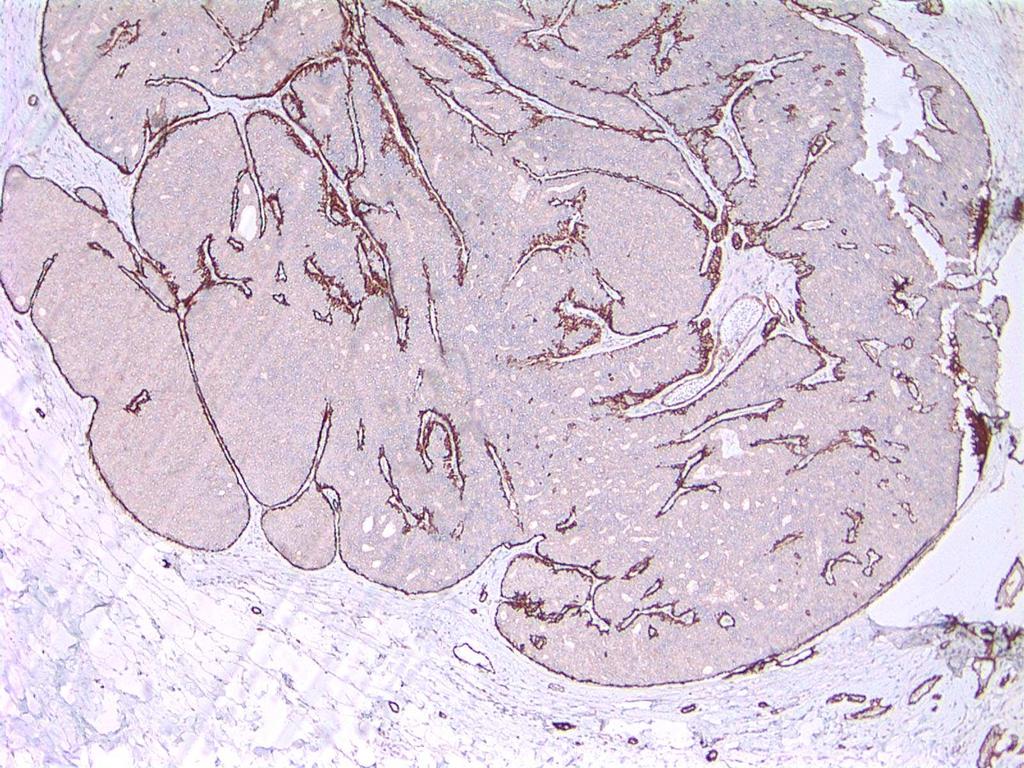
35 Encapsulated Papillary Carcinoma Considered to be a variant of DCIS Older women (mean age, mid 60s) ~50% central Mass, nipple discharge/bleeding Rounded, lobulated, circumscribed lesions on mammography Grossly well circumscribed Mean size, 2-3cm ~50% have adjacent DCIS (assoc. with increased local recurrence risk) ~1/3 of reported cases had associated invasive ca
36

37
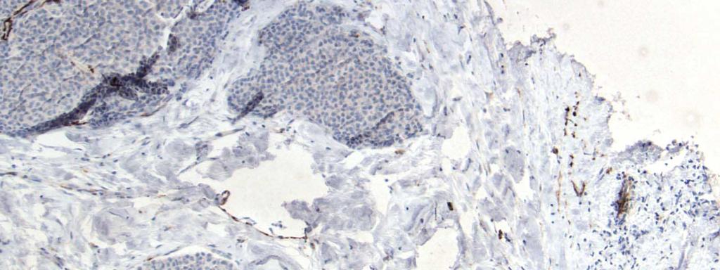
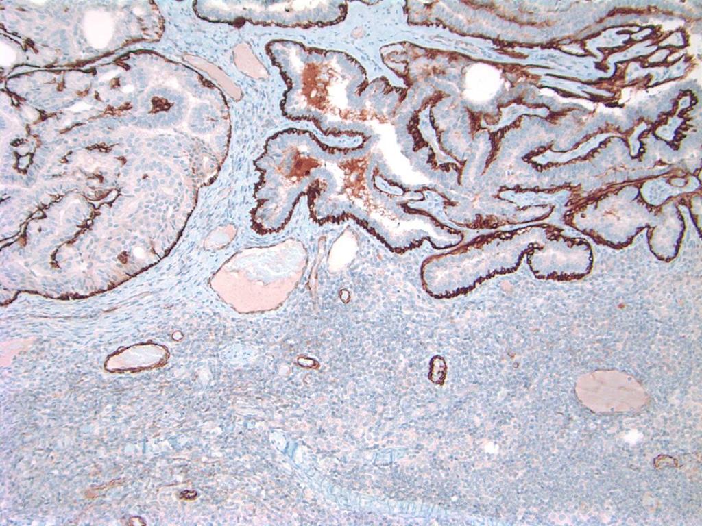
38 SMMHC
39 SMMHC
40 p63
41 CD10
42 CK5/6
43 Immunohistochemistry for Myoepithelial Cells to Distinguish In Situ from Invasive Carcinomas Invasive Cancer No Myoepithelial Cells
44 Questions Raised Are intracystic papillary carcinomas an exception to this principle? Does this tell us something about the nature of lesions we have traditionally considered to be intracystic papillary carcinomas? What does this mean for clinical practice?
45 Myoepithelial Markers in EPC Several studies have now demonstrated an absence of MECs in encapsulated papillary carcinomas Hill and Yeh, AJSP 2005 Collins et al, AJSP 2006 Esposito, AJCP 2009 Wynveen, AJSP 2010 Rakha AJSP 2011
46 Possible explanations MEC attenuated in EPCs MEC demonstrated in papillomas of similar size MEC protein expression altered in EPCs Multiple antibodies to a variety of MEC components evaluated EPCs are really invasive lesions May be circumscribed, encapsulated invasive carcinomas rather than in situ carcinomas
47 Does the absence of a MEC layer define a lesion as being invasive rather than in situ?
48 What about the basement membrane?

49 AJCP, 2009 Am J Surg Pathol, 2010
50 Myoepithelial Markers in EPC Both studies showed reduced/absent MECs in EPC Both studies showed presence of collagen IV (basement membrane), albeit reduced in many cases?carcinoma in transition to invasive carcinoma

51 Collagen IV Wynveen, Am J Surg Pathol, 2010
52 What about the electron microscopy? 5 cases of EPC lack MECs on EM 3 cases of SPC show possible MECs Hugh et al, Mod Pathol, A
53 International J Surg Pathol, cases of encapsulated papillary carcinoma 1 case with 3 micrometastatic foci in a sentinel node 1 case with micrometastases in 2 of 11 axillary nodes
54 Molecular Characterization Duprez, J Pathol, 2012 Piscuoglio, Mod Pathol, A 49 papillary carcinomas and 49 IDCs Papillary carcinomas belonged to luminal molecular subtype, lower rate of p53 expression than grade matched IDCs Less genomic aberrations, but patterns were similar to matched IDCs (16q loss) PI3KCA mutations seen in 43% of papillary carcinomas (vs. 29% IDCs) Genomic profiles of EPC and SPC similar


55 J Pathol, 2012
56 Outcome Studies Older studies of EPCs Many large, requiring mastectomy Some pts had lymph node and/or distant metastases and/or died of disease At least some invasive
57 Outcome Studies More recent f/u studies Clinical outcome excellent with adequate local therapy alone (akin to DCIS)
58 Outcome of Patients with EPC/SPC 11 Studies, 231 patients* Outcome Parameter Positive ALN 1 Local Recurrence 2 Distant Mets 1 Died of Disease 0 # *treatment included mastectomy, excision+rt, and excision alone; from Rakha, Am J Surg Pathol, 2011
59 Encapsulated Papillary Carcinoma of the Breast: An Invasive Tumor with Excellent Prognosis Rakha, 2011, Am J Surg Pathol Reviewed 208 pure EPCs and 30 solid papillary carcinomas Absent MEC layer found in majority of PC but not papillary DCIS Represent a special type of invasive carcinoma with indolent behavior and extremely favorable prognosis Adequately treated with local therapy
60 MMP1 MMP2 Pure DCIS EPC IDC Compared to DCIS NS NS Compared to IDC MMP7 NS Compared to IDC MMP9 E-cadherin TGFβ Compared to DCIS NS J Clin Pathol, 2012

61 EPC exhibits an expression pattern of invasion associated markers which is intermediate in nature between DCIS and invasive carcinoma, providing further support for the unique biologic features of EPC, and which may explain its clinically indolent behavior J Clin Pathol, 2012
62 Recommendations Regardless of whether these are truly in situ or invasive lesions, continue to manage as for DCIS Avoid over-diagnosis as frankly invasive papillary carcinoma!!!
63 Terminology Intracystic Papillary Carcinoma Encysted Papillary Carcinoma Encapsulated Papillary Carcinoma Hill and Yeh, Am J Clin Pathol, 2005

64 Circumscribed Nodule of Papillary Carcinoma No MEC Within or at Periphery Encapsulated Papillary Carcinoma
65 Staging of EPCs is controversial In the absence of conventional invasive carcinoma, the consensus of the WHO Working Group was that EPCs should be staged and managed as Tis disease WHO, 2012
66 Papillary Lesions of the Breast Intraductal papilloma Papilloma with atypia (atypical papilloma) Papilloma with DCIS Papillary DCIS Encapsulated papillary carcinoma Solid papillary carcinoma

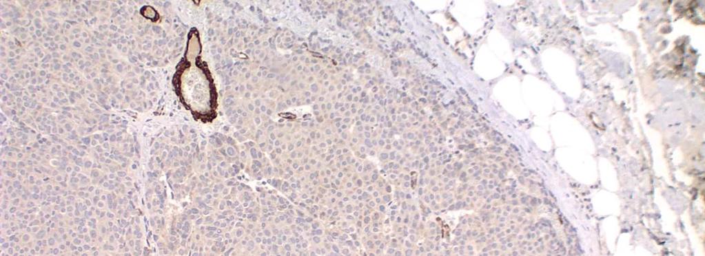
 Intra- and extra-cellular mucin production Frequently associated with")
67 Solid Papillary Carcinoma Considered to be a variant of DCIS Older women (mean age, early 70s) Mass, nipple discharge/bleeding Grossly well circumscribed Single or multiple circumscribed nodules of neoplastic cells, solid pattern Delicate to hyalinzed fibrovascular stromal network Endocrine differentiation common ( E-DCIS ) Intra- and extra-cellular mucin production Frequently associated with invasive mucinous ca and IDC


68

69


70

71
72 Synapto

73 SMMHC

74 SMMHC

75 Solid Papillary Carcinoma: No MEC SMA Rosen s Breast Pathology, 2 nd ed, 2001, Fig E

76

77 Lymph node
78 Solid Papillary Carcinoma In situ or Invasive Lesions? No myoepithelial cells at periphery Perineural invasion Lymph node metastases Excellent prognosis with adequate local therapy Are at least some low grade invasive carcinomas?
79 When there is doubt about the presence of invasion, solid papillary carcinomas should be regarded for staging purposes as a form of in situ carcinoma WHO, 2012
80 Papillary Lesions of the Breast Intraductal papilloma Papilloma with atypia (atypical papilloma) Papilloma with DCIS Lymph Papillary node DCIS papillomas Intracystic papillary carcinoma Solid papillary carcinoma Invasive papillary carcinoma


81 Lymph Node Papilloma Ichihara et al, AJSP, 2008


82 Lymph Node Papilloma Jaffer et al, Arch Pathol Lab Med, 2008
83 Lymph Node Papilloma Boulos, AJSP, 2014

84
85
86 Papillary Lesions of the Breast Intraductal papilloma Papilloma with atypia (atypical papilloma) Implications of papillary lesions Papilloma with DCIS on core needle biopsy Papillary DCIS Encapsulated papillary carcinoma Solid papillary carcinoma
87 Recommend excision for all patients with a targeted benign papilloma on CNB
88 Core Needle Biopsy Diagnoses for Atypical Papillary Lesions Atypical papillary lesion; excision recommended Papillary carcinoma, at least in situ; final categorization deferred to excision
89 Recommendations for CNB EPC/SPC on CNB requires excision No need to make a definitive diagnosis of EPC/SPC on CNB SLN not usually recommended
90 Summary Papillary lesions of the breast may be benign, atypical, in situ or invasive And perhaps even somewhere in between! Be aware of the existence of lymph node papillomas Targeted papillary lesions on core needle biopsy need excision Beware of post CNB changes in the excisional biopsy specimen, especially following biopsy of papillary lesions Important to communicate with our clinical colleagues the nature of the lesion excised; in particular whether myoepithelial cells are present or absent at the periphery of circumscribed nests of papillary carcinoma
Papillary Lesions of the Breast
 Papillary Lesions of the Breast Texas Society of Pathologists 2013 Laura C. Collins, M.D. Associate Professor of Pathology Associate Director, Division of Anatomic Pathology Beth Israel Deaconess Medical
Papillary Lesions of the Breast: WHO Update
 Papillary Lesions of the Breast: WHO Update Stuart J. Schnitt, M.D. Department of Pathology Beth Israel Deaconess Medical Center and Harvard Medical School Boston, MA, USA Papillary Lesions of the Breast
Papillary Lesions of the breast
 Papillary Lesions of the breast Emad Rakha Professor of Breast Pathology The University of Nottingham Papillary lesions of the breast are a heterogeneous group of disease, which are characterised by neoplastic
04/10/2018. Intraductal Papillary Neoplasms Of Breast INTRADUCTAL PAPILLOMA
 Intraductal Papillary Neoplasms Of Breast Savitri Krishnamurthy MD Professor of Pathology Deputy Division Head The University of Texas MD Anderson Cancer Center 25 th Annual Seminar in Pathology Pittsburgh,
Papillary Lesions of the Breast A Practical Approach to Diagnosis. (Arch Pathol Lab Med. 2016;140: ; doi: /arpa.
 Papillary Lesions of the Breast A Practical Approach to Diagnosis (Arch Pathol Lab Med. 2016;140:1052 1059; doi: 10.5858/arpa.2016-0219-RA) Papillary lesions of the breast Span the spectrum of benign,
Proliferative Epithelial lesions of the Breast. Sami Shousha, MD, FRCPath Charing Cross Hospital & Imperial College, London
 Proliferative Epithelial lesions of the Breast Sami Shousha, MD, FRCPath Charing Cross Hospital & Imperial College, London Amman, November2013 Proliferative Epithelial Lesions of the Breast Usual type
Benign Mimics of Malignancy in Breast Pathology
 Arthur Purdy Stout Society of Surgical Pathologists Companion Meeting Benign Mimics of Malignancy in Breast Pathology Stuart J. Schnitt, M.D. Beth Israel Deaconess Medical Center and Harvard Medical School,
Breast pathology. 2nd Department of Pathology Semmelweis University
 Breast pathology 2nd Department of Pathology Semmelweis University Breast pathology - Summary - Benign lesions - Acute mastitis - Plasma cell mastitis / duct ectasia - Fat necrosis - Fibrocystic change/
Papillary lesions of the breast: selected diagnostic and management issues
 Histopathology 2008, 52, 20 29. DOI: 10.1111/j.1365-2559.2007.02898.x REVIEW Papillary lesions of the breast: selected diagnostic and management issues L C Collins & S J Schnitt Department of Pathology,
Proliferative Breast Disease: implications of core biopsy diagnosis. Proliferative Breast Disease
 Proliferative Breast Disease: implications of core biopsy diagnosis Jean F. Simpson, M.D. Breast Pathology Consultants, Inc. Nashville, TN Proliferative Breast Disease Must be interpreted in clinical and
Papillary Lesions in Breast Pathology Practice: Diagnostic Challenges and Practical Approach. A Six- Year Experience from a Tertiary Care Hospital
 Open Access Journal Research Article DOI: 10.23958/ijirms/vol02-i05/12 Papillary Lesions in Breast Pathology Practice: Diagnostic Challenges and Practical Approach. A Six- Year Experience from a Tertiary
Ductal Carcinoma in Situ. Laura C. Collins, M.D. Department of Pathology Beth Israel Deaconess Medical Center and Harvard Medical School Boston, MA
 Ductal Carcinoma in Situ Laura C. Collins, M.D. Department of Pathology Beth Israel Deaconess Medical Center and Harvard Medical School Boston, MA Definition of DCIS WHO 2012 A neoplastic proliferation
Columnar Cell Lesions
 Columnar Cell Lesions Laura C. Collins, M.D. Department of Pathology Beth Israel Deaconess Medical Center and Harvard Medical School Boston, MA Question? Columnar cell lesions are: a) Annoying lesions
Flat Epithelial Atypia
 Flat Epithelial Atypia Richard Owings, M.D. University of Arkansas for Medical Sciences Department of Pathology Flat epithelial atypia can be a difficult lesion May be a subtle diagnosis Lots of changes
CLINICAL SIGNIFICANCE OF BENIGN EPITHELIAL CHANGES
 Papillomas. Papillomas are composed of multiple branching fibrovascular cores, each having a connective tissue axis lined by luminal and myoepithelial cells ( Fig. 23-11 ). Growth occurs within a dilated
Breast Pathology. Breast Development
 Breast Pathology Lecturer: Hanina Hibshoosh, M.D. Reading: Kumar, Cotran, Robbins, Basic Pathology, 6th Edition, pages 623-635 Breast Development 5th week - thickening of the epidermis - milk line 5th
3/27/2017. Disclosure of Relevant Financial Relationships. Papilloma???
 Management of Papillary Lesions Diagnosed at Rad Path Concordant Core Biopsy (CNB) Disclosure of Relevant Financial Relationships USCAP requires that all planners (Education Committee) in a position to
Epithelial Columnar Breast Lesions: Histopathology and Molecular Markers
 29th Annual International Conference Advances in the Application of Monoclonal Antibodies in Clinical Oncology and Symposium on Cancer Stem Cells 25 th -27t h June, 2012, Mykonos, Greece Epithelial Columnar
Enterprise Interest None
 Enterprise Interest None B3 lesions of the breast What are they at surgery? Case 4 Edi Brogi MD PhD Attending Pathologist - Director of Breast Pathology Memorial Sloan Kettering Cancer Center New York
In Situ Breast Carcinoma. James L. Connolly, M.D Beth Israel Deaconess Medical Center Professor of Pathology Harvard Medical School Boston, MA
 In Situ Breast Carcinoma James L. Connolly, M.D Beth Israel Deaconess Medical Center Professor of Pathology Harvard Medical School Boston, MA Content In Situ Ductal Carcinoma In Situ Lobular Carcinoma
Columnar Cell Lesions. Columnar Cell Lesions and Flat Epithelial Atypia
 Columnar Cell Lesions and Stuart J. Schnitt, M.D. Beth Israel Deaconess Medical Center and Harvard Medical School Boston, MA, USA Columnar Cell Lesions Lesions characterized by columnar epithelial cells
Case study 1. Rie Horii, M.D., Ph.D. Division of Pathology Cancer Institute Hospital, Japanese Foundation for Cancer Research
 NCCN/JCCNB Seminar in Japan April 15, 2012 Case study 1 Rie Horii, M.D., Ph.D. Division of Pathology Cancer Institute Hospital, Japanese Foundation for Cancer Research Present illness: A 50y.o.premenopausal
Columnar Cell Lesions and Flat Epithelial Atypia
 Columnar Cell Lesions and Flat Epithelial Atypia Laura C. Collins, M.D. Department of Pathology Beth Israel Deaconess Medical Center and Harvard Medical School, Boston, MA Terminology for Columnar Cell
Treatment options for the precancerous Atypical Breast lesions. Prof. YOUNG-JIN SUH The Catholic University of Korea
 Treatment options for the precancerous Atypical Breast lesions Prof. YOUNG-JIN SUH The Catholic University of Korea Not so benign lesions? Imaging abnormalities(10% recall) lead to diagnostic evaluation,
1 NORMAL HISTOLOGY AND METAPLASIAS
 1 NORMAL HISTOLOGY AND METAPLASIAS, MD Anatomy and Histology 1 Metaplasias 2 ANATOMY AND HISTOLOGY The female breast is composed of a branching duct system, which begins at the nipple with the major lactiferous
American Journal of Cancer Case Reports. Invasive Papillary Carcinoma of Male Breast: A Rare Case Report
 American Journal of Cancer Case Reports http://ivyunion.org/index.php/ajccr SantraAetal. American Journal of Cancer Case Reports 2014, 3:56-61 Page 1 of 6 Vol 3 Article ID 20140617, 6 pages Case Report
BREAST PATHOLOGY. Fibrocystic Changes
 BREAST PATHOLOGY Lesions of the breast are very common, and they present as palpable, sometimes painful, nodules or masses. Most of these lesions are benign. Breast cancer is the 2 nd most common cause
Basement membrane in lobule.
 Bahram Memar, MD Basement membrane in lobule. Normal lobule-luteal phase Normal lobule-follicular phase Lactating breast Greater than 95% are adenocarcinomas in situ carcinomas and invasive carcinomas.
Breast: Difficulties in Core Biopsies
 Breast: Difficulties in Core Biopsies Anna Marie Mulligan, MB, MSc, FRCPath University Health Network and University of Toronto E-mail: annamarie.mulligan@uhn.ca No conflicts of interest Role of Core Needle
Disclosures 5/27/2012. Outline of Talk. Outline of Talk. When Is LCIS Clinically Significant? Classic LCIS. Classic LCIS
 When Is LCIS Clinically Significant? Disclosures I have nothing to disclose Yunn-Yi Chen, MD, PhD Professor Outline of Talk Outline of Talk Classic LCIS Classic LCIS Definition of lobular differentiation
Interpretation of Breast Pathology in the Era of Minimally Invasive Procedures
 Shahla Masood, M.D. Professor and Chair Department of Pathology and Laboratory Medicine University of Florida College of Medicine Jacksonville Medical Director, UF Health Breast Center Chief of Pathology
04/10/2018 HIGH RISK BREAST LESIONS. Pathology Perspectives of High Risk Breast Lesions ELEVATED RISK OF BREAST CANCER HISTORICAL PERSPECTIVES
 Pathology Perspectives of High Risk Breast Lesions Savitri Krishnamurthy MD Professor of Pathology Deputy Division Head Director of Clinical Trials, Research and Development The University of Texas MD
Notice of Faculty Disclosure
 California Society of Pathology Diagnostic Problems in Surgical Pathology December 2015 Case 2 Laura C. Collins, M.D. Associate Professor of Pathology Associate Director of Anatomic Pathology Beth Israel
Good afternoon everyone. First of all many thanks to Dr. Bonaventura and Dr. Arn for inviting
 PATHOLOGY IN-SITU CARCINOMA, ROHIT BHARGAVA, MD 1 Good afternoon everyone. First of all many thanks to Dr. Bonaventura and Dr. Arn for inviting me here, it s great to be here and I m going to talk about
Are Encapsulated Papillary Carcinomas of the Breast In Situ or Invasive?
 Anatomic Pathology / Encapsulated Papillary Breast Carcinomas Are Encapsulated Papillary Carcinomas of the Breast In Situ or Invasive? A Basement Membrane Study of 27 Cases Nicole Nicosia Esposito, MD,
Lesion Imaging Characteristics Mass, Favoring Benign Circumscribed Margins Intramammary Lymph Node
 Lesion Imaging Characteristics Mass, Favoring Benign Circumscribed Margins Intramammary Lymph Node Oil Cyst Mass, Intermediate Concern Microlobulated Margins Obscured Margins Mass, Favoring Malignant Indistinct
Are Encapsulated Papillary Carcinomas of the Breast In Situ or Invasive?
 Anatomic Pathology / Encapsulated Papillary Breast Carcinomas Are Encapsulated Papillary Carcinomas of the Breast In Situ or Invasive? A Basement Membrane Study of 27 Cases Nicole Nicosia Esposito, MD,
Papillary lesions of the breast: A Radiopathological Pictorial Review and Diagnostic Work-up
 Papillary lesions of the breast: A Radiopathological Pictorial Review and Diagnostic Work-up R M Lorente-Ramos 1, MD, PhD; J Azpeitia Armán 1, MD; I Casado Fariñas 2, MD; T Rivera García 2, MD; Cueva Pérez
Benign Breast Disease and Breast Cancer Risk
 Benign Breast Disease and Breast Cancer Risk Jean F. Simpson, M.D. Vanderbilt University Nashville, Tennessee December 1, 2011 Nashville Nashville Lebanon 1 Cedars of Lebanon State Park The American University
Diseases of the breast (1 of 2)
") Diseases of the breast (1 of 2) Introduction A histology introduction Normal ducts and lobules of the breast are lined by two layers of cells a layer of luminal cells overlying a second layer of myoepithelial
Diseases of the breast (1 of 2) Introduction A histology introduction Normal ducts and lobules of the breast are lined by two layers of cells a layer of luminal cells overlying a second layer of myoepithelial
Controversies and Problematic Issues in Core Needle Biopsies (To excise or not to excise)
") Controversies and Problematic Issues in Core Needle Biopsies (To excise or not to excise) Laura C. Collins, M.D. Beth Israel Deaconess Medical Center and Harvard Medical School Boston, MA Schematic Representation
Controversies and Problematic Issues in Core Needle Biopsies (To excise or not to excise) Laura C. Collins, M.D. Beth Israel Deaconess Medical Center and Harvard Medical School Boston, MA Schematic Representation
The Hot Topic for today is a biopsy from a 58-year-old woman who had worrisome mammographic calcifications on screening.
 The Hot Topic for today is a biopsy from a 58-year-old woman who had worrisome mammographic calcifications on screening. 1 My name is Dan Visscher; I am a consultant in the Division of Anatomic Pathology
6/3/2010. Outline of Talk. Lobular Breast Cancer: Definition of lobular differentiation. Common Problems in Diagnosing LCIS in Core Biopsies
 Outline of Talk Lobular Breast Cancer: Common Problems in Diagnosing LCIS in Core Biopsies Definition of lobular differentiation Variants of LCIS that: carry risk for unsampled invasive cancer mimic DCIS
Atypical papillary lesions after core needle biopsy and subsequent breast carcinoma
 Asian Biomedicine Vol. 5 No. 2 April 2011; 243-248 DOI: 10.5372/1905-7415.0502.031 Original article Atypical papillary lesions after core needle biopsy and subsequent breast carcinoma Tuenchit Khamapirad
GOALS AND OBJECTIVES BREAST PATHOLOGY
 GOALS AND OBJECTIVES BREAST PATHOLOGY LEVEL: PGY2, PGY3, PGY5 A number of these rotations are introductory in nature, as they are major subspecialties, and are followed by two more blocks in PGY-3, during
Question 1 A. ER-, PR-, HER+ B. ER+, PR+, HER2- C. ER-, PR+, HER2- D. ER-, PR-, HER2- E. ER-, PR+, HER2+
 Triple Negative Breast Cancer Laura C. Collins, M.D. Department of Pathology Beth Israel Deaconess Medical Center and Harvard Medical School, Boston, MA Question 1 The tumor depicted on the next slide
Image guided core biopsies:
 Recommendations on the Surgical, Radiologic and Pathologic Approaches to Breast Disease: Using best practices based on multidisciplinary methodologies developed through the Allina Breast Committee. Image
Case Report Synchronous Bilateral Solid Papillary Carcinomas of the Breast
 Case Reports in Surgery Volume 2013, Article ID 812129, 4 pages http://dx.doi.org/10.1155/2013/812129 Case Report Synchronous Bilateral Solid Papillary Carcinomas of the Breast Noriko Yoshimura, 1 Shigeru
Case Report Basaloid ductal carcinoma in situ arising in salivary gland metaplasia of the breast: a case report
 Int J Clin Exp Pathol 2014;7(9):6370-6374 www.ijcep.com /ISSN:1936-2625/IJCEP0001480 Case Report Basaloid ductal carcinoma in situ arising in salivary gland metaplasia of the breast: a case report Eun
LYMPHATIC DRAINAGE AXILLARY (MOSTLY) INTERNAL MAMMARY SUPRACLAVICULAR
 INTERNAL MAMMARY SUPRACLAVICULAR") BREAST LYMPHATIC DRAINAGE AXILLARY (MOSTLY) INTERNAL MAMMARY SUPRACLAVICULAR HISTOLOGY LOBE: (10 in whole breast) LOBULE: (many per lobe) ACINUS/I, aka ALVEOLUS/I: (many per lobule) DUCT(S): INTRA- or
BREAST LYMPHATIC DRAINAGE AXILLARY (MOSTLY) INTERNAL MAMMARY SUPRACLAVICULAR HISTOLOGY LOBE: (10 in whole breast) LOBULE: (many per lobe) ACINUS/I, aka ALVEOLUS/I: (many per lobule) DUCT(S): INTRA- or
Pathology of Lobular & Ductal Preneoplasia. Syed A Hoda, MD Weill-Cornell, New York, NY
 Pathology of Lobular & Ductal Preneoplasia Syed A Hoda, MD Weill-Cornell, New York, NY Proliferative Epithelial Changes in Breast A wide range of proliferative epithelial changes occur in the breast There
Histological Type. Morphological and Molecular Typing of breast Cancer. Nottingham Tenovus Primary Breast Cancer Study. Survival (%) Ian Ellis
 Ian Ellis") Morphological and Molecular Typing of breast Cancer Ian Ellis Molecular Medical Sciences, University of Nottingham Department of Histopathology, Nottingham University Hospitals NHS Trust Histological Type
Morphological and Molecular Typing of breast Cancer Ian Ellis Molecular Medical Sciences, University of Nottingham Department of Histopathology, Nottingham University Hospitals NHS Trust Histological Type
HISTOMORPHOLOGICAL SPECTRUM OF BREAST LESIONS
 HISTOMORPHOLOGICAL SPECTRUM OF BREAST LESIONS Kiran H. S, Jayaprakash Shetty, Chandrika Rao Assistant Professor, Department of Pathology, Yenepoya Medical College, Mangalore. Professor, Department of Pathology,
COMPANION MEETING BREAST. Auditorium 11:15 1:00 am. Convenor: A/Professor Gelareh Farshid, SA Pathology, SA
 Australasian Division of the International Academy of Pathology Limited ABN 73 008 593 815 36 TH Annual Scientific Meeting Darling Harbour Convention Centre, Sydney, Australia June 3-5, 2011 COMPANION
Case year old female presented with asymmetric enlargement of the left lobe of the thyroid
 Case 4 22 year old female presented with asymmetric enlargement of the left lobe of the thyroid gland. No information available relative to a prior fine needle aspiration biopsy. A left lobectomy was performed.
Case 4 Diagnosis 2/21/2011 TGB
 Case 4 22 year old female presented with asymmetric enlargement of the left lobe of the thyroid gland. No information available relative to a prior fine needle aspiration biopsy. A left lobectomy was performed.
Update on 2015 WHO Classification of Lung Adenocarcinoma 1/3/ Mayo Foundation for Medical Education and Research. All rights reserved.
 1 Our speaker for this program is Dr. Anja Roden, an associate professor of Laboratory Medicine and Pathology at Mayo Clinic as well as consultant in the Anatomic Pathology Laboratory and co-director of
ARTHUR PURDY STOUT SOCIETY COMPANION MEETING: DIFFICULT NEW DIFFERENTIAL DIAGNOSES IN PROSTATE PATHOLOGY. Jonathan I. Epstein.
 1 ARTHUR PURDY STOUT SOCIETY COMPANION MEETING: DIFFICULT NEW DIFFERENTIAL DIAGNOSES IN PROSTATE PATHOLOGY Jonathan I. Epstein Professor Pathology, Urology, Oncology The Reinhard Professor of Urological
Papillary lesions of the breast - Imaging findings and diagnostic challenges
 Papillary lesions of the breast - Imaging findings and diagnostic challenges Poster No.: R-0146 Congress: RANZCR-AOCR 2012 Type: Educational Exhibit Authors: P. Jagmohan, F. J. Pool Keywords: Breast, Mammography,
Cytyc Corporation - Case Presentation Archive - March 2002
 FirstCyte Ductal Lavage History: 68 Year Old Female Gail Index: Unknown Clinical History: Negative Mammogram in 1995 6 yrs. later presents with bloody nipple discharge Subsequent suspicious mammogram Suspicious
Case 1. ACCME/Disclosure. Clinical History. Dr. Mulligan has nothing to disclose
 Breast Evening Specialty Conference USCAP, 2016 Case 1 Anna Marie Mulligan University Health Network, Toronto University of Toronto ACCME/Disclosure Dr. Mulligan has nothing to disclose Clinical History
ACRIN 6666 Therapeutic Surgery Form
 S1 ACRIN 6666 Therapeutic Surgery Form 6666 Instructions: Complete a separate S1 form for each separate area of each breast excised with the intent to treat a cancer (e.g. each lumpectomy or mastectomy).
A 60-year old Man with Left Jaw Mass. Simon Chiosea, MD University of Pittsburgh medical Center 3/15/2016
 ACCME/Disclosures The USCAP requires that anyone in a position to influence or control the content of CME disclose any relevant financial relationship WITH COMMERCIAL INTERESTS which they or their spouse/partner
Follicular Derived Thyroid Tumors
 Follicular Derived Thyroid Tumors Jennifer L. Hunt, MD, MEd Aubrey J. Hough Jr, MD, Endowed Professor of Pathology Chair of Pathology and Laboratory Medicine University of Arkansas for Medical Sciences
ACCME/Disclosures. Cribriform Lesions of the Prostate. Case
 Cribriform Lesions of the Prostate Ming Zhou, MD, PhD Departments of Pathology and Urology New York University Langone Medical Center New York, NY Ming.Zhou@NYUMC.ORG ACCME/Disclosures The USCAP requires
Case Report Intracystic Papillary Carcinoma of the Breast: Report of Three Cases and Literature Review
 Case Reports in Obstetrics and Gynecology Volume 2012, Article ID 979563, 4 pages doi:10.1155/2012/979563 Case Report Intracystic Papillary Carcinoma of the Breast: Report of Three Cases and Literature
Minimizing Errors in Diagnostic Pathology
 Shahla Masood, M.D. Professor and Chair Department of Pathology and Laboratory Medicine University of Florida College of Medicine-Jacksonville Medical Director, Shands Jacksonville Breast Health Center
EQA Circulation 43 Educational Cases
 EQA Circulation 43 Educational Cases E1-E2 Monica Agarwal Monklands Hospital E1 38 yrs male Submandibular gland tumour E1 Formal excision following diagnosis of poorly differentiated carcinoma on core
In situ lobular neoplasia of the breast with marked myoepithelial proliferation
 In situ lobular neoplasia of the breast with marked myoepithelial proliferation Sami Shousha To cite this version: Sami Shousha. In situ lobular neoplasia of the breast with marked myoepithelial proliferation.
Low-grade Adenosquamous Carcinoma Coexisting with Sclerosing Adenosis of the Breast: A Case Report
 31 Case Report J. St. Marianna Univ. Vol. 8, pp. 31 35, 2017 Low-grade Adenosquamous Carcinoma Coexisting with Sclerosing Adenosis of the Breast: A Case Report Ryoko Oi 1, 2, Ichiro Maeda 1, Yoshio Aida
Overview of Pathology Evaluation of Breast Lesions and Quality Assurance
 Overview of Pathology Evaluation of Breast Lesions and Quality Assurance 2 Michael O. Idowu, Jaime A. Singh, and Margaret M. Grimes Masses/Densities/Distortions: General Considerations Radiologic evaluation
PSA. HMCK, p63, Racemase. HMCK, p63, Racemase
 Case 1 67 year old male presented with gross hematuria H/o acute prostatitis & BPH Urethroscopy: small, polypoid growth with a broad base emanating from the left side of the verumontanum Serum PSA :7 ng/ml
Promise of a beautiful day
 Promise of a beautiful day Ductal carcinoma in Situ Lobular Carcinoma in Situ Natural History Manosmed Tartous Oct 2009 Gérard ABADJIAN MD Pathology Department Hôtel-Dieu de France. Associate Professor
Atypical proliferative lesions diagnosed on core biopsy - 6 year review
 Atypical proliferative lesions diagnosed on core biopsy - 6 year review Dr Angela Harris, Dr Julie Weigner & Dr Ricardo Vilain NSW Health Pathology Pathology North, Hunter Anatomical Pathology & Cytology
Cystic Hypersecretory Carcinoma of the Breast:
 J Korean Soc Radiol 2010;62:287-294 Cystic Hypersecretory Carcinoma of the Breast: Sonographic Features with a Histological Correlation 1 Sang Yu Nam, M.D., Boo-Kyung Han, M.D., Jung Hee Shin, M.D., Eun
Title malignancy. Issue Date Right 209, 12, (2013)
") NAOSITE: Nagasaki University's Ac Title Author(s) A case of intracystic apocrine papi malignancy Hayashi, Hiroko; Ohtani, Hiroshi; Y Citation Pathology - Research and Practice, Issue Date 2013-12 URL Right
NAOSITE: Nagasaki University's Ac Title Author(s) A case of intracystic apocrine papi malignancy Hayashi, Hiroko; Ohtani, Hiroshi; Y Citation Pathology - Research and Practice, Issue Date 2013-12 URL Right
Lesions Mimicking Adenoid Cystic Carcinoma. Diagnostic Problems in Salivary Gland Pathology An Update 5/29/2009
 Diagnostic Problems in Salivary Gland Pathology An Update Lesions Mimicking Adenoid Cystic Carcinoma Stacey E. Mills, M.D. W.S. Royster Professor of Pathology Director of Surgical and Cytopathology University
A712(18)- Test slide, Breast cancer tissues with corresponding normal tissues
- Test slide, Breast cancer tissues with corresponding normal tissues") A712(18)- Test slide, Breast cancer tissues with corresponding normal tissues (formalin fixed) For research use only Specifications: No. of cases: 12 Tissue type: Breast cancer tissues with corresponding
A712(18)- Test slide, Breast cancer tissues with corresponding normal tissues (formalin fixed) For research use only Specifications: No. of cases: 12 Tissue type: Breast cancer tissues with corresponding
High risk lesions of the breast : Review of the current diagnostic and management strategies
 High risk lesions of the breast : Review of the current diagnostic and management strategies Poster No.: C-1204 Congress: ECR 2016 Type: Educational Exhibit Authors: P. Jagmohan, F. J. Pool, P. G. Pillay,
ACCME/Disclosures. Diagnosing Mesothelioma in Limited Tissue Samples. Papanicolaou Society of Cytopathology Companion Meeting March 12 th, 2016
 Diagnosing Mesothelioma in Limited Tissue Samples Papanicolaou Society of Cytopathology Companion Meeting March 12 th, 2016 Sanja Dacic, MD, PhD University of Pittsburgh ACCME/Disclosures GENERAL RULES
Invasive Papillary Breast Carcinoma
 410 This is an Open Access article licensed under the terms of the Creative Commons Attribution- NonCommercial-NoDerivs 3.0 License (www.karger.com/oa-license), applicable to the online version of the
Case #1: 75 y/o Male (treated and followed by prostate cancer oncology specialist ).
.") SOLID TUMORS WORKSHOP Cases for review Prostate Cancer Case #1: 75 y/o Male (treated and followed by prostate cancer oncology specialist ). January 2009 PSA 4.4, 20% free; August 2009 PSA 5.2; Sept 2009
SOLID TUMORS WORKSHOP Cases for review Prostate Cancer Case #1: 75 y/o Male (treated and followed by prostate cancer oncology specialist ). January 2009 PSA 4.4, 20% free; August 2009 PSA 5.2; Sept 2009
CNB vs Surgical Excision
 Update on Core Needle Biopsy of Non-palpable Breast Lesions Nour Sneige, M.D. UT MD Anderson Cancer Center Houston, Tx Image-Guided CNB of Breast Lesions An alternative to surgical biospy CNB vs Surgical
A712(19)- Test slide, Breast cancer tissues with corresponding normal tissues
- Test slide, Breast cancer tissues with corresponding normal tissues") A712(19)- Test slide, Breast cancer tissues with corresponding normal tissues (formalin fixed) For research use only Specifications: No. of cases: 12 Tissue type: Breast cancer tissues with corresponding
A712(19)- Test slide, Breast cancer tissues with corresponding normal tissues (formalin fixed) For research use only Specifications: No. of cases: 12 Tissue type: Breast cancer tissues with corresponding
Quality ID #263: Preoperative Diagnosis of Breast Cancer National Quality Strategy Domain: Effective Clinical Care
 Quality ID #263: Preoperative Diagnosis of Breast Cancer National Quality Strategy Domain: Effective Clinical Care 2018 OPTIONS FOR INDIVIDUAL MEASURES: REGISTRY ONLY MEASURE TYPE: Process DESCRIPTION:
RSNA, /radiol Appendix E1. Methods
 RSNA, 2016 10.1148/radiol.2016151097 Appendix E1 Methods US and Near-infrared Data Acquisition Four optical wavelengths (740 nm, 780 nm, 808 nm, and 830 nm) were used to sequentially deliver the light
Incidence of ductal lesions
 Ductal Proliferative Lesions of the Breast: From FEA to ADH to DCIS Incidence of ductal lesions Pre-mammography: DCIS < 3% of breast cancers, large palpable masses, with invasion Mammography: DCIS 25%
Intraductal carcinoma of the prostate on needle biopsy: histologic features and clinical significance
 & 2006 USCAP, Inc All rights reserved 0893-3952/06 $30.00 www.modernpathology.org Intraductal carcinoma of the prostate on needle biopsy: histologic features and clinical significance Charles C Guo 1 and
University Journal of Pre and Para Clinical Sciences
 ISSN 2455 2879 Volume 2 Issue 1 2016 Metaplastic carcinoma breast a rare case report Abstract : Metaplastic carcinoma of the breast is a rare malignancy with two distinct cell lines described as a breast
CASE REPORT Malignant transformation of breast ductal adenoma: a diagnostic pitfall
 Malaysian J Pathol 2015; 37(3) : 281 285 CASE REPORT Malignant transformation of breast ductal adenoma: a diagnostic pitfall Hiroko HAYASHI, Hiroshi OHTANI,* Junzo YAMAGUCHI,** and Isao SHIMOKAWA Department
Rare Breast Cancers and Male Breast Cancer
 Johns Hopkins Hospital, Baltimore MD Rare Breast Cancers and Male Breast Cancer David Euhus, MD, FACS Professor of Surgery (PAR) Division of Surgical Oncology Breast Cancer Primary Cancers (97% of breast
Maram Abdaljaleel, MD Dermatopathologist and Neuropathologist University of Jordan, School of Medicine
 Maram Abdaljaleel, MD Dermatopathologist and Neuropathologist University of Jordan, School of Medicine The most common non-skin malignancy of women 2 nd most common cause of cancer deaths in women, following
Update in Salivary Gland Pathology. Benjamin L. Witt University of Utah/ARUP Laboratories February 9, 2016
 Update in Salivary Gland Pathology Benjamin L. Witt University of Utah/ARUP Laboratories February 9, 2016 Objectives Review the different appearances of a selection of salivary gland tumor types Establish
COMMON CONSULTATION CONUNDRUMS IN BREAST PATHOLOGY
 COMMON CONSULTATION CONUNDRUMS IN BREAST PATHOLOGY SANDRA J. SHIN CHIEF OF BREAST PATHOLOGY ASOCIATE PROFESSOR OF PATHOLOGY AND LABORATORY MEDICINE NEW YORK PRESBYTERIAN HOSPITAL-WEILL CORNELL MEDICAL
Atypical Ductal Hyperplasia and Papillomas: A Comparison of Ultrasound Guided Breast Biopsy and Stereotactic Guided Breast Biopsy
 Atypical Ductal Hyperplasia and Papillomas: A Comparison of Ultrasound Guided Breast Biopsy and Stereotactic Guided Breast Biopsy Breast Cancer is the most common cancer diagnosed in women in the United
Immunohistochemical studies (ER & Ki-67) in Proliferative breast lesions adjacent to malignancy
 in Proliferative breast lesions adjacent to malignancy") IOSR Journal of Dental and Medical Sciences (IOSR-JDMS) e-issn: 2279-0853, p-issn: 2279-0861.Volume 13, Issue 3 Ver. IV. (Mar. 2014), PP 84-89 Immunohistochemical studies (ER & Ki-67) in Proliferative
IOSR Journal of Dental and Medical Sciences (IOSR-JDMS) e-issn: 2279-0853, p-issn: 2279-0861.Volume 13, Issue 3 Ver. IV. (Mar. 2014), PP 84-89 Immunohistochemical studies (ER & Ki-67) in Proliferative
Diagnosis of Fibroepithelial and Mesenchymal Lesions on Core Needle Biopsy
 Diagnosis of Fibroepithelial and Mesenchymal Lesions on Core Needle Biopsy Emmanuel Agosto-Arroyo, MD Assistant Member Department of Anatomic Pathology 3/3/2018 Disclosure There are no conflicts of interest.
Disclosure. Relevant Financial Relationship(s) None. Off Label Usage None MFMER slide-1
 None. Off Label Usage None MFMER slide-1") Disclosure Relevant Financial Relationship(s) None Off Label Usage None 2013 MFMER slide-1 Case Presentation A 43 year old male, with partial nephrectomy for a right kidney mass 2013 MFMER slide-2 2013
Disclosure Relevant Financial Relationship(s) None Off Label Usage None 2013 MFMER slide-1 Case Presentation A 43 year old male, with partial nephrectomy for a right kidney mass 2013 MFMER slide-2 2013
Breast Cancer. Most common cancer among women in the US. 2nd leading cause of death in women. Mortality rates though have declined
 Breast Cancer Most common cancer among women in the US 2nd leading cause of death in women Mortality rates though have declined 1 in 8 women will develop breast cancer Breast Cancer Breast cancer increases
IBCM 2, April 2009, Sarajevo, Bosnia and Herzegovina
 Preoperative diagnosis and treatment planning in breast cancer The pathologist s perspective L. Mazzucchelli Istituto Cantonale di Patologia Locarno, Switzerland IBCM 2, 23-25 April 2009, Sarajevo, Bosnia