Adipocytic Tumours in children
|
|
|
- Scot Gibson
- 6 years ago
- Views:
Transcription


1 Università degli Studi di Padova Dipartimento di Medicina Sezione di Anatomia Patologica Generale e Citopatologia Adipocytic Tumours in children Rita Alaggio Basel Seminars in Pathology Paediatric Pathology & Genetics Basel, June 22nd 25th, 2016
2 Outline Classification of adipocytic tumors Lipoblastoma Lipoblastoma: Differential Diagnostic Challenges Liposarcoma
3 WHO 2013 Classification BENIGN Lipoma Lipoblastomatosis Lipomatosis of Nerve Lipoblastoma Angiomyolipoma Myolipoma of soft tissue Chondroid lipoma Spindle cell/pleomorphic lipoma Hybernoma MALIGNANT Atypical lipomatous tumor Dedifferentiated liposarcoma Myxoid liposarcoma Pleomorphic liposarcoma


4 WHO 2013 Classification BENIGN Lipoma Lipoblastomatosis Lipomatosis of Nerve Lipoblastoma Angiomyolipoma Myolipoma of soft tissue Chondroid lipoma Spindle cell/pleomorphic lipoma Hybernoma MALIGNANT Atypical lipomatous tumor Dedifferentiated liposarcoma Myxoid liposarcoma Pleomorphic liposarcoma

5 Outline Classification of adipocytic tumors Lipoblastoma Lipoblastoma: Differential Diagnostic Challenges Liposarcoma
6

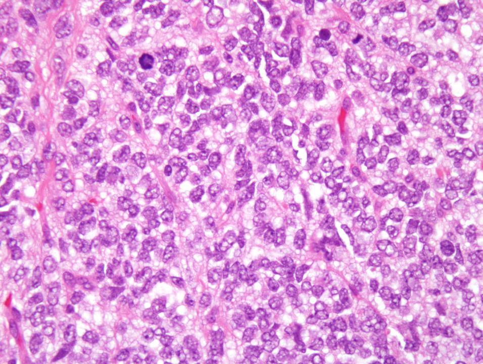
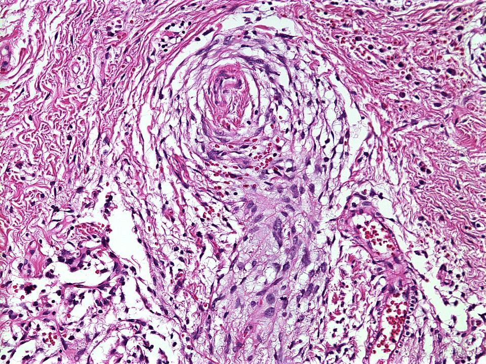
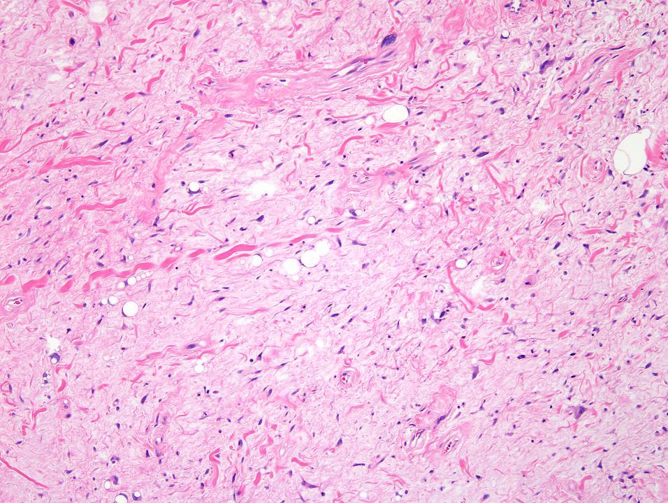
7 Case 1 Multivacuolated and univacuolated lipoblasts
8 Clinical History Boy, 1.5 yr expanding mass in the paravertebral region Main diameter 3 cm Treatment: resection
9 Clinical History Boy, 1.5 yr expanding mass in the paravertebral region Main diameter 3 cm Treatment: resection Diagnosis: Lipoblastoma
10 Lipoblastoma Lipoblastoma, a benign neoplasm of embryonal white fat, is a localized or diffuse (lipoblastomatosis) tumor with a tendency for local recurrence if incompletely excised. - WHO, 2013
11 Lipoblastoma Clinical Features 90% before 3 years 40% 1 st year of life Rare in adolescents and adults Sex: male predilection Site: trunk, extremities Others: retroperitoneum, pelvis, abdomen, head/neck, organs (lung, heart) Benign clinical course 13-46% of relapse in incomplete resection

12 Lipoblastoma (Courtesy of C. Coffin)

13 Lipoblastomatosis (Courtesy of C. Coffin)

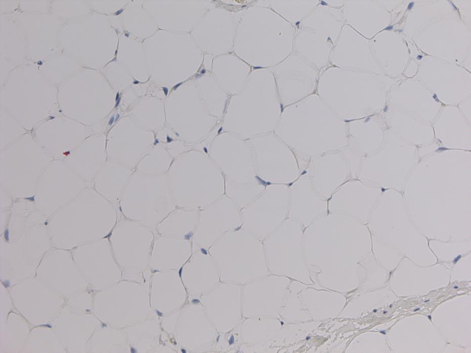
14 Lipoblastoma Histology Lobulated architecture with fibrovascular septa Lipoblasts in various stages of differentiation to mature fat with orientation from periphery to the center: primitive stellate or spindle cells multivacuolate lipoblasts signet ring cells Floret cells Hibernoma like cells Myxoid foci with plexiform vascular pattern, primitive mesenchymal cells Chronic inflammation, mast cells Fibrolipoma-like areas possible
15 Lipoblastoma Histology Lobulated architecture with fibrovascular septa Lipoblasts in various stages of differentiation to mature fat with orientation from periphery to the center: primitive stellate or spindle cells multivacuolate lipoblasts signet ring cells Floret cells Hibernoma like cells Myxoid foci with plexiform vascular pattern, primitive mesenchymal cells Chronic inflammation, mast cells Fibrolipoma-like areas possible
16 Lipoblastoma Histology Lobulated architecture with fibrovascular septa Lipoblasts in various stages of differentiation to mature fat with orientation from periphery to the center: primitive stellate or spindle cells multivacuolate lipoblasts signet ring cells Floret cells Hibernoma like cells Myxoid foci with plexiform vascular pattern, primitive mesenchymal cells Fibrolipoma-like areas (eg in maturing lipoblastoma) Chronic inflammation, mast cells
17 Abdominal Lipoblastoma, age 2 yr. Resection

18 Abdominal Lipoblastoma, age 2 yr. Recurrence, 5yr
19 Lipoblastoma Immunohistochemistry S100, CD34: positive nonspecific Desmin: in primitive mesenchymal cells: a potential pitfall PLAG1 protein: not liable p16: negative

")
20 p16 -ve Desmin p16 +ve (2 cases/30) S100 Lipoblastoma (Cappellesso et al Hum Pathol 2016)
21 Lipoblastoma Genetics Chromosome 8 aberrations typical, including extra copies in 10-15% Breakpoints cluster to 8q11-13 involving PLAG1 in 8q12.1 Translocation partners vary: 3q12-13, COL1A2 in 7q21.3 HAS2 in 8q24.13 rarely COL3A1 and RAB2A* others *Yoshida et al. Genes, Chromosomes & Cancer 53: (2014)
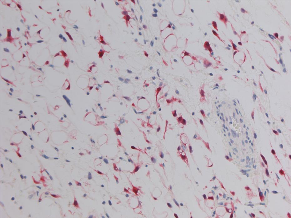
22 Lipoblastoma: Genetic Alterations Chromosomal Rearrangements Increase of PLAG1 copy number IGF2 Activation Adipocytes Proliferation

23 Chromosomal Rearrangements of PLAG1 HAS2 (hyaluronic acid synthase 2) Check and transport of Ialuronic Acid COL1A2 (collagen 1 alpha 2) Involved in the formation of the collagen in connective tissue, bone and tendons Potential role in the genesis of myxoid component? Potential role in collagen septae formation?
24 Lipoblastoma Associated Conditions Developmental delay or abnormalities Congenital malformations Seizures Sturge-Weber syndrome Autism Familial lipomas

25 Outline Classification of adipocytic tumors Lipoblastoma Lipoblastoma: Differential Diagnostic Challenges Liposarcoma
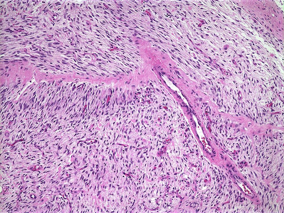
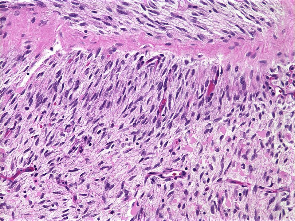
26 Lipoblastoma Differential Diagnostic Challenges Lipoma and variants Primitive myxoid mesenchymal tumor of infancy Lipofibromatosis Fibrous Hamartoma Liposarcomas, especially myxoid and myxoid-pleomorphic Other fatty and myxoid tumors

27 Lipoma Hibernoma
28 Genetic aberrations in pediatric lipoma Similar to adult lipomas In literature less than 30 pediatric lipomas investigated: 11 with aberrations of the 12q14 q15 region, with rearrangement of the HMGA2 gene in 3, 1 with anomaly of 6p21 region, resulting in a rearrangement of HMGA1 NFIB identified as a fusion partner of HMGA2 in pediatric lipoma. NFIB is a regulator of adipocyte differentiation; its inhibition suppresses induction of adipogenic transcription factors, (PPARγ, C/EBPa etc), and reduces accumulation of lipids during differentiation. Cancer Genetics 208 (2015)
29 Lipoblastoma Differential Diagnostic Challenges Lipoma and variants Primitive myxoid mesenchymal tumor of infancy Lipofibromatosis Fibrous Hamartoma Liposarcomas, especially myxoid and myxoid-pleomorphic Other fatty and myxoid tumors

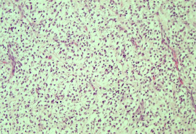
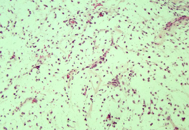
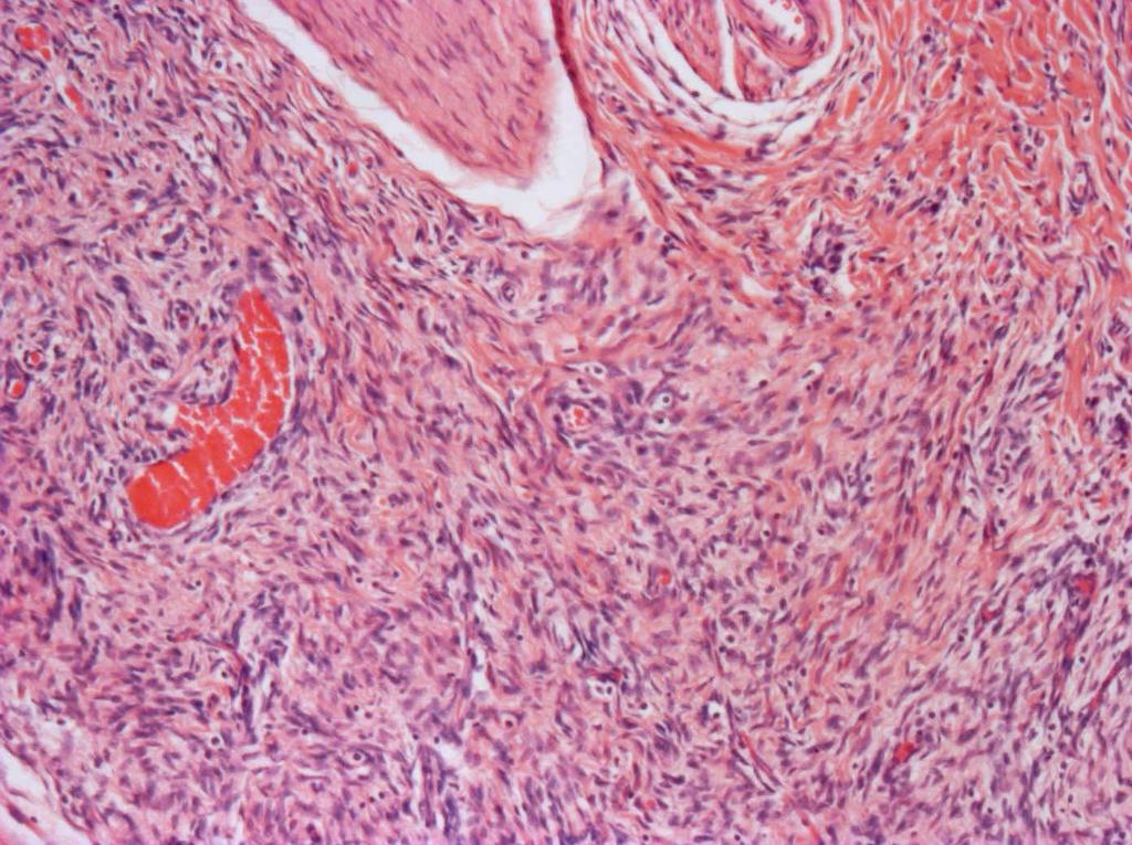
30 Vimentin Primitive myxoid mesenchymal tumor of infancy
31

32 Initial diagnosis of Lipoblastoma (2/30 cases in Italian series)
33 Lipoblastoma Differential Diagnostic Challenges Lipoma and variants Fibrous Hamartoma Primitive myxoid mesenchymal tumor of infancy Lipofibromatosis Liposarcomas, especially myxoid and myxoid-pleomorphic Other fatty and myxoid tumors
34

35
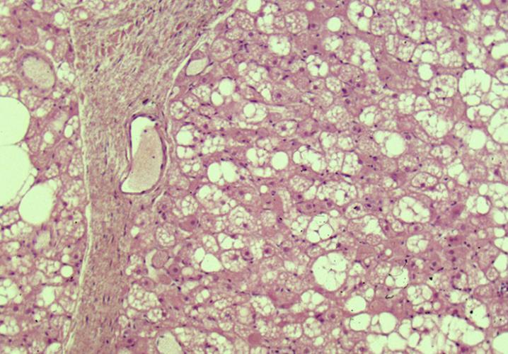
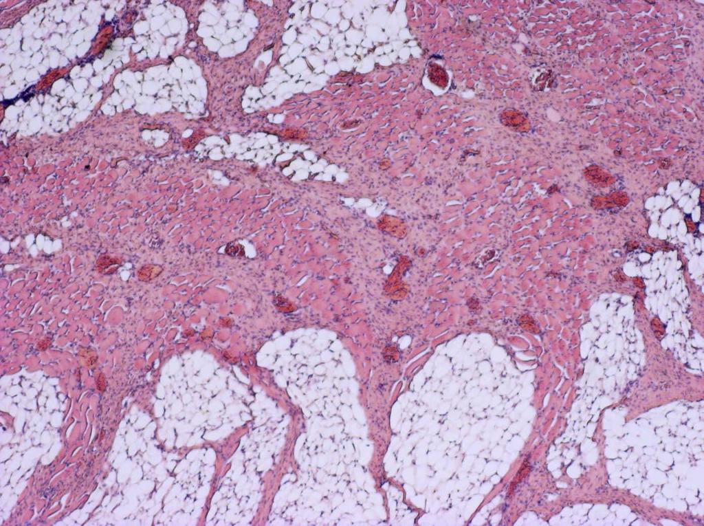
36 Fibrous Hamartoma In our series 2 originally diagnosed as lipoblastoma (out of 30 lipoblastomas) Slight male predilection Site: axilla, upper extremities, trunk, groin, external genitalia poorly circumscribed, nodular lesion Organoid lesion, 3 components Dense fibrous trabeculae Primitive mesenchymal cells in nests and bands in the context of a myxoid matrix Mature adipose tissue Rare multicentric cases
37
38
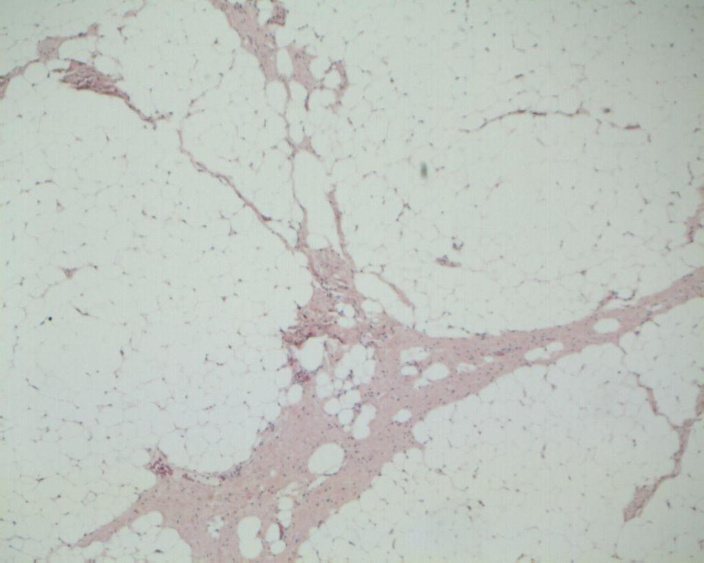
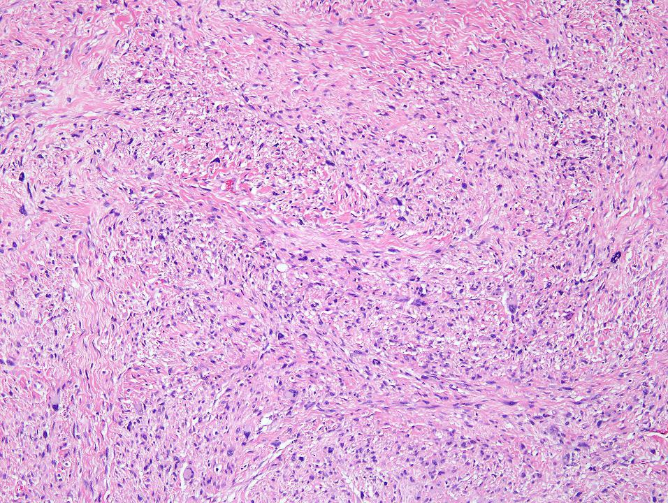
39
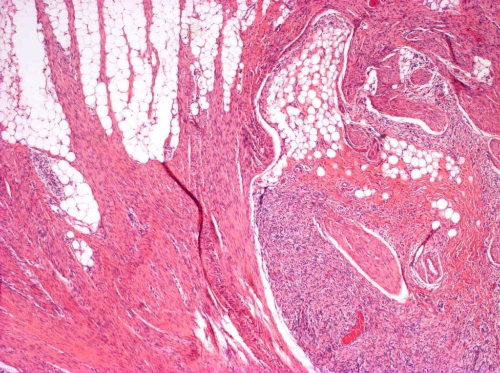
40 Lipofibromatosis Histology Abundant adipose tissue (>50%) Slender fascicles of fibroblasts among the adipose lobules Myxoid component lipoblast-like cells at the periphery along the fibrous septae Infiltrative growth pattern with entrapment of nerve trunks and muscle Dendritic cells (occasional) DD with Lipoblastoma: more prominent connectival component and fibroblastic proliferation
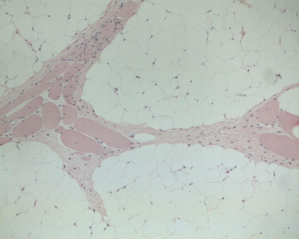
41
42 Lipofibromatosis Clinical Features Myofibroblastic lesion in the past diagnosed as: fibrous hamartoma, fibromatosis, CIFS, lipoblastoma e calcifying aponeurotic fibroma Frequently congenital Site: extremities (frequent), thorax, abdomen, head (less frequent) Tendency to relapse Treatment: conservative surgery
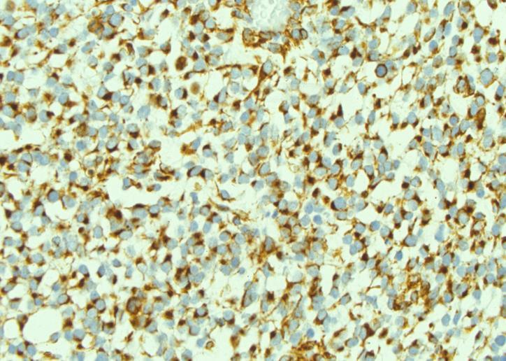
43 Lipofibromatosis Molecular/Cytogenetic Features NTRK1 Associated Gene Fusions in Pediatric Fibroblastic / Myofibroblastic Neoplasms: A Molecular Study of 58 Cases 3 LPF with TPM3-NTRK1 NTRK1 by IHC strong expression 18 LPFs recurrent complex FISH abnormalities at the 1q locus (including NTRK1 and a number of known NTRK1-fusion partners in other cancers) 10/11 FHI recurrent abnormalities in the same 1q region. Agaram NP et al. USCAP 2016
44

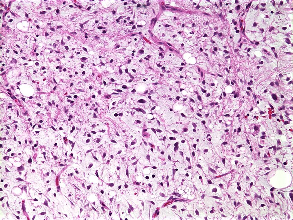
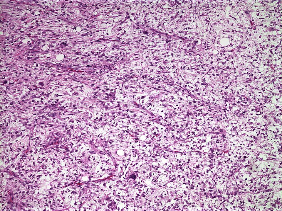
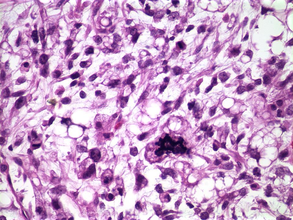
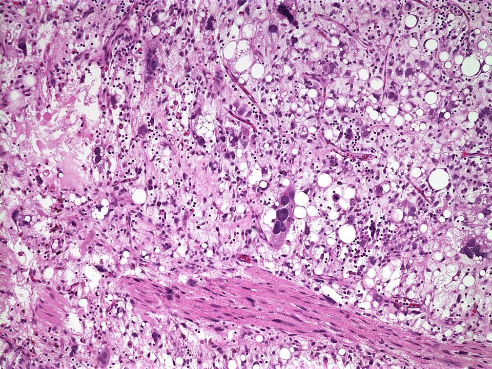
45 LIPOSARCOMA

46 Outline Classification of adipocytic tumors Lipoblastoma Lipoblastoma: Differential Diagnostic Challenges Liposarcoma
47 Liposarcoma in children Extremely rare in pediatric patients (2% of sarcomas) Distribution among soft tissue sarcomas: 5%, 6%, 22%, and 66% in age categories: <5 years, 5 9 years, years, and years, respectively (SEER data). Most frequent histotype represented by Myxoid Liposarcoma (MLPS)* Prognosis for conventional MLPS in young patients very favorable* Progression to Round cell-lps very uncommon in MLPS in young patients A subset of LPS show features of a Pleomorphic-MLPS, is typical of mediastinal region, with aggressive clinical behaviour. Spindle cell-lps also present (not clear if they belong to MLPS) Lipoma-like and pleomorphic LPS exceptional in children *Alaggio R, Liposarcomas in young patients: a study of 82 cases occurring in patients younger than 22 years of age.am J Surg Pathol May;33(5):
48 Myxoid Liposarcoma (60 cases: 10-22y, M:F=2:1)
49
50

51 Atypical lipomatous tumor (4 cases: 12-19y, M:F=1:1)
52

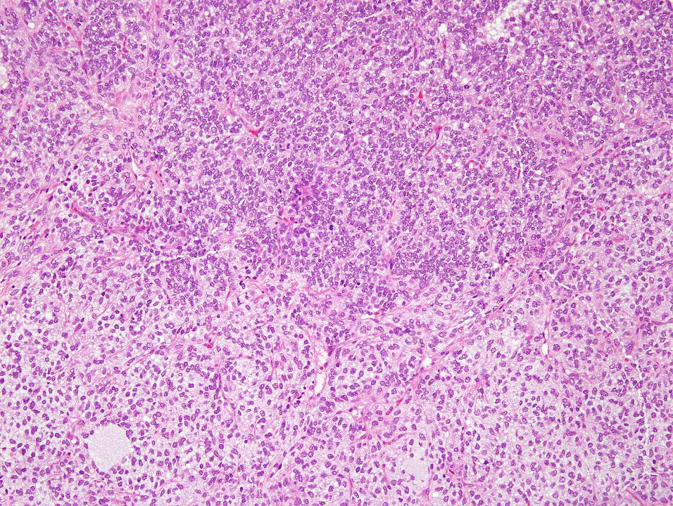
53 Pleomorphic Liposarcoma (2 cases: 5-21y, M:F=1:1)

54 Male, 10 year-old Familiar/Personal history negative Mass in the back noted few months before and recently increased in size.


55 MRI solid lesion deeply located in the subcutis, strongly adherent to the fascia.


56 Treatment Primary excision with macroscopic residual disease. After the histological diagnosis, the child underwent a primary re-excision.
 was completely excised The child is alive")
57 The mass (main diameter 3.6 cm) was completely excised The child is alive and diseasefree after 8 yr.

58 Spindle cell Liposarcoma (6 cases: 11-17y, M:F=1:5)
59
60

61 Important differential diagnosis Spindle cell lipoma Typical of adult age Bland cytology Nuclear palisading Ropey collagen bundles Mastocytes CD34+++

62 Pleomorphic Myxoid Liposarcoma (10 cases: 10-22y, M:F=2:3)
63
64
65
66
67
68
69 Outcome M/RCLPS P-MLPS SC-LPS ALT/DL PLPS Follow-up 31/60 ( mos; median 72 mos) 6/10 (8-108 mos; median 36 mos) 4/6 ( mos; median 42 mos) 4/4 ( mos; median 72 mos) 1/2 (72 mos) ANED 29/31 (94%) 1/6 (17%) 3/4 (75%) 4/4 (100%) 1/1 (100%) AWD 1/31 (3%) 1/6 (17%) 1/4 (25%) 0/4 (0%) 0/1 (0%) DOD 1/31 (3%) 4/6 (67%) 0/4 (0%) 0/4 (0%) 0/1 (0%) LR 3/31 (9%) 3/4 (75%) 1/4 (25%) 2/4 (50%) 0/1 (0%) Mets 0/31 (0%0 2/4 (50%) 1/4 (25%) 0/4 (0%) 0/1 (0%)
70 Pediatric Liposarcoma Genetic aberrations
71
72 CONCLUSIONS
73 Differential Diagnosis between Lipoblastoma and Liposarcoma may be challenging Lipoblastoma Myxoid LPS Usual age: Newborn-5 yr Adults Lobules Well-defined Indistinct Mature fat Central Peripheral, sparse Mucin pools Cellularity Focal Low More abundant Higher (focal) Atypia Absent Present (focal) Mitoses Genetics: Normal (if any) 8q11-13 PLAG1 t(7;8) Abnormal 12q13 DDIT3 t(12;16) or t(12;22) Courtesy of Cheryl Coffin

74 Lipoblasts do not mean liposarcoma in children Liposarcoma in children differs from its adult counterpart with minor aggressivity and peculiar histology -myxoid with pleomorphic cells -spindle cell


75 Fig.14-case 14; mixed myxoid round cell liposarcoma. At the right a myxoid area with intermingled round cell lipoblasts. At the left an area where the cells simulate the appearance and arrangement of a fibrosarcoma Arthur Purdy Stout. Annals of Surgery, January 1944 in some tumors (myxoid liposarcomas) areas showing a distinct spindle cell pattern were present. Unlike the spindle cells of the pleomorphic type, however, the cells were small and slender and generally oriented along a single plane Enzinger &Winslow Virch Arch Path Anat, 1962
Update On Lipomatous Tumors: Old Standbys and New Concepts
 Update On Lipomatous Tumors: Old Standbys and New Concepts John R. Goldblum, M.D. Chairman, Department of Anatomic Pathology Cleveland Clinic Professor of Pathology Cleveland Clinic Lerner College of Medicine
Mayo Medical Laboratories
 Mayo Medical Laboratories Virtual Lectures 2014 MFMER 2016 MFMER slide-1 Virtual Lectures Planning Committee Disclosure Summary As a provider accredited by ACCME, College of Medicine, Mayo Clinic (Mayo
Fun with Fat. General Rules. Case
 Fun with Fat General Rules Imaging: location (deep vs. superficial) Superficial lesions are seldom liposarcomas Deep lesions may be benign or malignant Myxoid stroma is common in benign and malignant lesions
ACCME/Disclosures ALK FUSION-POSITIVE MESENCHYMAL TUMORS. Tumor types with ALK rearrangements. Anaplastic Lymphoma Kinase. Jason L.
 Companion Meeting of the International Society of Bone and Soft Tissue Pathology The Evolving Concept of Mesenchymal Tumors ALK FUSION-POSITIVE MESENCHYMAL TUMORS Jason L. Hornick, MD, PhD March 13, 2016
Part 1. Slides 1-38, Rita Alaggio Soft tissue tumors Trondheim 14. mars 2013
 Part 1 Slides 1-38, Rita Alaggio Soft tissue tumors Trondheim 14. mars 2013 Pediatric Pathology Soft Tissue Tumors AN UPDATE Rita Alaggio Azienda Ospedaliera Università di Padova Soft Tissue Tumors More
Essential Dermatopathology. Jinah Kim, MD, PhD Department of Pathology and Dermatology Stanford University Medical Center
 Essential Dermatopathology Jinah Kim, MD, PhD Department of Pathology and Dermatology Stanford University Medical Center OBJECTIVES Review clinical, pathologic and molecular aspects of bone and fat tumors
Musculoskeletal Sarcomas
 Musculoskeletal Sarcomas Robert C. Orth, M.D., Ph.D. Edward B. Singleton Department of Pediatric Radiology Texas Children s Hospital Page 0 xxx00.#####.ppt 9/23/2012 9:01:18 AM No disclosures Page 1 xxx00.#####.ppt
Cutaneous Mesenchymal Neoplasms with EWSR1 Rearrangement
 Cutaneous Mesenchymal Neoplasms with EWSR1 Rearrangement By Konstantinos Linos MD, FCAP, FASDP Bone, Soft Tissue and Dermatopathology Assistant Professor of Pathology Dartmouth-Hitchcock Medical Center
Tumors of Adipose Tissue Tumors Epidemiology Clinical Features. Morphology. Mature Adipocytes Separated by delicate fibrous septa
 Tumors of Adipose Tissue Lipoma Liposarcoma Most commonly happens in female The most common soft tissue tumor o Originates from matured Adipocytes Most commonly happes at the 4 th and 5 th decade of life
No financial or other disclosures
 Case 2014-5 Esther N. Bit-Ivan, DO Northwestern University Jason Wang, MD Jason Park, MD Korgun Koral, MD Children s Medical Center Charles Timmons, MD Veena Rajaram, MD No financial or other disclosures
5/10. Pathology Soft tissue tumors. Farah Bhani. Mohammed Alorjani
 5/10 Pathology Soft tissue tumors Mohammed Alorjani Farah Bhani Slides are included in this sheet. Objectives: Soft tissue tumors 1. Describe soft tissue tumors. 2. Understand the classification of soft
Newer soft tissue entities
 Newer soft tissue entities Examples among fibroblastic tumors Turku, May 6, 2010 Markku Miettinen, M.D. AFIP, Washington, DC Fibroblastic neoplasms Solitary fibrous tumor /Hemangiopericytoma Low-grade
Financial disclosures
 Cutaneous Mesenchymal Neoplasms with EWSR1 Rearrangement By Konstantinos Linos MD, FCAP, FASDP Bone, Soft Tissue and Dermatopathology Assistant Professor of Pathology Dartmouth-Hitchc Geisel School of
57th Annual HSCP Spring Symposium 4/16/2016
 An Unusual Malignant Spindle Cell Lesion to Involve the Breast Erinn Downs-Kelly, D.O. Associate Professor of Pathology University of Utah & ARUP Laboratories No disclosures Case 39 y/o female with no
A 25 year old female with a palpable mass in the right lower quadrant of her abdomen
 May 2016 A 25 year old female with a palpable mass in the right lower quadrant of her abdomen Contributed by: Paul Ndekwe, MD, Resident Physician, Indiana University School of Department of Pathology and
Diplomate of the American Board of Pathology in Anatomic and Clinical Pathology
 A 33-year-old male with a left lower leg mass. Contributed by Shaoxiong Chen, MD, PhD Assistant Professor Indiana University School of Medicine/ IU Health Partners Department of Pathology and Laboratory
An Overview of Genital Stromal Tumors
 An Overview of Genital Stromal Tumors By Konstantinos Linos MD, FCAP, FASDP Bone, Soft Tissue and Dermatopathology Assistant Professor of Pathology Dartmouth-Hitchcock Medical Center Geisel School of Medicine
Chondroid lipoma: A rare recently described benign lipomatous tumor
 Al-Malki et al. 7 case report peer Reviewed open OPEN ACCESS Chondroid lipoma: A rare recently described benign lipomatous tumor Salman T. Al-Malki, Abdullah S. Al-Khamiss Abstract Introduction: lipomatous
3/27/2017. Disclosure of Relevant Financial Relationships
 Ophthalmic Pathology Evening Specialty Conference USCAP 2017 5 th March, 2017 Mukul K. Divatia, MD Assistant Professor Department of Pathology & Genomic Medicine Weill Cornell Medical College Houston Methodist
IN THE NAME OF GOD Dr. Kheirandish Oral and maxillofacial pathology
 IN THE NAME OF GOD Dr. Kheirandish Oral and maxillofacial pathology ORAL FOCAL MUCINOSIS Uncommon Tumorlike Cutaneous myxoid cyst Overproduction of hyaluronic acid by firoblasts Young adults Female Gingiva
The Relevance of Cytologic Atypia in Cutaneous Neural Tumors
 The Relevance of Cytologic Atypia in Cutaneous Neural Tumors Recent Findings - New Developments New Problems Zsolt B. Argenyi, M.D. Professor of Pathology & Dermatology Director of Dermatopathology Department
Spindle Cell Lesions Of The Breast. Emad Rakha Professor of Breast Pathology and Consultant Pathologist
 Spindle Cell Lesions Of The Breast Emad Rakha Professor of Breast Pathology and Consultant Pathologist * SCLs comprise a wide spectrum of diseases, ranging from reactive processes to aggressive malignant
WHAT IS MDM2? (MDMTWOMICS) MDM2 IN SARCOMAS? (MDMTWOMAS) MDM2MICS? NO CONFLICT OF INTERESTS 5/07/2018 MDM2 IN SOFT TISSUE AND BONE SARCOMAS
 MDM2 IN SARCOMAS? (MDMTWOMAS) MDM2MICS? NO CONFLICT OF INTERESTS 5/07/2018 MDM2 IN SOFT TISSUE AND BONE SARCOMAS") IN SOFT TISSUE AND BONE SARCOMAS WHAT IS? (MDMTWOMICS) Raf Sciot, M.D., PhD. Department of Pathology, University Hospitals Katholieke Universiteit Leuven, LEUVEN, Belgium IN SARCOMAS? (MDMTWOMAS) MICS?
IN SOFT TISSUE AND BONE SARCOMAS WHAT IS? (MDMTWOMICS) Raf Sciot, M.D., PhD. Department of Pathology, University Hospitals Katholieke Universiteit Leuven, LEUVEN, Belgium IN SARCOMAS? (MDMTWOMAS) MICS?
Case Report Intramuscular dendritic fibromyxolipoma of the right thigh region: a case report and review of the literature
 Int J Clin Exp Med 2018;11(5):5281-5285 www.ijcem.com /ISSN:1940-5901/IJCEM0067254 Case Report Intramuscular dendritic fibromyxolipoma of the right thigh region: a case report and review of the literature
J of Evolution of Med and Dent Sci/ eissn , pissn / Vol. 3/ Issue 46/Sep 22, 2014 Page 11296
 CT SPECTRUM OF GIANT RETROPERITONEAL LIPOSARCOMAS WITH HISTOPATHOLOGICAL CORRELATION Shashikumar M. R 1, Rajendra Kumar N. L 2, C. P. Nanjaraj 3, Nishanth R. K 4, Vishwanath Joshi 5 HOW TO CITE THIS ARTICLE:
1/10/2018. Soft Tissue Tumors Showing Melanocytic Differentiation. Overview. Desmoplastic/ Spindle Cell Melanoma
 2016 MFMER slide-1 2016 MFMER slide-2 2016 MFMER slide-3 Soft Tissue Tumors Showing Melanocytic Differentiation Andrew L. Folpe, M.D. Professor of Laboratory Medicine and Pathology Mayo Clinic, Rochester,
Myxo-inflammatory Fibroblastic sarcoma
 AKA Myxo-inflammatory Fibroblastic sarcoma Acral Myxoinflammatory fibroblastic sarcomaam.j.surg.path1998; 22; 911-924 Inflammatory myxoid tumour of soft parts with bizarre giant cells [Pathol.Res.Pract.
Diagnostic Approach to Soft Tissue Tumors
 SECTION 2 Diagnostic Approach to Soft Tissue Tumors Overview Biopsy and Resection of Soft Tissue Tumors 20 Clinical Approach Age- and Location-Based Approach to Diagnosis 24 Histologic Approach Pattern-Based
21/07/2017. Hobnail endothelial cells are not the same as epithelioid endothelial cells
 UPDATE IN CUTANEOUS VASCULAR S DERMATOPATHOLOGY SESSION BELFAST PATHOLOGY JUNE 21/2017 Dr E Calonje St John s Institute of Dermatology, London, United Kingdom THE FAMILY OF VASCULAR S WITH EPITHELIOID
Desmoplastic Melanoma R/O BCC. Clinical Information. 74 y.o. man with lesion on left side of neck r/o BCC
 R/O BCC Sabine Kohler, M.D. Professor of Pathology and Dermatology Dermatopathology Service Stanford University School of Medicine Clinical Information 74 y.o. man with lesion on left side of neck r/o
Evening Specialty Conference Bone and Soft Tissue Pathology. Diagnostic pitfalls in bone and soft tissue pathology
 Evening Specialty Conference Bone and Soft Tissue Pathology. Case 1 Elizabeth G Demicco, MD, PhD Mount Sinai Hospital, New York Disclosure of Relevant Financial Relationships USCAP requires that all planners
CASE REPORT PLEOMORPHIC LIPOSARCOMA OF PECTORALIS MAJOR MUSCLE IN ELDERLY MAN- CASE REPORT & REVIEW OF LITERATURE.
 PLEOMORPHIC LIPOSARCOMA OF PECTORALIS MAJOR MUSCLE IN ELDERLY MAN- CASE REPORT & REVIEW OF LITERATURE. M. Madan 1, K. Nischal 2, Sharan Basavaraj. C. J 3. HOW TO CITE THIS ARTICLE: M. Madan, K. Nischal,
CYSTIC TUMORS OF THE KIDNEY JOHN N. EBLE, M.D. CYSTIC NEPHROMA
 Page 1 CYSTIC TUMORS OF THE KIDNEY JOHN N. EBLE, M.D. Department of Pathology & Laboratory Medicine Phone (317) 274-4806 Medical Science A-128 FAX: (317) 278-2018 635 Barnhill Drive jeble @iupui.edu Indianapolis,
Molecular pathology in soft tissue tumors. Sylvia Höller Pathologie
 Molecular pathology in soft tissue tumors Sylvia Höller Pathologie When do we perform molecular testing? Morphology and IHC are not clearly fitting with an entity some translocations are entity specific
PLEOMORPHIC ADENOMA ( BENIGN MIXED TUMOR )
") ( BENIGN MIXED TUMOR ) Grossly, the tumor is freely movable, solid, sometimes lobulated and occasionally cystic. If recurrent, multinodular masses are common. Histologically, within a fibrous capsule,
( BENIGN MIXED TUMOR ) Grossly, the tumor is freely movable, solid, sometimes lobulated and occasionally cystic. If recurrent, multinodular masses are common. Histologically, within a fibrous capsule,
SOFT TISSUE TUMOR PATHOLOGY: AN UPDATE
 SOFT TISSUE TUMOR PATHOLOGY: AN UPDATE Jason L. Hornick, MD, PhD July 18, 2013 Department of Pathology Brigham and Women s Hospital Harvard Medical School Boston, MA, USA I have no disclosures. New Soft
Giant intramuscular calcifying aponeurotic fibroma of gluteus maximus: case report
 Annals of Tropical Paediatrics (2010) 30, 259 263 Giant intramuscular calcifying aponeurotic fibroma of gluteus maximus: case report S. ARORA, D. SABAT, S. K. ARORA*, V. KUMAR & R. K. SARAN { Department
Division of Pathology
 Case 38 Adult woman with a 35mm right breast lump at the 10 o clock position. Excision performed. (Case contributed by Dr Mihir Gudi, KKH) Division of Pathology Merlion, One Fullerton Singapore Diagnosis
Malignant Peripheral Nerve Sheath Tumor
 C H A P T E R 120 Malignant Peripheral Nerve Sheath Tumor Currently, malignant peripheral nerve sheath tumor (MPNST) is the most commonly used generic name for the neoplasms known in the past as neurosarcoma,
Special slide seminar
 Special slide seminar Tomáš Rozkoš The Fingerland Department of Pathology Charles University Medical Faculty and Faculty Hospital in Hradec Králové Czech Republic Case history, 33 years old resistance
Early View Article: Online published version of an accepted article before publication in the final form.
 Early View Article: Online published version of an accepted article before publication in the final form. Journal Name: Journal of Case Reports and Images in Pathology Type of Article: Case Report Title:
Case 1. Disclosure. Imaging. Clinical history 5/10/2016. USCAP 2016 Annual Meeting Evening Specialty Conference Bone and Soft tissue Pathology
 Disclosure Dr. Agaram has nothing to disclose Case 1 Narsi Agaram, MBBS USCAP 2016 Annual Meeting Evening Specialty Conference Bone and Soft tissue Pathology Clinical history Imaging 1998 A three month
Selected Pseudomalignant Soft Tissue Tumors of the Skin and Subcutis
 Selected Pseudomalignant Soft Tissue Tumors of the Skin and Subcutis Andrew L. Folpe, M.D. Professor of Laboratory Medicine and Pathology Mayo Clinic, Rochester, MN folpe.andrew@mayo.edu 2016 MFMER slide-1
Case 1 10/2/17. Myxoid Soft Tissue Tumors & Tumor-like Lesions. Myxofibro- or Fibromyxo-?: Myxoid Soft Tissue Tumours We Are All Mixed Up About
 Myxoid Soft Tissue Tumors & Tumor-like Lesions Myxofibro- or Fibromyxo-?: Myxoid Soft Tissue Tumours We Are All Mixed Up About Rajiv M. Patel, M.D. RCPA NZ ASM 2017 (4:15-5:00pm, Saturday, 23-09-17) Heterogenous
We are IntechOpen, the world s leading publisher of Open Access books Built by scientists, for scientists. International authors and editors
 We are IntechOpen, the world s leading publisher of Open Access books Built by scientists, for scientists 3,900 116,000 120M Open access books available International authors and editors Downloads Our
A case of fat-free pleomorphic lipoma occurring in the upper back and axilla simultaneously
 Wang et al. World Journal of Surgical Oncology 2013, 11:145 WORLD JOURNAL OF SURGICAL ONCOLOGY CASE REPORT Open Access A case of fat-free pleomorphic lipoma occurring in the upper back and axilla simultaneously
Solitary Fibrous Tumor of the Kidney with Massive Retroperitoneal Recurrence. A Case Presentation
 246) Prague Medical Report / Vol. 113 (2012) No. 3, p. 246 250 Solitary Fibrous Tumor of the Kidney with Massive Retroperitoneal Recurrence. A Case Presentation Sfoungaristos S., Papatheodorou M., Kavouras
Pitfalls in the diagnosis of well-differentiated hepatocellular lesions
 2013 Colorado Society of Pathology Pitfalls in the diagnosis of well-differentiated hepatocellular lesions Sanjay Kakar, MD University of California, San Francisco Outline Hepatocellular adenoma: new WHO
Takayuki Ohguri 1 Takatoshi Aoki 1 Masanori Hisaoka 2 Hideyuki Watanabe 1 Katsumi Nakamura 1 Hiroshi Hashimoto 2 Toshitaka Nakamura 3 Hajime Nakata 1
 Takayuki Ohguri 1 Takatoshi Aoki 1 Masanori Hisaoka 2 Hideyuki Watanabe 1 Katsumi Nakamura 1 Hiroshi Hashimoto 2 Toshitaka Nakamura 3 Hajime Nakata 1 Received July 1, 2002; accepted after revision November
Aspen conference on pediatric disease. July through August Bone and Soft Tissue Update. David M. Parham, MD. Rhabdomyoma and rhabdomyosarcoma
 Aspen conference on pediatric disease July through August 2014 Bone and Soft Tissue Update David M. Parham, MD Rhabdomyoma and rhabdomyosarcoma Embryonic rhabdomyogenesis is a highly conserved process
Rhabdomyomas and Rhabdomyosarcomas (RMS) David M. Parham, MD Chief of Anatomic Pathology
 David M. Parham, MD Chief of Anatomic Pathology") Rhabdomyomas and Rhabdomyosarcomas (RMS) David M. Parham, MD Chief of Anatomic Pathology Tumors of skeletal muscle: Rhabdomyomas and rhabdomyosarcomas Embryonal muscle 2 3 4 5 6 7 8 Rhabdomyoma Benign
Rhabdomyomas and Rhabdomyosarcomas (RMS) David M. Parham, MD Chief of Anatomic Pathology Tumors of skeletal muscle: Rhabdomyomas and rhabdomyosarcomas Embryonal muscle 2 3 4 5 6 7 8 Rhabdomyoma Benign
Case Report Spindle cell lipoma of the wrist, occurring in a distinctly rare location: a case report with review of literature
 Int J Clin Exp Pathol 2015;8(3):3299-3303 www.ijcep.com /ISSN:1936-2625/IJCEP0004992 Case Report Spindle cell lipoma of the wrist, occurring in a distinctly rare location: a case report with review of
Papillary Lesions of the Breast A Practical Approach to Diagnosis. (Arch Pathol Lab Med. 2016;140: ; doi: /arpa.
 Papillary Lesions of the Breast A Practical Approach to Diagnosis (Arch Pathol Lab Med. 2016;140:1052 1059; doi: 10.5858/arpa.2016-0219-RA) Papillary lesions of the breast Span the spectrum of benign,
GIANT RETROPERITONEAL LIPOSARCOMA: IMAGING AND LITERATURE Amit Kumar 1, Sanjay K. Suman 2, Bipin Kumar 3, Sumit Kumar 4
 GIANT RETROPERITONEAL LIPOSARCOMA: IMAGING AND LITERATURE Amit Kumar 1, Sanjay K. Suman 2, Bipin Kumar 3, Sumit Kumar 4 HOW TO CITE THIS ARTICLE: Amit Kumar, Sanjay K. Suman, Bipin Kumar, Sumit Kumar.
ESS: Pathologic Insights
 GEIS XVI INTERNATIONAL SYMPOSIUM Seville 4th October 2018 ESS: Pathologic Insights Sílvia Bagué The Royal Marsden Hospital London (United Kingdom) I have no conflicts of interest Endometrial stromal sarcoma
Mammary analogue secretory carcinoma of salivary gland A case report of new entity
 Case Report Mammary analogue secretory carcinoma of salivary gland A case report of new entity Vaibhav Bhika Bari 1*, Sandhya Unmesh Bholay 2 1 Assistant Professor, 2 Associate Professor Rajiv Gandhi Medical
Low-Grade Periductal Stromal of Breast: a case report
 Low-Grade Periductal Stromal of Breast: a case report Rosanna Nenna 1 Cosimo Damiano Inchingolo 1 Domenico Palmieri 2 Annalisa De Lucia 1 Giusy Elicio 1 Pina Miscioscia 1 ( 1 ) U.O.C. di Anatomia Patologica,
أملس عضلي غرن = Leiomyosarcoma. Leiomyosarcoma 1 / 5
 Leiomyosarcoma 1 / 5 EPIDEMIOLOGY Exact incidence is unknown, but older studies suggest that leiomyosarcomas comprise approximately 3 percent of soft-tissue sarcomas. Superficial leiomyosarcoma occurs
Atypical Palisaded Myofibroblastoma of Lymph Node: Report of a rare case.
 ISPUB.COM The Internet Journal of Pathology Volume 10 Number 1 Atypical Palisaded Myofibroblastoma of Lymph Node: Report of a rare case. V Kinnera, R Nandyala, M Yootla, K Mandyam Citation V Kinnera, R
Taku Naiki, 1 Shuzo Hamamoto, 1 Noriyasu Kawai, 1 Aya Naiki-Ito, 2 Yoshiyuki Kojima, 1 Takahiro Yasui, 1 Keiichi Tozawa, 1 and Kenjiro Kohri 1
 International Scholarly Research Network Volume 2011, Article ID 261735, 4 pages doi:10.5402/2011/261735 Case Report Giant Retroperitoneal Mucinous Tumor Supportively Diagnosed as a Dedifferentiated Liposarcoma
Financial disclosures
 An update on immunohistochemical markers in mesenchymal neoplasms By Konstantinos Linos MD, FCAP, FASDP Assistant Professor of Pathology Geisel School of Medicine at Dartmouth Dartmouth-Hitchcock Medical
Not the usual liposarcoma... could it be a fatty benign tumor?
 Not the usual liposarcoma... could it be a fatty benign tumor? Poster No.: C-0635 Congress: ECR 2017 Type: Educational Exhibit Authors: A. Cano Rodríguez, J. M. Morales Pérez, C. Le Cacheux, J. I. 1 1
Respiratory Tract Cytology
 Respiratory Tract Cytology 40 th European Congress of Cytology Liverpool, UK Momin T. Siddiqui M.D. Professor of Pathology and Laboratory Medicine Director of Cytopathology Emory University Hospital, Atlanta,
Klinisch belang van chromosomale translocatie detectie in sarcomen
 Translocations in sarcomas Klinisch belang van chromosomale translocatie detectie in sarcomen Judith V.M.G. Bovée, M.D., Ph.D. Department of Pathology Leiden University Medical Center RNA binding DNA binding
Retroperitoneal Sarcomas - A pictorial review
 Retroperitoneal Sarcomas - A pictorial review Poster No.: C-1409 Congress: ECR 2013 Type: Educational Exhibit Authors: D. Douraghi-Zadeh, K. L. Shahabuddin, R. H. Thomas, E. Moskovic; London/UK Keywords:
Primary Breast Liposarcoma
 Primary Breast Liposarcoma Bhagyam Nagarajan 1*, GayatriAutkar 1, Keyuri Patel 1, Meghal Sanghvi 1 1. Department of Radiology, Wockhardt Hospital, Mumbai, India * Correspondence: Dr Bhagyam Nagarajan,
Classification (1) Classification (3) Classification (2) Spindle cell lesions. Spindle cell lesions of bladder (Mills et al.
 Classification (3) Classification (2) Spindle cell lesions. Spindle cell lesions of bladder (Mills et al.") Non-epithelial tumours and nonepithelial tumour-like lesions of the bladder Dr Jonathan H Shanks The Christie NHS Foundation Trust, Manchester, UK Classification (1) Myofibroblastic proliferations and
Non-epithelial tumours and nonepithelial tumour-like lesions of the bladder Dr Jonathan H Shanks The Christie NHS Foundation Trust, Manchester, UK Classification (1) Myofibroblastic proliferations and
CASE REPORT Benign epithelioid peripheral nerve sheath tumour resembling schwannoma
 Malaysian J Pathol 2014; 36(3) : 217 221 CASE REPORT Benign epithelioid peripheral nerve sheath tumour resembling schwannoma Thejasvi KRISHNAMURTHY MD and SR NIVEDITHA MD, DNB Department of Pathology,
A Cooperative Approach to Diagnosis of Rare Diseases: Primitive Myxoid Mesenchymal Tumor of Infancy
 310 Available online at www.annclinlabsci.org A Cooperative Approach to Diagnosis of Rare Diseases: Primitive Myxoid Mesenchymal Tumor of Infancy David W Cuthbertson 1, Kevin Caceres 1, John Hicks 2, and
Update on Cutaneous Mesenchymal Tumors. Thomas Brenn
 Update on Cutaneous Mesenchymal Tumors Thomas Brenn Cutaneous Mesenchymal Tumours Wide morphological and biological spectrum Myofibroblastic, smooth muscle, neural, vascular, apidocytic, undifferentiated;
USCAP 2012: Companion Meeting of the AAOOP. Update on lacrimal gland neoplasms: Molecular pathology of interest
 USCAP 2012: Companion Meeting of the AAOOP Vancouver BC, Canada, March 17, 2012 Update on lacrimal gland neoplasms: Molecular pathology of interest Valerie A. White MD, MHSc, FRCPC Department of Pathology
Mesenchymal Tumors. Cavernous Hemangioma (CH) VASCULAR TUMORS MESENCHYMAL TUMORS OF THE LIVER: WHAT S NEW AND UNUSUAL (MY PERSPECTIVE)
 VASCULAR TUMORS MESENCHYMAL TUMORS OF THE LIVER: WHAT S NEW AND UNUSUAL (MY PERSPECTIVE)") Mesenchymal Tumors MESENCHYMAL TUMORS OF THE LIVER: WHAT S NEW AND UNUSUAL (MY PERSPECTIVE) CURRENT ISSUES IN ANATOMIC PATHOLOGY MAY 23, 2014 Linda Ferrell, MD, UCSF Focus on Vascular Tumors Benign and
Mesenchymal Tumors MESENCHYMAL TUMORS OF THE LIVER: WHAT S NEW AND UNUSUAL (MY PERSPECTIVE) CURRENT ISSUES IN ANATOMIC PATHOLOGY MAY 23, 2014 Linda Ferrell, MD, UCSF Focus on Vascular Tumors Benign and
MRI of a Subcutaneous Myolipoma in the Ankle: a Case Report
 Case Report http://dx.doi.org/10.3348/kjr.2011.12.5.641 pissn 1229-6929 eissn 2005-8330 Korean J Radiol 2011;12(5):641-645 MRI of a Subcutaneous Myolipoma in the Ankle: a Case Report Yeon Soo Lee, MD 1,
Financial disclosures
 Mesenchymal Neoplasms with Melanocytic Differentiation By Konstantinos Linos MD, FCAP, FASDP Bone, Soft Tissue and Dermatopathology Assistant Professor of Pathology Dartmouth-Hitchcock Medical Center Geisel
Soft tissue lipomas, lipoma variants and liposarcomas: MRI evaluation and review of literature
 Soft tissue lipomas, lipoma variants and liposarcomas: MRI evaluation and review of literature Poster No.: R-0122 Congress: RANZCR-AOCR 2012 Type: Authors: Keywords: DOI: Educational Exhibit A. A. Tandon,
Case Report Dendritic fibromyxolipoma in the latissimus dorsi: a case report and review of the literature
 Int J Clin Exp Pathol 2015;8(7):8650-8654 www.ijcep.com /ISSN:1936-2625/IJCEP0010580 Case Report Dendritic fibromyxolipoma in the latissimus dorsi: a case report and review of the literature Shuguang Liu
Slide Seminar Spanish Society of Pathology
 Slide Seminar Spanish Society of Pathology John R. Goldblum, M.D. Chairman, Department of Anatomic Pathology Cleveland Clinic Professor of Pathology Cleveland Clinic Lerner College of Medicine 1921 Original
Surgical Pathology Evening Specialty Conference USCAP 2015
 Surgical Pathology Evening Specialty Conference USCAP 2015 John R. Goldblum, M.D. Chairman, Department of Pathology, Cleveland Clinic Professor of Pathology, Cleveland Clinic Lerner College of Medicine
Dermatopathology. Dr. Rafael Botella Estrada. Hospital La Fe de Valencia
 Dermatopathology Dr. Rafael Botella Estrada. Hospital La Fe de Valencia Melanoma and mimics Dr. Martin Mihm Malignant lesions result from the accumulation of mutations Class I lesions (benign) Class II
SMOOTH MUSCLE TUMOURS
 SMOOTH MUSCLE TUMOURS NORMAL SMOOTH MUSCLE Cytology Immunohistochemistry Ultrastructure Masson Trichrome Smooth Muscle Ultrastructure Many myofilaments running parallel to the long axis of the smooth
The Enigmatic Spitz Lesion
 The Enigmatic Spitz Lesion The Dawn of Spitz S Spitz Sophie Spitz Melanomas of Childhood ; Am J Pathol 1948 1910-1956 13 children (18 mo - 12 yrs) 12/13 had a benign clinical course Sophie Spitz Born 1910
Index. J Juvenile hyaline fibromatosis, 27 Juvenile xanthogranuloma, 57 Juxta-articular myxoma, 152
 A Adenomatoid tumor, 76, 77 Adipose tissue tumors benign tumors angiolipoma, 6 chondroid lipoma, 9 fibrolipoma, 5 hibernoma, 8 lipoblastoma, 9 lipoma (see Lipoma) myelolipoma, 6 pleomorphic lipoma, 8 spindle
RARE TUMORS OF INFANCY. RAJKUMAR VENKATRAMANI, MD, MS Director, Rare Tumors Program, Texas Children s Hospital
 RARE TUMORS OF INFANCY RAJKUMAR VENKATRAMANI, MD, MS Director, Rare Tumors Program, Texas Children s Hospital OBJECTIVES Review the epidemiology and clinical presentation of soft tissue sarcomas in infancy.
A case of giant cell tumour of soft parts in a horse Francesco Cian 1, Sarah Whiteoak 2, Jennifer Stewart 1
 A case of giant cell tumour of soft parts in a horse Francesco Cian 1, Sarah Whiteoak 2, Jennifer Stewart 1 1 Animal Health Trust, Newmarket, UK 2 608 Equine and Farm Vets, Rowington, UK Signalment: Horse,
Pediatric Retroperitoneal Masses Radiologic-Pathologic Correlation
 Acta Radiológica Portuguesa, Vol.XVIII, nº 70, pág. 61-70, Abr.-Jun., 2006 Pediatric Retroperitoneal Masses Radiologic-Pathologic Correlation Marilyn J. Siegel Mallinckrodt Institute of Radiology, Washington
Affiliazione autori0. Riccardo Ricci Journal Club GIPAD, settore GIST Anatomia Patologica, Università Cattolica, Roma
 GIST Manifesting as a Retroperitoneal Tumor: Clinicopathologic Immunohistochemical, and Molecular Genetic Study of 112 Cases American Journal of Surgical Pathology, 2017, 41:577-585 Miettinen M*; Felisiak-Golabek
Gross appearance of peritoneal cysts. They have a thin, translucent wall and contain a clear fluid.
 Gross appearance of peritoneal cysts. They have a thin, translucent wall and contain a clear fluid. So-called multicystic benign mesothelioma. A, Gross appearance. So-called multicystic benign mesothelioma.
GUT-C 11/30/2017. Debasmita Das, M.D. PGY-1 Danbury Hospital
 GUT-C 11/30/2017 Debasmita Das, M.D. PGY-1 Danbury Hospital CLINICAL SUMMARY 8/2017 59 year old female Presented to the ED with 1 month history of general malaise, fever and weight loss PMH: Significant
Gastrointestinal stromal tumor
 Gastrointestinal stromal tumor 영남의대병리학교실 최준혁 Classification of gastrointestinal mesenchymal tumor Gastrointestinal stromal tumor(gist) Smooth muscle tumors : leiomyoma, leiomyosarcoma Neurogenic tumors
Disclosures. An update on ancillary techniques in the diagnosis of soft tissue tumors. Ancillary techniques. Introduction
 Disclosures An update on ancillary techniques in the diagnosis of soft tissue tumors. I have nothing to disclose. Andrew Horvai, MD, PhD Clinical Professor, Pathology Introduction Ancillary techniques
Enterprise Interest Nothing to declare
 Enterprise Interest Nothing to declare Diagnoses one would not like to miss in soft tissue pathology early in your career Marta Sbaraglia, MD Department of Pathology Hospital of Treviso University of Padua
Tumores de células pequeñas, redondas y azules: diagnóstico diferencial cuando el tiempo apremia
 Tumores de células pequeñas, redondas y azules: diagnóstico diferencial cuando el tiempo apremia Sílvia Bagué Servei de Patologia Hospital de Sant Pau Barcelona Soft tissue sarcomas Heterogeneous group
Pleomorphic Liposarcoma: A Clinicopathologic Analysis Of 19 Cases
 Pleomorphic Liposarcoma: A Clinicopathologic Analysis Of 19 Cases Katharine A. Downes, M.D., John R. Goldblum, M.D., Elizabeth A. Montgomery, M.D., Cyril Fisher, M.D., F.R.C.Path. Departments of Anatomic
Evidence by Spectral Karyotyping that 8q11.2 Is Nonrandomly Involved in Lipoblastoma
 Journal of Molecular Diagnostics, Vol. 2, No. 2, May 2000 Copyright American Society for Investigative Pathology and the Association for Molecular Pathology Evidence by Spectral Karyotyping that 8q11.2
Case Report Fibrolipoma with Osseous and Cartilaginous Metaplasia of Hoffa s Fat Pad: A Case Report
 Case Reports in Orthopedics Volume 2012, Article ID 547963, 5 pages doi:10.1155/2012/547963 Case Report Fibrolipoma with Osseous and Cartilaginous Metaplasia of Hoffa s Fat Pad: A Case Report Ioannis Gigis
Update in Salivary Gland Pathology. Benjamin L. Witt University of Utah/ARUP Laboratories February 9, 2016
 Update in Salivary Gland Pathology Benjamin L. Witt University of Utah/ARUP Laboratories February 9, 2016 Objectives Review the different appearances of a selection of salivary gland tumor types Establish
Female 18. Deeply pigmented lesion on trunk.?warty naevus?seborrhoeic keratosis?malignant melanoma. The best diagnosis is:
 Female 18. Deeply pigmented lesion on trunk.?warty naevus?seborrhoeic keratosis?malignant melanoma. The best diagnosis is: A. deep penetrating naevus B. naevoid malignant melanoma C. pigment synthesising
Diagnostic problems in uterine smooth muscle tumors
 Diagnostic problems in uterine smooth muscle tumors Marina Kos Ljudevit Jurak Clinical Department of Pathology, Clinical Hospital Center Sestre milosrdnice, Zagreb Institute of Pathology, University of
Cellular Neurothekeoma
 Cellular Neurothekeoma Scott W Binder, MD Pritzker Professor of Pathology & Dermatology Sr. Vice Chair Director, Pathology Clinical Services Chief, Dermatopathology Geffen/UCLA School of Medicine Clinical
Mesenchymal Tumors MESENCHYMAL TUMORS OF THE LIVER: WHAT S NEW AND UNUSUAL (MY PERSPECTIVE)
") MESENCHYMAL TUMORS OF THE LIVER: WHAT S NEW AND UNUSUAL (MY PERSPECTIVE) CURRENT ISSUES IN ANATOMIC PATHOLOGY MAY 23, 2014 Linda Ferrell, MD, UCSF Mesenchymal Tumors Focus on Vascular Tumors Benign and
MESENCHYMAL TUMORS OF THE LIVER: WHAT S NEW AND UNUSUAL (MY PERSPECTIVE) CURRENT ISSUES IN ANATOMIC PATHOLOGY MAY 23, 2014 Linda Ferrell, MD, UCSF Mesenchymal Tumors Focus on Vascular Tumors Benign and